international journal of chemtech research - sphinxsai.com282-299)v10n15ct.pdf · such compositions...
TRANSCRIPT
Formulation, Development and Evaluation of Film-Forming Gel for Prolonged Dermal Delivery of Miconazole Nitrate
R.B. Saudagar1*, P. A. Gangurde2
1Department of Pharmaceutical Chemistry, KCT’S R.G. Sapkal College of Pharmacy,
Anjaneri, Nasik 422 213, Maharashtra, India. 2Department of Quality Assurance and Techniques, KCT’s R.G. Sapkal College of
Pharmacy, Anjaneri, Tal. Trimbakeshwar, Dist. Nashik-422213, Maharashtra, India.
Abstract : The localized treatment of diseases of body tissues requires that the pharmaceutical
active be maintained at the site of treatment for an effective period of time. Sweat, clothing, movements and getting washed away easily on contact with water are some of the problems
that have limited the effectiveness and residence time of conventional topical formulations for
treatment of fungal infections of skin. This necessitates longer treatment duration. Hence, a composition that adheres to skin surface afflicted and provides localized delivery of an
antifungal agent is needed. The present work aims at designing a dosage form of Miconazole
nitrate referred to as a ‘film-forming gel’ which on application forms a thin, transparent film on skin surface. Eudragit RS PO and hydroxypropyl cellulose were used in combination to
provide a matrix film that would permit the release of the antifungal agent for a prolonged
time. The formulations were prepared using 32 full factorial design. They were tested for
drying time, drug release, antifungal activity, skin irritation and stability studies. The gel was characterised for pH, viscosity, drug content, effective dosage volume and mechanical
properties of the film formed after application; water vapour permeability were also tested.
All the formulations showed results within acceptable range for various tests. The optimized formulation showed drug release of 99.76% and antifungal activity in terms of efficacy as
98.78%. Such a formulation can be claimed to decrease duration of therapy, will be more
accepted by the patients and be a breakthrough in treating fungal infections of the skin. Keywords : Miconazole nitrate ; Fungal skin infections; Film-forming gel; Eudragit RS
PO;Hydroxypropyl cellulose.
Introduction:
The skin is a very important route for the dermal or transdermal delivery of pharmaceutically active substances. Film forming polymeric solutions are a novel approach in this area that might present an alternative
to the conventional dosage forms used on the skin, such as ointments, creams, gels or patches. The polymeric
solution is applied to the skin as a liquid and forms an almost invisible film in situ by solvent evaporation.
Transdermal drug delivery system (TDDS) can provide some desirable performances over conventional pharmaceutical dosage formulations, such as avoiding gut and hepatic first-pass metabolism, improving drug
bioavailability, reducing dose frequency and stabilizing drug delivery profiles [1].
Fungal diseases can be classified into 3 groups: the superficial, subcutaneous, and deep or systemic
mycoses. Superficial infections are confined to skin, hair, nails or mucous membranes. The most common
International Journal of ChemTech Research CODEN (USA): IJCRGG, ISSN: 0974-4290, ISSN(Online):2455-9555
Vol.10 No.15, pp 282-299, 2017

R.B. Saudagar et al /International Journal of ChemTech Research, 2017,10(15): 282-299. 283
fungal skin infections are the dermatophytoses, pityriasis versicolor, and candidiasis. Approximately 90% of
fungal skin infections are caused by ‘dermatophytes’, which are parasitic fungi affecting the skin, hair, nails. [1]
Miconazole nitrate is an imidazoleantifungal agent widely utilized in the treatmentan antifungal agent is
often prescribed for the treatment of various topical fungal infections such as Candidiasis, Coccidio Crypto
coccosis, Para coccidio idomycosis, and infections due to Pseudeli escheria. The drug undergoes substantial first pass metabolism and only half the amount of it is bioavailable systemically. To avoid this, delivery of
Miconazole Nitrate through skin delivers the potential advantage of bypassing the hepato-gastro first pass
metabolism associated with oral administration.
Topical therapy is an attractive choice for the treatment of the cutaneous infections due to its
advantages such as targeting of drugs to the site of infection and reduction of the risk of systemic side effects.
[4] Systemic treatment is usually reserved for infections of the nails, extensive cutaneous infections or those which have not responded to topical therapy. Conventional topical formulations are unable to retain the drug
over the skin for a prolonged period and hence necessitate longer treatment duration or have to be supplemented
by oral therapy.[5] For effective local delivery of an antifungal that is applied to the surface of the skin, the agent must be partitioned firstly from the vehicle into the stratum corneum, and then partitioned to the local
tissues including the viable epidermis, dermis, subcutaneous tissue and appendages. This is problematic since
antifungal compounds are generally hydrophobic and a means is needed to perform this partitioning to deliver
therapeutically effective concentrations of active agent in situ. For effective delivery of drugs via dermal route, much effort has been invested in providing chemical enhancers for drug penetration, such as DMSO and
azones. Many of these substances cause irritation and are not desirable due to their toxicity. Hence, there is a
need for improved compositions for topical delivery of antifungal agents that would minimize the systemic exposure of the therapeutic agent. [4,6] The need for multiple applications a day is frequently associated with
poor compliance of patients. Thus, prolonging the contact time of active substances to the skin and thereby
reducing the application frequency is subject of intensive research. [7] Sustained release delivery systems with
features of both semisolid formulations and patches may be employed here. The concept of film forming formulations is very recent. Film forming formulations may be solutions, gels or emulsions. Film forming
formulations are defined as non-solid dosage forms that produce a substantial film in situ after application on
the skin or any other body surface. Such compositions can either be liquids or semisolids with a film forming polymer as basic material for the matrix. The formed film is sufficiently substantial to provide a sustained drug
release to the skin. [8,9] Very few examples of film forming gel formulations have been reported in literature.
BeeGentleTM and GELNIQUE are commercially available film forming gel formulations. [10,11]We hypothesized that incorporation of the drug in a film forming gel would facilitate prolonged contact of the drug
on the skin and the film formed on drying would improve its skin retention ability, thereby improving the
topical treatment of fungal skin infections. This approach does not only sustain the release of drug and enhance
percutaneous absorption, but may even allow for drug targeting to the skin or even its substructure, thereby enhancing drug efficacy and improving patient compliance by reducing application frequency. In this study a
dermal gel containing miconazole nitrate was prepared using the film forming polymer, Eudragit RS PO
(Eudragit) and gelling agent, Hydroxypropyl cellulose (HPC). HPC also played the role of a secondary film forming polymer. Triethyl citrate (TEC) was used as a plasticizer. It is more efficient to use a multi-factorial
design than one-factor-at-a-time experimentation since it can give a combination of variables that give better
results for the optimization study. [9] Using the response surface analysis technique, we evaluated the effects of two factors: the amount of Eudragit and the amount of HPC on the drug release rate and antifungal activity by
utilizing 32 full factorial design.
Materials and Methods:
Materials:
Miconazole Nitrate was received from college Eudragit and HPC was received as gift sample
fromModern science Pvt. Ltd., Nashik Mumbai. TEC was received as gift sample from Evonik Degussa India
Pvt. Ltd., Mumbai. All other chemicals were of analytical grade and were obtained commercially.

R.B. Saudagar et al /International Journal of ChemTech Research, 2017,10(15): 282-299. 284
Preparation of dermal gel:
The polymeric solutions of Eudragit RS PO and Hydroxypropyl cellulose were prepared in ethanol using dispersion method. Eudragit RS PO was sprinkled over 10 mL of ethanol containing triethyl citrate (7.5 %
w/w of Eudragit RS PO). Hydroxypropyl cellulose was sprinkled over 10 mL of ethanol separately. Both
solutions were allowed to swell for 24 hours to produce clear solutions. The polymeric solutions were mixed properly with continuous stirring. Accurately weighed quantity (0.6 g) of the Miconazole nitrate was dissolved
in 5 mL ethanol. The drug solution and polymeric dispersion were mixed properly with continuous stirring and
volume was made upto the mark using ethanol. [8,12-15]
32 factorial design was followed for the development of the formulations. In this design, 2 factors were
evaluated each at 3 levels and experimental trials were performed at all 9 possible combinations as reflected in
table I.
Evaluation of formulations:
The formulations were tested clarity, pH, and viscosity. Clarity was checked visually and pH of the
formulations was checked using digital pH meter 335. [5,13] The rheological properties of gels were determined by the Brookfield viscometer; type DV-II + PRO using spindle SC4-18.Viscosity values of the
formulations were recorded at varying shear rates. [13]
Drug content:
To determine drug content 1 g of gel was taken in a 100 mL volumetric flask containing 10 mL phosphate buffer solution pH 4 and volume was made up to the mark with phosphate buffer solution pH 4 to get
a concentration of 100μg/mL. An aliquot of 0.5 mL was transferred to a 10 mL volumetric flask and volume
was made up with phosphate buffer solution pH 4. The absorbance of prepared solution was measured at λ max
of 271 nm by using UV visible spectrophotometer. [13]
Drying time:
For the assessment of the drying time the formulation was applied to the inner sides of the forearm of a
volunteer, who participated in the study on informed consent basis. After 2 minutes a glass slide was placed on
the film without pressure. If no remains of liquid were visible on the glass slide after removal, the film was considered dry. If remains of liquid were visible on the glass slide the experiment was repeated until the film
was found to be completely dry. [8]
Effective dosage volume:
The calculation of an effective dosage volume of the formulation to be applied per cm2 area of as skin
is necessary for ensuring that the drug is available at the site of fungal infection in concentrations above its minimum inhibitory concentration (MIC). [17] Pre-experiments were carried out to determine dose of drug to
be delivered per cm2. For the calculation of an effective dosage volume, the formulation was applied to the
inner sides of the forearm of a volunteer, who participated in the study on informed consent basis. Varying volumes of formulation (0.1 to 2 mL) were applied to an area of 2 cm2 on the inner sides of the forearm of the
volunteer. The volume that did not flow away from the application site was noted. The amount of drug in this
volume was calculated.
Formulation code F1 F2 F3 F4 F5 F6 F7 F8 F9
Ingredient %
Miconazole nitrate (w/v) 2 2 2 2 2 2 2 2 2
Eudragit RS PO (w/v) 7 7 7 12 12 12 17 17 17
HPC (w/v) 4 6 8 4 6 8 4 6 8
Triethyl citrate (w/w) 0.37 0.37 0.37 0.93 0.93 0.93 1.5 1.5 1.5
Ethanol : Water (v/v) 70:30 70:30 70:30 70:30 70:30 70:30 70:30 70:30 70:30

R.B. Saudagar et al /International Journal of ChemTech Research, 2017,10(15): 282-299. 285
Integrity of formulation on skin:
The formulation was applied to the forearm of a volunteer as described for the assessment of the drying time. The dry film was then worn overnight by the test subject. After 24 hours the test area was examined
visually for completeness of the film, appearance of cracks or flaking. [8]
Properties of film:
For the assessment of properties of the film, films were produced with a solvent evaporation technique
by pouring 1 mL of the preparations into a stainless steel mould lined by Teflon (6 cm x 10 cm). The films were left to dry for 72 hours at room temperature (three hours ventilated in the open air to allow the evaporation of
ethanol.The stickiness of the outer surface was tested by pressing cotton wool on the dry film under low
pressure. Depending on the quantity of cotton fibres that were retained by the film the stickiness was rated high (dense accumulation of fibres on the film), medium (thin fibre layer on the film) or low (occasional or no
adherence of fibres).The cosmetic attractiveness of the film was assessed by visual examination of the dry
films. Transparent films with a low skin fixation had a high attractiveness as they were almost invisible. Opaque films and films with a medium skin fixation were considered less attractive as they exhibited an increased
visibility and a slightwrinkling of the skin. Whitish films and films causing heavy wrinkling of the skin due to
strong skin fixation displayed only a low attractiveness. The mechanical properties of the film were tested. The
films were cut into size of 10 x 40 mm and the thicknessof the film using a digital vernier calliper. Each film was measured at five positions (central and the fourcorners) and the mean thickness was calculated. Folding
endurance was measured manuallyfor the prepared films. A strip of film (10 x 40 mm) was cut and repeatedly
folded at the same place till it broke. The number of times the film could be folded at the same place without breaking/cracking gave the value of folding endurance.18Films were evaluated for tensile strength and %
elongation using an apparatus assembled in the laboratory. Films of dimension 10 x 40 mm were attached to a
support that was inextensible but flexible and this support was in turn held between two clamps separated by a distance of 3 cm. Clamps were designed to secure the patch without crushing it during the test. These were
supported on a metal base. One of the clamps was fixed; the other one was movable and weights could be added
to the movable clamp. During measurement, the films were pulled by the movable clamp with the addition of
weights. The strength and elongation were measured when the films broke and tensile strength and % elongation were calculated using the following formulae. [19-22]
Tensile strength = Tensile load at break Cross sectional area
% Elongation = Maximum length recorded at break-Original lengthx 100
Original length
Weight variation test:
For each formulation, three film samples (10 x 40 mm) were used. Each film sample was weighed individually and the average weight was calculated. [23]
Drug content of films: [23,24]
Prepared film was put into 100 mL phosphate buffer solution pH 4 and stirred vigorously for 2 hours.
Then the whole solution was sonicated for 15 minutes. The above solution was filtered and drug was estimated spectrophotometrically at λ max
Water vapour permeability: [8]
The water vapour permeability (WVP) was investigated according to a method modified from the
British Pharmacopoeia. Films were produced with a solvent evaporation technique as described earlier. Circular
samples with a diameter of 2.0 cm were cut from the dry film sheets with the help of a scalpel. For the sample preparation 10 ml glass vials with an opening of 1.2 cm diameter (A = 1.13 cm2) were filled with
approximately 8 g of distilled water, covered with the circular film samples and the vial was sealed tightly with
an aluminium foil. To start the experiment, the top of the vial cap was opened and the weight of the vial was determined with an analytical scale. The vials (three replicates per formulation) were then placed into a
desiccator containing a desiccant to create a climate of low relative humidity (approximately 0%). They were
R.B. Saudagar et al /International Journal of ChemTech Research, 2017,10(15): 282-299. 286
kept at a determined temperature (37°C) for 72 hours and weighed. From the weight loss of the vials W (g) the
WVP was calculated as the amount of water that had permeated through the film in relation to the surface area
(A cm2) and the time (t, 24 hours) using the following formula: WVP = W/ (A*t) (g cm-2 24 hrs-1)
In-vitro Drug Release Study (Diffusion study): [5,23,25]
Laboratory-assembled apparatus resembling a Franz diffusion cell was used to determine the release
profile of drug from film forming gel. The cell consisted of two chambers, the donor and the receptor
compartment between which a diffusion membrane (egg membrane) was mounted. The donor compartment,
with inner diameter 24 mm, was open i.e. exposed to the atmosphere at one end and the receptor compartment was such that it permitted sampling. The diffusion medium used was phosphate buffer solution pH 5.8 (PBS). 1
mL of thedrug containing film forming gel was placed in the donor compartment over the drug release
membrane and wasseparated from the receptor compartment by the egg membrane. The egg membrane was previously soaked for24 hr. in PBS. The donor and receptor compartments were held together using a clamp.
The position of the donor compartment was adjusted so that egg membrane just touches the diffusion medium.
The whole assembly was fixed on a magnetic stirrer. The receptor compartment with 100 mL of PBS was placed on thermostatically controlled magnetic stirrer. It was maintained at 37 ± 0.5
0C and stirred constantly at
50 rpm. Samples of 1 mL were collected at predetermined time intervals and analysed for drug content by UV
Spectrophotometer at λ max against blank. The receptor phase was replenished with an equal volume of
phosphate buffer at each time of sample withdrawal.
Antifungal Activity: [26]
An agar diffusion method was used for the determination of antifungal activity of formulations.
Standard Petridishes (7.5 cm diameter) containing medium to a depth of 0.5 cm were used. The sterility of the
lots was controlled before use. Inoculum was prepared by suspending 1-2 colonies of Candida albicans (NCIM no.3102) from 24 hr. cultures in Sabouraud's medium into tubes containing 10 mL of sterile saline. The tubes
were diluted with saline. The inoculum (0.5 mL) was spread over the surface of agar and the plates were dried
at 35°C for 15 min prior to placing the formulation. Bores of 0.5 cm diameter were prepared and 20μl samples
of formulation (1% w/v) were added in the bores. After incubation at 35°C for 2 days, the zone of inhibition around the bores was measured.
Optimization study: [5]
Optimization of the formulations was studied by 32 full factorial design. The amounts of eudragit RS
PO (X1) and hydroxypropyl cellulose (X2) were selected as independent variables and the dependent variables were % drug release and antifungal activity. The data obtained were treated using Design expert version 8.0.4.1
software and analysed statistically using analysis of variance (ANOVA). The data were also subjected to 3-D
response surface methodology to study the effect of eudragit RS PO and hydroxypropyl cellulose on the
dependent variables.
Evaluation of Optimized formulation:
The batch which was selected from the solutions obtained by optimization study was further evaluated
for skin irritation, best fit kinetic model and stability study.
Best fit kinetic model: [5]
To examine the drug release kinetics, the release data of optimized formulation was fitted to models representing zero order, first order, Higuchi’s square root of time kinetics and Korsemeyer Peppas kinetics. The
coefficient of determination (r2) values were calculated from the plots of %CDR vs. t for zero order, log %CDR
remaining vs. t for first order and %CDR vs. t1/2 for Higuchi model, where %CDR is the amount of drug
released at time t, log %CDR is the amount of drug remaining after time t. The best fit kinetic model was
determined from r2 values.
R.B. Saudagar et al /International Journal of ChemTech Research, 2017,10(15): 282-299. 287
Stability study: [32]
The formulations were evaluated mainly for their physical characteristics at the predetermined intervals of 1 month up to 3 months and after 6 months. Physical appearance/clarity, pH, viscosity, drug content
andantifungal activity were evaluated.
Results And Discussion:
All formulations were found to be clear on visual inspection. The pH of the formulations was found to
bebetween 5.71 and 5.89. Ideally, the dermal gel should possess pH in the range of 5-6, so as to minimizediscomfort or irritation due to acidic pH and microbial growth due to basic pH. Hence, the
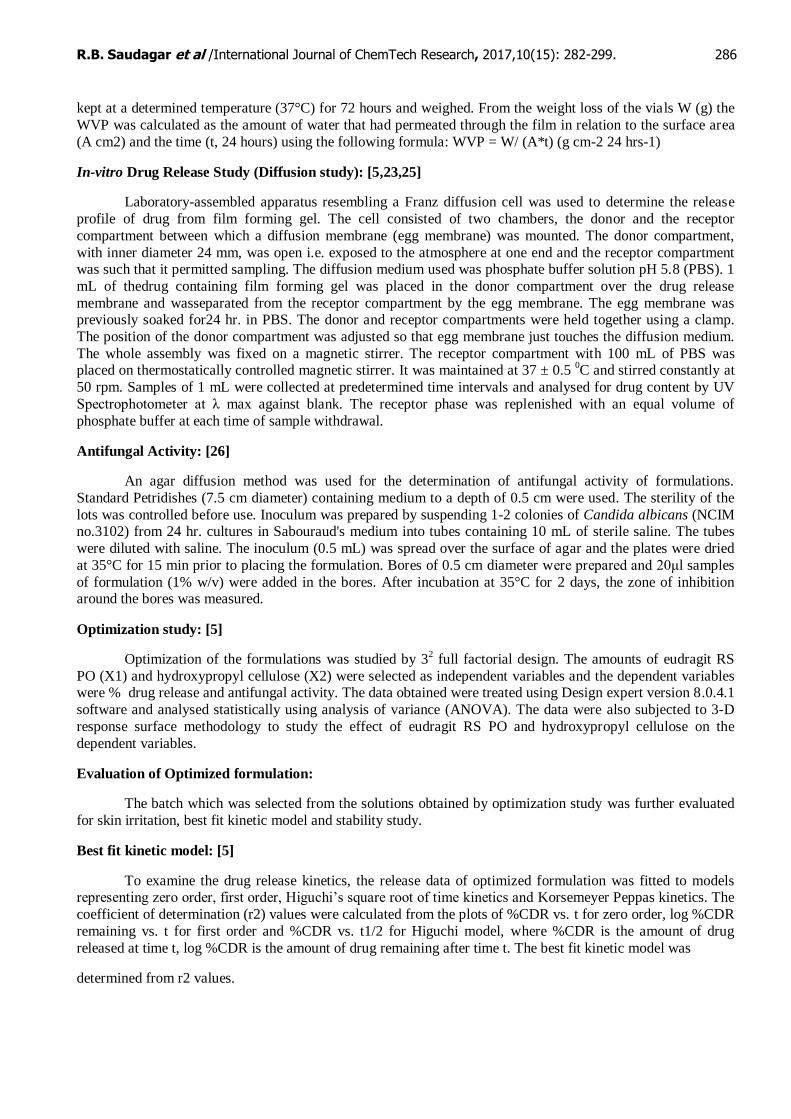
formulationsdisplayed pH values within acceptable range. The viscosity profile of formulations F1 to F9 has
been shown infigure I.
Figure I: Viscosity profile of formulations
Drug content:
The Drug content of formulations is shown in table IV. The percentage drug content of all prepared
dermalformulations was found to be in the range of 98-102 %. Therefore uniformity of content was maintained in allformulations.
Sr.
no.
Formulation code Drug content (%) (±S.D.)
1. F1 98.45 ±0.05
2. F2 98.75±0.2
3. F3 100.05±0.05
4. F4 99.23±0.22
5. F5 99.76±0.33
6. F6 98±0.25
7. F7 98.15±0.25
8. F8 101.33±0.38
9. F9 100.35±0.1
0
2000
4000
6000
8000
1 2 3 4
VIS
CO
SITY
(CP
)
SPPED (RPM)
speed F1 F2 F3
F4 F5 F6 F7
R.B. Saudagar et al /International Journal of ChemTech Research, 2017,10(15): 282-299. 288
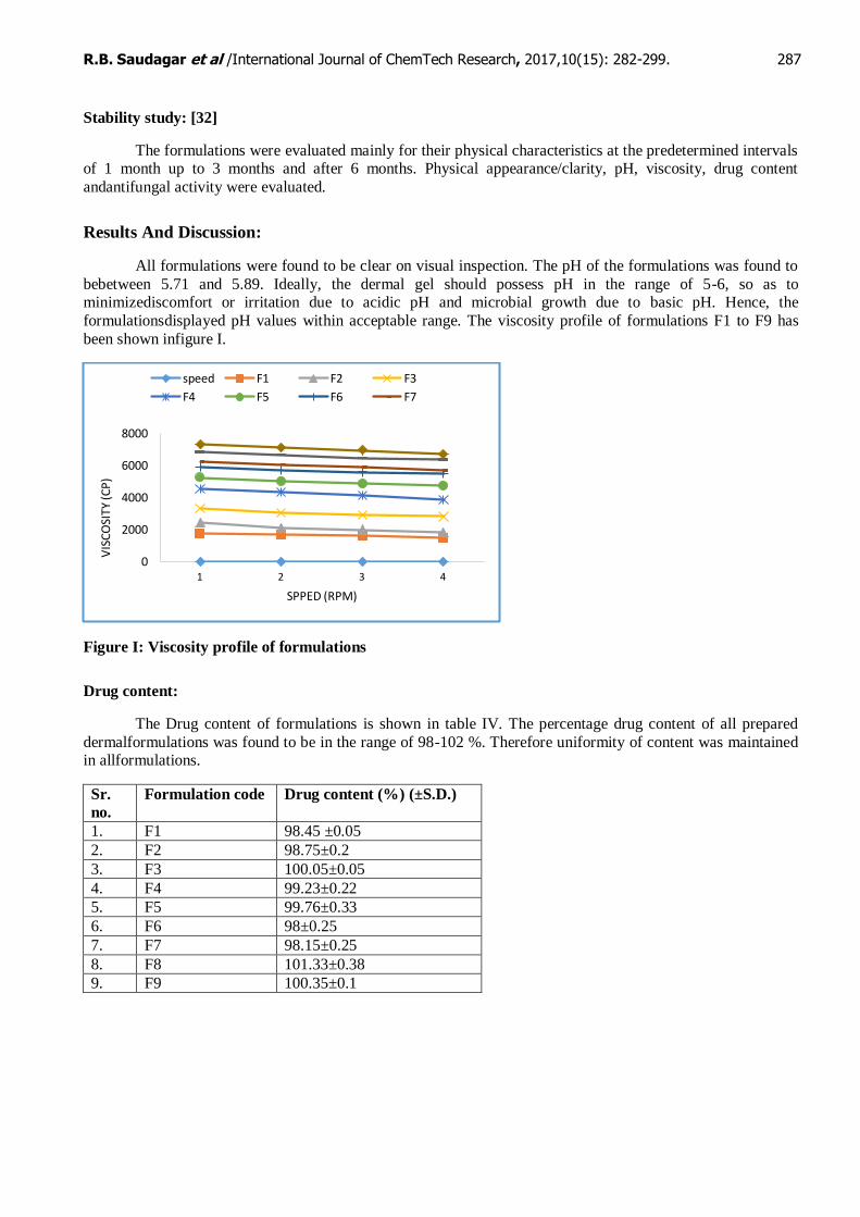
Drying time:
The drying time or film formation time for formulations F1 to F9 has been tabulated in table V.
Sr.
no.
Formulation code Drying time
1. F1 2 min 29 sec ±5 sec
2. F2 2 min 56 sec ± 5 sec
3. F3 3 min 05 sec ± 5 sec
4. F4 3 min 21 sec ± 5 sec
5. F5 3 min 42 sec ± 5 sec
6. F6 4 min 02 sec ± 5 sec
7. F7 4 min 19 sec ± 5 sec
8. F8 4 min 48 sec ± 5 sec
9. F9 5 min 29 sec ± 5 sec
Ideally, the dermal gel should dry to form a thin invisible film on the surface of skin at the application sitewithin 5 minutes, so as to minimize discomfort to patient. [8]
Effective dosage volume [33,34]
This test was carried out to define the volume of formulation that would cover a unit area of the skin to
deliveran effective dose. As part of a pre-experiment it was found that a concentration of 0.0509±0.002 mg/cm2
wasmade available by commercial cream. Thus, a volume of formulation that could deliver equivalent amount ofdrug was needed to be calculated. 1 mL of formulation covered an area of 2 cm2. This volume contains 1 mg
of the drug. That means the formulation delivers 0.5 mg/cm2 which is effective as stated in literature (0.5 to
25mg/cm2).
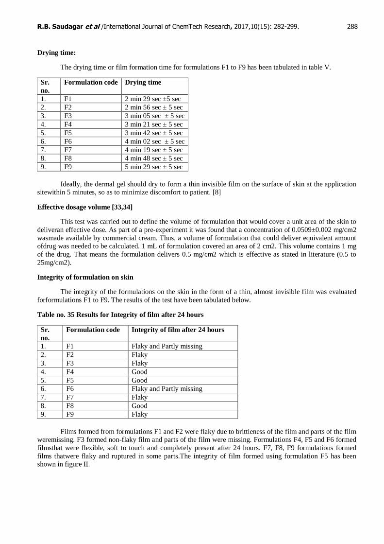
Integrity of formulation on skin
The integrity of the formulations on the skin in the form of a thin, almost invisible film was evaluated
forformulations F1 to F9. The results of the test have been tabulated below.
Table no. 35 Results for Integrity of film after 24 hours
Sr.
no.
Formulation code Integrity of film after 24 hours
1. F1 Flaky and Partly missing
2. F2 Flaky
3. F3 Flaky
4. F4 Good
5. F5 Good
6. F6 Flaky and Partly missing
7. F7 Flaky
8. F8 Good
9. F9 Flaky
Films formed from formulations F1 and F2 were flaky due to brittleness of the film and parts of the film weremissing. F3 formed non-flaky film and parts of the film were missing. Formulations F4, F5 and F6 formed
filmsthat were flexible, soft to touch and completely present after 24 hours. F7, F8, F9 formulations formed
films thatwere flaky and ruptured in some parts.The integrity of film formed using formulation F5 has been shown in figure II.

R.B. Saudagar et al /International Journal of ChemTech Research, 2017,10(15): 282-299. 289
A] On applicationB] After 24 hours (with a peeled portion)
Properties of film:
Outward stickiness
The results for outward stickiness of the formulations have been tabulated in table VII.
Table no. 36 Results for Outward stickiness
Sr.
no.
Formulation
code
Observation
1. F1 Low
2. F2 Low
3. F3 Medium
4. F4 Low
5. F5 Low
6. F6 Medium
7. F7 Low
8. F8 Low
9. F9 Medium
Cosmetic attractiveness
Results for cosmetic attractiveness of the film formed after drying have been given in table VII.
Table VIII: Results for Cosmetic attractiveness
Sr.
no.
Formulation
code
Observation
1. F1 Medium
2. F2 Medium
3. F3 Medium
4. F4 High
5. F5 High
6. F6 High
7. F7 Low
8. F8 Low
9. F9 Low
R.B. Saudagar et al /International Journal of ChemTech Research, 2017,10(15): 282-299. 290
Mechanical properties of the film
Film thickness
Table IX shows the values for film thickness of films formed from various formulations.
Table IX: Results for Film thickness
Sr.
no.
Formulation code Observed value (±S.D.) (mm)
1. F1 0.491 ±0.001
2. F2 0.534 ±0.002
3. F3 0.631 ±0.001
4. F4 0.495 ±0.001
5. F5 0.531 ±0.001
6. F6 0.635 ±0.001
7. F7 0.48 ±0.001
8. F8 0.537 ±0.001
9. F9 0.641 ±0.001
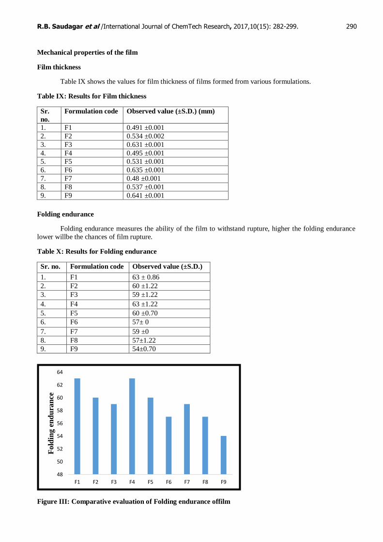
Folding endurance
Folding endurance measures the ability of the film to withstand rupture, higher the folding endurance
lower willbe the chances of film rupture.
Table X: Results for Folding endurance
Sr. no. Formulation code Observed value (±S.D.)
1. F1 63 ± 0.86
2. F2 60 ±1.22
3. F3 59 ±1.22
4. F4 63 ±1.22
5. F5 60 ±0.70
6. F6 57± 0
7. F7 59 ±0
8. F8 57±1.22
9. F9 54±0.70
Figure III: Comparative evaluation of Folding endurance offilm
48
50
52
54
56
58
60
62
64
F1 F2 F3 F4 F5 F6 F7 F8 F9
Fold
ing e
nd
ura
nce
R.B. Saudagar et al /International Journal of ChemTech Research, 2017,10(15): 282-299. 291
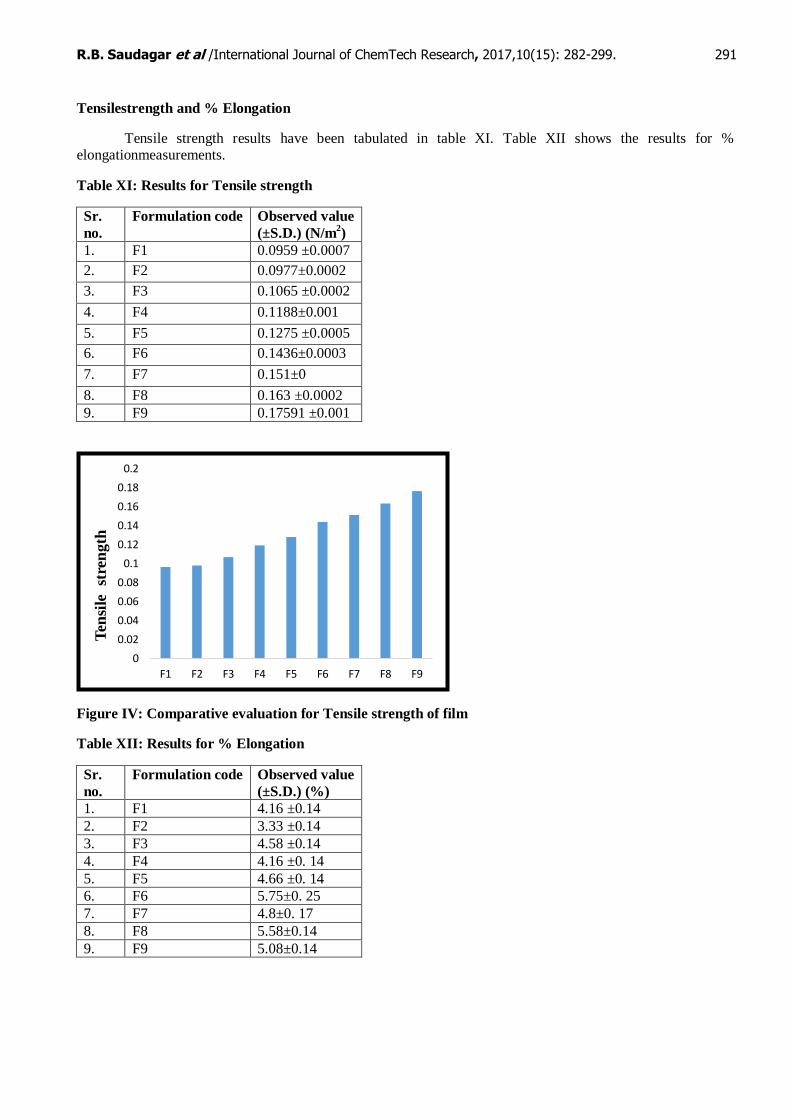
Tensilestrength and % Elongation
Tensile strength results have been tabulated in table XI. Table XII shows the results for % elongationmeasurements.
Table XI: Results for Tensile strength
Sr.
no.
Formulation code Observed value
(±S.D.) (N/m2)
1. F1 0.0959 ±0.0007
2. F2 0.0977±0.0002
3. F3 0.1065 ±0.0002
4. F4 0.1188±0.001
5. F5 0.1275 ±0.0005
6. F6 0.1436±0.0003
7. F7 0.151±0
8. F8 0.163 ±0.0002
9. F9 0.17591 ±0.001
Figure IV: Comparative evaluation for Tensile strength of film
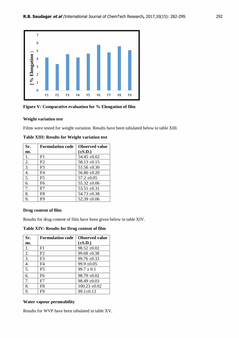
Table XII: Results for % Elongation
Sr.
no.
Formulation code Observed value
(±S.D.) (%)
1. F1 4.16 ±0.14
2. F2 3.33 ±0.14
3. F3 4.58 ±0.14
4. F4 4.16 ±0. 14
5. F5 4.66 ±0. 14
6. F6 5.75±0. 25
7. F7 4.8±0. 17
8. F8 5.58±0.14
9. F9 5.08±0.14
0
0.02
0.04
0.06
0.08
0.1
0.12
0.14
0.16
0.18
0.2
F1 F2 F3 F4 F5 F6 F7 F8 F9
Ten
sile
st
ren
gth
R.B. Saudagar et al /International Journal of ChemTech Research, 2017,10(15): 282-299. 292
Figure V: Comparative evaluation for % Elongation of film
Weight variation test
Films were tested for weight variation. Results have been tabulated below in table XIII.
Table XIII: Results for Weight variation test
Sr.
no.
Formulation code Observed value
(±S.D.)
1. F1 54.45 ±0.02
2. F2 58.13 ±0.15
3. F3 51.56 ±0.30
4. F4 56.86 ±0.20
5. F5 57.2 ±0.05
6. F6 55.32 ±0.06
7. F7 53.51 ±0.31
8. F8 54.73 ±0.38
9. F9 52.39 ±0.06
Drug content of film
Results for drug content of film have been given below in table XIV.
Table XIV: Results for Drug content of film
Sr.
no.
Formulation code Observed value
(±S.D.)
1. F1 98.52 ±0.02
2. F2 99.68 ±0.38
3. F3 99.76 ±0.33
4. F4 99.9 ±0.05
5. F5 99.7 ± 0.1
6. F6 98.79 ±0.02
7. F7 98.49 ±0.03
8. F8 100.23 ±0.02
9. F9 99.1±0.13
Water vapour permeability
Results for WVP have been tabulated in table XV.
0
1
2
3
4
5
6
7
F1 F2 F3 F4 F5 F6 F7 F8 F9
( %
Elo
ngati
on
)
R.B. Saudagar et al /International Journal of ChemTech Research, 2017,10(15): 282-299. 293
Table XV: Results for WVP determination
Sr.
no.
Formulation
code
Observed value (±S.D.) (g
cm-2
24h-1
)
1. F1 0.0463±0.005
2. F2 0.0505 ±0.008
3. F3 0.0540 ±0.001
4. F4 0.0539 ±0.009
5. F5 0.0502±0.003
6. F6 0.051 ±0.004
7. F7 0.0506 ±0.002
8. F8 0.0525 ±0.002
9. F9 0.0516 ±0.005
According to the British Pharmacopoeia a material can be considered permeable to water vapour when
the WVP exceeds 0.05 g cm-2 24h-1. [8] The films displayed such WVP values that show permeability above
the limit set in the Pharmacopoeia and can therefore be considered non-occlusive.
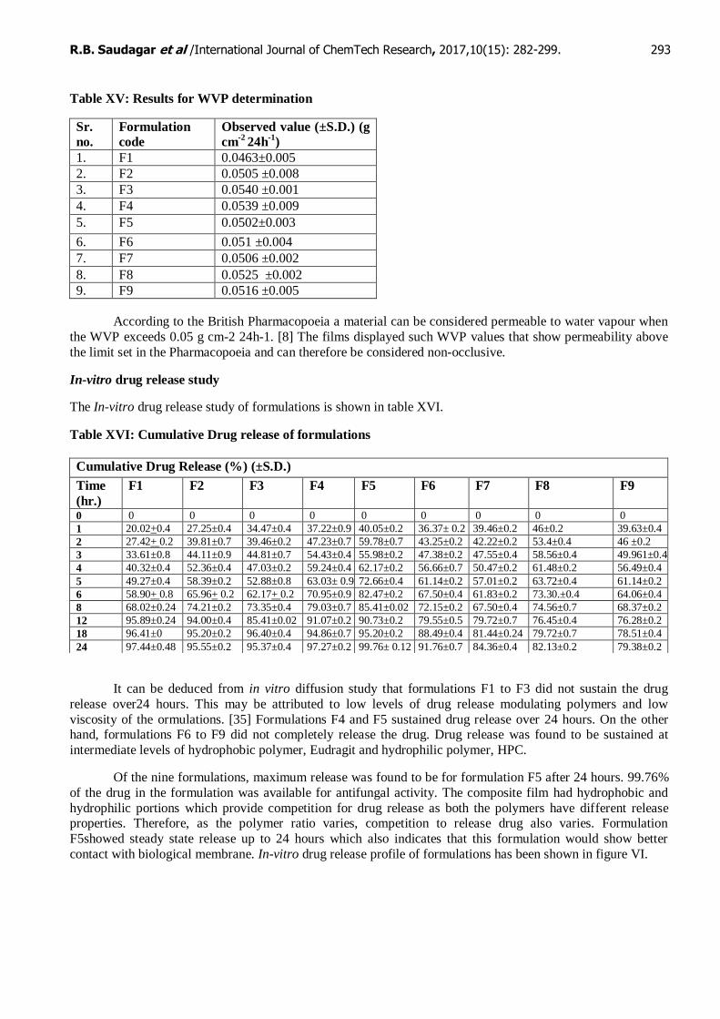
In-vitro drug release study
The In-vitro drug release study of formulations is shown in table XVI.
Table XVI: Cumulative Drug release of formulations
It can be deduced from in vitro diffusion study that formulations F1 to F3 did not sustain the drug
release over24 hours. This may be attributed to low levels of drug release modulating polymers and low
viscosity of the ormulations. [35] Formulations F4 and F5 sustained drug release over 24 hours. On the other hand, formulations F6 to F9 did not completely release the drug. Drug release was found to be sustained at
intermediate levels of hydrophobic polymer, Eudragit and hydrophilic polymer, HPC.
Of the nine formulations, maximum release was found to be for formulation F5 after 24 hours. 99.76%
of the drug in the formulation was available for antifungal activity. The composite film had hydrophobic and
hydrophilic portions which provide competition for drug release as both the polymers have different release properties. Therefore, as the polymer ratio varies, competition to release drug also varies. Formulation
F5showed steady state release up to 24 hours which also indicates that this formulation would show better
contact with biological membrane. In-vitro drug release profile of formulations has been shown in figure VI.
Cumulative Drug Release (%) (±S.D.)
Time
(hr.)
F1 F2 F3 F4 F5 F6 F7 F8 F9
0 0 0 0 0 0 0 0 0 0
1 20.02+0.4 27.25±0.4 34.47±0.4 37.22±0.9 40.05±0.2 36.37± 0.2 39.46±0.2 46±0.2 39.63±0.4
2 27.42+ 0.2 39.81±0.7 39.46±0.2 47.23±0.7 59.78±0.7 43.25±0.2 42.22±0.2 53.4±0.4 46 ±0.2
3 33.61±0.8 44.11±0.9 44.81±0.7 54.43±0.4 55.98±0.2 47.38±0.2 47.55±0.4 58.56±0.4 49.961±0.4
4 40.32±0.4 52.36±0.4 47.03±0.2 59.24±0.4 62.17±0.2 56.66±0.7 50.47±0.2 61.48±0.2 56.49±0.4
5 49.27±0.4 58.39±0.2 52.88±0.8 63.03± 0.9 72.66±0.4 61.14±0.2 57.01±0.2 63.72±0.4 61.14±0.2
6 58.90+ 0.8 65.96+ 0.2 62.17+ 0.2 70.95±0.9 82.47±0.2 67.50±0.4 61.83±0.2 73.30.±0.4 64.06±0.4
8 68.02±0.24 74.21±0.2 73.35±0.4 79.03±0.7 85.41±0.02 72.15±0.2 67.50±0.4 74.56±0.7 68.37±0.2
12 95.89±0.24 94.00±0.4 85.41±0.02 91.07±0.2 90.73±0.2 79.55±0.5 79.72±0.7 76.45±0.4 76.28±0.2
18 96.41±0 95.20±0.2 96.40±0.4 94.86±0.7 95.20±0.2 88.49±0.4 81.44±0.24 79.72±0.7 78.51±0.4
24 97.44±0.48 95.55±0.2 95.37±0.4 97.27±0.2 99.76± 0.12 91.76±0.7 84.36±0.4 82.13±0.2 79.38±0.2

R.B. Saudagar et al /International Journal of ChemTech Research, 2017,10(15): 282-299. 294
Figure VI: In-vitro drug release profile of formulations F1 to F9
Antifungal activity
The results of antifungal activity of formulations have been shown in table XVII.
Table XVII Zone of inhibition and % efficacy of formulations
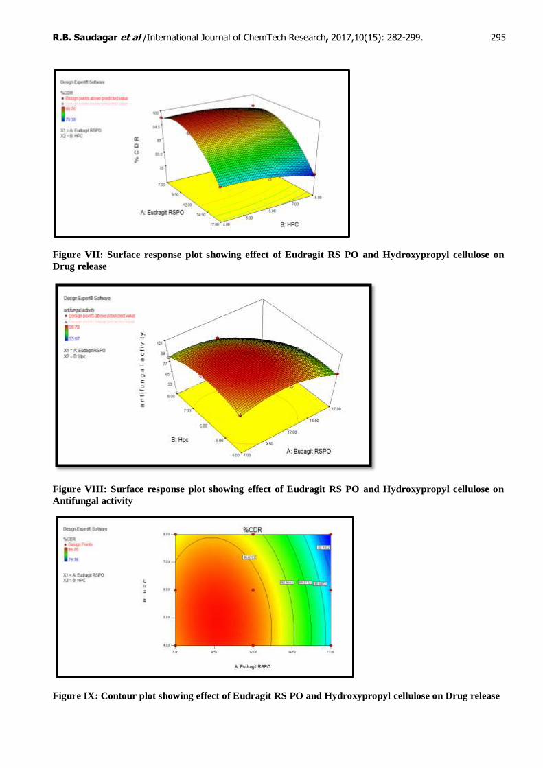
Optimization: [5]
From design expert version 7.1.6 thirty nine solutions were found. The batch with Eudragit 12.5 % w/v
and HPC 6 % w/v with desirability 1 was found to be optimum. From this data formulation F5 was selected as
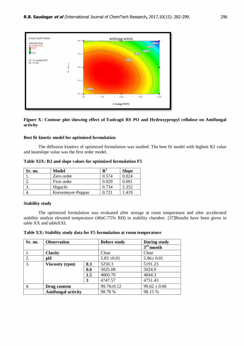
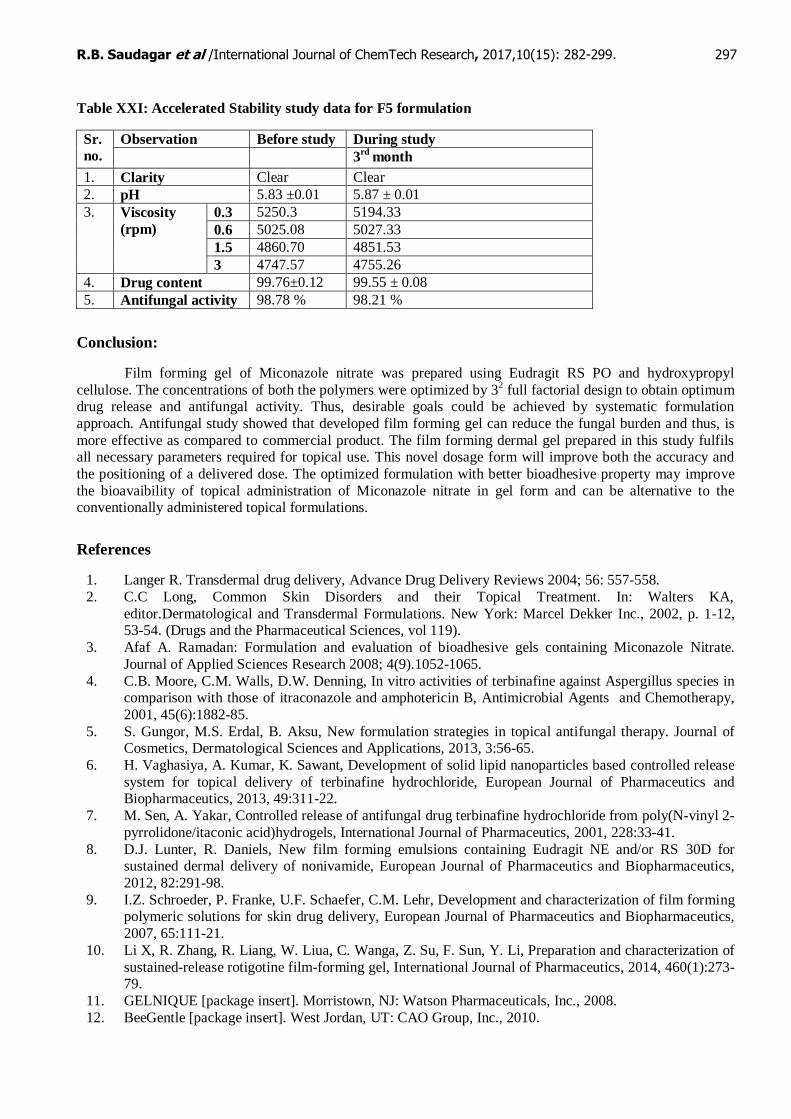
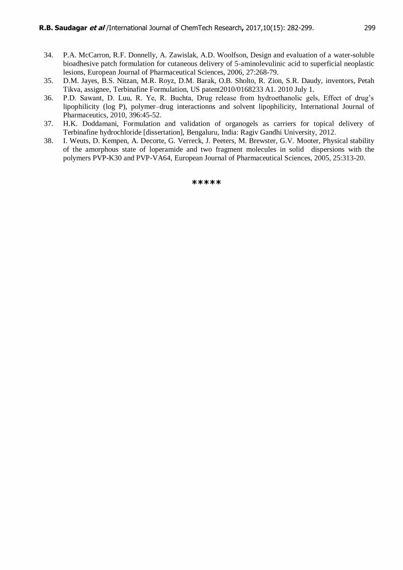
the optimum formulation. The figures below show the effect of concentration of Eudragit RS PO and
Hydroxypropyl cellulose on drug release and antifungal activity. It is shown that both the independent variables have a significant effect on the dependent variables and drug release and antifungal activity decrease as
concentration of polymers increases. Figure VII: Surface response plot showing effect of Eudragit RS PO and
Hydroxypropyl cellulose on Drug release.
0
50
100
150
0 10 20 30
% C
DR
Time (hrs)
In vitro drug release
Sr. no. Formulation Code Candida albicans NCIM no. 3102
Zone of Inhibition
(mm) ±SD
% Efficacy
1. Standard value 14 100
2. F1 12.33±0.57 88.07
3. F2 13.33±0.57 95.21
4. F3 11.56±0.31 82.57
5. F4 12.82±0.27 91.57
6. F5 13.83±0.15 98.78
7. F6 11.3±0.3 80.71
8. F7 11.10±0.10 79.28
9. F8 10.60±0.13 75.71
10. F9 7.43±0.51 53.07
11. Ethanol (control) 1.27±0.23 5.28
12. Drug suspension 13.55±0.05 96.78
13. Commercial cream 11.1±0.1 79.28
R.B. Saudagar et al /International Journal of ChemTech Research, 2017,10(15): 282-299. 295
Figure VII: Surface response plot showing effect of Eudragit RS PO and Hydroxypropyl cellulose on
Drug release
Figure VIII: Surface response plot showing effect of Eudragit RS PO and Hydroxypropyl cellulose on
Antifungal activity
Figure IX: Contour plot showing effect of Eudragit RS PO and Hydroxypropyl cellulose on Drug release
R.B. Saudagar et al /International Journal of ChemTech Research, 2017,10(15): 282-299. 296
Figure X: Contour plot showing effect of Eudragit RS PO and Hydroxypropyl cellulose on Antifungal
activity
Best fit kinetic model for optimized formulation
The diffusion kinetics of optimized formulation was studied. The best fit model with highest R2 value
and leastslope value was the first order model.
Table XIX: R2 and slope values for optimized formulation F5
Sr. no. Model R2 Slope
1. Zero order 0.574 0.024
2. First order 0.929 0.091
3. Higuchi 0.734 2.352
4. Korsemeyer-Peppas 0.721 1.419
Stability study
The optimized formulation was evaluated after storage at room temperature and after accelerated
stability studyat elevated temperature (40oC/75% RH) in stability chamber. [37]Results have been given in table XX and tableXXI.
Table XX: Stability study data for F5 formulation at room temperature
Sr. no. Observation Before study During study
3rd
month
1. Clarity Clear Clear
2. pH 5.83 ±0.01 5.86± 0.01
3. Viscosity (rpm) 0.3 5250.3 5191.23
0.6 5025.08 5024.9
1.5 4860.70 4844.3
3 4747.57 4751.43
4. Drug content 99.76±0.12 99.62 ± 0.06
Antifungal activity 98.78 % 98.15 %
R.B. Saudagar et al /International Journal of ChemTech Research, 2017,10(15): 282-299. 297
Table XXI: Accelerated Stability study data for F5 formulation
Sr.
no.
Observation Before study During study
3rd
month
1. Clarity Clear Clear
2. pH 5.83 ±0.01 5.87 ± 0.01
3. Viscosity
(rpm)
0.3 5250.3 5194.33
0.6 5025.08 5027.33
1.5 4860.70 4851.53
3 4747.57 4755.26
4. Drug content 99.76±0.12 99.55 ± 0.08
5. Antifungal activity 98.78 % 98.21 %
Conclusion:
Film forming gel of Miconazole nitrate was prepared using Eudragit RS PO and hydroxypropyl
cellulose. The concentrations of both the polymers were optimized by 32 full factorial design to obtain optimum
drug release and antifungal activity. Thus, desirable goals could be achieved by systematic formulation
approach. Antifungal study showed that developed film forming gel can reduce the fungal burden and thus, is
more effective as compared to commercial product. The film forming dermal gel prepared in this study fulfils all necessary parameters required for topical use. This novel dosage form will improve both the accuracy and
the positioning of a delivered dose. The optimized formulation with better bioadhesive property may improve
the bioavaibility of topical administration of Miconazole nitrate in gel form and can be alternative to the conventionally administered topical formulations.
References
1. Langer R. Transdermal drug delivery, Advance Drug Delivery Reviews 2004; 56: 557-558.
2. C.C Long, Common Skin Disorders and their Topical Treatment. In: Walters KA,
editor.Dermatological and Transdermal Formulations. New York: Marcel Dekker Inc., 2002, p. 1-12, 53-54. (Drugs and the Pharmaceutical Sciences, vol 119).
3. Afaf A. Ramadan: Formulation and evaluation of bioadhesive gels containing Miconazole Nitrate.
Journal of Applied Sciences Research 2008; 4(9).1052-1065.
4. C.B. Moore, C.M. Walls, D.W. Denning, In vitro activities of terbinafine against Aspergillus species in comparison with those of itraconazole and amphotericin B, Antimicrobial Agents and Chemotherapy,
2001, 45(6):1882-85.
5. S. Gungor, M.S. Erdal, B. Aksu, New formulation strategies in topical antifungal therapy. Journal of Cosmetics, Dermatological Sciences and Applications, 2013, 3:56-65.
6. H. Vaghasiya, A. Kumar, K. Sawant, Development of solid lipid nanoparticles based controlled release
system for topical delivery of terbinafine hydrochloride, European Journal of Pharmaceutics and Biopharmaceutics, 2013, 49:311-22.
7. M. Sen, A. Yakar, Controlled release of antifungal drug terbinafine hydrochloride from poly(N-vinyl 2-
pyrrolidone/itaconic acid)hydrogels, International Journal of Pharmaceutics, 2001, 228:33-41.
8. D.J. Lunter, R. Daniels, New film forming emulsions containing Eudragit NE and/or RS 30D for sustained dermal delivery of nonivamide, European Journal of Pharmaceutics and Biopharmaceutics,
2012, 82:291-98.
9. I.Z. Schroeder, P. Franke, U.F. Schaefer, C.M. Lehr, Development and characterization of film forming polymeric solutions for skin drug delivery, European Journal of Pharmaceutics and Biopharmaceutics,
2007, 65:111-21.
10. Li X, R. Zhang, R. Liang, W. Liua, C. Wanga, Z. Su, F. Sun, Y. Li, Preparation and characterization of
sustained-release rotigotine film-forming gel, International Journal of Pharmaceutics, 2014, 460(1):273-79.
11. GELNIQUE [package insert]. Morristown, NJ: Watson Pharmaceuticals, Inc., 2008.
12. BeeGentle [package insert]. West Jordan, UT: CAO Group, Inc., 2010.

R.B. Saudagar et al /International Journal of ChemTech Research, 2017,10(15): 282-299. 298
13. J.L. Zatz, G.P. Kushla, Gels In: H.A. Lieberman, M.M. Rieger, G.S. Banker, editors. Pharmaceutical
Dosage Forms-Disperse System. 2nd ed. New York: Marcel Dekker Inc. p.399-405. (Drugs and the
Pharmaceutical Sciences, vol 2). 14. K. Saroha, S. Singh, A. Aggarwal, S. Nanda, Transdermal Gels- An alternative vehicle for drug
delivery, International Journal of Pharmaceutical, Chemical and Biological Sciences 2013, 3(3):495-03.
15. M.A. Attia, H.Y. Badawy, Film forming gel for treatment of oral mucositis: In vitro studies,
International Journal of Drug Delivery, 2010, 2:314-321. 16. R. Guo , Du X., Zhang R., Deng L., Dong A., Zhang J., Bioadhesive film formed from a novel organic–
inorganic hybrid gel for transdermal drug delivery system, European Journal of Pharmaceutics and
Biopharmaceutics, 2011, 79:574-83. 17. L.K. Souza, C.H. Bruno, L. Lopes, S.H. Pulcinelli, C.V. Santilli, L.A. Chiavacci, Ureasil–polyether
hybrid film-forming materials, Colloids and Surfaces B: Biointerfaces, 2013, 101:156-61.
18. D.M. Jayes, B.S. Nitzan, M.R. Royz, D.M. Barak, O.B. Sholto, R. Zion, S.R. Daudy, inventors, Petah Tikva, assignee, Terbinafine Formulation, US patent2010/0168233 A1. 2010 July 1.
19. S.M. Mohamed, A.M.E. Masoud, M.D. Elgadir, M.A. Mahdy, Preparation and release charcteristics of
itraconazole polymeric films for topical application, International Journal of Pharmacy and
Pharmaceutical Sciences, 2013, 5(3):167-70. 20. B.K. Dey, P.K. Kar, L.K. Nath, Formulation, design, preparation and in vitro-in vivo evaluation of
propranolol hydrochloride transdermal patches using hydrophilic and hydrophobic polymer complex,
Research Journal of Pharmacy and Technology, 2009, 2(1):155-60. 21. Y.B. Ubarchande, T. Regupathy, C. Vijaya, S.V. Deshmane, Formulation and evaluation of
mucoadhesive buccal films of losartan potassium, Research Journal of Pharmacy and Technology,
2009, 2(4):833-36. 22. C. Suja, C. Ramasamy, R. Narayanacharyula, Development and evaluation of lisinopril transdermal
patches, Research Journal of Pharmacy and Technology. 2011, 4(8):1260-64.
23. S.V. Kulkarni, R.P. Kumar, N. Patel, R.B. Someshwara, A.P. Kumar, Development and evaluation of
diltiazem HCl transdermal patches by using glycerol and castor oil as plasticizers, Research Journal of Pharmacy and Technology, 2010, 3(3):905-09.
24. J.R. Kumar, S. Muralidharan, S.A. Dhanaraj, Formulation and in-vitro evaluation of terbinafine
hydrochloride transdermal patches, Journal of Pharmaceutical Sciences & Research, 2012, 4(6):1840-43.
25. D.K. Jain, G.N. Darwhekar, S. Chaurasia, Formulation development and evaluation of transdermal
patches of losartan, International Journal of Pharmtech Research, 2012, 4(2):757-64.
26. D.S. Kumar, R. Sairam, S. Anandbabu, L. Karpagavalli, A. Maheswaran, N. Narayanan, Formulation and evaluation of transdermal patches of salbutamol, Research Journal of Pharmaceutical, Biological
and Chemical Sciences, 2012, 3(3):1132-38.
27. P. Verma, K. Pathak, Nanosized ethanolic vesicles loaded with econazole nitrate for the treatment of deep fungal infections through topical gel formulation, Nanomedicine: Nanotechnology, Biology, and
Medicine, 2012, 8:489-96.
28. S. Amin, S.R. Mir, K. Kohli, A. Ali, Novel polymeric matrix films for transdermal delivery of metoclopramide, International Journal of Pharmaceutical Frontier Research, 2012, 2(1):48-60.
29. R. Vijaya, S. Sureshkumar, S. Umamaheswari, M. Prakash, T. Senbagapriya, A. Umamaheswari,
Preparation of amitriptyline hydrochloride films using eudragit RL 100 and hydroxypropyl methyl
cellulose polymers and their in vitro evaluation for effective transdermal delivery, International Journal of Life Science and Pharma Research, 2012, 2(2):7-15.
30. M. Bharkatiya, R.K. Nema, M. Bhatnagar, Designing and characterization of drug free patches for
transdermal application, International Journal of Pharmaceutical Sciences and Drug Research, 2010, 2(1):35-39.
31. R. Khullar, D. Kumar, N. Seth, S. Saini, Formulation and evaluation of mefenamic acid emulgel for
topical delivery, Saudi Pharmaceutical Journal, 2012, 20:63-67. 32. W. Zhua, C. Guoa, A. Yua, Y. Gaoa, F. Caoa, G. Zhai, Microemulsion-based hydrogel formulation of
penciclovir for topical delivery, International Journal of Pharmaceutics, 2009, 378:152-58.
33. International Conference on Harmonization Steering Committee, ICH Harmonized Tripartite Guideline-
Stability Testing of New Drug Substances and Products, ICH Q1A (R2), February 6, 2003.
R.B. Saudagar et al /International Journal of ChemTech Research, 2017,10(15): 282-299. 299
34. P.A. McCarron, R.F. Donnelly, A. Zawislak, A.D. Woolfson, Design and evaluation of a water-soluble
bioadhesive patch formulation for cutaneous delivery of 5-aminolevulinic acid to superficial neoplastic
lesions, European Journal of Pharmaceutical Sciences, 2006, 27:268-79. 35. D.M. Jayes, B.S. Nitzan, M.R. Royz, D.M. Barak, O.B. Sholto, R. Zion, S.R. Daudy, inventors, Petah
Tikva, assignee, Terbinafine Formulation, US patent2010/0168233 A1. 2010 July 1.
36. P.D. Sawant, D. Luu, R. Ye, R. Buchta, Drug release from hydroethanolic gels, Effect of drug’s
lipophilicity (log P), polymer–drug interactionns and solvent lipophilicity, International Journal of Pharmaceutics, 2010, 396:45-52.
37. H.K. Doddamani, Formulation and validation of organogels as carriers for topical delivery of
Terbinafine hydrochloride [dissertation], Bengaluru, India: Ragiv Gandhi University, 2012. 38. I. Weuts, D. Kempen, A. Decorte, G. Verreck, J. Peeters, M. Brewster, G.V. Mooter, Physical stability
of the amorphous state of loperamide and two fragment molecules in solid dispersions with the
polymers PVP-K30 and PVP-VA64, European Journal of Pharmaceutical Sciences, 2005, 25:313-20.
*****