intrauterinegrowthrestriction 130120060505-phpapp02 - copy
TRANSCRIPT

Intrauterine Growth Intrauterine Growth RestrictionRestriction
(IUGR)(IUGR)
Dr. sadaf ijazDr. sadaf ijaz

Definition
• Fetuses of birth weight less than 10th percentile of those born at same gestational age
ortwo standard deviations below the population mean are considered growth restricted.
« small for gestational age »(SGA) fetuses, all of which may not necessarily growth restricted as many of these may be just constitutionally small and not at risk of any adverse outcome.)

Rcog guidelines
• SGA is defined as EFW or AC less than 10th centile and sever SGA when these are less than 3rd centile.
• 50 -70% of these may be just constitutionally small and not at risk of any adverse outcome.

Incidence
• 3 - 5 % of all pregnancies.• 1/3 of infants with BW < 2800 grams are growth
retarded and not premature.• 9 - 27 % have anatomic and/or genetic
abnormalities.• Perinatal mortality is 6 - 10 times higher for
these fetuses.• 30% of all stillborn infants are growth restricted.

• « Therefore the term IUGR should more strictly refer to fetuses that are small for gestational age and display other signs of chronic hypoxia or failure to thrive. »

Normal Fetal Growth• Fetal growth depends on two components:
– Genetic Potential – Substrate supply
The genetic potential is derived from both parents and is mediated through growth factors such as insulin-like growth factor.
Adequate substrate supply is essential to achieve genetic potential. This is derived from placenta which is dependent upon the uterine and placental vascularity

A comparision between normal and IUGR babies.

Normal and IUGR placenta

Normal Fetal Growth
• Normal fetal growth is characterized by
cellular hyperplasia followed by
hyperplasia and hypertrophy and lastly by
hypertrophy alone.
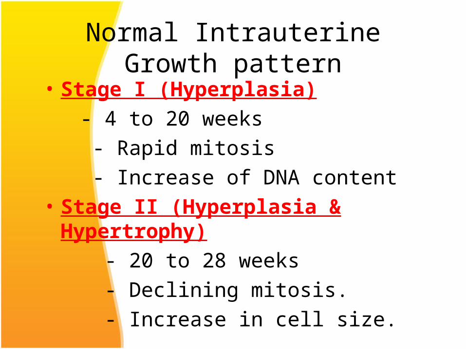
Normal Intrauterine Growth pattern
• Stage I (Hyperplasia)
- 4 to 20 weeks
- Rapid mitosis
- Increase of DNA content
• Stage II (Hyperplasia & Hypertrophy)
- 20 to 28 weeks
- Declining mitosis.
- Increase in cell size.

Normal Intrauterine Growth pattern
• Stage III ( Hypertrophy) - 28 to 40 weeks - Rapid increase in cell size. - Rapid accumulation of fat, muscle and
connective tissue.• 95% of fetal weight gain occurs during last 20
weeks of gestations.

Weight gain• Fetal growth accelerates from about 5g
per day at 14 -15 wks of gestation to
• 10g per day at 20 wks
• Peaks at 30 -35g per day at 32-34wks
• After which growth rate decreases.
Fetal Growth indices

• Symphysiofundal height increases by about 1cm per wk between 14 and 32 wks.
• Abdominal girth increases by 1 inch per wk after 30 wks. It is about 30 inches at 30wks in an average built woman.

Classification of Inrauterine Growth Restriction
1. Type 1 or symmetrical or intrinsic IUGR
2. Type 2 or assymetric IUGR
3. Intermediate IUGR

Classification
SymmetricSymmetricll AAsymmetricalsymmetrical
Baby's brain is abnormally large when compared to the liver.may occur when the fetus experiences a problem during later development .and is seen in undernourished fetus who is compensating by directing most of its energy to maintaining vitals organs,
The baby's head and body are proportionately small. may occur when the fetus experiences a problem during early development.
In a normal infant, the brain weighs about three times more than the liver.
In asymmetrical IUGR, the brain can weigh five or six times more than the
liver.

Types of IUGR
• Symmetric IUGR: weight,length and head circumference are all below the 10th percentile. (33 % of IUGR Infants)
• Asymmetric IUGR: weight is below the 10 th percentile and head circumference and length are preserved. (55 % of IUGR)
• Combined type IUGR: Infant may have skeletal shortening, some reduction of soft tissue mass.(12% of IUGR)

Ponderal Index
• Way of characterizing the relationship of height to mass for an individual.
• PI = 100 x
• Typical values are 2.0 to 2.5• PI is normal in symmetric IUGR.• PI is low in asymmetric IUGR.
Mass (gm) Height (cm)3

Classification of IUGR
• Type 1 or symmetrical IUGR- (20-30%)
Occurs as a result of growth inhibition early in pregnancy i.e. the hyperplastic stage. Any pathological insult at this phase leads to reduced no. of cells in fetus and overall decreased growth potential.
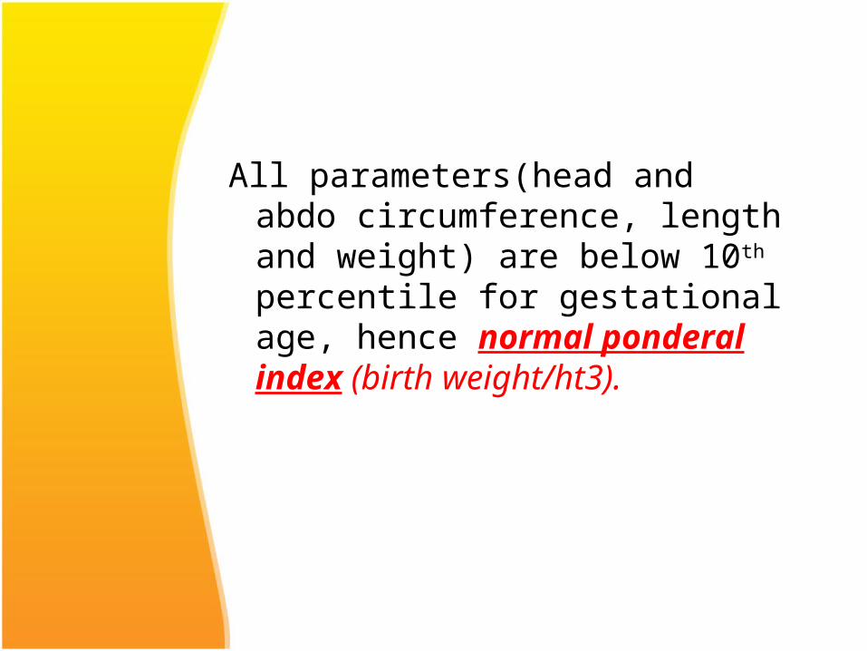
All parameters(head and abdo circumference, length and weight) are below 10th percentile for gestational age, hence normal ponderal index (birth weight/ht3).

Type 2 or asymmetric IUGR (70-80%)
Occurs as a result of restriction of nutrient supply in utero i.e. uteroplacental insufficiency.

• The onset of growth restriction occurs usually after 28 wks of gestation i.e. in the stage of hypertrophy. The fetus has near normal total no. of cells but cell size is reduced.
• There is brain sparing effect so that the head growth remains normal but the abdominal girth slows down.

• The Ponderal index is low.
• This asymmetry results from redistribution of fetal cardiac output with increased flow to brain and heart at the expense of reduced splanchnic circulation.

• Liver size is reduced because of diminished glycogen stores.
• In case of severe placental insufficiency the head growth may also be affected.
This type of growth restriction leads to decreased amniotic fluid, chronic hypoxia and may result in fetal death.

Intermediate IUGR (5-10% of all growth
restricted fetuses)• It is a combination of type 1 and type 2.• Fetal growth restriction occurs during intermediate
phase of growth affecting both hyperplasia and hypertrophy, resulting in decrease in cell no. as well as size.
• Causes includeChronic HTLupus nephritisMaternal vascular diseases that are severe and
have onset in early 2nd trimester

• IUGR may also be classified simply as Early onset (onset before 32
weeks) Late onset (onset after 32
weeks)

Etiology
• IUGR is a manifestation of fetal, maternal and placental disorders that affect fetal growth.

Maternal Fetal Placental Constitutional Cong anomalies Abn Trophoblast Nutritional Chromosomal Velamentous cord Genetic Infections Circumvallate p Collagen vascular ds Multiple preg Pl Praevia Autoimmune disease Pl infarction HTN, Diabetes Tumors Renal disease Environmental Drugs ,anemia cadiac,smoking cocaine,chemotheraputic agnts genetic

Complications of IUGRPerinatal mortality and morbidity of IUGR infants is 10 times greater than normal infants.
• Antepartum period- increased incidence of-
-still births
-oligohydramniosIUGR is found in 30% of unexplained stillbirths.
• During labour- higher incidence of-
-meconium aspiration
-fetal distress
-intrapartum fetal death

Complications of IUGR contd..
• Neonatal period- increased incidence of-
Hypoxic ischemic encephalopathy
Persistent fetal circulation insufficiency
They have difficulty in temperature regulation because of absent brown fat and small body mass relative to surface area.

Lack of glycogen stores may predispose to hypoglycemia
Chronic intrauterine hypoxia may lead to polycythemia, necrotizing enterocolitis, other metabolic abnormalities.

Complications cont..• Childhood- increases mortality from-
Infectious diseasesCongenital anomalies
Incidence of cerebral palsy are 4-6 times higher.
Subtle impairment of cognitive performance and educational underachievement.

Long term complications-• Increased risk of coronary heart
disease • Hypertension, • Type II diabetes mellitus,• Dyslipidaemia and stroke.

Diagnosis of IUGR
Identifying mothers at risk:Poor maternal nutrition Poor BMI at conceptionPre-eclampsiaRenal disordersDiseases causes vascular
insufficiencyInfections (TORCH)Poor maternal wt. gain during
pregnancy

Determination of gestational age is utmost importance-
– Can be calculated from the date of LMP- not reliable.uncertain in 20-40% of cases.
– Ultrasound dating before 21 wks of pregnancy provides more accurate estimate.

Diagnosis of IUGR
1. Clinically- Serial measurement of fundal height and abdominal girth. Symphysio-fundal height normally increases
by 1cm per wk b/w 14 and 32 wks. A lag in fundal ht. of 4wks is suggestive
of moderate IUGR. A lag of >6 wks is suggestive of severe
IUGR. Sensitivity 60-85% & PPV 20-80% Poor screening tool

2. Sonographic evaluation- most useful tool for diagnosis of IUGR To differentiate between symmetrical and
asymmetrical IUGR To monitor the fetal condition.
Fetal biometry:i. BPD(Biparietal Diameter)- determines gestational
age and type of IUGR.When growth rate of BPD is below 5th percentile, 82% of births are below 10th percentile(i.e. IUGR).

ii. Head circumference.
iii. Transverse cerebellar diameter(TCD)
iv. Abdominal circumference(AC).

An increase in fetal AC of less than 10 mm in 14 days has sensitivity of 85% and specificity of 74% for identification of IUGR.
iv. Measurement ratios- there are some age independent ratios to detect IUGR.HC/AC: HC/AC >2 SD above mean is predictive
of IUGR.

Placental Morphology: Acceleration of placental maturation may occur with IUGR and PIH.(Placenta is graded from grade 0 to grade III)
Amniotic fluid volume: type 2 IUGR is usually associated with oligohydramnios.Amniotic fluid index(AFI) between 8 and 25
is normal.

• Placental grading (Grannum classification) refers to a ultrasound grading system of the placenta based on its maturity. This primarily affects the extent of calcifications. In some countries the use of placental grading has fallen out of obstetric practice.
• The grading system is as follows:• grade 0: <18 weeks
– uniform echogenicity– smooth chorionic plate
• grade I: 18-29 weeks– occasional parenchymal calcification/hyperechoic
areas– subtle indentations of chorionic plate

• grade II: >30 weeks– occasional basal calcification/hyperechoic areas– deeper indentations of chorionic plate (does not reach
up to basal plate)• seen as comma type densities at the chorionic
plate• grade III: >39 weeks
– significant basal calcification – chorionic plate interrupted by indentations (frequently
calcified) that reach up to basal plate: cotyledons– an early progression to a grade III placenta
is concerning and is sometimes associated with placental insufficiency

3. Doppler Ultrasonography: doppler flow studies are important adjuncts to fetal biometry in
identifying the IUGR fetuses at risk of adverse outcome.Most widely used arterial indices are :Pulsatility index (PI): Systolic and diastolic peak
velocity / time averaged maximum velocityResistance Index(RI): Systolic and diastolic peak
velocity/ systolic peak velocitySystolic to diastolic ratio(S/D): Systolic peak velocity /
diastolic peak velocity

Umblical Artery doppler- In IUGR there is increased umblical artery resistance (increased S/D ratio), absent end diastolic flow and finally reversed end diastolic flow.Perinatal mortality rate increases significantly in fetuses with absent end diastolic flow (9-41%) and reversed end diastolic flow (33-73%) in umblical artery.


Middle cerebral artery Doppler- in a normal fetus has relatively little flow during diastole. Increased resistance to blood flow in placenta results in redistribution of cardiac output to favour cardiac and cerebral circulations leading to increased flow in the diastolic phase with decreased S/D ratio.


Normal Flow velocity waveforms of middle Cerebral Artery Doppler

Cerebral artery doppler

Cerebral/Placental ratio: ratio between MCA PI and umblical artery PI is more sensiive predictor than either MCA and umblical artery velocimetry alone to detect redistribution of blood flow. Cut off values below 1.0 to 1.1 are considered to be
diagnostic of brain spairing effect.
• MCA peak systolic velocimetry: is good indicator of fetal anaemia and is less useful in IUGR.

Ductus venosus doppler:
Perinatal mortality in growth restricted fetuses has been found to be significantly worse when abnormalities in fetal venous circulation are detected.
In the normal fetus, flow in the ductus venosus is forwards , moving towards the heart during entire cardiac cycle.

Triphasic waveform

• Abnormal waveforms in fetal ductus venosus flow assessment can occur in a number of situations which include
• aneuploidic anomalies– Down syndrome:
• congenital cardiac anomalies– fetal pulmonary arterial anomalies
• congenital pulmonary stenosis• pulmonary atresia
• fetal arteriovenous malformations l• fetal tumours that lead to arterio-venous shunting
– sacrococcygeal teratoma• twin to twin transfusion syndrome: recipient twin• maternal diabetes:

• Abnormal waveforms include• reduction flow in ductus venosus A wave• absent flow in ductus venosus A wave• reversal of flow in ductus venosus A wave• abnormal indices• Growth restricted fetuses with abnormal ductus
venous flow have worse perinatal outcome compared to those where flow abnormality is confined to the umbilical or middle cerebral artery .

When circulatory compensation of the fetus fails, the ductus venosus waveform shows absent or reverse blood flow during atrial conraction. Perinatal mortality being 63-100%.
Therefore it is recommended that fetus should be delivered before the development of absent of reversed blood flow of DV.

: Representation of fetal umbilical and hepatic venous system. The arrows indicate the direction of flow. FO, foramen ovale; RA, right atrium; DV, ductusvenous; UV, umbilical vein; HV, hepatic veins; IVC, inferior venacava; PS, portal sinus; LPV, left portal vein; RPV, right portalvein; EPV, extrahepatic portal vein; GB, gallbladder


MANAGEMENT
Principles:1. Identify the cause of growth restriction.2. Treat the cause if found.3. General management

MANAGEMENT First step is to identify the aetiology of IUGR:-Maternal history pertaining to the risk factors of
IUGR.Clinical examination- maternal habitus, height,
weight, BP etc.Laboratory investigations- Hb, blood sugar, renal
function tests, serology for TORCHSpecific investigations for thrombophilias in pts with history suggestive of early onset growth restriction.

MANAGEMENT
Fetal evaluation: thorough ultrasound for growth restriction, amniotic fluid, congenital anomalies and doppler evaluation

Management cont..
Treatment of underlying cause: such as hypertension, cessation of smoking, protein energy supplementation in poorly nourished and underweight women.

General Management: Bed rest in left lateral position to increase
uteroplacental blood flow Maternal nutritional supplementation with high
caloric and protein diets, antioxidents. Maternal oxygen therapy

Management cont..
Pharmacological therapy:Aspirin in low doses(1-2 mg/kg body wt.), betamimetics etc
Thus there is no form of therapy currently available which can reverse IUGR, the only intervention possible in most cases is delivery.

• Delivery: Since IUGR fetus is at increased risk of intrauterine hypoxia and intrauterine demise, the decision needs to delicately balance the risk to the fetus in utero with continuation of pregnancy and that of prematurity if delivered before term.
RISK OF PREMATURITYDIFFICULT EXTRA
UTERINE EXISTENCE
RISK OF IUD
• HOSTILE INTRA UTERINE ENVIRONMENT
Judge Optimum Time Of DeliveryJudge Optimum Time Of Delivery

• The optimum timing of delivery is determined by gestational age, underlying aetiology, possibility of extrauterine survival and fetal condition.
• Strict fetal surveillance is needed to monitor fetal well being and to detect signs of fetal compromise

Fetal Surveillance
1. Daily fetal movement score2. Non stress test(NST)3. Biophysical profile(BPP)4. Amniotic fluid index(AFI)5. Growth parameters6. Doppler studies
Sonography is usually repeated every 2 wks.

Fetal biophysical profile

Management cont..
• Role of steroids:Antenatal glucocorticoid administration reduces the incidence of respiratory distress syndrome, intraventricular hemorrhage and death in IUGR fetusus weighing less than 1500gm.

Mode of Delivery Fetuses with significant IUGR should be preferably
delivered in well equiped centres which can provide intrapartum continuous fetal heart monitoring , fetal blood sampling and expert neonatal care.
Vaginal delivery: can be allowed as long as there is no obstetric indication for caesarian section and fetal heart rate is normal.
• Fetuses with major anomaly incompatible with life should also be delivered vaginally.

Caesarian section: In all cases of IUGR with features of acidosis caesarian section should be done without trial of labour. These include:Repetitive late decelerationspoor biophysical profilereversal of end diastolic flow in umblical arteryabnormal venous dopplerblood gas analysis showing acidic pH on
cordocentesis.

ConclusionOne of the primary aims of antenatal care is to
identify fetuses which show a significant growth lag, since they are at a high risk of hypoxic complications in the perinatal period.
Management options are limited to close fetal monitoring and termination of pregnancy balancing the risk of prematurity and that of intrauterine demise.


GREEN TOP GUIDELINES
• All women should be assessed at booking for risk factors for a SGA fetus/neonate to identify those who require increased surveillance
• measurement of fetal size and assessment of wellbeing with umbilical artery Doppler from 26–28 weeks of pregnancy.
• Women who have three or more minor risk factors should be referred for uterine artery Doppler at 20–24 weeks of gestation

• A low level (< 0.415 MoM) of the first trimester marker PAPP–A should be considered a major risk factor for delivery of a SGA neonate.
• In high risk populations uterine artery Doppler at 20–24 weeks of pregnancy has a moderate predictive value for a severely SGA neonate.
• In women with an abnormal uterine artery Doppler at 20–24 weeks of pregnancy, subsequent normalisation of flow velocity indices is still associated with an increased risk of a SGA neonate.

• Women with an abnormal uterine artery Doppler at 20–24 weeks and/or notching should be referred for serial ultrasound measurement of fetal size and assessment of wellbeing with umbilical artery Doppler commencing at 26–28 weeks of pregnancy.
• Women with a normal uterine artery Doppler do not require serial measurement of fetal size and serial assessment of wellbeing with umbilical artery Doppler unless they develop specific pregnancy complications, for example antepartum haemorrhage or hypertension.

• Serial ultrasound measurement of fetal size and assessment of wellbeing with umbilical artery Doppler should be offered in cases of fetal echogenic bowel.
• Abdominal palpation has limited accuracy for the prediction of a SGA neonate
• Serial measurement of symphysis fundal height (SFH) is recommended at each antenatal appointment from 24 weeks of pregnancy as this improves prediction of a SGA neonate.

• SFH should be plotted on a customised chart .
• Women with a single SFH which plots below the 10th centile or serial measurements which demonstrate slow or static growth by crossing centiles should be referred for ultrasound measurement of fetal size.
• Women in whom measurement of SFH is inaccurate (for example: BMI > 35, large fibroids, hydramnios) should be referred for serial assessment of fetal size using ultrasound.

Optimum method of diagnosing a SGA fetus and FGR
• Fetal abdominal circumference (AC) or estimated fetal weight (EFW) < 10th centile can be used to diagnose a SGA fetus.

• Routine measurement of fetal AC or EFW in the third trimester does not reduce the incidence of a SGA neonate nor does it improve perinatal outcome.
• Where the fetal AC or EFW is < 10th centile or there is evidence of reduced growth velocity, women should be offered serial assessment of fetal size and umbilical artery Doppler.

Investigations that are indicated in SGA fetuses
• Referral for a detailed fetal anatomical survey and uterine artery Doppler by a fetal medicine specialist if severe SGA is identified at the 18–20 week scan.
• Karyotyping should be offered in severely SGA fetuses with structural anomalies and in those detected before 23 weeks of gestation, especially if uterine artery Doppler is normal.

• Serological screening for congenital cytomegalovirus (CMV) and toxoplasmosis infection should be offered in severely SGA fetuses.
• Testing for syphilis and malaria should be considered in high risk populations.
• Uterine artery Doppler has limited accuracy .