iteration industrial tomography … · iterative industrial tomography intensified ... the...
TRANSCRIPT
ITERATIVE INDUSTRIAL TOMOGRAPHY INTENSIFIED BY SUPERCOMPUTERS
Vengrinovich V.L., Zolotarev S.A., Institute of Applied Physics, Minsk, Belarus
Introduction Manufacturing of automotive and aircraft engines, helicopter blades, turbine blades and many other complex machineries can not be provided without the use of modern means of measuring linear dimensions. But such measurements can not be applied when the spatial complexity of the intrinsic part’s structure is too high, like in precise assemblies or it is inaccessible for direct measurements. Non-contact methods, x-ray tomography being one of them, become just an alternative. The opportunities of contemporary industrial x-ray tomography is irreplaceable for the visualization and virtual size measurement of shaped castings with complex geometry, complex assemblies, etc. Modern industrial x-ray tomography gives also start to reverse technologies establishing feedback from a machine component to a design in the production line, enabling also a new level of manufacturing facilities. To succeed this, measurement accuracy becomes a key point. Unfortunately there are many restrictions to accuracy improvement due to some imperfections of x-ray source - too large focal spots, limited source energy, as well as peculiarities of industrial parts – large shapes manifold, large range of materials densities and length non uniformity in different directions. These restrictions lead, eventually, to a lack of input data needed for reliable reconstruction. Thus new approaches, mathematical background, algorithms and data acquisition improvements should be developed to meet these challenges. The report considers basic principles of industrial computer tomography in very prevailing case when the input acquisition x-ray data are acquired with the lack of observation angles, number of projections or x-ray source energy respectively. The common way to fill up the absent or fuzzy data is to use the iterative methods for inverse problem solution using any kind of a priory knowledge available. In this case the final image is achieved by a successive approximation approach. In [1] we synthesized the fundamentals of the method. However, in practice the iterative procedure is characterized: 1) by very low rate of convergence (and therefore their performance on conventional computers requires a significant time to achieve proper results, and 2) requires much memory space to store every iteration step as an image vector, projection data and the necessary matrix of coefficients. A natural way to overcome these shortcomings and improve the image quality is to provide high-speed multiprocessor computational systems, as well as the development of new parallel algorithms for 3D tomographic reconstruction. We describe here the application to the reconstruction problem the parallel computing supercomputer family «SKIF» with a broad spectrum of performance (up to billions operations per second), set up jointly by teams from Russia and Belarus. Advanced parallel iterative reconstruction technology of three-dimensional images has unconditional advantages over traditional sequential iterative tomographic reconstruction calculus. Algorithm description In the laboratory of computer diagnostics we developed the effective synchronous parallel computing algorithms and software codes for the programming language C + + and MPI for a three-dimensional tomographic reconstruction within cone-beam circular acquisition geometry. Debugging the code and numerical calculations were carried out on K-1000 supercomputer family «SKIF». The number of the «SKIF» processor cluster nodes which carried out numerical calculations was usually in the range of 30 to 90. To implement the parallel three-dimensional tomographic reconstruction the voxel form of parallelism was used. The experimental data to validate the technique were acquired with the help of x-ray scanning machine Securescan and Pulmoskan - 760U developed by the Minsk company «Adani». The scanners are supplied with Г-shaped linear detectors with a 3136 recording elements size of 0.8 mm, x-ray tube and linear
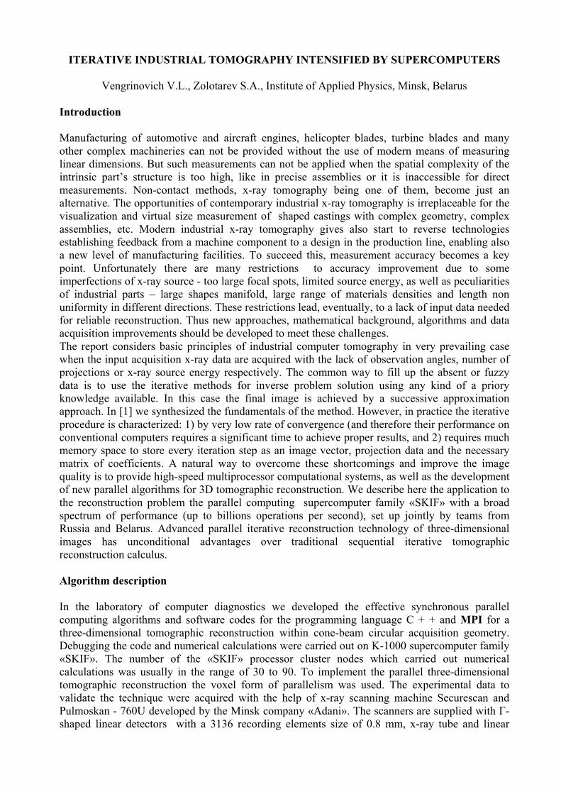
scanning system (along the line perpendicular to the line of detectors). The three-dimensional tomograms size was 580x280x580 volumetric elements with a resolution of 0.16 mm x 0.16 mm x 0.16 mm. The technique enables to reduce 50 - 100 times the time of the tomographic reconstruction and provides an opportunity to restore objects with dimensions 512 x 1024 voxels with simultaneous storage all the necessary data in the memory unit of 4 Gb. Industrial applications Below are several industrial application examples of the new parallel computing technology. The first one illustrates the case of a spatially vast object with a very small fill by a material itself. The calculations need to process by iteration the whole space occupied by the object while being interested in a very limited volume occupied by the material itself, and moreover, in defects inside it. Figure 1 shows two randomly selected x-ray projections of the casted aluminum bearing separator, which typical baffle size is about 3 mm. The number of acquired projections was 60 (instead of 500 usually used) within 1800.The fast parallel computing technique was applied for its 3-D image reconstruction.
Fig. 1. Two randomly selected x-ray projections of the aluminum bearing separator, which typical baffle size of about 3 mm.
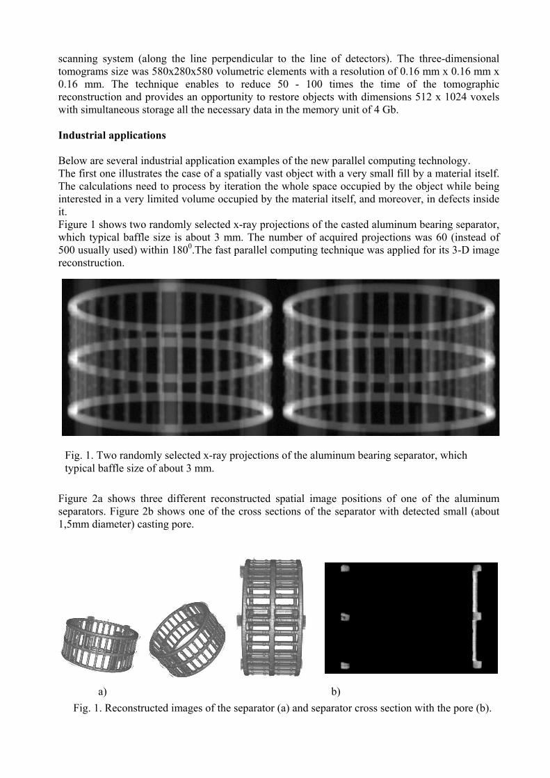
Figure 2a shows three different reconstructed spatial image positions of one of the aluminum separators. Figure 2b shows one of the cross sections of the separator with detected small (about 1,5mm diameter) casting pore.
a) b) Fig. 1. Reconstructed images of the separator (a) and separator cross section with the pore (b).
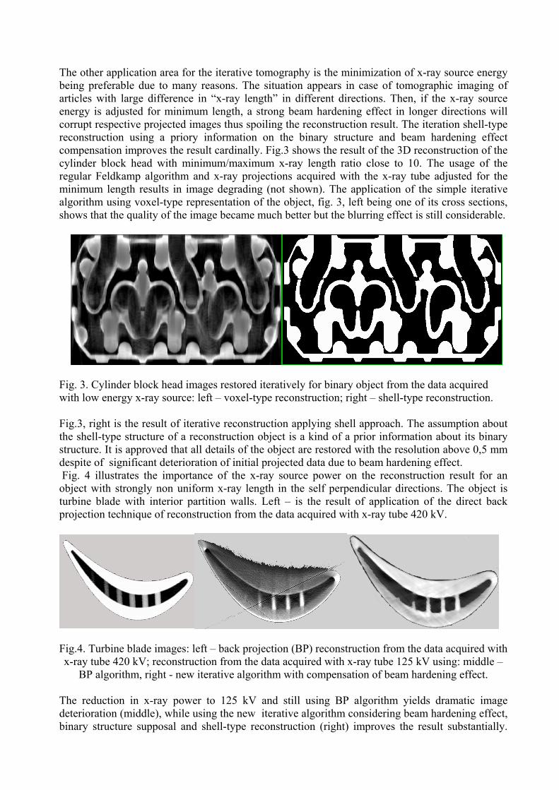
The other application area for the iterative tomography is the minimization of x-ray source energy being preferable due to many reasons. The situation appears in case of tomographic imaging of articles with large difference in “x-ray length” in different directions. Then, if the x-ray source energy is adjusted for minimum length, a strong beam hardening effect in longer directions will corrupt respective projected images thus spoiling the reconstruction result. The iteration shell-type reconstruction using a priory information on the binary structure and beam hardening effect compensation improves the result cardinally. Fig.3 shows the result of the 3D reconstruction of the cylinder block head with minimum/maximum x-ray length ratio close to 10. The usage of the regular Feldkamp algorithm and x-ray projections acquired with the x-ray tube adjusted for the minimum length results in image degrading (not shown). The application of the simple iterative algorithm using voxel-type representation of the object, fig. 3, left being one of its cross sections, shows that the quality of the image became much better but the blurring effect is still considerable.
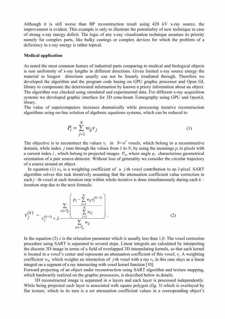
Fig. 3. Cylinder block head images restored iteratively for binary object from the data acquired with low energy x-ray source: left – voxel-type reconstruction; right – shell-type reconstruction. Fig.3, right is the result of iterative reconstruction applying shell approach. The assumption about the shell-type structure of a reconstruction object is a kind of a prior information about its binary structure. It is approved that all details of the object are restored with the resolution above 0,5 mm despite of significant deterioration of initial projected data due to beam hardening effect. Fig. 4 illustrates the importance of the x-ray source power on the reconstruction result for an object with strongly non uniform x-ray length in the self perpendicular directions. The object is turbine blade with interior partition walls. Left – is the result of application of the direct back projection technique of reconstruction from the data acquired with x-ray tube 420 kV.
Fig.4. Turbine blade images: left – back projection (BP) reconstruction from the data acquired with x-ray tube 420 kV; reconstruction from the data acquired with x-ray tube 125 kV using: middle –
BP algorithm, right - new iterative algorithm with compensation of beam hardening effect.
The reduction in x-ray power to 125 kV and still using BP algorithm yields dramatic image deterioration (middle), while using the new iterative algorithm considering beam hardening effect, binary structure supposal and shell-type reconstruction (right) improves the result substantially.
Although it is still worse than BP reconstruction result using 420 kV x-ray source, the improvement is evident. This example is only to illustrate the potentiality of new technique in case of strong x-ray energy deficit. The logic of any x-ray visualization technique assumes its priority namely for complex parts, like bulky castings or complex devices for which the problem of a deficiency in x-ray energy is rather topical. Medical application As noted the most common feature of industrial parts comparing to medical and biological objects is non uniformity of x-ray lengths in different directions. Given limited x-ray source energy the material in longest directions usually can not be linearly irradiated through. Therefore we developed the algorithm and the program code basing on GPU graphic processor and Open GL library to compensate the deteriorated information by known a priory information about an object. The algorithm was checked using simulated and experimental data. For different x-ray acquisition systems we developed graphic interface for 3D cone-beam Tomography using GPU and OpenGL library. The value of supercomputers increases dramatically while processing iterative reconstruction algorithms using on-line solution of algebraic equations systems, which can be reduced to
1
Ni i
jj jP w v
== ∑ (1)
The objective is to reconstruct the values vj in N=n3 voxels, which belong to a reconstructive domain, while index j runs through the values from 1 to N, by using the meanings pi in pixels with a current index i , which belong to projected images Pφ, where angle φ, characterises geometrical orientation of a pair source-detector. Without loss of generality we consider the circular trajectory of a source around an object.
In equation (1) wij is a weighting coefficient of a j-th voxel contribution to an I-pixel. SART algorithm solves this task iteratively assuming that the attenuation coefficient value correction in each j –th voxel at each iteration step within whole iterative is done simultaneously during each k – iteration step due to the next formula:
( )kjv =
( 1)
1
( 1) 1i
i
N ki il l
lN
p Pil
k lj
ijp P
p w v
wv
w
ϕ
ϕ
λ
−
=
∈
− =
∈
⎛ ⎞−⎜ ⎟
⎜ ⎟⎜ ⎟⎜⎝+
∑∑
∑
∑
⎟⎠ . (2)
In the equation (2) λ is the relaxation parameter which is usually less than 1,0. The voxel correction procedure using SART is separated to several steps. Linear integrals are calculated by interpreting the discrete 3D image in terms of a field of overlapped 3D interpolating kernels, so that each kernel is located in a voxel’s center and represents an attenuation coefficient of this voxel, vj. A weighting coefficient wij, which weights an interaction of j-th voxel with a ray ri, in this case stays as a linear integral on a segment of a ray intersecting with voxel kernel function [10]. Forward projecting of an object under reconstruction using SART algorithm and texture mapping, which hardwarily realized on the graphic processors, is described below in details.
3D reconstructed image is separated in n layers and each layer is processed independently. While being projected each layer is associated with square polygon (fig. 5) which is overlayed by flat texture, which in its turn is a set attenuation coefficient values in a corresponding object’s
layer. By rotating a polygon with overlayed texture on to the angle φ of x-ray source geometrical position and by perspective projecting with cone-beam angle γ, one displays voxels in applicable layer which are respectively weighted, onto pixels of projected image.
The fig. 1 illustrates the projecting algorithm within which mapping of a layer on the screen (buffer frame) is done with the help of graphic video card, while the layers projections accumulation is realized by soft in the computer’s RAM due to a limited number of bits used to the data presentation in a buffer frame. After all texture polygons will be accumulated in a buffer it will contain the whole projection of an object under reconstruction for the projecting angle φ. The reverse projecting is clear and not described here in details. As opposed to the forward procedure, within reverse one a correcting image is prospectively projected onto a polygon, which is located on
Screen, projecting image
Layer of a volume Texture polygon, cone-beam angle γ
Fig.5. Forward projecting. Projection geometry
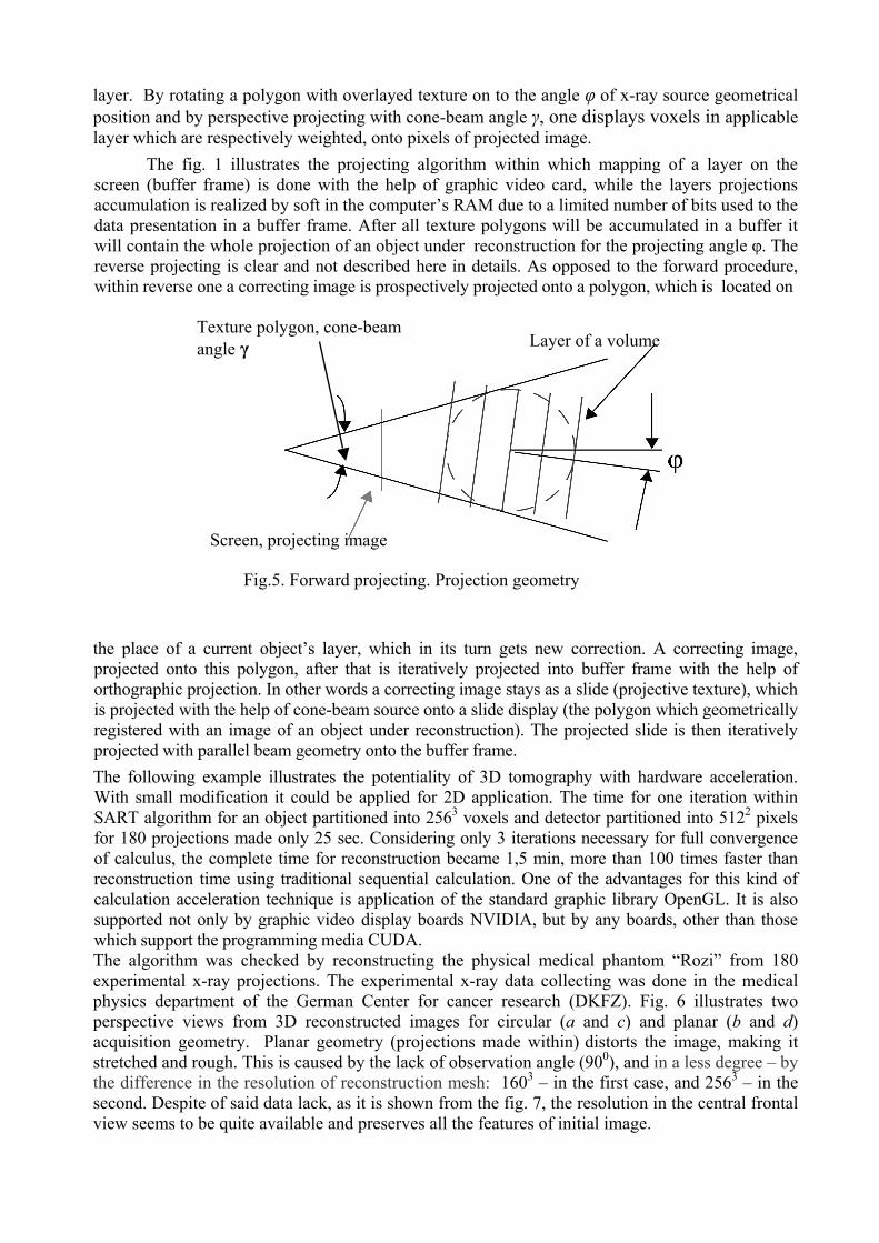
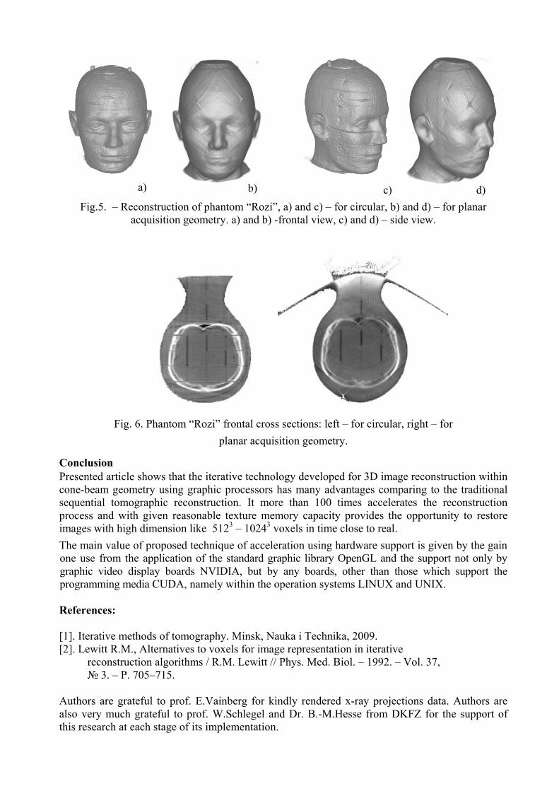
the place of a current object’s layer, which in its turn gets new correction. A correcting image, projected onto this polygon, after that is iteratively projected into buffer frame with the help of orthographic projection. In other words a correcting image stays as a slide (projective texture), which is projected with the help of cone-beam source onto a slide display (the polygon which geometrically registered with an image of an object under reconstruction). The projected slide is then iteratively projected with parallel beam geometry onto the buffer frame. The following example illustrates the potentiality of 3D tomography with hardware acceleration. With small modification it could be applied for 2D application. The time for one iteration within SART algorithm for an object partitioned into 2563 voxels and detector partitioned into 5122 pixels for 180 projections made only 25 sec. Considering only 3 iterations necessary for full convergence of calculus, the complete time for reconstruction became 1,5 min, more than 100 times faster than reconstruction time using traditional sequential calculation. One of the advantages for this kind of calculation acceleration technique is application of the standard graphic library OpenGL. It is also supported not only by graphic video display boards NVIDIA, but by any boards, other than those which support the programming media CUDA. The algorithm was checked by reconstructing the physical medical phantom “Rozi” from 180 experimental x-ray projections. The experimental x-ray data collecting was done in the medical physics department of the German Center for cancer research (DKFZ). Fig. 6 illustrates two perspective views from 3D reconstructed images for circular (a and c) and planar (b and d) acquisition geometry. Planar geometry (projections made within) distorts the image, making it stretched and rough. This is caused by the lack of observation angle (900), and in a less degree – by the difference in the resolution of reconstruction mesh: 1603 – in the first case, and 2563 – in the second. Despite of said data lack, as it is shown from the fig. 7, the resolution in the central frontal view seems to be quite available and preserves all the features of initial image.
d)c)b)a)
Fig.5. – Reconstruction of phantom “Rozi”, a) and c) – for circular, b) and d) – for planar acquisition geometry. a) and b) -frontal view, c) and d) – side view.
Fig. 6. Phantom “Rozi” frontal cross sections: left – for circular, right – for planar acquisition geometry.
Conclusion Presented article shows that the iterative technology developed for 3D image reconstruction within cone-beam geometry using graphic processors has many advantages comparing to the traditional sequential tomographic reconstruction. It more than 100 times accelerates the reconstruction process and with given reasonable texture memory capacity provides the opportunity to restore images with high dimension like 5123 – 10243 voxels in time close to real. The main value of proposed technique of acceleration using hardware support is given by the gain one use from the application of the standard graphic library OpenGL and the support not only by graphic video display boards NVIDIA, but by any boards, other than those which support the programming media CUDA, namely within the operation systems LINUX and UNIX. References: [1]. Iterative methods of tomography. Minsk, Nauka i Technika, 2009. [2]. Lewitt R.M., Alternatives to voxels for image representation in iterative reconstruction algorithms / R.M. Lewitt // Phys. Med. Biol. – 1992. – Vol. 37, № 3. – P. 705–715. Authors are grateful to prof. E.Vainberg for kindly rendered x-ray projections data. Authors are also very much grateful to prof. W.Schlegel and Dr. B.-M.Hesse from DKFZ for the support of this research at each stage of its implementation.