karl storz endoskope | united states - collection on · 2020-01-28 · weight reduction and its...
TRANSCRIPT


Contributing Authors:Prof. Karl Miller, M.D.
Prof. Hans Lönroth, M.D., PhDProf. Matthias Kemen, M.D., and Gerasimos Tzivras, M.D.
Raul J. Rosenthal, M.D.J.C. Ruiz de Adana, M.D., J. López-Herrero, M.D.
and A. Hernández-Matias, M.D.Daniel Krawczykowski, M.D.
Prof. Roberto Maria Tacchino, M.D.Francesco Greco, M.D., and Daniele Matera, M.D.
Prof. Holger Till, M.D., and Susann Blüher, M.D.
Foreword by:Prof. Rudolf A. Weiner, M.D.
The nominal fee of € 5 per copy will bedonated to the Nonprofi t Organization
“Moby Dick” (www.mobydicknetzwerk.de)
COLLECTION ONLAPAROSCOPYIN BARIATRICS

Collection on Laparoscopy in BariatricsII
All rights reserved. No part of this publication may be translated, re print ed or reproduced, transmitted in any form or by any means, electronic or mechanical, now known or hereafter invented, including photocopying and recording, or utilized in any information storage or retrieval system without the prior written permission of the copyright holder.
Important notice:
Medical knowledge is ever changing. As new research and clinical experience broaden our knowledge, changes in treatment and therapy may be required. The authors and editors of the material herein have consulted sources believed to be reliable in their efforts to provide information that is complete and in accord with the standards accepted at the time of publication. However, in view of the possibility of human error by the authors, editors, or publisher of the work herein, or changes in medical knowledge, neither the authors, editors, publisher, nor any other party who has been involved in the preparation of this work, warrants that the information contained herein is in every respect accurate or complete, and they are not responsible for any errors or omis-sions or for the results obtained from use of such information. The information contained within this brochure is intended for use by doctors and other health care professionals. This material is not intended for use as a basis for treatment decisions, and is not a substitute for professional consultation and/or use of peer-reviewed medical literature.
Some of the product names, patents, and registered designs referred to in this booklet are in fact registered trademarks or proprietary names even though specifi c reference to this fact is not always made in the text. Therefore, the appearance of a name without designation as proprietary is not to be construed as a representation by the publisher that it is in the public domain.
Collection on Laparoscopy in Bariatrics Adjustable Gastric Banding in the Management of Obesity Prof. Karl Miller, M.D.General Hospital Hallein, Austria Laparoscopic Gastric Bypass Prof. Hans Lönroth, M.D., PhDSahlgrenska University Hospital Göteborg, Sweden Bariatric Surgery Prof. Matthias Kemen, M.D., and Gerasimos Tzivras, M.D.,Herne Protestant Hospital, Department of Surgery, Herne, Germany Laparoscopic Sleeve GastrectomyRaul J. Rosenthal, M.D., Cleveland Clinic, Weston, USA Laparoscopic Sleeve GastrectomyJ.C. Ruiz de Adana, M.D., J. López-Herrero, M.D.,and A. Hernández-Matias, M.D.Madrid University Hospital of Getafe, Spain Biliopancreatic Diversion Sleeve Gastrectomy with/withoutDuodenal SwitchDaniel Krawczykowski, M.D.Polyclinic Priollet-Courlancy, Châlons-en-Champagne, France Laparoscopic Biliopancreatic Diversion Prof. Roberto Maria Tacchino, M.D., Francesco Greco, M.D.,and Daniele Matera, M.D.Catholic University of Sacred Heart Rome, ItalyBariatric Surgery in Childhood and Adolescence,Indications and TechniquesProf. Holger Till, M.D., and Susann Blüher, M.D.Leipzig University Clinical CenterWomen’s and Children’s Hospital, Leipzig, Germany Foreword Prof. Rudolf Weiner, M.D.,Sachsenhausen Hospital, General Surgery, Frankfurt a. M., Germany
© 2014 Published by ® TuttlingenISBN 978-3-89756-537-1, Printed in GermanyP.O. Box, D-78503 Tuttlingen, GermanyTelephone: +49 74 61/1 45 90Fax: +49 74 61/708-529E-mail: [email protected]
Other foreign language editions are available upon request.For additional information please contact ® Tuttlingen, Germany, at the address indicated above.
Layout and image reproduction:® Tuttlingen, Germany
Printed by:Straub Druck + Medien AGD-78713 Schramberg, Germany
08.14-0.75
Your donation is dedicated toMoby Dick, a Health Program for Obese Children and Teenagers.
For further information, see page 119.

IIITable of Contents
Table of ContentsForeword, Prof. Rudolf A. Weiner, M.D.. . . . . . . . . . . . . . . . . . . . . . . . . . . . . . . . . . . . . . . V
Chapter I Adjustable Gastric Banding in the Treatment of Obesity
Prof. Karl Miller, M.D. Head, Department of Surgery, Hallein General Hospital, Austria
Introduction . . . . . . . . . . . . . . . . . . . . . . . . . . . . . . . . . . . . . . . . . . . . . . . . . . . . . . . . . . . . . . 8Patient Selection . . . . . . . . . . . . . . . . . . . . . . . . . . . . . . . . . . . . . . . . . . . . . . . . . . . . . . . . . . 8
Preoperative Details . . . . . . . . . . . . . . . . . . . . . . . . . . . . . . . . . . . . . . . . . . . . . . . . . . . . . 9Perioperative Care . . . . . . . . . . . . . . . . . . . . . . . . . . . . . . . . . . . . . . . . . . . . . . . . . . . . . . 9
Operative Technique . . . . . . . . . . . . . . . . . . . . . . . . . . . . . . . . . . . . . . . . . . . . . . . . . . . . . . . 9Perigastric Technique . . . . . . . . . . . . . . . . . . . . . . . . . . . . . . . . . . . . . . . . . . . . . . . . . . . . 10Pars fl accida Technique . . . . . . . . . . . . . . . . . . . . . . . . . . . . . . . . . . . . . . . . . . . . . . . . . . 11Combined Technique . . . . . . . . . . . . . . . . . . . . . . . . . . . . . . . . . . . . . . . . . . . . . . . . . . . . 12
Complications and Their Prevention. . . . . . . . . . . . . . . . . . . . . . . . . . . . . . . . . . . . . . . . . . . 13Perioperative Complications. . . . . . . . . . . . . . . . . . . . . . . . . . . . . . . . . . . . . . . . . . . . . . . 13Late Complications . . . . . . . . . . . . . . . . . . . . . . . . . . . . . . . . . . . . . . . . . . . . . . . . . . . . . . 14
Summary . . . . . . . . . . . . . . . . . . . . . . . . . . . . . . . . . . . . . . . . . . . . . . . . . . . . . . . . . . . . . . . . 16References. . . . . . . . . . . . . . . . . . . . . . . . . . . . . . . . . . . . . . . . . . . . . . . . . . . . . . . . . . . . . . . 17
Chapter II Laparoscopic Gastric Bypass
Hans Lönroth, MD, PhDChairman, Department of Surgery, Sahlgrenska University Hospital, Goteborg, Sweden
Background . . . . . . . . . . . . . . . . . . . . . . . . . . . . . . . . . . . . . . . . . . . . . . . . . . . . . . . . . . . . . . 22Port Sites and Placement of Trocars . . . . . . . . . . . . . . . . . . . . . . . . . . . . . . . . . . . . . . . . . . 22Creation of the Gastric Pouch. . . . . . . . . . . . . . . . . . . . . . . . . . . . . . . . . . . . . . . . . . . . . . . . 23Division of the Omentum. . . . . . . . . . . . . . . . . . . . . . . . . . . . . . . . . . . . . . . . . . . . . . . . . . . . 23Creation of the Gastroenterostoma . . . . . . . . . . . . . . . . . . . . . . . . . . . . . . . . . . . . . . . . . . . 23Controversies. . . . . . . . . . . . . . . . . . . . . . . . . . . . . . . . . . . . . . . . . . . . . . . . . . . . . . . . . . . . . 26Acknowledgement. . . . . . . . . . . . . . . . . . . . . . . . . . . . . . . . . . . . . . . . . . . . . . . . . . . . . . . . . 27References. . . . . . . . . . . . . . . . . . . . . . . . . . . . . . . . . . . . . . . . . . . . . . . . . . . . . . . . . . . . . . . 27
Chapter III Bariatric Surgery
Prof. Matthias Kemen, M.D. and Gerasimos Tzivras, M.D.Herne Protestant Hospital, (Teaching Hospital for the University of Essen)Department of Surgery, Herne, Germany
Introduction . . . . . . . . . . . . . . . . . . . . . . . . . . . . . . . . . . . . . . . . . . . . . . . . . . . . . . . . . . . . . . 32Laparoscopic Adjustable Gastric Banding (LAGB). . . . . . . . . . . . . . . . . . . . . . . . . . . . . . . . 34
Theory and Practice . . . . . . . . . . . . . . . . . . . . . . . . . . . . . . . . . . . . . . . . . . . . . . . . . . . . . 34Laparoscopic Gastric Bypass with Roux-en-Y Limb (LGBRY) . . . . . . . . . . . . . . . . . . . . . . 37
Image Sequence Illustrating the Roux-en-Y Gastric Bypass . . . . . . . . . . . . . . . . . . . . . 39Laparoscopic Roux-en-Y Gastric Bypass . . . . . . . . . . . . . . . . . . . . . . . . . . . . . . . . . . . . 44The Jejunojejunal Anastomosis . . . . . . . . . . . . . . . . . . . . . . . . . . . . . . . . . . . . . . . . . . . . 44Laparoscopic Roux-en-Y Gastric Bypass . . . . . . . . . . . . . . . . . . . . . . . . . . . . . . . . . . . . 46Steps from Identifying the Ligament of Treitz to Division of the Stomach . . . . . . . . . . . 46Laparoscopic Roux-en-Y Gastric Bypass with an Intragastric Balloon . . . . . . . . . . . . . 48Laparoscopic Roux-en-Y Gastric Bypass . . . . . . . . . . . . . . . . . . . . . . . . . . . . . . . . . . . . 51
Laparoscopic Sleeve Gastrectomy. . . . . . . . . . . . . . . . . . . . . . . . . . . . . . . . . . . . . . . . . . . . 54
Chapter IV Laparoscopic Sleeve Gastrectomy
Raul J. Rosenthal, M.D., FACSSection Head of Minimally Invasive Surgery and Medical Director of theBariatric and Metabolic Institute, Cleveland Clinic, Weston, Florida, U.S.A.
Introduction . . . . . . . . . . . . . . . . . . . . . . . . . . . . . . . . . . . . . . . . . . . . . . . . . . . . . . . . . . . . . . 60History of Sleeve Gastrectomy . . . . . . . . . . . . . . . . . . . . . . . . . . . . . . . . . . . . . . . . . . . . . . . 60Technique . . . . . . . . . . . . . . . . . . . . . . . . . . . . . . . . . . . . . . . . . . . . . . . . . . . . . . . . . . . . . . . 61Results . . . . . . . . . . . . . . . . . . . . . . . . . . . . . . . . . . . . . . . . . . . . . . . . . . . . . . . . . . . . . . . . . . 63Conclusion . . . . . . . . . . . . . . . . . . . . . . . . . . . . . . . . . . . . . . . . . . . . . . . . . . . . . . . . . . . . . . . 63References. . . . . . . . . . . . . . . . . . . . . . . . . . . . . . . . . . . . . . . . . . . . . . . . . . . . . . . . . . . . . . . 64

Collection on Laparoscopy in BariatricsIV
Chapter V Laparoscopic Sleeve Gastrectomy
J.C. Ruiz de Adana, J. López-Herrero, A. Hernández-Matías Department of General Surgery, Madrid University Hospital of Getafe (Madrid), Spain
Introduction . . . . . . . . . . . . . . . . . . . . . . . . . . . . . . . . . . . . . . . . . . . . . . . . . . . . . . . . . . . . . . 68Patient Selection and Indications . . . . . . . . . . . . . . . . . . . . . . . . . . . . . . . . . . . . . . . . . . . . . 68Potential Complications and Precautions. . . . . . . . . . . . . . . . . . . . . . . . . . . . . . . . . . . . . . . 72Recommended Literature . . . . . . . . . . . . . . . . . . . . . . . . . . . . . . . . . . . . . . . . . . . . . . . . . . . 73
Chapter VI Biliopancreatic Diversion Sleeve Gastrectomywith/without Duodenal Switch
Daniel Krawczykowski, M.D.Polyclinique Priollet-Courlancy, Châlons-en-Champagne, France
Defi nition and Historical Background of Biliopancreatic Diversion with Duodenal Switch 76Biliopancreatic Diversion with Duodenal Switch: Results and Strategic Approach . . . . . . 77The Two Components . . . . . . . . . . . . . . . . . . . . . . . . . . . . . . . . . . . . . . . . . . . . . . . . . . . . . . 80
Sleeve Gastrectomy . . . . . . . . . . . . . . . . . . . . . . . . . . . . . . . . . . . . . . . . . . . . . . . . . . . . . 80Duodenal Switch . . . . . . . . . . . . . . . . . . . . . . . . . . . . . . . . . . . . . . . . . . . . . . . . . . . . . . . 88The Technique . . . . . . . . . . . . . . . . . . . . . . . . . . . . . . . . . . . . . . . . . . . . . . . . . . . . . . . . . 89
Conclusion . . . . . . . . . . . . . . . . . . . . . . . . . . . . . . . . . . . . . . . . . . . . . . . . . . . . . . . . . . . . . . . 90Literature . . . . . . . . . . . . . . . . . . . . . . . . . . . . . . . . . . . . . . . . . . . . . . . . . . . . . . . . . . . . . . . . 92
Chapter VII Laparoscopic Biliopancreatic Diversion
Roberto Maria Tacchino, Francesco Greco, and Daniele Matera Department of Surgery, Catholic University of Sacred Heart, Rome, Italy
Introduction . . . . . . . . . . . . . . . . . . . . . . . . . . . . . . . . . . . . . . . . . . . . . . . . . . . . . . . . . . . . . . 100Preoperative Management . . . . . . . . . . . . . . . . . . . . . . . . . . . . . . . . . . . . . . . . . . . . . . . . . . 100Instrumentation . . . . . . . . . . . . . . . . . . . . . . . . . . . . . . . . . . . . . . . . . . . . . . . . . . . . . . . . . . . 100Patient Positioning. . . . . . . . . . . . . . . . . . . . . . . . . . . . . . . . . . . . . . . . . . . . . . . . . . . . . . . . . 101Team Positioning . . . . . . . . . . . . . . . . . . . . . . . . . . . . . . . . . . . . . . . . . . . . . . . . . . . . . . . . . . 101Trocar Placement . . . . . . . . . . . . . . . . . . . . . . . . . . . . . . . . . . . . . . . . . . . . . . . . . . . . . . . . . 102Gastric Resection . . . . . . . . . . . . . . . . . . . . . . . . . . . . . . . . . . . . . . . . . . . . . . . . . . . . . . . . . 103Small Bowel Measurement . . . . . . . . . . . . . . . . . . . . . . . . . . . . . . . . . . . . . . . . . . . . . . . . . . 105The Double-Loop Technique: Gastro-ileal Anastomosis . . . . . . . . . . . . . . . . . . . . . . . . . . . 106Ileo-ileal Anastomosis . . . . . . . . . . . . . . . . . . . . . . . . . . . . . . . . . . . . . . . . . . . . . . . . . . . . . . 107End of Procedure. . . . . . . . . . . . . . . . . . . . . . . . . . . . . . . . . . . . . . . . . . . . . . . . . . . . . . . . . . 108
Chapter VIII Bariatric Surgery in Childhood and AdolescenceIndications and Techniques – An Overview of the Current Debate
Prof. Holger Till, M.D. and Susann Blüher, M.D.Leipzig University Clinical Center, Women’s and Children’s Hospital, Leipzig, Germany
Introduction . . . . . . . . . . . . . . . . . . . . . . . . . . . . . . . . . . . . . . . . . . . . . . . . . . . . . . . . . . . . . . 112Indications . . . . . . . . . . . . . . . . . . . . . . . . . . . . . . . . . . . . . . . . . . . . . . . . . . . . . . . . . . . . . . . 113Material and Methods . . . . . . . . . . . . . . . . . . . . . . . . . . . . . . . . . . . . . . . . . . . . . . . . . . . . . . 114Results . . . . . . . . . . . . . . . . . . . . . . . . . . . . . . . . . . . . . . . . . . . . . . . . . . . . . . . . . . . . . . . . . . 115
Operative Techniques. . . . . . . . . . . . . . . . . . . . . . . . . . . . . . . . . . . . . . . . . . . . . . . . . . . . 115Discussion . . . . . . . . . . . . . . . . . . . . . . . . . . . . . . . . . . . . . . . . . . . . . . . . . . . . . . . . . . . . . . . 115References . . . . . . . . . . . . . . . . . . . . . . . . . . . . . . . . . . . . . . . . . . . . . . . . . . . . . . . . . . . . . . 116
Instrument Sets for Laparoscopic Bariatric SurgeryTelescopes, Operating Instruments, Accessories and Units
Instrument Set for Gastric Banding. . . . . . . . . . . . . . . . . . . . . . . . . . . . . . . . . . . . . . . . . . . . 123Instrument Set for Laparoscopic Roux-Y-Gastric Bypass. . . . . . . . . . . . . . . . . . . . . . . . . . 125Instrument Set for Bileopancreatic Diversion Sleeve Gastrectomy . . . . . . . . . . . . . . . . . . . 127Instrument Set for Duodenal Switch . . . . . . . . . . . . . . . . . . . . . . . . . . . . . . . . . . . . . . . . . . . 129Instrument Set for Bariatric Surgery in Pediatrics. . . . . . . . . . . . . . . . . . . . . . . . . . . . . . . . . 131

VTable of Contents
ForewordThe worldwide epidemic of obesity has reached such alarming proportions that a recent WHO report coined a new word to describe this phenomenon: “globesity.” It is also clear that the public and policy makers have only just begun to realize the increasing problems that emerge due to being overweight and their implications for public health.
Thus far, there have been no society-wide concepts for dealing with the steady rise of obesity in industrialized countries and developing nations. The persistence of morbid obesity implies that conservative treatment options have failed and that surgical intervention is the only approach that can offer a permanent solution to the comorbidities associated with obesity and its sequelae. In patients who are more than 30–40 kg overweight, bariatric surgery is by far superior to all conservative treatment options including multimodal regimens, behavioral therapy, exercise therapy, and even pharmacotherapy. This superiority concerns both the degree of weight reduction and its permanence.
While conservative therapies can bring about a weight loss of up to 10 kg in one year, this result is only temporary, lasting 1–2 years, and the dieters usually regain the lost weight. On the contrary, we know from the Swedish Obese Subjects (SOS) Study that modern methods of bariatric surgery can achieve a permanent weight reduction in the long term.
The acceptance of bariatric surgery is closely tied to the development of laparoscopyand the minimally invasive approach. While open weight-reduction surgery was associated with a high rate of general complications such as pulmonary complications, incision dehiscence, and pulmonary embolism, the advent of laparoscopic surgical techniques has led to a high degree of acceptance among surgeons and patients alike. The morbidity of bariatric procedures has been reduced by a factor of 10, and today laparoscopic techniques are regarded as safe and established procedures. This book, a collection of articles written by an international team of experts, covers all relevant details and technical aspects of bariatric surgery including the most important standard procedures. Given the variety and complexity of surgical treatment options – always combining the principles of restriction (reducing food ingestion) and malabsorption (decreasing the absorption of fat and nutrients) – it is clear at the outset that there is no single operation that is ideal for everyone. The most appropriate procedure should be selected based on a careful consideration of individual factors (age, sex, severity of overweight and comorbid conditions, willingness and ability of the patient to cooperate) and then carried out with high technical profi ciency.
Beside the standard procedures, there are also a number of new therapeutic approaches and expert options that are not included in this volume because they have not yet been proven by clinical trials. Furthermore, this collection is highlighted by numerous intraoperative, high-defi nition videoendoscopic photographs using instruments, camera systems and complex operating room systems manufactured by KARL STORZ. They vividly illustrate the steps involved in bariatric procedures, pointing out high-risk phases of the operation and offering strategies for the prevention of complications. This collection provides the laparoscopist with an excellent overview of the currently established laparoscopic surgical procedures.
Obesity surgery will continue to expand more widely in the future. Given the current lack of effective options for the prevention of obesity and its ineffective conservative management, bariatric surgery will continue to remain as the most rapidly growing subspecialty of modern visceral medicine and surgery.
Prof. Rudolf A. Weiner, M.D.
President of the International Federation for Surgery of Obesityand Metabolic Disorders 2014–2015Past President-elect, IFSO European Chapter 2010–2012President, IFSO World Congress 2011
VForeword


11Adjustable Gastric Bandingin the Treatment of ObesityProf. Karl Miller, M.D. Head, Department of Surgery, Hallein General Hospital, Austria
Collection on Laparoscopy in Bariatrics8
IntroductionAs laparoscopic techniques have become prevalent in almost every surgical specialty, the number of bariatric procedures has increased dramatically during the past 10 years. The least invasive weight-loss operation is adjustable gastric banding.
Patient SelectionThe indication for bariatric surgery is documented in evidence-based guidelines. A body mass index (BMI = body weight in kilograms divided by height in meters squared) of 40 or more represents clinically severe overweight, which requires medical treatment and justifi es surgical treatment if the patient desires it and the attending surgeon feels that it is indicated. Patients with a BMI of 35–40 should be considered for surgical treatment if they have comorbid conditions that could improve signifi cantly in response to weight loss. The patient should have a BMI of 40 kg/m2 or more, or a body weight that is 45 kg or more above the ideal value for his or her constitution according to a standard table. (A BMI of 40 means that the patient is approximately 45 kg above the ideal weight for an individual of normal height.) If the BMI is 35–40 kg/m2 (i.e., less than 45 kg above the ideal weight), a serious medical problem exists that could be signifi cantly improved by weight reduction to justify the risk of a proposed operation. The patient must be able to care for himself or have one or more caregivers who can provide the necessary follow-up care. A high degree of patient motivation and an interdisciplinary approach are far more important for success than rigorous exclusion criteria, which may be amended or discarded from one year to the next. For patients who undergo adjustable gastric banding, follow-up measures such as band adjustments, psychological counseling, and nutritional counseling are an essential part of management. In cases where follow-up care cannot or will not be provided, the patient should not be selected for bariatric surgery. Other issues are addressed in the guidelines of the American Society for Bariatric Surgery (ASBS) and the recommendations of the International Federation for the Surgery of Obesity (IFSO), which state that centers providing bariatric surgery should have adequate experience in open and laparoscopic intest inal surgery. They should also have an infrastructure that includes trained nutritional counselors, psychologists, a motivated nursing staff, and preferably a patient support group. Other essentials are properly equipped examination tables, operat ing tables, beds, and necessary instruments for cases that require conversion to open surgery. Facilities for perioperative monitoring should also be available. The importance of operator training and experience is self-evident. Explaining the pro cedure and its risks to the patient and securing informed consent are time- consuming but imperative. There is scarcely any other situation in which the success or failure of an operation depends so strongly on patient cooperation. The patient should be educated about obesity as a disease and informed about current surgical treatment options, laparoscopic banding, possible complications, warning signs, and post operative care. Patients with an extreme form of morbid obesity (triple obesity, BMI > 60), a severe eating disorder (e.g., binge eating), or insulin-dependent type II diabetes mellitus should be referred for a more complex surgical procedure such as gastric bypass or biliopancreatic diversion.

9Adjustable Gastric Banding in the Treatment of Obesity
Preoperative Details
Endocrine disorders should be adequately treated and controlled. A medical eva luation, abdominal ultrasound scans, and spirometry are recommended. Any gallstones should be synchronously removed, because dramatic weight loss is often complicated by cholelithiasis. The patient is presented to the anesthesiologist with the results of the preoperative workup several days before the operation. Nutrition al counseling is mandatory, and a psychological evaluation is recommended.
Perioperative Care
Unlike many other operations, bariatric surgery marks the starting point rather than the endpoint of treatment. Regular follow-up visits and patient cooperation are crucial for success. Perioperative antibiotics (single-shot cephalosporin prophylaxis) and low-molecular heparin (moderate to high risk) are recommended. On the day of the surgery, the patient may drink small sips of tea following the procedure. A water-soluble oral contrast examination is performed on the fi rst postoperative day, and the liquid phase of the postoperative diet is begun, with gradual transition to a regular diet. Further nutritional counseling should be given prior to the fi rst band adjustment whenever possible. Additional follow-ups depend on the type of procedure that was performed and, of course, on individual patient requirements.
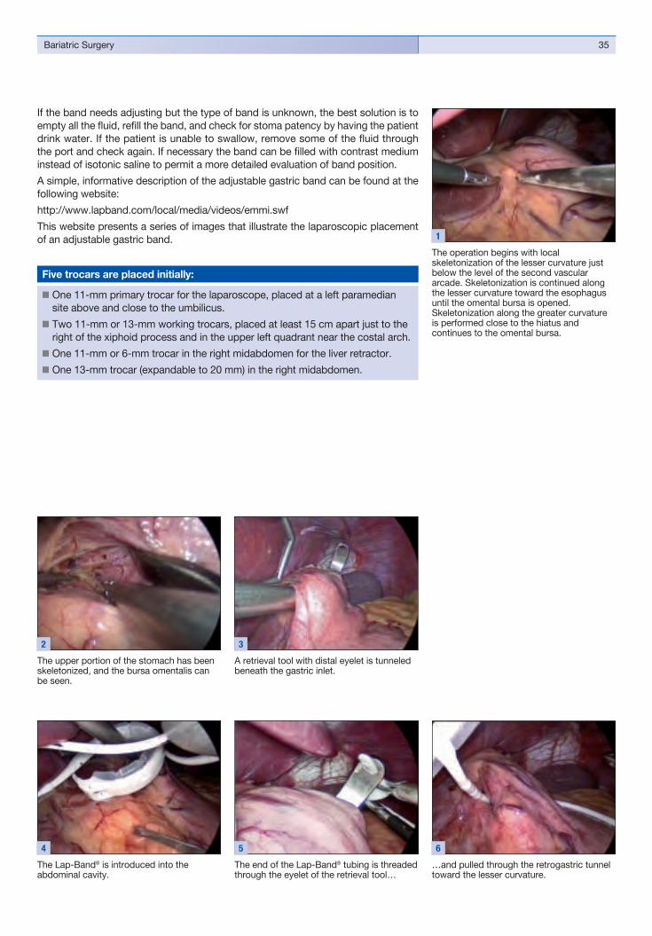
Operative TechniqueThe patient is placed in a supine, slightly hyperextended position with the legs abducted. A pneumoperitoneum (12 mmHg) is created in the left mid- to upper abdomen. Once the trocars have been placed, the patient is moved to a reverse Trendelenburg position to prevent injury to the liver. A variety of trocar patterns have been described in the literature. The sites depend on the body habitus of the patient. Trocar placement in extremely obese patients will play a key role in the success of the operation. Unlike the situation in normal-weight patients, the axial alignment of trocars in the abdominal wall of obese patients can no longer be changed following placement. Preoperative ultrasound scans can be very helpful for determining the size of the left lobe of the liver. The port for the laparoscope is placed slightly to the left of the midline, approximately one handwidth below the xiphoid process. Whenever possible, this port should be created under vision to avoid hepatic injury. The liver retractor is placed in the right mid- to upper abdomen or in the epi gastrium. A long needle is introduced into the epigastrium to the right of the midline to locate the optimum site for a dissecting instrument and for optional insertion of the stapler. The site for another dissecting instrument and eventual band insertion is placed in the left mid- to upper abdomen. The 16-mm trocar can be placed at this site for implantation of the gastric band. An optional 6-mm trocar may be placed in the area of the left costal arch for applying traction to the stomach wall or retracting the greater omentum. After the trocars have been placed, the left lobe of the liver is retracted proximally until the diaphragm can be seen. The band should always be placed at the level of the gastric inlet, never around the eso phagus. Three main techniques are available for implanting the band. Perigastric placement following the creation of a retrogastric tunnel has been largely superseded in recent years by the “pars fl accida” technique. A combined technique (pars fl accida + perigastric) has been specially developed for cases in which there is a heavy accumulation of fat about the gastric cardia.

Collection on Laparoscopy in Bariatrics10
Perigastric Technique
A gastric tube tipped with an infl atable balloon is advanced into the stomach. The balloon is infl ated with 15–20 cc of air and withdrawn to the gastroesophageal junction. A window approximately 0.5 cm large is dissected free on the lesser curvature of the stomach at the level of the balloon equator. The dissection proceeds back along the stomach wall to the angle of His, taking care not to open the lesser sac. On the greater curvature side, the left crus of the diaphragm and the gastroesophageal junction are dissected free at the angle of His. The gastrophrenic ligament should not be divided, as the band will be placed within the ligament. The dissector (we prefer the Goldfi nger®, Ethicon Johnson & Johnson, Obtech) is passed along the window dissected on the lesser curvature and angled within the gastro-phrenic ligament to place it in the angle of His. The gastric band should be introduced into the abdomen only through a 16-mm or 19-mm trocar. Another technique is to pass the band around the gastroesophageal junction with an atraumatic defl ectable grasper. Before the band is locked into place, the balloon at the end of the stomach tube is infl ated with 15 mL of air. This creates a small gastric pouch, below which the band is fastened with a self-locking mechanism. There is no need to secure the locked band with reinforcing sutures. Slippage is prevented by anchoring the band to the anterior gastric wall with three or four interrupted seromuscular sutures. The end of the tubing is brought out of the body through the 19-mm or 16-mm trocar and connected to the access port, which is secured with four nonabsorbable sutures on or below the anterior rectus sheath at the 19-mm or 16-mm trocar site. Another option is to use a port stapler (Figs. 1a, b).
The access port is fi xed to the rectus fascia with nonabsorbable sutures (a) or by using a port stapler (Velocity®) (b).
1a 1b
11Adjustable Gastric Banding in the Treatment of Obesity
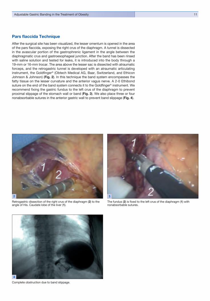
Retrogastric dissection of the right crus of the diaphragm (2) to the angle of His. Caudate lobe of the liver (1).
The fundus (2) is fi xed to the left crus of the diaphragm (1) withnonabsorbable sutures.
Complete obstruction due to band slippage.
4
Pars fl accida Technique
After the surgical site has been visualized, the lesser omentum is opened in the area of the pars fl accida, exposing the right crus of the diaphragm. A tunnel is dissected in the avascular portion of the gastrophrenic ligament in the angle between the diaphragmatic crus and gastroesophageal junction. After the band has been rinsed with saline solution and tested for leaks, it is introduced into the body through a 19-mm or 16-mm trocar. The area above the lesser sac is dissected with atraumatic forceps, and the retrogastric tunnel is developed with an atraumatic articu lating instrument, the Goldfi nger® (Obtech Medical AG, Baar, Switzerland, and Ethicon Johnson & Johnson) (Fig. 2). In this technique the band system encompasses the fatty tissue on the lesser curvature and the anterior vagus nerve. A 2-0 Ethibond suture on the end of the band system connects it to the Goldfi nger® instrument. We recommend fi xing the gastric fundus to the left crus of the diaphragm to prevent proximal slippage of the stomach wall or band (Fig. 3). We also place three or four nonabsorbable sutures in the anterior gastric wall to prevent band slippage (Fig. 4).
2 3

Collection on Laparoscopy in Bariatrics12
� Reduce the pouch volume to 15 cc.� Place the band above the lesser sac.� The pars fl accida technique is preferred.� Place the band within the gastrophrenic ligament.� Place sutures in the anterior gastric wall to prevent band slippage.� Opening the lesser sac places the band so far from the gastroesophageal
junction that fi xation sutures should additionally be placed in the posterior gastric wall.
� Do not infl ate the band during the initial postoperative weeks, as this may cause profuse vomiting, which in turn may dislodge the fi xation sutures and cause band slippage.
� If the band is too narrow, use the combined implantation technique.
Combined Technique
The gastric band may be too narrow in patients who have copious fat about the cardia. After the band has been placed by the pars fl accida technique and before it is latched into place, a tunnel is dissected between the stomach wall and fat pad (Fig. 5). The gastric band is pulled through the tunnel with another instrument (dissector or Goldfi nger®) and then fi nally locked close to the stomach wall (Fig. 6).
Today the following recommendations can be offered based on developments in surgical technique:
A tunnel is developed between the stomach wall and fat pad.
5
The band is placed close to the stomach wall using the “combined technique”.
6

13Adjustable Gastric Banding in the Treatment of Obesity
Complications: Incidence (%) Perioperative complications: Death 0 – 2.1 Stomach wall lesion 0 – 3.5 Pneumothorax 0 – 0.2 Bleeding 0.5 – 2.0 Late complications: Pouch dilatation with or without band slippage 0 – 13.4 Band erosion 0 – 4.6 Complications relating to the access port or band system 0.5 – 10.4 Wound infection 0 – 7.7 Motility disorder (clinically apparent) 0 – 1.5
Table 1: Complications of adjustable gastric banding15-37
Complications and Their PreventionComplications are subdivided into perioperative complications and late compli -ca tions (Table 1). Highest priority is given to the prevention of complications. Reference was made earlier to the importance of thorough training and an inter-disciplinary approach to treatment. The “pars fl accida technique” has led to a signifi cant reduction of surgical complications such as band erosion and slippage.
Perioperative Complications
Mortality
Perioperative deaths have been described in the literature as a result of gastric wall perforation, gastric wall necrosis, cardiogenic shock, and pulmonary embolism. A perioperative mortality rate of 0–0.1% has been reported in large series of patients fi tted with an adjustable gastric band.
Gastric Wall Lesions
It is relatively easy to injure the gastric wall under conditions where vision is obscured. It is common for surgeons to perforate the stomach wall during the “learning curve” of the fi rst 50 or so operations. The incidence of gastric wall perforations ranges from 0% to 3.5% in the literature. If the perforation site is distal to the gastric band, the affected site can be oversewn and the band reimplanted. A meticulous operat ing technique with good vision and the use of suitable atraumatic instruments can prevent this dreaded complication. We recommend testing for leaks under diffi cult viewing conditions by instilling methylene blue through a stomach tube before the band is placed around the gastroesophageal junction (5 mL of methylene blue diluted in 15 mL of saline solution).
Other Perioperative Complications
Other complications (bleeding, pneumothorax) like those occurring in conventional obesity surgery have been reported. Published reports indicate that laparoscopic obesity surgery is associated with fewer perioperative complications than conventional bariatric procedures. Effective hemostatic agents such as FloSeal® (Baxter) should always be on hand.

Collection on Laparoscopy in Bariatrics14
Late Complications
Pouch Dilatation with Gastric-Wall or Band SlippageNumerous authors have reported pouch dilatation on the posterior gastric wall in the area of the lesser sac. This type of complication has been greatly reduced by placing sutures in the posterior gastric wall or placing the band above the lesser sac and within the gastrophrenic ligament. In a study of 350 patients, O’Brien showed that even when the band was placed in the lesser sac, the incidence of pouch dilatation and band slippage was reduced from 30% to 2.5% when sutures were placed in the posterior gastric wall. Gastric pouch dilatation typically occurs on an average of 8 months after the operation.
Pouch Dilatation without Gastric-Wall or Band SlippageThere have been only a few published reports of pouch dilatation occurring in the absence of gastric-wall or band slippage. It has been suggested that making the pouch too large is responsible for this complication. Desaive published the results of his study on two different perioperative pouch sizes, 15 and 25 cc. The reoperation rate for pouch dilatation was reduced from 33% with the 25-cc pouch to 5.1% with the 15-cc pouch. The perioperative pouch can be measured with a calibration balloon supplied with the band system (BioEnterics Corporation) or a nasogastric tube with a lateral balloon at its tip (Ethicon Endo-Surgery and Obtech AG). The volume of the pouch should never exceed 15 cc.Pouch dilatation is often characterized by early eating diffi culties. Possible causes of the dilatation may include eating past the point of satiety, eating too fast, induced vomiting, or drinking too many carbonated beverages. Chelala showed that vomiting is a frequent cause of pouch dilatation. Thus, it is best to wait several weeks after implantation before the band is tightened any further.
Treatment of Pouch Dilatation and Band SlippageThis complication can be avoided by correct band placement (above the lesser sac) and by perioperative measurement of the gastric pouch. The diagnosis is confi rmed by radiographs showing asymmetrical pouch enlargement. Extreme forms lead to complete obstruction corresponding to an “internal” herniation of the stomach wall. These forms require immediate intervention.When dilatation is detected early, the stoma can be enlarged by band adjustment (removing fl uid). This measure can reverse the pouch dilatation in some cases. Alvarez-Cordero successfully treated 3 of 8 dilatations by removing fl uid from the band system. If this is unsuccessful, the band should be repositioned.If the same band cannot be repositioned, the old band can be removed and a new band placed at the correct level. Band repositioning can be successfully performed laparoscopically.

15Adjustable Gastric Banding in the Treatment of Obesity
Band ErosionIn cases where the band has eroded into the stomach (Fig. 7), the system should generally be removed. In a series of 500 patients, Dargent reported three cases of band erosion that developed 17, 18, and 21 months after surgery. One patient underwent a two-thirds gastrectomy, and the other two underwent simple laparoscopic band removal. Gastric band erosion is typically manifested either by asymp tomatic weight gain or by acid refl ux with upper abdominal pain. An analysis of 3800 Lap-Band patients documented an erosion rate of 0.6%. Band erosion may possibly be caused by increased pressure within the band system (overfi lling), trauma to the gastric wall during dissection, or by clips or sutures. A defi nite etiology has not yet been established, however. The gastric band can be removed gastroscopically with a band cutter (A.M.I. GmbH, Feldkirch, Austria) or laparoscopically.
Complications Relating to the Access Port and Tubing SystemNonabsorbable sutures should be used to prevent tilting of the access port. The point where the tubing system exits the abdomen and the port site should be spaced several centimeters apart to prevent kinking of the tubing. Access port infections may be caused by band erosion allowing infectious organisms to spread from the stomach to the port pocket. For this reason, gastroscopy should be performed whenever an access port infection is found. Breaks in aseptic technique during needle insertions have also been postulated as a cause. A leak in the band system is usually manifested by an asymptomatic weight gain. Generally the leak can be detected by the injection of Jopamiro® or Uromiro®. But there are also very small leaks that take several hours or days to become “symptomatic” (i.e., the patient is again able to eat much more food several days after the surgery). A minileak can be demonstrated in these cases by thallium-201 scintigraphy. When a leak is detected, the access port can be replaced under local anesthesia or the entire system can be replaced laparoscopically in patients with a leaky band.The initial management of access port infections without band erosion consists of removing the port, fi lling the band system with the last documented fl uid volume, sealing the tubing system, and burying the tubing in the peritoneum. Laparoscopy can then be repeated 6–8 weeks later to implant a new access port or band.
Complete intragastric band erosion.
7

Collection on Laparoscopy in Bariatrics16
Esophageal Mobility DisordersGreenstein claims that a preexisting hiatal hernia and/or esophageal dysmotility are associated with an increased reoperation rate. Patients with esophageal dysmotility in this study had a reoperation rate of 33%, as did patients with a hiatal hernia. It should be noted, however, that the reoperation rate in his series was 18% and that all revisions were done in the fi rst 30 patients. We have not seen this correlation in our patients. Up to 60% of morbidly obese patients have a clinically silent motility disorder. If a clinically apparent dysmotility occurs after surgery (radiographic and manometric fi ndings as in achalasia), we recommend draining the fl uid from the band system or removing the entire band system laparoscopically and proceeding with a different bariatric procedure such as gastric bypass.
Adjuvant Medical TherapyOne option in patients with a defective band system is to administer orlistat (Xenical®, 3 x 120 mg) to prevent weight gain until the band can be revised. We were able to show in a pilot study that patients on orlistat therapy continued to lose weight despite an incompetent band or after the removal of their band.
SummaryObesity and morbid obesity represent a chronic, multifactorial disease that requires treatment. We believe that laparoscopically implantable and adjustable gastric banding has proven to be an effi cient method for the treatment of most morbidly obese patients. It does not require opening the stomach or small intestine, and it preserves the integrity of digestive anatomy and physiology. Patients do not experience long-term metabolic complications. Weight loss and food intake can be tailored to the needs of the individual patient. Eighty percent of patients may expect to lose 50–60% of their excess weight. It is much easier to remove the band and return patients to their original status than with other bariatric procedures.Laparoscopic adjustable gastric banding does involve a diffi cult learning curve but thereafter is easy to perform and carries relatively little risk when safety recommendations are followed. Unlike many other weight-loss operations, this procedure marks the starting point of treatment. Regular follow-ups and a cooperative patient are essential for a successful outcome.

17Adjustable Gastric Banding in the Treatment of Obesity
References1. Council on Scientifi c Affairs. Treatment of obesity in adults. JAMA 1988; 260:
2547-2551.2. SEGAL L, CARTER R, ZIMMET P: The Cost of Obesity, The Australian
Perspective, PharmacoEconomics 5 (Suppl. 1): 1994, 45-52.3. MARTIN LF, HUNTER S, LAUVE R, O’LEARY JP: Severe Obesity:
Expensive to Society, Frustrating to Treat, But Important to Confront, Southern Medical Journal, 1995, 88, 9, 895-902.
4. National Institute of Health Consensus Statement, Gastrointestinal Surgery for Severe Obesity, Obes Surg 1991;1:243-56.
5. FINIGAN KM, MARTIN LF, ROBINSON AF, ROTH N: Improvement InQuality of Life One Year After Gastric Lap-Band®, Obesity Surgery, 1997;7, 281.
6. MILLER K, MAYER E, PICHLER M, HELL E: Quality-of-Life Outcomes ofPatients with the LAP-BAND® Versus Non-Operative Treatment of Obesity. Preliminary Results of an Ongoing Long-term Follow-up Study, ObesitySurgery, 1997; 7: 280.
7. PORIES WJ, SWANSON MS, MACDONALD KG et al: Who would have thought it? an operation proves to be the most effective therapy for adult-onset diabetes mellitus. Ann Surg 1995;222:339-52.
8. CHUA TY, MENDIOLA RM: Laparoscopic vertical banded gastroplasty:the Milwaukee experience. Obes Surg 1995; 5:77-80.
9. WITTGROVE AC, CLARK GW, SCHUBERT KR: Laparoscopic gastric bypass, Roux-en-Y: technique and results in 75 patients with 3-30 months follow-up. Obes Surg 1996; 6: 500-504.
10. CLEATOR IGM, LITWIN D, PHANG PT, BROSSEUK DT, RAE AJ: Laparoscopic ileogastrostomy for morbid obesity. Obes Surg 1994; 4: 358-360.
11. CHAPMAN AE, KIROFF G, GAME P et al: Laparoscopic adjustable gastric banding in the treatment of obesity: a systematic literature review. Surgery 2004;135:326-51.
12. FRIED M, HAINER V, BASDEVANT A et al: Clinical Guidelines Interdisciplinary European guidelines on surgery of severe obesity. Int J Obes 2007; 10: 1-9.
13. SUGERMAN HJ, BREWER WH, SHIFFMAN ML et al: A multicenter,placebo-controlled, randomized, double-blind, prospective trial of prophylactic ursodiol for the prevention of gallstone formation following gastric-bypass-induced rapid weight loss. Am J Surg 1995; 169 : 91-6.
14. MILLER K, HELL E, LANG B, LENGAUER E: Gallstone formation prophylaxis following gastric restrictive procedures for weight loss: a randomized doubleblind placebo controlled trial. Ann Surg 2003; 238: 697-702
15. FRIED M, MILLER K, KORMANOVA K: Literature Review of ComparativeStudies of Complications with Swedish Band and Lap-Band®. Obes Surg 2004, 14: 256-60
16. DARGENT J: Laparoscopic Adjustable Gastric Banding: Lessons from the fi rst 500 Patients in a Sinle Institution. Obes Surg 1999, 9: 446-452.
17. FAVRETTI F, CADIERE GB, SEGATO G, DE MARCHI F et al: Lap-band for the treatment of morbid obesity. A 6-year experience of 509 patients. Obes Surg 1999; 9: 327.
18. KLAIBER CH, METZGER A, FORSELL P: Laparoskopisches gastric banding. Chirurg 2000; 71: 146-151.
19. MILLER K, PUMP A, HELL E: Vertical banded gastroplasty versus adjustable gastric banding: prospective long-term follow-up study.Surg Obes Relat Dis. 2007 Jan-Feb;3(1):84-90.

Collection on Laparoscopy in Bariatrics18
20. BELVA PH, TAKIEDDINE M, LEFEBVRE JC, VANEUKEM P: LaparoscopicLAP-BAND Gastroplasty: European Results, Obesity Surgery, 8, 1998, 364.
21. DE JONG JR. van RAMSHORST B: Re-interventions after Laparoscopic Gastric Banding. bes Surg 1998; 8: 386.
22. ELMORE U, RESTUCCIA A, PERROTTA N, POLITO D, DE LEO A,SILECCHIA G, BASSO N: Laparoscopic Adjustable Silicon Gastric Banding (LASGB): Analyses of 64 Consecutive Patients, Obes Surg 1998; 8: 399.
23. ANGRISANI L, LORENZO M, SANTORO T, NICODEMI O, DA PRATO D,CIANNELLA M, PERSICO G, TESAURO B: Follow-up of LAP-BANDComplications, Obes Surg 1998; 8: 384.
24. CHAPMAN AE, KIROFF G, GAME P et al: Laparoscopic adjustable gastric banding in the treatment of obesity: a systematic literature review. Surgery 2004;135:326-51.
25. O’BRIAN P, BROWN W, SMITH A, CHAPMAN L, KOTZANDER A, DIXON J, STEPHENS M: The LAP-BAND provides effective control of morbid obesity – a prospective study of 350 patients followed for up to 4 years, Obes Surg 1998; 8 : 398.
26. MIZRAHI S, AVINOAH E: Technical tips for laparoscopic gastric banding: 6 years’ experience in 2800 procedures by a single surgical team. Am J Surg 2007;193:160-5.
27. DESAIVE C: Infl uence of the Initial Volume of the Gastric Pouch on the Rateof Complication after Adjustable Silicone Gastric Banding. Obes Surg 5,1995, 247.
28. CHELALA E, CADIÉRE GB, FAVRETTI F, HIMPENS J, VERTRUYEN M, BRUYNS J, MAROQUIN L, LISE M: Conversions and Complications in 185 Laparoscopic Adjustable Silicone Gastric Banding Cases, SurgicalEndoscopy, 1997; 11, 268-271.
29. ALVAREZ-CORDERO R, RAMIREZ-WIELLA G, ARAGON-VIRUETTE E, TOLEDO-DELGADO A: Laparoscopic Gastric Banding: Initial Two Year Experience.Obes Surg 1998; 8: 360.
30. MILLER K, HELL E: Laparoscopic adjustable gastric banding: a prospective4-year follow-up study. Obes Surg 1999; 9: 183-187.
31. FORSEL P, HELLERS G, HELL E: The swedish adjustable gastric banding (SAGB) for morbid obesity – weight loss, complications, pouch volume, and stoma diameter in a four-year follow up. Acta Chir Austriaca 1998; 30:161-165.
32. SILECCHIA G, POLITO D, DE LEO A, TRENTINO P, RESTUCCIA A, BASSO N: Major Complications Following Laparoscopic Adjustable Silicone GastricBanding (LAGB): A Proposal for a Minimally Invasive Treatment. Obes Surg 1997; 7: 304.
33. MILLER K, RETTENBACHER L, HELL E: Adjustments and leak detection of the adjustable silicone gastric band (ASGB) and Lap-band TM adjustable gastric (LAGB) band system. Obes Surg 1996; 6: 406-411.
34. MILLER K, HELL E: Laparoscopic treatment of complications after adjustable gastric banding. Obes Surg 1999; 9: 352-353.
35. GREENSTEIN RJ, NISSAN A, JAFFIN B: Esophageal Anatomy and Function in Laparoscopic Gastric Restrictive Bariatric Surgery: Implications for PatientSelection. Obes Surg 1998 ; 8 199-206.
36. MILLER K, AMERHAUSER A, RETTENBACHER L, HELL E: EsophagealMotility after Vertical Banded Gastroplasty and Laparoscopic Adjustable Gastric Banding. The European Journal of Coelio-surgery 1999; 29: 61.
37. MILLER K, HELL E: Orlistat Treatment After Failure of the Adjustable Gastric Band System. Obes Surg 1999;4:333.

19Adjustable Gastric Banding in the Treatment of Obesity


22Laparoscopic Gastric Bypass Hans Lönroth, MD, PhDChairman, Department of SurgerySahlgrenska University Hospital, Goteborg, Sweden

Collection on Laparoscopy in Bariatrics22
Schematic depiction of port placement in laparoscopic gastric bypass surgery. The camera port is placed above the umbilicus slightly to the left of the ligamentum teres, and no further than 20 cm from the xiphoid process. A liver retractor is placed in the upper midline, close to the xiphoid.
1
BackgroundSurgical treatment is the most effective method to induce weight loss in morbidly obese patients (Sjöström et al). Gastric bypass is one of the most well-documented operations in obesity treatment. Mason and Griffen presented this operation already in the 1960s. Since then, and particularly due to the advent of laparoscopic surgery, it has been further developed with the introduction of laparoscopic gastric bypass (Wittgrove et al, Lönroth et al). Gastric bypass is known to yield excellent long- term results regarding weight reduction with only few eating disturbances. However, it is considered a large operation with the potential of severe complications as well as some metabolic disturbances, mainly Vitamin B12 and iron defi ciency. The benefi ts of laparoscopy are undisputed and the procedure has shown to be also effective in the treatment of morbidly obese (Nguyen et al). To date, there are still some differences in performing the gastric bypass procedure, with a multitude of centers employing varying techniques. A few alternative options in the sequential design of the laparoscopic gastric bypass procedure will be discussed below.
Port Sites and Placement of TrocarsTo gain access to the abdominal cavity can sometimes be diffi cult in morbidly obese patients. There are several ways described for access such as open Hasson technique at the umbilicus, use of the Verres needle, trocars with shielded tip, radial expandable trocars, optical trocars etc. According to a recent Cochrane review, however, there is no clear evidence as to which technique is to be preferred from a safety standpoint. Using the Hasson technique through a periumbilical incision can be diffi cult in patients with a very thick abdominal wall. Sheathed trocars and optical trocars are also reported to be potentially hazardous. As shown in Fig. 1, the suggested port placement involves the surgeon standing on the patient’s right side. If the surgeon uses a French position between the patient’s legs, the left-hand working port may have to be placed in a more lateral position to the right side of the patient. The camera port should be placed no more than a maximum of 20 cm below the xiphoid process and slightly to the left of the midline in order to spare the ligament um teres. In the majority of obese patients, the optical trocar provides a clear image of the various layers of the abdominal wall allowing for a safe access.

23Laparoscopic Gastric Bypass
Sapala JA et al 2001.
N = 1120 Low incidence of � pouch dilatation � staple-line dehiscence � refl ux esophagitis � marginal ulceration
Table 1: Micropouch Gastric Bypass
Creation of the Gastric PouchIn purely restrictive procedures the pouch volume is of outmost importance. An overly large pouch leads to impaired restriction with susceptibility to overeating, impaired emptying of the pouch with retention and vomiting and also an increased tendency to pouch dilatation which can be explained by the elevated tension of the pouch wall due to the oversized diameter of the pouch. In the early days of the gastric bypass procedure, the pouch was created with non-cutting staplers involv ing the risk of staple-line ruptures and gastro-gastric fi stulas. With modern cutting staplers, the risk of fi stula formation is low (Fobi 2001). Another problem, that can be seen, is stoma ulceration at the site of the gastro-entero- anastomosis. This, how ever, is correlated with the existence of parietal cells in the pouch. Thus, patients with stoma ulcers have an increased acid secretion in the pouch as compared to non-ulcer control patients (Hedberg et al 2005).
Therefore, in the preoperative decision-making, the size of the gastric pouch should be determined with the goal of minimizing the risk of pouch dilatation, staple-line dehiscence, refl ux esophagitis and marginal ulceration. The creation of a micro-pouch is capable of eliminating these problems without impairing the patient’s ability for reasonable food intake (Table 1) (Sapala et al 2001). On the other hand, pouch size seems to be not as critical in patients undergoing gastric bypass surgery, as compared to purely restrictive banding operations. No clear correlation with regard to pouch volume and postoperative weight loss has been demonstrated so far.
Division of the OmentumDivision of omentum provides an easy access to the ligament of Treitz and also shortens the distance between the proximal jejunum and the gastric pouch when using an antecolic route of the Roux-limb construction. Even though division of the omentum may not always be necessary on a routine basis, it adds very little to the complexity of the operation or OR-time.
Creation of the GastroenterostomaThere is some controversy about the creation of the stoma between the pouch and the small intestine. Näslund et al. failed to demonstrate any correlation between weight loss and stoma diameter in patients who underwent gastric bypass surgery, while a clear correlation could be established between stoma size and weight loss in the patient group treated by gastric banding. However, the number of patients in this randomized study was small.
Several techniques to create a gastroenterostoma have been described in the litera ture, including the original circular stapling (Wittgrove et al.), totally hand-sewn anastomosis (Higa et al.) or a combined technique involving linear stapling and anterior running suture of the stoma (Olbers, Lönroth). In an attempt to improve the long-term outcome of the procedure, modifi ed techniques have evolved using banded or silastic ring-banded gastroentero stomas. Arasaki and co-workers discovered, that silastic ring-banded patients with a ring size of 62 mm lost more weight as compar ed to those with a ring size of 77 mm. However, the follow-up time was short and the patients with the smaller ring size frequently experienced concomitant vomiting. In another study analyzing data of a 48-months follow-up period, the mean weight loss was 77% with a range of 25–123% weight loss (Fobi et al. 1999). This outcome, however, is similar to that reported in most consecutive series published so far, even with stoma sizes excessively larger than 10 mm in diameter (Olbers, Lönroth).
Collection on Laparoscopy in Bariatrics24
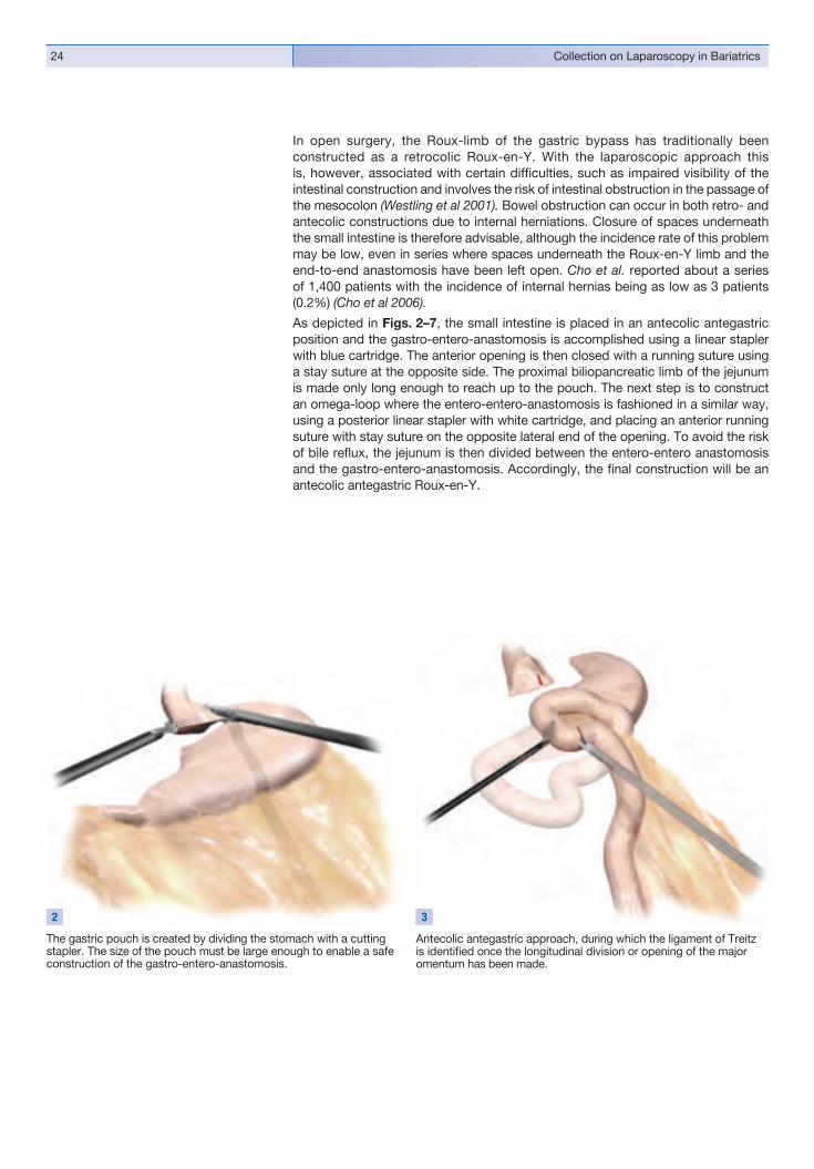
The gastric pouch is created by dividing the stomach with a cutting stapler. The size of the pouch must be large enough to enable a safe construction of the gastro-entero-anastomosis.
2
Antecolic antegastric approach, during which the ligament of Treitz is identifi ed once the longitudinal division or opening of the major omentum has been made.
3
In open surgery, the Roux-limb of the gastric bypass has traditionally been constructed as a retrocolic Roux-en-Y. With the laparoscopic approach this is, however, associated with certain diffi culties, such as impaired visibility of the intestinal construction and involves the risk of intestinal obstruction in the passage of the mesocolon (Westling et al 2001). Bowel obstruction can occur in both retro- and antecolic constructions due to internal herniations. Closure of spaces underneath the small intestine is therefore advisable, although the incidence rate of this problem may be low, even in series where spaces underneath the Roux-en-Y limb and the end-to-end anastomosis have been left open. Cho et al. reported about a series of 1,400 patients with the incidence of internal hernias being as low as 3 patients (0.2%) (Cho et al 2006).
As depicted in Figs. 2–7, the small intestine is placed in an antecolic antegastric position and the gastro-entero-anastomosis is accomplished using a linear stapler with blue cartridge. The anterior opening is then closed with a running suture using a stay suture at the opposite side. The proximal biliopancreatic limb of the jejunum is made only long enough to reach up to the pouch. The next step is to construct an omega-loop where the entero-entero-anastomosis is fashioned in a similar way, using a posterior linear stapler with white cartridge, and placing an anterior running suture with stay suture on the opposite lateral end of the opening. To avoid the risk of bile refl ux, the jejunum is then divided between the entero-entero anastomosis and the gastro-entero-anastomosis. Accordingly, the fi nal construction will be an antecolic antegastric Roux-en-Y.

25Laparoscopic Gastric Bypass
The gastro-entero-anastomosis is here constructed by posteriorstapling and placement of an anterior running suture, which can be made in a single-layer fashion with sero-muscular sutures in the intestine and full thickness suture of the stomach wall.
4
The jejunum is divided between the two anastomotic constructions to prevent bile refl ux.
6
Final situation after completion of the antecolic antegastric Roux-en-Y gastric bypass with creation of a minipouch.
7
Construction of the entero-entero-anastomosis with side-to-side posterior stapling and placement of an anterior running suture.
5

Collection on Laparoscopy in Bariatrics26
ControversiesThe length of the different intestinal limbs in the Roux-en-Y construction is frequently under debate. A short bilio-pancreatic limb may reduce the risk of internal herniation underneath the Roux-limb, while a very long bileopancreatic limb may add a mal ab sorptive component to the gastric bypass. Various lengths of the Roux-limb have been tested, but the outcome is still unclear (Tables 2a, b). In general, most Roux-limbs are made 70 cm or longer. A longer Roux-limb does not guarantee a better outcome as far as weight loss is concerned.
Different types of staple-line reinforcement have been tested for their effi ciency to reduce the risk of anastomotic leakage or bleeding. According to data publish ed in the medical literature so far, no evidence could be provided supporting the use of these sometimes costly materials. With rates of leakage or bleeding ranging below 1%, the clinical benefi t of staple line reinforcement could not be conclusive ly demonstrated. A study by Nguyen (Nguyen 2005) reported about a signifi cant reduction in the number of bleeding sites along those staple lines, that were re inforced using buttressing material (Seemgard®). To date, however, it has not been demonstrated whether this has any clinical impact on the postoperative bleeding frequency.
Fibrin glue is also tested for its potential to reduce the incidence of complications, such as leaks, bleedings but also bowel obstruction. Silecchia and co-workers (Silecchia 2006) found that the total number of complications in the group treated with fi brin glue (Tissucol®) was reduced while the total number of each defi ned complication such as anastomotic leakage, bleeding or internal herniation with bowel obstruction was low. The average leakage rate published in more than 30 different reports, have been found to be less than 3% without the extra use of any leak- preventing materials (Baker 2004).
Prevention of leaks is best done by intraoperative leak testing with either methylene blue or air bubbling. This should be done as a matter of routine during all operations. Postoperative X-ray with gastrographine contrast may also be of value, although it is not 100% sensitive.
Postoperative intestinal obstruction can be caused by stenosis of the gastro-entero-anastomosis or entero-entero-anastomosis. Internal herniation, either behind the entero-entero-anastomosis or behind the Roux-limb, may also occur as a result of intestinal obstruction involving the risk of strangulation. This condition can be hard to diagnose while patients can have severe pain, but without signs of peritonitis and even negative fi ndings in CT-scanning. Among the prophylactic measures that may be taken to prevent the occurrence of this complication, closure of internal hernias during the primary operation is generally recommended as well as timely intervention by diagnostic laparoscopy in patients with acute abdominal pain of unclear etiology. Patients with intestinal obstructions as well as peritonitis with intestinal paralysis may also need a gastrostomy to relieve the dilatation of the gastric remnant.
The rate of complications is related to the experience of the surgeon and the number of patients operated on per year at the institution. In high volume centers, the gastric bypass procedure is a well-documented and well-functioning weight- reducing operation, that can be suggested to most patients who are in need of surgical treatment.
Choban 2002 75 cm vs 150 cm150 cm vs 250 cmInconclusive
Table 2a: Roux-en-Y Gastric Bypass: Short Limb vs Long Limb Inabnet 2005
*) Excess Weight Loss
Patient population A B
N = 25 23
Length of Limb (cm) 50 100
EWL% (per year) * 75% 72% NS
Table 2b: Length of the Roux-limb (BMI < 50)
Brolin 1992 75 cm vs 150 cmp < 0.03
27Laparoscopic Gastric Bypass
AcknowledgementFigs. 1 – 7 published with permission of Johnson & Johnson, Sweden.
ReferencesARASAKI CH, DEL GRANDE JC, YANAGITA ET et al: Incidence of regurgitation after the banded gastric bypass. Obes Surg 2005;15:1408–1417.
BAKER RS, FOOTE J, KEMMETER P, BRADY R, VROEGOP T, SERVELD M:The science of stapling and leaks. Obes Surg 2004;14:1290–1298.
BROLIN RE, KENLER HA, GORMAN JF, CODY RP: Long-limb gastric bypass in the super obese. A prospective randomized study. Ann Surg 1992;215:4:387–95
BROLIN RE: Long limb Roux-en-Y gastric bypass revisited. Surg Clin N Am 2005;85:807–817.
CHOBAN PS, FLANCBAUM L: The effect of Roux limb lengths on outcome after Roux-en-Y gastric bypass. A prospective, randomized clinical trial. Obes Surg 2002;12:4:540–5.
FOBI MAL, LEE H, IGWE D, STANCZYK M, TAMBI JN: Prospective comparative evaluation of stapled versus transacted silastic ring gastric bypass: 6-year follow-up. Obes Surg 2001;11:18–24.
HEDBERG J, HEDENSTRÖM H, NILSSON S, SUNDBOM M, GUSTAVSSON S: Role of gastric acid in stomal ulcer after gastric bypass. Obes Surg 2005;15:10:1375–8.
HIGA KD, HO T, BOONE KB: Laparoscopic Roux-en-Y gastric bypass: technique and 3-year follow-up. J Laparoendosc Adv Surg Tech A 2001;11:6:377–82.
INABNET WB, QUINN T, GAGNER M, URBAN M, POMP A: LaparoscopicRoux-en-Y gastric bypass in patients with BMI<50: A prospective randomized trialcomparing short and long limb lengths. Obes Surg 2005;15:51–57.
LÖNROTH H, DALENBÄCK J, HAGLIND E, LUNDELL L: Laparoscopic gastric bypass. Another option in bariatric surgery. Surg Endosc 1996;10:6:636–8.
NGUYEN NT, LONGORIA M, WELBOURNE S, SABIO A, WILSON SE: Glycolide copolymer staple-line reinforcement reduces staple site bleeding duringlaparoscopic gastric bypass. Arch Surg 2005;140:773–778.
NGUYEN NT, Wolfe BM: Laparoscopic versus open gastric bypass. Seminars in Lap Surg 2002;9:2:86–93.
NÄSLUND I: The size of the gastric outlet and the outcome of surgery for obesity. Acta Chir Scand 1986;152:205–10.
OLBERS T, BJÖRKMAN S, LINDROOS AK, MALECKAS A, LÖNN L, SJÖSTRÖM L,LÖNROTH H: Body composition, dietary intake, and energy expenditure after laparoscopic Roux-en-Y gastric bypass and laparoscopic vertical banded gastroplasty, A randomized clinical trial. Ann Surg 2006;244:5:715–722.
OLBERS T, FAGEVIK-OLSÉN M, MALECKAS M, LÖNROTH H: Randomized clinical trial of laparoscopic Roux-en-Y gastric bypass versus laparoscopic vertical banded gastroplasty for obesity. Br J Surg 2005;92:557–562.
Collection on Laparoscopy in Bariatrics28
OLBERS T, LÖNROTH H, FAGEVIK-OLSÉN M, LUNDELL L: Laparoscopic gastric bypass: development of technique, respiratory function and long-term outcome. Obes Surg 2003;13:3:364–70.SAPALA JA, WOOD MH, SAPALA MA, SCHUHKNECHT MP, FLAKE TM: The micropouch gastric bypass: technical considerations in primary and revisionally operations. Obes Surg 2001;11:3–17.SILECCHIA G, BORU CE, MOUIEL J, ROSSI M et al: Clinical evaluation of fi brin glue in prevention of anastomotic leak and internal hernia after laparoscopic gastric bypass: Preliminary results of prospective, randomized multicentre trial. Obes Surg 2006;15:125–131.SJÖSTRÖM L, LINDROOS AK, PELTONEN M et al: Lifestyle, diabetes andcardiovascular risk factors 10 years after bariatric surgery. N Engl J Med 2004;351:2683–93.SJÖSTRÖM L, NARBRO K, SJÖSTRÖM D et al: Effects of Bariatric surgery on mortality in Swedish obese subjects. N Engl J Med 2007;357:741–52.WESTLING A, GUSTAVSSON S: Laparoscopic vs open Roux-en-Y gastric bypass: a prospective, randomized trial. Obes Surg 2001;11:284–292.WITTGROVE AC, CLARK GW, TREMBLAY LJ: Laparoscopic gastric bypass,Roux-en-Y: preliminary report of fi e cases. Obes Surg 1994;4:4:353–357.

29Laparoscopic Gastric Bypass


33Bariatric SurgeryProf. Matthias Kemen, M.D. and Gerasimos Tzivras, M.D.Herne Protestant Hospital, (Teaching Hospital for the University of Essen)Department of Surgery, Herne, Germany

Collection on Laparoscopy in Bariatrics32
IntroductionObesity, once considered an “aristocratic” symbol of affl uence, has now assum ed the character of a life-threatening disease. It has become a serious epidemiologic health issue (Table 1) that is causing enormous psychosocial and economic problems in our consumer society.
While the growing problems of obesity have been well documented and explained in western history and their conservative treatment options are widely known, it is only in recent years that physicians have made signifi cant progress toward mastering the challenging problem of bariatric surgery (obesity surgery, weight-loss surgery).
This challenge is compounded by the fact that morbid obesity is usually associat ed with intractable comorbid conditions. When we look at the numerous obesity- related diseases listed in the guidelines of the German Obesity Society, the German Diabetes Association, and the German Society for Nutrition (Table 2), we can appreciate the great complexity of the obesity problem. This problem has assumed truly alarming proportions, as evidenced by the fact that type 2 diabetes mellitus, once rare in children and adolescents, is now frequently diagnosed and treated in this age group.
Unfortunately, the conservative management of morbid obesity tends to yield poor long-term results. Everyone is familiar with the “yo-yo” effect in which an obese patient loses weight initially while dieting but then quickly regains the lost pounds.
Postoperative results have confi rmed that bariatric surgery can break this vicious cycle and restore the overweight patient to a healthy, normal-weight life. A striking benefi t of bariatric surgery is that it not only rids patients of excess pounds but also frees them from obesity-related diseases. For example, we have seen many cases in which obese diabetics no longer needed antidiabetic medication (including insulin!) after their operation. The metabolic syndrome can be successfully treated by bariatric surgery.
Table 1:Epidemiology of obesity in Germany. Distribution by Body Mass Index (BMI).(From “BDA Obesity Manual”, published by the Professional Society of General Practitioners in Germany (BDA), www.ifap.de).
18.6 million, BMI 25 – 30
12.0 million, BMI 25 – 30 + comorbidity
9.2 million, BMI 30 – 40
1.2 million, BMI > 40
3%
46%
22%
29%

33Bariatric Surgery
Table 2:Comorbid conditions and complications of overweight/obesity (after WHO 2000, based on guidelines of the German Obesity Society, German Diabetes Association, and GermanSociety for Nutrition).
� Abnormalities of carbohydrate metabolism (e.g., insulin resistance, impaired glucose tolerance, type 2 diabetes mellitus) (Colditz et al. 1995, Chan et al. 1994)
� Other metabolic disorders (e.g., dyslipidemia, hyperuricemia, impaired hemostasis)
� Arterial hypertension
� Cardiovascular diseases (e.g., coronary heart disease, stroke, heart failure)
� Cancer (e.g., endometrium, cervix, ovaries, breast, prostate, kidney, colon)
� Hormonal disorders (e.g., hyperandrogenemia, polycystic ovary syndrome, low testosterone levels in males)
� Pulmonary complications (e.g., dyspnea, hypoventilation and sleep apnea syndrome)
� Gastrointestinal diseases (e.g., cholecystolithiasis, acute and chronic cholecystitis,nonalcoholic steatohepatitis, refl ux disease)
� Degenerative diseases of the musculoskeletal system (e.g., osteoarthritis, spinal syndromes)
� Increased surgical and anesthesia risk
� Physical complaints (e.g., hyperhydrosis, joint pain, exertional dyspnea)
� Psychosocial complaints including proneness to depression and anxiety, social discrimination, and low self-esteem
� Impaired activities of daily living (ADLs)
Table 2: Comorbid conditions and complications of overweight/obesity
Bariatric surgery has been so effective in reversing comorbid conditions that it is sometimes referred to as “metabolic surgery.” In selecting patients for bariatric surgery, some surgeons feel that it is important to consider not only the patients‘ BMI but also their metabolic abnormalities. For example, the Italian professor Nic-ola Scopinaro (Fig. 1) gave the following lecture at the Sixth International Obesity Surgery Expert Meeting in Saafelden, Austria (March 9—12, 2008):
Biliopancreatic Diversion and Type 2 Diabetes in Patients with a BMI < 35 kg/m2
The patients with type 2 diabetes mellitus studied by Scopinaro no longer required insulin after biliopancreatic diversion. This pioneer of obesity surgery and author of the surgical procedure named after him generally prefers a relatively “aggressive” approach that is ordinarily used only in extremely obese patients. The goal is to correct the diabetic metabolic disorder, and the results reported by Scopinaro show that this can be accomplished by surgical means.
This chapter reviews the three standard procedures used in bariatric surgery – adjustable gastric banding, sleeve gastrectomy, and the Roux-en-Y gastric bypass. All of these procedures are performed laparascopically at the surgical unit of the Protestant Hospital in Herne, Germany.
A detailed analysis of issues relating to bariatric surgery, such as the description of postoperative complications, is beyond the scope of this chapter.
Prof. Nicola Scopinaro giving a lectureat the Sixth International Obesity Surgery Expert Meeting in Saafelden, Austria(March 9–12, 2008).
1

Collection on Laparoscopy in Bariatrics34
Laparoscopic Adjustable Gastric Banding (LAGB)One of the fastest-growing forms of bariatric surgery is the laparoscopic implantation of an adjustable gastric band (Lap-Band®).
This procedure has been performed “offi cially” in the United States since it received FDA approval in June of 2001, and it has been practiced in Europe and Australia since 1993.
Adjustable gastric banding was performed in more than 200,000 patients throughout the world from June, 2006, through 2008. The goal of this procedure is to achieve long-term weight reduction in severely obese patients.
Whenever patients consider a bariatric surgical procedure, they should fi rst gather information on the various aspects of the surgery and on available techniques. In the course of the doctor-patient consultation, the attending surgeon can give the patient valuable information that may be of signifi cant help in the decision-making process.
Laparoscopic adjustable gastric banding (often referred to as the Lap-Band®) employs an adjustable silicone band that is placed around the upper part of the stomach. The hollow interior of the band can be fi lled with a variable amount of saline solution, enabling the surgeon to adjust the size of the stoma outlet as desired.
Tightening the gastric band creates a small upper pouch at the top of the stomach. Since the pouch has a capacity of only about 25–30 mL, it fi lls quickly during food ingestion and signals the brain that the stomach is full. This early feeling of satiety helps the patient to achieve gradual yet constant and sustainable weight loss.
Theory and Practice
Gastric banding is a drastic step for overweight patients. It is associated with the same risks as all major surgical procedures on the gastrointestinal tract.
Weight reduction by this purely restrictive procedure ranges from a few kilograms to several kilograms, but generally less weight is lost than with a combined restrictive and malabsorptive procedure such as the Roux-en-Y gastric bypass (LGBRY), biliopancreatic diversion (BPD), or duodenal switch (BPS-DS).
Some patients achieve a normal body weight while others remain obese, though less so than before gastric banding. A major factor in achieving the desired weight reduction is active, consistent cooperation from the patient, who must adhere rigorously to postoperative guidelines on dietary modifi cation and exercise. Genetic predisposition is another factor that should be considered in patient selection. Experience has shown that “big eaters” without a genetic predisposition are good candidates for gastric banding.
The gastric band is fi lled or emptied with a special transdermal needle placed through a small access port that is secured in an epifascial tunnel in the upper left quadrant of the abdomen. The band creates an adjustable stoma between the upper pouch and lower stomach, which in turn produces the desired reduction of food intake capacity. Several adjustable gastric bands are available on the market. The fi ll volume varies considerably in different bands, depending on the manufacturer.
35Bariatric Surgery
If the band needs adjusting but the type of band is unknown, the best solution is to empty all the fl uid, refi ll the band, and check for stoma patency by having the patient drink water. If the patient is unable to swallow, remove some of the fl uid through the port and check again. If necessary the band can be fi lled with contrast medium instead of isotonic saline to permit a more detailed evaluation of band position.
A simple, informative description of the adjustable gastric band can be found at the following website:
http://www.lapband.com/local/media/videos/emmi.swf
This website presents a series of images that illustrate the laparoscopic placement of an adjustable gastric band.
The operation begins with local skeletonization of the lesser curvature just below the level of the second vascular arcade. Skeletonization is continued along the lesser curvature toward the esophagus until the omental bursa is opened.Skeletonization along the greater curvature is performed close to the hiatus and continues to the omental bursa.
1
The upper portion of the stomach has been skeletonized, and the bursa omentalis can be seen.
2
A retrieval tool with distal eyelet is tunneled beneath the gastric inlet.
3
The Lap-Band® is introduced into the abdominal cavity.
4
� One 11-mm primary trocar for the laparoscope, placed at a left paramedian site above and close to the umbilicus.
� Two 11-mm or 13-mm working trocars, placed at least 15 cm apart just to the right of the xiphoid process and in the upper left quadrant near the costal arch.
� One 11-mm or 6-mm trocar in the right midabdomen for the liver retractor.� One 13-mm trocar (expandable to 20 mm) in the right midabdomen.
Five trocars are placed initially:
The end of the Lap-Band® tubing is threaded through the eyelet of the retrieval tool…
5
…and pulled through the retrogastric tunnel toward the lesser curvature.
6

Collection on Laparoscopy in Bariatrics36
The Lap-Band® is passed around the gastric inlet…
7
…and tightened with the closure tool.
8
The buckle of the Lap-Band® is locked into place.
9
The Lap-Band® is correctly positioned around the gastric inlet.
10
The Lap-Band® is fi xed to the anteriorstomach wall with interrupted seromuscular sutures.
11 12 13
14 15

37Bariatric Surgery
The free end of the tubing is brought out of the abdominal cavity through the working trocar.
16 17
The free end of the tubing is passed behind the stomach and connected to the Lap-Band® access port, which is secured mechanically or manually in an epifascial tunnel in the upper left quadrant of the abdomen. The trocars are removed from the abdominal cavity under laparoscopic vision, and the pneumoperitoneum is released. The trocar incisions are closed with absorbable sutures.
Laparoscopic Gastric Bypasswith Roux-en-Y Limb (LGBRY)As early as 1991, the National Institutes of Health (NIH) held a consensus meeting with the goal of establishing mandatory guidelines and indications for the surgical treatment of morbid obesity. According to these guidelines, candidates for bariatric surgery should meet one of the following criteria:
1. A BMI of 40 or higher,
or
2. A BMI of 35 or higher plus one or more comorbid conditions.
The consensus conference also emphasized the need for interdisciplinary cooperation in the care of bariatric surgical patients by a team of doctors and therapists in a comprehensive program that would address the multifactorial aspects of the disease in treatment and follow-up: comorbidity, diet, exercise, behavior, and psychological aspects.
The selected bariatric procedure is basically considered a “tool” for helping overweight patients change their lifestyle and eating habits and achieve effective, sustainable obesity management and personal behavior modifi cation.

Collection on Laparoscopy in Bariatrics38
Additional conclusions:
� Bariatric surgery is currently the most effective method for the treatment of morbid obesity.
� Gastric bypass is one of the four main surgical options(along with biliopancreatic diversion, duodenal switch, and gastric banding)for the treatment of morbid obesity.
� Laparoscopic surgery is as safe and effective as conventional open surgery. � Patients should undergo a comprehensive preoperative diagnostic workup and
counseling and, as mentioned, should receive multidisciplinary care to achieve optimum results.
Gastric bypass is a restrictive and malabsorptive bariatric procedure in which a small pouch is created from the upper portion of the stomach while the much larger, lower portion of the stomach is isolated. The small intestine is divided, and gastro intestinal continuity is restored by a Roux-en-Y gastroenterostomy. Gastric bypass is performed laparascopically throughout the world, and a great variety of techniques have been developed.
This operation generally results in a dramatic degree of weight loss, leading to a marked regression or even resolution of numerous comorbid conditions. It is not unusual for insulin-dependent diabetics to discontinue their antidiabetic medication, giving rise to the term “metabolic surgery” for bariatric operations. It has also been shown that long-term mortality is reduced by up to 40% in gastric bypass patients.
It should be added, however, that 2% of the complications that arise during the initial postoperative months have a fatal outcome.
The following series of images illustrate the steps involved in a laparoscopic Roux-en-Y gastric bypass.
Five trocars are placed initially:
� One 11-mm primary trocar for the laparoscope, placed at a left paramedian site above and close to the umbilicus.
� Two 11-mm or 13-mm working trocars, placed at least 15 cm apart just to the right of the xiphoid process and in the upper left quadrant near the costal arch.
� One 11-mm or 6-mm trocar in the right midabdomen for the liver retractor.� One 13-mm trocar (expandable to 20 mm) in the right midabdomen.

39Bariatric Surgery
Trocar sites for laparoscopic Roux-en-Ygastric bypass: � One 11-mm primary trocar placed at a left
paramedian site above and close to the umbilicus.
� Two 13-mm working trocars, placed at least 15 cm apart just to the right of the xiphoid process and in the upper left quadrant near the costal arch.
� One 11-mm or 6-mm trocar in the right midabdomen for the liver retractor.
� One 13-mm trocar in the right midabdomen.� Two 6-mm trocars, one in the left mid/
lower abdomen just below the umbilical fossa and the other at an approximately symmetrical site on the right side.
1
Image Sequence Illustrating the Roux-en-Y Gastric Bypass
A swath is dissected through to the greater omentum to the transverse colon.
2
The ligament of Treitz is located and identifi ed.
3
Starting at the ligament of Treitz, a 75-cm length of small intestine is measured off(in 5-cm increments) in a counterclockwise direction.
4 5
The loop of jejunum is brought up to the stomach…
6
…and fi xed there with a simple interrupted suture.
7

Collection on Laparoscopy in Bariatrics40
Lateral upward traction is placed on the bowel loop.
12
The jejunal limb is opened with an ultrasound dissector.
13
The linear stapler is introduced through an incision in the small bowel wall…
14
…and fi red to create the jejunojejunalanastomosis.
15
That loop is brought up to the stomach and fi xed to the previously transposed jejunum with a simple interrupted suture.
9 10
11
Another 150 cm of bowel is measured off (in 5-cm increments) in a clockwise direction.
8
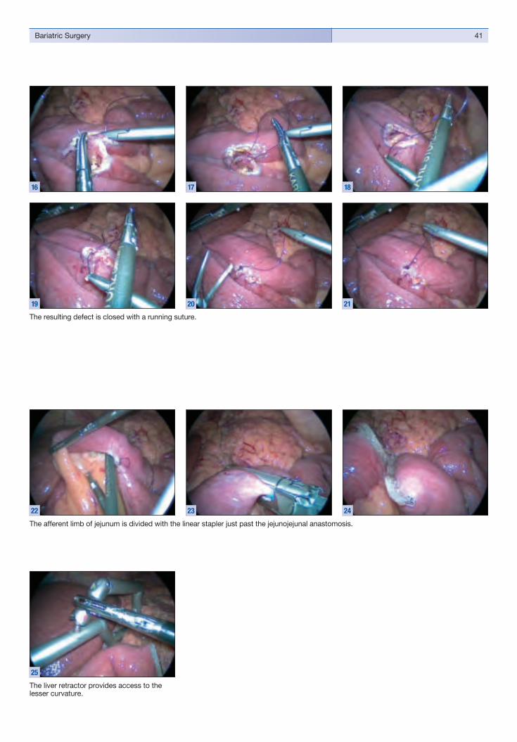
41Bariatric Surgery
The liver retractor provides access to the lesser curvature.
25
The afferent limb of jejunum is divided with the linear stapler just past the jejunojejunal anastomosis.
22 23 24
16 17 18
The resulting defect is closed with a running suture.
19 20 21

Collection on Laparoscopy in Bariatrics42
The lesser curvature is skeletonized at the approximate level of the second to third vascular arcade, opening the omental bursa.
26 27 28
The gastric pouch is divided superiorly with the roticulator.
31 32 33
The 45-mm linear stapler is introduced transversely and fi red.
29 30

43Bariatric Surgery
The jejunum and gastric stump are opened on the lesser curvature side with the ultrasound dissector.
38 39 40
The divided afferent limb of jejunum is brought up and sutured to the gastric stump.
34 35
36 37

Collection on Laparoscopy in Bariatrics44
The defect at the GE is closed with sutures (and tested for leaks).
43 44
The gastroenterostomy (GE) is created with the 45-mm linear stapler.
41
The gastric tube is advanced into the jejunum under vision.
42
Laparoscopic Roux-en-Y Gastric BypassThe Jejunojejunal Anastomosis
The limbs of jejunum are tacked together with a simple interrupted suture, which is retracted upward and to the right.
1
The jejunal limbs are incised with the ultrasound dissector.
2
The 45-mm linear stapler is introduced through the incision in the small bowel wall.
3

45Bariatric Surgery
The defect at the GE is closed with sutures (and tested for leaks).
11 12 13
Small remaining tissue bridges are secured with clips.
10
The lesser curvature of the stomach isskeletonized at the approximate level of the second to third vascular arcade, opening the omental bursa.
6
The 45-mm linear stapler is introduced transversely and fi red.
7
The efferent limb of jejunum is dissected free just past the jejunojejunal anastomosis(toward the stomach) and transected with the linear stapler.
4 5
The gastric pouch is divided superiorly with the roticulator.
8 9
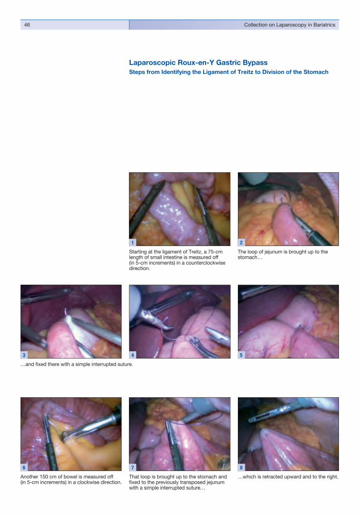
Collection on Laparoscopy in Bariatrics46
Laparoscopic Roux-en-Y Gastric BypassSteps from Identifying the Ligament of Treitz to Division of the Stomach
Starting at the ligament of Treitz, a 75-cm length of small intestine is measured off(in 5-cm increments) in a counterclockwise direction.
1
The loop of jejunum is brought up to the stomach…
2
…and fi xed there with a simple interrupted suture.
3 4 5
Another 150 cm of bowel is measured off(in 5-cm increments) in a clockwise direction.
6
That loop is brought up to the stomach and fi xed to the previously transposed jejunum with a simple interrupted suture…
7
…which is retracted upward and to the right.
8
2
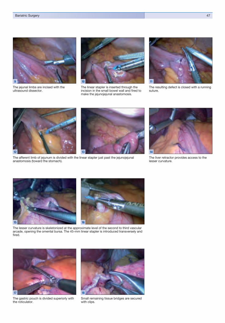
47Bariatric Surgery
The resulting defect is closed with a running suture.
11
The liver retractor provides access to the lesser curvature.
14
The jejunal limbs are incised with theultrasound dissector.
9
The linear stapler is inserted through theincision in the small bowel wall and fi red to make the jejunojejunal anastomosis.
10
Small remaining tissue bridges are secured with clips.
18
The gastric pouch is divided superiorly with the roticulator.
17
The afferent limb of jejunum is divided with the linear stapler just past the jejunojejunalanastomosis (toward the stomach).
12 13
The lesser curvature is skeletonized at the approximate level of the second to third vascular arcade, opening the omental bursa. The 45-mm linear stapler is introduced transversely and fi red.
15 16

Collection on Laparoscopy in Bariatrics48
Laparoscopic Roux-en-Y Gastric Bypasswith an Intragastric Balloon
Laparoscopic view of the intragastric balloon, which is pierced with a needle tip and removed gastroscopically by the internist during the operation.
1 2 3
Dissection and transection of the jejunal limb, which is brought upward to the stomach for creating the gastroenterostomy (GE).
6 7 8
The defect left by creation of the stapled jejunojejunal anastomosis is closed with a running suture.
4 5

49Bariatric Surgery
The lesser curvature is dissected and skeletonized at the approximate level of the second to third vascular arcade.
9 10 11
The pouch is created by progressively dividing the stomach up to the esophagus with the 65-mm linear stapler.
13 14 15
The jejunal limb of the GE is sutured to the gastric stump.
16 17 18
The stomach is divided transversely on the lesser curvature side with the 45-mm linear stapler.
12

Collection on Laparoscopy in Bariatrics50
The jejunal limb and gastric stump are incised, and the 45-mm linear stapler is introduced and fi red to create the gastroenterostomy.
19 20 21
The defect at the GE is closed with sutures (and tested for leaks).
22 23 24
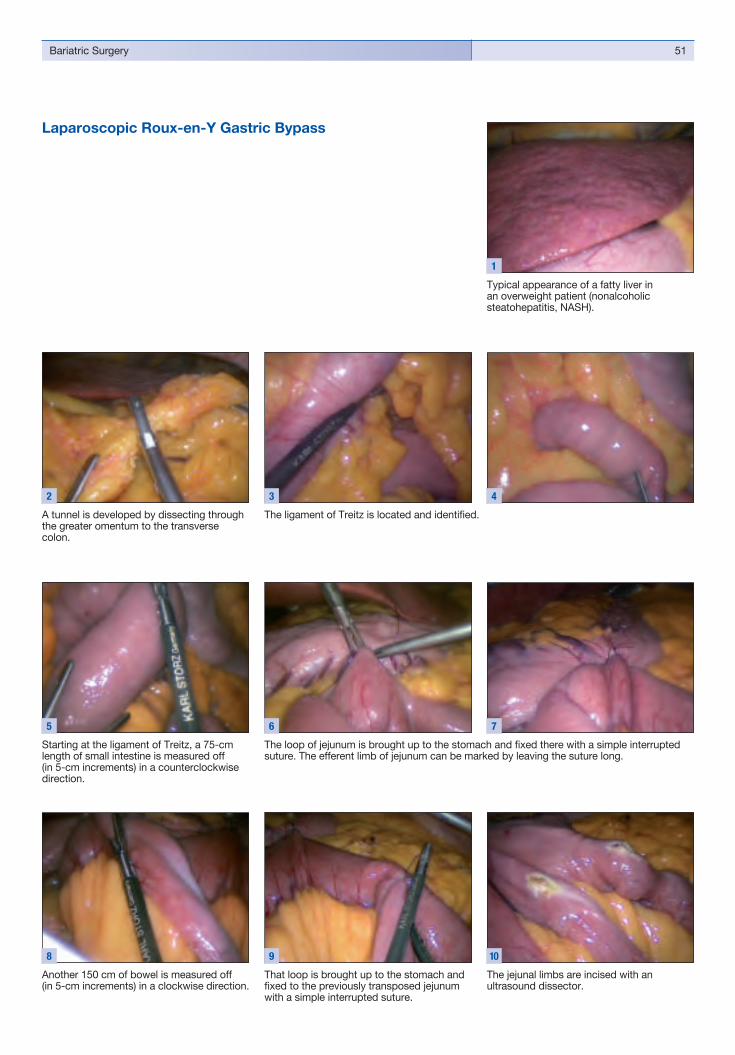
51Bariatric Surgery
Laparoscopic Roux-en-Y Gastric Bypass
Typical appearance of a fatty liver in an overweight patient (nonalcoholic steatohepatitis, NASH).
1
A tunnel is developed by dissecting through the greater omentum to the transverse colon.
2
Starting at the ligament of Treitz, a 75-cm length of small intestine is measured off(in 5-cm increments) in a counterclockwise direction.
5
Another 150 cm of bowel is measured off(in 5-cm increments) in a clockwise direction.
8
That loop is brought up to the stomach and fi xed to the previously transposed jejunum with a simple interrupted suture.
9
The jejunal limbs are incised with anultrasound dissector.
10
The ligament of Treitz is located and identifi ed.
3 4
The loop of jejunum is brought up to the stomach and fi xed there with a simple interrupted suture. The efferent limb of jejunum can be marked by leaving the suture long.
6 7

Collection on Laparoscopy in Bariatrics52
The jejunal limb to the stomach is divided with the linear stapler just past the jejunojejunal anastomosis.
16 17 18
The lesser curvature is skeletonized at the approximate level of the second to thirdvascular arcade, opening the omental bursa.
19
The linear stapler is inserted through theincision in the bowel wall…
11
…and fi red to make the jejunojejunalanastomosis.
12
The resulting defect is closed with a running suture.
13
Any defects in the serosa are repaired with interrupted seroserous sutures.
14 15
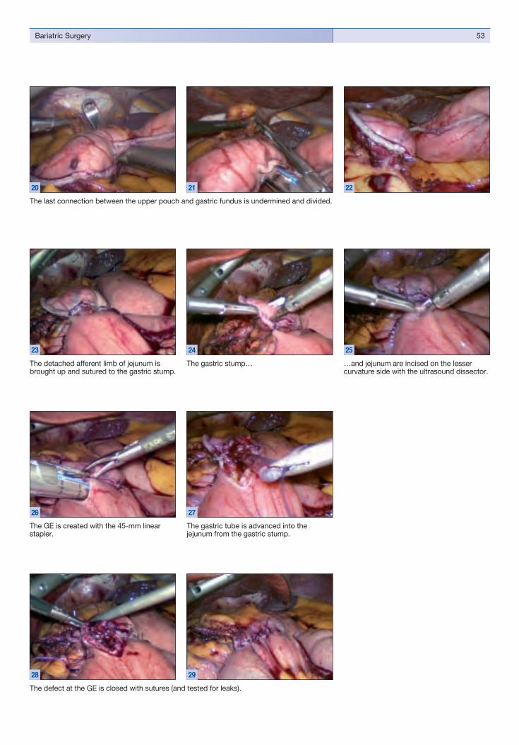
53Bariatric Surgery
The last connection between the upper pouch and gastric fundus is undermined and divided.
20 21 22
The defect at the GE is closed with sutures (and tested for leaks).
28 29
The detached afferent limb of jejunum is brought up and sutured to the gastric stump.
23
The gastric stump…
24
…and jejunum are incised on the lessercurvature side with the ultrasound dissector.
25
The GE is created with the 45-mm linearstapler.
26
The gastric tube is advanced into the jejunum from the gastric stump.
27

Collection on Laparoscopy in Bariatrics54
Laparoscopic Sleeve GastrectomySleeve gastrectomy is an irreversible, restrictive bariatric procedure in which approx-imately 85% of the stomach is removed laparascopically on the greater curvature side, leaving a banana-shaped gastric sleeve of greatly reduced capacity.
Laparoscopic sleeve gastrectomy is generally performed in extremely overweight patients in an initial sitting in cases where other restrictive or malabsorptive bariatric procedures (e.g., gastric bypass, biliopancreatic diversion, etc.) are deemed too risky. The procedure may then be converted to a gastric bypass, for example, in a later sitting.
The sleeve gastrectomy alone may be an adequate, relatively low-risk bariatric procedure for patients who are obese but not extremely obese. Some surgeons even prefer it over gastric banding.
The following series of images illustrate the steps involved in a laparoscopic sleeve gastrectomy.
Five trocars are placed initially:
� One 11-mm primary trocar for the laparoscope, placed at a left paramedian site above and close to the umbilicus.
� Two 11-mm or 13-mm working trocars, placed at least 15 cm apart just to the right of the xiphoid process and in the upper left quadrant near the costal arch.
� One 11-mm or 5-mm trocar in the right midabdomen for the liver retractor.� One 13-mm trocar (expandable to 20 mm) in the right midabdomen.
Trocar sites for laparoscopic sleeve gastrectomy: � One 11-mm primary trocar placed at a
left paramedian site above and close to the umbilicus.
� Two 13-mm working trocars, placed at least 15 cm apart just to the right of the xiphoid process and in the upper left quadrant near the costal arch.
� One 11-mm or 6-mm trocar in the right midabdomen for the liver retractor.
� One 13-mm working trocar in the right midabdomen.
1
The operation begins with distal skeletoni-zation along the greater curvature of the stomach.
2
Retrogastric skeletonization.
5
Skeletonization of the greater curvature is continued toward the esophagus.
3 4
Skeletonization of the greater curvature is continued toward the esophagus.
6 7

55Bariatric Surgery
The greater curvature has been completely skeletonized.
8
A large-caliber gastric tube is passed into the dissected stomach and placed along the lesser curvature.
9
Remaining retrogastric adhesions are cleared until the posterior surface of the stomach wall has been dissected free.
12
The sleeve gastrectomy is begun by placing a 45-mm linear stapler across the distal greater curvature (junction of middle and distal antrum). Another option is to use a 60-mm linear stapler placed close to the esophagus and parallel to the intragastric tube.
10 11
Skeletonization of the greater curvature is continued toward the esophagus.
13 14
15 16
Collection on Laparoscopy in Bariatrics56
The sleeve gastrectomy is complete except for a narrow tissue bridge.
17
The cut surface of the stomach wall is che-cked for hemostasis.
21
If necessary, bleeding sites are oversewn with interrupted seroserous sutures.
22
The specimen has been completely detached.
20
Thin remaining bridges of stomach wall can be clipped and divided.
18 19

57Bariatric Surgery
Gastric specimen ready for extraction.
23
The staple line is tested for leaks with methylene blue.
24
The 13-mm working trocar in the upper left quadrant is replaced with a 20-mm trocar.
25 26
The specimen is extracted from the abdominal cavity.
27 28
Concluding RemarksA number of surgical solutions have been developed to solve the challenging problem of weight reduction in morbid obesity. Several studies have shown that, as a general rule, the malabsorptive procedures appear to be more effective despite their higher risks.
It is important to consider that weight reduction by bariatric surgery can also reverse the comorbid conditions that are associated with obesity, leading to a signifi cant decline in overall mortality.
Despite the potential for postoperative complications, which require complex management in specially equipped intensive care units, bariatric surgery still offers the only hope for a defi nitive cure in morbidly obese patients.


44Laparoscopic Sleeve Gastrectomy Raul J. Rosenthal, M.D., FACSSection Head of Minimally Invasive Surgery and Medical Director of theBariatric and Metabolic Institute, Cleveland Clinic, Weston, Florida, U.S.A.

Collection on Laparoscopy in Bariatrics60
IntroductionSurgery is the most effective treatment for weight loss and improvement in some comorbid conditions in patients with morbid obesity1, 6. Currently, laparoscopic Roux-en-Y gastric bypass (LRYGBP) and laparoscopic adjustable gastric banding (LAGB) are the most commonly performed bariatric procedures in the United States. The addition of laparoscopic sleeve gastrectomy (LSG) as an alternative surgical option for morbid obesity has occurred only over the last few years.
History of Sleeve GastrectomySleeve gastrectomy (SG) was fi rst described in 1988 when Scopinaro’s technique of biliopancreatic diversion (BPD) with distal gastrectomy and gastroileostomy was modifi ed by Hess and simultaneously by Marceau4, 7,13. The BPD-duodenal switch (BPD-DS) combines a vertical SG with a gastric volume of approximately 100 to150 mls and a duodenal switch with a common channel of 100 cm and an aliment ary limb of 150 cm. The SG performed the restrictive component of the pro cedure replacing the need for a distal gastrectomy. This technique resulted in greater weight loss with reduced morbidity seen compared to the original Scopinaro procedure including reduction in ulcerogenicity and malabsorption with hypoproteinaemia, hypocalcaemia, and the dumping syndrome8. The fi rst part of the duodenum, pylorus, antrum, lesser curvature, and vagal nerve integrity are maintained creating moderate restriction while allowing a relatively normal eating behaviour. The BPD-DS is effective at providing and maintaining excess weight loss (EWL) in the long term5,
8,13. Hess et al. have reported maintenance of greater than 50% EWL in 94% of 182 patients who underwent BPD-DS with long term follow up of more than 10 years, and greater than 80% EWL in 52% of patients5. With advances in minimally invasive surgery, Gagner described the fi rst laparoscopic BPD-DS in 19996, 8.
Due to its acceptably low complication rate and relative ease of technical per formance, SG was subsequently recommended as an initial step in the management of super-obese patients or those with a high operative risk due to extensive comorbid conditions to minimize the associated morbidity and mortality. A more defi nitive procedure would be deferred to a later date following an initial weight loss. Regan et al. described a two stage procedure in super-super obese patients who underwent LSG as a fi rst-stage procedure followed by LRYGB as a second-stage for more defi nitive treatment of obesity5. Their initial experience suggested that the two stage approach was feasible, and effective in the short term in achieving signifi cant average excess weight loss of 51 kg in 6 patients with an initial body mass index (BMI) of 58–71 kg/m2. The average excess weight loss was 37 kg following LSG over an 11-months period between procedures11. Cottam et al. recently published their initial experience on 126 patients with a mean BMI of 65 kg/m2 (range 45–91) who underwent a two stage approach2. Laparoscopic SG was performed as the fi rst stage, followed by LRYGB after signifi cant weight loss and reduction in co-morbidities. One patient died, and major complications occurred in 13%. The mean excess weight after LSG at 1 year was 46%. Thirty-six patients with a mean BMI of 49 kg/m2 went on to have the second-stage LRYGB2.
As experience with the technique increased the role of LSG evolved and many began to consider it as a primary restrictive bariatric procedure. Early reports of prospective and retrospective studies were encouraging9,10. Mognol et al. reported their initial experience of 10 patients with a mean BMI of 64 kg/m2 (range 61–80) who underwent LSG. The mean operative time was 2 hours (range 1.5–2.5), all procedures were completed laparoscopically, and there were no postoperative complications or mortality. At 1 year the average EWL was 51%9. A further retrospective analysis of 60 patients who underwent LSG demonstrated an EWL of 83% at 1 year with a median weight loss of 25 kg. More importantly, there was a signifi cant improvement in comorbid conditions including diabetes, hypertension, dyslipidemia, and joint pain10. Schauer et al. recommend a SG or LAGB with an interval LRYGB, if weight loss does not continue to achieve a BMI < 35 kg/m2. 3

61Laparoscopic Sleeve Gastrectomy
1
Operating room setup.
2
Preparation for surgery. The costal margin and sites of port placement are marked out with a skin pencil.
TechniqueWe use a 7-trocar technique for LSG with identical port placement to our LRYGB procedure14. After induction of anesthesia and endotracheal intubation, the abdominal cavity is accessed through a 1-cm supraumbilical incision using a bladeless trocar (Figs. 1–3).
The abdominal cavity is insuffl ated with carbon dioxide to a pressure of 15 mmHg. The operating ports are inserted under direct vision. The liver is retracted cranially, and the gastroesophageal (GE) junction is exposed (Fig. 4).
A point on the greater curvature, approximately 6 cm proximal to the pylorus, is identifi ed as the distal extent of the resection (Figs. 5, 6).
Trocar placement for laparoscopic sleeve gastrectomy.
3
Gastroesophageal (GE) junction is exposed.
6
Pylorus identifi cation.
5
Start point on the greater curvature 6 cm from pylorus.
4
6 mm
6 mm
13 mm
13 mm
11mm
13 mm
13 mm

Collection on Laparoscopy in Bariatrics62
The ultrasonic scalpel is used to divide the vessels along the greater curvature up to the angle of His (Figs. 7, 8).
A 48–52 Fr bougie is inserted transorally to the level of the distal stomach. Linear cutting staplers are used to vertically transect the stomach creating a narrow gastric tube with an estimated capacity of 150 mls (Figs. 10, 11).
The staple line is then oversewn with a running 2/0 silk suture. The resected stomach is placed in a specimen bag and extracted (Figs. 12–15).
All patients have a routine gastrografi n swallow study on postoperative day 1 and are commenced on oral fl uids, if normal.
Gastric vessels are taken down with the ultrasonic scalpel.
7
The vessels are resected very close to the greater curvature.
8
Final view after greater curvature skeletization.
9
Once the bougie is placed, the fi rst linear cutting stapler is used.
10
The stomach is fully transected with linear staplers.
11
The stomach is fully transected with linear staplers.
12
14
Staple line is oversewn with a running suture.
13
The resected stomach is extracted.
15

63Laparoscopic Sleeve Gastrectomy
ResultsIn our center, we have performed 105 LSG as a one-stage restrictive procedure for weight loss. The majority, n=92 were performed as primary procedures. Thirteen patients underwent LSG as a secondary procedure after failed LAGB (n=11), previous jejunoileal bypass with weight regain (n=2), or a failed LRYGB (n=1). We recently published our initial experience with LSG as a primary procedure for weight loss12,15. A retrospective analysis of 30 patients who underwent LSG between November 2004 and March 2006, who had completed a minimum of 6 months follow up, demonstrated a mean weight loss of 22.6 kg and 30.5 kg at 3 and 6 months respectively. The mean EWL was 41% and 53% at 3 and 6 months respectively. The mean operative time was 80 min (range 65–130), and all procedures were completed laparoscopically. The mean hospital stay was 3.2 days (range 2–25). One patient required a second laparoscopy on postoperative day 1 for management of a leak from the staple line close to the GE junction detected on the gastrografi n study. Three patients required readmission for mild dehydration.
ConclusionLaparoscopic SG is an effective primary restrictive procedure to achieve weight loss. It is a relatively straightforward procedure that can be performed laparoscopically with an extremely low conversion rate. Operative time is reasonable ranging from 1 to 2 hours. Our mean operative time for LSG was 80 min. Meticulous attention to oversewing the staple line was the main factor contributing to our extended operative time, but we feel this time is well spent to achieve haemostasis, and prevent a leak which could result in signifi cant morbidity. Laparoscopic SG is technically feasible in high-risk patients with signifi cant morbidity, and also in very obese patients with BMI > 70 kg/m2. As our experience with LSG is expanding, we are attempting to identify defi nitive indications for LSG in our patient population. Currently, we perform LSG in very high-risk patients with signifi cant comorbidity, patients aged >70 years with signifi cant comorbidity, low BMI 35–40 with comorbidity, or according to patient preference. We do not perform LSG as a staged procedure. We offer LRYGB, LSG and LAGB to all patients, but recommend LRYGB as the procedure of choice in patients with a BMI > 50 kg/m2 with comorbidity.
In conclusion, LSG can be performed with minor complications and low mortality. It has been demonstrated to be a safe and effective procedure in the short term. Prospective studies are required after LSG to determine the long-term outcome, and effi cacy of maintenance of weight loss and resolution of comorbid conditions.

Collection on Laparoscopy in Bariatrics64
References1. Gastrointestinal surgery for severe obesity. NIH Consensus Development
Conference Consensus Statement 9(1):1–20, 1991.
2. COTTAM D, QURESHI FG, MATTAR SG, SHARMA S, HOLOVER S, BONANOMI G, RAMANATHAN R and SCHAUER P: Laparoscopic sleevegastrectomy as an initial weight-loss procedure for high-risk patients withmorbid obesity. Surg Endosc 20(6):859–863, 2006.
3. DEMARIA EJ, SCHAUER P, PATTERSON E, NGUYEN NT, JACOB BP, INABNET WB. and BUCHWALD H: The optimal surgical management of the super-obese patient: the debate. Presented at the annual meeting of theSociety of American Gastrointestinal and Endoscopic Surgeons, Hollywood, Florida, USA, April 13–16, 2005. Surg Innov 12(2):107–121, 2005.
4. HESS DS. and HESS DW: Biliopancreatic diversion with a duodenal switch. Obes Surg 8(3):267–282, 1998.
5. HESS DS, HESS DW and OAKLEY RS: The biliopancreatic diversion with the duodenal switch: results beyond 10 years. Obes Surg 15(3):408–416, 2005.
6. MAGGARD MA, SHUGARMAN LR, SUTTORP M, MAGLIONE M,SUGERMAN HJ, LIVINGSTON EH, NGUYEN NT, LI Z, MOJICA WA, HILTON L,RHODES S, MORTON SC and SHEKELLE PG: Meta-analysis: surgical treatment of obesity. Ann Intern Med 142(7):547–559, 2005.
7. MARCEAU P, BIRON S, ST GEORGES R, DUCLOS M, POTVIN M and BOURQUE RA: Biliopancreatic Diversion with Gastrectomy as Surgical Treatment of Morbid Obesity. Obes Surg 1(4):381–387, 1991.
8. MARCEAU P, HOULD FS, SIMARD S, LEBEL S, BOURQUE RA, POTVIN Mand BIRON S: Biliopancreatic diversion with duodenal switch. World J Surg 22(9):947–954, 1998.
9. MOGNOL P, CHOSIDOW D and MARMUSE JP: Laparoscopic sleevegastrectomy as an initial bariatric operation for high-risk patients: initial results in 10 patients. Obes Surg 15(7):1030–1033, 2005.
10. MOON HAN S, KIM WW and OH JH: Results of laparoscopic sleeve gastrectomy (LSG) at 1 year in morbidly obese Korean patients. Obes Surg 15(10):1469–1475, 2005.
11. REGAN JP, INABNET WB, GAGNER M and POMP A: Early experience with two-stage laparoscopic Roux-en-Y gastric bypass as an alternative in the super-super obese patient. Obes Surg 13(6):861–864, 2003.
12. ROA PE, KAIDAR-PERSON O, PINTO D, CHO M, SZOMSTEIN S and ROSENTHAL RJ: Laparoscopic sleeve gastrectomy as treatment for morbid obesity: technique and short-term outcome. Obes Surg 16(10):1323–1326, 2006.
13. SCOPINARO N, ADAMI GF, MARINARI GM, GIANETTA E, TRAVERSO E, FRIEDMAN D, CAMERINI G, BASCHIERI G and SIMONELLI A:Biliopancreatic diversion. World J Surg 22(9):936–946, 1998.
14. SZOMSTEIN S, ARIAS F and ROSENTHAL R: How We Do LaparoscopicRoux-En-Y Gastric Bypass. Contemporary Surgery 62(3):106–111, 2006.
15. TUCKER ON, SZOMSTEIN S and ROSENTHAL RJ: Indications for sleeve gastrectomy as a primary procedure for weight loss in the morbidly obese. J Gastrointest Surg 12(4):662–667, 2008.

65Laparoscopic Sleeve Gastrectomy


55Laparoscopic Sleeve Gastrectomy J.C. Ruiz de Adana, J. López-Herrero, A. Hernández-Matías Department of General SurgeryMadrid University Hospital of Getafe (Madrid), Spain

Collection on Laparoscopy in Bariatrics68
IntroductionSleeve gastrectomy is a bariatric technique which converts the stomach into a (sleeve-shaped) narrow tube along the lesser curvature of the stomach and involves the dissection of the gastric remnant. The procedure is technically feasible and reproducible, however it can be fraught with diffi culties in cases of extreme obesity, central obesity and in the presence of a large left hepatic lobe. It is associated with a reduced risk of major complications as compared to a gastric bypass or any other malabsorptive procedure, due to the fact that no anastomosis or manipulation of the inframesocolic compartment is necessary. Still, the procedure has an overall post-operative mortality of 0.5% and a long-term revision rate of 4.4%.
The reduction in ghrelin associated with extensive resection of the gastric fundus may explain the better results in comparison with other restrictive techniques (vertic al gastroplasty and gastric banding). The mean rates of excess weight loss (EWL) are 49% after 6 months, 56% after one year and 66% after three years but are much higher in patients with a body mass index (BMI) of between 35 and 45 (Table 1). Most of the comorbidities associated with obesity, such as hypertension, dys lipidemia or diabetes mellitus, improve and are cured in 60–100% of cases. Laparoscopic sleeve gastrectomy has a low incidence of complications, revisions and mortality, although it is crucial that long-term studies (> 5 years) must be obtained to determine the effi cacy of the procedure in terms of long-term main tenance of excess weight loss.
� As fi rst-line surgical treatment modality in super-obese patients (BMI > 50) or those with signifi cant risk factors, and as part of a staged strategy of treatment, after initial weight loss and regression of obesity-related comorbidities: with duodenal switch or bypass surgery (two-stage surgery).
� As a last-resort technique in patients with severe hepatopathy, AIDS, transplants, intestinal infl ammatory disease, of over 65 years of age and in patients with pathologies or history of gastric tumors who require regular endoscopic checks. Also in the presence of adverse intraoperative circumstances such as intolerance of pneumoperitoneum, hemodynamic instability, etc. calling for a reduced duration of surgery and/or a less complicated technique
� As revision surgery after failure of a gastric banding procedure.� Selectively in young patients or those with a BMI < 45 who do not accept the
consequences of another type of bariatric surgery and who are willing to give their explicit informed consent for the implementation of this surgical treatment option without long-term scientifi c evidence for the effi cacy of the technique.
Patient Selection and Indications
Authors Number of Preoperative Follow-up EWL* cases BMI (%) (months) (%)
Lee et al. (2007) 216 49 12 59
Cottam et al. (2006) 126 65 12 46
Hamoui et al. (2006) 118 55 24 47
Moon Han et al. (2005) 60 37 12 83
Himpens et al. (2006) 40 39 36 66
Table 1: Short- and medium-term weight loss after sleeve gastrectomy
* Excess Weight Loss

69Laparoscopic Sleeve Gastrectomy
� Patient positioning: in French (reverse Trendelenburg) position, with the operating surgeon between the patient’s legs and the video monitor at the patient’s head (Figs. 1a–c).
� Trocar ports: 5 trocars, sized 11–13 mm (one of 15 mm, if green loads are used). The camera port (T1) (30º-laparoscope with coupled videocamera) is placed along the midline, at approximately 15 cm from the xiphoid. An auxiliary left subcostal port (T4) and two pararectal operating ports (T2) (T3) of 11–13 mm in diameter, unless (green) loads of 4.5-mm staples (16 mm) are used (Fig. 2). The last trocar for liver retraction is usually placed in the subxiphoid area (T5), however, as an alternative option, it may also be in the right subcostal area, and 6 or 11 mm in diameter, depending on the size of the retractor to be used (Fig. 1c).
� Instrumentation and Accessories:
1� 30º-laparoscope, diameter 10 mm, length 31 cm.
2� Four blunt-tipped trocars sized 11–13 mm, one of them used for the camera, and one or two 16-mm trocars for the stapler, if green cartridges with 4.5-mm staples are applied.
3� Linear endostapler with loads of 45/60 mm in length and staples sized 3.5 mm (blue), and of larger diameter, 3.8 mm (gold) or 4.5 mm (green) for section of the pyloric antrum.
4� Vessel sealing device or ultrasonic dissector.
5� Clip applicator and needle holder.
6� 2/0 monofi lament suture or
7� Bioabsorbable staple line reinforcement material (e.g, Gore Seamguard®).
8� Liver retractor.
9� 32–36 Fr esophageal catheter or bougie.� Technique of Laparoscopic Sleeve Gastrectomy: The gastric reservoir is converted into a narrow tube (sleeve) extending from the
eso phagus to the duodenum along the entire lesser curvature of the stomach. The most important technical aspects are:
� the size of the calibrated bougie used to create the gastric tube,
� proper wound closure technique along the staple lines to prevent formation of leaks and hemorrhages and,
� the distance from the pyloris to the fi rst line of staples, which varies among authors and is still a matter of controversy.
Surgical Technique
Schematic drawing of the operating room setup for laparoscopic sleeve gastrectomy.co = camera operatorfa = fi rst assistants = operating surgeonan = anesthesistit = instrument trayvm = video monitor
3
Port placement for laparoscopic sleeve gastrectomy.Trocar T1 11–13 mm (camera port)Trocar T2 11–13 mm (operating port)Trocar T3 11–13 mm (operating port)Trocar T4 11–13 mm (operating port)Trocar T5 6 or 11 mm (liver retractor)
2
1b
1c
Figs. 1a–cOperating room setup for the laparoscopic sleeve gastrectomy. The operating surgeon assumes a position between the patient’s legs.
1a
T1T2
T3T4
T5
itvm
vm
s
fa
an
co

Collection on Laparoscopy in Bariatrics70
Once the stomach contents have been fully evacuated by aspiration, the left crus of the diaphragm is exposed and a cotton pledget is positioned, thus permitting sub sequent localization of this anatomical site. (Figs. 4a,b). The vessel sealing device or the ultrasonic dissector is used to release the entire vascular attachments of the larger curvature in close proximity to the serosa of the gastric wall. Dissection then proceeds cranially toward the angle of His until the cotton pledget, the landmark for the left crus of the diaphragm, can be visualized (Figs. 5a–d). There is controversy in the literature as to how far dissection should be carried out toward the duodenum (Figs. 6a–c). It can either be extended up to a point 2 cm from the pylorus to create a regular shaped small sleeve (restrictive component), or ended at a distance of
Dissection and extirpation of the entire greater curvature of the stomach.
5c
5a
5d
5b
Dissection is extended toward the pyloris.
6a 6b 6c
The left crus of the diaphragm is exposed and a cotton pledget is left in place making subsequent localization of this anatomical site easier.
4b
4a
6 cm from the sphincter. In this way, the antrum, and hence its capability to distend during ingestion, can be maintained, facilitating gastric mobility, rapid transit of food and sensation of early satiety (neurohormonal component).Subsequently, any adhesions along the posterior gastric wall (Figs. 7a, b) have to be released until it is possible to fully mobilize the entire stomach and refl ect it like “the page of a book”, with the hepatogastric ligament acting as binding margin (Figs. 8a, b). At this level, an iatrogenic lesion of the coronary artery pedicle would necessitate a total gastrectomy. As per our protocol, the standard nasogastric tube is removed and replaced with a calibration bougie, that must be small in size (32–36 Fr) to obtain a sleeve that is as narrow as possible while simultaneously preventing stenosis in the area of the incisura angularis or formation of an esophageal lesion.

71Laparoscopic Sleeve Gastrectomy
Gastric transection is started at the predefi ned point on the pyloric antrum by introducing the endostapler through the right pararectal trocar (Figs. 9a–f). At this level the tissue thickness of the gastric wall is increased and staples larger than 3.5 mm (gold or green cartridges) should be used to avoid rupture, incomplete transection or endostapler jamming. Frequently, it can be diffi cult for the bougie to reach the pylorus until the fi rst cut has been made, but it is absolutely essential to mark the lesser curvature if the transection margin abuts on the angular incisure to reduce to a minimum the risk of post-operative stenosis. Gastric transection is continued using the linear stapler loaded with cartridges of staples sized 3.5 mm or 3.8 mm. The stapler is introduced through the left pararectal trocar and is applied while advanc ing toward the esophagogastric junction. The fi rst assistant mobilizes the fundus toward the spleen through the left subcostal port allowing the operating surgeon to create several stomas of 45 or 60 mm in length, matched to the size of the bougie, with the goal of removing the largest possible volume of the gastric fundus, the principal source of ghrelin. The staples must be applied so that they are evenly aligned to each other, making sure that no dehiscences occur along the staple line. Most authors re commend the use of buttressing material along the entire length of the staple line, immediately after completion of gastric transection, because this measure is considered to minimize the incidence of post-operative hemorrhage (0–6%) and leaks (0–3%), particularly at a level just below the gastric cardia.
Upon completion of transection and longitudinal stapling over a 34 Fr calibrated bougie,the stomach presents as a narrow gastric sleeve.
9e
9b
9f
9c9a
9d
Complete mobilization of the stomach.
8b
8a
Adhesiolysis along the posterior gastric surface.
7a 7b

Collection on Laparoscopy in Bariatrics72
In women, the resected stomach can be extracted transvaginally via the fundus of the posterior cul-de-sac.
11a 11b 11c
This can be accomplished by using loads armed with bioabsorbable staple line reinforcement sleeves (e.g, Gore Seamguard® Glycolide copolymer) or by oversewing the staple line with continuous running sutures made of monofi lament material (Figs. 10a,b). It is advisable to confi rm integrity of the gastic sleeve with insuffl ated air and methylene blue and to leave in place a nasogastric catheter and aspiration drain during the early postoperative period (24–36h). Finally, the resected stomach can be extracted in women transvaginally via the fundus of the posterior sac (Figs. 11a–c) or by increasing the size of the trocar incision.
Potential Complications and PrecautionsThe gastric sleeve maintains gastrointestinal continuity with excellent safety and good quality of life, generates no gastrointestinal malabsorption, causes only minor nutritional defi ciencies and can be converted to another weight loss procedure within the gamut of bariatric surgery, however it is an irreversible procedure and associated with potential postoperative complications that can occur secondary to the longitudinal transection and stapling of the whole stomach. Even though the procedure involves no anastomosis, the incidence of leaks is 0–3%, particularly at a level just below the gastric cardia. Such occurrence can prove to be very serious and diffi cult to control because the site of the leakage adjoins a narrow tube of low distension capacity and is exposed to a high degree of pressure. The formation of leaks can prevented by fully mobilizing the posterior gastric wall to allow the endostapler to be correctly applied, using staples adjusted to the thickness of the tissue, and by reinforcing the mechanical suture line employing the methods described above. Once manifested, a fi stula takes a long time to respond to conservative treatment and requires to be approached by invasive measures, such as endoscopic-assisted surgical repair, placement of a self-exanding stent or, recently, surgical shunt implantation to a Roux-en-Y loop. Iatrogenic bleeding from the spleen can occur if the organ is approached too close during dissection of the short gastric vessels, an incident, that can mandate a splenectomy. Surgical dissection of the greater omentum must be performed painstakingly, because an iatrogenic lesion of the pedicle of the coronary artery would necessitate a total gastrectomy, which – unlike other bariatric procedures – is an irreversible procedure.
The staple line along the gastric remnant is secured by using loads armed with bioabsorb able staple line reinforcement material or by oversewing with continuous running sutures.
10a 10b
73Laparoscopic Sleeve Gastrectomy
Stenosis and gastroesophageal refl ux (GERD) are late complications associated with this technique. The fi rst problem can be prevented by taking great care during insertion of the calibrating bougie and visual assessment of the posterior stomach surface prior to stapling, particularly in close proximity to the angular incisure. The second problem is often linked with the fi rst one but extensive dissection of the esophago gastric junction must be avoided and the hiatus repaired concurrently in the presence of hernia or if the crura are found to be very far apart. Finally, as mentioned above, the inherent problems associated with dilation of the sleeve can result in weight regain, and this can be prevented by fashioning the size of the sleeve, as has been stated, with a small-diameter calibrating bougie and using laparoscopic re-sleeve gastrectomy (LRSG) and/or a derived procedure (gastric bypass or duodenal switch).
Recommended Literature1. AGGARWAL S, KINI S, HERRON DM: Laparoscopic sleeve gastrectomy for
morbid obesity: a review. Surg Obes Relat Dis 2007; 3: 189–194.
2. BALTASAR A, BOU R, BENGOECHEA M, SERRA C, CIPAGAUTA L: Use of a Roux limb to correct esophagogastric junction fi stulas after sleeve gastrectomy. Obes Surg 2007; 17: 1408–1410.
2. Clinical Issues Committee of the American Society for Metabolic and Bariatric Surgery (position statement). Sleeve gastrectomy as a bariatric procedure.Surg Obes Relat Dis 2007; 3: 573–576.
3. COTTAM D, QURESHI FG, MATTAR SG, SHARMA S, HOLOVER S, BONANOMI G, RAMANATHAM R, SCHAUER P: Laparoscopic sleeve gastrectomy as an initial weight-loss procedure for high-risk patients with morbid obesity.Surg Endosc 2006; 20: 859–863.
4. GUMBS AA, GAGNER M, DAKIN G, POMP A: Sleeve gastrectomy for morbid obesity. Obes Surg 2007; 17: 062–969.
5. HAMOUI N, ANTHONE GJ, KAUFMAN HS, CROOKES PF: Sleeve gastrectomy in the high-risk patient. Obes Surg 2006; 16 (11): 1445–49.
6. HIMPENS J, DAPRI G, CADIERE GB: A prospective randomized study between laparoscopic gastric banding and laparoscopic isolated sleeve gastrectomy: results after 1 and 3 years. Obes Surg 2006; 16 (11): 1450–56.
7. LEE CM, CIRANGLE PT, JOSSART GH: Vertical gastrectomy for morbid obesity in 216 patients: report of two-year results (abstract). Surg Endosc 2006; 20: 255.
8. MOO HAN S, KIM WW, OH JH: Results of laparoscopic sleeve gastrectomy (LSG) at 1 year in morbidly obese Korean patients. Obes Surg 2005; 15: 1469–75.
9. SANCHEZ SANTOS R, RUIZ DE GORDEJUELA AG, GOMEZ N, PUJOL J, MORENO P, FRANCOS JM, RAFECAS A, MASDEVALL C: Factores asociados a morbimortalidad tras el bypass gastrico. Alternativas para disminuir riesgos: gastroplastia tubular. Cir Esp 2006; 80 (2): 90–5.
10. SERRA C, PEREZ N, BOU R, BENGOECHEA M, BALTASAR A: Gastrectomia tubular laparoscópica. Una operación bariátrica con diferentes indicaciones. Cir Esp 2006; 79: 289–92.


66Biliopancreatic Diversion Sleeve Gastrectomywith/without Duodenal Switch Daniel Krawczykowski, M.D.Polyclinique Priollet-Courlancy, Châlons-en-Champagne, France
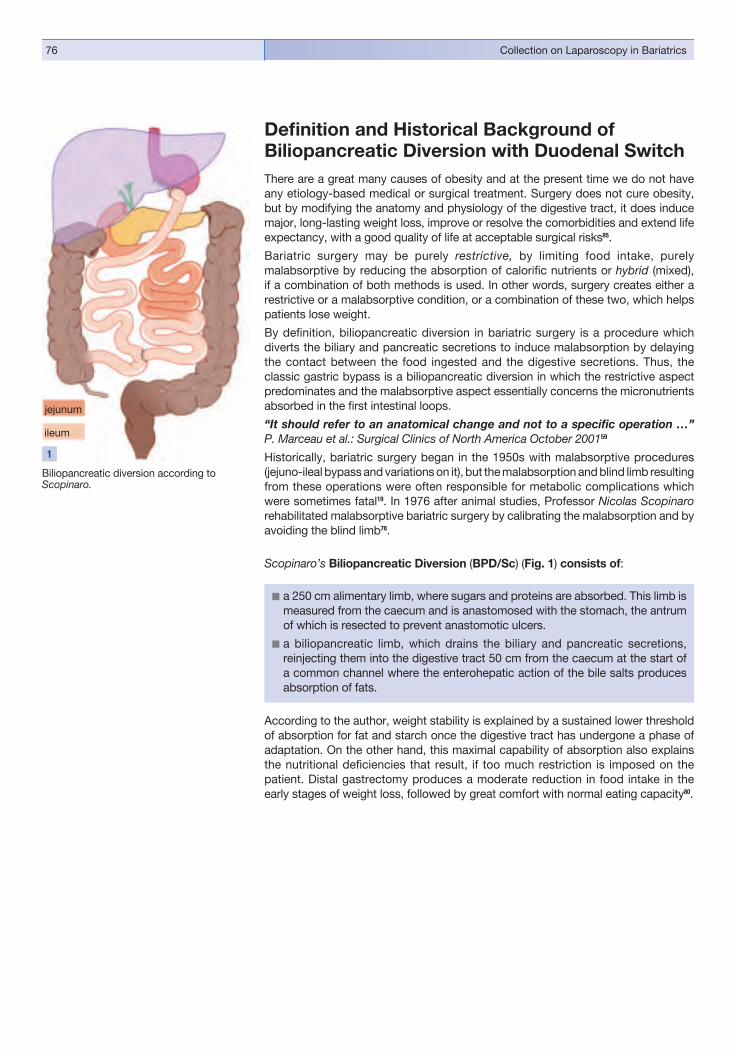
Collection on Laparoscopy in Bariatrics76
Defi nition and Historical Background of Biliopancreatic Diversion with Duodenal SwitchThere are a great many causes of obesity and at the present time we do not have any etiology-based medical or surgical treatment. Surgery does not cure obesity, but by modifying the anatomy and physiology of the digestive tract, it does induce major, long-lasting weight loss, improve or resolve the comorbidities and extend life expectancy, with a good quality of life at acceptable surgical risks85.
Bariatric surgery may be purely restrictive, by limiting food intake, purely malabsorptive by reducing the absorption of calorifi c nutrients or hybrid (mixed), if a combination of both methods is used. In other words, surgery creates either a restrictive or a malabsorptive condition, or a combination of these two, which helps patients lose weight.
By defi nition, biliopancreatic diversion in bariatric surgery is a procedure which diverts the biliary and pancreatic secretions to induce malabsorption by delaying the contact between the food ingested and the digestive secretions. Thus, the classic gastric bypass is a biliopancreatic diversion in which the restrictive aspect predominates and the malabsorptive aspect essentially concerns the micronutrients absorb ed in the fi rst intestinal loops.
“It should refer to an anatomical change and not to a specifi c operation …”P. Marceau et al.: Surgical Clinics of North America October 200159
Historically, bariatric surgery began in the 1950s with malabsorptive procedures (jejuno-ileal bypass and variations on it), but the malabsorption and blind limb resul t ing from these operations were often responsible for metabolic complications which were sometimes fatal19. In 1976 after animal studies, Professor Nicolas Scopinaro rehabilitated malabsorptive bariatric surgery by calibrating the malabsorption and by avoiding the blind limb78.
Scopinaro’s Biliopancreatic Diversion (BPD/Sc) (Fig. 1) consists of:
� a 250 cm alimentary limb, where sugars and proteins are absorbed. This limb is measured from the caecum and is anastomosed with the stomach, the antrum of which is resected to prevent anastomotic ulcers.
� a biliopancreatic limb, which drains the biliary and pancreatic secretions, reinjecting them into the digestive tract 50 cm from the caecum at the start of a common channel where the enterohepatic action of the bile salts produces absorption of fats.
According to the author, weight stability is explained by a sustained lower threshold of absorption for fat and starch once the digestive tract has undergone a phase of adaptation. On the other hand, this maximal capability of absorption also explains the nutritional defi ciencies that result, if too much restriction is imposed on the patient. Distal gastrectomy produces a moderate reduction in food intake in the early stages of weight loss, followed by great comfort with normal eating capacity80.
Biliopancreatic diversion according to Scopinaro.
1
jejunum
ileum

77Biliopancreatic Diversion Sleeve Gastrectomy with / without Duodenal Switch
1. To treat the problems of diarrhea and protein malnutrition, Marceau elongated the common channel to 1 m57; Hess chose to measure the alimentary limb and common channel at 40% and 10%, respectively, of the total small bowel length38.
2. Following the description of the duodenal switch by T. DeMeester22 to treat pathological duodeno-gastric refl ux, a duodenal switch (DS) and a 60–65% sleeve gastrectomy (SG) were carried out to replace the distal gastrectomy and the gastroileal anastomosis of the BPD/Sc50. This second modifi cation reduces the amount of acid at the proximal anastomosis, produces early satiety, maintains antro-pyloric function, and keeps a 4 cm length of duodenum, which improves the absorption of iron, calcium, and magnesium, removes the risk of dumping syndrome and the risk of anastomotic ulcers.
This operation, and the adaptations that the author introduced later, produce a considerable loss of weight (> 70%) which is sustainable (> 25 years)81.
In 1988, D. Hess in the United States and in 1990, P. Marceau in Canada, made modifi cations to the BPD/Sc to reduce the level of complications (Fig. 2).
Biliopancreatic Diversion with Duodenal Switch: Results and Strategic ApproachBiliopancreatic diversion with duodenal switch (BPD/DS) is a mixed or hybrid procedure: moderately restrictive (sleeve gastrectomy) and partially malabsorptive (duodenal switch). D. Hess and P. Marceau described performing BPD/DS by laparotomy, M. Gagner74 by laparoscopy in 2000, and R. Rabkin by a combined laparoscopic-assisted approach in 200372. Currently, BPD/DS is employed either as fi rst-line surgery or as revision surgery by numerous teams in North and South America, Europe, Israel and Australia, whatever the patient’s age or BMI, as long as they meet the clinical indication criteria for surgery1, 5, 20, 26, 32, 33, 37, 42, 43, 58, 69, 71,72, 88, 91. The number of surgeons belonging to the American Society for Metabolic and Bariatric Surgery (ASMBS) who carry out BPD/DS has doubled between 2002 and 2003. In France, even though bariatric surgery has evolved rapidly in recent years, BPD/DS is reserved for patients with a BMI > 50 or where other techniques have failed.
Biliopancreatic diversion with duodenal switch.
2

Collection on Laparoscopy in Bariatrics78
The morbidity and mortality rates of BPD/DS are equivalent to those for major digestive surgery, and are comparable with those for gastric bypass surgery. The incidence rate of 1% is subject to experience of the team (Tables 1, 2).
The side effects are known58 and should be distinguished from complications (diarrhea, severe denutrition). They include flatulence, steatorrhea (often 2 to 4 bowel movements per day, usually steatorrheic, foul-smelling, sometimes associat ed with urgency) and metabolic changes: decrease in levels of albumin, calcium, iron, hemoglobin and liposoluble vitamins (A, D, E, K). The problems related to malabsorption, must be investigated and can be corrected by administration of supplements. Transi tory discomfort can be relieved with an appropriate diet (reduction in fat content for steatorrhea and in carbohydrates for fl atulence), and by taking metro nidazole and pancreatic enzymes. Apart from these side effects, the comfort of feeding is excellent, the restriction allowing a normal diet without vomiting, with patient satisfaction rates beyond 95%60. The rate of surgical revision for excessive weight loss, protein malnutrition or diarrhea (lengthening of the alimentary limb and common channel by a more proximal ileo-ileal anastomosis), is 0.6% when the channel is calibrat ed at 100 cm60 and between 3.7 and 5.7%, when it is shorter or if the restriction was greater2, 35, 38. Patients with serious denutrition, with uncontrollable diarrhea or an intercurrent disabling condition should receive intravenous albumin and have their digestive functions restored by creating a latero- lateral anastomosis between the jejunum below the angle of Treitz and the proximal alimentary limb38, or a restoration of gastrointestinal continuity19. Taking into account patients lost to follow-up, the reversal rate for D. Hess is 0.61% after 15 years38.
Buchwald’s meta-analysis12 has established that, at the present time, BPD/DS is the bariatric procedure yielding the best results regarding the rates of initial weight loss and long-term maintenance of weight loss: 70% of patients who underwent BPD/DS presented with sustained excess body weight loss at 10 years after surgery.
Authors Marceau60 Hess38 Anthone2 Rabkin82 Baltasar5 Krawczykowski43
Patients 1423 1300 701 345 125 49
Pre-op. BMI ± Sd 51.5± – 52.3 ± 9.6 50 50 48.4 ± 6.7 (range) (33–103) (40–90) – (36–118) (39–100) –
Mean Age 40.1 – 42.3 ± 10.4 43 37 41.7 ± 10.6 (range) (15–70) – – (19–67) (16–68) –
Operative Mortality Rate (%) 1.1 0.57 1.4 0 1.6 0 – – – – (+ 2 late deaths) (2 late deaths)
Total – – 10 – 5.6 –
Leaks – 0.7 3.2 – 4 8.1
Occlusions – – 1.5 – 1.6 –
Hemorrhage – – 0.3 – – –
Rhabdomyolysis – – 0.6 – – –
Pancreatitis – – – – – 2
PE* or Pneumopathy – – – – – 2
Table 1: BPD/DS: Rates of Operative Mortality and Morbidity
Operativemorbidity
(%)
)* Pulmonary embolism.

79Biliopancreatic Diversion Sleeve Gastrectomy with / without Duodenal Switch
Authors Marceau Hess Anthone Krawczykowski 200760 200538 20052 (not published)
Patients 1423 618 451 284 182 48 (701) 25
Length of follow-up years 7.3 (2–15) < 5 5–10 >10 >10 5 4
Follow-up Rate (%) 97 – – – 92 – 88
Gastric Remnant Volume – – – – 100 100 120 Estimation (ml)
Length of 100 100 100 100 50–75–100 100 100 Common Channel (cm)
IEWL* (%) 73 ± 19 77 69.4 68.9 75 63.3 ± 17.1 78.4 ± 19.2
Total Reversal (%) 0.2 – – – 0.61 – 0
Revision for Diarrhea 0.7 – – – 3.7 5.7 0
or Malnutrition (%)
Revision for 1.5 – – – – – 4 Inadequate IEWL* (%)
Table 2: BPD/DS: Long-term Results
)* Initial Excess Weight Loss.
The benefi cial effects of BPD/DS are remarkable in cases of dyslipidemia (cured in nearly 99% of cases), diabetes (92% of patients have been able to stop all treatment) and other comorbidities associated with obesity: sleep apnea, cardiac risks etc. The effi cacy of this treatment, greater even than that of gastric bypass, has been shown even in the super-obese71.
“… BPD/DS should be considered the new gold standard. Others should strive to match those results.” Hess 2004, ASBS Consensus Conference30
BPD/DS is particularly useful in a strategic management of obesity and/or its comorbidities in that it allows the gastric and intestinal operations to be performed as isolated procedures. Certain authors, therefore, advocate the use of a staged approach, i.e.: a SG in high-risk patients or, on the contrary, in patients with a moderate BMI, or for regaining weight after pure malabsorptive surgery; a DS in the case of insuffi cient weight loss or persistence of diabetes or hypercholesterolemia after a SG, a band gastroplasty or a vertical banded gastroplasty.
There are several benefi ts that accrue from a two-stage strategic approach:
� The restrictive surgery (SG) can be offered to patients reluctant or unfi t to undergo a complex operation as a primary procedure.
� The malabsorptive surgery (DS) can be added without reoperating on the esophagogastric junction.
� The overall complication rate is lower when the two components of theBPD/DS are performed separately.73

Collection on Laparoscopy in Bariatrics80
The Two ComponentsSleeve Gastrectomy (Fig. 3)
Sleeve Gastrectomy (SG) involves the resection of the greater curvature of the stomach. This is the restrictive component of BPD/DS described in 1993 byP. Marceau. SG should, therefore, not be considered a new operation, however the use of SG as isolated procedure is a new surgical approach to obesity.
Nowadays, the use of isolated SG (ISG) is increasingly widespread in bariatric surgery, but has not been included in the offi cial register of surgical procedures13. So far, only a few clinical reports have been published about this procedure as an entity in itself, medium to long-term results are not known, and some technic al details and the indications are still the subject of controversy. Nevertheless, the good results in terms of short-term weight loss (49 to 80% EWL at one year) and comorbidity84, along with the acceptable rate of complications assure its place in the panoply of surgical procedures6,11, 27, 34, 36, 44, 47, 54, 64. A First Inter national Consensus Summit for Sleeve Gastrectomy was held in New York City in October 200721.
This longitudinal gastrectomy has some affi nities with the vertical gastroplasty called “Magenstrasse and Mill Operation” which was practiced by Johnston41 since 1992, but in this latter the stomach is transected and not resected.
Speaking physiologically, the operation is restrictive (satiety occurs quickly) and “anorexigenic” (the patients feels little hunger and little interest in eating as Ghrelin secretion is reduced52. Other effects are being studied, such as accelerated gastric emptying62 and the remarkable short-term results on diabetes, which seem to equal those of gastric bypass90, 93.
Other advantages of this procedure are no foreign body, no adjustment required, very rare vomiting, no dumping syndrome, no major nutritional defi ciencies to be expected. We have presented our results at the 6th International Obesity Surgery Expert Meeting in Saalfelden 2008: at 2 years after primary ISG, there was no major hypoalbuminaemia, no vitamine B12 defi ciency, no anemia, but the low serum level of ferritin increased from 4.2% before the operation to 19.5% at 2 years, probably because of low desire to eat red meat. However, a case of Wernicke’s syndrome has been reported by Makarewicz56. Albeit rare, patients suffering from vomiting, less than 2.5%, have to be checked for thiamine defi ciency.
Stomach calibration is variable, depending on the authors. The residual stomach volume is between 60 and 250 ml, depending on the bougie used to calibrate the sleeve (between 28 and 60 French) and the starting point for the resection (between 2 and 8 cm from the pylorus). If malabsorptive surgery is envisaged later, mainly in super-obese patients, the restriction must be moderate to avoid the problems of denutrition68, 89. Baltasar identifi ed a wide antral reservoir as a major factor that contributes to the failure of this technique. He considers, that gastric resection must start upstream of the pylorus over a 34 Fr.-calibration tube to produce a stomach with a volume of between 50 and 60 cc, and he proposes a re-SG, if the gastric pouch exceeds 4 cm in diameter at the esophagogastric junction7. Weiner stated, that a removed gastric volume of less than 500 cc is a predictor of failure in treatment92. In agreement with other authors86, we advocate the notion, that a “tight” sleeve gastrectomy calibrated using a 36 Fr bougie, with the left crus entirely freed and the gastric fundus entirely removed, should not dilate and per se represents the ideal ISG.
Sleeve gastrectomy.
3
81Biliopancreatic Diversion Sleeve Gastrectomy with / without Duodenal Switch
Indications for Sleeve Gastrectomy found in the literature are:
1. As fi rst-stage procedure prior to DS or bypass:a) For high-risk patients: BMI> 65 for whom mortality of 6.25% and morbidity
of 38% have been reported for a BPD/DS in a single procedure26, 73, 74, or for patients with cardiac, respiratory, or hepatic conditions3, 6,17, 35, 65.
b) For patients who refuse a band or following failure or complications of a gastroplasty band6,10, 44, 48.
2. As isolated surgery for patients with a low BMI,35-45 but also for patients on anti-infl ammatory treatment, or with anemia, Crohn’s disease, AIDS and those for whom a malabsorption procedure would be dangerous27, 36, 40, 54.
3. Secondary to malabsorptive surgery, such as a jejuno-ileal bypass55.
The drawbacks of Sleeve Gastrectomy are:
Irreversible nature of gastric resection. SG is indeed so far the only defi nitive non-reversible bariatric surgery.
The risk of hemorrhage from the line of section. This risk can be reduced by reinforcing the stapling with a running suture, fi brin glue17 or by using Gore Seamguard® bioresorbable staple line reinforcement15; bovine pericardium reinforcement has been used but seems to migrate14. At present, we make separate Vicryl 2/0 stitches, if focal hemostasis is needed sporadically.
Bleeeding may also occur from spleen or liver laceration as well as from trocar-sites6,51.
Leaks are the major concern of this surgery. They may occur in the range of 0 to 10%, mainly when SG is performed secondary to gastric banding61. Leaks usually occur at the esophagogastric junction. Evaluation of staple line patency should be performed by instillation of methylene blue and/or by insuffl ation of air. However, using these tests does not allow to reliably detect all sites of leakage, which will become apparent during oral refeeding on the 2nd or even up to the 5th day. Nevertheless, some authors reported about late leaks occurring after 3 weeks, 2 cases in our series, one in Weiner’s series92 and one in Silecchia’s84.
The clinical signs of leakage can be mild and well-tolerated, which usually is the case, if they occur delayed, and should be controllable by administration of IV anti biotics and parenteral nutrition84. A severe complication should be assumed, if the patient presents with hyperthermia, tachycardia (> 120/min) unexplained by dehydration or pain, tachy pnea (>20/min), a decrease in left basal breath sounds, left lumbar pain or pain in the left hypochondrium. If not adequately treated by drainage, leaks may lead to a cascade of complications: subphrenic collection/abscess, reactive pleural effusion, fi stulation into the pleural space and into the left lung, pseudoaneurysm (infectious) of the splenic artery with the potential risk of massive hemorrhage and, in turn, to rapidly deteriorating sepsis ending up with failure of multiple organs. A progressive rise in leukocytosis and serum CRP level, often associated with a fall in serum albumin, requires diagnostic assessment and appropriate treatment. Para clinical examinations, particularly to exclude a pulmonary embolism, must not delay this revision, even if the gastrographin swallow, that we normally make on D1 or D2, is considered fi ne and does not show a leak.
Collection on Laparoscopy in Bariatrics82
The principal objectives in the management of this type of complication are patient safety and complete closure of the leak. The former can be achieved by creating a gastro-cutaneous fi stula, that prevents fl uid collection by directing the juices out of the body, the latter by reducing the output, keeping the patient well-nourished and treating the underlying cause that might compromise wound healing (formation of strictures).
Whenever possible, suturing the leak and placing an omental patch should be attempted, however these measures are often found to be ineffective. Usually, we debride the false membranes, perform thorough lavage, place a drain in contact with the leak, and, given the time for improvement, we perform an alimentary jejuno stomy.
When leak drainage is found to be inadequate or protracted (more than 3 weeks), this can be controlled successfully by placing coated self-expanding stents18, 25, 83. Among other options, that may be adopted for this purpose, drainage via a Roux-en-Y limb8 or total gastrectomy have been described82.
The mechanism and correlated factors favoring the formation of leaks are still unclear: ischemia, thickness of the stomach, technical problems concerning the staples and stapling are issues of prime concern. A long gastric sleeve and, even more so, a distal stenosis, contribute to a delayed healing of leaks.
Prevention of Leaks. Fistula-related complications may be prevented by observ ing the principles for use of viscerosynthesis material: using stapling equipment and staples according to the standards laid down by the manufacturers (60 mm Ethicon Endo-Surgery Echelon Endopath® Stapler with green or gold cartridges or green Covidien Endo GIA staples), keeping to a tissue compression time before stapling, avoiding crossing staples, dissecting to preserve good vascularization4. It can be construed, that the calibration of the sleeve on a small-diameter Faucher bougie (36 Fr and below) promotes the formation of leaks. The fi nal stapling must be made towards the greater curvature to avoid encroachment of the eso phagus.
Unstapling may occur perioperatively, particularly in the antral part of the stomach which is sometime thick. This is dealt with using a running suture, because re- stapling could narrow the sleeve.
Gastric strictures. The incidence of gastric strictures is between 0.7 and 4%17, 51. If stapling is applied too close to the incisura angularis, this is supposed to promote stricture formation, but it is also argued, that this might be related to oversewing of the staple-line17. Gastric strictures can delay the healing of a proxim al fi stula and, in theory, can contribute to dilatation upstream. Hematoma and oedema may lead to gastric strictures that are of transient nature. Endoscopic dilation has been advocated for non- resol ving strictures, and furthermore in the case of a proximal non-healing leak17, 51. A seromyotomy may also be considered an option18.
Dilatation of the gastric sleeve probably occurs more frequently, if the calibration was wide92. Re-sleeve gastrectomies have been described with good results in patients regaining weight, particularly after BPD/DS where the stomach was calibrated with a 60-Fr bougie31 but also for failure after ISG7. A wide proximal gastric pouch with a funnel-like appearance or the appearance of a proximal gastric diverticulum is the result of an incomplete dissection and resection of the fundus. These fi ndings are frequently encountered in revision procedures after gastric banding10. The clinical consequences of incomplete dissections are yet unknown, but it must be assumed that progressive dilatation of the proximal stomach portion will occur, with a resumption of weight gain following in its wake.

83Biliopancreatic Diversion Sleeve Gastrectomy with / without Duodenal Switch
Weight regain or insuffi cient weight loss: This is not considered a complication, but rather the fate of any restrictive surgery. Nevertheless, the excess weight loss (EWL) obtained with an ISG is, in the short term, greater than with an intragastric balloon63, comparable to that of a bypass53 and, at 3 years, greater than that obtained with gastric banding40. However, ISG remains essentially a restrictive procedure and, as for all restrictive surgery, mid- and long-term results depend on several factors. Langer53 described a case where, after an incomplete resection of the fundus, the ghrelin level did not fall post-operatively and after moderate weight reduction the patient returned to his original weight. A re-sleeve gastrectomy or the addition of malabsorptive surgery should be envisaged if the pouch poses a problem or if the patient regains weight. In agreement with other authors, it seems to us, that progressive dilatation of the stomach is not the main aspect of the problem: such dilatation is of rare incidence, and signifi cant weight loss can be obtained with relatively wide calibrations of 48-Fr53. Weight gain rather seems to be related to the ingestion of high-caloric foods with a soft or liquid consistency86 and, in our experience, to the lack of physical activity, that is suited to weight loss. That being the case, and if patient compliance could not be established, or if the patient is reluctant to bring about a radical change in the quality of his/her diet and physical activity, an additional malabsorptive procedure may be offered, as long as the patient is willing to accept the risks of a new operation, metabolic monitor ing and taking vitamin and trace element supplements daily.
Gastro-esophageal refl ux. The procedure has the reputation of promoting gastro-esophageal refl ux (GERD) by removing the anti-refl ux system. Indeed, it is true that early on, certain patients (± 20%) complain of refl ux occurring, which usually responds to proton pump inhibitors (PPI). Nevertheless, this problem, which appears in the early postoperative period, sometimes resolves after the phase of initial weight loss. On the other hand, some patients on medication for a symptomatic refl ux prior to ISG will experience an improvement or a resolution of the GERD after the SG40. In patients presenting with severe symptoms of GERD after ISG, a gastric bypass surgery can be performed53. A repeat SG has been performed by M. Gagner for a patient complaining of GERD and a regain weight after a BPD/DS67.
Rhabdomyolysis has been reported, but it is related to the operating time and not specifi c to this procedure28.
Mucocele of the gastric tube has been described after conversion of a vertical banded gastroplasty to BPD/DS9,77.
Our experience: We have been performing ISGs in a two-stage BPD/DS program since December 2001 in patients with a complication or failure of gastric banding, and since December 2002, in patients who do not wish to have a gastric band34, 47, 48, 49.In our patient group, analyzed until March 2008, we had 143 patients who underwent an ISG as fi rst-line treatment and 66 as second-line treatment after gastric banding (failure to lose weight or complication). Our results in terms of weight loss and morbidity are comparable to those found in the literature, as shown in Table 3 (see p. 72). We have had no mortality, and in our experience, leaks have a higher incidence in revision SGs, particularly if the band had already been removed, probably because the dissection is more diffi cult. Our current technique has allowed us to reduce the incidence of fi stulas to 1.8%, since we switched over to using the 60-mm staplerinstead of the 45-mm stapler (Ethicon Endo-Surgery Echelon™ ENDOPATH® Stapler), i.e. 1 case in the last 56 SGs). After 3 years, 23% of the patients who had an ISG as initial treatment (mean initial BMI of 50) underwent an additional DS, while this rate was as high as 43% in the patient group treated with a gastric band (mean initial BMI of 43.5). This leads us to the assumption, that even with a moderate BMI (<50), certain patients must benefi t from malabsorptive surgery.

Collection on Laparoscopy in Bariatrics84
Johnston41 230 40–64 36 5–6 – 62 4 2.5 0 – 34 – – 60 – – – – 32 – – 58 – – – at 3 Y
Almogy3 21 57.7 – – 100 47 at 1 20 – 0 (53–71.5) late 14
Baltasar6 31 3 series 32 2 < 60 42–100 3.2 0 3.2
Bernande10 8 ** 50.5 34 – – 57 at 1 Y 0 0 0 (37–74)
Braghetto11 50 * 37.9 32 2–3 75 ± 22 – 2 ? 0 (32.9–46.8)
Cottam17 126 * 65.4 ± 9 46–50 5 – 45 ± 17 14 1.6 0 (45–91) at 1 Y Late 0.75
Felberbauer27 126 48.1±8.7 48 Crow’s foot – 3.17 2.4 0
Hamoui34 118 * 55 – – – 15.3 1.7 0.85 (37–108)
Han36 130 * 30–56 48 – 50–60 83.3 at 1 Y 2.9 0.7 0.7
Himpens40 40 * 39 34 Crow’s foot – 5 1 gastric 0 ischemia
Krawczykowski 143 * 44.4 ± 5.5 36 Crow’s foot – 8.4 4.9 0 (not published) (34.6–60.3)
66 ** 44.4 ± 6.2 36 Crow’s foot – 19.7 15 0 (35.9–61)
Langer52 23 * 48.5 ± 6.9 48 Crow’s foot – 56 at 1 Y – – – (40–73)
Lee54 216 * 49 ± 11 32 6–8 60–80 59 ± 17 4.6 1.4 0 at 1 Y
Regan73 14 * 58–71 60 Crow’s foot – 33 21.4 0 0 at 11mths.
Skrekas84 93 90 * 46.9±6.5 36 3 <100 67.2 8.6 4.3 0 3 ** (37–66) at 12 mths.
Table 3: Sleeve Gastrectomy: Morbidity, Mortality and Results
)* Primary )** Secondary )*** Initial Excess Weight Loss Y = years
PatientsAuthors Initial BMI Leaks(%)
Mortality(%)
At 20 mths.64% of Pt loss> 50%
6.8% loss < than 25%
49.4(26.8–79.6)
at 1 Y47.3at 2 Y
57.7(0–125.5)
at 1 Y66
(-3.1–152.4)at 3 Y
77.7 at 1 Y73.3 at 2 Y67.2 at 3 Y
68.8 at 1 Y69.6 at 2 Y54.4 at 3 Y
Bougie Calibration
(Fr)
Startingfrom pylorus
(cm)
Size ofResidualStomach
IEWL***(%)
Morbidity (%)Major Com-plications

85Biliopancreatic Diversion Sleeve Gastrectomy with / without Duodenal Switch
The Technique We use 8 trocars and a 0° laparoscope, and no extra-long instruments. We work along two curvi linear paths (Fig. 4, highlighted in blue). The fi rst (superior) path provides a perfect view of the esophagogastric junction, while the second (inferior) one allows to visualize the greater curvature of the stomach. The patient is placed in a reverse Trendelenburg position. The surgeon assumes a standing position between the lower limbs of the patient, the assistant holding the 0° scope is to the right of the patient and the scrub nurse to the left. Insuffl ation is performed with a Veress needle in the left hypochondrial area until a pneumoperitoneum of 15 mmHg is established. We introduce the fi rst 11-mm trocar (T1) on the median line at 10 cm from the xiphoid process, then, under visual control, a 6-mm trocar (T2) below the xiphoid for the retractor that holds aside the left hepatic lobe and, usually, gives a perfect view of the esophagogastric junction. Between these 2 trocars, a right para median 6-mm trocar (T3) is placed 10 cm from the median line for the forceps held by the surgeon allowing exposure by traction on the stomach. On the left side, on the midclavicular line and in line with trocars T1 and T3, thus slightly offset downwards, a 13-mm trocar with a reducer (T4) is inserted. In the fi rst instance, this allows the introduction of scissors or the ultrasonic dissector, and during the fi nal stage of the operation, the stapler. On the same T1–T3 line, at the point of intersection with the anterior axillary line, a 6-mm trocar (T5) is inserted allowing traction to be applied to the omentum during the following operative maneuvers: dissection of the angle of His; division of the stomach from the omentum; holding the stomach during stapling. On the inferior path, 5 cm on either side of the umbilicus, two 13-mm trocars (T6 and T7) are inserted to accomodate the laparoscope/stapler, and vice versa. Finally, on-axis with the midclavicular line, a 6-mm trocar (T8) is placed to provide good exposure during dissection and after stapling of the greater curvature.
Port placement for Isolated Sleeve Gastrectomy (ILS).
4
T1 Laparoscope with coupled video camera
T2 Liver Retractor
T3 First Surgeon; Retractor
T4 First Surgeon; Dissector, Stapler
T5 Assistant Surgeon; Dissector, Stapler
T6 Laparoscope; Stapler
T7 Laparoscope; Stapler
T8 First Surgeon; Retractor

Collection on Laparoscopy in Bariatrics86
We always start with dissection of the angle of His, all the more so, if the patient has a gastric band or a history of surgery of the esophagogastric junction (Fig. 5a). The strict observance of this principle is the critical factor that contributes to the feasibility of the operation. Dissection is performed using scissors or the ultra sonic dissector. The nasogastric catheter, which was used to drain the stomach, is remov ed. The objective is to start the dissection of the anterior part of the left crus of the diaphragm and to reduce any hiatal hernia, if present. The fat pad cover ing the esophagogastric junction is usually dissected and excised, particularly if it is bulky (Fig. 5b). We then move to the greater curvature of the stomach and in continuation with Latarjet’s nerve, between 6 and 8 cm upstream of the pylorus, which may be palpated, we begin to free the stomach from its contacts using an ultrasonic dissector (Figs. 5c, d). The stomach is dissected as far as the angle of His. Particular care is taken while passing near the short gastric vessels, moving the laparoscope from trocar T7 to trocar T1 (Fig. 5e). Any posterior attachments of the stomach including the branches of the posterior gastric artery are freed from the stomach (Fig. 5f). The fundus often has a posterior development which must be perfectly dissected (Fig. 5g). The left crus must be completely in view (Fig. 5h). Under control and assist ance of the surgeon the anesthetist passes down a 36 Fr.-Faucher tube, which is then guided toward the pylorus. The tube is held in contact with the lesser curvature. The stomach is stretched out and stapling is initiated by fi ring the60-mm Ethicon Endo-Surgery Endopath® Echelon Stapler loaded with green or gold cartridges, depending on the thickness of the antrum. The latter maneuv er is perform ed with the scope in T6 and the stapler in T7 port (Fig. 5i). Stenos is at the incisura angularis must be avoided. The stapler is fi red twice, and then the laparo scope is placed in T7 or T1, depending on the patient’s size. Loaded with gold cartridges, the stapler is again fi red via port T6 or T4, thereby staying in close contact with the calibration tube (Fig. 5j). Stapling is completed with the stomach stretched out by applying traction to the posterior wall of the fundus toward the patient’s left shoulder. This technique is employed to prevent taking in too much tissue and risking unstapling or creating too wide a residual stomach. The last stapling must be shifted onto the external side of the angle of His to avoid injuring the eso phagus (Fig. 5k). We discontinued our practice of reinforcing the staple line with a running suture because, in our experience, this takes generally nearly half an hour, the appear ance of the sleeve is sometimes irregular, and the incidence of fi stula formation is equivalent to the rate found with the use of 45-mm staples. We assume, that placing a running suture on a narrow sleeve is a predisposing factor for complications: the start of the running suture may be the origin of tension on the bottom of the eso phagus, and may cause shearing of the gastric serous membrane.
According to our protocol, we therefore perform a methylene blue leak test while compressing the stomach with a clamp forceps at the pylorus. In this way, not only the suture line can be checked for signs of leakage or bleeding, this also gives us an idea of the residual gastric volume (usually between 100 and 120 ml). In addition, an intraoperative air insuffl ation test is performed. The stomach is extracted in a surgic al bag via trocar port T4, which is enlarged and closed with Vicryl at the end of the procedure (Figs. 5l–m). We systematically take a tissue sample for hepat ic biopsy; cholecystectomy is only performed if gall stones are present. We only drain occasionally. The naso gastric catheter remains in place for 24 hours to prevent vomiting and to check on the absence of bleeding. The patients are given pro phylactic anti biotic medication (second generation cephalosporin) before the incision. Thrombosis prophylaxis is started the day before the operation and continued for a month, and patients wear support stockings. Patients are encouraged to walk about, once they have regained consciousness. They are allowed to begin a liquid diet after radio logical checks and are discharged on the 5th postoperative day with a blended soft diet and purées up to the 3rd week.
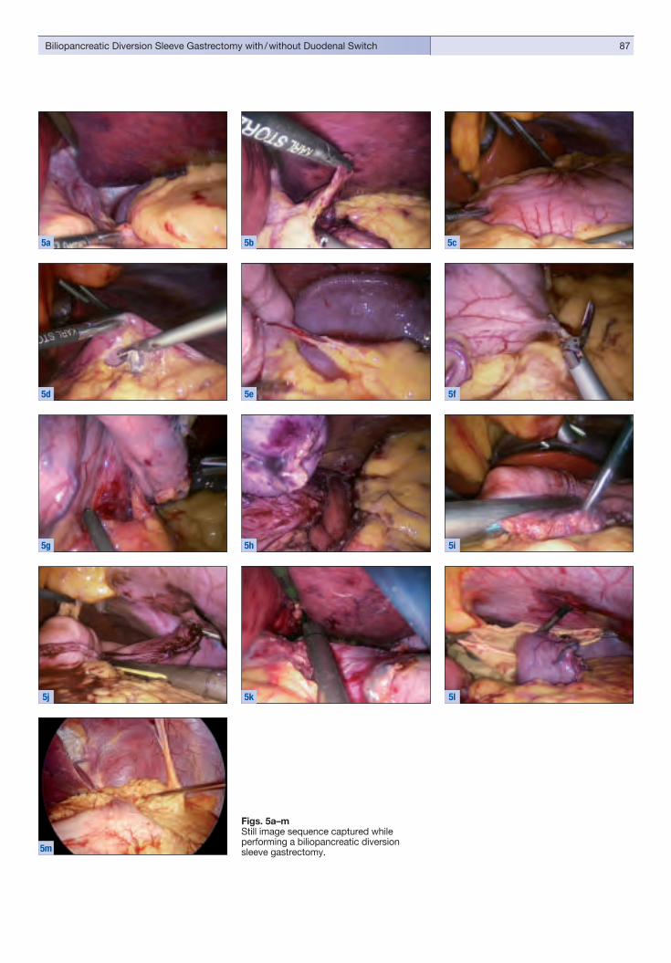
87Biliopancreatic Diversion Sleeve Gastrectomy with / without Duodenal Switch
5a 5b 5c
5d 5e 5f
5g 5h 5i
5j 5k 5l
5m
Figs. 5a–mStill image sequence captured while performing a biliopancreatic diversionsleeve gastrectomy.

Collection on Laparoscopy in Bariatrics88
Duodenal Switch (Fig. 6)
Isolated Duodenal Switch (IDS) has been described by DeMeester in the surgical treatment of primary bile refl ux gastritis. In this indication the goal of surgery is to divert the bile and pancreatic secretions with a long common channel22.
In bariatric surgery, DS is the malabsorptive component of BPD/DS. The common channel is short (50 –150 cm) and the DS is classically combined with an SG. Nevertheless, variants have been suggested to reduce the morbidity related to SG or to improve the results of preceeding restrictive surgery. Thus, DS has been carried out:
Isolated duodenal switch.
6
Banded duodenal switch.
7
1. As Isolated Duodenal Switch (IDS) for surgically curable metabolic conditions associated with moderate obesity. In terms of Excess Weight Loss (EWL) the outcome is equivalent to less than 5 BMI points, but with regard to metabolic symptoms, the results are remarkable, with a curative rate of nearly 99% of cases of hypercholesterolemia and, to a lesser degree, of diabetes16, 66.
2. After reestablishing normal gastric capacity19.
3. Simultaneously to placement of a resorbable89 or silicone gastric band (banded DS)30 (Fig. 7).
4. In addition to a well-tolerated purely restrictive surgery, which has however proven ineffective, either in terms of weight loss or comorbidity: two-step band ed DS45, 46, 69, 87, VBG94 and secondary DS, SG and additional DS resulting in the classic two-stage BPD/DS49 73.
Our experience: In our 2-stage BPD/DS program with a total number of 209 patients treated with ISG as fi rst- or second-line procedure, 21 patients re ceived an additional DS. The second operation was relatively easy to perform, we had few post-operative complications, and 6 months after the second procedure the initial excess weight loss (IEWL) was equivalent to that of the single-step BPD/DS.
We also performed additional or isolated DS secondary to failure of weight reduction with non-SG restrictive surgery: 32 patients had gastric banding, 2 patients vertical banded gastroplasty and 3 patients who wanted prior or simultaneous removal of their gastric band. Postoperative morbidity was mainly related to the presence of the band (secondary removal of the band). The rate of IEWL was lower than that for BPD/DS (Fig. 8).
Generally speaking, it is not advisable to add a DS to a gastric band. Indeed, in our experience, even if the band is defl ated, nearly half of the patients with restricted food intake will request it to be removed in the year the DS was performed. The potential risk of anastomotic (duodeno-ileal) ulceration must be added to the side effects and complications already mentioned for biliopancreatic diversion, though these fi ndings are based on two series only16, 66. Retaining a 4 to 5 cm duodenal stump reduces this risk66. Finally, in addition to the metabolic risks linked to mal absorption, the patient is exposed to the risk of complications specifi c to the band (adjustments, slipping, erosion). On the other hand, we did not have any severe hypoalbuminemia in this group of patients.
Peterli reported the same experiences as we had, and he also prefers to convert a failed band gastroplasty into a classic BPD/DS with an SG as restriction70.
Common channel

89Biliopancreatic Diversion Sleeve Gastrectomy with / without Duodenal Switch
The Technique
We use between 5 and 7 trocars and a 0°-laparoscope, but no extra-long laparo-scopic instruments. The patient is in the reverse Trendelenburg position. The surgeon assumes a standing position between the lower limbs of the patient, the assist ant holding the 0°-scope is to the right of the patient and the scrub nurse to the left. Insuffl ation is performed with a Veress needle in the left hypochondrial area until a pneumoperitoneum of 15 mmHg is established. We introduce a fi rst right paraumbilical 13-mm trocar (T1) for the laparoscope, a second left paraumbilic al 13-mm trocar (T2) for the dissection and transection of the duodenum, then for the duodeno-ileal anastomosis. A third 13-mm trocar (T3) is positioned on the left midclavicular line 5 to 6 cm higher than the fi rst two trocars, in the fi rst instance to be used to stretch the stomach, then for performing the ileo-ileal anastomosis. A fourth 6-mm trocar (T4) allows the tissues to be presented. A fi fth 6-mm trocar (T5) is placed under the xiphoid for retracting the right lobe of the liver. If required, we introduce a 6-mm suprapubic trocar and one into the left fl ank (T6 and T7) (Fig. 9).
Port placement for duodenal switch.
9
Comparison of % IEWL (±SD) between primary BPD/DS, two-stage BPD/DS and Banded DS.
8
32 LASGBfollowed by
Duodenal Switch (banded DS)
30 to 60%
40 to 80%
80%
21 SGfollowed by
Duodenal Switch
23 one-stageBPD-DS
T1 Laparoscope with coupled video camera
T2 First Surgeon; dissector, stapler, needle holder for duodeno-ileal anastomosis
T3 First Surgeon; grasping forceps, stapler, needle holder for ileo-ileal anastomosis
T4 First Surgeon; Retractor
T5 Liver Retractor
T6 Assistant Surgeon; Retractor
T7 Assistant Surgeon; Retractor

Collection on Laparoscopy in Bariatrics90
Once the pylorus has been localized endoscopically, and a springing sensation can be felt while advancing the tip of an instrument through the sphincter, we measure 4 cm downstream (Fig. 10a) and, with the aid of a monopolar hook or ultrasonic dissector (Ethicon® Endo-Surgery, Norderstedt, Germany), we incise the peritoneal layer on the inferior and superior surfaces of the duodenum. The scrub nurse expands the stomach with a grasping forceps through trocar T3. We dissect the posterior surface of the duodenum aiming for the right edge of the hepatic pedicle (Figs. 10b, c). Stapling is performed with one, or occasionally, with two 45 mm cartridges of blue staples (3.5 mm ATB 45 Ethicon® Endosurgery) after removal of the naso gastric catheter (Fig. 10d). It is sometimes convenient to perform a cholecystectomy at this point.
The surgical team then moves to the left of the patient repositioned fl at for the lower procedures. We systematically carry out an appendectomy, then we unroll and measure the entire ileum on its antimesenteric side (stretched) from the ileo caecal valve using forceps graduated with marks at 5 and 10 cm (Fig. 10e).At a distance of 100 cm proximal from the ileocaecal valve we pass a thread through, that serves as a marker (common channel) and we measure an addition al 150 cm to obtain an alimentary limb measuring a total of 250 cm. We transect the small bowel at this level with a white cartridge (Fig. 10f). It is not necessary to incise the mesentery widely. It is important to confi rm the absence of intestinal torsion and to make sure, that the alimentary limb is in the patient’s right side. The ileo-ileal anasto-mosis is then made either manually with a Vicryl 2/0 running suture or, mechanic ally, with white cartridges placing the stitches at the angles of the anastomosis to relieve tension on the staples (Fig. 10g). The alimentary limb is approximat ed to the proximal duodenal stump in an antecolic position. The assistant facilitates this maneuver by supporting the distal anastomosis. The team returns to the initial position with the surgeon placed between the legs of the patient in the reverse Trende lenburg position. The duodeno-ileal anastomosis is made with a Vicryl 2/0 double running suture. It is termino-lateral or sometimes termino-terminal. The patency and permeability of the anastomosis are tested by instillation of methylene blue and, at the end of the procedure, by insuffl ation of air (Figs. 10h–l). The ileo-ileal mesenteric opening and the space between the mesentery of the mounted limb and the greater omentum are closed with a Prolene 1 running suture (Fig. 10m). We usually leave an aspiration drain in contact with the proximal anastomosis and a nasogastric catheter for 24 hours (Fig. 10n). The change to water solubles is made on the 1st or 2nd post-operative day. The antibiotic and antithrombosis prophylaxis and dietary program are the same as for SG.
ConclusionIn bariatric surgery, BPD/DS is the most effective operative procedure in terms of weight loss, maintenance of weight loss, healing rate of comorbidities and dietary comfort. This operation is a modifi cation of BPD/Sc, the 30th anniversary of which was celebrated at the 23rd Annual Meeting of the ASBS in 2006. The signifi cant new feature is the use of the individual components of BPD/DS (i.e., SG and DS) in an isolated manner as 1st or 2nd line treatment in a strategic approach to obesity, which is a chronic incurable disease. With awareness of their results, side effects and complications, these components can be better integrated into the decision-making process.

91Biliopancreatic Diversion Sleeve Gastrectomy with / without Duodenal Switch
10a 10b 10c
10d 10e 10f
10g 10h 10i
10j 10k 10l
10m 10n
Figs. 10a–mStill image sequence captured while performing isolated duodenal switch.

Collection on Laparoscopy in Bariatrics92
Literature1. ANTHONE GJ, LORD RV, DeMEESTER TR, CROOKES PF: The duodenal
switch operation for the treatment of morbid obesity. Ann Surg 2003; 238:618–627.
2. ANTHONE G: The duodenal switch operation for morbid obesity.Surg Clin N Am 2005; 85: 819–833.
3. ALMONGY G, CROOKES PF, ANTHONE G: Longitudinal gastrectomy as a treatment for the high-risk super-obese patient. Obes Surg 2004; 14: 492–497.
4. BAKER RS, FOOTE J, KEMMETER P, BRADY R, VROEGOP T, SERVELD M: The science of stapling and leaks. Obes Surg 2004; 14: 1290–1298.
5. BALTASAR A, BOU R, BENGOCHEA M, ARLANDIS F, ESCRIVA C, MIRO J, MARTINEZ R, PEREZ N: Duodenal switch: an effective therapy for morbid obesity – Intermediate results. Obes Surg 2001; 11: 54–58.
6. BALTASAR A, SERRA C, PÉREZ N, BOU R, BENGOCHEA M, FERRI L: Laparoscopic sleeve gastrectomy: a multi-purpose bariatric operation. Obes Surg 2005; 15: 1124–1128.
7. BALTASAR A, SERRA C, PÉREZ N, BOU R, BENGOCHEA M: Re-sleeve gastrectomy. Obes Surg 2006; 16: 1535–1538.
8. BALTASAR A, BOU R, BENGOCHEA M, SERRA C, CIPAGAUTA L: Use of a Roux limb to correct esophagogastric junction fi stulas after sleeve gastrectomy. Obes Surg 2007; 17: 1408–1410.
9. BALTASAR A: Mucocele of the gastric tube after conversion of VBG to DS. Obes Surg 2006; 16: 528.
10. BERNANTE P, FOLETTO M, BUSETTO L, POMERRI F, PESENTI F, PELIZZO MR, NITTI D: Feasibility of laparoscopic sleeve gastrectomy as a revision procedure for prior laparoscopic gastric banding. Obes Surg 2006;16: 1327–1330.
11. BRAGHETTO I, KORN O, VALLADARES H, GUTIÉRREZ L, CSENDES A, DEBANDI A, CASTILLO J, RODRIGUEZ A, BURGOS AM, BRUNET L: Laparoscopic sleeve gastrectomy: surgical technique, indications and clinical results. Obes Surg 2007; 17: 1442–1450.
12. BUCHWALD H, AVIDOR Y, BRAUNWALD M, JENSEN MD, PORIES W, FAHRBACH K, SCHOELLES K: Bariatric surgery. A systematic review and meta-analysis. JAMA 2004; 292: 1724–1738.
13. BUCHWALD H: Consensus conference statement: Bariatric surgery for morbid obesity: Health implications for patients, health professionals, and third-party payers. J Am Coll Surg 2005; 200: 593–604.
14. CONSTEN EC, DAKIN GF, GAGNER M: Intraluminal migration of bovine pericardial strips used to reinforce the gastric staple-line in laparoscopic bariatric surgery. Obes Surg 2004; 14: 549–554.
15. CONSTEN ECJ, GAGNER M, POMP A, INABMET WB: Decreased bleed ing after laparoscopic sleeve gastrectomy with or without duodenal switch formorbid obesity using a stapled buttressed absorbable polymer membrane. Obes Surg 2004; 14: 1360–1366.
16. COSSU ML, NOYA G, TONOLO GC, PROFILI S, MELONI GB, RUGGIU M, BRIZZI P, COSSU F, PILO L, TILOCCA PL: Duodenal switch without gastric resection: results and observations after 6 years. Obes Surg 2004; 14:1354–1359.
17. COTTAM D, QURESHI FG, MATTAR SG, SHARMA S, HOLOVER S,BONANOMI G, RAMANATHAN R, SCHAUER P: Laparoscopic sleevegastrectomy as an initial weight-loss procedure for high-risk patients withmorbid obesity. Surg Endosc 2006; 20: 859–863.

93Biliopancreatic Diversion Sleeve Gastrectomy with / without Duodenal Switch
18. DAPRI G, VAZ C, CADIÈRE GB, HIMPENS J: A prospective randomized study comparing two different techniques for laparoscopic sleeve gastrectomy. Obes Surg 2007; 17: 1435–1441.
19. DAPRI G, CADIÈRE GB, HIMPENS J: Laparoscopic restoration of gastrointestinal continuity after duodenal switch. Surg Obes Relat Dis 2008; 4: 451–454.
20. DEITEL M: Jejunocolic and jejunoileal bypass: an historical perspective.In Deitel M. Surgery for the morbidly obese patient. FD-Communications Inc. 1989; 7: 81–90.
21. DEITEL M, CROSBY RD, GAGNER M: The fi rst international consensussummit for sleeve gastrectomy, New York City, October 25–27, 2007.Obes Surg 2008; 18: 487–496.
22. DeMEESTER TR, FUCHS KH, BALL CS, ALBERTUCCI M, SMYRK TC, MARCUS JN: Experimental and clinical results with proximal end-to-endduodenojejunostomy for pathologic duodenogastric refl ux. Ann Surg 1987; 206: 414–426.
23. DI BETTA E, MITTEMPERGHER F, DI FABIO F, CASELLA C, TERRAROLI C,SALERNI B: Duodenal switch without gastric resection after failed gastric restrictive surgery for morbid obesity. Obes Surg 2006; 16: 258–261.
24. DOLAN K, FIELDING G: Bilio pancreatic diversion following failure oflaparoscopic adjustable gastric banding. Surg Endosc 2004; 18: 60–63.
25. EISENDRATH P, CREMER M, HIMPENS J, CADIÈRE GB, LE MOINE O, DEVIÈRE J: Endotherapy including temporary stenting of fi stulas of the upper gastrointestinal tract after laparoscopic bariatric surgery. Endoscopy 2007; 39: 625–630.
26. FAZYLOV RM, SAVEL RH, HOROVITZ JH, PAGALA MK, COPPA GF, NICASTRO J, LAZZARO RS, MACURA JM: Association of super-super – obesity and male gender with elevated mortality in patients undergoing the duodenal switch procedure. Obes Surg 2005; 15: 618–623.
27. FELBERBAUER FX, LANGER F, SHAKERI-MANESCH S, SCHMALDIENST E, KEES M, KRIWANEK S, PRAGER M, PRAGER G: Laparoscopic sleevegastrectomy as an isolated bariatric procedure: Intermediate-term results from a large series in three Austrian centers. Obes Surg; 2008: 18 : 814–818.
28. FORESTIERI P, FORMATO A, PILONE V, ROMANO A, MONDA A,TRAMONTANO S: Rhabdomyolis after sleeve gastrectomy: increase in muscle enzymes does not predict fatal outcome. Obes Surg 2008; 18: 349–351.
29. GABRIEL SG, KARAINDROS CA, PAPAIOANNOU MA, TASSIOULIS AA, GABRIEL SG, SIGALAS VI, GIANNAKAKIS PP: Biliopancreatic diversion with duodenal switch combined with laparoscopic adjustable gastric banding. Obes Surg 2005; 15: 517–522.
30. GAGNER M, STEFFEN R, BIERTHO L, HORBER F: Laparoscopic adjustable gastric banding with duodenal switch for morbid obesity: technique andpreliminary results. Obes Surg 2003; 13: 444–449.
31. GAGNER M, ROGULA T: Laparoscopic reoperative sleeve gastrectomy for poor weight loss after biliopancreatic diversion with duodenal switch. Obes Surg 2003; 13: 649–654.
32. GAGNER M, GUMBS A: Gastric banding: conversion to sleeve, bypass,or DS. Surg Endosc 2007; 21: 1931–1935.
33. GUEDEA ME, ARRIBAS del AMO D, SOLANAS JAG, MARCO CA,BERNADO AJ, RODRIGO MA, DIAGO VA, DIEZ MM: Results ofbiliopancreatic diversion after fi ve years. Obes Surg 2004; 14: 766–772.
34. HAMOUI N, ANTHONE GJ, KAUFMAN HS, CROOKES PF: Sleevegastrectomy in the high-risk patient. Obes Surg 2006; 16: 1445–1449.
Collection on Laparoscopy in Bariatrics94
35. HAMOUI N, CHOCK B, ANTHONE GJ, KAUFMAN HS, CROOKES PF: Revision of the duodenal switch: indications, technique, and outcomes.J Am Coll Surg 2007; 204: 603–608.
36. HAN SM, KIM WW, OH JH: Results of laparoscopic sleeve gastrectomy at1 year in morbidly obese Korean patients. Obes Surg 2005; 15: 1469–1475.
37. HESS DS, HESS DW: Biliopancreatic diversion with duodenal switch.Obes Surg 1998; 8: 267–282.
38. HESS DS, HESS DW, OAKLEY RS: The biliopancreatic diversion with the duodenal switch: results beyond 10 years. Obes Surg 2005; 15: 408–416.
39. HESS D: 2004 ASBS Consensus Conference Biliopancreatic diversion with duodenal switch Surg Obes Relat Dis 2005; 1: 329–333.
40. HIMPENS J, DAPRI G, CADIERE GB: A prospective randomized study between laparoscopic gastric banding and laparoscopic isolated sleevegastrectomy: results after 1 and 3 years. Obes Surg 2006; 16: 1450–1456.
41. JOHNSTON D, DACHTER J, SUE-LING H, KING R, MARTIN L: The Magenstrasse and Mill operation for morbid obesity. Obes. Surg. 2003; 13: 10–16.
42. KESHISHIAN A, ZAHRIYA K, HARTOONIAN T, AYAGIAN C: Duodenal switch is a safe operation for patients who failed other bariatric operations. Obes Surg 2004; 14: 1187–1192.
43. KRAWCZYKOWSKI D, TOPART P: Biliopancreatic diversion with duodenal switch: two French centers experience. (Abstract) Obes Surg 2004;14: 930.
44. KRAWCZYKOWSKI D, LECKO M, NORE O: Preliminary results with laparoscopic sleeve gastrectomy. Chir Gastroenterol 2005; 21(suppl 1):26–30.
45. KRAWCZYKOWSKI D, LECKO M: Is banded duodenal switch an optionfor inadequate weight loss with gastric banding? (abstract) Obes Surg 2006; 16: 216.
46. KRAWCZYKOWSKI D, LECKO M, NORE O: How does additional duodenal switch contribute to weight loss after a restrictive procedure? (abstract) Obes Surg 2006; 16: 399.
47. KRAWCZYKOWSKI D, LECKO M: Is isolated laparoscopic sleeve gastrectomy an option in bariatric surgery? (abstract) Surg Obes Relat Dis 2006; 2: 339.
48. KRAWCZYKOWSKI D, LECKO M, NORE O: A comparative study between primary and secondary isolated laparoscopic sleeve gastrectomy: preliminary results. (abstract) Obes Surg 2006; 16: 1009.
49. KRAWCZYKOWSKI D: One or two stages BPD/DS. (abstract) Surg Obes Relat Dis 2008, 4: 334.
50. LAGACE M, MARCEAU P, MARCEAU S, HOULD FS, POTVIN M, BOURQUE RA, BIRON S: Biliopancreatic diversion with a new type of gastrectomy: some previous conclusions revisited. Obes Surg 1995; 5:411–418.
51. LALOR PF, TUCKER ON, SZOMSTEIN S, ROSENTHAL RJ: Complications after laparoscopic sleeve gastrectomy. Surg Obes Relat Dis 2008; 4: 33–38.
52. LANGER FB, HODA R, BOHDJALIAN A, FELBERBAUER FX, ZACHERL J, SCHINDLER K, LUGER A, LUDVIK B, PRAGER G: Sleeve gastrectomyand gastric banding: effects on plasma ghrelin levels. Obes Surg 2005; 15: 1024–1029.
53. LANGER FB, BOHDJALIAN A, FELBERBAUER FX, FLEISCHMANN E,REZA HODA MA, LUDVIK B, ZACHERL J, JAKESZ R, PRAGER G: Does gastric dilation limit the success of sleeve gastrectomy as a sole operation for morbid obesity? Obes Surg 2006; 16: 166–171.
54. LEE C, CIRANGLE PT, JOSSART G: Vertical gastrectomy for morbid obesity in 216 patients: report of two-years results. Surg Endosc 2007; 21:1810–1816.
95Biliopancreatic Diversion Sleeve Gastrectomy with / without Duodenal Switch
55. LUTRZYKOWSKI M: Vertical gastric resection (sleeve gastrectomy) in a morbidly obese patient with past jejunoileal bypass. Obes Surg 2007; 17:423–425.
56. MAKAREWICZ W, KASKA L, KOBIELA J, STANKIEWICZ M,WUJTEWICZ MA, LACHINSKI AJ, SLEDZINSKI Z: Wernicke’s Syndrome after Sleeve Gastrectomy. Obes Surg 2007; 5: 704–706
57. MARCEAU P, BIRON S, BOURQUE RA: Biliopancreatic diversion with a new type of gastrectomy. Obes Surg 1993; 3: 29–36.
58. MARCEAU P, HOULD F, SIMARD S, LEBEL S, BOURQUE R, POTVIN M, BIRON S: Biliopancreatic diversion with duodenal switch: World J Surg 1998, 22, 947–954.
59. MARCEAU P, HOULD F, LEBEL S, MARCEAU S, BIRON S: Malabsorptive obesity surgery. Surg Clin N Am 2001; 81: 1113–1127.
60. MARCEAU P, BIRON S, HOULD FS, LEBEL S, MARCEAU S,LESCELLEUR O, BIERTHO L, SIMARD S: Duodenal switch: long-term results. Obes Surg 2007; 17: 1421–1429.
61. McBEAN E, SZOMSTEIN S, ROSENTHAL R: Laparoscopic sleevegastrectomy: an alternative approach for failed laparoscopic adjustable gastric banding in the treatment of morbid obesity. (abstract) Surg Obes Relat Dis 2008, 4: 353–354.
62. MELISSAS J, KOUKOURAKI S, ASKOXYLAKIS J, STATHAKI M, DASKALAKIS M, PERISINAKIS K, KARKAVITSAS N: Sleeve gastrectomy –A restrictive procedure? Obes Surg 2007; 17: 57–62.
63. MILONE L, STRONG V, GAGNER M: Laparoscopic sleeve gastrectomy is superior to endoscopic intragastric balloon as a fi rst stage procedure for super-obese patients (BMI>50) Obes Surg 2005; 15: 612–617.
64. NOCCA D, KRAWCZYKOWSKI D, BOMANS B, NOËL P, PICOT MC,BLANC PM, de SEGUIN de HONS C, MILLAT B, GAGNER M, MONNIER L, FABRE JM: A prospective multicenter study of 163 sleeve gastrectomies: Results at 1 and 2 years. Obes Surg 2008; 18: 560–565.
65. NGUYEN NT, LONGORIA M, GELFAND DV, SABIO A, WILSON S: Staged laparoscopic Roux-en-Y: a novel two-stage bariatric operation as an alternative in the super-obese with massively enlarged liver. Obes Surg 2005; 15: 1077–1081.
66. NOYA G, COSSU M, COPPOLA M, TONOLO G, ANGIUS M, FAIS E,RUGG M: Biliopancreatic diversion preserving the stomach and pylorus for the treatment of hypercholesterolemia and diabetes type II : results in the fi rst 10 cases. Obes. Surg. 1998, 8, 67–72.
67. PARIKH M, GAGNER M: Laparoscopic hiatal hernia repair and repeat sleeve gastrectomy for gastroesophageal refl ux disease after duodenal switch. Surg Obes Relat Dis 2008;4: 73–75.
68. PAPADIA F: Biliopancreatic diversion and gastric restriction. (Correspondence) Obes Surg 2004; 14: 145–146.
69. PETERLI R, DONADINI A, PETERS T, ACKERMANN C, TONDELLI P: Re-operations following adjustable gastric banding. Obes Surg 2002; 12: 851–856.
70. PETERLI R, WOLNERHANSSEN BK, PETERS T, KERN B, ACKERMANN C, von FLUE M: Prospective study of a two-stage operative concept in thetreatment of morbid obesity: primary Lap-Band® followed if needed by sleeve gastrectomy with duodenal switch. Obes Surg 2007; 17: 334–340.
71. PRACHAND VN, DaVEE RT, ALVERDY JC: Duodenal switch providessuperior weight loss in the super-obese ( BMI > 50 kg/m²) compared with gastric bypass. Ann Surg 2006; 244: 611–619.
Collection on Laparoscopy in Bariatrics96
72. RABKIN RA, RABKIN JM, METCALF BM, LAZO M, ROSSI M,LEHMAN-BECKER LB: Laparoscopic technique for performing duodenal switch with gastric reduction. Obes Surg 2003; 13: 263–268.
73. REGAN JP, INABNET WB, GAGNER M, POMP A: Early experience withtwo stage laparoscopic Roux-en-Y gastric bypass as an alternative in the super-super obese patient. Obes Surg 2003; 13: 861–864.
74. REN C, PATTERSON E, GAGNER M: Early results of laparoscopic biliopancreatic diversion with duodenal switch: a case series of 40 consecutive patients. Obes Surg 2000; 10: 514–523.
75. ROA PE, KAIDAR-PERSON O, PINTO D, CHO M, SZOMSTEIN S,ROSENTHAL RJ: Laparoscopic sleeve gastrectomy as treatment for morbid obesity: technique and short-term outcome. Obes Surg 2006; 16: 1323–1326.
76. SABENCH PERREFERRER F, HERNANDEZ GONZALEZ M,BLANCO BLASCO S, MORANDEIRA RIVAS A, DEL CASTILLO DEJARDIN D: Infl uence of sleeve gastrectomy on several experimental models of obesity: metabolic and hormonal implications. Obes Surg 2008; 18: 97–108.
77. SANCHEZ-PERNAUTE A, PÉREZ-AGUIRRE E, TALAVERA P, ROBIN A, DIEZ-VALLADARES L, CABRERIZO L, RUBIO MA, MÉNDEZ R, SANTOS E, TORRES A: Mucocele of the gastric tube after conversion of vertical banded gastroplasty to duodenal switch: not just a radiological image. Obes Surg. 2006; 16: 524–527.
78. SCOPINARO N, ADAMI GF, MARINARI GM, GIANETTA E, TRAVERSO E, FRIEDMAN D, CAMERINI G, BASCHIERI G, SIMONELLI A: Biliopancreatic diversion. World J Surg 1998; 22: 936–946.
79. SCOPINARO N, ADAMI GF, MARINARI GM, TRAVERSO E, PAPADIA F, CAMERINI G: Biliopancreatic diversion: two decades of experience. In Deitel M. & Cowan S.M.JR. Update: Surgery for the morbidly obese patient.FD-Communications Inc. 2000; 23: 227–258.
80. SCOPINARO N, MARINARI GM, PRESTOLESI F, PAPADIA F, MURELLI F, MARINI P, ADAMI GF: Energy and nitrogen absorption after biliopancreatic diversion. Obes Surg 2000; 10: 436–441.
81. SCOPINARO N: Biliopancreatic diversion: mechanisms of action andlong-term results. Obes Surg 2006; 16: 683–689.
82. SERRA C, BALTASAR A, PÉREZ N: Total gastrectomy for complications of duodenal switch with reversal. Obes Surg 2000; 16: 1082–1086.
83. SERRA C, BALTASAR A, ANDREO L, PEREZ N, BENGOCHEA M,CHISBERT JJ: Treatment of gastric leaks with coated self-expanding stents after sleeve gastrectomy. Obes Surg 2007; 17: 866–872.
84. SILECCHIA G, BORU C, PECCHIA A, RIZZELLO M, CASELLA G,LEONETTI F, BASSO N: Effectiveness of laparoscopic sleeve gastrectomy (First stage of biliopancreatic diversion with duodenal switch) on co-morbidities in super-obese high-risk patients. Obes Surg 2006; 16: 1138–1144.
85. SJOSTROM L, NARBRO K, SJOSTROM CD, KARASON K, LARSSON B, WEDEL H, LYSTIG T, SULLIVAN M, BOUCHARD C, CARLSSON B, BENGTSSON C, DAHLGREN S, GUMMESSON A, JACOBSON P,KARLSSON J, LINDROOS A-K, LONROTH H, NASLUND I, OLBERS T, STENLOF K, TORGERSON J, AGREN G, CARLSSON LMS: Effects of bariatric surgery on mortality in Swedish obese subjects. N Engl J Med 2007; 357: 2633–2634.
86. SKREKAS G, LAPATSANIS D, STAFYLA V, PAPALAMBROS A: One year after laparoscopic „Tight“ sleeve gastrectomy: technique and outcome.Obes Surg 2008; 18: 810–813.

97Biliopancreatic Diversion Sleeve Gastrectomy with / without Duodenal Switch
87. SLATER GH, FIELDING GA: Combining laparoscopic adjustable gastric banding and biliopancreatic diversion after failed bariatric surgery. Obes Surg 2004; 14: 677–682.
88. TOPART P, KRAWCZYKOWSKI D: Biliopancreatic diversion with duodenal switch with or without gastric resection for failed gastric banding. (abstract) Surg Obes Relat Dis 2006; 2: 336.
89. VASSALO C, NEGRI L, DELLA VALLE A, BERBIGLIA G, PESSINA A, RAMAIOLI F, DONO C, FARISEO M: Biliopancreatic diversion with transitory gastric restriction and duodenal bulb preservation: 88 patients since 1992. Obes Surg 2004; 14: 773–776.
90. VIDAL J, IBARZABAL A, NICOLAU J, VIDOV M, DELGADO S, MARTINEZ G, BALUST J, MORINIGO R, LACY A: Short-term effects of sleeve gastrectomy on type 2 diabetes mellitus in severely obese subjects. Obes Surg 2007; 17: 1069–1074.
91. WEINER RA, BLANCO-ENGERT R, WEINER S, POMHOFF I, SCHRAMM M: Laparoscopic biliopancreatic diversion with duodenal switch: three different duodeno-ileal anastomotic techniques and initial experience. Obes Surg 2004; 14: 334–340.
92. WEINER RA, WEINER S, POMHOFF I, JACOBI C, MAKAREWICZ W, WEIGAND G: Laparoscopic sleeve gastrectomy – Infl uence of sleeve size and resected gastric volume. Obes Surg 2007; 17: 1297–1305.
93. WHEELER A, MORALES M, FEARING N, SCOTT J de la TORRE R, RAMASWAMY A: Laparoscopic sleeve gastrectomy in the super morbidly obese is effective treatment for diabetes mellitus and obstructive sleep apnea. Surg Obes Relat Dis 2008, 4: 351–352 (abstract).
94. YASHKOV YI, OPPEL TA, SHISHLO LA, VINNITSKY LI: Improvement of weight loss and metabolic effects of vertical banded gastroplasty by an added duodenal switch procedure. Obes Surg 2001; 11: 635–639.


77Laparoscopic Biliopancreatic Diversion Roberto Maria Tacchino, Francesco Greco, and Daniele Matera
Department of SurgeryCatholic University of Sacred Heart, Rome, Italy

Collection on Laparoscopy in Bariatrics100
IntroductionScopinaro’s biliopancreatic diversion (BPD) consists in a distal gastrectomy with a long Roux-en-Y reconstruction: the ileo-ileal anastomosis and the gastro-ileal anastomosis are placed 50 cm and 250 cm distal from the ileocecal (ICV) valve respectively, measured with intestine stretched. The initial weight loss after BPD is due to a temporary mechanism based on the reduced gastric volume which results in a signifi cant decrease in calorie intake in the early postoperative period. After that, the essential weight loss and maintenance mechanism is due to biliopancreatic juice diversion: absorption is restricted to the common channel (50 cm from the ICV) for fats, and to the alimentary limb (250 cm from GEA* to ICV) for proteins and starches. BPD is recognized as the most powerful surgical procedure for weight loss and weight maintenance, assuring an average excess weight loss (EWL) of 70–80% at20 years postoperatively.
Preoperative Management
� Pureed diet is recommended at the day before surgery.� No intestinal preparation is required.� Antibiotic prophylaxis with a second-generation cephalosporin is started during
the intervention and continued for 48 hours.� Thromboembolic prophylaxis should be administered in the form of inter-
mittent pneumatic compression devices for the lower limbs and heparin calcium 5000 U.I. in double subcutaneous dose/day, started perioperatively and continued until discharge.
� Early mobilization after intervention is prescribed.
Instrumentation
1x HOPKINS® 30° Forward-Oblique Telescope, diameter 5 mm 26046 BA
1x VERESS Pneumoperitoneum Needle 26120 JLL
2x 11-mm Trocar 30103 MP
3x 6-mm Trocar 30160 MP
2x Grasping Forceps 33322 ON
1x Scissors 34321 MS
1x Grasping Forceps 33310 AF
1x KOH Macro Needle Holder 26173 KAF (and/or KOH Macro Needle Holder 26173 KAR curved)
1x Suction and Irrigation Device 37112 A and 37360 LH
1x RoBi® KELLY Grasping Forceps 38321 ML
1x Suture Passer 26173 AM
1x Dissector 33322 MD
1x Clip Applicator 30444 GA
Recommended Instrument Set KARL STORZ Item Number
)* gastroenteric anastomosis

101Laparoscopic Biliopancreatic Diversion
Patient PositioningThe patient is placed in supine position with the left arm abducted and right arm alongside the body.
The laparoscopic tower is placed in proximity of the right patient’s shoulder.
Team PositioningThe key steps of surgery can be outlined as follows:
Operating room setup. The surgeon stands at the left side of the patient; the fi rst assistant assumes a position on the right of the patient during step I and III, and changes to the left in step II.2
1
1. Gastric resection.
2. Ileal measurement.
3. Performing of anastomosis. The surgeon stands to the left of the patient while the fi rst assistant assumes
a position on the patient’s right during step I and III, and changes to the left in step II.
The scrub nurse with the instrument tray are placed to the right bottom of the table.
video monitor
fi rst assistant (camera port) scrub nurse
instrument tray
anesthesist
operating surgeon
Laparoscopic Biliopancreatic Diversion: Step I and III
operating surgeon
Laparoscopic Biliopancreatic Diversion: Step II
video monitor
fi rst assistant (camera port)
scrub nurse
instrument tray
anesthesist

Collection on Laparoscopy in Bariatrics102
Trocar Placement The double-loop technique in laparoscopic BPD, which has been developed by our team, is performed with only four trocars, two 13-mm trocars and two 6-mm trocars.The pneumoperitoneum is established by inserting the Veress needle in the left upper quadrant and insuffl ating the peritoneal cavity until a maximum intraabdominal pressure of 15 mmHg is reached.Access to the abdominal cavity is obtained with an optical trocar system once adequate distension of the abdominal cavity has been achieved. This fi rst trocar (T1) is passed almost perpendicularly through the abdominal wall in the right upper quadrant on the midclavicular line, approximately four fi nger-widths from the right subcostal margin.T1 is the port, that will be used by the operating surgeon for handling of operating instruments (vessel sealing system), for the grasping forceps and for the stapler (transection of the duodenum, ileo-ileal anastomosis).After videoendoscopic inspection of the abdominal cavity, the second trocar(T2, 6 mm) is inserted through the left fl anc at an angle of approximately 30° toward the stomach. The laparoscope with coupled video camera is placed in T2 and the third trocar(T3, 13 mm) is inserted 2–3 cm below the left subcostal margin on the anterior axillary line in the direction of the center of the greater gastric curvature.The last trocar (T4, 6 mm) is inserted laterally in the right fl ank just below the costal margin.
Step I and III:
Trocar T1 and T3 will be used for operating instruments, T2 will be the camera port and T4 will be the port used for liver retraction, usually provided by the fi rst assistant (Fig.3).
Step II:
T1 and T2 will be used for bowel measurement, T3 will be the camera port and T4 will be occupied by the clinch holding the stomach remnant (Fig. 3).
Trocar PlacementTrocar T1 13 mmTrocar T2 6 mmTrocar T3 13 mmTrocar T4 6 mmSteps I and III: T1 and T3 are used foroperating instruments, T3 is the camera port and T4 is the port used for liver retraction, which is performed by the fi rst assistant.Step II: T1 and T2 are used for bowel measure ment, T3 is the camera port andT4 is occupied by the clinch holding the stomach remnant. 3

103Laparoscopic Biliopancreatic Diversion
4
Dissection of the greater curvature of the stomach starts in the center, in closeproximity to the gastric wall. The omental bursa is entered with an atraumatic grasper.
5
Dissection of the greater curvature is carried cranially as far as the plane of the fi rst short gastric vessel.
8
Dissection of the greater curvature of the stomach continues down to the pylorus.
6 7
Dissection of the lesser curvature starts from the opening at the level of the pylorus, and is completed at the level of the descending branch of the left gastric artery.
9 10
Gastric ResectionThe patient is placed in a slight reverse Trendelenburg position.
Once the stomach has been emptied with an orogastric tube, the margin of the greater curvature is gently grasped by the fi rst assistant with a clinch (T4) and ele vated vertically to expose the junction between the great omentum and the gastric wall (Fig. 4).
Dissection is performed with the Ligasure held in the right hand (T3), starting at the middle of the greater curvature close to the gastric wall. The omental bursa is entered by using an atraumatic grasper (T1) with the left hand (Fig. 5).
As the pylorus region is reached, the fi rst assistant elevates the stomach with the clinch (T4) vertically to expose the inferior aspect of the gastric wall, which is freed from its posterior attachments: the pancreas is clearly visualized and the lesser omentum is put under tension (Figs. 6, 7).
Dissection of the lesser omentum starts from the posterior aspect at the level of the pyloric artery that is now coagulated.
Dissection is continued along the greater curvature. The clinch (T4) holds the gastric wall margin at the starting point of dissection and exposes the great omentum while stretching the stomach to the left of the screen. Proceeding cranially and keeping close to the gastric wall, the LigaSure™ (T1) is repeatedly applied until dissection is complete at the level of the fi rst short gastric vessel (Fig. 8).
Dissection of the lesser curvature is completed: with the clinch placed in T4, the gast ric wall is mobilized caudally, pulling it gently toward the left leg of the patient and then changing the direction of traction from the right to the left of the screen. Dissection of the lesser curvature begins at the opening of the lesser curvature, level with the pylorus, and is fi nished at the plane of the descending branch of the left gastric artery (Figs. 9, 10).

Collection on Laparoscopy in Bariatrics104
Likewise, the posterior margin of the lesser curvature is freed from fat: the stomach is elevated vertically (with the clinch in T4) and the dissection of the posterior aspect is completed.
The orogastric tube is removed. The clinch in T4 exposes the pylorus region by retrac ting the liver (Fig. 11). The 60-mm linear stapler (blue cartridge) is introduced in T1; the a traumatic grasper in T3 mobilizes the antrum vertically; the stapler is placed just below the pylorus and fi red. Integrity of the staple line is checked and, if leaks or sites of hemorrhage are detected, they are managed with a coagulation electrode or oversewn with a continuous absorbable suture (Figs. 12, 13).
The stomach is now transected: with the stapler introduced in T3, two or three loads of 60-mm linear staples (blue cartridge) are fi red (Figs. 14, 15).
With the clinch placed in T4 the right cut edge of the transected stomach is fi rmly held in place to expose the staple line and checked for bleeding or malfunction of the stapler. In case of need, the site of dehiscence or bleeding is clipped/coagulated or reinforced with a continuous absorbable suture.
The gastrotomy is created with a coagulating scissors (T3), 2 cm from the staple line, on the posterior surface of the stomach (Fig. 16).
The stomach is released, grasped by the clinch in T4, and the fi rst assistant, hand ling the camera port, moves to the left side of the patient.
The clinch exposes the pylorus region by retracting the liver and elevating the antrum with an atraumatic grasper.
11
The gastrotomy is created with a coagulating scissors on the posterior surface of the stomach.
16
The 60-mm linear stapler is placed just below the pylorus and fi red. Integrity of the stapleline is checked for leaks or signs of hemorrhage. If need arises, the situation is managed with electro coagulation or application of a continuous absorbable suture.
12 13
The stomach is transected with multiple loads from the 60-mm linear stapler (blue cartridge).
14 15
105Laparoscopic Biliopancreatic Diversion
The omentum is gently mobilized toward the right hypochondrium to expose the bowel.
17
Measurement of a distance of 50 cm from the ileocecal valve. The bowel is stretched upward and held against the 5 cm-marking on the graduated shaft of the other grasper (placed in T1).
18
The fi nal measurement point, located at 50 cm from the ICV, is marked with a stitch of 2/0 silk suture. An enterotomy is made just distal to this point at the anti-mesenteric margin of the ileum.
19
The same technique is employed bymeasuring 200 centimeters upstream, and placing a second stitch just proximal to the second enterotomy.
20
Small Bowel MeasurementDuring the second step of the intervention, the surgeon stands on the left side of the patient and proceeds with measuring of the small bowel with the aid of two atraumatic graspers (guided through T1 with the right hand, and T2 with the left hand).
The fi rst assistant, operating the video camera via T3, stands on the left side, next to the patient’s shoulder in cephalic position relative to the operating surgeon, behind the left extended arm.
The patient is placed in a moderate Trendelenburg position. The omentum is gently mobilized toward the right hypochondrium to expose the bowel (Fig. 17). The ileocecal valve is identifi ed and the distal ileum is grasped via T1: the small bowel is gently stretched upward with a grasping forceps in T2 and held against the 5-cm graduation mark of the other grasper (placed in T1), that assumes an almost perpendicular position. By repeating this maneuver ten times, a length of 50 cm from the ileocecal valve is measured (Fig. 18). While fi rmly holding the limb with the grasper (placed in T2) the last measurement point is marked with a stitch of 2/0 silk suture by using a needle holder that has been passed through T1.
An enterotomy is made just distal to this point: the bowel is suspended vertically by the grasper in T1. For creation of the enterotomy, a coagulating scissors, placed in T2, is applied to the anti-mesenteric margin of the ileum (Fig. 19).
The same technique is employed by measuring 200 cm upstream, and placing a second stitch just proximal to the enterotomy (Fig. 20).
Collection on Laparoscopy in Bariatrics106
The Double-Loop Technique:Gastro-ileal AnastomosisAt a point, 250 centimeters from the ICV, the limb is fi rmly held by the surgeon with the grasper placed in T1; the fi rst assistant (operating the video camera) passes from the left to the right of the patient, and the camera changes its position from T3 to T2.
The operating surgeon introduces a 60-mm linear stapler (blue cartridge) via T3. The bowel is picked up at the site of the silk stitch and brought toward the stapler. The anvil of the stapler is inserted through the enterotomy at 250 cm from the ICV (Fig. 21). The patient is placed in a mild reverse Trendelenburg position and the stapler ismaneuvered upward and rotated by about 45° toward the stomach in direction of the gastro tomy. The clinch in T4 holds the stomach vertically and exposes the open ing in the posterior wall. A latero-lateral gastro-ileal anastomosis is created by advanc ing the loaded linear stapler into the stomach and pushing the release button. (Fig. 22).
The insertion opening is subsequently closed as follows (Fig. 23):
The 60-mm linear stapler (blue cartridge) is introduced through the enterotomy at the point 250 cm from the ICV, and is lifted toward the stomach.
21
A latero-lateral gastro-ileal anastomosis is performed: the loaded linear stapler has entered the stomach and is fi red.
22
The insertion opening for creation of the g astro-ileal anastomosis is closed with a continuous absorbable suture.
23
1. A continuous suture is made using the EndoStitch™ through T3 and clinch (T1) with 18 cm absorbable suture material: the fi rst stitch is passed from the external margin of the opening proximal to T3 (stomach) and through the inside out in the limb (ileum): performing a double-layer continuous suture.
2. Three extra-long (120 cm, absorbable suture material) stay sutures are passed with the EndoStitch™ through the corners and the center of the opening and are pulled outside through port T3. The 60-mm blue roticulator stapler is introduced through T3, articulated parallel to the opening and opened to enclose the three stitches that are gently pulled and put under tension. While making sure, that both bowel limbs have been approximated correctly, the stapler is closed and fi red.

107Laparoscopic Biliopancreatic Diversion
Ileo-ileal AnastomosisAn enterotomy opening is created on the afferent loop, 10 to 15 cm proximal to the gastro-ileal anastomosis (coagulating scissors in the right hand via T3; grasper in the left hand via T1) (Fig. 24).
The efferent bowel loop is gently mobilized (using the graspers in T3 and T1) cranially until the enterotomy at 50 cm from the ICV is visualized (Fig. 25). With the clinch in T4, the fi rst assistant picks up the 2/0 silk traction suture proximal to the opening and provides for proper spacial alignment to the 60-mm roticulator stapler, which is introduced via T1. Once, the anvil of the stapler has been inserted through the entero tomy, the bowel is suspended by the clinch (T4); the stapler is introduced in the bowel in the direction of the IVC and articulated upward (Step I) by the scrub nurse. Assisted by the grasper in T3, the loaded stapler is inserted through the entero tomy on the afferent loop. The two openings are aligned, the stapler is completely articulated (Step II) and fi red (Figs. 26–28).
The enterotomy can be closed as described for the gastro-ileal anastomosis:
1. A continuous suture is made using the EndoStitch™ through T3, or
2. Application of one load from the roticulator stapler (60-mm cartridge, white) introduced through T3 (Fig. 29).
Once the anvil of the stapler has been introduced through the opening on the efferent loop, . . .
The two openings are aligned with each other and the stapler is fi red, thus completing the ileo-ileo anastomosis.
An enterotomy opening is created on the afferent loop, 10 to 15 cm from the gastro-ileal anastomosis.
24
The insertion opening of the ileo-ilealanastomosis is closed with a continuous absorb able suture.
29
The efferent bowel loop is gently mobilized cranially until the enterotomy can be visualized at 50 cm from the ICV.
25 26
. . . the loaded stapler can be introduced via the enterotomy on the afferent loop.
27 28

Collection on Laparoscopy in Bariatrics108
End of ProcedureA methylene blue test is performed after insertion of a nasogastric tube to check for anastomotic leaks. The nasogastric tube is left in place for the fi rst 24–48 hours.
A linear stapler, loaded with a 60-mm white cartridge, is introduced via T3 and the bowel limb between the gastro-ileal and the ileo-ileal anastomosis is interrupted, so to create the Roux-en-Y confi guration (Fig. 30).
The surgical specimen is extracted via port T3. Once the trocar has been removed, a Kelly grasping forceps is introduced from outside and the fascial incision is enlarged (Fig. 31).
Two transabdominal absorbable sutures are placed via T3 and T1 with the aid of the suture passer to prevent formation of incisional hernias (Fig. 32).
A dissector is passed through T4. A Jackson-Pratt aspiration drain is then grasped from inside out via T3. The drain is maneuvered to its fi nal position below the liver, in proximity to the gastro-ileal anastomosis and the duodenal stump, and then exits via T4 (Fig. 33).
The abdomen is defl ated and all trocars are removed. The trocar incisions T3 and T1 are closed with two transabdominal absorbable sutures to prevent formation of incisional hernias.
All skin incisions are clipped with the clip applier.
The bowel between the gastro-ileal andthe ileo-ileal anastomosis is interrupted so to create the Roux-en-Y confi guration.
30
Once the trocar has been removed, the fascial incision is enlarged to enable removal of the specimen from the left subcostal port (T3).
31
Two transabdominal absorbable sutures are placed through the 13-mm ports with the aid of the suture passer to prevent formation of incisional hernias.
32
A Jackson-Pratt aspiration drain is placed below the liver in proximity to the gastro-ilealanastomosis and the duodenal stump.
33

109Laparoscopic Biliopancreatic Diversion


88Bariatric Surgery in Childhood and AdolescenceIndications and Techniques – An Overview of the Current DebateProf. Holger Till, M.D. and Susann Blüher, M.D.Leipzig University Clinical Center, Women’s and Children’s Hospital, Leipzig, Germany

Collection on Laparoscopy in Bariatrics112
IntroductionMorbid obesity has increased dramatically in children and adolescents throughout the world during the past 10 years. Both the incidence and severity (BMI) of obesity have continued to rise1. This trend is also present in Germany, where the KIGGS study of children and adolescent health in 2006 confi rmed a growing number of overweight and obese children during recent years2. It is particularly alarming to note that obese children and adolescents, like adults, develop the “metabolic syndrome,” characterized by multiple obesity-related conditions such as arterial hypertension, insulin resistance, type 2 diabetes, dyslipidemia, hyperuricemia, as well as orthopedic and emotional disorders3–6.
The fi rst-line treatment for all obese patients, including children and adolescents, is an interdisciplinary lifestyle-intervention program that includes nutritional analysis and education, increased physical activity, individual and family counseling, and interdisciplinary rehabilitation services. On the other hand, the international literature shows that, as in adults, even the best conservative weight-loss programs in children and adolescents often fail to achieve long-term success.
By contrast, reports on experience with bariatric surgery have shown that adults undergoing this surgery not only achieve signifi cant postoperative weight loss but can also maintain their lower BMI at a relatively stable level for 10–15 years7. Additionally, these patients experience a signifi cant improvement in comorbid conditions, resulting in a 30–40% reduction of overall mortality in severely obese patients compared with conservative therapy8, 9.
The procedures used in bariatric surgery are classifi ed as restrictive (reducing stomach capacity, as by gastric banding) or malabsorptive (bypassing portions of the small intestine to decrease nutritional absorption). Another type of procedure is the sleeve gastrectomy (SG), which greatly reduces the capacity of the stomach without bypassing the intestine10. This method has become established as the fi rst (restrictive) weight-loss step in patients diagnosed with super-super obesity(BMI > 50). While the SG often serves as a prelude to an eventual bypass proced ure, some patients do not require a second operation, raising the question of what role SG may assume as a stand-alone procedure10. Given the familiar long-term complications of standard weight-loss operations in young patients, we were the fi rst to offer SG as a stand-alone procedure for children and adolescents with morbid obesity11.
This article reviews the currently accepted medicolegal principles and selection criteria for bariatric surgery as well as the operative techniques that have been described for children and adolescents.

113Bariatric Surgery in Childhood and Adolescence
IndicationsToday, there are (still) no specifi c guidelines in Germany for recommending bariatric surgery in children and adolescents. It is helpful, however, to review the guidelines of the Bariatric Scientifi c Collaborative Group (BSCG)13, 14, which defi ne the following criteria:
Inclusion criteria
� BMI > 45 kg/m2 or 3.5 SDS or BMI > 40 kg/m2 and at least one severe or two less severe comorbid conditions (Table 1).
� No weight reduction achieved with a 6- to 12-month interdisciplinaryoutpatient treatment program (offi ce visits, rehabilitation measures, exercise, nutritional analysis and education, psychotherapeutic counseling).
� Patient selection for bariatric surgery by an interdisciplinary team of experts (including a pediatrician, child psychiatrist, and clinical ethics committee).
� Compliance with necessary follow-up care and replacement therapy. � Compliance with further management at an obesity center
(for children and adolescents).
Exclusion criteria
� Severe psychiatric disorder (unstable psychosis, borderline personality,severe depression and personality disorders).
� Diagnosed eating disorder.� Alcohol or drug abuse.� Inability to participate in long-term interdisciplinary follow-up care.
Table 1Comorbid conditions in obese patients (adapted from BLÜHER et al., Bariatric surgery inmorbidly obese adolescents – proposal for an interdisciplinary concept.Adipositas – Ursachen, Klinik und Folgeerkrankungen 2008; 1:36—40.
Severe � Type 2 diabetes or massive hyperinsulinism, impaired glucose tolerance (IGT) � Obstructive sleep apnea � Arterial hypertension
Less severe � Osteoarthritis � Varicose veins � Dyslipidemia � Hyperuricemia � Gastroesophageal refl ux � Limitation of motion � Panniculitis � Steatohepatitis
Comorbid conditions in obese patients

Collection on Laparoscopy in Bariatrics114
Material and MethodsThe international literature (PubMed database) was analyzed with respect to patient selection criteria, operative techniques, and results of bariatric surgery in children and adolescents. We researched technical details of the standard procedures, looking for any modifi cations relative to the techniques used in adults.
We shall also review the technique and results of sleeve gastrectomy based on our own small clinical series. Our surgical technique was basically the same as that recommended by Deitel12. Five trocars (2 x 6 mm, 2 x 11 mm, 1 x 15 mm) are placed in a semicircular pattern in the upper abdomen (Fig. 1). The procedure also requires a liver retractor (e.g., Nathanson) in the epigastrium or an additional trocar in the right upper abdomen. A Ch 26 gastroscope is introduced and placed along the lesser curvature of the stomach to protect that region during the operation. Then, the omental bursa is opened approximately 6 cm oral to the pylorus (to obtain a suffi cient antrum).
Next the lesser curvature is skeletonized to the left crus of the diaphragm, and the posterior gastric wall is dissected free. The antrum, body, and fundus of the stom-ach are then resected with an Endo-GIA stapler (Fig. 2) (staple size matched to gastric wall thickness; green or blue cartridge, depending on manufacturer). This resection leaves a gastric remnant shaped like a hockey stick (Fig. 3) with a capac-ity of only 50–100 mL. The staple line is then secured by oversewing it with running sutures (Fig. 4). It is tested for leaks by the instillation of methylene blue. The speci-men is extracted in a retrieval bag.
Trocar sites for a sleeve gastrectomy in a 16-year-old girl. Five trocars (two 6-mm,two 11-mm, one 15-mm) are placed in the upper abdomen.
1
The antrum, body and fundus of thestomach are resected with the Endo-GIA. The staple size should be appropriate forthe thickness of the gastric wall.
2
The rest of the stomach is converted into a tube shaped like a hockey stick.
3
The staple line is secured by oversewing it with a running suture.
4

115Bariatric Surgery in Childhood and Adolescence
Results
Operative Techniques
On searching the PubMed database for the literature on bariatric surgery in children and adolescents from 1994 to 2007, we found 802 sites that contained 47 relevant articles (3 cohort studies, 15 case series, 6 case descriptions, 13 survey articles,4 guidelines, and 6 letters from readers or expert opinions). By far the most common bariatric procedures, based on a detailed analysis of 22 selected original works, were gastric banding (total of 274 operations, 248 of which were laparoscopic) and the Roux-en-Y gastric bypass (total of 292 operations, 62 of which were laparo-scopic). Technical details such as trocar sites, band position, and gastric pouch size were basically identical to the reports from the adult literature. The most frequent complications of gastric banding were band slippage (1.8%), need for reversal or revision (due to various causes, 2.1%), iron defi ciency anemia (1.5%), and hair loss (1.8%). The main complications associated with the Roux-en-Y bypass were ulcers and abdominal wall hernias (2.4% each), iron defi ciency (3.4%), and protein defi ciency (4.8%). Revision was necessary in 7.5% of cases. All the procedures were of comparable effi cacy in achieving postoperative weight loss, and they all allowed for sustainable weight loss over a period of several years.
Our own small series of sleeve gastrectomies involved a total of four children and adolescents (average age 14.5 years, range of 8–17 years) with an average pre operative BMI of 48.4 (range of 40.6–56.3) and multiple comorbidity in the form of metabolic syndrome. Postoperative radiographs in all patients confi rmed a watertight staple line. No postoperative complications were documented over an average follow-up period of 18.75 months (range of 13–27 months, unpublished data). All the patients experienced signifi cant weight loss (current average BMI = 34.8) and showed continuous improvement or even complete resolution of their comorbid conditions.
DiscussionWhen all conservative treatment options have been unsuccessful in morbidly obese children and adolescents, then (and only then) does bariatric surgery offer a reasonable alternative. The indications for bariatric surgery have been internation ally established and should be swiftly adopted in Germany in the form of guidelines. This is because the Roux-en-Y bypass, gastric banding and, more recently, the sleeve gastrectomy appear to produce effective postoperative weight loss while also improv ing obesity-related conditions. On the other hand, experience with bariatric surgery in children and adolescents is still limited, and little is known about the long-term complications associated with specifi c procedures15–19. Thus, these patients should be treated according to the American model (e.g., Cincinnati) at reputable centers which have access to age-specifi c (pediatric) specialists and are associat ed with a high-volume center that performs bariatric surgery in adults. This would yield data on effi cacy, advantages/disadvantages, complications, and long-term results so that current concepts of morbid obesity surgery in children and adolescents could be continually analyzed and improved15. In addition, a central registry of obesity surgery should be established in Germany for patients under 18 years of age so that the specifi c case histories and results in this population could be reviewed and critically assessed.

Collection on Laparoscopy in Bariatrics116
References 1. SINHA R, FISCH G, TEAGUE B, TAMBORLANE WV, BANYAS B, ALLEN K,
SAVOYE M, RIEGER V, TAKSALI S, BARBETTA G, SHERWIN RS andCAPRIO S: Prevalence of Impaired Glucose Tolerance AmongChildren and Adolescents With Marked Obesity. N Engl J Med 2002; 346:802–810.
2. KALIES H, LENZ J and von KRIES R: Prevalence of Overweight and Obesity and Trends in Body Mass Index in German Pre-School Children, 1982–1997.Int J Obes Relat Metab Disord 2002; 26: 1211–1217.
3. REICH A, MÜLLER G, GELBRICH G, DEUTSCHER K, GÖDICKE R, KIESS W: Obesity and blood pressure – results from the examination of 2365schoolchildren in Germany. Int J Obes 2003; 27: 1459–1464.
4. KIESS W, BÖTTNER A, BLÜHER S, RAILE K, GALLER A, KAPELLEN TM:Type 2 diabetes mellitus in children and adolescents – the beginning of acatastrophe? Nephrol Dial Transplant 2004; 19: 2693–2696.
5. BLÜHER S, KIESS W: Obesity and type 2 diabetes mellitus in childhood and adolescence. In: Mantzoros C (ed.): Obesity and Diabetes. Humana Press Inc., Totowa, NJ, USA, 2005. ISBN: 1-59259-985-0.
6. KROMEYER-HAUSCHILD K, ZELLNER K: Trends in overweight and obesity and changes in the distribution of body mass index in schoolchildren of Jena, East Germany. European Journal of Clinical Nutrition 2007; 61: 404–411.
7. MAGGARD, MA et al: Meta-analysis: surgical treatment of obesity. Ann Intern Med. 2005 Apr 5;142(7):547–59.
8. SJÖSTRÖM L, NARBRO K, SJÖSTRÖM CD et al: Effects of bariatric surgeryon mortality in Swedish obese subjects. N Engl J Med 2007; 357: 753–761.
9. ADAMS TD, GRESS RE, SMITH SC, et al: Long-term mortality after gastric bypass surgery. N Engl J Med 2007; 357: 753–761.
10. WEINER RA, WEIGAND G, WEINER S, THEODORIDOU S, POMHOFF I:Laparoskopische Schlauchmagenbildung in der Behandlung der morbidenAdipositas. Adipositas 2008; 2:11–18.
11. TILL H, BLÜHER S, HIRSCH W, KIESS W: Effi cacy of laparoscopic sleevegastrectomy (LSG) as a stand-alone technique for children with morbid obesity. Obes Surg 2008 May 6.
12. DEITEL M, CROSBY AD, GAGNER M: The fi rst international consensus summit for sleeve gastrectomy (SG), New York City, October 25–27, 2007.Obes Surg 2008 Mar 21.
13. INGE TH, KREBS NF, GARCIA VF et al: Bariatric Surgery for SeverelyOverweight Adolescents: Concerns and Recommendations. Pediatrics 2004; 114:217–23.
14. FRIED M, HAINER V, BASDEVANT A, BUCHWALD H, DEITEL M, FINER N, GREVE JW, HORBER F, MATHUS-VLIEGEN E, SCOPINARO N, STEFFEN R, TSIGOS C, WEINER R, WIDHALM K: Interdisciplinary European guidelines for surgery for severe (morbid) obesity. Obes Surg 2007; 17: 260–270.
15. HELMRATH M A, BRANDT ML, INGE TH: Adolescent obesity and bariatric surgery. Surg Clin N Am 2006; 86: 441–454.
16. INGE TH, XANTHAKOS SA, ZELLER MH: Bariatric surgery for pediatric extreme obesity: now or later? International Journal of Obesity 2007; 31: 1–14.
17. STANFORD A, GLASCOCK JM, EID GM, KANE T, FORD HR, IKRAMUDDIN S et al: Laparoscopic Roux-en-Y Gastric Bypass in Morbidly Obese Adolescents. Journal of Pediatric Surgery 2003; 38: 430–433.
18. SUGERMAN HJ, SUGERMAN EL, DEMARIA EJ, KELLUM JM, KENNEDY C, MOWERY Y et al: Bariatric Surgery for Severely Obese Adolescents.Journal of Gastrointestinal Surgery 2003; 7:102–108.
19. DOLAN K, CREIGHTON L, HOPKINS G, FIELDING G: Laparoscopic Gastric Banding in Morbidly Obese Adolescents. Obesity Surgery 2003; 13: 101–104.

117Bariatric Surgery in Childhood and Adolescence


119KARL STORZ Fundraising Activity in Aid of “Moby Dick”
KARL STORZ Fundraising Activity “Moby Dick”
The nominal fee of 5 € per copy will go to “Moby Dick”, a Health Program for Obese Children and Teenagers:
Moby Dick Club e.V.Deutsche Apotheker- und ÄrztebankBank Code: 300 606 01Account No.: 0006532403IBAN: DE23 3006 0601 0006 5324 03BIC (Swift Code): DAAEDEDD
For more detailed information about “Moby Dick”,please refer to the following websites:
www.mobydickhamburg.dewww.mobydicknetzwerk.de
Thank you for your donation!


Instrument Sets for Laparoscopic Bariatric SurgeryTelescopes, Operating Instruments, Accessories and Units

Collection on Laparoscopy in Bariatrics122
Instruments for Gastric BandingRecommended Set, Standard-sized Instruments
1 2 3
4
5
6
7
8
9
bl
bm
bn
bo
bp
bq
br
bs
bt
bu
cl
cm
cn
co
cp
cq
cr
cs
ct
cu
dl
dm dn
do
dp
31
1
***

123Instruments and Videoendoscopic Equipment
Instruments for Gastric BandingRecommended Set, Standard-sized Instruments
1 30103MC Trocar, with conical tip, insufflation stopcock, multifunctional valve, size 11 cm, working length 10.5 cm, color code: green2 30141DB Reducer, attachable, 11/5 mm3 30160MC Trocar, with conical tip, insufflation stopcock, multifunctional valve, size 6 mm, working length 10.5 cm, color code: black4 26003BA HOPKINS® Forward-Oblique Telescope 30º, enlarged view, diameter 10 mm, length 31 cm,
autoclavable, fiber optic light transmission incorporated, color code: red5 30140HB Reduction Sleeve, reusable, instrument diameter 5 mm, trocar cannula outer diameter 13 mm, color code: black6 495NE Fiber Optic Light Cable, diameter 4.8 mm, length 300 cm7 33300CM Metal Outer Sheath, with cm-marking, insulated, with LUER-Lock connector for cleaning, size 5 mm, length 36 cm,
for use with Forceps Insert 33310KW 8 34351MW CLICKLINE® Scissors, rotating, dismantling, insulated, with connector pin for unipolar coagulation, double action jaws,
serrated, curved, conical, size 5 mm, length 36 cm 34310MWP Scissors Insert, for CLICKLINE® Instruments, serrated, curved, conical, package of 12 (not illustrated)9 33325UL CLICKLINE® REDDICK-OLSEN Dissecting and Grasping Forceps, rotating, dismantling, insulated, with connector pin
for unipolar coagulation, double action jaws, robust, size 5 mm, length 36 cm, for use with trocars size 6 mmbl 33325ML CLICKLINE® KELLY Dissecting and Grasping Forceps, rotating, dismantling, insulated, with connector pin for unipolar
coagulation, double action jaws, long, size 5 mm, length 36 cm, for use with trocars size 6 mmbm 33325DF CLICKLINE® Dissecting and Grasping Forceps, rotating, dismantling, insulated, with connector pin for unipolar
coagulation, double action jaws, atraumatic, size 5 mm, length 36 cm, for use with trocars size 6 mmbn 34331EK CLICKLINE® Hook Scissors, rotating, dismantling, without connector pin for unipolar coagulation,
with irrigation connection for cleaning, single action jaws, jaws not crossing, size 5 mm, length 36 cmbo 33333ON CLICKLINE® Grasping Forceps, rotating, dismantling, without connector pin for unipolar coagulation,
single action jaws, with fine serration, fenestrated, size 5 mm, length 36 cmbp 33333AF CLICKLINE® Grasping Forceps, rotating, dismantling,without connector pin for unipolar coagulation,
with irrigation connection for cleaning, double action jaws, atraumatic, fenestrated, size 5 mm, length 36 cmbq 38651ON ROBI® Grasping Forceps, CLERMONT-FERRAND model, rotating, dismantling, with connector pin for bipolar coagulation,
with fine atraumatic serration, fenestrated jaws, double action jaws, size 5 mm, length 36 cm, color code: light bluebr 38651MW ROBI® METZENBAUM Scissors, CLERMONT-FERRAND model, rotating, dismantling, with connector pin for bipolar
coagulation, curved slender blades, double action jaws, size 5 mm, length 36 cm, color code: light blue bs 38651MD ROBI® KELLY Grasping Forceps, CLERM ONT-FERRAND model, rotating, dismantling, with connector pin for bipolar
coagulation, suitable for dissection, double action jaws, size 5 mm, length 36 cm, color code: light bluebt 26173AM BERCI Fascial Closure Instrument for subcutaneous ligature of trocar incisions, size 2.8 mm, length 17 cmbu 30173FAL KOH Macro Needle Holder, dismantling, with LUER-Lock irrigation connector for cleaning, single action jaws, straight jaws,
with tungsten carbide inserts, with axial handle, disengageable ratchet, ratchet position left, size 5 mm, length 33 cm
cl 30173LAR Same, jaws curved to left, ratchet position rightcm 30460AL PILLING Titanium Clips, (medium-large), box with 16 cartridges, 10 clips each, sterile, for use with applicator 30444LRcn 30444LR Applicator, dismantling, rotating, for PILLING ligating clips 26060AL (medium-large),
with ratchet to lock the jaw holding the clipco 26596SK KOECKERLING Knot Tier, for extracorporeal knotting size 5 mm, length 36 cmcp 26005M Unipolar High Frequency Cord, with 5 mm plug for HF unit, models KARL STORZ AUTOCON® system (50, 200, 350),
AUTOCON® II 400 (111, 115) and Erbe type ICC, length 300 cmcq 33500CM Metal Outer Sheath, with cm-marking, with LUER-Lock connector for cleaning, size 10 mm, length 36 cm,
for use with Forceps Insert 33510BLScr 33533BLS CLICKLINE® BABCOCK Grasping Forceps, rotating, dismantling, without connector pin for unipolar coagulation,
with irrigation connection for cleaning, double action jaws, rounded, long, size 10 mm, length 36 cmcs 33533DU CLICKLINE® DUVAL Grasping Forceps, rotating, dismantling, without connector pin for unipolar coagulation,
with irrigation connection for cleaning, double action jaws, size 10 mm, length 36 cmct 32540PT Surgical Sponge Holder, for atraumatic dissection of tissue layers, size 10 mm, length 30 cmcu 30775UF Coagulating and Dissecting Electrode, insulated sheath, with connector pin for unipolar coagulation, L-shaped,
size 5 mm, length 36 cmdl 30775UFE Exchangeable Electrode Tip, L-shaped, autoclavable, package of 6dm 37360LH Suction and Irrigation Cannula with lateral holes, size 5 mm, length 36 cm, for use with suction and irrigation handlesdn 30805 Handle with Two-Way Stopcock, suitable for irrigation and suction, for use with 5 mm suction and irrigation tubes,
autoclavabledo 30623 URL CUSCHIERI Retractor, size 10 mm, length 36 cm, large contact surfacedp 30623 GB Retractor for Gastric Banding, size 10 mm, length 36 cm
Optional solution for suction and irrigation
* 37113A Pistol Grip Handle, with clamping valve, for suction and irrigation, autoclavable
** 37360LH Suction and Irrigation Cannula with lateral holes, size 5 mm, length 36 cm, for use with suction and irrigation handles
It is recommended to check the suitability of the product for the intended procedure prior to use.

Collection on Laparoscopy in Bariatrics124
Instruments for Laparoscopic Roux-Y-Gastric Bypass Recommended Set, Standard-sized Instruments
1
1
1
1
2
2
2
2
3
6
7
8
9
bl
bm
bn
bo
bp
bp
bq
br
bs
bt
clcm
cn
co
cp
cq
cr
cs
cu
**
4
5
bu
ct *

125Instruments and Videoendoscopic Equipment
Instruments for Laparoscopic Roux-Y-Gastric Bypass Recommended Set, Standard-sized Instruments
1 30108MTR TERNAMIAN EndoTIP Cannula, with thread and rotating stopcock, size 13.5 mm, working length 11.5 cm, color code: blue
2 30142HB Double Reducer, 13/10 mm, 13,5/10 mm, 13/5 mm und 13.5/5 mm
3 26003BA HOPKINS® Forward-Oblique Telescope 30º, enlarged view, diameter 10 mm, length 31 cm, autoclavable, fiber optic light transmission incorporated, color code: red
4 30106KP Trocar, with pyramidal tip, insufflation stopcock, automatic valve, size 22 mm, working length 12 cm
5 30103TM TERNAMIAN EndoTIP Cannula, with thread and rotatable insufflation stopcock, size 11 mm, working length 10.5 cm, color code: green
6 30103CS Telescope Stopper, sterile, size 11 mm, for single use, package of 12
7 30140HB Reduction Sleeve, reusable, instrument diameter 5 mm, trocar cannula outer diameter 13 mm, color code: black
8 495NE Fiber Optic Light Cable, diameter 4.8 mm, length 300 cm
9 33300CM Metal Outer Sheath, with cm-marking, insulated, with LUER-Lock connector for cleaning, size 5 mm, length 36 cm, for use with Forceps Insert 33310KW
bl 34351MA CLICKLINE® Scissors, rotating, dismantling, insulated, with connector pin for unipolar coagulation, double action jaws, serrated, curved, round cupped jaws, length of blades 17 mm, size 5 mm, length 36 cm
bm 33325KW CLICKLINE® MATKOWITZ Grasping Forceps, double-action jaws, diameter 5 mm, length 36 cm, with connector pin for unipolar coagulation
bn 33325ML CLICKLINE® KELLY Dissecting and Grasping Forceps, rotating, dismantling, insulated, with connector pin for unipolar coagulation, double action jaws, long, size 5 mm, length 36 cm, for use with trocars size 6 mm
bo 33361AV CLICKLINE® Anvil Grasper, rotating, double action jaws, size 5 mm, length 36 cm
bp 33363ON CLICKLINE® Grasping Forceps, rotating, dismantling, without connector pin for unipolar coagulation, single action jaws, with fine serration, fenestrated, size 5 mm, length 36 cm, 2 pcs. required
bq 38651ON ROBI® Grasping Forceps, CLERMONT-FERRAND model, rotating, dismantling, with connector pin for bipolar coagulation, with fine atraumatic serration, fenestrated jaws, double action jaws, size 5 mm, length 36 cm, color code: light blue
br 38651MW ROBI® METZENBAUM Scissors, CLERMONT-FERRAND model, rotating, dismantling, with connector pin for bipolar coagulation, curved slender blades, double action jaws, size 5 mm, length 36 cm, color code: light blue
bs 38651MD ROBI® KELLY Grasping Forceps, CLERMONT-FERRAND model, rotating, dismantling, with connector pin for bipolar coagulation, suitable for dissection, double action jaws, size 5 mm, length 36 cm, color code: light blue
bt 26173AM BERCI Fascial Closure Instrument for subcutaneous ligature of trocar incisions, size 2.8 mm, length 17 cm
bu 30460AL PILLING Titanium Clips, (medium-large), box with 16 cartridges, 10 clips each, sterile, for use with applicator 30444LR
cl 30444LR Applicator, dismantling, rotating, for PILLING ligating clips 26060AL (medium-large), with ratchet to lock the jaw holding the clip
cm 30173FAL KOH Macro Needle Holder, dismantling, with LUER-Lock irrigation connector for cleaning, single action jaws, straight jaws, with tungsten carbide inserts, with axial handle, disengageable ratchet, ratchet position left, size 5 mm, length 33 cm
cn 30173LAR Same, jaws curved to left, ratchet position right
co 33533BLS CLICKLINE® BABCOCK Grasping Forceps, rotating, dismantling, without connector pin for unipolar coagulation, with irrigation connection for cleaning, double action jaws, rounded, long, size 10 mm, length 36 cm
cp 33500CM Metal Outer Sheath, with cm-marking, with LUER-Lock connector for cleaning, size 10 mm, length 36 cm, for use with Forceps Insert 33510BLS
cq 37360LH Suction and Irrigation Cannula with lateral holes, size 5 mm, length 36 cm, for use with suction and irrigation handles
cr 30805 Handle with Two-Way Stopcock, suitable for irrigation and suction, for use with 5 mm suction and irrigation tubes, autoclavable
cs 30775UF Coagulating and Dissecting Electrode, insulated sheath, with connector pin for unipolar coagulation, L-shaped, size 5 mm, length 36 cm
ct 30775UFE Exchangeable Electrode Tip, L-shaped, autoclavable, package of 6
cu 30623URL CUSCHIERI Retractor, size 10 mm, length 36 cm, large contact surface
Optional solution for suction and irrigation
* 37113A Pistol Grip Handle, with clamping valve, for suction and irrigation, autoclavable
** 37360LH Suction and Irrigation Cannula with lateral holes, size 5 mm, length 36 cm, for use with suction and irrigation handles

Collection on Laparoscopy in Bariatrics126
Instruments for Bileopancreatic Diversion Sleeve Gastrectomy Recommended Set, Standard-sized Instruments
1 23
4
5
6
7
8
9
bl
bm
bn
bo
bp
bq
br
bs
btbu
cl
cm
cn
co
cp
cq
cr
cs
ct cu
dl
2
2
2
1
1
***
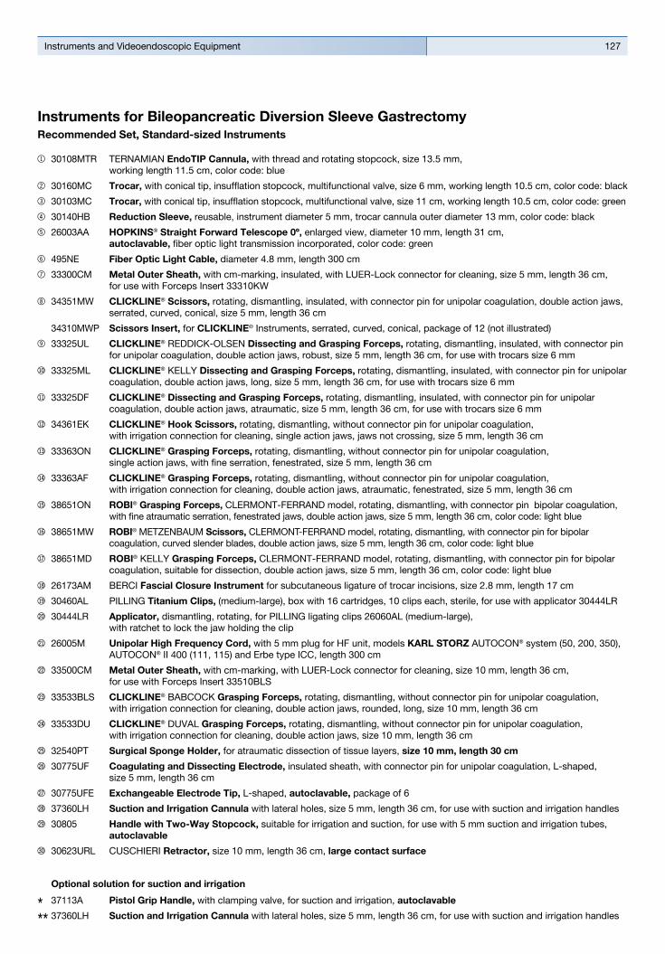
127Instruments and Videoendoscopic Equipment
Instruments for Bileopancreatic Diversion Sleeve Gastrectomy Recommended Set, Standard-sized Instruments
1 30108MTR TERNAMIAN EndoTIP Cannula, with thread and rotating stopcock, size 13.5 mm, working length 11.5 cm, color code: blue
2 30160MC Trocar, with conical tip, insufflation stopcock, multifunctional valve, size 6 mm, working length 10.5 cm, color code: black3 30103MC Trocar, with conical tip, insufflation stopcock, multifunctional valve, size 11 cm, working length 10.5 cm, color code: green4 30140HB Reduction Sleeve, reusable, instrument diameter 5 mm, trocar cannula outer diameter 13 mm, color code: black5 26003AA HOPKINS® Straight Forward Telescope 0º, enlarged view, diameter 10 mm, length 31 cm,
autoclavable, fiber optic light transmission incorporated, color code: green6 495NE Fiber Optic Light Cable, diameter 4.8 mm, length 300 cm7 33300CM Metal Outer Sheath, with cm-marking, insulated, with LUER-Lock connector for cleaning, size 5 mm, length 36 cm,
for use with Forceps Insert 33310KW 8 34351MW CLICKLINE® Scissors, rotating, dismantling, insulated, with connector pin for unipolar coagulation, double action jaws,
serrated, curved, conical, size 5 mm, length 36 cm 34310MWP Scissors Insert, for CLICKLINE® Instruments, serrated, curved, conical, package of 12 (not illustrated)9 33325UL CLICKLINE® REDDICK-OLSEN Dissecting and Grasping Forceps, rotating, dismantling, insulated, with connector pin
for unipolar coagulation, double action jaws, robust, size 5 mm, length 36 cm, for use with trocars size 6 mmbl 33325ML CLICKLINE® KELLY Dissecting and Grasping Forceps, rotating, dismantling, insulated, with connector pin for unipolar
coagulation, double action jaws, long, size 5 mm, length 36 cm, for use with trocars size 6 mmbm 33325DF CLICKLINE® Dissecting and Grasping Forceps, rotating, dismantling, insulated, with connector pin for unipolar
coagulation, double action jaws, atraumatic, size 5 mm, length 36 cm, for use with trocars size 6 mmbn 34361EK CLICKLINE® Hook Scissors, rotating, dismantling, without connector pin for unipolar coagulation,
with irrigation connection for cleaning, single action jaws, jaws not crossing, size 5 mm, length 36 cmbo 33363ON CLICKLINE® Grasping Forceps, rotating, dismantling, without connector pin for unipolar coagulation,
single action jaws, with fine serration, fenestrated, size 5 mm, length 36 cmbp 33363AF CLICKLINE® Grasping Forceps, rotating, dismantling, without connector pin for unipolar coagulation,
with irrigation connection for cleaning, double action jaws, atraumatic, fenestrated, size 5 mm, length 36 cmbq 38651ON ROBI® Grasping Forceps, CLERMONT-FERRAND model, rotating, dismantling, with connector pin bipolar coagulation,
with fine atraumatic serration, fenestrated jaws, double action jaws, size 5 mm, length 36 cm, color code: light bluebr 38651MW ROBI® METZENBAUM Scissors, CLERMONT-FERRAND model, rotating, dismantling, with connector pin for bipolar
coagulation, curved slender blades, double action jaws, size 5 mm, length 36 cm, color code: light bluebs 38651MD ROBI® KELLY Grasping Forceps, CLERMONT-FERRAND model, rotating, dismantling, with connector pin for bipolar
coagulation, suitable for dissection, double action jaws, size 5 mm, length 36 cm, color code: light bluebt 26173AM BERCI Fascial Closure Instrument for subcutaneous ligature of trocar incisions, size 2.8 mm, length 17 cmbu 30460AL PILLING Titanium Clips, (medium-large), box with 16 cartridges, 10 clips each, sterile, for use with applicator 30444LRcl 30444LR Applicator, dismantling, rotating, for PILLING ligating clips 26060AL (medium-large),
with ratchet to lock the jaw holding the clipcm 26005M Unipolar High Frequency Cord, with 5 mm plug for HF unit, models KARL STORZ AUTOCON® system (50, 200, 350),
AUTOCON® II 400 (111, 115) and Erbe type ICC, length 300 cmcn 33500CM Metal Outer Sheath, with cm-marking, with LUER-Lock connector for cleaning, size 10 mm, length 36 cm,
for use with Forceps Insert 33510BLSco 33533BLS CLICKLINE® BABCOCK Grasping Forceps, rotating, dismantling, without connector pin for unipolar coagulation,
with irrigation connection for cleaning, double action jaws, rounded, long, size 10 mm, length 36 cmcp 33533DU CLICKLINE® DUVAL Grasping Forceps, rotating, dismantling, without connector pin for unipolar coagulation,
with irrigation connection for cleaning, double action jaws, size 10 mm, length 36 cmcq 32540PT Surgical Sponge Holder, for atraumatic dissection of tissue layers, size 10 mm, length 30 cmcr 30775UF Coagulating and Dissecting Electrode, insulated sheath, with connector pin for unipolar coagulation, L-shaped,
size 5 mm, length 36 cmcs 30775UFE Exchangeable Electrode Tip, L-shaped, autoclavable, package of 6ct 37360LH Suction and Irrigation Cannula with lateral holes, size 5 mm, length 36 cm, for use with suction and irrigation handlescu 30805 Handle with Two-Way Stopcock, suitable for irrigation and suction, for use with 5 mm suction and irrigation tubes,
autoclavabledl 30623URL CUSCHIERI Retractor, size 10 mm, length 36 cm, large contact surface
Optional solution for suction and irrigation
* 37113A Pistol Grip Handle, with clamping valve, for suction and irrigation, autoclavable
** 37360LH Suction and Irrigation Cannula with lateral holes, size 5 mm, length 36 cm, for use with suction and irrigation handles

Collection on Laparoscopy in Bariatrics128
Instruments for Duodenal SwitchRecommended Set, Standard-sized Instruments
13
4
5
6
7
8
9
bl
bm
bn
bo
bp
bq
brbs
bt
bu
cl
cm
cn
co
cp cq
cr
3
3
1
2
***
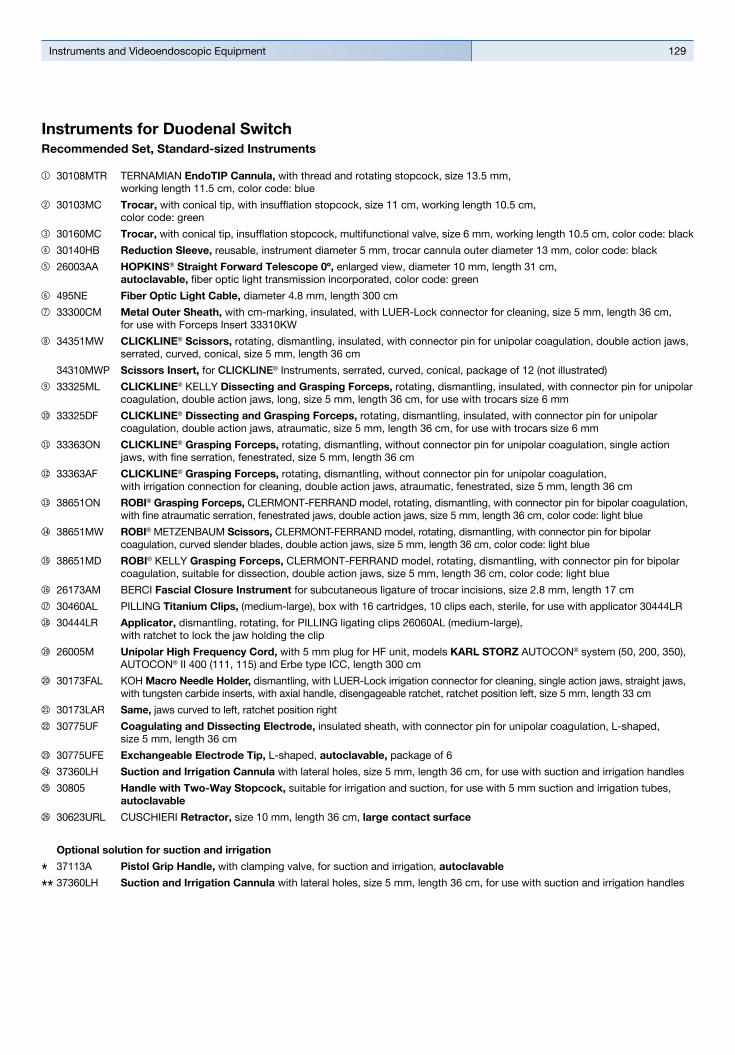
129Instruments and Videoendoscopic Equipment
Instruments for Duodenal SwitchRecommended Set, Standard-sized Instruments
1 30108MTR TERNAMIAN EndoTIP Cannula, with thread and rotating stopcock, size 13.5 mm, working length 11.5 cm, color code: blue
2 30103MC Trocar, with conical tip, with insufflation stopcock, size 11 cm, working length 10.5 cm, color code: green
3 30160MC Trocar, with conical tip, insufflation stopcock, multifunctional valve, size 6 mm, working length 10.5 cm, color code: black4 30140HB Reduction Sleeve, reusable, instrument diameter 5 mm, trocar cannula outer diameter 13 mm, color code: black5 26003AA HOPKINS® Straight Forward Telescope 0º, enlarged view, diameter 10 mm, length 31 cm,
autoclavable, fiber optic light transmission incorporated, color code: green6 495NE Fiber Optic Light Cable, diameter 4.8 mm, length 300 cm7 33300CM Metal Outer Sheath, with cm-marking, insulated, with LUER-Lock connector for cleaning, size 5 mm, length 36 cm,
for use with Forceps Insert 33310KW 8 34351MW CLICKLINE® Scissors, rotating, dismantling, insulated, with connector pin for unipolar coagulation, double action jaws,
serrated, curved, conical, size 5 mm, length 36 cm 34310MWP Scissors Insert, for CLICKLINE® Instruments, serrated, curved, conical, package of 12 (not illustrated) 9 33325ML CLICKLINE® KELLY Dissecting and Grasping Forceps, rotating, dismantling, insulated, with connector pin for unipolar
coagulation, double action jaws, long, size 5 mm, length 36 cm, for use with trocars size 6 mmbl 33325DF CLICKLINE® Dissecting and Grasping Forceps, rotating, dismantling, insulated, with connector pin for unipolar
coagulation, double action jaws, atraumatic, size 5 mm, length 36 cm, for use with trocars size 6 mmbm 33363ON CLICKLINE® Grasping Forceps, rotating, dismantling, without connector pin for unipolar coagulation, single action
jaws, with fine serration, fenestrated, size 5 mm, length 36 cmbn 33363AF CLICKLINE® Grasping Forceps, rotating, dismantling, without connector pin for unipolar coagulation,
with irrigation connection for cleaning, double action jaws, atraumatic, fenestrated, size 5 mm, length 36 cmbo 38651ON ROBI® Grasping Forceps, CLERMONT-FERRAND model, rotating, dismantling, with connector pin for bipolar coagulation,
with fine atraumatic serration, fenestrated jaws, double action jaws, size 5 mm, length 36 cm, color code: light bluebp 38651MW ROBI® METZENBAUM Scissors, CLERMONT-FERRAND model, rotating, dismantling, with connector pin for bipolar
coagulation, curved slender blades, double action jaws, size 5 mm, length 36 cm, color code: light bluebq 38651MD ROBI® KELLY Grasping Forceps, CLERMONT-FERRAND model, rotating, dismantling, with connector pin for bipolar
coagulation, suitable for dissection, double action jaws, size 5 mm, length 36 cm, color code: light bluebr 26173AM BERCI Fascial Closure Instrument for subcutaneous ligature of trocar incisions, size 2.8 mm, length 17 cmbs 30460AL PILLING Titanium Clips, (medium-large), box with 16 cartridges, 10 clips each, sterile, for use with applicator 30444LRbt 30444LR Applicator, dismantling, rotating, for PILLING ligating clips 26060AL (medium-large),
with ratchet to lock the jaw holding the clipbu 26005M Unipolar High Frequency Cord, with 5 mm plug for HF unit, models KARL STORZ AUTOCON® system (50, 200, 350),
AUTOCON® II 400 (111, 115) and Erbe type ICC, length 300 cmcl 30173FAL KOH Macro Needle Holder, dismantling, with LUER-Lock irrigation connector for cleaning, single action jaws, straight jaws,
with tungsten carbide inserts, with axial handle, disengageable ratchet, ratchet position left, size 5 mm, length 33 cmcm 30173LAR Same, jaws curved to left, ratchet position rightcn 30775UF Coagulating and Dissecting Electrode, insulated sheath, with connector pin for unipolar coagulation, L-shaped,
size 5 mm, length 36 cmco 30775UFE Exchangeable Electrode Tip, L-shaped, autoclavable, package of 6cp 37360LH Suction and Irrigation Cannula with lateral holes, size 5 mm, length 36 cm, for use with suction and irrigation handlescq 30805 Handle with Two-Way Stopcock, suitable for irrigation and suction, for use with 5 mm suction and irrigation tubes,
autoclavablecr 30623URL CUSCHIERI Retractor, size 10 mm, length 36 cm, large contact surface
Optional solution for suction and irrigation
* 37113A Pistol Grip Handle, with clamping valve, for suction and irrigation, autoclavable
** 37360LH Suction and Irrigation Cannula with lateral holes, size 5 mm, length 36 cm, for use with suction and irrigation handles
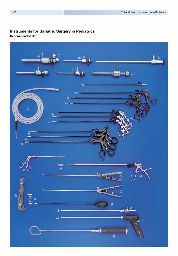
Collection on Laparoscopy in Bariatrics130
Instruments for Bariatric Surgery in PediatricsRecommended Set
1 2
3
4
4
5
6
7
8
9
bl
bm
bn
bo
bp
bq
br
bsbt
bu
cl
cm
cn
co
cp
cqcr
***
cs

131Instruments and Videoendoscopic Equipment
Instruments for Bariatric Surgery in PediatricsRecommended Set
1 30108MTR TERNAMIAN EndoTIP Cannula, with thread and rotating stopcock, size 13.5 mm, working length 11.5 cm, color code: blue
2 30142HB Double Reducer, 13/10 mm, 13,5/10 mm, 13/5 mm und 13.5/5 mm3 30103MC Trocar, with conical tip, insufflation stopcock, multifunctional valve, size 11 cm, working length 10.5 cm, color code: green4 30160MC Trocar, with conical tip, insufflation stopcock, multifunctional valve, size 6 mm, working length 10.5 cm, color code: black5 26003BA HOPKINS® Forward-Oblique Telescope 30º, enlarged view, diameter 10 mm, length 31 cm, autoclavable,
fiber optic light transmission incorporated, color code: red6 30105KP Trocar, with pyramidal tip, insufflation stopcock, automatic valve, size 15 mm, working length 12 cm7 495NL Fiber Optic Light Cable, diameter 3.5 mm, length 180 cm8 33300CM Metal Outer Sheath, with cm-marking, insulated, with LUER-Lock connector for cleaning, size 5 mm, length 36 cm,
for use with Forceps Insert 33310KW9 34351MW CLICKLINE® Scissors, rotating, dismantling, insulated, with connector pin for unipolar coagulation, double action jaws,
serrated, curved, conical, size 5 mm, length 36 cm 34310MWP Scissors Insert, for CLICKLINE® Instruments, serrated, curved, conical, package of 12 (not illustrated)bl 33325ML CLICKLINE® KELLY Dissecting and Grasping Forceps, rotating, dismantling, insulated, with connector pin for unipolar
coagulation, double action jaws, long, size 5 mm, length 36 cm, for use with trocars size 6 mmbm 33325DF CLICKLINE® Dissecting and Grasping Forceps, rotating, dismantling, insulated, with connector pin for unipolar
coagulation, double action jaws, atraumatic, size 5 mm, length 36 cm, for use with trocars size 6 mmbn 33363ON CLICKLINE® Grasping Forceps, rotating, dismantling, without connector pin for unipolar coagulation, single action
jaws, with fine serration, fenestrated, size 5 mm, length 36 cmbo 33363AF CLICKLINE® Grasping Forceps, rotating, dismantling, without connector pin for unipolar coagulation,
with irrigation connection for cleaning, double action jaws, atraumatic, fenestrated, size 5 mm, length 36 cmbp 38651ON ROBI® Grasping Forceps, CLERMONT-FERRAND model, rotating, dismantling, with connector pin for bipolar coagulati-
on, with fine atraumatic serration, fenestrated jaws, double action jaws, size 5 mm, length 36 cm, color code: light bluebq 38651MW ROBI® METZENBAUM Scissors, CLERMONT-FERRAND model, rotating, dismantling, with connector pin for bipolar
coagulation, curved slender blades, double action jaws, size 5 mm, length 36 cm, color code: light bluebr 38651MD ROBI® KELLY Grasping Forceps, CLERMONT-FERRAND model, rotating, dismantling, with connector pin for bipolar
coagulation, suitable for dissection, double action jaws, size 5 mm, length 36 cm, color code: light bluebs 26173AM BERCI Fascial Closure Instrument for subcutaneous ligature of trocar incisions, size 2.8 mm, length 17 cmbt 30460AL PILLING Titanium Clips, (medium-large), box with 16 cartridges, 10 clips each, sterile, for use with applicator 30444LRbu 30444LR Applicator, dismantling, rotating, for PILLING ligating clips 26060AL (medium-large),
with ratchet to lock the jaw holding the clipcl 26005M Unipolar High Frequency Cord, with 5 mm plug for HF unit, models KARL STORZ AUTOCON® system (50, 200, 350),
AUTOCON® II 400 (111, 115) and Erbe type ICC, length 300 cmcm 30173FAL KOH Macro Needle Holder, dismantling, with LUER-Lock irrigation connector for cleaning, single action jaws, straight jaws,
with tungsten carbide inserts, with axial handle, disengageable ratchet, ratchet position left, size 5 mm, length 33 cmcn 30173LAR Same, jaws curved to left, ratchet position rightco 30775UF Coagulating and Dissecting Electrode, insulated sheath, with connector pin for unipolar coagulation, L-shaped,
size 5 mm, length 36 cmcp 30775UFE Exchangeable Electrode Tip, L-shaped, autoclavable, package of 6cq 37360LH Suction and Irrigation Cannula with lateral holes, size 5 mm, length 36 cm, for use with suction and irrigation handlescr 30805 Handle with Two-Way Stopcock, suitable for irrigation and suction, for use with 5 mm suction and irrigation tubes,
autoclavablecs 30623U CUSCHIERI Retractor, size 5 mm, length 36 cm
Optional solution for suction and irrigation
* 37113A Pistol Grip Handle, with clamping valve, for suction and irrigation, autoclavable
** 37360LH Suction and Irrigation Cannula with lateral holes, size 5 mm, length 36 cm, for use with suction and irrigation handles

Collection on Laparoscopy in Bariatrics132
HOPKINS® Telescopes Diameter 10 mm, length 31 cm and 42 cm
26003BA HOPKINS® Forward-Oblique Telescope 30º, enlarged view, diameter 10 mm, length 31 cm, autoclavable, fiber optic light transmission incorporated, color code: red
26003AA HOPKINS® Straight Forward Telescope 0º, enlarged view, diameter 10 mm, length 31 cm, autoclavable, fiber optic light transmission incorporated, color code: green
26003AEA HOPKINS® Straight Forward Telescope 0°, enlarged view, diameter 10 mm, length 42 cm, autoclavable, fiber optic light transmission incorporated, color code: green
26003AA
26003BEA HOPKINS® Forward-Oblique Telescope 30°, enlarged view, diameter 10 mm, length 42 cm, autoclavable, fiber optic light transmission incorporated, color code: red
30103CS Telescope Stopper, sterile, size 11 mm, for single use, package of 12
26003AE
26003AE ENDOCAMELEON® HOPKINS® Telescope, diameter 10 mm, length 32 cm, autoclavable, variable direction of view from 0°–120°, adjustment knob for selecting the desired direction of view, fiber optic light transmission incorporated, color code: gold

133Instruments and Videoendoscopic Equipment
TERNAMIAN EndoTIP Cannulassize 11 mm and 13.5 mm
30103TM
30103TM TERNAMIAN EndoTIP Cannula, with thread and rotatable insufflation stopcock, size 11 mm, working length 10.5 cm, color code: green,
including: Multifunctional Valve
Cannula
31103T4 Same, working length 15 cm
30108MTR
30108MTR TERNAMIAN EndoTIP Cannula, with thread and rotating stopcock, size 13.5 mm, working length 11.5 cm, color code: blue,
including: Multifunctional Valve
Cannula
31108MTR Same, working length 15 cm
Trocarssize 6 and 11 mm
30103MC Trocar, with conical tip, with insufflation stopcock, size 11 cm, working length 10.5 cm, color code: green,
including: Trocar only Cannula, without valve Multifunctional Valve
31103MC Same, working length 15 cm
30103MC
30160MC Trocar, with conical tip, insufflation stopcock, size 6 mm, working length 10.5 cm, color code: black,
including: Trocar only Cannula, without valve Multifunctional Valve
31160MC Same, working length 15 cm
30103MC

Collection on Laparoscopy in Bariatrics134
30105KP Trocar, with pyramidal tip, with insufflation stopcock, size 15 mm, working length 12 cm,
including: Cannula, without valve Trocar only Automatic Valve
30106KP Same, working length 15 cm
30105KP
30140HB Reduction Sleeve, reusable, instrument diameter 5 mm, trocar cannula outer diameter 13 mm, color code: black
30142HB Double Reducer, 13/10 mm, 13.5/10 mm, 13/5 mm and 13.5/5 mm
30141DB Reducer, attachable, 11/5 mm
Trocars and Accessoiressize 15 and 22 mm
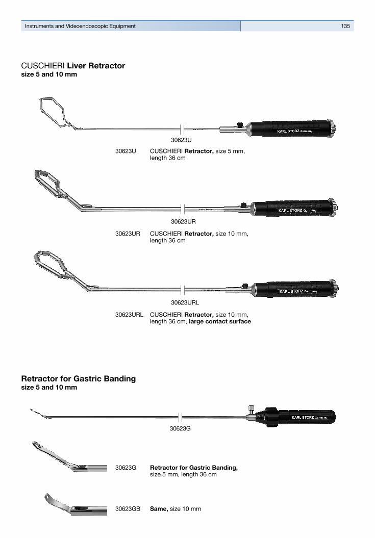
135Instruments and Videoendoscopic Equipment
30623URL CUSCHIERI Retractor, size 10 mm, length 36 cm, large contact surface
30623URL
30623U CUSCHIERI Retractor, size 5 mm, length 36 cm
30623U
30623G Retractor for Gastric Banding, size 5 mm, length 36 cm
30623GB Same, size 10 mm
30623G
Retractor for Gastric Bandingsize 5 and 10 mm
CUSCHIERI Liver Retractorsize 5 and 10 mm
30623UR CUSCHIERI Retractor, size 10 mm, length 36 cm
30623UR

Collection on Laparoscopy in Bariatrics136
34351MA
CLICKLINE® ScissorsSize 5 mm, length 36 cm and 43 cm
34351MW CLICKLINE® Scissors, rotating, dismantling, insulated, with connector pin for unipolar coagulation, double action jaws, serrated, curved, conical, size 5 mm, length 36 cm,
including: Plastic Handle, insulated, without ratchet Outer Sheath, insulated Scissors Insert
34451MW Same, length 43 cm
34310MWP Scissors Insert, for CLICKLINE® Instruments, serrated, curved, conical, package of 12
34351MA CLICKLINE® Scissors, rotating, dismantling, insulated, with connector pin for unipolar coagulation, double action jaws, serrated, curved, round cupped jaws, length of blades 17 mm, size 5 mm, length 36 cm,
including: Plastic Handle, insulated, without ratchet Outer Sheath, insulated Scissors Insert
34451MA Same, length 43 cm

137Instruments and Videoendoscopic Equipment
38651MW ROBI® METZENBAUM Scissors, CLERMONT-FERRAND model, rotating, dismantling, with connector pin for bipolar coagulation, with LUER-Lock irrigation connector for cleaning, double action jaws, curved, slender blades, for cutting and bipolar coagulation, size 5 mm, length 36 cm,
including: Ring Handle Metal Outer Sheath, insulated Scissors Insert
38751MW Same, length 43 cm
CLICKLINE® Hook Scissors and ROBI® METZENBAUM ScissorsSize 5 mm, length 36 cm and 43 cm
34361EK
38651MW
34361EK CLICKLINE® Hook Scissors, rotating, dismantling, without connector pin for unipolar coagulation, with irrigation connection for cleaning, single action jaws, jaws not crossing, size 5 mm, length 36 cm,
including: Metal Handle, with hemostat style ratchet,
with larger contact area Outer Sheath, insulated Scissors Insert
34461EK Same, length 43 cm

Collection on Laparoscopy in Bariatrics138
33325UL CLICKLINE® REDDICK-OLSEN Dissecting and Grasping Forceps, rotating, dismantling, insulated, with connector pin for unipolar coagulation, double action jaws, robust, size 5 mm, length 36 cm, for use with trocars size 6 mm,
including: Metal Handle, with hemostat style ratchet,
with larger contact area Outer Sheath, insulated Forceps Insert
33425UL Same, length 43 cm
33325DF CLICKLINE® Dissecting and Grasping Forceps, rotating, dismantling, insulated, with connector pin for unipolar coagulation, double action jaws, atraumatic, size 5 mm, length 36 cm, for use with trocars size 6 mm,
including: Metal Handle, with hemostat style ratchet,
with larger contact area Outer Sheath, insulated Forceps Insert
33425DF Same, length 43 cm
33325KW CLICKLINE® MATKOWITZ Grasping Forceps, double-action jaws, diameter 5 mm, length 36 cm, with connector pin for unipolar coagulation,
including: Metal Handle, with hemostat style ratchet,
with larger contact area Outer Sheath, insulated Forceps Insert
33425KW Same, length 43 cm
33325ML CLICKLINE® KELLY Dissecting and Grasping Forceps, rotating, dismantling, insulated, with connector pin for unipolar coagulation, double action jaws, long, size 5 mm, length 36 cm, for use with trocars size 6 mm,
including: Metal Handle, with hemostat style ratchet,
with larger contact area Outer Sheath, insulated Forceps Insert
33425ML Same, length 43 cm
CLICKLINE® Dissecting and Grasping ForcepsSize 5 mm, length 36 cm and 43 cm
33325DF

139Instruments and Videoendoscopic Equipment
33563BLS CLICKLINE® BABCOCK Grasping Forceps, rotating, dismantling, without connector pin for unipolar coagulation, with irrigation connection for cleaning, double action jaws, rounded, long, size 10 mm, length 36 cm,
including: Metal Handle, with hemostat style ratchet,
with larger contact area Outer Sheath, insulated Forceps Insert
33363AF CLICKLINE® Grasping Forceps, rotating, dismantling,without connector pin for unipolar coagulation, with irrigation connection for cleaning, double action jaws, atraumatic, fenestrated, size 5 mm, length 36 cm,
including: Metal Handle, with hemostat style ratchet,
with larger contact area Outer Sheath, insulated Forceps Insert
33463AF Same, length 43 cm
33363ON CLICKLINE® Grasping Forceps, rotating, dismantling, without connector pin for unipolar coagulation, single action jaws, with fine serration, fenestrated, size 5 mm, length 36 cm,
including: Metal Handle, with hemostat style ratchet,
with larger contact area Outer Sheath, insulated Forceps Insert
33463ON Same, length 43 cm
33563DU CLICKLINE® DUVAL Grasping Forceps, rotating, dismantling, without connector pin for unipolar coagulation, with irrigation connection for cleaning, double action jaws, size 10 mm, length 36 cm,
including: Metal Handle, with hemostat style ratchet,
with larger contact area Outer Sheath, insulated Forceps Insert
CLICKLINE® Dissecting and Grasping ForcepsSize 5 mm and 10 cm, length 36 cm and 43 cm
33363AF

Collection on Laparoscopy in Bariatrics140
38651MD ROBI® KELLY Dissecting and Grasping Forceps, CLERMONT-FERRAND Model, rotating, dismantling, with connector pin for bipolar coagulation, with LUER-Lock irrigation connector for cleaning, double action jaws, suitable for dissection, size 5 mm, length 36 cm,
including: Ring Handle, without ratchet, with larger contact area Metal Outer Sheath, insulated Forceps Insert
38751MD Same, length 43 cm
38651ON ROBI® Grasping Forceps, CLERMONT-FERRAND Model, rotating, dismantling, with connector pin for bipolar coagulation, with LUER-Lock irrigation connector for cleaning, double action jaws, fenestrated, with fine atraumatic serration, size 5 mm, length 36 cm,
including: Ring Handle, without ratchet, with larger contact area Metal Outer Sheath, insulated Forceps Insert
38751ON Same, length 43 cm
38651MD
33361AV CLICKLINE® Grasping Forceps, rotating, dismantling, without connector pin for unipolar coagulation, with irrigation connector for cleaning, double action jaws, for stapler pressure plates, size 5 mm, length 36 cm,
including: Metal Handle, with hemostat style ratchet,
with larger contact area Outer Sheath, insulated Forceps Insert
CLICKLINE® Grasping Forceps and ROBI® Grasping ForcepsSize 5 mm, length 36 cm and 43 cm
33361AV

141Instruments and Videoendoscopic Equipment
33300CMM
33300CM
Metal Outer Sheath with cm-marking Size 5 mm
33300CM Metal Outer Sheath, with cm-marking, insulated, with LUER-Lock connector for cleaning, size 5 mm, length 36 cm
33400CM Same, length 43 cm
33300CMM Metal Outer Sheath, with cm-marking, with LUER-Lock connector for cleaning, size 5 mm, length 36 cm,
33400CMM Same, length 43 cm
30775UF Coagulating and Dissecting Electrode, insulated sheath, with connector pin for unipolar coagulation, L-shaped, size 5 mm, length 36 cm
30778UF Same, length 43 cm
30775UFE Exchangeable Electrode Tip, L-shaped, autoclavable, package of 6
Dissecting Electrodeswith Exchangeable Electrode Tip Insulated Sheath with Connector Pin for Uniplar Coagulation
30775UF

Collection on Laparoscopy in Bariatrics142
KOH Macro Needle Holder, size 5 mm, dismantling,including:
## Handle## Outer Sheath## Working Insert
Cleaning and sterilization are gaining increasing importance for KARL STORZ as a manufacturer of surgical instruments.
Similar to all our surgical instruments, the cleaning and hygiene of our needle holders also play an important role.
For cleaning and sterilization, the KOH macro needle-holders can be disassembled into their main compo-nents.
## Choice of six different handles and three different working inserts
## With tungsten carbide inserts## In the event of damage, only the component with the defect needs to be replaced
## Can be disassembled into three separate components
## Autoclavable## Cleaning adaptor
This unique reusable three-piece design offers the user the following benefits:
KOH Macro Needle Holderdismantable
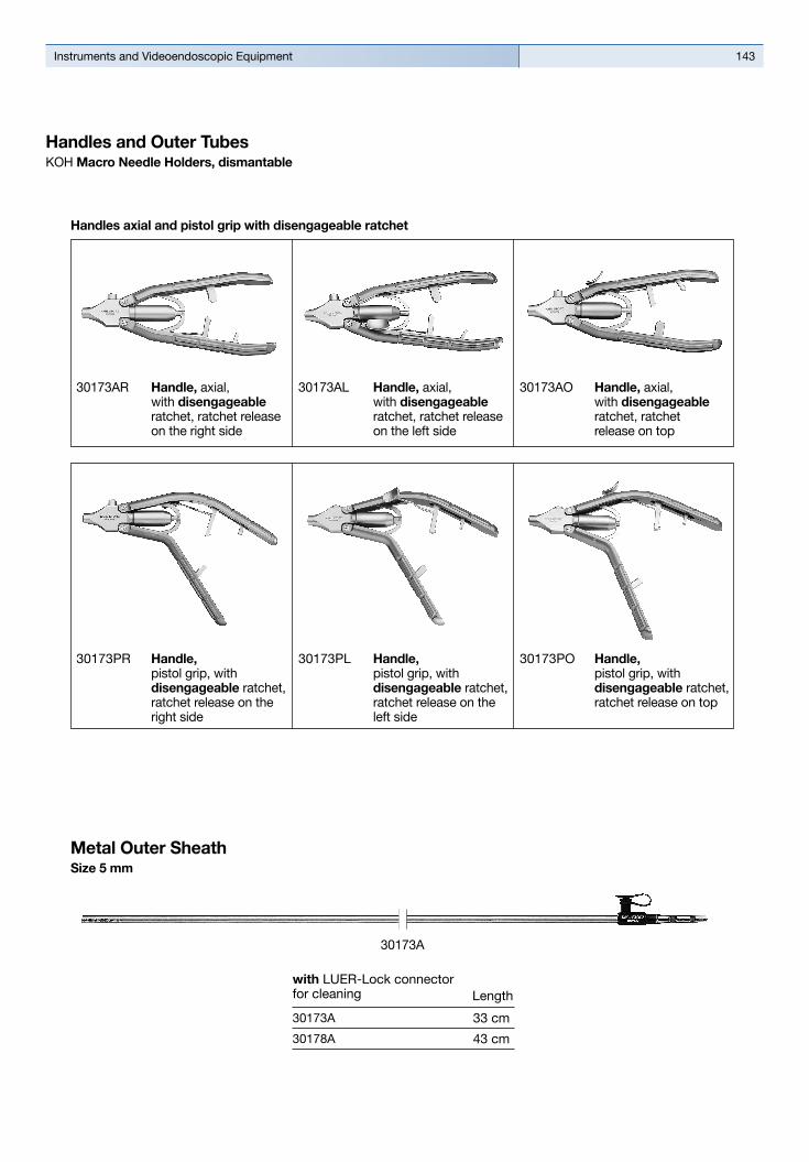
143Instruments and Videoendoscopic Equipment
30173AR Handle, axial, with disengageable ratchet, ratchet release on the right side
30173AL Handle, axial, with disengageable ratchet, ratchet release on the left side
30173AO Handle, axial, with disengageable ratchet, ratchet release on top
Handles axial and pistol grip with disengageable ratchet
30173PR Handle, pistol grip, with disengageable ratchet, ratchet release on the right side
30173PL Handle, pistol grip, with disengageable ratchet, ratchet release on the left side
30173PO Handle, pistol grip, with disengageable ratchet, ratchet release on top
30173A
Metal Outer SheathSize 5 mm
with LUER-Lock connector for cleaning
43 cm
33 cm30173A
30178A
Length
Handles and Outer TubesKOH Macro Needle Holders, dismantable

Collection on Laparoscopy in Bariatrics144
Handle
30173AR 30173AL 30173AO
Complete Instrument
30173RAR 30173RAL 30173RAO
30178RAR 30178RAL 30178RAO
KOH Macro Needle Holder, dismantling, jaws curved to right, with tungsten carbide inserts, for use with suture material size 0/0 – 7/0
30173R
30178R
Insert No.
43 cm
Working Length
33 cm
Single-action jaws
Size 5 mm
30173LAR 30173LAL 30173LAO
30178LAR 30178LAL 30178LAO
KOH Macro Needle Holder, dismantling, jaws curved to left, with tungsten carbide inserts, for use with suture material size 0/0 – 7/0
30173L
30178L
30173FAR 30173FAL 30173FAO
30178FAR 30178FAL 30178FAO
KOH Macro Needle Holder, dismantling, straight jaws, with tungsten carbide inserts, for use with suture material size 0/0 – 7/0
30173F
30178F
KOH Macro Needle Holderdismantable

145Instruments and Videoendoscopic Equipment
Handle
30173PR 30173PL 30173PO
Complete Instrument
30173RPR 30173RPL 30173RPO
30178RPR 30178RPL 30178RPO
KOH Macro Needle Holder, dismantling, jaws curved to right, with tungsten carbide inserts, for use with suture material size 0/0 – 7/0
30173R
30178R
Insert No.
43 cm
Working Length
33 cm
30173LPR 30173LPL 30173LPO
30178LPR 30178LPL 30178LPO
KOH Macro Needle Holder, dismantling, jaws curved to left, with tungsten carbide inserts, for use with suture material size 0/0 – 7/0
30173L
30178L
30173FPR 30173FPL 30173FPO
30178FPR 30178FPL 30178FPO
KOH Macro Needle Holder, dismantling, straight jaws, with tungsten carbide inserts, for use with suture material size 0/0 – 7/0
30173F
30178F
Single-action jaws
Size 5 mm
KOH Macro Needle Holderdismantable

Collection on Laparoscopy in Bariatrics146
KOH Macro Needle Holder
Size 5 mm
26173KAL KOH Macro Needle Holder, with tungsten carbide insert, axial handle, with disengageable ratchet, ratchet position right, jaws curved to left, size 5 mm, length 33 cm, for use with suture material size 0/0 to 7/0 and needle sizes BV, SH or CT-1
26178KAL Same, length 43 cm
26173KAR KOH Macro Needle Holder, with tungsten carbide insert, axial handle, with disengageable ratchet, ratchet position left, jaws curved to right, size 5 mm, length 33 cm, for use with suture material size 0/0 to 7/0 and needle sizes BV, SH or CT-1
26178KAR Same, length 43 cm
Multiple puncture approach Operating instruments, length 33 and 43 cm, for use with trocar size 6 mm
26173KAF

147Instruments and Videoendoscopic Equipment
Original Size
30444LR Applicator, Dismantling, Rotating, for PILLING ligating clips 26060AL (medium-large), with ratchet to lock the jaw holding the clip
including: Metal handle, with ratchet Metal outer sheath Insert
30460AL PILLING Titanium Clips, (medium-large), box with 16 cartridges, 10 clips each, sterile, for use with applicator 30444LR
Caution:The use of other clips than indicated, can lead to damage of the mouthpiece.
Clip Applicatorsize 10 mmMultiple puncture approach Operating instrument, length 33 cm,for use with trocar size 11 mm
30444LR
26173AM BERCI Fascial Closure Instrument for subcutaneous ligature of trocar incisions, size 2.8 mm, length 17 cm
BERCI Fascial Closure Instrument for subcutaneous ligature of trocar incisions
26173AM

Collection on Laparoscopy in Bariatrics148
37112A Suction and Irrigation Handle, straight, with clamping valve, for suction and irrigation, autoclavable, including Cleaning Adaptor 8236091, for use with Suction and Irrigation Tubes diameter 3 mm, 5 mm, 10 mm, 37360 LH, 37560 LH
37113A Same, pistol grip
37113A
37112A
Handles for Suction and Irrigationsize 5 mm and 10 mm
031133-10* Tubing Set, for single use, sterile, package of 10, for use with Suction and Irrigation Handles 37112A (straight) and 37113A (pistol grip)
031134-10* Tubing Set, for single use, sterile, package of 10, for use with Suction and Irrigation Handles 37112A (straight) and 37113A (pistol grip) in combination with silicone tube inner diameter 5 mm at the patient end
031218-10
031219-10* Tubing Set, with two puncture cannulas, for single use, sterile, package of 10, for use with Handles 37112A (straight) and 37113A (pistol grip) in combination with ENDOMAT® LC
031218-10* Tubing Set, with two puncture cannulas, for single use, sterile, package of 10, for use with Handles 37112A (straight) and 37113A (pistol grip) in combination with HAMOU® ENDOMAT®
031219-10
*
Single-Use Tubing Setsfor use with Suction and Irrigation Handles 37112A and 37113A

149Instruments and Videoendoscopic Equipment
32540PTL Surgical Sponge Holder, for atraumatic dissection of tissue layers, size 10 mm, length 43 cm
including: Handle Outer Sheath, insulated Sponge Holder Insert
32540PT Surgical Sponge Holder, tissue layers, size 10 mm, length 30 cm
including: Handle Outer Sheath, insulated Sponge Holder Insert
32540PT
Surgical Sponge Holderssize 5 mm and 10 mm
Irrigation and Suction Tubessize 5 mm, trocar size 6 mm
37360LH
30805 Handle with Two-Way Stopcock, suitable for irrigation and suction, for use with 5 mm suction and irrigation tubes, autoclavable
37360LH Suction and Irrigation Cannula with lateral holes, size 5 mm, length 36 cm, for use with suction and irrigation handles
37460LH Same, length 43 cm

Collection on Laparoscopy in Bariatrics150
Knot Tiersize 5 mm, trocar size 6 mm
26596SK KOECKERLING Knot Tier, for extracorporeal knotting size 5 mm, length 36 cm
26596SK
AccessoriesUnipolar High Frequency Cord
unipolar
High Frequency Surgery Units
KARL STORZInstrument
26005M Unipolar High Frequency Cord, with 5 mm plug for AUTOCON® II 400 SCB system (111, 115, 122, 125), AUTOCON® II 200, AUTOCON® II 80, KARL STORZ AUTOCON® system (50, 200, 350) and Erbe type ICC, length 300 cm
Bipolar High Frequency Cord
bipolarHigh Frequency Surgery Units
KARL STORZInstrument
26176LE Bipolar High Frequency Cord, for AUTOCON® II 400 SCB system (111, 113, 115, 122, 125), AUTOCON® II 200, AUTOCON® II 80, KARL STORZ Coagulator 26021 B/C/D, 860021 B/C/D, 27810 B/C/D, 28810 B/C/D, AUTOCON® series (50, 200, 350), Erbe-Coagulator, T and ICC series, length 300 cm

151Instruments and Videoendoscopic Equipment
Mobile Equipment Cart
Monitor:9627NB 27" FULL HD Monitor
Camera System: TC200DE IMAGE1 S CONNECT, connect moduleTC300 IMAGE1 S H3-LINK, link moduleTH100 IMAGE1 S H3-Z
Three-Chip FULL HD Camera Head
Light Source:20 1331 01-1 XENON 300 SCB Cold Light Fountain 495NCSC Fiber Optic Light Cable
HF-Device:20 5352 01-125 AUTOCON® II 40020 0178 30 Two-Pedal Footswitch
Insufflation:UI400S1 ENDOFLATOR® 40 UP501S3 S-PILOT ™
Pump System:26 3311 01-1 HAMOU® ENDOMAT®
Equipment Cart:UG120 COR™ Equpiment Cart, narrow, highUG500 Monitor HolderUG609 Bottle Holder, for CO2-Bottles29005DFH Foot-Pedal Holder,
for Two- and Three-Pedal FootswitchesUG310 Isolation Transformer, 200 V – 240 V UG410 Earth Leakage Monitor, 200 V – 240 V
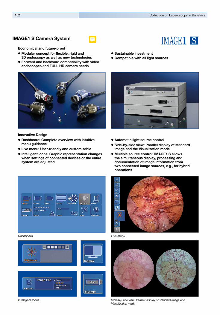
Collection on Laparoscopy in Bariatrics152
Innovative Design## Dashboard: Complete overview with intuitive menu guidance
## Live menu: User-friendly and customizable## Intelligent icons: Graphic representation changes when settings of connected devices or the entire system are adjusted
## Automatic light source control## Side-by-side view: Parallel display of standard image and the Visualization mode
## Multiple source control: IMAGE1 S allows the simultaneous display, processing and documentation of image information from two connected image sources, e.g., for hybrid operations
Dashboard Live menu
Side-by-side view: Parallel display of standard image and Visualization mode
Intelligent icons
Economical and future-proof## Modular concept for flexible, rigid and 3D endoscopy as well as new technologies
## Forward and backward compatibility with video endoscopes and FULL HD camera heads
## Sustainable investment## Compatible with all light sources
IMAGE1 S Camera System
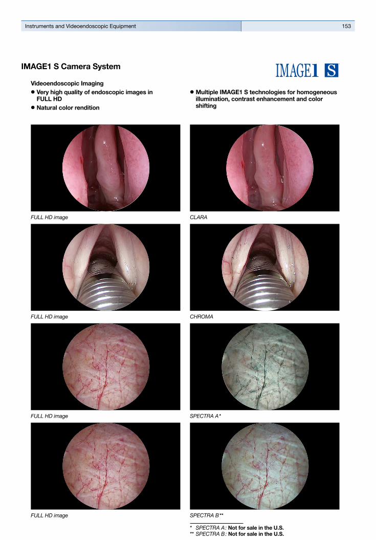
153Instruments and Videoendoscopic Equipment
Videoendoscopic Imaging## Very high quality of endoscopic images in FULL HD
## Natural color rendition
## Multiple IMAGE1 S technologies for homogeneous illumination, contrast enhancement and color shifting
FULL HD image CHROMA
FULL HD image SPECTRA A *
FULL HD image
FULL HD image CLARA
SPECTRA B **
* SPECTRA A : Not for sale in the U.S.** SPECTRA B : Not for sale in the U.S.
IMAGE1 S Camera System

Collection on Laparoscopy in Bariatrics154
TC 200EN* IMAGE1 S CONNECT, connect module, for use with up to 3 link modules, resolution 1920 x 1080 pixels, with integrated KARL STORZ-SCB and digital Image Processing Module, power supply 100 – 120 VAC/200 – 240 VAC, 50/60 Hz
including: Mains Cord, length 300 cm DVI-D Connecting Cable, length 300 cm SCB Connecting Cable, length 100 cm USB Flash Drive, 32 GB, USB silicone keyboard, with touchpad, US
* Available in the following languages: DE, ES, FR, IT, PT, RU
Specifications:
HD video outputs
Format signal outputs
LINK video inputs
USB interface SCB interface
- 2x DVI-D - 1x 3G-SDI
1920 x 1080p, 50/60 Hz
3x
4x USB, (2x front, 2x rear) 2x 6-pin mini-DIN
100 – 120 VAC/200 – 240 VAC
50/60 Hz
I, CF-Defib
305 x 54 x 320 mm
2.1 kg
Power supply
Power frequency
Protection class
Dimensions w x h x d
Weight
TC300 IMAGE1 S H3-LINK, link module, for use with IMAGE1 FULL HD three-chip camera heads, power supply 100 – 120 VAC/200 – 240 VAC, 50/60 Hz, for use with IMAGE1 S CONNECT TC200ENincluding:Mains Cord, length 300 cm
Link Cable, length 20 cm
For use with IMAGE1 S IMAGE1 S CONNECT Module TC200EN
IMAGE1 S Camera System
TC 300 (H3-Link)
TH100, TH101, TH102, TH103, TH104, TH106 (fully compatible with IMAGE1 S) 22 2200 55-3, 22 2200 56-3, 22 2200 53-3, 22 2200 60-3, 22 2200 61-3, 22 2200 54-3, 22 2200 85-3 (compatible without IMAGE1 S technologies CLARA, CHROMA, SPECTRA*)
1x
100 – 120 VAC/200 – 240 VAC
50/60 Hz
I, CF-Defib
305 x 54 x 320 mm
1.86 kg
Camera System
Supported camera heads/video endoscopes
LINK video outputs
Power supply
Power frequency
Protection class
Dimensions w x h x d
Weight
Specifications:
TC200EN
TC300
* SPECTRA A : Not for sale in the U.S.** SPECTRA B : Not for sale in the U.S.

155Instruments and Videoendoscopic Equipment
TH104
TH104 IMAGE1 S H3-ZA Three-Chip FULL HD Camera Head, 50/60 Hz, IMAGE1 S compatible, autoclavable, progressive scan, soakable, gas- and plasma-sterilizable, with integrated Parfocal Zoom Lens, focal length f = 15 – 31 mm (2x), 2 freely programmable camera head buttons, for use with IMAGE1 S and IMAGE 1 HUB™ HD/HD
IMAGE1 FULL HD Camera Heads
Product no.
Image sensor
Dimensions w x h x d
Weight
Optical interface
Min. sensitivity
Grip mechanism
Cable
Cable length
IMAGE1 S H3-ZA
TH104
3x 1/3" CCD chip
39 x 49 x 100 mm
299 g
integrated Parfocal Zoom Lens, f = 15 – 31 mm (2x)
F 1.4/1.17 Lux
standard eyepiece adaptor
non-detachable
300 cm
Specifications:
TH100 IMAGE1 S H3-Z Three-Chip FULL HD Camera Head, 50/60 Hz, IMAGE1 S compatible, progressive scan, soakable, gas- and plasma-sterilizable, with integrated Parfocal Zoom Lens, focal length f = 15 – 31 mm (2x), 2 freely programmable camera head buttons, for use with IMAGE1 S and IMAGE 1 HUB™ HD/HD
IMAGE1 FULL HD Camera Heads
Product no.
Image sensor
Dimensions w x h x d
Weight
Optical interface
Min. sensitivity
Grip mechanism
Cable
Cable length
IMAGE1 S H3-Z
TH100
3x 1/3" CCD chip
39 x 49 x 114 mm
270 g
integrated Parfocal Zoom Lens, f = 15 – 31 mm (2x)
F 1.4/1.17 Lux
standard eyepiece adaptor
non-detachable
300 cm
Specifications:
For use with IMAGE1 S Camera System IMAGE1 S CONNECT Module TC200EN, IMAGE1 S H3-LINK Module TC300 and with all IMAGE 1 HUB™ HD Camera Control Units
IMAGE1 S Camera Heads
TH100
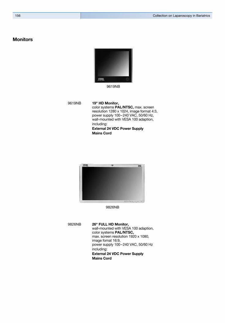
Collection on Laparoscopy in Bariatrics156
9826NB
9826NB 26" FULL HD Monitor, wall-mounted with VESA 100 adaption, color systems PAL/NTSC, max. screen resolution 1920 x 1080, image fomat 16:9, power supply 100 – 240 VAC, 50/60 Hzincluding:External 24 VDC Power SupplyMains Cord
9619NB
9619NB 19" HD Monitor, color systems PAL/NTSC, max. screen resolution 1280 x 1024, image format 4:3, power supply 100 – 240 VAC, 50/60 Hz, wall-mounted with VESA 100 adaption,including:
External 24 VDC Power SupplyMains Cord
Monitors

157Instruments and Videoendoscopic Equipment
Monitors
Optional accessories:9826SF Pedestal, for monitor 9826NB9626SF Pedestal, for monitor 9619NB
26"
9826NB
l
–
l
l
l
l
l
–
l
–
l
l
l
l
l
l
19"
9619NB
l
–
–
l
l
l
l
l
l
l
–
l
l
l
l
l
KARL STORZ HD and FULL HD Monitors
Wall-mounted with VESA 100 adaption
Inputs:
DVI-D
Fibre Optic
3G-SDI
RGBS (VGA)
S-Video
Composite/FBAS
Outputs:
DVI-D
S-Video
Composite/FBAS
RGBS (VGA)
3G-SDI
Signal Format Display:
4:3
5:4
16:9
Picture-in-Picture
PAL/NTSC compatible
19"
optional
9619NB
200 cd/m2 (typ)
178° vertical
0.29 mm
5 ms
700:1
100 mm VESA
7.6 kg
28 W
0 – 40°C
-20 – 60°C
max. 85%
469.5 x 416 x 75.5 mm
100 – 240 VAC
EN 60601-1, protection class IPX0
Specifications:
KARL STORZ HD and FULL HD Monitors
Desktop with pedestal
Product no.
Brightness
Max. viewing angle
Pixel distance
Reaction time
Contrast ratio
Mount
Weight
Rated power
Operating conditions
Storage
Rel. humidity
Dimensions w x h x d
Power supply
Certified to
26"
optional
9826NB
500 cd/m2 (typ)
178° vertical
0.3 mm
8 ms
1400:1
100 mm VESA
7.7 kg
72 W
5 – 35°C
-20 – 60°C
max. 85%
643 x 396 x 87 mm
100 – 240 VAC
EN 60601-1, UL 60601-1, MDD93/42/EEC, protection class IPX2

Collection on Laparoscopy in Bariatrics158
Cold Light Fountains and Accessories
For use with telescopes, diameter 10 mm:495NE Fiber Optic Light Cable,
with straight connector, diameter 4.8 mm, length 300 cm
For use with telescopes, diameter 5 mm:495NL Fiber Optic Light Cable,
with straight connector, diameter 3.5 mm, length 180 cm
20134001 Cold Light Fountain XENON NOVA® 300, power supply: 100–125VAC/220–240VAC, 50/60 Hz
including: Mains Cord
20132028 XENON Spare Lamp, only, 300 watt, 15 volt
Cold Light Fountain XENON NOVA® 300
20 133101-1 Cold Light Fountain XENON 300 SCB
with built-in antifog air-pump, and integrated KARL STORZ Communication Bus System SCB power supply: 100 –125 VAC/220 –240 VAC, 50/60 Hz
including: Mains Cord Silicone Tubing Set, autoclavable, length 250 cm SCB Connecting Cord, length 100 cm20133027 Spare Lamp Module XENON
with heat sink, 300 watt, 15 volt20133028 XENON Spare Lamp, only,
300 watt, 15 volt
Cold Light Fountain XENON 300 SCB

159Instruments and Videoendoscopic Equipment
ENDOFLATOR® 40 with KARL STORZ SCBwith High Flow Insufflation (40 l/min.)
UI400S1 ENDOFLATOR® 40 SCB, Set, with integrated SCB module, power supply 100 – 240 VAC, 50/60 Hz
including: ENDOFLATOR® 40 Mains Cord, length 300 cm SCB Connecting Cable, length 100 cm Universal Wrench Insufflation Tubing Set, with gas filter, sterile,
for single use, package of 5 *
Subject to the customer’s application-specific requirements additional accessories must be ordered separately.
*
HAMOU® ENDOMAT® with KARL STORZ SCBSuction and Irrigation System
26 3311 01-1 HAMOU® ENDOMAT® SCB, power supply 100 – 240 VAC, 50/60 Hz
including: Mains Cord 5x HYST Tubing Set*, for single use 5x LAP Tubing Set*, for single use SCB Connecting Cable, length 100 cm VACUsafe Promotion Pack Suction*, 2 l
Subject to the customer’s application-specific requirements additional accessories must be ordered separately.
*

Collection on Laparoscopy in Bariatrics160
Notes:


WITH COMPLIMENTS OFKARL STORZ––ENDOSKOPE