lectins: function, structure, biological properties and · universidade federal rural do...
TRANSCRIPT

Lectins: Function, structure, biological properties and potential applications
ABSTRACT Lectins are a special class of proteins widely distributed in nature, which selectively recognize and reversibly bind to carbohydrates and glycoconjugates through their binding sites. These proteins, which can be detected through haemagglutination assays, interact with different carbohydrates present in cell surfaces. Lectins are generally classified according to their structure, specificity for carbohydrates and species location. Depending on their properties and distribution in tissues, lectins can play important physiological roles. The characteristic property of lectins to recognize other molecules in a distinct way makes it relevant in research involving purification, structural analysis, in vitro/in vivo applications of these macromolecules and biotechnological uses in different areas such as molecular and cell biology, immunology, pharmacology, medicine, clinical analysis, nanotechnology as well as in systems for drug release. Lectins can be used for analysis of structure and physiology of cells, tissues and pathogenic microorganisms. In agriculture, these proteins are used as insecticidal agents. Lectins have already been shown to exhibit different biological activities and effects, such as mitogenic and antiproliferative activities
on cell lines of human cancer, inhibition of bacterial and fungal growth, action as promoting agents in cell aggregation, immunomodulatory activities and toxic effects. These proteins are promising as drugs for treatment and in diagnosis of human diseases; they are important tools in cytochemistry, histochemistry and immunohisto-chemistry and are also useful in forensic medicine. In summary, this review provides an overview of lectin research, with focus on physiological functions, structural performance, classification, potential biotechnological properties and applications. KEYWORDS: lectins, physiological functions, biotechnological applications 1. Introduction
1.1. Lectins: carbohydrate ligant proteins Lectins are proteins of non-immune origin with wide distribution in nature, which recognize and reversibly bind to carbohydrates and glycoconjugates, and are free or linked to cell surfaces through specific binding sites [1]. The ability to agglutinate cells distinguishes these proteins from other macromolecules able to bind carbohydrates. In addition, their non-immune origin differentiates them from anti-carbohydrate immunoglobulins that agglutinate cells. While antibodies are structurally similar, lectins differ in amino acid
1CEB - Centre of Biological Engineering, University of Minho, 4710-057 Braga, Portugal. 2Departamento de Ciências Animais, Laboratório de Biologia Celular e Molecular, Universidade Federal Rural do Semi-Árido (UFERSA), 59625-900 Mossoró, RN, Brazil; 3Departamento de Bioquímica, Laboratório de Bioquímica de Proteínas, Universidade Federal de Pernambuco (UFPE), 50670-901 Recife, PE, Brazil.
A. F. S. Santos1, M. D. C. da Silva2, T. H. Napoleão3, P. M. G. Paiva3, M. T. S. Correia3 and L. C. B. B. Coelho3,*
*Corresponding author: [email protected]
Current Topics in Peptide & Protein R e s e a r c h
Vol. 15, 2014

composition, involvement of metals, three-dimensional structure and molecular weight. Moreover, lectins exist in animals as well as in organisms without immune systems, such as plants and bacteria. The first lectins discovered in plants were ricin (present in extracts of Ricinus communis) and abrin (obtained from extracts of Abrus precatorius), which are capable of agglutinating blood cells and are ribosome-inactivating proteins (RIPs) [2, 3], consisting of heterodimeric proteins with two peptide chains held together by a disulfide bridge [4]. Lectins able to agglutinate erythrocytes were previously called (phyto-)haemaglutinins, phytoaglutinins or plant aglutinins [5]. Boyd and Shapleigh [6] used the term lectin (originated from the Latin “lectus”, meaning “selected”) to designate the class of proteins which shows common characteristics of selectivity in the interaction with carbohydrates. This term was generalized by Sharon and Lis [7] to include all proteins from variable sources, non-immune origin and able to bind carbohydrates, with or without specificity for blood type erythrocytes. In 1980, lectins were designated by Goldstein et al. [8] as (glyco-)proteins of non-immune origin, showing two or more binding sites able to reversibly interact with carbohydrates, precipitate glycoconjugates and agglutinate animal or plant cells. Investigations of lectins have indicated their presence in animals, lichens, and bacteria as well as higher fungi [9]. Physiological functions suggested for many lectins are based on their general properties and locations in different tissues. The existence of binding sites for specific carbohydrates, the major characteristic of lectins, is undoubtedly an important factor for the determination of their physiological role.
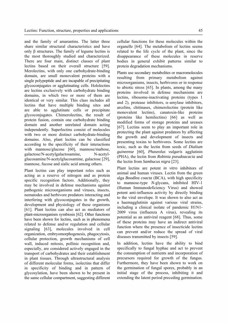
1.2. Detection and specificity The lectins are, in most cases, di- or multivalent and able to interact with carbohydrates or glycoproteins in solution or linked to cell membranes and their binding sites interact with cells forming various reversible linkages. Because of this ability, lectins are easily detected through agglutination assays. The haemagglutination assay (Figure 1A) allows for easy viewing of the agglutinating
42 A. F. S. Santos et al.
erythrocyte property of lectins. The use of erythrocytes from different human or other animal origin, treated enzymatically [10, 11], chemically [12, 13] as well as untreated [12, 14] has revealed that lectins may be specific to different erythrocytes. The lectin from Cordyceps militaris fungus agglutinates erythrocytes of mice and rats but is not able to agglutinate ABO group cells [11]; lectin from Gracilaria ornata agglutinates animal erythrocytes (rabbit and chicken) but not human erythrocytes [10]. Phthirusa pyrifolia leaf lectin (PpyLL) was not specific to human erythrocytes (types A, B, and O), and no haemagglutinating activity (HA) was detected with human AB erythrocytes; also, this lectin showed high affinity to rabbit erythrocytes while not agglutinating chicken or rat cells [15]. Other lectins which are non-specific to blood groups are the coagulant Moringa oleifera seed lectin, cMoL [12] and Crataeva tapia bark lectin, CrataBL [13]. The inhibition of haemagglutination (Figure 1B) in the presence of carbohydrate(s) confirms lectin detection indicated by previous haemagglutination assays; in addition, lectin sample evaluations use assays of polysaccharides or glycoprotein precipitation [16]. Wang et al. [17] showed another strategy to detect specific carbohydrate binding ability of lectins where an enzyme-linked adsorbent assay applied different monosaccharide-polyacrylamide conjugates as capturing agents for screening lectins in biological samples. This assay, more sensitive than the conventional haemagglutinating assay, provided an alternative and efficacious detection for specific carbohydrate- binding substances. Lectins are specific to monosaccharides or oligosaccharides. However, some lectin inhibition occurs only with oligosaccharides [18], glycoproteins and/or polysaccharides [19]. Many lectins can be inhibited by mono- or disaccharides, such as the lectin from eggs of Scomberomorous niphonius fish, a rhamnose binding lectin [20], lectins of the mushroom Agrocybe cylindracea, which are specific to lactose [21] and from Lyophyllum decastes with specificity for galabiose, a rare disaccharide in human tissues [16]. Generally, simple carbohydrates need higher concentrations to promote inhibition when compared to complex oligosaccharides. The phloem lectin of snake gourd
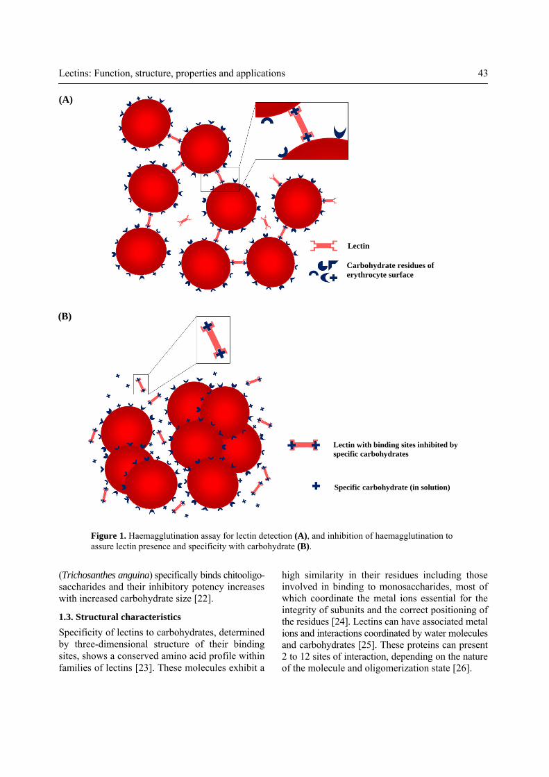
Lectin with binding sites inhibited by specific carbohydrates
Specific carbohydrate (in solution)
Lectin
Carbohydrate residues of erythrocyte surface
Lectins: Function, structure, properties and applications 43
high similarity in their residues including those involved in binding to monosaccharides, most of which coordinate the metal ions essential for the integrity of subunits and the correct positioning of the residues [24]. Lectins can have associated metal ions and interactions coordinated by water molecules and carbohydrates [25]. These proteins can present 2 to 12 sites of interaction, depending on the nature of the molecule and oligomerization state [26].
(Trichosanthes anguina) specifically binds chitooligo- saccharides and their inhibitory potency increases with increased carbohydrate size [22].
1.3. Structural characteristics Specificity of lectins to carbohydrates, determined by three-dimensional structure of their binding sites, shows a conserved amino acid profile within families of lectins [23]. These molecules exhibit a
(A)
(B)
Figure 1. Haemagglutination assay for lectin detection (A), and inhibition of haemagglutination to assure lectin presence and specificity with carbohydrate (B).

44 A. F. S. Santos et al.
binding of the complex. The specific (hydrogen bridges and electrostatic) or non-specific (van der Waals) interactions constitute the main contributions to the strength and stability of the molecular complex [28]. 2. Plant lectins Plants are a rich source of lectins and serve as the main source for isolation and analysis of these molecules. Lectins are present in various plant tissues and have different biological properties (Table 1). The major source of lectins is often seeds [29, 30, 31, 12, 32, 33, 34]; quiescent seeds of legumes are particularly rich in these proteins. Plant lectins have been purified from barks [35, 13], heartwood [36], leaves [37, 38, 15] and fruits [39]. Other lectin sources are roots [40], tubers [41], bulbs [42], rhizomes [43, 44, 45, 46], coleoptiles [47], cotyledons [48] and phloem exudate [49]. Plant lectins isolated from algae [10, 50], lichens and ferns have distinct properties [51, 52, 53, 54]. Plant lectins have been classified into seven families of proteins which are related structurally and evolutionarily. These are lectins of phloem from Cucurbitaceae [22], chitin-binding lectins containing domains of hevein, legume lectins [56], ribosome-inactivating protein type 2 [57], mannose-binding lectins of monocots, jacalin-related lectins [58]
Structural differences occur in lectins from the primary structure to the last degree of molecular organization; they may differ in amino acid sequence, change in the number of subunits, and in the nature of the polypeptides. Interactions between the subunits seem to play a dominant role in the stability of these proteins [27]. The specificities and affinities of the associated sites are achieved mainly by hydrogen bridges, including the aid of van der Waals and hydrophobic interactions with residues of aromatic amino acids that are close to the hydrophobic portions of monosaccharides [24], which contribute to the stability and specificity of the complexes formed. Simulations of molecular dynamics have shown lectins to be structurally flexible to experimental differences in the analysis of adaptable molecules. The presence of ions in the environment may determine whether there is an interaction between lectin and carbohydrate and if this interaction is improved or not by a specific ion. In addition, the concentration of ions may provide for a meeting of carbohydrate monomers, forming micelles in the hydrophobic sites of lectins. The complexity of molecule binding is stabilized by many or at least one hydrogen bridge. These assays have demonstrated that specific and favorable electrostatic interactions among amino acid residues of the lectin and monomers of the ligand facilitate the Table 1. Lectins in different plant tissues with distinct biological activities.
Tissues Biological activities References
Seeds Anticoagulant and antiplatelet aggregating properties; coagulant, mitogenic, antibacterial, antifungal and antitumor activities [29, 31, 12, 32, 33, 34]
Bark Antifungal and insecticidal activities [35, 13]
Heartwood Termiticidal activity [36]
Stem Antiviral and apoptosis-inducing activities [55]
Leaves Antiviral, antibacterial and antifungal activities [37, 15]
Fruits Mitogenic and antiviral activities [39]
Roots Antifungal and termiticidal activities [40]
Tubers Insecticidal and antitumor activities [41]
Bulbs Proteolytic activity [42]
Rhizomes Antiproliferative, immunostimulatory, antiviral, antifungal, antitumor and apoptosis-inducing activities [43, 44, 45, 46]
Lectins: Function, structure, properties and applications 45
cellular functions for these molecules within the organelle [64]. The metabolism of lectins seems related to the life cycle of the plant, since the disappearance of these molecules in reserve bodies in general exhibit patterns similar to protein degradation mechanisms. Plants use secondary metabolites or macromolecules resulting from primary metabolism against microorganisms, insects, herbivores or in response to abiotic stress [65]. In plants, among the many proteins involved in defense mechanisms are lectins, ribosome-inactivating proteins (types 1 and 2), protease inhibitors, α-amylase inhibitors, arcelins, chitinases, chimerolectins (protein like monovalent lectins), canatoxin-like proteins (proteins like hemilectins) [66] as well as modified forms of storage proteins and ureases [67]. Lectins seem to play an important role in protecting the plant against predators by affecting the growth and development of insects and presenting toxins to herbivores. Some lectins are toxic, such as the lectin from seeds of Dialium guineense [60], Phaseolus vulgaris agglutinin (PHA), the lectin from Robinia pseudoacacia and the lectin from Sambucus nigra [23]. Plant lectins are potent in vitro inhibitors of animal and human viruses. Lectin from the green alga Boodlea coacta (BCA), with high specificity to mannose-type N-glycans, inhibited HIV-1 (Human Immunodeficiency Virus) and showed potent anti-influenza activity by directly binding to the viral envelope. It was shown to also act as a haemagglutinin against various viral strains, including a clinical isolate of pandemic H1N1-2009 virus (influenza A virus), revealing its potential as an antiviral reagent [68]. Thus, some of these proteins may have an indirect antiviral function where the presence of insecticide lectins can prevent and/or reduce the spread of viral diseases transmitted by insects [59]. In addition, lectins have the ability to bind specifically to fungal hyphae and act to prevent the consumption of nutrients and incorporation of precursors required for growth of the fungus. Furthermore, they have been shown to work on the germination of fungal spores, probably in an initial stage of the process, inhibiting it and extending the latent period preceding germination.
and the family of amarantins. The latter three share similar structural characteristics and have only β structures. The family of legume lectins is the most thoroughly studied and characterized. There are four main, distinct classes of plant lectins based on their overall structure [59]. Merolectins, with only one carbohydrate-binding domain, are small monovalent proteins with a single polypeptide and are incapable of precipitating glycoconjugates or agglutinating cells. Hololectins are lectins exclusively with carbohydrate binding domains, in which two or more of them are identical or very similar. This class includes all lectins that have multiple binding sites and are able to agglutinate cells or precipitate glycoconjugates. Chimerolectins, the result of protein fusion, contain one carbohydrate binding domain and another unrelated domain acting independently. Superlectins consist of molecules with two or more distinct carbohydrate-binding domains. Also, plant lectins can be classified according to the specificity of their interactions with mannose/glucose [60], mannose/maltose, galactose/N-acetylgalactosamine, N-acetyl-glucosamine/N-acetylglucosamine, galactose [29], mannose, fucose and sialic acid among others. Plant lectins can play important roles such as acting as a reserve of nitrogen and as protein specific recognition factors. Additionally, they may be involved in defense mechanisms against pathogenic microorganisms and viruses, insects, nematodes and herbivore predators interacting and interfering with glycoconjugates in the growth, development and physiology of these organisms [61]. Plant lectins can also act as mediators of plant-microorganism symbiosis [62]. Other functions have been shown for lectins, such as in phenomena related to defense and/or regulation and cellular signaling [63], molecules involved in cell organization, embryomorphogenesis, phagocytosis, cellular protection, growth mechanisms of cell wall, induced mitosis, pollinic recognition and, especially, are considered actively engaged in the transport of carbohydrates and their establishment in plant tissues. Through ultrastructural analysis of different molecular forms, isolectins that differ in specificity of binding and in pattern of glycosylation, have been shown to be present in the same cellular compartment, suggesting different
46 A. F. S. Santos et al.
phagocytosis of microorganisms), selectins (that mediate the adhesion of leukocytes to endothelial cells and carry them to lymphoid tissues and sites of inflammation), and secreted molecules in the extracellular matrix and serum. Rhamnose-binding lectin (RBL) is an animal lectin family proposed based on their characteristic sugar-binding specificity and their molecular structure, which consists of two to three homologous, tandemly repeated CRDs [94]. Lectins with specificity for methyl-α-D-mannopyranoside are present in Rachycentron canadum [95] and Oreochromis niloticus sera; this latter lectin has also shown specificity for D-mannose [33]. Mannose-binding lectin (MBL) is an important component of innate immunity capable of activating the lectin pathway of the complementary system. Zhang et al. [96] has isolated an MBL gene from channel catfish (Ictalurus punctatus). Animal lectins seem to be involved in the mechanisms of endocytosis, intracellular translocation of glycoproteins [97], binding to glycoconjugates [92], apoptosis processes [98], defense against microorganisms, regulating the processes of cell adhesion and migration as well as in binding of bacteria to epithelial cells [99]. Moreover, they have a function in the immune system of crustaceans [78], birds and mammals [100]. In vertebrates, the class of integral lectins of membranes appears to be involved with binding of glycoconjugates to membranes of the cell surface or vesicles, resulting in the location of glycoconjugates in specific sites (endocytosis) or in the transport of the same to other cellular compartments (intracellular translocation) [92]. Meanwhile, the class of soluble lectins moves in the aqueous compartments within and among cells, interacting with other soluble substances and binding to membrane glycoconjugates. The fact that these proteins seem to be initially concentrated within the cells, and secreted by them, suggests that they have a common function to bind to glycoconjugates present on and around cells [92]. Among C-type lectins, the most studied molecules are the mannose-specific lectins in the serum of mammals, which act against pathogens; they bind to oligomannosides of bacterial and fungal cell surfaces, neutralizing them by cell lysis or opsonization [24]. A class of lectins that
Several plant lectins show antifungal activity against pathogenic species that attack plants [69, 31]. A coagulant lectin isolated from M. oleifera seeds (cMoL) acted as an anticoagulant protein on in vitro samples of blood [70]. 3. Animal lectins The presence of lectins in invertebrate animals occurs in almost all phyla in the haemolymph and coelomic fluid. Many lectins have been isolated from invertebrates, such as protozoa [71, 72], insects [73], molluscs [74, 75], crustaceans [76, 77, 78], sea cucumbers [79], polychaetes [80] and sea sponges [81]. In vertebrates, lectins have been isolated and characterized from fish [82, 83], snakes [84, 85] and other animals. In humans in particular, there are many well-characterized lectins of different tissues and cells, such as lungs [86, 87], serum [88, 89] and dendrites [90, 91]. In vertebrates, there are two classes of lectins, based on their location: integral lectins of membranes and soluble lectins present in intra- and intercellular fluids [92]. Integral lectins are those that are located in membranes as structural components, and differ in specificity to carbohydrates as well as their physical and chemical properties. Soluble lectins can move freely in the intra- and intercellular environments. Gabius [93] divided animal lectins into five main groups according to their structural characters: C-type lectins, I-type lectins, galectin (or S-type), pentranxins and P-type lectins. C-type lectins are those that depend on the presence of Ca2+ ions to bind carbohydrates, show different specificities and have conserved carbohydrate recognition domains. I-type lectins are those that have a carbohydrate recognition domain (CRD) similar to immuno-globulins. Galectins or S-type lectins are thiol-dependent, also have a CRD and are specific to β-galactosides. Pentranxins are constituted by many subunits producing pentameric lectins. P-type lectins are those consisting of a similar, but not well defined, CRD and are specific to glycoproteins containing mannose 6-phosphate. Sharon [24] includes in the group of C-type lectins, endocytic receptors (such as liver lectins), macrophagic receptors (that can work in

Lectins: Function, structure, properties and applications 47
and cell adhesion [99]. Lectins of the bacterial surface appear to be involved in mediating the initiation of bacterial infection through adhesion to epithelial cells, as occurs in infections of the urinary and gastrointestinal tracts. Fungal lectins may be involved in cell wall biosynthesis, differentiation of mycelium, adhesion of spores from pathogenic species to insects and nematodes, recognition between fungi and mycoparasites, as well as acting as storage proteins and presenting pesticide activity [112]. Some authors have reported that lichen lectins of fungal origin can have an important role in the establishment of symbiosis, as a recognition factor between species serving as a factor of interspecific recognition [113]. 5. Lectins and plant evolution Among the arguments indicating that lectins have a role in plant defense, the most important is the ability of these molecules to link to glycoconjugates of other organisms. In general, these molecules bind to simple carbohydrates, but they have a very high affinity for oligosaccharides that are unusual or totally absent in plants. Examples of these proteins are chitin-binding plant lectins, such as the lectin from Viscum album [114], and lectins that bind to sialic acid. Chitin-binding lectin recognizes the typical cell wall of fungi and exoskeleton of invertebrates; sialic acid ligand lectins recognize a carbohydrate that is absent in plants but is the main component of animal glycoproteins. Besides this binding ability, plant lectins have high stability, even under unfavorable conditions such as changes in pH, temperature or exposure to proteases of insects and animals. These molecules seem to be preferentially associated with certain parts of plants that are more susceptible to attack by other organisms (generally storage organs and seeds) [59]. Lectins, especially those of seeds and other storage tissues, appear to play a role as reserve proteins due to their biochemical properties, abundance and role in the regulation of plant development. However, these proteins seem to play a dual role: they act as defense proteins and are toxic to various predators, while they also store nitrogen. The different specificities of lectins to bind carbohydrates and recognize a wide variety of molecules present in
act as receptors, called CTLR, function as signaling molecules of the cell surface and are able to recognize a range of highly conserved molecules of pathogens and to stimulate an appropriate immune response [101]. Other organisms such as insects and protozoa (invertebrate animals) have lectins in their cell structures; in some protozoa, such proteins appear to be related to the mechanism of recognition of parasitized hosts by these animals. Immunohistochemical and immuno- cytochemical assays with tissues from cattle to detect binding sites of lectins from Tritrichomonas sp. have confirmed the lectin as a mediator of the adhesion of these parasites to host tissues [102]. 4. Lectins from fungi, bacteria and viruses Fungal lectins from species producing fruiting bodies or mushrooms have been isolated and characterized, and they presented different biological properties [16, 11, 19, 103, 21, 18, 104, 14]. In addition to fungi or, more rarely, yeasts [105], some bacteria and cyanobacteria have been shown to be sources of lectins [106]; these proteins can also be found in viruses [107]. The opportunistic human pathogen Pseudomonas aeruginosa produces two lectins, which bind to galactose- and fucose/mannose-containing glycoconjugates. [106]. The A33 protein from the Vaccinia virus, which is a type II membrane protein found in the outer envelope of extracellular and cell-associated virus particles, evolved from a C-type lectin-like protein [107]. A fungal lectin from Psathyrella velutina, called integrin-type lectin, showed structural similarities with the extracellular domain of integrin class proteins involved in cell adhesion [108]. Fungal lectins, as in animals, are classified according to their structure, such as the lectin from Coprinopsis cinerea, related to galectins, a lectin class characterized by many conserved residues [18]. Finally, fungal lectins, as with plant lectins, can be classified according to their binding specificity: to mannose [14], arabinose [109], N-acetyl-D-galactosamine [110], melibiose, xylose [111], or lactose [21]. Lectins in microorganisms appear to play several important roles, like the interaction of host cells, recognition in immunological processes, phagocytosis

48 A. F. S. Santos et al.
Therefore, the differential binding of free serum PSA to M. amurensis agglutinin lectin between prostate cancer and benign prostate hypertrophy could be a potential measure for diagnosis of prostate cancer. Affinity matrices of seed lectins from C. mollis were able to isolate glycoproteins from complex protein mixtures [122]. A lectin from Crataeva tapia bark improved tissue damage and plasma hyperglycemia in alloxan-induced diabetic mice [123].
6.1. Lectins against microorganisms Lectins, especially those with specificity to mannose or N-acetyl-glucosamine, have a remarkable anti-HIV (Human Immunodeficiency Virus) activity as shown in cell culture assays [80], not only inhibiting cell infection but also preventing the spread of viral infection from infected cells to uninfected T lymphocytes [26]. Some lectins can also act by inhibiting the activity of HIV-1 reverse transcriptase [39, 111, 103]. Different lectins have shown activity against bacteria [124, 125, 75, 126], protozoa [81], fungi [127, 69, 31] and nematodes [61], indicating that these proteins can be valuable in future clinical treatments. Antimicrobial proteins attract the interest of many researchers for their potential as tools against fungal and bacterial infections in plant or human pathologies. Such proteins, once incorporated into transgenic plants, promote resistance against pathogens; also, lectins can serve as active principles of antifungal or antibacterial clinical use. Antifungal proteins or peptides are ubiquitous in nature and are expressed in different structures or organs of many plants and animals [128]. Among several antifungal protein classes are lectins [69, 31], such as the ribosome-inactivating proteins. Antibacterial proteins or peptides found in different organisms act as enzymes or represent many protein groups such as C-type lectins present in shrimp and scallops [129, 77] and plant lectins [39]. The broad range of lectins on the market and the possibility of isolating new, biologically active molecules with potent action in a wide spectrum of applications have resulted in lectins being
microorganisms and other animals as well as their dual role in nitrogen storage and protection against predators are characteristics resulting from the large adaptive evolution of plants [23]. A structural study of the lectin from Parkia platycephala seeds showed a circular arrangement of β-prism-type domains, conferring adaptability to the molecule; the carbohydrate binding is flexible and the cyclic structure of the molecule indicates convergent evolution in the construction of related lectins with functions in host defense against pathogens and predators [115]. 6. Biological properties and applications Lectins stand out in biotechnology due to their many applications and other potential uses that are being evaluated daily and studied by researchers in lectinology. Lectins and their characteristic properties, mainly due to their ability to bind glycoconjugates, stand out as important tools in research covering various areas of science, especially in biochemistry, cellular and molecular biology, immunology, pharmacology, medicine and clinical analysis. Lectins have a variety of effects on cells, such as agglutination, mitogenic stimulation, redistribution of cell surface components, modifying the activity of membrane enzymes, inhibition of bacterial and fungal growth, cell aggregation, toxicity, immunomodulation, among others [41, 31, 103, 75, 116, 117]. Based on the carbohydrate, specific binding lectins can recognize tissues and aid in the diagnosis of diseases. Cratylia mollis seed lectin is a versatile tool for biomedical studies [118, 119]. The lectin is able to detect dengue glycoproteins in clinical samples [120]. Ohyama et al. [121] studied different lectins that were tested for their binding to carbohydrates on serum prostate-specific antigen (PSA); they found that such lectins have a predominant core structure of N-glycans with the sialic acid alpha2-3 galactose linkage as an additional terminal carbohydrate. Among the lectins examined, the M. amurensis agglutinin-bound fraction of free serum PSA increased in prostate cancer patients compared to patients with benign prostate hypertrophy. The binding of PSA to M. amurensis agglutinin, which recognizes alpha2,3-linked sialic acid, was also confirmed by surface plasmon resonance analysis.

Lectins: Function, structure, properties and applications 49
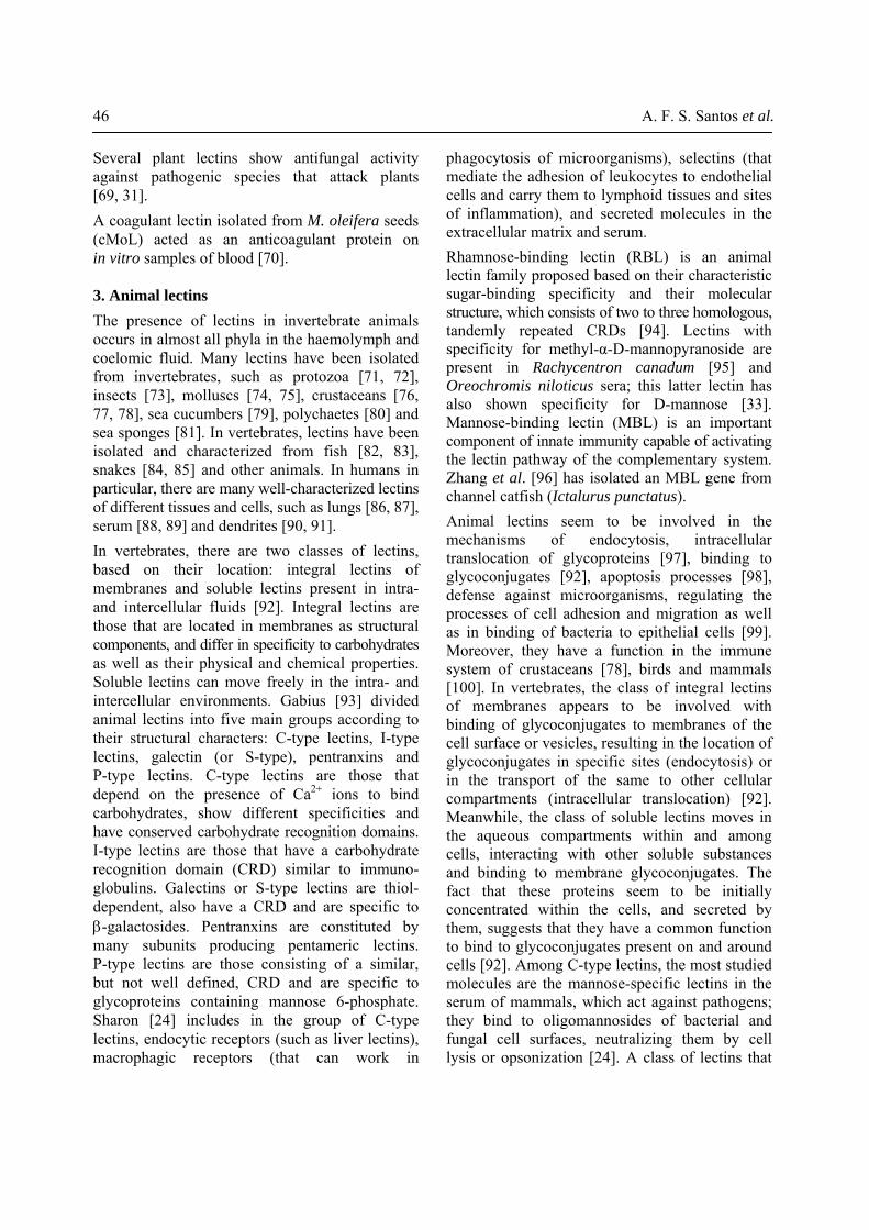
lines Mo-T, Jurkat [104], L1210, M1, hepatoma cells (Hep G2) [132] and breast cancer [34]. Mitogenic activity is a common property of many lectins, especially those of fungal origin, acting on splenocytes [103, 133, 111, 39, 134, 132] and human T lymphocytes [74, 135]. BlL, a galactose-binding C-type lectin purified from Bothrops leucurus snake venom, exhibits anticancer activity by inducing melanoma cell death [85]. A lectin from C. mollis seeds induced a transition in membrane permeability [136]; a gel formulation used the lectin to evaluate bone repair in rats [137]. The healing process restores the anatomical and functional integrity of tissues; wound healing is a complex series of biochemical and cellular events in response to tissue damage. A wound constitutes a continuous anatomic-physiological solution from the lesion area and a scar corresponds to the biological attempt from the organism to restore its integrity. This is known as the cicatricial process, which is divided into three phases: inflammatory, proliferative and maturation. Such phases are not isolated processes and frequently overlap one another. The inflammatory phase begins immediately after a lesion develops and is characterized by vasoconstriction mediated by neurogenic and chemical factors during a period from seconds to minutes. This is followed by local vasodilation, which through increasing vascular permeability, results in an influx blood flow to the area, bringing with it seric proteins, coagulation factors and platelets. The increase of extravascular liquid, characterized by elevation of oncotic pression in the lesion area, is called edema and its clinical appearance is swelling or inflammation. Besides inflammation, blushing, pain and increase of temperature occur in lesion. The proliferative phase or fibroplasia, characterized by tissue formation, induces reepithelization and wound contraction. The tissue formed has a humid aspect, is contractile (due to the presence of fibroblasts), and fills the space left by the wound, acting as a barrier to microorganisms. Reepithelization starts by migration of basal epithelial cells (keratinocytes), of centripetal shape, from the lesion border; the composition of the extracellular matrix is gradually substituted by a collagenous matrix. The maturation phase or wound contraction is marked by remodelling of
continuously evaluated with regard to various biological properties, among them, against micro-organisms. A type of pathogenic fungus extremely important in agriculture is the genus Fusarium. Some purified plant lectins have antifungal properties against members of this genus [130]. Lectins isolated from seeds of Pisum sativum [31] and Astragalus mongholicus [127] and another lectin isolated from Sebastiania jacobinensis bark [35] are inhibitors of F. oxysporum. Two chitin-binding lectins purified from Artocarpus heterophyllus and A. altilis inhibited F. moniliforme [69]; the lectin from S. jacobinensis bark [35] also inhibited this species of fungus. Souza et al. [40] studied a lectin from Bauhinia monandra secondary roots that showed antifungal activity against phytopathogenic species of Fusarium and was more active on F. solani. Lectins with antifungal activity are mainly chitin-binding lectins; these proteins are able to bind chitin from the fungal cell wall [130]. There is a growing interest in evaluating the effect of lectins against bacteria of medical importance. Such lectins are able to interact and inhibit the development of microbial cells as lectins recognize carbohydrates or glycoconjugates in cell surfaces, according to their specificity. Lectins from different sources can act as antibacterial agents against Gram-positive and Gram-negative bacteria, such as a lectin from the marine species Holothuria scabra [125], the lectin from Araucaria angustifolia seeds [124] and C-type lectins from chicken and goose eggs (both active against B. subtilis, S. aureus and P. aeruginosa) [126]. The lichen lectin from Cladonia verticillaris (ClaveLL) has shown antibacterial activity against Gram-positive (Bacillus subtilis, Staphylococcus aureus and Enterococcus faecalis) and Gram-negative (Escherichia coli and Klebsiella pneumoniae) strains as well as antifungal activity against Trichophyton rubrum [54]. Athamna et al. [131] have shown that plant lectins are capable of differentiating mycobacterial species that are potential pathogens.
6.2. Lectins and cells Lectins show mitogenic activity in mononuclear blood cells and antiproliferative activity in cell lines of human cancer [43, 41], such as leukemia

50 A. F. S. Santos et al.
drugs [146]; these molecules also act in systems for drug release. Bauhinia monandra leaf lectin (BmoLL) and Lens culinaris lectin (LCL) incorporated and adsorbed on the surface of nanoparticles are potential tools as oral drugs with controlled release [147]. Production and use of intelligent drugs aiming at more target-specific and less aggressive therapeutic treatment for patients with cancer is of great interest to the pharmaceutical industry and medicine.
6.3. Lectins and insects Lectins, beyond their effects on cells, also have the ability to act as insecticidal molecules against a variety of species [148, 10, 41, 36, 51]. In addition, their genes can produce transgenic plants of economic interest [149]. A lectin from Galanthus nivalis, GNA, was fused to an insect-specific, spider neurotoxin, SFI1 (Segestria Florentine toxin 1). The lectin acted as a carrier to the neurotoxin, leading it to the haemolymph of Lepidoptera larvae; the fusion of SFI1/GNA showed the potential use of the lectin as a pesticide in the cultivation of plants [150]. Several lectins have revealed deleterious effects against different life stages (larva [151, 152, 10], nymph [153] and adult [154]) and on oviposition [155] of insects [156]. These effects indicate insecticidal activity against Coleoptera [155, 10], Homoptera [148], Hemiptera and/or Diptera [43, 154], Lepidoptera [151, 152] and/or Hymenoptera [157] besides other orders of representative insects. Two reports of lectin toxicity with lethal effects on an insect of the Isoptera order (Nasutitermes corniger) were evaluated [36, 51]. Both M. urundeuva heartwood lectin (MuHL) and Cladonia verticillaris lichen lectin (ClaveLL) showed termiticidal effects yet yielded different LC50 values for soldier and worker castes. While ClaveLL was more effective against workers, MuHL was more toxic to soldiers. Both ClaveLL and MuHL did not show repellent properties [36, 51]. The mechanisms of insecticidal action of lectins are unknown, although entomotoxic activity seems to depend on the carbohydrate recognition property that they exhibit [156]. In general, lectins able to bind N-acetyl-D-glucosamine with affinity to chitin have insecticidal properties. However, plant lectins with different specificities have also shown
tissues in a sense to restore the shape and function of injured tissue. Deposition, grouping and remodelling of collagen (initially of type III, but which by degradation is substituted by type I collagen) associated with endothelial regression and the reappearance of connective tissue, collagen fibers and a few blood vessels, are characteristics of matured healing of the damaged tissue. Figure 2 contains a schematic representation of different phases from the cicatricial process of wounds in mice, in its histopathological and macroscopic perspective. These wounds were treated with isoforms from C. mollis seed lectin (Cramoll 1,4) at the second (inflammatory phase), seventh (prolipherative phase) and twelfth (maturation phase) days of treatments. Besides Cramoll 1,4, lectins from seeds of Eugenia malaccensis and Parkia pendula have been evaluated in relation to their efficacy in the cicatricial process [138, 139, 32]. The antitumor action of lectins has been observed in sarcoma 180 [103, 140] and in some cell lines of human tumors [141]. A modulating effect on the immune response is a property observed in some lectins [142, 143]. Other lectins, in particular plant lectins, and especially RIPs type 2, may have cytotoxic activity in vitro, inducing apoptosis [57]. Bauhinia forficata lectin from seeds shows selective cytotoxic effect and adhesion inhibition in MCF7 breast cancer cells [34]. Examples of commercial lectins with cytotoxic action in mammalian cells are ricin, abrin and, of lesser intensity, concanavalin A, and wheat germ agglutinin (WGA) among others [144]. The toxic effect of lectins on cells is usually selective; they are much more active in transformed cells, which are more sensitive to lectin effects compared to normal cells. A lectin isolated from Viscum album showed a cytotoxic property in Molt4 human cells, a cell line derived from T cells of leukemia [114]. A protein from the seeds of Pouteria torta (pouterin), with lectin-like activity, induced tumor necrosis factor receptor 1 (TNFR1)-mediated apoptosis in HeLa cells from human cervical cancer [144]. A mannose-binding lectin from Sophora flavescensa is a potent anti-tumor agent with cytotoxic and apoptotic effects on HeLa cells [145].Some lectins have the ability to mediate mucoadhesion, cytoadhesion and cytoinvasion of

lectins, they may decrease food intake through binding to the midgut epithelium or to the peritrophic matrix [161]. They could cross the midgut epithelial barrier and pass into the insect circulatory system resulting in a toxic action interfering with endogenous lectins involved in self-defense which are present in the haemolymph [162]. Alternatively, they may be internalized by endocytotic vesicles into the epithelial cells and block nuclear localization and sequence-dependent nuclear protein importation, inhibiting cell proliferation [163]. Biotechnological and industrial interest is growing in the discovery of new natural pesticides to promote biological control that prevent undesirable environmental impacts [164, 165, 166]. In this context, genes coding for inhibitors of digestive enzymes, amylase inhibitors, peptides, lectins and other molecules can produce transgenic plants with resistance to microorganisms and insects [149, 148]. Genes of new lectins with defense properties are being widely incorporated into transgenics as an alternative to check resistance in vegetables of economic interest and can produce plants less susceptible to microrganism attacks. These lectins are biotechnological strategies for the preservation of endangered species of economic importance, through incorporation of their genes into transgenic plants to serve in the control of pests.
6.4. Lectins and cytochemistry/histochemistry Detection of carbohydrades by lectins involves their binding to glycoconjugates in cells, tissue sections or free carbohydrate/glycoconjugates. The binding is detected by techniques of direct visualization using labelled lectins [167] or by an indirect way through the application of immunological techniques. Radio-marked lectins and conjugated lectins are sensitive and specific reagents for the detection of glycoproteins and other glycoconjugates [168, 169]. Lectins are cytochemical, histochemical or immunohistochemical tools for locating glycoconjugates in different animal tissues [170] and for the detection and characterization of glycosilated residues and different glycoconjugates present in human or animal cells and tissue surfaces [171, 168, 169, 172]. These proteins
ability as insecticidal molecules, suggesting that this property is not exclusive to N-acetyl-D-glucosamine-specific lectins [155, 10]. For example, a Xerocomus chrysenteron (mushroom) lectin with affinity for D-galactose and lactose and, to a lesser extent, N-acetyl-D-galactosamine had an effect on Drosophila melanogaster and pea aphid Acyrthosiphon pisum [112]. A lectin from Gracilaria ornata, named GOL, which is only inhibited by some glycoproteins, caused a significant reduction in larval survival; also, GOL significantly affected the percentage of adult emergence [10]. In other work, Sadeghi et al. [155] used 14 plant lectins (of four lectin families) with several specificities; they showed that all lectins had deterrent effects against oviposition of C. maculatus adults. Talisia esculenta lectin (TEL), isolated from seeds with chitin-binding properties and inhibited by mannose and glucose, produced elevated mortality in Callosobruchus maculatus and Zabrotes subfasciatus larvae [158]. Lectin effects such as repellence or toxicity against insects are, according to the most widely used methods, generally based on bioassays with lectin incorporation into the experimental environment [159] or into artificial diets offered to target insects [160]. Lectin toxicity seems to depend upon the necessary resistance of the molecule against assimilatory proteins and proteolytic degradation by digestive enzymes and on binding to insect gut structures, in the peritrophic membrane and/or chitinous structures in the midgut region, thereby inhibiting digestion and absorption and causing nutritional deprivation [151, 152, 10]. According to Macedo et al. [152], BmoLL is able to bind to chitin as well as to midgut extracts and membrane proteins of Callosobruchus maculatus larvae. Insecticidal activity may involve binding to chitin components, interactions with glycoconjugates on epithelial cell surfaces, binding to glycosylated digestive enzymes and/or assimilatory proteins, resistance against enzymatic digestion and decrease of the α-amylase activity in the midgut of insects. The mechanism of action of insecticidal lectins is still unknown, but probably has different modes of action according to cell localization [154]. Some mechanisms of lectin action promote insect mortality. According to subcellular targets of
Lectins: Function, structure, properties and applications 51

52 A. F. S. Santos et al.
Figure 2
Reepithelization
Maturation Phase
Collagen Type III
Granulation Tissue Fibroblast
Inflammatory Phase
Proliferative Phase
Edema HeatRedness Pain
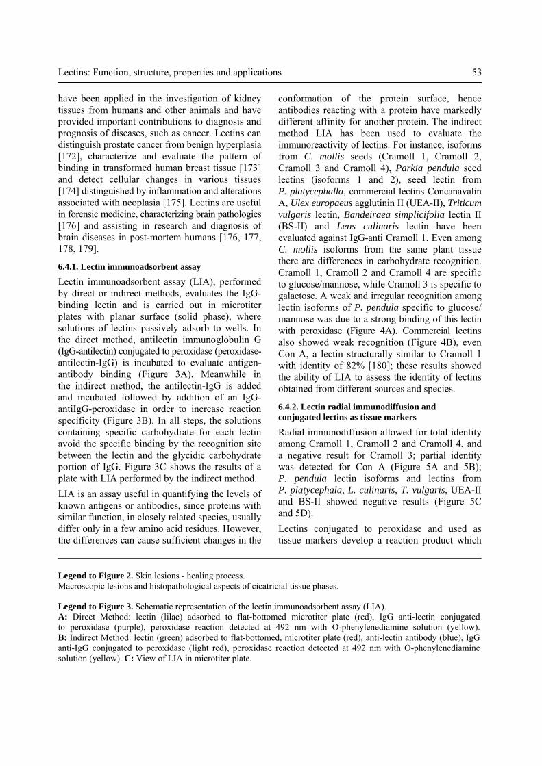
Figure 3
have been applied in the investigation of kidney tissues from humans and other animals and have provided important contributions to diagnosis and prognosis of diseases, such as cancer. Lectins can distinguish prostate cancer from benign hyperplasia [172], characterize and evaluate the pattern of binding in transformed human breast tissue [173] and detect cellular changes in various tissues [174] distinguished by inflammation and alterations associated with neoplasia [175]. Lectins are useful in forensic medicine, characterizing brain pathologies [176] and assisting in research and diagnosis of brain diseases in post-mortem humans [176, 177, 178, 179].
6.4.1. Lectin immunoadsorbent assay
Lectin immunoadsorbent assay (LIA), performed by direct or indirect methods, evaluates the IgG-binding lectin and is carried out in microtiter plates with planar surface (solid phase), where solutions of lectins passively adsorb to wells. In the direct method, antilectin immunoglobulin G (IgG-antilectin) conjugated to peroxidase (peroxidase- antilectin-IgG) is incubated to evaluate antigen-antibody binding (Figure 3A). Meanwhile in the indirect method, the antilectin-IgG is added and incubated followed by addition of an IgG-antiIgG-peroxidase in order to increase reaction specificity (Figure 3B). In all steps, the solutions containing specific carbohydrate for each lectin avoid the specific binding by the recognition site between the lectin and the glycidic carbohydrate portion of IgG. Figure 3C shows the results of a plate with LIA performed by the indirect method. LIA is an assay useful in quantifying the levels of known antigens or antibodies, since proteins with similar function, in closely related species, usually differ only in a few amino acid residues. However, the differences can cause sufficient changes in the
conformation of the protein surface, hence antibodies reacting with a protein have markedly different affinity for another protein. The indirect method LIA has been used to evaluate the immunoreactivity of lectins. For instance, isoforms from C. mollis seeds (Cramoll 1, Cramoll 2, Cramoll 3 and Cramoll 4), Parkia pendula seed lectins (isoforms 1 and 2), seed lectin from P. platycephalla, commercial lectins Concanavalin A, Ulex europaeus agglutinin II (UEA-II), Triticum vulgaris lectin, Bandeiraea simplicifolia lectin II (BS-II) and Lens culinaris lectin have been evaluated against IgG-anti Cramoll 1. Even among C. mollis isoforms from the same plant tissue there are differences in carbohydrate recognition. Cramoll 1, Cramoll 2 and Cramoll 4 are specific to glucose/mannose, while Cramoll 3 is specific to galactose. A weak and irregular recognition among lectin isoforms of P. pendula specific to glucose/ mannose was due to a strong binding of this lectin with peroxidase (Figure 4A). Commercial lectins also showed weak recognition (Figure 4B), even Con A, a lectin structurally similar to Cramoll 1 with identity of 82% [180]; these results showed the ability of LIA to assess the identity of lectins obtained from different sources and species.
6.4.2. Lectin radial immunodiffusion and conjugated lectins as tissue markers
Radial immunodiffusion allowed for total identity among Cramoll 1, Cramoll 2 and Cramoll 4, and a negative result for Cramoll 3; partial identity was detected for Con A (Figure 5A and 5B); P. pendula lectin isoforms and lectins from P. platycephala, L. culinaris, T. vulgaris, UEA-II and BS-II showed negative results (Figure 5C and 5D). Lectins conjugated to peroxidase and used as tissue markers develop a reaction product which
Lectins: Function, structure, properties and applications 53
Legend to Figure 2. Skin lesions - healing process. Macroscopic lesions and histopathological aspects of cicatricial tissue phases.
Legend to Figure 3. Schematic representation of the lectin immunoadsorbent assay (LIA). A: Direct Method: lectin (lilac) adsorbed to flat-bottomed microtiter plate (red), IgG anti-lectin conjugated to peroxidase (purple), peroxidase reaction detected at 492 nm with O-phenylenediamine solution (yellow). B: Indirect Method: lectin (green) adsorbed to flat-bottomed, microtiter plate (red), anti-lectin antibody (blue), IgG anti-IgG conjugated to peroxidase (light red), peroxidase reaction detected at 492 nm with O-phenylenediamine solution (yellow). C: View of LIA in microtiter plate.

54 A. F. S. Santos et al.
Figure 4. Lectin immunoadsorbent assay (LIA) by the indirect method using IgG-anti-Cramoll 1. Top, Cramoll 1, Cramoll 2, Cramoll 3 and Cramoll 4 (Cramoll: isoform lectin from Cratylia mollis seeds), Parkia pendula lectin isoforms 1 and 2, and P. platycephala lectin. Bottom, Con A: seed lectin from Canavalia ensiformis (concanavalin A); UAS-II: isolectin II from Ulex europaeus seeds (U. europaeus aglutinin); BS-II: isolectin II from Bandeiraea simplicifolia seeds and Lens culinaris lectin. A492: absorbance at 492 nm.
Figure 5. Radial immunodiffusion in agarose gel containing IgG-anti-Cramoll 1 in the central wells. A, Cramoll 1 (1), Cramoll 2 (2), Cramoll 3 (3) and Con A (4). B, Cramoll 1 (1), Cramoll 4 (2), Cramoll 2 (3) and Con A (4). C, Con A (1), isoform 1 from P. pendula seed lectin (2), isoform 1 from P. pendula seed lectin (3) and P. platycephalla seed lectin (4). D, Lens culinaris lectin (1), Triticum vulgaris lectin (2), Bandeiraea simplicifolia lectin II, BS-II (3) and Ulex europaeus agglutinin, UEA-II (4).

antimicrobial and insecticidal to mitogenic and antitumoral activities. Lectins have already been shown to have an important function in the cicatricial process from inflammatory lesions and are used in the diagnosis of diseases. This study is a reference for everyone with interest in the development and challenges of this important class of proteins. ACKNOWLEDGMENTS Authors acknowledge the Conselho Nacional de Desenvolvimento Científico e Tecnológico (CNPq) for fellowships to PMGP, MTSC and LCBBC. In addition, we are grateful to Grant SFRH/BPD/ 37349/2007 from the Foundation for Science and Technology and POPH/FSE, awarded to AFSS. CONFLICT OF INTEREST STATEMENT The authors declare that there are no conflicts of interest pertaining to the material in this manuscript.
can be evaluated by optical microscopy. Figure 6A shows a diagram of the steps used for studies of marking tissues. Staining intensity information from normal and transformed tissue cells confirm that differences are due to carbohydrate changes in tissue labelling evaluated through inhibition using carbohydrates specific to a particular lectin. Figure 6B shows a section of infiltrating ductal carcinoma tissue marked with Cramoll 1 conjugated to peroxidase; Figure 6C detected inhibition of the reaction using mannose, a carbohydrate specific to Cramoll 1. CONCLUSIONS This review provides an overview of current lectin research. The review is about the structural characteristics of these proteins, their distribution in nature with different biological applications and their role in plant evolution. Topics include different properties of these proteins, from
Lectins: Function, structure, properties and applications 55
Figure 6. Conjugated lectins as tissue markers. A, Schematic representation of tissue labelling using peroxidase conjugated lectin. B, Mammary tumor (infiltrating ductal carcinoma) labelled with a lectin (Cramoll 1) conjugated to peroxidase. C, Lectin (Cramoll 1) inhibition activity using a specific carbohydrate (mannose).
3. Bradberry, S. 2007, Medicine, 35, 576. 4. Bayer, H., Ey, N., Wattenberg, A., Voss,
C. and Berger, M. R. 2012, Protein Expres. Purif., 82, 97.
5. Sharon, N. and Lis, H. 1988, Trends Biochem. Sci., 12, 488.
6. Boyd, W. C. and Shapleigh, E. 1954, Science, 119, 419.
7. Sharon, N. and Lis, H. 1972, Science, 177, 949.
8. Goldstein, I. J., Hughes, R. C., Monsigny, M., Osawa, T. and Sharon, N. 1980, Nature, 285, 66.
9. Santos, A. F. S., Napoleão, T. H., Bezerra, R. F., Carvalho, E. V. M. M., Correia, M. T. S., Paiva, P. M. G. and Coelho, L. C. B. B. 2013, Advances in Medicine and Biology, L. V. Berhardt (Ed.), Nova Publishers Inc, New York, 63, 33.
10. Leite, Y. F. M. M., Silva, L. M. C. M., Amorim, R. C. N., Freire, E. A., Jorge, D. M. M., Grangeiro, T. B and Benevides, N. M. B. 2005, Biochim. Biophys. Acta, 1724, 137.
11. Jung, E. C., Kim, K. D., Bae, C. H., Kim, J. C., Kim, D. K. and Kim, H. H. 2007, Biochim. Biophys. Acta, 1770, 833.
12. Santos, A. F. S., Luz, L. A., Argolo, A. C. C., Teixeira, J. A., Paiva, P. M. G. and Coelho, L. C. B. B. 2009, Process Biochem., 44, 504.
13. Araújo, R. M. S., Ferreira, R. S., Napoleão, T. H., Carneiro-da-Cunha, M. G., Coelho, L. C. B. B., Correia, M. T. S., Oliva, M. L. V. and Paiva, P. M. G. 2012, Plant Sci., 183, 20.
14. Francis, F., Jaber, K., Colinet, F., Portetelle, D. and Haubruge, E. 2011, Fungal Biology, 115, 1093.
15. Costa, R. M. P. B., Vaz, A. F. M., Oliva, M. L. V., Coelho, L. C. B. B., Correia, M. T. S. and Carneiro-da-Cunha, M. G. 2010, Process Biochem., 45, 526.
16. Goldstein, I. J., Winter, H. C., Aurandt, J., Confer, L., Adamson, J. T., Hakansson, K. and Remmer, H. 2007, Arch. Biochem. Biophys., 467, 268.
17. Wang, T., Lee, M. and Su, N. 2009, Food Chem., 113, 1218.
ABBREVIATIONS A492 : Absorbance at 492 nm BCA : Lectin from the green alga Boodlea coacta BlL : Bothrops leucurus lectin BmoLL : Bauhinia monandra leaf lectin BS-II : Isolectin II from Bandeiraea simplicifolia seeds ClaveLL : Cladonia verticillaris lichen lectin cMoL : Coagulant Moringa oleifera seed lectin Con A : Seed lectin from Canavalia ensiformis (Concanavalin A) Cramoll : Isoform lectin from Cratylia mollis seeds CrataBL : Crataeva tapia bark lectin CRD : Carbohydrate recognition domain CTLR : Class of lectins that act as receptors GNA : Lectin from Galanthus nivalis GOL : Gracilaria ornata lectin HA : Haemagglutinating activity HIV : Human Immunodeficiency Virus H1N1-2009 virus: Influenza A virus IgG : Immunoglobulin G LCL : Lens culinaris lectin LIA : Lectin immunoadsorbent assay MBL : Mannose-binding lectin MuHL : Lectin from the M. urundeuva heartwood PHA : Phaseolus vulgaris agglutinin Pouterin : Protein from the seeds of Pouteria torta PpyLL : Phthirusa pyrifolia leaf lectin PSA : Prostate-specific antigen RBL : Rhamnose-binding lectin RIPs : Ribosome-inactivating proteins SFI1 : Segestria florentine toxin 1 TEL : Talisia esculenta lectin TNFR1 : Tumor necrosis factor receptor 1 UAS-II : Isolectin II from Ulex europaeus seeds UEA-II : Ulex europaeus agglutinin II WGA : Wheat germ agglutinin REFERENCES 1. Correia, M. T. S., Coelho, L. C. B. B. and
Paiva, P. M. G. 2008, Recent Trends Toxicology, 37, 47.
2. Olsnes, S. 2004, Toxicon, 44, 361.
56 A. F. S. Santos et al.

35. Vaz, A. F. M., Costa, R. M. P. B., Melo, A. M. M. A., Oliva, M. L. V., Santana, L. A., Silva-Lucca, R. A., Coelho, L. C. B. B. and Correia, M. T. S. 2010, Food Chem., 119, 1507.
36. Sá, R. A., Napoleão, T. H., Santos, N. D. L., Gomes, F. S., Albuquerque, A. C., Xavier, H. S., Coelho, L. C. B. B., Bieber, L. W. and Paiva, P. M. G. 2008, Int. Biodeter. Biodegr., 62, 460.
37. Ooi, L. S. M., Sun, S. S. M. and Ooi, V. E. C. 2004, Int. J. Biochem. Cell B, 36, 1440.
38. Rameshwaram, N. R. and Nadimpalli, S. K. 2008, J. Chromatogr. B, 861, 209.
39. Wang, H. and Ng, T. B. 2006, Plant Physiol. Bioch., 44, 181.
40. Souza, J. D., Silva, M. B. R., Argolo, A. C. C., Napoleão, T. H., Sá, R. A., Correia, M. T. S., Paiva, P. M. G., Silva, M. D. C. and Coelho, L. C. B. B. 2011, Int. Biodeter. Biodegr., 65, 696.
41. Kaur, M., Singh, K., Rup, P. J., Saxena, A. K., Khan, R. H., Ashraf, M. T., Kamboj, S. S. and Singh, J. 2006, Arch. Biochem. Biophys., 445, 156.
42. Parisi, M. G., Moreno, S. and Fernández, G. 2008, Plant Physiol. Bioch., 46, 403.
43. Kaur, A., Singh, J., Kamboj, S. S., Sexana, A. K., Pandita, R. M. and Shamnugavel, M. 2005, Phytochemistry, 66, 1933.
44. Chu, K. T. and Ng, T. B. 2006, Biochem. Bioph. Res. Co., 340, 118.
45. Yao, Q., Wu, C., Luo, P., Xiang, X., Liu, J., Mou, L. and Bao, J. 2010, Process Biochem., 45, 1477.
46. Shao, B., Wang, S., Zhou, J., Ke, L. and Rao, P. 2011, Process Biochem., 46, 1554.
47. Martinez, M. and Cordoba, F. 2000, Prep. Biochem. Biotech., 30, 199.
48. Oliveira, J. T. A., Melo, V. M. M., Câmara, M. F. L., Vasconcelos, I. M., Beltramini, L. M., Machado, O. L. T., Gomes, V. M., Pereira, S. P., Fernandes, C. F., Nunes, E. P., Capistrano, G. G. G. and Monteiro-Moreira, A. C. O. 2002, Phytochemistry, 61, 301.
49. Ota, E., Tsuchiya, W., Yamazaki, T., Nakamura, M., Hirayama, C. and Konno, K. 2013, Phytochemistry, 89, 15.
18. Wälti, M. A., Walser, P. J., Thore, S., Grünler, A., Bednar, M., Künzler, M. and Aebi, M. 2008, J. Mol. Biol., 379, 146.
19. Thakur, A., Rana, M., Lakhanpal, T. N., Ahmad, A. and Khan, M. I. 2007, BBA-Gen. Subjects, 1770, 1404.
20. Terada, T., Watanabe, Y., Tateno, H., Naganuma, T., Ogawa, T., Muramoto, K. and Kamiya, H. 2007, Biochim. Biophys. Acta, 1770, 617.
21. Liu, C., Zhao, X., Xu, X. C., Li, L. R., Liu, Y. H., Zhong, S. D. and Bao, J. K. 2008a, Int. J. Biol. Macromol., 42, 138.
22. Narahari, A., Nareddy, P. K. and Swamy, M. J. 2011, Biochimie, 93, 1676.
23. Peumans, W. J. and van Damme, E. J. M. 1998, Biotechnol. Genet. Eng., 15, 199.
24. Sharon, N. 1993, Trends Biochem. Sci., 18, 221.
25. Sharon, N. and Lis, H. 2002, J. Agr. Food Chem., 50, 6586.
26. Balzarini, J. 2006, Antivir. Res., 71, 237. 27. Mitra, N., Srinivas, V. R., Ramya, T. N.
C., Ahmad, N., Reddy, G. B. and Surolia, A. 2002, Biochemistry, 41, 9256.
28. Konidal, P. and Niemeyer, B. 2007, Biophys. Chem., 128, 215.
29. Wong, J. H., Wong, C. C. T. and Ng, T. B. 2006, Biochim. Biophys. Acta, 1760, 808.
30. Singha, B., Adhya, M. and Chatterjee, B. P. 2007, Carbohyd. Res., 342, 1034.
31. Sitohy, M., Doheim, M. and Badr, H. 2007, Food Chem., 104, 971.
32. Brustein, V. P., Souza-Araújo, F. V., Vaz, A. F. M., Araújo, R. V. S., Paiva, P. M. G., Coelho, L. C. B. B., Carneiro-Leão, A. M. A., Teixeira, J. A., Carneiro-da-Cunha, M. G. and Correia, M. T. S. 2012, Inflammopharmacology, 20, 315.
33. Silva, M. C. C., Santana, L. A., Mentele, R., Ferreira, R. F., Miranda, A., Silva-Lucca, R. A., Sampaio, M. U., Correia, M. T. S. and Oliva, M. L. V. 2012, Process Biochem., 47, 1049.
34. Silva, M. C. C., Paula, C. A. A., Ferreira, J. G., Paredes-Gamero, E. J., Vaz, A. M. S. F., Sampaio, M. U., Correia, M. T. S. and Oliva, M. L. V. 2014a, Biochim. Biophys. Acta, 1840, 2262.
Lectins: Function, structure, properties and applications 57
63. Jiang, J. F., Han, Y., Xing, L. J., Xu, Y. Y., Xu, Z. H. and Chong, K. 2006, Toxicon, 47, 133.
64. Santos, A. C. O., Peixoto, C. A. and Coelho, L. C. B. B. 2004, Micron, 35, 613.
65. Zhao, J., Davis, L. C. and Verpoorte, R. 2005, Biotechnol. Adv., 23, 283.
66. Stanisçuaski, F., Silva, C. T. F., Mulinari, F., Alves, M. P. and Carlini, C. R. 2005, Toxicon, 45, 753.
67. Carlini, C. R. and Grossi-de-Sá, M. F. 2002, Toxicon, 40, 1515.
68. Sato, Y., Hirayama, M., Morimoto, K., Yamamoto, N., Okuyama, S. and Hori, K. 2011, J. Biol. Chem., 286(22), 19446.
69. Trindade, M. B., Lopes, J. L. S., Soares-Costa, A., Monteiro-Moreira, A. C., Moreira, R. A., Oliva, M. L. V. and Beltramini, L. M. 2006, BBA-Proteins Proteom, 1764, 146.
70. Luz, L. A., Silva, M. C. C., Ferreira, R. S., Santana, L. A., Silva-Lucca, R. A., Mentele, R., Oliva, M. L. V., Paiva, P. M. G. and Coelho, L. C. B. B. 2013, Int. J. Biol. Macromol., 58, 31.
71. Brown, A. C., Harrison, L. M., Kapulkin, W., Jones, B. F., Sinha, A., Savage, A., Villalon, N. and Cappello, M. 2007, Mol. Biochem. Parasit., 151, 141.
72. Heron, B. T., Sateriale, A., Teixeira, J. E. and Huston, C. D. 2011, Int. J. Parasitol., 41, 137.
73. Ourth, D. D., Narra, M. B. and Chung, K. T. 2005, Biochem. Bioph. Res. Co., 335, 1085.
74. Banerjee, S., Chaki, S., Bhowal, J. and Chatterjee, B. P. 2004, Arch. Biochem. Biophys., 421, 125.
75. Takahashi, K. G., Kuroda, T. and Muroga, K. 2008, Comp. Biochem. Phys. B, 150, 45.
76. Yang, H., Luo, T., Li, F., Li, S. and Xu, X. 2007, Fish Shellfish Immun., 22, 88.
77. Sun, W. D., Fu, L. D., Jia, Y. P., Du, X. J., Wang, Q., Wang, Y. H., Zhao, X. F., Yu., X. Q. and Wang, J. X. 2008, Mol. Immu., 45, 348.
78. Sanchez-Salgado, J. L., Pereyra, M. A., Vivanco-Rojas, O., Sierra-Castillo, C., Alpuche-Osorno, J. J., Zenteno, E. and Agundis, C. 2014, Fish Shellfish Immun., 39, 450.
50. Rivanor, R. L. C., Chaves, H. V., Val, D. R., Freitas, A. R., Lemos, J. C., Rodrigues, J. A. G., Pereira, K. M. A., Araújo, I. W. F., Bezerra, M. M. and Benevides, N. M. B. 2014, Int. Immunopharmacol., 21, 34.
51. Silva, M. D. C., Sá, R. A., Napoleão, T. H., Gomes, F. S., Santos, N. D. L., Albuquerque, A. C., Xavier, H. S., Paiva, P. M. G., Correia, M. T. S. and Coelho, L. C. B. B. 2009, Int. Biodeter. Biodegr., 63(3), 334.
52. Albuquerque, L. P., Santana, G. M. S., Napoleão, T. H., Coelho, L. C. B. B., Silva, M. V. and Paiva, P. M. G. 2013, Appl. Biochem. Biotech., 172, 1098.
53. Albuquerque, L. P., Pontual, E. V., Santana, G. M. S., Silva, L. R. S., Aguiar, J. S., Coelho, L. C. B. B., Rêgo, M. J. B. M., Pitta, M. G. R., Silva, T. G., Melo, A. M. M. A., Napoleão, T. H. and Paiva, P. M. G. 2014, Acta Trop., 138, 23.
54. Ramos, D. B. M., Gomes, F. S., Napoleão, T. H., Paiva, P. M. G., Silva, M. D. C. and Coelho, L. C. B. B. 2014, Chinese J. Biol., Article ID 219392.
55. Peng, H., Lv, H., Wang, Y., Liu, Y., Li, C., Meng, L., Chen, F. and Bao, J. K. 2009, Peptides, 30, 1805.
56. Garcia-Pino, A., Buts, L., Wyns, L. and Loris, R. 2006, J. Mol. Biol., 361,153.
57. Stirp, F., Bolognesi, A., Bortolotti, M., Farini, V., Lubelli, C., Pelosi, E., Polito, L., Dozza, B., Strocchi, P., Chambery, A., Parente, A. and Barbieri, L. 2007, Toxicon, 50, 94.
58. Barre, A., Peumans, W. J., Rossignol, M., Borderies, G., Culerrier, R., van Damme, E. J. M. and Rougé, P. 2004, Biochimie, 86, 685.
59. Peumans, W. J. and van Damme, E. J. M. 1995, Plant Physiol., 109, 347.
60. Bari, A. U., Silva, H. C., Silva, M. T. L., Júnior, F. N. P., Cajazeiras, J. B., Sampaio, A. H., Leal, R. B., Teixeira, E. H., Rocha, B. A. M., Nascimento, K. S., Nagano, C. S. and Cavada, B. S. 2013, J. Mol. Recognit., 26, 351.
61. Ripoll, C., Favery, B., Lecomte, P., van Damme, E., Peumans, W., Abad, P. and Jouanin, L. 2003, Plant Sci., 164, 517.
62. Limpens, E. and Bisseling, T. 2003, Curr. Opin. Plant Biol., 6, 343.
58 A. F. S. Santos et al.
95. Coriolano, M. C. and Coelho, L. C. B. B. 2012, Advances in Environmental Research. N. S. Gotsiridze-Columbus. (Ed.), Online Research Updates or Nova Science Publishers, New York Inc., 26, 1.
96. Zhang, H., Peatman, E., Liu, H., Niu, D., Feng, T., Kucuktas, H., Waldbieser, G., Chen, L. and Liu, Z. 2012, Res. Vet. Sci., 92, 408.
97. Yamashita, K., Hara-Kuge, S. and Ohkura, T. 1999, Biochim. Biophys. Acta, 1473, 147.
98. Liu, Z., Zhang, Q., Peng, H. and Zhang, W. 2012, Appl. Biochem. Biotechnol., 168, 629.
99. Ponchel, G. and Irache, J. M. 1998, Adv. Drug Deliver Rev., 34, 191.
100. Holmskov, U., Malhotra, R., Sim, R. B. and Jensenius, J. C. 1994, Immunol. Today, 15, 67.
101. Willcocks, S., Yamakawa, Y., Stalker, A., Coffey, T. J., Goldammer, T. and Werling, D. 2006, Vet. Immunol. Immunop., 113, 234.
102. Babal, P. and Russell, L. C. 1999, J. Parasitol., 85, 33.
103. Li, Y. R., Liu, Q. H., Wang, H. X. and Ng, T. B. 2008, BBA-Gen Subjects, 1780, 51.
104. Pohleven, J., Obermajer, N., Sabotiča, J., Anžlovar, S., Sepčić, K., Kos, J., Kralj, B., Štrukelj, B. and Brzin, J. 2009, Biochim. Biophys. Acta, 1790, 173.
105. Singh, R. S., Bhari, R. and Kaur, H. P. 2011, Biotechnol. Adv., 29(6), 726.
106. Imberty, A., Wimmerová, M., Mitchell, E. P. and Gilboa-Garber, N. 2004, Microbes Infect., 6, 221.
107. Krupovič, M., Cvirkaite-Krupovič, V. and Bamford, D. H. 2010, Virus Res., 151, 97.
108. Cioci, G., Mitchell, E. P., Chazalet, V., Debray, H., Oscarson, S., Lahmann, M., Gautier, C., Breton, C., Perez, S. and Imberty, A. 2006, J. Mol. Biol., 357, 1575.
109. Wang, H. and Ng, T. B. 2005, Biochem. Bioph. Res. Co., 337, 621.
110. Chumkhunthod, P., Rodtong, S., Lambert, S. J., Fordham-Skelton, A. P., Rizkallah, P. J., Wilkinson, M. C. and Reynolds, C. D. 2006, BBA-Gen Subjects, 1760, 326.
111. Zheng, S., Li, C., Ng, T. B. and Wang, H. X. 2007, Process Biochem., 42, 1620.
79. Gowda, N. M., Goswami, U. and Khan, M. I. 2008a, Fish Shellfish Immun., 24, 450.
80. Molchanova, V., Chikalovets, I., Chernikov, O., Belogortseva, N., Li, W., Wang, J. H., Yang, D. Y. O., Zheng, Y. T. and Lukyanov, P. 2007, Comp. Biochem. Phys. C, 145, 184.
81. Moura, R. M., Queiroz, A. F. S., Fook, J. M. S. L. L., Dias, A. S. F., Monteiro, N. K. V., Ribeiro, J. K. C., Moura, G. E. D. D., Macedo, L. L. P., Santos, E. A. and Sales, M. P. 2006, Comp. Biochem. Phys. A, 145, 517.
82. Carvalho, E. V. M. M., Bezerra, R. F., Bezerra, R. S., Araújo, J. M., Santos, A. J. G., Correia, M. T. S. and Coelho, L. C. B. B. 2012, Fisheries Sci., 78, 879.
83. Cammarata, M., Parisi, M. G., Benenati, G., Vasta, G. R. and Parrinello, N. 2014, Dev. Comp. Immunol., 44, 332.
84. Nunes, E. S., Souza, M. A. A., Vaz, A. F. M., Coelho, L. C. B. B., Aguiar, J. S., Silva, T. G., Guarnieri, M. C., Melo, A. M. M. A., Oliva, M. L. V. and Correia, M. T. S. 2012, Radiat. Phys. Chem., 81, 484.
85. Aranda-Souza, M. A., Rossato, F. A., Costa, R. A. P., Figueira, T. R., Castilho, R. F., Guarniere, M. C., Nunes, E. S., Coelho, L. C. B. B., Correia, M. T. S. and Vercesi, A. E. 2014, Toxicon, 82, 97.
86. Kishore, U., Greenhough, T. J., Waters, P., Shrive, A. K., Ghai, R., Kamran, M. F., Bernal, A. L., Reid, K. B. M., Madan, T. and Chakraborty, T. 2006, Mol. Immunol., 43, 1293.
87. Sorensen, G. L., Husby, S. and Holmskov, U. 2007, Immunobiology, 212, 381.
88. Bouwman, L. H., Roep, B. O. and Roos, A. 2006, Hum. Immunol., 67, 247.
89. Wallis, R. 2007, Immunobiology, 212, 289. 90. Kanazawa, N., Tashiro, K. and Miyachi, Y.
2004, Immunobiology, 209, 179. 91. Kanazawa, N. 2007, J. Dermatol. Sc., 45, 77. 92. Barondes, S. H. 1984, Science, 223, 1259. 93. Gabius, H. J. 1997, Eur. J. Biochem., 243,
543. 94. Jimbo, M., Usui, R., Sakai, R., Muramoto,
K. and Kamiya, H. 2007, Comp. Biochem. Physiol. B, 147, 164.
Lectins: Function, structure, properties and applications 59
124. Santi-Gadelha, T., Gadelha, C. A. A., Aragão, K. S., Oliveira, C. C., Mota, M. R. L., Gomes, R. C., Pires, A. F., Toyama, M. H., Toyama, D. O., Alencar, N. M. N., Criddle, D. N., Assreuy, A. M. S. and Cavada, B. S. 2006, Biochem. Bioph. Res. Co., 350, 1050.
125. Gowda, N. M., Goswami, U. and Khan, M. I. 2008b, J. Invertebr. Pathol., 99(2), 141.
126. Wellman-Labadie, O., Lakshminarayanan, R. and Hincke, M. T. 2008, FEBS Lett., 582, 699.
127. Yan, Q., Jiang, Z., Yang, S., Deng, W. and Han, L. 2005, Arch. Biochem. Biophys., 442, 72.
128. Ng, T. B. 2004, Peptides, 25, 1215. 129. Wang, H., Song, L., Li, C., Zhao, J.,
Zhang, H., Ni, D. and Xu, W. 2007, Mol. Immunol., 44, 722.
130. Santos, A. F. S., Napoleão, T. H., Paiva, P. M. G. and Coelho, L. C. B. B. 2012, Lectins: Important Tools for Biocontrol of Fusarium Species. T. F. Rios and E. R. Ortega (Ed.), Nova Science Publishers, Inc., New York, 161.
131. Athamna, A., Cohen, D., Athamna, M., Ofek, I. and Stavri, H. 2006, J. Microbiol. Meth., 65, 209.
132. Ngai, P. H. K. and Ng, T. B. 2004, Biochem. Bioph. Res. Co., 314, 988.
133. Wong, J. H., Chan, E. H. Y. and Ng, T. B. 2008, BBA-Gen Subjects, 1780, 1017.
134. Ho, J. C. K., Sze, S. C. W., Shen, W. Z. and Liu, W. K. 2004, BBA-Gen Subjects, 1671, 9.
135. Maciel, E. V. M., Araujo-Filho, V. S., Nakazawa, M., Gomes, Y. M., Coelho, L. C. B. B. and Correia, M. T. S. 2004, Biologicals, 32, 57.
136. Fernandes, M. P., Leite, A. C. R., Araújo, F. F. B., Saad, S. T. O., Baratti, M. O., Correia, M. T. S., Coelho, L. C. B. B., Gadelha, F. R. and Vercesi, A. E. 2014, J. Eukaryot. Microbiol., 61, 381.
137. Santos-Oliveira, R., Carneiro-Leão, A. M. A., Cavalcanti, C. L. B., Coelho, L. C. B. B., Cruz, A. F., De Pontes Filho, N. T., De Santana, M. F., Correia, M. T. S. and Lima-Ribeiro, M. H. M. 2013, Int. J. Appl. Basic Med. Res., 3, 88.
112. Trigueros, V., Lougarre, A., Ali-Ahmed, D., Rahbé, Y., Guillot, J., Chavant, L., Fournier, D. and Paquereau, L. 2003, Biochim. Biophys. Acta, 1621, 292.
113. Feoktistov, A. S., Kitashov, A. V. and Lobakova, E. S. 2009, Mosc. Univ. Biol. Sci. Bull, 64, 23.
114. Peumans, W. J., Verhaert, P., Pfüller, U. and van Damme, E. J. M. 1996, FEBS Letters, 396, 261.
115. del Sol, F. G., Nagano, C., Cavada, B. S. and Calvete, J. J. 2005, J. Mol. Biol., 353, 574.
116. Araújo, L. C. C., Aguiar, J. S., Napoleão, T. H., Mota, F. V. B., Barros, A. L. S., Moura, M. C., Coriolano, M. C., Coelho, L. C. B. B., Silva, T. G. and Paiva, P. M. G. 2013, PLoS One, 8, e81973.
117. Silva, L. C. N., Alves, N. M. P., Castro, M. C. A. B., Pereira, V. R. A., PAZ, N. V. N., Coelho, L. C. B. B., Figueiredo, R. C. B. Q. and Correia, M. T. S. 2015, Int. J. Biol. Macromol., 72, 848.
118. Oliveira, P. S. S., Rego, M. J. B. M., Ramos, R., Galdino, S. L., Cavalcanti, M. B., Correia, M. T. S., Coelho, L. C. B. B. and Pitta, M. G. R. 2013, BioMed. Res. Int., 2013, Article ID 263968.
119. Silva, L. C. N., Bezerra Filho, C. M., Paula, R. A., Coelho, L. C. B. B., Silva, M. V. and Correia, M. T. S. 2014b, Curr. Bioactive Compounds, 10, 44.
120. Avelino, K. Y. P. S., Andrade, C. A. S., De Melo, C. P., Nogueira, M. L., Correia, M. T. S., Coelho, L. C. B. B. and Oliveira, M. D. L. 2014, Synthetic Met., 194, 102.
121. Ohyama, C., Hosono, M., Nitta, K., Oh-eda, M., Yoshikawa, K., Habuchi, T., Arai, Y. and Fukuda, M. 2004, Glycobiology, 14(8), 671.
122. Napoleão Thiago Henrique, Belmonte, B. R., Pontual, Emmanuel Viana, De Albuquerque, L. P., Sá, R. A., Paiva, L. M., Coelho, L. C. B. B. and Paiva, P. M. G. 2013, J. Stored Prod. Res., 54, 26.
123. Rocha, A. A., Araújo, T. F. S., Fonseca, C. S. M., Mota, D. L., Medeiros, P. L., Paiva, P. M. G., Coelho, L. C. B. B., Correia, M. T. S. and Lima, V. L. M. 2013, Evid-Based Compl. Alt. Med., 2013, Article ID 869305.
60 A. F. S. Santos et al.

152. Macedo, M. L. R., Freire, M. G. M., Silva, M. B. R. and Coelho, L. C. B. B. 2007, Comp. Biochem. Physiol. A, 146, 486.
153. Bandyopadhyay, S., Roy, A. and Das, S. 2001, Plant Sci., 161, 1025.
154. Sauvion, N., Nardon, C., Febvay, G., Gategouse, A. M. R. and Rahbé, Y. 2004, J. Insect Physiol., 50, 1137.
155. Sadeghi, A., van Damme, E. J. M., Peumans, W. J. and Smagghe, G. 2006, Phytochemistry, 67, 2078.
156. Paiva, P. M. G., Pontual, E. V., Napoleão, T. H. and Coelho, L. C. B. B. 2012, Larvae: Morphology, Biology and Life Cycle, H. Pourali and V. N. Raad. (Ed.), Nova Science Publishers, Inc., New York, 37.
157. Couty, A., de la Vinã, G., Clark, S. J., Kaiser, L., Pham-Delègue, M. H. and Poppy, G. M. 2001, J. Insect Physiol., 47, 553.
158. Macedo, M. L. R., Freire, M. G. M., Novello, J. C. and Marangoni, S. 2002, Biochim. Biophys. Acta, 1571, 83.
159. Su, N, Tamashiro, M., Yates, J. R., Haverty, M. I. 1982, J. Econ. Entomol., 75, 188.
160. Kang, H. Y., Matsushima, N., Sameshima, K. and Takamura, N. 1990, Mokuzai Gakkaishi, 36, 78.
161. Fitches, E., Gatehouse, A. M. R. and Gatehouse, J. A. 1997, J. Insect Physiol., 14, 727.
162. Fitches, E., Woodhouse, S. D., Edwards, J. P. and Gatehouse, J. A. 2001, J. Insect Physiol., 47, 777.
163. Yu, L. G., Fernig, D. G., White, M. R. H., Spiller, D. G., Appleton, P., Evans, R. C., Grierson, I., Smith, J. A., Davies, H., Gerasimenko, O. V., Petersen, O. H., Milton, J. D. and Rhodes, J. M. 1999, J. Biol. Chem., 274, 4890.
164. Napoleão, T. H., Santos-Filho, T. G., Pontual, E. V., Ferreira, R. S., Coelho, L. C. B. B. and Paiva, P. M. G. 2013, Appl. Biochem. Biotech., 171, 744.
165. Agra-Neto, A. C., Napoleão, T. H., Pontual, E. V., De Lima Santos, N. D., De Andrade Luz, L., De Oliveira, C. M. F., De Melo-Santos, M. A. V., Coelho, L. C. B. B., Do Amaral Ferraz Navarro, D. M. and Paiva, P. M. G. 2014, Parasitol. Res., 113, 175.
138. Melo, C. M. L., Porto, C. S., Melo-Júnior, M. R., Mendes, C. M., Cavalcanti, C. C. B., Coelho, L. C. B. B., Porto, A. L. F., Carneiro-Leão, A. M. A. and Correia, M. T. S. 2011, Int. J. Pharm., 408, 113.
139. Coriolano, M. C., Melo, C. M. L., Silva, F. O., Schirato, G. V., Porto, C. S., Santos, P. J. P., Correia, M. T. S., Porto, A. L. F., Carneiro-Leão, A. M. A. and Coelho, L. C. B. B. 2014, Appl. Biochem. Biotech., 172, 2682.
140. Andrade, C. A. S., Correia, M. T. S., Coelho, L. C. B. B., Nascimento, S. C. and Santos-Magalhães, N. S. 2004, Int. J. Pharm., 278, 435.
141. Liu, Q., Wang, H. and Ng, T. B. 2006, BBA-Gen Subjects, 1760, 1914.
142. Gavrovic-Jankulovic, M., Poulsen, K., Brckalo, T., Bobic, S., Lindner, B. and Petersen, A. 2008, Int. J. Biochem. Cell B, 40, 929.
143. Ghosh, D. and Maiti, T. K. 2007, Immunobiology, 212, 589.
144. Boleti, A. P. A., Ventura, C. A., Justo, G. Z., Silva, R. A., Sousa, A. C. T., Ferreira, C. V., Yano, T. and Macedo, M. L. R. 2008, Toxicon, 51, 1321.
145. Liu, Z., Liu, B., Zhang, Z. T., Zhou, T. T., Bian, H. J., Min, M. W., Liu, Y. H., Chen, J. and Bao, J. K. 2008b, Phytomedicine, 15, 867.
146. Gabor, F., Bogner, E., Weissenboeck, A. and Wirth, M. 2004, Adv. Drug Deliver. Rev., 56, 459.
147. Rodrigues, J. S., Santos-Magalhães, N. S., Coelho, L. C. B. B., Couvreur, P., Ponchel, G. and Gref, R. 2003, J. Control Release, 92, 103.
148. Dutta, I., Majumder, P., Saha, P., Ray, K. and Das, S. 2005, Plant Sci., 169, 996.
149. McCafferty, H. R. K., Moore, P. H. and Zhu, Y. J. 2008, Plant Sci., 175, 385.
150. Fitches, E., Edwards, M. G., Mee, C., Grishin, E., Gatehouse, A. M. R., Edwards, J. P. and Gatehouse, J. A. 2004, J. Insect. Physiol., 50, 61.
151. Coelho, M. B., Marangoni, S. and Macedo, M. L. R. 2007, Comp. Biochem. Phys. C, 146, 406.
Lectins: Function, structure, properties and applications 61
173. Beltrão, E. I. C., Correia, M. T. S., Figueredo-Silva, J. and Coelho, L. C. B. B. 1998, Appl. Biochem. Biotech., 74, 125.
174. Kunstfeld, R. and Petzelbauer, P. 2001, J. Am. Acad. Dermatol., 45, 601.
175. Brinck, U., Korabiowska, M., Bosbach, R. and Gabius, H. J. 1998, Acta Anat., 161, 219.
176. Ulfig, N., Bohl, J., Neudorfer, F. and Rezaie, P. 2004, Brain and Development, 26, 307.
177. Ishikawa, T., Zhu, B.L., Li, D. R., Zhao, D., Michiue, T. and Maeda, H. 2006, Legal Med., 8, 28.
178. Arendash, G. W., Jensen, M. T., Salem Jr., N., Hussein, N., Cracchiolo, J., Dickson, A., Leighty, R. and Potter, H. 2007, Neuroscience, 149, 286.
179. Uryu, K., Chen, X. H., Martinez, D., Browne, K. D., Johnson, V. E., Graham, D. I., Lee, V. M. Y., Trojanowski, J. Q. and Smith, D. H. 2007, Exp. Neurol., 208, 185.
180. Souza, S. R., Dutra, R. F., Correia, M. T. S., Pessoa, M. M. A., Lima-Filho, J. L. and Coelho, L. C. B. B. 2003, Bioresource Technol., 88, 255.
166. De Lima Santos, N. D., Da Silva Paixão, K., Napoleão, T. H., Trindade, P. B., Pinto, M. R., Coelho, L. C. B. B., Eiras, A. E., Navarro, D. M. A. F. and Paiva, P. M. G. 2014, Parasitol. Res., 113, 1837.
167. Tazaki, K. 1997, Biochim. Biophys. Acta, 1334, 19.
168. Szöke, T., Kayser, K., Trojan, I., Kayser, G., Furak, J., Tiszlavicz, L., Baumhäkel, J. D. and Gabius, H. J. 2007, Eur. J. Cardio-Thorac, 31, 783.
169. Thöm, I., Schult-Kronefeld, O., Burkholder, I., Goern, M., Andritzky, B., Blonski, K., Kugler, C., Edler, L., Bokemeyer, C., Schumacher, U. and Laack, E. 2007, Lung Cancer, 56, 391.
170. Franceschini, V., Lazzari, M. and Ciani, F. 2000, Anat. Embryol., 202, 49.
171. Hemmoranta, H., Satomaa, T., Blomqvist, M., Heiskanen, A., Aitio, O., Saarinen, J., Natunen, J., Partanen, J., Laine, J. and Jaatinen, T. 2007, Exp. Hematol., 35, 1279.
172. Lima, A. L. R., Cavalcanti, C. C. B., Silva, M. C. C., Paiva, P. M. G., Coelho, L. C. B. B., Beltrão, E. I. C. and Correia, M. T. S. 2010, J. Biomed. Biotechnol., 1.
62 A. F. S. Santos et al.