malaysian journal of microbiologymjm.usm.my/uploads/issues/388/7.corrected proof mjm 630...the...
TRANSCRIPT

Malaysian Journal of Microbiology, Vol 11(1) 2015, pp. 54-69 http://dx.doi.org/10.21161/mjm.63014
Malaysian Journal of Microbiology
Published by Malaysian Society of Microbiology (In since 2011)
54 ISSN (print): 1823-8262, ISSN (online): 2231-7538
Does competition for existence pushed the evolution of the ‘once’ saprophytic fungi to parasitic life?
Gokulshankar Sabesan
1*, Remya Vallathol
1, Ranjitsingh Amirtha Jacob Abbadorai
2 and Ranjith Mehenderkar
3
1Microbiology Unit, Faculty of Medicine, AIMST University, Semeling, Jalan Bedong, 08100 Kedah, Malaysia. 2Department of Advanced Zoology and Biotechnology, Sri Paramakalyani College, Alwarkurichi, Tamil Nadu
India. 3Microbiology Unit, Quest International University Perak, Ipoh, Perak, Malaysia.
Email: [email protected]
Received 3 April 2014; Received in revised form 24 November 2014; Accepted 9 December 2014
ABSTRACT Aims: The current study was planned to understand how the ‘once’ saprophytic fungi would have adapted/equipped
themselves to be pathogens in human environment and what environmental events that would have compelled/ facilitated certain fungi to become human pathogens. Methodology and results: Antibiosis of soil fungi such as Chrysosporium keratinophillum, Aspergillus niger, Aspergillus
fumigatus, Aspergillus flavus, Penicillium sp., Rhizopus oryzae and Curvularia lunata on different test fungi viz. different species of dermatophytes, Malassezia furfur, Cryptococcus neoformans and Histoplasma capsulatum was studied. In vitro susceptibility testing of secretory and intracellular substances of soil fungi was also tested on the test fungi of clinical significance. The ability of the test fungi for saprophytic survivability was tested on sterile and un-sterile soil. M. furfur and the anthrophilic dermatophytes were susceptible to the secretory substance and intracellular substances of the soil fungi. Conclusion, significance and impact study: The anthropophilic dermatophytes are not capable of existing in soil as saprophytes. The antagonistic effect of C. keratinophillum (sharing the same nutritional preference viz keratinophilic
nature and ecological niche) could be one among the possible early events that formed the basis for the evolution of obligate parasitism in certain dermatophytes. Other obligate anthropophilic fungi, M. furfur is also not capable of existing even in sterile soil as a saprophyte. Most of the soil fungi tested show inhibition on M. furfur in vitro. The ability of fungi to cause disease in humans appears to be an accidental phenomenon With the exception of a few dermatophytes, pathogenicity among the molds is not necessary for the maintenance of dissemination of the species (Rippon, 1988). Further, the fungi that are able to cause disease seem to do so because of some peculiar trait of their metabolism that is not shared by taxonomically similar species. The survival and growth of fungi at the elevated temperature of the body, the reduced oxidation-reduction environment of tissue and the ability to overcome the host’s defence mechanisms clearly sets ‘mere’ saprophytic and pathogenic fungi apart from each other. It is always of greater interest to know how these organisms would have adapted/equipped themselves to be pathogens in human environment and what environmental events that would have compelled/facilitated certain fungi to become human pathogens. This study reports the possible fungus-fungus interaction which would have been one of the driving forces of fungal evolution resulting in obligate anthropization of certain species. Keywords: Fungal interaction, dermatophytes, evolution
INTRODUCTION
Fungi are often found existing in the ecosystem as heterotrophs. Based on the nature of their heterotrophic existence fungi can be further classified as either ‘saprobes’ or as ‘parasites’. But such a classification is always uncertain as there is always not a sharp distinction between the parasites and saprophytes as the natural
habitats of most of the fungi that cause systemic infection are in organic wastes or debris, or in soils enriched by organic wastes (Rippon, 1988). Parasitism is truly severe and hence these fungi are termed as true pathogenic fungi which includes dimorphic organisms such Histoplasma capsulatum, Blastomyces dermatitidis, Coccidioides immitis and Paracoccidioides brasiliensis. (Crum et al., 2004; Kauffman, 2006). These dimorphic
*Corresponding author

Mal. J. Microbiol. Vol 11(1) 2015, pp. 54-69
55 ISSN (print): 1823-8262, ISSN (online): 2231-7538
fungi are present as saprophytes in the soil, guano of bats and pellets of various birds (Rippon, 1988), but as they encounter the human habitat (usually by chance) they exhibit true pathogenic potential irrespective of the immune status of the host. It is really amazing to know how these fungi evolved the 'super specialty' of existing in the saprophytic form and yet cause life threatening diseases in ‘immunocompetent’ people.
Opportunistic pathogens are organisms, which cause infections under certain host conditions such as immuno-suppression. These fungi are also commonly prevalent in soil and or in bird guano etc. as saprophytes. When host immunity is compromised/suppressed these fungi cause moderate to life threatening diseases. An example of such a pathogen is Cryptococcus neoformans. Studies have
shown that cryptococcal infection poses a major threat to the life of AIDS patients all over the world (Drouhet, 1997). It would be interesting to unravel how these geophilic fungi can spontaneously specialize themselves to colonize, invade and cause disease in human beings. Serious doubts exist that whether the lack in immune barrier is the major cause for these infections or the evolving adaptations in the fungi that contribute to the parasitism as such adaptations viz. melanin and capsule production, mannitol utilization etc. (Chaturvedi et al., 1996) in C. neoformans are unique to certain geophilic group among the whole of the fungal community. Decoding of the ‘adaptive ability’ of these fungi would definitely provide a better management strategy for containing the diseases caused by them. But what early events where responsible for the evolution of the saprophytes in to parasites is always a million dollar question. Though, when in soil the fungus needs to interact with a lot of micro and macro-organisms, the role of fungus–fungus interaction as an evolutionary factor cannot be undermined. Our understanding of this complex and dynamic system of fungus-fungus interactions in the environment is limited and therefore forms a new avenue of investigation to get a clearer picture on whether such interactions appropriated the fungal evolution.
The ecological status of some of the above organisms is never limited to its original habit. Besides the ability to survive freely as saprobes, these organisms cause infections in human beings at varying degree from mild to severe and opportunistic to true pathogenic-life threatening infections as seen in case of C. neoformans and H. capsulatum respectively (Rippon, 1988). While, in case of the dermatophytes, the anthropophilic and zoophilic group are not able to exist in soil for a prolonged period while their geophilic counterparts like Microsporum gyseum and Microsporum nanum despite their ability to survive in nature also can cause skin lesions of severe nature.
Several keratinophilic fungi biologically closely related to dermatophytes have been isolated from soil (Rippon, 1988). However among the dermatophytes known from clinical sources, only M. gypseum is reported to be prevalent in soil throughout the world. Other species of dermatophytes, which have been isolated from soil such as Microsporum cookei, Trichophyton ajelloi, Trichophyton
terrestre, Microsporum distortum etc. are not known to be pathogenic (Novak and Galgoczy, 1966). The saprophytic existence of obligate parasitic dermatophytes in soil is not exactly known. However, short-term survivability of some species cannot be ruled out (Ranganathan et al., 1996).
Unlike many other fungi, Malassezia yeasts are rarely found in the environment. Their habitat is primarily the skin and mucosae of mammals and birds (Midgley, 1989; Guillot and Bond, 1999). Lipid-dependent Malassezia organisms are frequently isolated from human skin.
The antibiosis/predation of other soil organisms on different groups of dermatophytes and Malassezia furfur should not been underestimated as possible reasons of the obligate anthropophization of these species.
Even though C. neoformans and H. capsulatum are
able to survive as saprobes, their isolations are mostly documented only from soils which are enriched by excreta of birds or bats. The objectives of this study were to determine whether these fungi are able to survive the competition of other soil organisms in environments free from the fecal pellets / guano of birds / bats warrants a detailed study. MATERIALS AND METHODS Cultures used
Soil fungi Chrysosporium keratinophillum, Aspergillus niger, Aspergillus fumigatus, Aspergillus flavus, Penicillium sp., Rhizopus oryzae, Curvularia lunata. Test fungi Two isolates of C. neoformans were of clinical origin and
the other two were non-pigment producing environmental isolates. The two isolates of H. capsulatum earlier collected from PGIMER, Chandigarh and maintained at Department of Microbiology, The New College, Chennai, India were used for the study. The test other organisms include dermatophytes [Geophilic: Microsporum gypseum (4 isolates); Zoophilic: M. canis (4 isolates); Anthropophilic: Trichophyton rubrum, T. mentagrophytes, T. tonsurans, T. violaceum and Epidermophyton floccosum (4 isolates each)] Malassezia furfur (4), Histoplasma capsulatum (2 isolates), Cryptococcus neoformans (4 isolates). All the dermatophytes and C. neoformans isolates used in the current study were obtained from the culture collection of Department of Microbiology The New College, Chennai. Antibiosis of soil fungi on test fungi
Antibiosis of soil fungi such as C. keratinophillum, A. niger, A. fumigatus, A. flavus, Penicillium sp., R. oryzae and C. lunata on different of test fungi viz. different species of dermatophytes, M. furfur, C. neoformans and H. capsulatum was studied according to the method described by Prochacki and Engelhardt-Zasada (1972).

Mal. J. Microbiol. Vol 11(1) 2015, pp. 54-69
56 ISSN (print): 1823-8262, ISSN (online): 2231-7538
The degree of antibiosis was recorded as per the below category:
I - Mutually intermingling growth II - Partial inhibition III - Dominance / Complete inhibition or over growth by antagonist IV - Mutual slight inhibition (<10 cm) V - Co-dominance / Mutual inhibition at a distance (>10 cm)
Figure 1: Schematic representation of the fungal interactions
In vitro susceptibility testing of secretory and intracellular substances of soil fungi on test fungi (Gokulshankar et al., 2005)
Twenty microliters of the fungal suspension of each of the soil fungi was prepared in distilled water and adjusted to an absorbance of 0.6 at 450 nm were inoculated into 50 mL of Sabouraud’s dextrose broth (pH 7) and incubated at 26 °C. The fungal mat of each of the soil fungi was collected separately by filtration on the 5
th, 10
th, 15
th, 20
th
and 25th day of growth and the respective culture filtrates
were filter sterilized separately. The filtrate was concentrated using a rotary vacuum system at 26 °C. The concentrate was weighed and dissolved in 5% dimethyl sulfoxide (DMSO) to a concentration of 4 mg/mL. This was used as a stock solution of the secretory substance (SS) of the soil fungi. Similarly the fungal mat of all the soil fungi were collected on the 5
th, 10
th, 15
th, 20
th and 25
th
days of growth was washed three times and ground in distilled water using a mortar and pestle. The fungal
homogenate was then sonicated to release the intra-cellular substances (ICS). After centrifugation at 10,000 g for 30 min the supernatant was concentrated and a stock of 4 mg/mL was prepared in 5% DMSO. This was used as the stock solution of ICS of the test fungi.
Broth dilution method was employed for susceptibility testing. Briefly, the stock solutions of SS and ICS were incorporated in 1 mL of Sabouraud’s dextrose growth (pH 7) separately and serially diluted to achieve different concentrations of SS and ICS ranging from 2 mg/mL to 62.5 µg/mL. Approximately 20 µL of the fungal suspension of test isolates of each species of dermatophytes, M. furfur, C. neoformans and H. capsulatum (10 days old) adjusted to an absorbance of 0.6 at 450 nm were inoculated into each tube. Sabouraud’s dextrose broth containing no SS and ICS, inoculated only with each of the test fungi separately was used as control. The assay was done in duplicates and the tubes were incubated at 26 °C for 21 days. The experiment was repeated thrice wherever necessary. The minimum inhibitory
Mal. J. Microbiol. Vol 11(1) 2015, pp. 54-69
57 ISSN (print): 1823-8262, ISSN (online): 2231-7538
concentrations (MIC) of the SS and ICS of the soil fungi on the test fungi were determined using standard procedure. Saprophytic survivability testing (Ranganathan, 1996)
Each of the test organisms viz. all the species of dermatophytes chosen for the study, M. furfur, C. neoformans and H. capsulatum were inoculated into 100 mL of Sabouraud’s broth in a 250 mL conical flask, kept in a rotary shaker for 18 days at 26 °C. Garden soil was collected and was divided in to aliquots of 100 g each. Each 100 g aliquot was transferred in to 250 mL beaker and the pH of the soil was adjusted to 7.5. Approximately 10 g of the fungal mat was inoculated into the soil and the soil was mixed well. The soil was moistened with sterile distilled water. For each organism eight beakers in duplicate were maintained so as to study the survivability period of the organism on 10, 20, 30, 40, 50, 60, 90 and 120
th days after inoculation. The viability of the organism
on the 1st day after inoculation was confirmed. All the beakers were covered with aluminum foil and were incubated at 26 °C for 120 days. For each organism eight beakers in duplicate were maintained so as to study the survivability period of the organism on the 10
th, 20
th, 30
th,
40th
, 50th
, 60th
, 90th and 120
th days after inoculation. The
viability of the organism was confirmed on the first day after inoculation. Similarly each of the organisms was maintained in sterile soil for comparison. The soil was sterilized at 160 °C for 2 h/day for 7 days. The pH of the soil was adjusted before sterilization.
Attempts were made to recover the organisms from sterile and un-sterile soil at different time intervals by plating techniques in appropriate media. For dermatophytes hair-baiting technique was also employed in addition to plating technique for recovery of the organisms as per standard procedure (Ulfig and Ulfig, 1990). RESULTS Antibiosis of soil fungi on test fungi
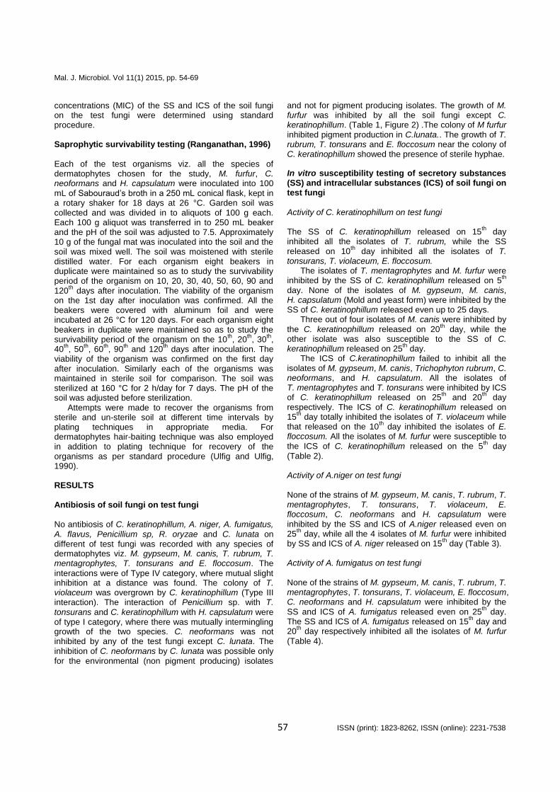
No antibiosis of C. keratinophillum, A. niger, A. fumigatus, A. flavus, Penicillium sp, R. oryzae and C. lunata on different of test fungi was recorded with any species of dermatophytes viz. M. gypseum, M. canis, T. rubrum, T. mentagrophytes, T. tonsurans and E. floccosum. The
interactions were of Type IV category, where mutual slight inhibition at a distance was found. The colony of T. violaceum was overgrown by C. keratinophillum (Type III interaction). The interaction of Penicillium sp. with T. tonsurans and C. keratinophillum with H. capsulatum were of type I category, where there was mutually intermingling growth of the two species. C. neoformans was not inhibited by any of the test fungi except C. lunata. The inhibition of C. neoformans by C. lunata was possible only for the environmental (non pigment producing) isolates
and not for pigment producing isolates. The growth of M. furfur was inhibited by all the soil fungi except C. keratinophillum. (Table 1, Figure 2) .The colony of M furfur inhibited pigment production in C.lunata.. The growth of T. rubrum, T. tonsurans and E. floccosum near the colony of C. keratinophillum showed the presence of sterile hyphae.
In vitro susceptibility testing of secretory substances (SS) and intracellular substances (ICS) of soil fungi on test fungi Activity of C. keratinophillum on test fungi The SS of C. keratinophillum released on 15
th day
inhibited all the isolates of T. rubrum, while the SS released on 10
th day inhibited all the isolates of T.
tonsurans, T. violaceum, E. floccosum. The isolates of T. mentagrophytes and M. furfur were
inhibited by the SS of C. keratinophillum released on 5th
day. None of the isolates of M. gypseum, M. canis, H. capsulatum (Mold and yeast form) were inhibited by the SS of C. keratinophillum released even up to 25 days.
Three out of four isolates of M. canis were inhibited by the C. keratinophillum released on 20
th day, while the
other isolate was also susceptible to the SS of C. keratinophillum released on 25
th day.
The ICS of C.keratinophillum failed to inhibit all the isolates of M. gypseum, M. canis, Trichophyton rubrum, C. neoformans, and H. capsulatum. All the isolates of T. mentagrophytes and T. tonsurans were inhibited by ICS of C. keratinophillum released on 25
th and 20
th day
respectively. The ICS of C. keratinophillum released on 15
th day totally inhibited the isolates of T. violaceum while
that released on the 10th
day inhibited the isolates of E. floccosum. All the isolates of M. furfur were susceptible to the ICS of C. keratinophillum released on the 5
th day
(Table 2). Activity of A.niger on test fungi None of the strains of M. gypseum, M. canis, T. rubrum, T. mentagrophytes, T. tonsurans, T. violaceum, E. floccosum, C. neoformans and H. capsulatum were inhibited by the SS and ICS of A.niger released even on 25
th day, while all the 4 isolates of M. furfur were inhibited
by SS and ICS of A. niger released on 15th
day (Table 3). Activity of A. fumigatus on test fungi None of the strains of M. gypseum, M. canis, T. rubrum, T. mentagrophytes, T. tonsurans, T. violaceum, E. floccosum, C. neoformans and H. capsulatum were inhibited by the SS and ICS of A. fumigatus released even on 25
th day.
The SS and ICS of A. fumigatus released on 15th day and
20th
day respectively inhibited all the isolates of M. furfur (Table 4).

Mal. J. Microbiol. Vol 11(1) 2015, pp. 54-69
58 ISSN (print): 1823-8262, ISSN (online): 2231-7538
Table 1: Types of Interactions between Colonies -Test fungi versus Soil fungi.
* Environmental non-pigment producing isolate of C. neoformans
I - Mutually intermingling growth II - Partial inhibition III - Dominance / Complete inhibition or over growth by antagonist IV - Mutual slight inhibition V - Co-dominance / Mutual inhibition at a distance
Table 2: Susceptibility pattern of test fungi to SS and ICS of C. keratinophillum.
Soil fungus
Test fungus
M.
gypse
um
M.
ca
nis
T.r
ub
rum
T.
me
nta
gro
p
hyte
s
T.
ton
su
rans
T.
vio
lace
um
E.
flo
ccosu
m
M.
furf
ur
*C.
ne
ofo
rma
n
s
H.
ca
psu
latu
m
C. keratinophillum
IV IV IV IV IV III IV IV IV I
A. niger
IV IV IV IV IV IV IV III IV IV
A. fumigatus
IV IV IV IV IV IV IV III IV IV
A. flavus
IV IV IV IV IV IV IV III IV IV
Penicillium sp
IV IV IV IV IV IV IV III IV IV
R. oryzae
IV IV IV IV IV IV IV III IV IV
C. lunata
IV IV IV IV IV IV IV II III IV
Test fungus
No. of isolates tested
SS released at different growth intervals (Days) / Inhibition of
test fungus (n)
ICS released at different growth intervals (Days) / Inhibition of test
fungus (n)
5 10 15 20 25 5 10 15 20 25
M. gypseum 4 4+ 4+ 4+ 4+ 4+ 4+ 4+ 4+ 4+ 4+ M. canis 4 3+ 2+ 2+ 1+ - 4+ 4+ 4+ 4+ 4+ T.rubrum 6 5+ 1+ - - - 6+ 6+ 6+ 6+ 6+ T. mentagrophytes 6 - - - - - 6+ 6+ 6+ 6+ - T. tonsurans 6 1+ - - - - 6+ 6+ 6+ - - T. violaceum 4 2+ - - - - 4+ 3+ - - - E. floccosum 4 1+ - - - - 3+ - - - - M. furfur 4 - - - - - - - - - - C. neoformans 4 4+ 4+ 4+ 4+ 4+ 4+ 4+ 4+ 4+ 4+ H. capsulatum Yeast form
2 2+ 2+ 2+ 2+ 2+ 2+ 2+ 2+ 2+ 2+
H. capsulatum
Mold form 2 2+ 2+ 2+ 2+ 2+
2+ 2+ 2+ 2+ 2+

Mal. J. Microbiol. Vol 11(1) 2015, pp. 54-69
59 ISSN (print): 1823-8262, ISSN (online): 2231-7538
Figure 2: Interactions between test fungi and soil fungi.

Mal. J. Microbiol. Vol 11(1) 2015, pp. 54-69
60 ISSN (print): 1823-8262, ISSN (online): 2231-7538
Table 3: Susceptibility pattern of test fungi to SS and ICS of A. niger.
Test fungus
No. of isolates tested
SS released at different growth intervals (Days) / Inhibition of test
fungus (n)
ICS released at different growth intervals (Days) / Inhibition of test
fungus (n)
5 10 15 20 25 5 10 15 20 25
M. gypseum 4 4+ 4+ 4+ 4+ 4+ 4+ 4+ 4+ 4+ 4+ M. canis 4 4+ 4+ 4+ 4+ 4+ 4+ 4+ 4+ 4+ 4+ T.rubrum 6 6+ 6+ 6+ 6+ 6+ 6+ 6+ 6+ 6+ 6+ T. mentagrophytes 6 6+ 6+ 6+ 6+ 6+ 6+ 6+ 6+ 6+ 6+ T. tonsurans 6 6+ 6+ 6+ 6+ 6+ 6+ 6+ 6+ 6+ 6+ T. violaceum 4 4+ 4+ 4+ 4+ 4+ 4+ 4+ 4+ 4+ 4+ E. floccosum 4 4+ 4+ 4+ 4+ 4+ 4+ 4+ 4+ 4+ 4+ M. furfur 4 3+ 2+ - - - 3+ 2+ - - - C. neoformans 4 4+ 4+ 4+ 4+ 4+ 4+ 4+ 4+ 4+ 4+ H. capsulatum Yeast form
2 2+ 2+ 2+ 2+ 2+ 2+ 2+ 2+ 2+ 2+
H. capsulatum
Mold form 2 2+ 2+ 2+ 2+ 2+ 2+ 2+ 2+ 2+ 2+
Activity of A. flavus on test fungi
All the isolates of M. gypseum, M. canis, T. rubrum, T. mentagrophytes, T. tonsurans, T. violaceum, E.floccosum, C. neoformans and H. capsulatum were resistant to the SS and ICS of A. flavus released on different time intervals. i.e. 5
th-25
th days. The SS of A. flavus released
on 5th
day inhibited all the 4 isolates of M. furfur, while the ICS of A. flavus released on the 10
th day inhibited all the
isolates of M. furfur (Table 5).
Activity of Penicillium sp. on test fungi The SS and ICS of Penicillium sp. released on 5
th day
inhibited all the 4 isolates of M. furfur, while SS and ICS released on the 15
th day inhibited the mold form of H.
capsulatum. The yeast form of H. capsulatum was not inhibited either by the SS and ICS of Penicillium sp. None of the isolates of M. gypseum, M. canis, T. rubrum, T. mentagrophytes, T. tonsurans, T. violaceum, E. floccosum, and C. neoformans were inhibited by the SS and ICS of Penicillium sp. (Tables 6).
Table 4: Susceptibility pattern of test fungi to SS and ICS of A. fumigatus.
Test fungus
No. of isolates tested
SS released at different growth intervals (Days) / Inhibition of test fungus (n)
ICS released at different growth intervals (Days) / Inhibition of test fungus (n)
5 10 15 20 25 5 10 15 20 25
M. gypseum 4 4+ 4+ 4+ 4+ 4+ 4+ 4+ 4+ 4+ 4+
M. canis 4 4+ 4+ 4+ 4+ 4+ 4+ 4+ 4+ 4+ 4+ T.rubrum 6 6+ 6+ 6+ 6+ 6+ 6+ 6+ 6+ 6+ 6+ T. mentagrophytes
6 6+ 6+ 6+ 6+ 6+ 6+ 6+ 6+ 6+ 6+
T. tonsurans 6 6+ 6+ 6+ 6+ 6+ 6+ 6+ 6+ 6+ 6+ T. violaceum 4 4+ 4+ 4+ 4+ 4+ 4+ 4+ 4+ 4+ 4+ E. floccosum 4 4+ 4+ 4+ 4+ 4+ 4+ 4+ 4+ 4+ 4+ M. furfur 4 3+ 2+ - - - 3+ 2+ 1+ - - C. neoformans 4 4+ 4+ 4+ 4+ 4+ 4+ 4+ 4+ 4+ 4+ H. capsulatum Yeast form
2 2+ 2+ 2+ 2+ 2+ 2+ 2+ 2+ 2+ 2+
H. capsulatum Mold form
2 2+ 2+ 2+ 2+ 2+ 2+ 2+ 2+ 2+ 2+

Mal. J. Microbiol. Vol 11(1) 2015, pp. 54-69
61 ISSN (print): 1823-8262, ISSN (online): 2231-7538
Table 5: Susceptibility pattern of test fungi to SS and ICS of A. flavus.
Test fungus
No. of isolates tested
SS released at different growth intervals (Days) / Inhibition of test
fungus(n)
ICS released at different growth intervals (Days) / Inhibition of test fungus (n)
5 10 15 20 25 5 10 15 20 25
M. gypseum 4 4+ 4+ 4+ 4+ 4+ 4+ 4+ 4+ 4+ 4+
M. canis 4 4+ 4+ 4+ 4+ 4+ 4+ 4+ 4+ 4+ 4+
T.rubrum 6 6+ 6+ 6+ 6+ 6+ 6+ 6+ 6+ 6+ 6+
T. mentagrophytes 6 6+ 6+ 6+ 6+ 6+ 6+ 6+ 6+ 6+ 6+
T. tonsurans 6 6+ 6+ 6+ 6+ 6+ 6+ 6+ 6+ 6+ 6+
T. violaceum 4 4+ 4+ 4+ 4+ 4+ 4+ 4+ 4+ 4+ 4+
E. floccosum 4 4+ 4+ 4+ 4+ 4+ 4+ 4+ 4+ 4+ 4+
M. furfur 4 - - - - - 2+ - - - -
C. neoformans 4 4+ 4+ 4+ 4+ 4+ 4+ 4+ 4+ 4+ 4+
H. capsulatum Yeast form
2 2+ 2+ 2+ 2+ 2+ 2+ 2+ 2+ 2+ 2+
H. capsulatum Mold form
2 2+ 2+ 2+ 2+ 2+ 2+ 2+ 2+ 2+ 2+
Activity of R. oryzae on test fungi The SS of R. oryzae released on 10
th day inhibited all the
isolates of M. furfur, while the ICS released on the 15th
day inhibited all the isolates of M. furfur. All other test fungi viz, M. gypseum, M. canis, T.rubrum, T. mentagrophytes, T. tonsurans, T. violaceum, E. floccosum, C. neoformans and H. capsulatum were resistant to the SS and ICS of R. oryzae (Tables 7). The SS and ICS of C. lunata released on 20
th day
inhibited all the isolates of M. canis and T. rubrum. The
SS released on 15th
day inhibited all the isolates of T. mentagrophytes and T. tonsurans while that released on the 10
th day inhibited all the isolates of T. violaceum and
E. floccosum. The environmental isolates of C. neoformans were susceptible to SS released on 10
th day
while the clinical isolates were not inhibited by the SS of C. lunata. All the isolates of M. furfur were susceptible to the SS released on 5
th day. The SS failed to inhibit yeast
form of the H. capsulatum while the mold form was
inhibited by SS released on 20th day
Table 6: Susceptibility pattern of test fungi to SS and ICS of Penicillium sp.
Test fungus
No. of isolates tested
SS released at different growth intervals (Days)/ Inhibition of test
fungus (n)
ICS released at different growth intervals (Days)/ Inhibition of test
fungus (n)
5 10 15 20 25 5 10 15 20 25
M. gypseum 4 4+ 4+ 4+ 4+ 4+ 4+ 4+ 4+ 4+ 4+ M. canis 4 4+ 4+ 4+ 4+ 4+ 4+ 4+ 4+ 4+ 4+
T.rubrum 6 6+ 6+ 6+ 6+ 6+ 6+ 6+ 6+ 6+ 6+
T. mentagrophytes 6 6+ 6+ 6+ 6+ 6+ 6+ 6+ 6+ 6+ 6+
T. tonsurans 6 6+ 6+ 6+ 6+ 6+ 6+ 6+ 6+ 6+ 6+
T. violaceum 4 4+ 4+ 4+ 4+ 4+ 4+ 4+ 4+ 4+ 4+
E. floccosum 4 4+ 4+ 4+ 4+ 4+ 4+ 4+ 4+ 4+ 4+
M. furfur 4 - - - - - - - - - -
C. neoformans 4 4+ 4+ 4+ 4+ 4+ 4+ 4+ 4+ 4+ 4+
H. capsulatum Yeast form
2 2+ 2+ 2+ 2+ 2+ 2+ 2+ 2+ 2+ 2+
H. capsulatum
Mold form 2 2+ 1+ - - - 2+ 2+ - - -

Mal. J. Microbiol. Vol 11(1) 2015, pp. 54-69
62 ISSN (print): 1823-8262, ISSN (online): 2231-7538
Table 7: Susceptibility pattern of test fungi to SS and ICS of R. oryzae.
Test fungus
No. of isolates tested
SS released at different growth intervals (Days)/ Inhibition of test
fungus (n)
ICS released at different growth intervals (Days)/ Inhibition of test fungus (n)
5 10 15 20 25 5 10 15 20 25
M. gypseum 4 4+ 4+ 4+ 4+ 4+ 4+ 4+ 4+ 4+ 4+ M. canis 4 4+ 4+ 4+ 4+ 4+ 4+ 4+ 4+ 4+ 4+
T.rubrum 6 6+ 6+ 6+ 6+ 6+ 6+ 6+ 6+ 6+ 6+
T. mentagrophytes 6 6+ 6+ 6+ 6+ 6+ 6+ 6+ 6+ 6+ 6+
T. tonsurans 6 6+ 6+ 6+ 6+ 6+ 6+ 6+ 6+ 6+ 6+
T. violaceum 4 4+ 4+ 4+ 4+ 4+ 4+ 4+ 4+ 4+ 4+
E. floccosum 4 4+ 4+ 4+ 4+ 4+ 4+ 4+ 4+ 4+ 4+
M. furfur 4 3+ - - - - 3+ 1+ - - -
C. neoformans 4 4+ 4+ 4+ 4+ 4+ 4+ 4+ 4+ 4+ 4+
H. capsulatum Yeast form
2 2+ 2+ 2+ 2+ 2+ 2+ 2+ 2+ 2+ 2+
H. capsulatum Mold form
2 2+ 2+ 2+ 2+ 2+ 2+ 2+ 2+ 2+ 2+
Table 8: Susceptibility pattern of test fungi to SS and ICS of C. lunata.
Test fungi
No. of isolates tested
SS released at different growth intervals (Days)/ Inhibition of test
fungi (n)
ICS released at different growth intervals (Days)/ Inhibition of test fungi
(n)
5 10 15 20 25 5 10 15 20 25
M. gypseum 4 4+ 4+ 4+ 4+ 4+ 4+ 4+ 4+ 4+ 4+ M. canis 4 3+ 2+ 1+ - - 3+ 2+ 1+ - -
T.rubrum 6 5+ 2+ 1+ - - 6+ 4+ 2+ - -
T. mentagrophytes 6 3+ 1+ - - - 4+ 3+ 1+ - -
T. tonsurans 6 2+ 1+ - - - 2+ 1+ - - -
T. violaceum 4 2+ - - - - 2+ - - - -
E. floccosum 4 1+ - - - - 2+ - - - -
M. furfur 4 - - - - - - - - - -
C. neoformans 4* 4+ 2+ 2+ 2+ 2+ 4+ 2+ 2+ 2+ 2+
H. capsulatum Yeast form
2 2+ 2+ 2+ 2+ 2+ 2+ 2+ 2+ 2+ 2+
H. capsulatum Mold form
2 2+ 2+ 1+ - - 2+ 2+ 1+ - -
*2 environmental and 2 clinical isolates
Minimum Inhibitory Concentration testing
The SS of C. keratinophillum recorded MIC of 1000 µg/mL against M. canis and T. rubrum, 500 µg/mL against T. mentagrophytes and T. tonsurans, 250 µg/mL against T. violaceum and E. floccosum and 125 µg/mL against M. furfur. The ICS of C. keratinophillum recorded MIC of 2500 µg/mL against M. canis, T. rubrum, T. mentagrophytes, T. tonsurans, T. violaceum and E. floccosum.
The MIC of ICS of C. keratinophillum against M. furfur was at 250 µg/mL. Both SS and ICS of C. keratinophillum
were not active up to a concentration of 2500 µg/mL against M. gypseum, C. neoformans and H. capsulatum (mold and yeast form).
The SS and ICS of A. niger, A fumigatus, A. flavus, Penicillium sp. and R. oryzae were not active up to a concentration of 2500 µg/mL against all the species of dermatophytes, C. neoformans and yeast form of H. capsulatum, M. furfur recorded MIC of 250 µg/mL for the SS of A. niger, A. fumigatus, A. flavus, Penicillium sp. and R. oryzae, while the MIC value was at 1000 µg/mL for the ICS released by the above fungi. The SS and ICS of Penicillium sp. recorded MIC value of 1000 µg/mL against the mold form of H. capsulatum.

Mal. J. Microbiol. Vol 11(1) 2015, pp. 54-69
63 ISSN (print): 1823-8262, ISSN (online): 2231-7538
The SS of C. lunata recorded MIC of 1000 µg/mL against M. canis, T.rubrum, C. neoformans (environmental isolates) and mold form of H. capsulatum, 500 µg/mL against T. mentagrophytes and 250 µg/mL against T. tonsurans, T. violaceum and E. floccosum while it was 125 µg/mL against M. furfur ICS of C. lunata recorded MIC of 1000 µg/mL against M. canis, T.rubrum, C. neoformans (environmental isolates) and mold form of H. capsulatum, 500 µg/mL against T. mentagrophytes, T. tonsurans, T. violaceum and E. floccosum, 250 µg/mL against M. furfur (Table 9).
The clinical isolates of C. neoformans did not record any susceptibility to SS and ICS of C. lunata up to a concentration of 2500 µg/mL. Saprophytic survivability testing of test fungi in unsterile soil
Only 2/4 strains of T. rubrum, T. mentagrophytes, and C. neoformans could be recovered on 10
th day by plating
technique. Recovery of T. tonsurans, T. violaceum, E. floccosum, M. furfur and yeast form of H. capsulatum was not possible on 10
th day.
One out of two strains of mold form of H. capsulatum could be recovered on 10
th day. On 20
th day it was
possible to recover only one isolate of T. mentagrophytes, while all other test organisms viz. T. rubrum, T. tonsurans, T. violaceum, E. floccosum, M. furfur, C. neoformans and H. capsulatum could not be recovered after 20 days. H. capsulatum (mold form) could be recovered up to 120
th
day. All the 4 isolates of M. gypseum could be recovered up to 120 days. Isolation of M. canis was not possible after 30 days. A similar pattern of results was obtained when hair-baiting technique was employed to recover the test dermatophytes from the unsterile soil.
Saprophytic survivability testing of test fungi in sterile soil
Recovery of 1/4 isolate of T. rubrum and 2/4 isolates of T. mentagrophytes was possible on the 30
th day. Two
isolates each of T. tonsurans and E. floccosum could be recovered on 10
th day after which their recovery was not
possible. All the 4 isolates of T. violaceum and M. furfur
could not be isolated even on the 10th
day. One isolate of T. mentagrophytes could be recovered on 40
th day while
recovery of T. rubrum after 30th day was not possible.
All the 4 isolates of C. neoformans could be recovered up to 120
th day. Isolation of H. capsulatum in the mold
form was possible up to 120 days, but the yeast form of H. capsulatum could not be recovered after 20 days by plating technique.
It was possible to isolate two strains of T. rubrum on 30
th day and one strain of T. mentagrophytes on 40
th day
while a single isolate each of T. tonsurans and E. floccosum was possible on 10
th day by hair baiting
technique. T. violaceum could not be isolated even on the 10
th day by hair baiting technique. All the 4 isolates of M. gypseum could be recovered up
to 120 days by both plating and baiting techniques.
Isolation of M. canis after 60 days was not possible (Tables 10 and 11). DISCUSSION
Fungal evolution and antibiosis
It has long been thought, and has been confirmed by modern phylogenetic studies (Makimura et al., 1998; Makimura et al., 1999; Gräser et al., 1999; Summerbell et al., 1999) that pathogenic dermatophytes probably arose
from soil-borne, nonpathogenic ancestors that are likely to be similar in habitat to today’s nonpathogenic dermatophytoids (eg. T. ajelloi, T. terrestre).
The present investigation revealed that the Secretory substances (SS) released by C. keratinophillum possess antidermatophytic activity against T. rubrum, T. tonsurans, T. mentagrophytes, T. violaceum and E. floccosum. The SS did not, however, inhibit the growth of M. gypseum and M. canis. M. gypseum is a geophilic fungi prevalent throughout the world (Rippon, 1988). The selective ability of M. gypseum to counter the antagonistic activity of the SS of C. keratinophillum may be one of the reasons for
worldwide distribution of this fungus in soil (Gokulshankar et al., 2005). The susceptibility of these anthropophilic dermatophytes to intra cellular substances (ICS) of C. keratinophillum is relatively less when compared to the SS secreted by the organism. It is interesting to note that when C. keratinophillum and an anthropophilic dermatophyte were co-inoculated on Sabouraud’s dextrose agar plate, C. keratinophillum did not inhibit the mycelial growth of T. rubrum, T. tonsurans, T. mentagrophytes and E. floccosum. However conidia formation did not occur on hyphae of T. rubrum, T. tonsurans and E. floccosum grown near C. keratinophillum. It is presumed from the present study that the nature and quantity of the SS released by C. keratinophillum during its early growth phase might not by very active to inhibit the growth of these dermatophytes. Further, when both C. keratinophillum and an
anthropophilic dermatophyte were inoculated simultaneously on Sabouraud’s agar plate, the growth of dermatophyte may also occur before the release of SS byC. keratinophillum. The absence of conidia formation on the hyphae of T. rubrum, T. tonsurans and E. floccosum grown near the C. keratinophillum indicates that SS of the organism possess anti-dermatophytic substances. Further, the results of susceptibility testing also showed that the SS released on the 5
th day was comparatively
less active against dermatophytes compared the SS released on the 10
th, 15
th, 20
th and 25
th day of growth. The
colony of T. violaceum was totally encircled by the colony of C. keratinophillum on co-inoculation, which is a strong indication of inhibition of the former by the later.
Earlier studies (Gokulshankar et al., 2001; Gokulshankar et al., 2005) on the co-inoculation of different species of dermatophytes and C. keratinophillum in sterilized soil revealed that none of the isolates of T. rubrum, T. mentagrophytes var. interdigitale and E. floccosum could be recovered from soil after 15
th day

Mal. J. Microbiol. Vol 11(1) 2015, pp. 54-69
64 ISSN (print): 1823-8262, ISSN (online): 2231-7538
either by baiting or plating technique. Whereas, these dermatophytes could be recovered from sterilized soil when inoculated alone up to 40 days by plating technique. Ranganathan (1996) have already recorded that none of these anthropophilic dermatophytes could exist in soil as saprophytes. However, the viability of these organisms at least in sterilized soil for a prolonged period cannot be ruled out. The inability to recover these dermatophytes even by plating technique from the sterilized soil when they were inoculated along with C. keratinophillum (Gokulshankar et al., 2005) strongly suggests the possibility of SS released by C. keratinophillum has a definite role in the complete elimination of the dermatophytes. Interestingly, the growth of M. gypseum was not affected by SS produced by C. keratinophillum.
This might be one of the reasons for the geophilic nature of the fungi. Why some species of dermatophytes have selected parasitic mode of existence while majority of dermatophytes species still live in soil as saprophytes is not known. The established homology between the obligate parasitic dermatophyte T. rubrum and strict saprophyte T. ajelloi (Rippon, 1988; Makimura et al., 1998; Makimura et al., 1999; Gräser et al., 1999; Summerbell et al., 1999) suggests the possibility of the past existence of the present day obligate parasitic dermatophytes in soil. Whenever, baiting technique is employed for isolation of dermatophytes from soil, Chrysosporium spp. is the predominant fungi to be isolated (Gokulshankar et al., 2001). Chrysosporium and allied genera accounted for 53.8% distribution, with C. indicum being the dominant species among the
keartinophylic fungi in soil (Deshmukh and Agarwal, 2004). Considering the global prevalence of Chrysosporium spp. and in combination with the results in the present study, it is speculated whether the anti-dermatophytic activity of these fungi might also be one of the early events for the evolutionary divergence of some geophilic archi-dermatophyte (saprophytic) to obligate parasitic dermatophyte species (Gokulshankar et al., 2005). The antibiosis of other soil microbes on dermatophytes also cannot be underestimated for such a parasitic evolution of dermatophytes. In our present study several other soil inhabiting fungi such as Aspergillus niger, A. fumigatus, A. flavus, Penicillium sp., R. oryzae and Curvularia lunata were also tested for their anti-dermatophyte activity. Among these the SS and ICS of C. lunata was found to have a definite role in inhibition.
M. furfur was inhibited by C. keratinophillum, A. niger, A. fumigatus, A. flavus, Penicillium sp., R. oryzae and C. lunata. Further the SS and ICS of all the soil fungi tested were inhibitory to M. furfur. This suggests the inability of
the organism to co-exist with any of these fungi. Since saprophytic existence of M. furfur is also not known, it makes one to contemplate whether the inhibition by other organisms would have forced M. furfur to adapt to an
obligate commensal/parasitic existence in the course of evolution? (If only there had been a past existence of this group in soil).
But it is really surprising to note that when C. lunata and M. furfur were co-inoculated, despite the overgrowth and inhibition of M. furfur by C. lunata, there is inhibition of pigment production in C. lunata by M. furfur. It is presumed that this could be because of the production of azelaic acid, a metabolite that can affect pigment formation in the skin, the possible mechanism for the depigmentation seen in some patients of tinea versicolor (Nazzaro-Porro and Passi, 1978). Curvularia also produces a black pigment (similar to melanin in human skin ?) (Lanisnik Rizner and Wheeler, 2003), which may be inhibited by M. furfur.
Saprophytic survivability of obligate parasitic dermatophytes and M. furfur in soil
The role of animals and soil in disseminating human dermatophytosis is known (Georg 1960a, b). However, the most frequent mode of transmission of the disease is through various exogenous sources such as floor, combs, linen etc. (Otecenasek, 1978). The viability of the fungal elements in these sources is not known. Prochacki and Engelhardt-Zasada (1972) have reported the antibiotic effect of T. megnini on various species of dermatophytes. The antibiotic effect of the major keratinophilic fungi in soil, Chrysosporium sp., on various species of dermatophytes during their survival in soil is not known. In the present study, the SS of C. keratinophillum showed inhibitory effect on the growth of the anthropophilic dermatophytes in the in vitro studies. In light of the above findings, it is speculated that Chrysosporium species may
pose a potential challenge to dermatophytes in soil, as both of these organisms are keratinophilic in nature. The role of other members of genus Chrysosporium coupled with the antibiosis of other soil bacteria/fungi could have compelled these dermatophytes to evolve parasitic adaptations. The reported low incidence of T. rubrum, T. mentagrophytes, T. tonsurans, T. violaceum and E. floccosum in soil may also be due to the well-defined anthropophization of these dermatophytes species.
The present study revealed that none of the strains of T. tonsurans, T. violaceum and E. floccosum could be recovered from unsterile soil on 10
th day. Further T.
rubrum and T. mentagrophytes were not recovered on the 20
th and 30
th day respectively from unsterile soil. The
inability to recover the organisms from unsterile soil suggests that the antibiotic effect of soil microbes might play a role in limiting their survivability in soil. However most of these organisms could not be recovered even from sterile soil after 30 days. This strongly suggests that, saprophytic survivability for these organisms in soil may not be possible due to their well-defined anthropophization. But the viability of the fungal elements especially arthroconidia and chalamydospores in soil for a shorter period cannot be ruled out. However, in our present investigation one isolate of T. mentagrophytes could be recovered from sterile soil on 40
th day by plating
and baiting technique. This shows that these organisms can survive in soil for longer period and thereby proves

Mal. J. Microbiol. Vol 11(1) 2015, pp. 54-69
65 ISSN (print): 1823-8262, ISSN (online): 2231-7538
Table 9: Minimum inhibitory concentration of SS and ICS of soil fungi on test fungi
Soil fungus MIC of the SS of Soil fungi on test fungi (µg/mL) MIC of the ICS of soil fungi on test fungi (µg/mL)
M. g
ypseu
m
M. canis
T.r
ub
rum
T .
me
nta
gro
ph
yte
s
T .to
nsura
ns
T .vio
laceu
m
E. floccosu
m
M .fu
rfur
C .ne
ofo
rman
s*
H. ca
psula
tum
-yea
st
H .ca
psula
tum
-mold
M. g
ypseu
m
M. canis
T.r
ub
rum
T .
me
nta
gro
ph
yte
s
T .to
nsura
ns
T .vio
laceu
m
E. floccosu
m
M .fu
rfur
C .ne
ofo
rman
s*
H. ca
psula
tum
-yea
st
H .ca
psula
tum
-mold
C. keratinophillum
N.A 1000 1000 500 500 250 250 125 N.A N.A N.A N.A 2500 2500 2500 2500 2500 1000 250 N.A N.A N.A
A.niger
N.A N.A N.A N.A N.A N.A N.A 250 N.A N.A N.A N.A N.A N.A N.A N.A N.A N.A 1000 N.A N.A N.A
A. fumigatus
N.A N.A N.A N.A N.A N.A N.A 250 N.A N.A N.A N.A N.A N.A N.A N.A N.A N.A 1000 N.A N.A N.A
A. flavus
N.A N.A N.A N.A N.A N.A N.A 250 N.A N.A N.A N.A N.A N.A N.A N.A N.A N.A 1000 N.A N.A N.A
Penicillium sp
N.A N.A N.A N.A N.A N.A N.A 250 N.A N.A 1000 N.A N.A N.A N.A N.A N.A N.A 1000 N.A N.A 1000
R oryzae
N.A N.A N.A N.A N.A N.A N.A 250 N.A N.A N.A N.A N.A N.A N.A N.A N.A N.A 1000 N.A N.A N.A
C .lunata
N.A 1000 1000 500 250 250 250 125 1000 N.A 1000 N.A 1000 1000 500 500 500 500 250 1000 N.A 1000
* MIC of environmental isolates of C. neoformans N.A: Not active up to a concentration of 2500 µg/mL

Mal. J. Microbiol. Vol 11(1) 2015, pp. 54-69
66 ISSN (print): 1823-8262, ISSN (online): 2231-7538
Table 10: Saprophytic survivability of test organisms in unsterile and sterile soil - recovery study by soil plating technique.
Test organisms
No. of isolates
Recovery from unsterile soil in days/ No. of isolates
Recovery from sterile soil in days/ No. of isolates
10 20 30 40 50 60 90 120 10 20 30 40 50 60 90 120
T. rubrum 4 2+ - - - - - - - 4+ 3+ 1+ - - - - -
T. mentagrophytes 4 2+ - - - - - - - 4+ 3+ 1+ - - - - -
T. tonsurans 4 2+ 1+ - - - - - - 3+ 2+ 2+ 1+ - - - -
T. violaceum 4 - - - - - - - - 2+ - - - - - - -
E. floccosum 4 - - - - - - - - - - - - - - - -
M. gypseum 4 - - - - - - - - 2+ - -- - - - - -
M. canis 4 4+ 4+ 4+ 4+ 4+ 4+ 4+ 4+ 4+ 4+ 4+ 4+ 4+ 4+ 4+ 4+
M. furfur 4 4+ 4+ 2+ - - - - - 4+ 4+ 3+ 3+ 2+ 2+ - -
C. neoformans 4 - - - - - - - - - - - - - - - -
H. capsulatum Yeast form
2 2+ - - - - - - - 4+ 4+ 4+ 4+ 4+ 4+ 4+ 4+
H. capsulatum
Mold form 2 - - - - - - - - 2+ 2+ - - - - - -
Note: +, could be recovered; -, could not be recovered

Mal. J. Microbiol. Vol 11(1) 2015, pp. 54-69
67 ISSN (print): 1823-8262, ISSN (online): 2231-7538
Table 11: Saprophytic survivability of test organisms in unsterile and sterile soil by hair baiting technique.
Test organisms No. of isolates
Recovery from unsterile soil in days/ No. of isolates
Recovery from sterile soil in days/ No. of isolates
10 20 30 40 50 60 90 120 10 20 30 40 50 60 90 120
T. rubrum 4 2+ - - - - - - - 4+ 3+ 2+ - - - - -
T. mentagrophytes 4 2+ 1+ - - - - - - 3+ 2+ 2+ 1+ - - - -
T. tonsurans 4 - - - - - - - - 1+ - - - - - - -
T. violaceum 4 - - - - - - - - - - - - - - - -
E. floccosum 4 - - - - - - - - 1+ - - - - - - -
M. gypseum 4 4+ 4+ 4+ 4+ 4+ 4+ 4+ 4+ 4+ 4+ 4+ 4+ 4+ 4+ 4+ 4+
M. canis 4 4+ 4+ 3+ - - - - - 4+ 4+ 4+ 4+ 3+ 3+ - -

Mal. J. Microbiol. Vol 11(1) 2015, pp. 54-69
68 ISSN (print): 1823-8262, ISSN (online): 2231-7538
the wide ecological niche of these organisms. Several workers have reported the isolation of T. mentagrophytes from soil from different parts of the world (Galgoczy, 1963; Grin and Ozegovic, 1963; Doss, 1992).
Similarly the recovery of zoophilic dermatophyte, M. canis was also not possible even in sterile soil after 60 days suggesting the inability of this organism for saprophytic existence. The organism could not be isolated either by hair baiting or plating technique. So the viability of the organism even as a spore in soil for prolonged period seemed impossible.
The recovery of M. gypseum from sterile and unsterile soil even up to 120 days by plating and baiting method substantiates the geophilic nature of this organism. A similar finding was reported earlier (Gokulshankar et al., 2001; Gokulshankar et al., 2005).
Hair baiting was found to be a superior method for isolating keratinophilic fungi especially dermatophytes from soil. The present study suggests that saprophytism may not be possible for T. rubrum, T. tonsurans, T. violaceum and E. floccosum. The ecological niche of different species of dermatophytes varies from species to species despite the fact that they are basically keratinophilic. No clear-cut answer has previously been given for how and why such divergence in their habitat preference has taken place. Probably the anti-dermatophytic activity of C. keratinophillum, the
predominnt keratinophilic fungi in soil, could have played a role in the parasitic divergence of the anthropophilic and zoophilic dermatophytes.
M. furfur is not capable of saprophytic existence both
in sterile and unsterile soil, even for a period of 10 days. This clearly indicates that the organism is capable of only obligate parasitic/commensalistic existence in human/animal hosts. Saprophytic existence of C. neoformans and H. capsulatum in soil
Saprophytic survival of C. neoformans was possible up to
120 days in sterile soil but not in unsterile soil. The reason could be because of predation by soil organism like nematodes and amoebae as reported by earlier workers (Steenbergen and Casadevall, 2003). However, presence of soil predators was not determined in the present study but it is possible that they could have been present. The source of soil used for testing the survivability of the C. neoformans isolates in sterile soil also was the same and
the elimination of predators would have occurred by the sterilization process adopted. That justifies the reason for the isolation of C. neoformans from sterile soil up to a period of 120 days. Both the melanin producing and non-melanin producing isolates could be isolated from sterile soil whereas the recovery of both of these isolates was not possible in unsterile soil.
Saprophytic survivability for H. capsulatum was
possible up to 120 days for the mold suspension however; the yeast suspension was not able to survive both in sterile and unsterile soil even for 20 days. This clearly illustrates that yeast morphogenesis is an adaptation
developed by the organism only for pathogenic intra-cellular state while a mold form is necessary for the saprobic existence. Conclusion
The competition/anatoginsm/fungus-fungus interaction
for existence in soil coupled with various biotic and abiotic factors could have played a definite role in the emergence parasitism in the once sapropobic fungi. References Chaturvedi, V., Flynn T., Niehaus W. G. and Wong, B.
(1996). Stress tolerance and pathogenic potential of a
mannitol mutant of Cryptococcus neoformans. Microbiology 142, 937-943.
Crum N. F., Lederman E. R., Stafford, C. M., Parrish, J. S. and Wallace, M. R. (2004). Coccidioidomycosis: A
descriptive survey of a reemerging disease. Clinical characteristics and emerging controversies. Medicine (Baltimore) 83, 149-175.
Deshmukh, S. K. and Agrawal, S. C. (2004). Isolation of
dermatophytes and other keratinophilic fungi from soils of Jammu, India. Mycoses 46, 226-228.
Doss, A. K. (1992). Studies on keratinophilic fungi and
certain Zygomycetes in relation to human mycosis in the city of Madras. Ph. D thesis, University of Madras, Madras.
Drouhet, E. (1997). Mile stones in the history of Cryptococcus and Cryptococcosis. Journal of Medical Mycology 7, 10-28.
Galgoczy, J. (1963). Investigations with dermatophyta
inoculated on to soil (in Hungarian with French, German and Russian summary). Borgy Vener Szle 39, 213-219.
Georg, L. K. (1960a). Animal ringworm in public health.
U.S Department (HEW Public Health series publication).
Georg, L. K. (1960b). Epidemiology of the
dermatophytes: Sources of infection, modes of transmission and epidemicity. Annals of the New York Academy of Sciences 89, 69-77.
Gokulshankar, S., Ranjith, M. S., Ranganathan, S., Selvakumar, B. N. and Mohammed, A. (2001). Influence of Chrysosporium spp. in the prevalence of dermatophytes in soil. Indian Journal of Dermatology 46(3), 142-145.
Gokulshankar, S., Ranjithsingh, A. J. A., Ranjith, M. S., Ranganathan, S. and Palaniappan, R. (2005). Role of Chrysosporium keratinophillum in the parasitic evolution of dermatophytes. Mycoses 48, 442-446.
Gräser, Y., El Fari, M., Vilgalys, R., Kuijpers, A. F., de Hoog, G. S., Presber, W. and Tietz, H. J. (1999).
Phylogeny and taxonomy of the family Arthrodermataceae (dermatophytes) using sequence analysis of the ribosomal ITS region. Medical Mycology 37, 105- 114.

Mal. J. Microbiol. Vol 11(1) 2015, pp. 54-69
69 ISSN (print): 1823-8262, ISSN (online): 2231-7538
Grin, E. and Ozegovic, L. (1963). Influence of the soil on
certain dermatophytes and their evolutional trend. Mycopathologia et Mycologia Applicata 21, 23-28.
Guillot, J. and Bond, R. (1999). Malassezia pachydermatis: A review. Medical Mycology 37, 295-306.
Kauffman, C. A. (2006). Endemic mycoses:
Blastomycosis, histoplasmosis, and sporotrichosis. Infectious Disease Clinics of North America. 20, 645-662.
Lanisnik Rizner, T. and Wheeler, M. H. (2003). Melanin
biosynthesis in the fungus Curvularia lunata (teleomorph: Cochliobolus lunatus). Canadian Journal of Microbiology 49,110-119.
Makimura, M., Mochizuki, T., Hasegawa, A., Uchida, K., Saito, H. and Yamaguchi, H. (1998). Phylogenetic classification of Trichophyton mentagrophytes complex strains based on DNA sequences of nuclear ribosomal internal transcribed spacer 1 regions. Journal of Clinical Microbioogy 36, 2629-2633.
Makimura, M., Tamura, Y. and Mochizuki, T. (1999).
Phylogenetic classification and species identification of dermatophyte strains based on DNA sequences of nuclear ribosomal internal transcribed spacer 1 region. Journal of Clinical Microbioogy 37, 920-924.
Midgley, G. (1989). The diversity of Pityrosporum (Malassezia) yeasts in vivo and in vitro. Mycopathologia 106, 143-153.
Nazzaro-Porro, M. and Passi, S. (1978). Identification of Tyrosinase inhibitor’s in cultures of Pityrosporum. The Journal of Investigative Dermatology 71, 205-208.
Novak, E. K. and Galgoczy, J. C. (1966). Notes on Dermatophytes of soil origin. Mycopathologia et Mycologia Applicata 28, 289-296.
Otecenasek, M. (1978). Ecology of dermatophytes. Mycopathologia 65, 67-72.
Prochacki, H. and Engelhardt-Zasada, C. (1972).
Studies on the antibiosis of dermatophytes. Mycopathologia et Mycologia Applicata 48, 187-190.
Ranganathan, S. (1996). Characterisation of
dermatophytes by biotyping, mating experiments, DNA typing and protease profile and the possible therapeutic efficasy of Azadirachta indica in the treatment of tinea infections. Ph.D thesis, University of Madras, Madras, India.
Rippon, J. W. (1988). Medical Mycology, the Pathogenic
fungi and Pathogenic Actionmycetes, 3rd
edn. W.B. Saunders, Philadelphia.
Steenbergen, J. N. and Casadevall, A. (2003). The
origin and maintenance of virulence for the human pathogenic fungus Cryptococcus neoformans. Microbes and Infection 5, 667-675.
Summerbell, R. C., Haugland, R., Li, A. and Gupta, A. K. (1999). Ribosomal RNA internal transcribed spacer
1 and 2 sequences of asexual, anthropophilic dermatophytes related to Trichophyton rubrum. Journal of Clinical Microbiology 37, 4005-4011.
Ulfig, K. and Ulfig, A. (1990). Keratinophilic fungi in bottom sediments of surface waters. Journal of Medical and Veterinary Mycology 28, 419-422.