marked copy: manuscript # ar-11-0718bogyolab.stanford.edu/pdf/liarthritis2011.pdf · (transgenic...
TRANSCRIPT

Marked copy: Manuscript # ar-11-0718
Treatment of Arthritis by Macrophage Depletion and Immunomodulation:
Testing an Apoptosis-Mediated Therapy in a Humanized Death Receptor
Mouse Model
Jun Li, M.D., Ph.D.1, Hui-Chen Hsu, Ph.D.1,2, PingAr Yang, B.S. 1,2, Qi Wu, B. S.
1,2, Hao Li, M.S.1, Laura E. Edgington, B.A. 3, Matthew Bogyo, Ph.D. 3, Robert P.
Kimberly, M.D. 1, and John D. Mountz, M.D., Ph.D. 1,2
1 Division of Clinical Immunology and Rheumatology, Department of Medicine,
University of Alabama at Birmingham, Birmingham, AL, 35294; 2 Department of
Medicine, Birmingham VA Medical Center, Birmingham, AL, 35233; 3 Department
of Pathology, Stanford University School of Medicine, Stanford, CA, 94305
This work was supported by a grant from Daiichi Sankyo Co., Ltd (to J.D.M.) and
Lupus Research Institute (to H-C.H.). Additional support was granted from the
American College of Rheumatology Research and Education Foundation for the
Within Our Reach: Finding a Cure for Rheumatoid Arthritis campaign, the Alliance
for Lupus Research – Target Identification in Lupus program, the Department of
Veterans Affairs Merit Review Grant 1I01BX000600-01, the National Institutes of
Health Grants 1AI 071110-01A1, ARRA 3RO1AI71110-02S1 (to J.D.M.), 1R01
EB005011-06 (to M.B.) and (1R01AI083705-01A2 to H-C.H.), and the Arthritis
Investigator Award supported by the Arthritis Foundation (to H-C.H.).
Full Length Arthritis & RheumatismDOI 10.1002/art.33423
© 2011 American College of RheumatologyReceived: May 05, 2011; Revised: Sep 02, 2011; Accepted: Oct 13, 2011
2
Corresponding author: John D. Mountz, M.D., Ph.D.
Phone: 205-934-8909, Fax: 205-996-6788, E-mail: [email protected]
All authors claim to have no financial interests which could create a potential
conflict of interest or the appearance of a conflict of interest with regard to the
work.
Page 2 of 48
John Wiley & Sons
Arthritis & Rheumatism

3
Objective. To determine the therapeutic efficacy and
immunomodulatory effect of an anti-human death receptor 5 (DR5) antibody,
TRA-8, in eliminating macrophage subsets in a collagen II-induced arthritis (CIA)
mouse model.
Methods. A chimeric human/mouse (hu/mo) DR5 transgenic (Tg)
mouse, under the regulation of the mouse 3-kb promoter and a Floxed-STOP
cassette, was generated and crossed with an ubiquitous Cre (Ubc.Cre) and a
lysozyme M Cre (LysM.Cre) Tg mouse to achieve inducible- or macrophage-
specific expression. CIA was induced in mice by chicken CII, which were then
treated with the anti-human DR5 antibody, TRA-8. The clinical scores,
histopathologic severity, macrophage apoptosis and depletion, and T cell subset
development were evaluated.
Results. In hu/mo DR5 Tg Ubc.Cre mice with CIA, Tg DR5 was most
highly expressed in CD11b+ macrophages with lower expression on CD4+ T
cells. In the hu/mo DR5 Tg LysM.Cre mice, Tg DR5 was restrictively expressed
in macrophages. Near infrared (NIFR) in vivo imaging of caspase activity and
TUNEL staining demonstrated that TRA-8 rapidly induced apoptosis of
macrophages in the inflammatory synovium. Depletion of pathogenic
macrophages by TRA-8 leads to significantly reduced clinical scores of arthritis,
decreased macrophage infiltration, synovial hyperplasia, osteoclast formation,
joint destruction, cathepsin activity, inflammatory cytokine expression in joints,
reduced Th17, and increased Treg cells in the draining lymph nodes (LN).
Page 3 of 48
John Wiley & Sons
Arthritis & Rheumatism

4
Conclusion. The anti-human DR5 antibody TRA-8 was efficacious in
reducing the severity of arthritis by targeted depleting macrophages and
immunomodulation. Our data provide pre-clinical evidence that TRA-8 is a
potential novel biologic agent for rheumatoid arthritis (RA) therapy.
Page 4 of 48
John Wiley & Sons
Arthritis & Rheumatism

5
Rheumatoid arthritis (RA) is characterized by synovial hyperplasia and
inflammation, with increased numbers of macrophages, fibroblasts, and
lymphocytes in the synovium (1-3). Although the earliest attempts to delete
CD4+ T cells in the treatment of RA were disappointing (4), specific therapies to
deplete B cells by anti-CD20 in RA are promising (5, 6). However, not all patients
respond, and disease relapses can occur after B cell repopulation (7).
Macrophages are of central importance in the pathogenesis of RA (8, 9), and
disease severity correlates with the number of activated macrophages in the
inflamed tissues and in circulation (10). The "professional" antigen-presenting
role of macrophages has also been implicated in the pathogenesis of RA (9).
Interactions between macrophages and fibroblasts, B, and T cells regulate
synovial inflammation (11-13) and suggest that the macrophage is an attractive
target for RA therapy. However, there has been no clinically proven efficacious
and safe therapy for specific elimination of inflammatory macrophages in RA.
Human death receptor 5 (DR5) is a pro-apoptotic molecule and mediates
apoptosis upon binding with its ligand, TRAIL, or an anti-DR5 agonistic antibody
(14). While DR5 is found on most examined cell types, its expression is
upregulated in cancer cells and it is a promising target for cancer therapy (15-
17). Moreover, increased DR5 expression and susceptibility to anti-human DR5-
mediated apoptosis are characteristics of the proliferating synovial fibroblasts in
RA (18), though the regulation of expression and apoptotic function of DR5 in
macrophages of human RA is unknown. Investigation of the therapeutic efficacy
Page 5 of 48
John Wiley & Sons
Arthritis & Rheumatism

6
of anti-DR5 in mouse disease models has been limited by two major obstacles.
Firstly, although an antibody (MD5-1) has been developed against MK (the
mouse homologue of human DR5), this antibody exhibits low cell-killing activity
without a cross-linker and has not been extensively analyzed (19). Secondly,
engineering a Tg mouse expressing human DR5 for testing of anti-human DR5
therapy has not been developed.
We have utilized a Tg mouse expressing a hu/mo-chimeric DR5 receptor
consisting of the extracellular domain of human DR5 and the transmembrane
and intracellular regions of mouse MK. This enables the binding of the anti-
human DR5 antibody to the extracellular domain and the induction of apoptosis
in mouse cells. Treatment with an anti-human DR5 antibody, TRA-8, successfully
prevented the development of, or ameliorated the severity of, CIA when
administered before or after the onset of arthritis, respectively. The major target
of TRA-8 in this disease model was shown to be macrophages in which DR5
expression is upregulated. Our data provide pre-clinical evidence that the anti-
human DR5 antibody, TRA-8, is a potential anti-arthritic biologic agent that
preferentially eliminates macrophages and exhibits subsequent
immunomodulatory effects.
Page 6 of 48
John Wiley & Sons
Arthritis & Rheumatism

7
MATERIALS AND METHODS
Mice. C57BL/6, UBC-cre/ESR1)1Ejb/J (Ubc.Cre), and B6.129-Lyzstm1(cre)Ifo/J (Lys
M.Cre) mice were obtained from the Jackson Laboratory. All animal procedures
were approved by The University of Alabama at Birmingham Institutional Animal
Care and Use Committee.
Cell lines, cell preparation, and culture. The mouse NIH3T3 cell line was
obtained from the American Type Culture Collection (ATCC, Manassas, VA) and
cultured in DMEM (Invitrogen) supplemented with 10% FBS, 100 units/ml
penicillin, 100 µg/ml streptomycin (Invitrogen), and 2 mM glutamine (Invitrogen)
at 37ºC, 5% CO2 in a humidified incubator. Single-cell suspensions from spleen
and inguinal lymph nodes were prepared and cultured in RPMI-1640 (Invitrogen)
supplemented with 10% FBS, 10 mM HEPES, and 0.1% 2-mercaptoethanol
(Invitrogen).
Expression constructs, transfection, and ATPLite analysis. Human and
mouse DR5 cDNAs were obtained from Open Biosystems (Huntsville, AL). The
extracellular domain of human DR5 was amplified by PCR using primer A
(huDR5For) 5’-ACTGTCGACGCCCCAAGTCAGCCTGGACACATA-3’ and
primer B (huDR5Rev) 5’-
TCCTATCCAGAGGCCTAGCTTATGCCAAGAACAGGGAGAGGCAGGAGTCC
CTGG-3’. Similarly, the transmembrane and intracellular domains of mouse DR5
were amplified by PCR using primer C (MoDR5For) 5’-
Page 7 of 48
John Wiley & Sons
Arthritis & Rheumatism

8
CCAGGGACTCCTGCCTCTCCCTGTTCTTGGCATAAGCTAGGCCTCTGGATA
GGA-3’ and primer D (MoDR5Rev) 5’-
GATGCGGCCGCTCAAACGCACTGAGATCCTCCTGG-3’. The fused chimeric
DR5 was then generated by PCR using a mixture of the A-B and C-D products
as template together with primers A and D. The purified final PCR product was
then digested by SalI and NotI and this chimeric DR5 fragment was used to
replace the human DR5 in the vector. A 3-kb putative mouse DR5 promoter was
cloned from mouse BAC RP24-355K8 (Children’s Hospital Oakland Research
Institute, Oakland, CA) and subcloned upstream of the chimeric DR5.
Transfection was performed using Lipofectamine 2000 (Invitrogen). Cell viability
was determined using an ATP luminescence assay kit (PerkinElmer, Waltham,
MA).
Establishment of the chimeric hu/mo-chim DR5 Tg mouse. The DNA used
for generation of DR5 Tg mice was based the 3-kb mouse promoter/chimeric
DR5 construct. To enable tissue-specific expression, a Floxed-STOP cassette
(Addgene) was introduced between the promoter and chimeric DR5. A 8.3-kb
DrdI-DrdI fragment was used to generate Tg mice on a C57BL/6 background
(Transgenic Mouse Facility, UAB). The Tg mice were genotyped using the
primers specific for the human DR5 extracellular domain.
Generation of hu/mo-chimeric DR5 Ubc.Cre double Tg and hu/mo-chimeric
DR5 LysM.Cre double Tg mice. The Floxed-STOP chimeric DR5 Tg mice were
Page 8 of 48
John Wiley & Sons
Arthritis & Rheumatism

9
bred with two different Cre-expressing mice: (i) Ubc.Cre mice which have strong
tamoxifen-inducible Cre activity in all tissues examined; and (ii) LysM.Cre mice
which express Cre in myeloid cells. For Cre induction in Ubc.Cre mice, mice
were treated with tamoxifen (5 mg/mouse/day) for five consecutive days via
gavage.
Quantitative reverse transcription PCR (qRT-PCR) analysis. Intracardial
perfusion was performed prior to the processing of organs and tissues. RNA was
isolated from synovium and other tissues using TRIzol reagent (Invitrogen). The
first-strand cDNA was synthesized by using random hexamer primers and
RevertAidTMM-MuLV Reverse Transcriptase (Fermentas Life Science). QRT-
PCR was performed using an IQ5 multicolor RT-PCR detection system as
described previously (20). Primers used are shown in the supplementary table 1.
Flow cytometric analysis. Single-cell suspensions were stained using
fluorochrome-conjugated mouse-specific Abs, including APC–anti-CD4
(Biolegend), FITC–anti-CD8 (BD Biosciences), Alexa 700–anti-CD19
(eBioscience), FITC–anti-CD11b (BD Biosciences), FITC–anti-CD11c (BD
Biosciences), PE–anti-mouse DR5 (Biolegend), APC–anti-Gr1(Biolegend),
PE/Cy7–anti-Ly6C (Biolegend), FITC-anti-IFN-γ (Biolegend), PE-anti-IL-17
(Biolegend), Alexa 647-anti-IL-23p19 (eBioscience), and PE-anti-Foxp3
(eBioscience). Tg chimeric DR5 was stained with biotin–anti human DR5
(Biolegend) followed by Streptavidin eFluor 450 (eBioscience). Prior to staining,
Page 9 of 48
John Wiley & Sons
Arthritis & Rheumatism

10
Fc receptors were blocked by anti-mouse CD16/32 (Biolegend). Intracellular and
intranuclear staining was performed following manufacturer’s instruction
(eBioscience). Before intracellular cytokine measurement, cells were stimulated
with 25 ng/ml PMA (Sigma) plus 500 ng/ml ionomycin (Sigma) for 2 h with the
addition of GolgiStop (BD Biosciences) for an additional 3h. Data were acquired
on a BD LSRII flow cytometer and analyzed using FlowJo software (Tree Star,
Inc.).
Induction of CIA. CIA was induced and scored in DR5 Tg mice of C57BL/6
background that were 8- to 16-weeks old as described (21). Briefly, mice were
immunized by intradermal administration of chicken Type II collagen (Chondrex,
Inc.) emulsified in complete Freund’s adjuvant (CFA), followed by injection of
chicken CII in incomplete Freund’s adjuvant (IFA) on day 30 after the primary
injection. To ensure a higher incidence of CII arthritis, an adenovirus expressing
mouse IL-17 (AdIL-17, 2x10⁹ pfu/mouse, a generous gift from Dr. Jay Kolls) (22)
was administered intravenously to all mice 2 days prior to the primary
immunization with CII.
TRA-8 treatment of CIA mice. TRA-8 (Daiichi-Sankyo, dissolved in PBS, 0.2 mg
per mouse) or IgG1 isotype control was administered i.v. or i.p. twice/week
starting on day 0 (early treatment) or on day 30 (late treatment) until mice were
sacrificed.
Page 10 of 48
John Wiley & Sons
Arthritis & Rheumatism

11
Histopathologic assessment and immunohistochemical staining. After
sacrifice, the knee, ankle, and foot joints were fixed in 4% formaldehyde and then
decalcified. Tissue sections (5 µm) were stained with hematoxylin and eosin and
examined by light microscopy. Immunohistochemical staining using anti-Mac-3
(clone M3/84, BD Biosciences) was performed as described previously (23).
TUNEL assay was performed by using the ApopTag Plus Peroxidase In Situ
Apoptosis Detection Kit (Millipore), following the manual. Sections were
counterstained with hematoxylin or methyl green. Cytospin preparations of LN
cells were fixed with 4% formaldehyde. Tyramide signal amplification was carried
out according to the manufacturer’s instruction (Invitrogen).
In vivo imaging of arthritis and apoptosis. For cathepsin activity
determination, mice were injected intravenously with 2 nmol ProSense 750
(ViSen, Bedford, MA) in 150 µl PBS. Twenty-four hours after injection, mice were
imaged using the Odyssey Infrared imaging System (LI-COR, Lincoln,
Nebraska). For apoptosis detection, a caspase-targeted activity-based probe,
AB50–Cy5, was used as described previously (24).
ELISA cytokine measurement. Cytokine levels in sera were measured by
ELISA according to the manufacturer’s manual (Biolegend).
Statistics. Figures are representative of at least 3 independent experiments.
Statistical analyses were performed using two-tailed Student’s t test, one-way
Page 11 of 48
John Wiley & Sons
Arthritis & Rheumatism

12
ANOVA, and bivariate correlation analysis. P values <0.05 were considered
statistically significant.
Page 12 of 48
John Wiley & Sons
Arthritis & Rheumatism

13
RESULTS
Hu/mo-chimeric DR5 proteins and their apoptosis-inducing function upon
TRA-8 binding in mouse cells.
Mouse and human DR5 exhibit only ~50% homology at the amino acid
level (Figure 1A) and TRA-8, which binds to the extracellular domain of human
DR5, however, does not recognize the extracellular domain of mouse DR5. We
also observed that the death domain of human DR5 does not initiate the
apoptotic cascade in mouse cells. To overcome this obstacle, we generated
constructs that express a hu/mo-chimeric DR5 consisting of the extracellular
domain of human DR5 with the transmembrane and intracellular domains of
mouse DR5 (Figure 1B, Left).
To achieve the desired regulation of hu/mo-chimeric DR5 protein
expression, hu/mo DR5 constructs with different regulatory elements were
produced that contain the CMV promoter, mouse 1-kb, 3kb DR5 putative
promoters, first intron, and 3’-untranslated region (3’-UTR) (Figure 1B, Right).
Hu/mo-chimeric DR5 expression was determined by cell-surface flow cytometry
using the anti-human DR5 antibody, which recognizes the extracellular domain of
human DR5 (Figure 1C). As shown, anti-human DR5 recognized the transfected
human (Construct 1) but not mouse DR5 protein (Construct 2). In mouse NIH3T3
cells, the CMV, 1-kb and 3-kb promoter resulted in high expression of the hu/mo-
chimeric DR5 (Constructs 3-5) whereas the first intron and 3’UTR of mouse DR5
exhibited negative regulatory effects on hu/mo-chimeric DR5 expression
Page 13 of 48
John Wiley & Sons
Arthritis & Rheumatism

14
(Constructs 6, 7). For functional studies, NIH 3T3 mouse cells were transfected
with these constructs (Figure 1D). TRA-8 (1 µg/ml) was added 24 h after
transfection, followed by overnight incubation. The ATPLite assay was used to
measure the cell viability. TRA-8 did not decrease viability in cells transfected
with the full-length human or mouse DR5 driven by the CMV promoter, which
lack either the mouse death domain that initiates apoptotic signaling or the
human extracellular domain that binds TRA-8 (Constructs 1 and 2, Figure 1D).
However, in cells transfected with the chimeric DR5, significant reduced cell
viability by TRA-8 was detected, with the 3-kb chimeric DR5 construct resulted in
the highest apoptosis inducing effect by TRA-8 (Constructs 3-5, Figure 1D).
Addition of the first intron and the 3’-UTR reduced the TRA-8 killing effect (Figure
1D). Thus, the chimeric DR5 regulated by the 3-kb promoter (Construct 5, Figure
1B, C, and D) is the optimal construct for DR5 expression and TRA-8-mediated
apoptosis and was selected for generation of Tg mice.
Expression of hu/mo-chimeric DR5 and its apoptosis-inducing function in
hu/mo DR5 Tg+ Ubc.Cre mice.
In order to enable temporal and spatial expression of the chimeric DR5, a
Floxed-STOP was inserted between the 3-kb promoter and the chimeric DR5.
Founder DR5 Tg mice were crossed with Ubc.Cre mice, which exhibit strong
tamoxifen-inducible Cre expression ubiquitously. To determine if the tissue
distribution of the chimeric Dr5 correlates with that of endogenous mouse Dr5,
the expression of chimeric Dr5 and mouse endogenous Dr5 in tissues harvested
Page 14 of 48
John Wiley & Sons
Arthritis & Rheumatism
15
from tamoxifen treated DR5 Ubc.Cre mice was determined (Figure 2A).
Tamoxifen treatment induced the expression of the hu/mo-chimeric DR5 Tg in
various tissues, including the lymph nodes (LN), brain, lung, spleen, and kidney
and the expression pattern exhibited a significant correlation with that of the
endogenous mouse DR5 (Figure 2A). Western blot analysis indicated that the
hu/mo DR5 Tg protein expression correlated with the mRNA expression with high
level of protein detected in the LN, spleen and lung (Data not shown).
To examine the chimeric DR5 expression in different immune cells and
TRA-8 induced apoptosis, CIA was induced in these mice. Chicken type II
collagen (cCII) induces arthritis in approximately 60–70% of mice with the H-2b
background (25). Prior to injection of cCII, we administered mice with an
adenovirus that expresses IL-17 (AdIL-17) to increase the incidence of arthritis
and thus facilitate the evaluation of the therapeutic effects of TRA-8 (22). In the
draining LN of the mice with CIA (two months after primary CII injection), the
expression of the hu/mo-chimeric DR5 was highest in the CD11b+Gr-1+
granulocytes and CD11b+Ly6C+ inflammatory macrophages, with lower
expression on CD4+ and CD8+ T cells, and minimal expression on CD19+ B cells
(Figure 2B). Importantly, both early and late TRA-8 treatment (0.2 mg
twice/week) reduced the percentage of CD11bhigh -activated macrophages in the
LN by ~50–60% (Figure 2C), suggesting that TRA-8 can potentially suppress the
frequency of macrophages during an active inflammatory stage in CIA.
Page 15 of 48
John Wiley & Sons
Arthritis & Rheumatism

16
To further assess the effects of TRA-8 on the macrophages, a single-cell
suspension prepared from the draining inguinal lymph nodes of the cCII-induced
hu/mo-chimeric DR5 Tg+ Ubc.Cre mice without in vivo TRA-8 treatment was
stimulated with LPS (5 µg/ml) for 2 days and then treated with or without TRA-8
for an additional 2 days. As shown in Figure 2D, for LPS-stimulated
macrophages obtained from hu/mo-chimeric DR5 Tg+ Ubc.Cre mice, thymidine
incorporation was significantly lower in TRA-8-treated than untreated mice.
TRA-8 treatment prevents the development of, and ameliorates established
arthritis in hu/mo DR5 Tg+ Ubc.Cre mice.
After CIA induction (Figure 3A and B, arrows), both the isotype-treated
hu/mo DR5 Tg+ Ubc.Cre (DR5 Tg+) mice and the control DR5 Tg− Ubc.Cre (DR5
Tg−) mice developed joint swelling and erythema indicated by the arthritis score
(Figure 3A and B, gray squares). Early TRA-8 treatment (0.2 mg twice/week) of
the hu/mo-chimeric DR5 Tg+ Ubc.Cre mice resulted in a significant reduction in
the early-stage arthritis, as well as the late-stage arthritis that was associated
with the CII boost (Figure 3A, filled circles). Moreover, initiation of the TRA-8
treatment (0.2 mg twice/week) 2 days before the CII boost significantly inhibited
the late phase of CIA (Figure 3A, open circles). In contrast, TRA-8 treatment of
the control DR5 Tg− Ubc.Cre mice did not affect the development of arthritis in
response to either the primary or secondary CII injection (Figure 3B).
Histopathologic analysis of the joints from cCII-injected hu/mo-chimeric DR5 Tg+
Ubc.Cre mice 2 months after TRA-8 treatment revealed a dramatic reduction in
Page 16 of 48
John Wiley & Sons
Arthritis & Rheumatism

17
the severity of synovial hyperplasia (H), and bone erosion (E), as well as
significant attenuation of inflammatory cell infiltration in the joints (Figure 3C, left
panels) compared to DR5 Tg− control (Figure D, left panels).
Immunohistochemical analysis of the synovium revealed that the numbers of
Mac-3+ macrophages (M) were much lower in the TRA-8-treated, CII-injected
hu/mo-chimeric DR5 Tg+ Ubc.Cre mice than their untreated counterparts (Figure
3C and D, right panel).
TRA-8 treatment specifically eliminates inflammatory macrophages and
ameliorates established arthritis in hu/mo DR5 Tg+ LysM.Cre mice.
To test whether targeted depletion of macrophages with TRA-8 is feasible
in vivo, we restricted expression of the chimeric DR5 to myeloid lineage cells by
crossing the Floxed-STOP chimeric DR5 Tg mice with mice in which Cre is
driven by the lysozyme M (Lys.M) promoter (26). CIA was induced as described
above. At day 60, FACS analysis showed that CD11b+ macrophages from the
draining LN of hu/mo-chimeric DR5 in DR5 Tg+ LysM.Cre (DR5 Tg+) mice
exhibited increased cell surface binding to anti-human DR5 compared with that of
the control mice (Figure 4A, upper panel). A similar result was also confirmed by
cytospin (Figure 4A, lower panel). There was a very low frequency of CD4+,
CD8+ T cells and CD19+ B cells that expressed hu/mo-chimeric DR5 (<1%) in
these mice (Figure 4B). TRA-8 treatment of these mice reduced the percentage
of CD11b+ macrophages from 4.3% to 1.5% (Figure 4C), whereas the TRA-8
depletion effect was not significant in DR5 Tg- mice (Figure 4C). Within the
Page 17 of 48
John Wiley & Sons
Arthritis & Rheumatism

18
CD11b+ cells, the percentage of the Ly6C+ inflammatory macrophage
subpopulation was reduced from 3.1% to 0.75% (Figure 4D) and this effect was
not significant in DR5 Tg- mice. TRA-8 treatment did not change in the
percentage of total CD4+ and CD8+ T cells, CD19+ B cells, or CD11c+ dendritic
cells in all these four groups of mice (data not shown).
TRA-8-induced apoptosis was assessed by a non-invasive in vivo imaging
method and a TUNEL staining. LysM.Cre hu/mo-chimeric DR5 Tg+ and control
LysM.Cre mice were induced to develop CIA. At 8 weeks after induction,
baseline-levels of caspase activity were measured in vivo using the caspase
imaging probe AB50–Cy5 (24). Mice were then treated with TRA-8 (0.2 mg on
day 0 and day 3) and apoptosis imaging was repeated on the same mice.
Apoptosis reached the peak 6 days after the first dose of TRA-8 administration
(Figure 5A, right panel and B). There was a significant increase in the caspase
probe signal in mice with arthritis post- compared with pre-TRA-8 treatment
(Figure 5A, right panel and 5B). Our result demonstrated that TRA-8 can induce
apoptosis in vivo in the joints of arthritic mice.
Apoptosis analysis by TUNEL assay was carried out on joint sections of
the same mice after acute TRA-8 treatment, as described above. At this early
time point after short term TRA-8 treatment, there was no significantly difference
in synovial hyperplasia (H), bone erosion (E), and macrophage infiltration (M)
between LysM.Cre hu/mo-chimeric DR5 Tg+ (Tg DR5+) and control LysM.Cre
Page 18 of 48
John Wiley & Sons
Arthritis & Rheumatism

19
DR5- Tg (Tg DR5-) mice (Figure 5C, left and middle panels). There was a
significantly increased TUNEL staining (TU) after short term TRA-8 treatment in
the hu/mo-chimeric DR5 Tg+ mice but not in control DR5 Tg– mice (Figure 5C,
right panels). Serial section staining indicated that the TUNEL staining was most
prominent in macrophages (M) within the intima and sublining of the synovium
(Figure 5C, right panels). Apoptosis of macrophages and fibroblasts was also
quantified by calculating the percentage of apoptotic cells in the Mac-3+ and Mac-
3- regions (Figure 5D).
Following establishment of CIA, TRA-8 treatment (0.2mg twice/week)
significantly attenuated the severity of the arthritis in the hu/mo-chimeric DR5 Tg+
LysM.Cre (Tg DR5+) mice (Figure 6A, upper panel), but not in control DR5 Tg–
LysM.Cre (Tg DR5-) mice (Figure 6A, lower panel), suggesting that targeted
depletion of macrophages can ameliorate CIA. Production of high levels of
cysteine cathepsins is clinically associated with arthritis severity (27).
Assessment of cathepsin activity using the ProSense 750TM protease probe
confirmed high levels of activity in the ankles, tarsal joints, and digits of control
DR5 Tg- LysM.Cre mice with and without TRA-8 treatment, and in hu/mo-
chimeric DR5 Tg+ LysM.Cre mice that were not treated with TRA-8. In marked
contrast, only minimal protease activity was found in the TRA-8-treated hu/mo-
chimeric DR5 Tg+ LysM.Cre mice (Figure 6B, C). Histopathologic analysis of the
joints from cCII-injected chimeric DR5 Tg mice (Figure 6D, left panels) treated
with TRA-8 for one month revealed a dramatic reduction in the severity of
Page 19 of 48
John Wiley & Sons
Arthritis & Rheumatism
20
synovial hyperplasia (H), inflammatory cell infiltration, bone erosion (E) in the
joints, and a significant decrease in Mac3+ macrophages (M) in synovium
compared to DR5 Tg- control mice (Figure 6D, right panels). TRAP staining (TR)
further indicated that TRA-8 treatment also lead to the reduced activity of
osteoclasts in the joints (Figure 6D).
TRA-8 treatment decreases the expression of pro-inflammatory cytokines
and exhibits immunomodulatory effects in hu/mo DR5 Tg+ LysM.Cre mice.
To further investigate the immune response regulated by the depletion of
inflammatory macrophages, sera and synovial cytokines were assessed on day
60. TRA-8 treatment significantly reduced the protein levels of IL-6 and IL-17A in
the sera (Figure 7A) as well as Tnfa, Il6, Il23a(p19) and Il17a transcripts in the
joints of DR5Tg+ LysM.Cre mice treated with TRA-8 (Figure 7B) compared to
control DR5Tg- mice with CIA. In the CD11b+ cell population from the draining
LN, the percentage of IL-23+ inflammatory macrophages was reduced from 5.8%
to 0.2% (Figure 7C), which is consistent with the observation that the expression
of Irf5, a signature transcription factor of inflammatory macrophages (28), is also
reduced in the synovium of the TRA-8 treated mice (Figure 7B). IL-23 is a pro-
inflammatory cytokine that has been proposed to play a central role in the
development of arthritis (29-31), which is related to the dysregulated balance
between IL-23/Th17 axis and regulatory T cells (Tregs) (32). We further identified
that TRA-8 treatment can restrain Th17 while promoting Tregs cell development.
Th17 cells were reduced from 2.9% to 1.4% whereas Tregs were increased from
Page 20 of 48
John Wiley & Sons
Arthritis & Rheumatism

21
3.9% to 6.3% (Figure 7D) in the DR5Tg+ LysM.Cre mice treated with TRA-8
compared with the control DR5Tg- mice with CIA. Consistent with this, there were
decreased expression of the Il-17a and increased expression of Foxp3 in the
synovium (Figure 7B). IFNγ-producing Th1 cells were also reduced by TRA-8
treatment in these mice (Figure 7D).
Page 21 of 48
John Wiley & Sons
Arthritis & Rheumatism

22
DISCUSSION
The present results are the first to show that in vivo administration of
TRA-8 can directly induce apoptosis of a subpopulation of macrophages and
attenuate CIA. The predominant cell types targeted by TRA-8 therapy are a
subpopulation of inflammatory macrophages which produce high levels of
cytokines, including TNF-α and IL-6 (33). TRA-8 therapy has a novel cell
depletion mechanism of directly triggering apoptosis of the targeted cells. TRA-8
and its humanized version, tigatuzumab (CS1008), have been shown to be
effective in elimination of tumor cells in xenograft cancer models and well-
tolerated in a phase I clinical trail (34, 35).
TRA-8 exhibits higher selectivity than TRAIL and it does not recognize
mouse MK (14). The existing anti-mouse MK monoclonal antibody (MD5-1) is not
efficacious in inducing apoptosis without cross-linkers (19). Furthermore, we
have found that the neither human DR5 nor mouse MK initiated TRA-8-mediated
apoptotic cascades in mouse cells (Figure 1D). To investigate the efficacy and
the mechanism of TRA-8 therapy in mouse models, we generated a chimeric
hu/mo-chimeric DR5 Tg comprised of the extracellular domain of human DR5
and the transmembrane and intracellular domains of mouse MK with regulatory
elements. The expression of the chimeric DR5 was comparable to endogenous
mouse MK. We observed that the chimeric DR5 is predominantly expressed in
CD11b+ macrophages of DR5 Tg mice crossed with both Ubc.Cre and LysM.Cre
Page 22 of 48
John Wiley & Sons
Arthritis & Rheumatism

23
mice after induction of CIA. This suggested that the mouse MK 3-kb promoter is
active in macrophage during arthritis pathogenesis.
DR5 is broadly expressed in most human tissues (36, 37). In tumors, its
expression is highest in melanoma and lung cancers (36). Although human DR5
is widely expressed, anti-DR5 treatment induced apoptosis only occurs in
selected cells (38). The present study indicated the high selectivity of TRA-8-
mediated killing in CD11bhi Ly6C+ macrophages. Ly6C is a
monocyte/macrophage differentiation antigen regulated by interferon-γ (39),
which has been shown to facilitate apoptosis signaling in macrophages and
cancer cells (40, 41). Inflammatory macrophages express much higher levels of
Ly6C distinguished from those residing normally in the tissues (42). In CIA and
K/BxN serum transfer-induced arthritis, reduction of Ly6C+ synovial macrophages
has been associated with GM-CSF blockade and reduction in disease severity
(43), suggesting that Ly6Chi macrophages are associated with GM-CSF-
mediated arthritis response. Our results suggest that TRA-8 treatment can
shape the heterogeneity of macrophages, leading to the homeostasis of the
myelomonocytic cells in the inflammatory conditions.
One of the major advantages of DR5 is the inflammation-dependent
selectivity which is reflected by the fact that while the chimeric DR5 was found to
be mainly expressed by macrophages, inflammatory Ly6C+ CD11b+
macrophages are more susceptible to DR5 mediated apoptosis. This suggested
Page 23 of 48
John Wiley & Sons
Arthritis & Rheumatism

24
that our approach of using TRA-8 can selectively deplete inflammatory
macrophages while sparing the anti-inflammatory macrophages. It has been
reported that one single dosage of intraarticular administration of clodronate
liposomes leads to macrophage depletion in the synovium of RA patients (44),
however, such therapy may not be safe for systemic administration and depletion
of macrophages based on their phagocytotic capacity will be difficult in terms of
specificity of killing and regulation of activity (45).
Dysregulated IL-23/Th17 axis and Tregs have been implicated in the
pathogenesis of RA. Interactions between CD4 T cells and macrophages have
been shown to be important for polarization of both lineages of cells (30, 46, 47).
Recently, it has been reported that human Tregs can be converted to Th17 cells
in the context of inflammatory signals and this differentiation process can be
enhanced by IL-1β, IL-23, and IL-21 (48). We have determined the polarization
condition of CD4 T cells in these mice after macrophage depletion by TRA-8.
Interestingly, we found that there was a significant decrease in the frequency of
Th17 and Th1 CD4 T cells in the LN isolated from the TRA-8 treated DR5 Tg+
LysM.Cre mice compared to control TRA-8 treated DR5 Tg- LysM.Cre mice with
CIA. In contrast, the frequency of Tregs in the LN of DR5 Tg+ LysM.Cre mice
was nearly 2 fold higher than that in the DR5 Tg- LysM.Cre mice. Our results
suggest that TRA-8, via diminishing the frequency of IL-23+ M1 macrophages,
can indirectly regulate the balance of pro-inflammatory versus anti-inflammatory
effector CD4 T cells.
Page 24 of 48
John Wiley & Sons
Arthritis & Rheumatism

25
In conclusion, the results of the present study indicate that the anti-human
DR5 antibody TRA-8 can specifically eliminate inflammatory macrophages
leading to the rebalance of the IL-23/Th17 axis and Tregs and resolution of
arthritis in a mouse arthritis model, which suggests that anti-human DR5 can be
developed into a novel biologic agent for the therapy of RA and other
macrophage-mediated inflammatory diseases or autoimmune diseases.
Page 25 of 48
John Wiley & Sons
Arthritis & Rheumatism

26
REFERENCES
1. Kinne RW, Brauer R, Stuhlmuller B, Palombo-Kinne E, Burmester GR.
Macrophages in rheumatoid arthritis. Arthritis Res 2000;2(3):189-202.
2. Pap T, Muller-Ladner U, Gay RE, Gay S. Fibroblast biology. Role of
synovial fibroblasts in the pathogenesis of rheumatoid arthritis. Arthritis Res
2000;2(5):361-7.
3. Lundy SK, Sarkar S, Tesmer LA, Fox DA. Cells of the synovium in
rheumatoid arthritis. T lymphocytes. Arthritis Res Ther 2007;9(1):202.
4. Moreland LW, Pratt PW, Mayes MD, Postlethwaite A, Weisman MH,
Schnitzer T, et al. Double-blind, placebo-controlled multicenter trial using
chimeric monoclonal anti-CD4 antibody, cM-T412, in rheumatoid arthritis patients
receiving concomitant methotrexate. Arthritis Rheum 1995;38(11):1581-8.
5. Edwards JC, Cambridge G. Sustained improvement in rheumatoid arthritis
following a protocol designed to deplete B lymphocytes. Rheumatology (Oxford)
2001;40(2):205-11.
6. Cohen SB, Emery P, Greenwald MW, Dougados M, Furie RA, Genovese
MC, et al. Rituximab for rheumatoid arthritis refractory to anti-tumor necrosis
factor therapy: Results of a multicenter, randomized, double-blind, placebo-
controlled, phase III trial evaluating primary efficacy and safety at twenty-four
weeks. Arthritis Rheum 2006;54(9):2793-806.
7. Calero I, Nieto JA, Sanz I. B cell therapies for rheumatoid arthritis: beyond
B cell depletion. Rheum Dis Clin North Am 2010;36(2):325-43.
Page 26 of 48
John Wiley & Sons
Arthritis & Rheumatism

27
8. Maruotti N, Cantatore FP, Crivellato E, Vacca A, Ribatti D. Macrophages
in rheumatoid arthritis. Histol Histopathol 2007;22(5):581-6.
9. Kinne RW, Stuhlmuller B, Burmester GR. Cells of the synovium in
rheumatoid arthritis. Macrophages. Arthritis Res Ther 2007;9(6):224.
10. Szekanecz Z, Koch AE. Macrophages and their products in rheumatoid
arthritis. Curr Opin Rheumatol 2007;19(3):289-95.
11. McInnes IB, Leung BP, Liew FY. Cell-cell interactions in synovitis.
Interactions between T lymphocytes and synovial cells. Arthritis Res
2000;2(5):374-8.
12. Kiener HP, Watts GF, Cui Y, Wright J, Thornhill TS, Skold M, et al.
Synovial fibroblasts self-direct multicellular lining architecture and synthetic
function in three-dimensional organ culture. Arthritis Rheum 2010;62(3):742-52.
13. Egan PJ, van Nieuwenhuijze A, Campbell IK, Wicks IP. Promotion of the
local differentiation of murine Th17 cells by synovial macrophages during acute
inflammatory arthritis. Arthritis Rheum 2008;58(12):3720-9.
14. Ichikawa K, Liu W, Zhao L, Wang Z, Liu D, Ohtsuka T, et al. Tumoricidal
activity of a novel anti-human DR5 monoclonal antibody without hepatocyte
cytotoxicity. Nat Med 2001;7(8):954-60.
15. MacFarlane M, Ahmad M, Srinivasula SM, Fernandes-Alnemri T, Cohen
GM, Alnemri ES. Identification and molecular cloning of two novel receptors for
the cytotoxic ligand TRAIL. J Biol Chem 1997;272(41):25417-20.
Page 27 of 48
John Wiley & Sons
Arthritis & Rheumatism

28
16. Bellail AC, Qi L, Mulligan P, Chhabra V, Hao C. TRAIL agonists on clinical
trials for cancer therapy: the promises and the challenges. Rev Recent Clin Trials
2009;4(1):34-41.
17. Yagita H, Takeda K, Hayakawa Y, Smyth MJ, Okumura K. TRAIL and its
receptors as targets for cancer therapy. Cancer Sci 2004;95(10):777-83.
18. Ichikawa K, Liu W, Fleck M, Zhang H, Zhao L, Ohtsuka T, et al. TRAIL-R2
(DR5) mediates apoptosis of synovial fibroblasts in rheumatoid arthritis. J
Immunol 2003;171(2):1061-9.
19. Takeda K, Yamaguchi N, Akiba H, Kojima Y, Hayakawa Y, Tanner JE, et
al. Induction of tumor-specific T cell immunity by anti-DR5 antibody therapy. J
Exp Med 2004;199(4):437-48.
20. Hsu HC, Yang P, Wu Q, Wang JH, Job G, Guentert T, et al. Inhibition of
the catalytic function of activation-induced cytidine deaminase promotes
apoptosis of germinal center B cells in BXD2 mice. Arthritis Rheum
2011;63(7):2038-48.
21. Inglis JJ, Simelyte E, McCann FE, Criado G, Williams RO. Protocol for the
induction of arthritis in C57BL/6 mice. Nat Protoc 2008;3(4):612-8.
22. Lubberts E, van den Bersselaar L, Oppers-Walgreen B, Schwarzenberger
P, Coenen-de Roo CJ, Kolls JK, et al. IL-17 promotes bone erosion in murine
collagen-induced arthritis through loss of the receptor activator of NF-kappa B
ligand/osteoprotegerin balance. J Immunol 2003;170(5):2655-62.
Page 28 of 48
John Wiley & Sons
Arthritis & Rheumatism

29
23. Wu Y, Liu J, Feng X, Yang P, Xu X, Hsu HC, et al. Synovial fibroblasts
promote osteoclast formation by RANKL in a novel model of spontaneous
erosive arthritis. Arthritis Rheum 2005;52(10):3257-68.
24. Edgington LE, Berger AB, Blum G, Albrow VE, Paulick MG, Lineberry N,
et al. Noninvasive optical imaging of apoptosis by caspase-targeted activity-
based probes. Nat Med 2009;15(8):967-73.
25. Inglis JJ, Criado G, Medghalchi M, Andrews M, Sandison A, Feldmann M,
et al. Collagen-induced arthritis in C57BL/6 mice is associated with a robust and
sustained T-cell response to type II collagen. Arthritis Res Ther 2007;9(5):R113.
26. Clausen BE, Burkhardt C, Reith W, Renkawitz R, Forster I. Conditional
gene targeting in macrophages and granulocytes using LysMcre mice.
Transgenic Res 1999;8(4):265-77.
27. Hansen T, Petrow PK, Gaumann A, Keyszer GM, Eysel P, Eckardt A, et
al. Cathepsin B and its endogenous inhibitor cystatin C in rheumatoid arthritis
synovium. J Rheumatol 2000;27(4):859-65.
28. Krausgruber T, Blazek K, Smallie T, Alzabin S, Lockstone H, Sahgal N, et
al. IRF5 promotes inflammatory macrophage polarization and TH1-TH17
responses. Nat Immunol 2011;12(3):231-8.
29. Cornelissen F, van Hamburg JP, Lubberts E. The IL-12/IL-23 axis and its
role in Th17 cell development, pathology and plasticity in arthritis. Curr Opin
Investig Drugs 2009;10(5):452-62.
Page 29 of 48
John Wiley & Sons
Arthritis & Rheumatism

30
30. Paradowska-Gorycka A, Grzybowska-Kowalczyk A, Wojtecka-Lukasik E,
Maslinski S. IL-23 in the pathogenesis of rheumatoid arthritis. Scand J Immunol
2010;71(3):134-45.
31. Hillyer P, Larche MJ, Bowman EP, McClanahan TK, de Waal Malefyt R,
Schewitz LP, et al. Investigating the role of the interleukin-23/-17A axis in
rheumatoid arthritis. Rheumatology (Oxford) 2009;48(12):1581-9.
32. Nistala K, Wedderburn LR. Th17 and regulatory T cells: rebalancing pro-
and anti-inflammatory forces in autoimmune arthritis. Rheumatology (Oxford)
2009;48(6):602-6.
33. Dunay IR, Damatta RA, Fux B, Presti R, Greco S, Colonna M, et al. Gr1(+)
inflammatory monocytes are required for mucosal resistance to the pathogen
Toxoplasma gondii. Immunity 2008;29(2):306-17.
34. Kim H, Morgan DE, Zeng H, Grizzle WE, Warram JM, Stockard CR, et al.
Breast tumor xenografts: diffusion-weighted MR imaging to assess early therapy
with novel apoptosis-inducing anti-DR5 antibody. Radiology 2008;248(3):844-51.
35. Forero-Torres A, Shah J, Wood T, Posey J, Carlisle R, Copigneaux C, et
al. Phase I trial of weekly tigatuzumab, an agonistic humanized monoclonal
antibody targeting death receptor 5 (DR5). Cancer Biother Radiopharm
2010;25(1):13-9.
36. Daniels RA, Turley H, Kimberley FC, Liu XS, Mongkolsapaya J, Ch'En P,
et al. Expression of TRAIL and TRAIL receptors in normal and malignant tissues.
Cell Res 2005;15(6):430-8.
Page 30 of 48
John Wiley & Sons
Arthritis & Rheumatism
31
37. Spierings DC, de Vries EG, Vellenga E, van den Heuvel FA, Koornstra JJ,
Wesseling J, et al. Tissue distribution of the death ligand TRAIL and its
receptors. J Histochem Cytochem 2004;52(6):821-31.
38. Amm HM, Zhou T, Steg AD, Kuo H, Li Y, Buchsbaum DJ. Mechanisms of
Drug Sensitization to TRA-8, an Agonistic Death Receptor 5 Antibody, Involve
Modulation of the Intrinsic Apoptotic Pathway in Human Breast Cancer Cells. Mol
Cancer Res 2011;9(4):403-417.
39. Jutila MA, Kroese FG, Jutila KL, Stall AM, Fiering S, Herzenberg LA, et al.
Ly-6C is a monocyte/macrophage and endothelial cell differentiation antigen
regulated by interferon-gamma. Eur J Immunol 1988;18(11):1819-26.
40. Kohara H, Kitaura H, Fujimura Y, Yoshimatsu M, Morita Y, Eguchi T, et al.
IFN-gamma directly inhibits TNF-alpha-induced osteoclastogenesis in vitro and in
vivo and induces apoptosis mediated by Fas/Fas ligand interactions. Immunol
Lett 2011;137(1-2):53-61.
41. Shin EC, Ahn JM, Kim CH, Choi Y, Ahn YS, Kim H, et al. IFN-gamma
induces cell death in human hepatoma cells through a TRAIL/death receptor-
mediated apoptotic pathway. Int J Cancer 2001;93(2):262-8.
42. Chan J, Leenen PJ, Bertoncello I, Nishikawa SI, Hamilton JA.
Macrophage lineage cells in inflammation: characterization by colony-stimulating
factor-1 (CSF-1) receptor (c-Fms), ER-MP58, and ER-MP20 (Ly-6C) expression.
Blood 1998;92(4):1423-31.
43. Cook AD, Turner AL, Braine EL, Pobjoy J, Lenzo JC, Hamilton JA.
Regulation of systemic and local myeloid cell subpopulations by bone marrow
Page 31 of 48
John Wiley & Sons
Arthritis & Rheumatism
32
cell-derived granulocyte-macrophage colony-stimulating factor in experimental
inflammatory arthritis. Arthritis Rheum 2011;63(8):2340-51.
44. Barrera P, Blom A, van Lent PL, van Bloois L, Beijnen JH, van Rooijen N,
et al. Synovial macrophage depletion with clodronate-containing liposomes in
rheumatoid arthritis. Arthritis Rheum 2000;43(9):1951-9.
45. Van Rooijen N. The liposome-mediated macrophage 'suicide' technique. J
Immunol Methods 1989;124(1):1-6.
46. Sato K, Suematsu A, Okamoto K, Yamaguchi A, Morishita Y, Kadono Y, et
al. Th17 functions as an osteoclastogenic helper T cell subset that links T cell
activation and bone destruction. J Exp Med 2006;203(12):2673-82.
47. Notley CA, Inglis JJ, Alzabin S, McCann FE, McNamee KE, Williams RO.
Blockade of tumor necrosis factor in collagen-induced arthritis reveals a novel
immunoregulatory pathway for Th1 and Th17 cells. J Exp Med
2008;205(11):2491-7.
48. Koenen HJ, Smeets RL, Vink PM, van Rijssen E, Boots AM, Joosten I.
Human CD25highFoxp3pos regulatory T cells differentiate into IL-17-producing
cells. Blood 2008;112(6):2340-52.
Page 32 of 48
John Wiley & Sons
Arthritis & Rheumatism

33
AUTHOR CONTRIBUTIONS
J.L., H-C. H., H. L. and J.D.M. designed experiments, analyzed data, and wrote
the manuscript. J.L. designed, generated, and tested all constructs used for this
work. J.L. and PA. Y. paired, bred, and genotyped all mice used for this study.
J.L., PA. Y., and Q.W. carried out all in vivo and in vitro experiments. R.P.K
designed experiments and was involved in interpretation of data. L.E. and M.B.
provided the AB50–Cy5 apoptosis imaging probes, assisted the in vivo apoptosis
imaging analysis, and were involved in writing the manuscript. J.D.M. directed all
of the experiments.
ACKNOWLEDGEMENTS
We thank Mr. Larry Johnson and Mr. Jingju Zhang at the UAB Arthritis and
Musculoskeletal Disease Center – Gene Targeting Core Facility for generation of
the human/mouse DR5 transgenic mice (P30 AR48311). We thank Ms. Enid
Keyser of the UAB Arthritis and Musculoskeletal Disease Center Analytic and
Preparative Cytometry Facility for operating the FACS instrument (P30
AR48311). Confocal imaging was carried out at the UAB Arthritis and
Musculoskeletal Disease Center High Resolution Imaging Facility (P30
AR48311). Joint processing and sections were carried out at the UAB Center for
Metabolic Bone Disease Core Laboratory. AdIL-17 is a generous gift from Dr. Jay
Kolls at the Louisiana State University – Health Sciences Center. Dr. Fiona
Hunter provided editorial assistance in the preparation of the manuscript.
Page 33 of 48
John Wiley & Sons
Arthritis & Rheumatism

34
FIGURE LEGENDS
Figure 1. Human, mouse and chimeric DR5 and their expression and function in
inducing apoptosis by TRA-8. A, Amino acid alignment of human and mouse
DR5. Junction between human and mouse DR5 in the chimeric molecule,
transmembrane, and death domain are as indicated. B, Schematic diagram
representing the human, mouse and chimeric DR5 (left), and human, mouse and
chimeric DR5 constructs generated with different promoters and regulatory
elements (right). C, FACS analysis of cell surface expression of DR5 recognized
by an anti-human DR5 antibody in NIH3T3 cells transiently transfected with the
indicated constructs. Percentage of the hu/mo DR5+ cells was shown. N ≥ 3. * P
< 0.05 versus result from construct 1. D, TRA-8 mediated killing in NIH3T3
transiently transfected with the indicated constructs. Cell viability was determined
using the ATPLite assay. For panels C and D, the data represent the mean ±
s.e.m. (n ≥ 3). Hu, human; mo, mouse; chim, chimeric. * P < 0.05 and ** P <
0.01 versus isotype control.
Figure 2. Hu/mo-chimeric Tg DR5 expression and function in Ubc.Cre DR5 Tg
mice. A, Real-time PCR analysis of the chimeric DR5 expression in the indicated
tissues obtained from the Ubc.Cre DR5 double-positive transgenic mice after
induction of Cre expression by tamoxifen. Expression is represented as the ratio
of copy numbers of chimeric Dr5 or endogenous mouse Dr5 to those of Gapdh.
The correlation R2 and correlation P value are shown in the upper-right of the
panel. B, Transgenic chimeric DR5 cell surface expression on different immune
Page 34 of 48
John Wiley & Sons
Arthritis & Rheumatism

35
cells was analyzed by FACS (left panels). The chimeric DR5 expression is
representative of three experiments (right panels). C, The percentage of
CD11bhigh spleen macrophages from mice with and without TRA-8 treatment (0.2
mg, i.v., twice/week for 2 months) was determined by FACS. Data represent the
mean ± s.e.m. (n ≥ 3). D, Proliferation of LPS-stimulated spleen cells from DR5
Tg− Ubc.Cre and hu/mo DR5 Tg+ Ubc.Cre mice treated with TRA-8 or isotype
control antibody was determined using the [3H]-thymidine incorporation assay (n
≥ 3). * P < 0.05 versus isotype control.
Figure 3. TRA-8 treatment prevents the development and attenuates the severity
of CIA in hu/mo-chimeric DR5 Tg+ Ubc.Cre mice. CIA was induced in the
indicated DR5 Tg+ (A) and DR5 Tg- mice (B) as described in detail in the
methods section. Chicken type II collagen emulsified in CFA and IFA was
administered intradermally on day 0 and day 32 respectively (arrows). Early and
late TRA-8 treatment (0.2 mg/mouse, once per week) was initiated on day 0 and
day 30 respectively (arrow heads) and continued until sacrifice. Clinical scores
(0-3 per paw; n=6 per group) were assessed daily until the mice were sacrificed.
Data are presented as mean arthritis score ± s.e.m.. ** P < 0.01 versus TRA-8-
treated groups at the same time point. C and D, Representative H&E and Mac-3
immunohistochemical staining of the knee joint sections from a TRA-8 treated
hu/mo DR5 Tg+ Ubc.Cre (C) or a TRA-8 treated DR5 Tg− Ubc.Cre (D) mouse.
The magnification of the objective lens used to acquire the indicated images is
Page 35 of 48
John Wiley & Sons
Arthritis & Rheumatism
36
shown in the left. E, erosion; H, hyperplasia; M, macrophages, (Scale bar, 100
µm).
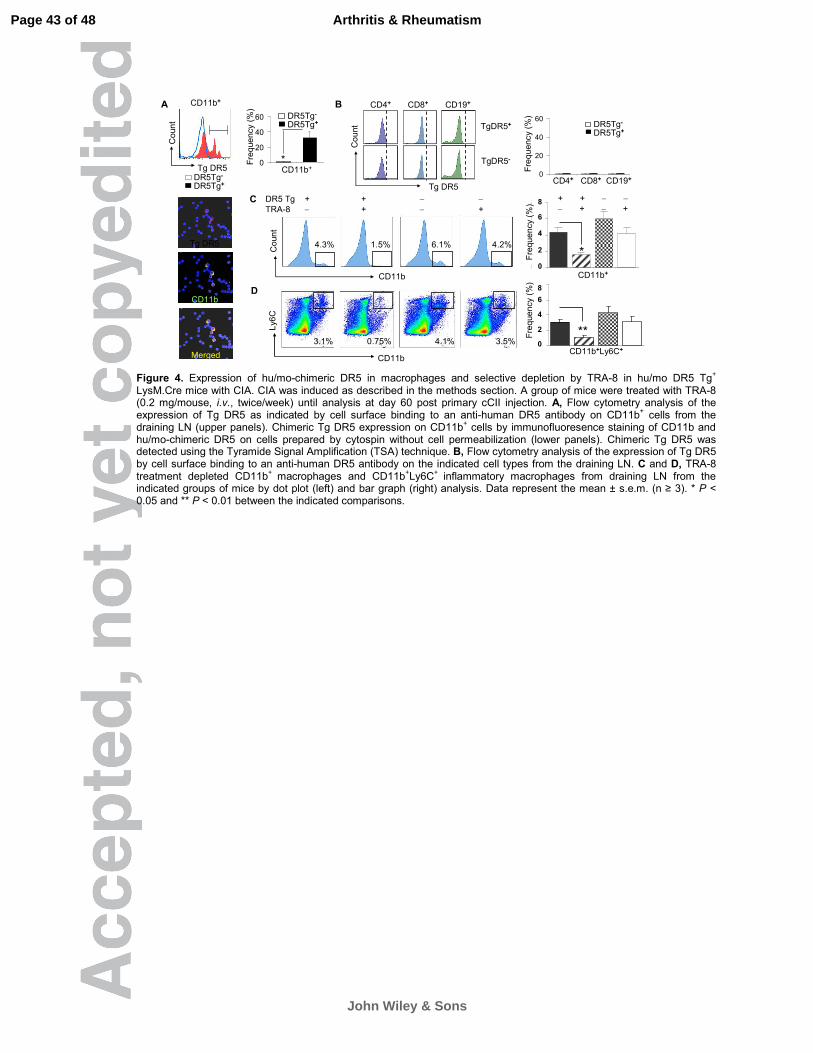
Figure 4. Expression of hu/mo-chimeric DR5 in macrophages and selective
depletion by TRA-8 in hu/mo DR5 Tg+ LysM.Cre mice with CIA. CIA was induced
as described in the methods section. A group of mice were treated with TRA-8
(0.2 mg/mouse, i.v., twice/week) until analysis at day 60 post primary cCII
injection. A, Flow cytometry analysis of the expression of Tg DR5 as indicated by
cell surface binding to an anti-human DR5 antibody on CD11b+ cells from the
draining LN (upper panels). Chimeric Tg DR5 expression on CD11b+ cells by
immunofluoresence staining of CD11b and hu/mo-chimeric DR5 on cells
prepared by cytospin without cell permeabilization (lower panels). Chimeric Tg
DR5 was detected using the Tyramide Signal Amplification (TSA) technique. B,
Flow cytometry analysis of the expression of Tg DR5 by cell surface binding to
an anti-human DR5 antibody on the indicated cell types from the draining LN. C
and D, TRA-8 treatment depleted CD11b+ macrophages and CD11b+Ly6C+
inflammatory macrophages from draining LN from the indicated groups of mice
by dot plot (left) and bar graph (right) analysis. Data represent the mean ± s.e.m.
(n ≥ 3). * P < 0.05 and ** P < 0.01 between the indicated comparisons.
Figure 5. Apoptosis induced by TRA-8 in the joints of hu/mo-chimeric DR5 Tg+
LysM. Cre mice with CIA. A, In vivo imaging of TRA-8 induced apoptosis in
joints. B, Quantitation of the imagines. DR5 Tg+ LysM.Cre and control LysM.Cre
Page 36 of 48
John Wiley & Sons
Arthritis & Rheumatism

37
mice were induced to develop CIA. At 8 week after induction, baseline levels of
the caspase activity were measured using the caspase-targeted activity based
probe AB50-Cy5 (left panel). Mice were then treated with TRA-8 (0.2 mg, day 0
and 3) and apoptosis imaging using AB50-Cy5 was performed on the same mice
on day 6 after initiating TRA-8 treatment (right panel). C, Immediately after the
second AB50-Cy5 imaging, the joints were removed, fixed and processed for
H.&E. (left), Mac-3 staining (middle, counterstained with methyl green), and
TUNEL (right, counterstained with hematoxyline). D, Quantitative analysis of the
TUNEL+ cells of the indicated cell types in synovium. The values on the Y-axis
represent the percentages of TUNEL-positive cells of the indicated cell types.
Five randomly chosen fields of synovium were evaluated for each section. E,
erosion; H, hyperplasia; M, macrophages, and TU, TUNEL. Scale bar, 100 µm.
Data represent mean ± s.e.m. (n ≥ 3). ** P < 0.01 between the indicated
comparisons.
Figure 6. TRA-8 treatment ameliorates the severity of CIA in hu/mo-chimeric
DR5 Tg+ LysM.Cre mice. A, CIA was induced in hu/mo DR5 Tg+ LysM.Cre
(upper panel) and control DR5 Tg– LysM.Cre mice (lower panel). Arrows indicate
the intradermal injection of chicken type II collagen on day 0 and 30. TRA-8
treatment was initiated on day 28 (arrow heads). Clinical scores (n = 6 per group)
were assessed until the mice were sacrificed on day 60. Data are presented as
the mean clinical score ± s.e.m. * P < 0.05 and ** P < 0.01 versus TRA-8 treated
group at the indicated time point. B, Cathepsin activity in joints was measured by
Page 37 of 48
John Wiley & Sons
Arthritis & Rheumatism

38
in vivo imaging using the NIRF-probe ProSense 750. C, Quantitative analysis of
ProSense 750 intensity. Data are presented as the mean ± s.e.m. * P < 0.05
between the indicated comparisons. D, Histological assessment of
representative knee joints from TRA-8-treated DR5 Tg+ LysM.Cre DR5 (left
panels) and DR5 Tg− LysM.Cre mice (right panels), which included H&E , Mac-3,
and TRAP staining as indicated (scale bar: 100 µm). Both groups were treated
with TRA-8 weekly for one month. E, erosion; H, hyperplasia; M, macrophages,
and TR, TRAP.
Figure 7. TRA-8 treatment decreases the expression of pro-inflammatory
cytokines and exhibits immunomodulatory effects in hu/mo DR5 Tg+ LysM.Cre
mice. CIA was induced in hu/mo DR5 Tg+ LysM.Cre and control DR5 Tg–
LysM.Cre mice as described in the methods section. TRA-8 treatment was
initiated on day 28 until analysis at day 60 post primary cCII injection. A, Sera
levels of IL-6 and IL-17A of indicated mice with TRA-8 treatment were analyzed
by ELISA. B, Absolute copy numbers of Tnfa, Il6, Il17, Il23(p19), Irf5 and Foxp3
of synovium of the indicated mice were determined by qRT-PCR and
represented as copy number x105/Gapdh. C, Percentage of IL-23+ CD11b+
macrophages from the draining LN of the indicated mice treated with TRA-8 on
day 60 (CD11b+ gated) was determined by flow cytometry. D, Percentage of
Th17 (IL-17+) and Th1 (IFN-γ+) cells (CD4+ gated) and Tregs (CD4+, Foxp3+)
from the draining LN of the indicated mice treated with TRA-8 on day 60 were
Page 38 of 48
John Wiley & Sons
Arthritis & Rheumatism

39
analyzed by flow cytometry. Data are presented as the mean ± s.e.m. * P < 0.05
and ** P < 0.01 between the indicated comparisons.
Page 39 of 48
John Wiley & Sons
Arthritis & Rheumatism

A
Transmembrane Domain Death Domain
HUMAN MEQRGQNAPAASGARKRHGPGPREARGARPGLRVPKTLVLVVAAVLLLVSAESALITQQD 60
MOUSE MEPPGPSTPTASAAARADHYTP----GLRP---LPKRRLLYSFALLLAVLQAVFVPVTAN 53
** * .:*:**.* : . * * ** :** :* *:** * : . :
HUMAN LAPQQRAAPQQKRSSPSEGLCPPGHHISEDGRDCISCKYGQDYSTHWN-DLLFCLRCTRC 119
MOUSE PAHNRPAGLQRPEESPSRGPCLAGQYLSEG--NCKPCREGIDYTSHSNHSLDSCILCTVC 111
* :: *. *: ..***.* * .*:::**. :* .*: * **::* * .* *: ** *
HUMAN DSGEVELSPCTTTRNTVCQCEEGTFREEDSPEMCRKCRTGCPRGMVKVGDCTPWSDIECV 179
MOUSE KEDKVVETRCNITTNTVCRCKPGTFEDKDSPEICQSCSN-CTDGEEELTSCTPRENRKCV 170
...:* : *. * ****:*: ***.::****:*:.* . *. * :: .*** .: :**
HUMAN HKESGTKHSGEAPAVEETVTSSPGTPASPCSLSGIIIGVTVAAVVLIVAVFVCKSLLWKK 239
MOUSE SK----------------------TAWASWHKLGLWIGLLVPVVLLIGALLVWKTGAWRQ 208
* *. :. *: **: *..*:** *::* *: *::
HUMAN VLPYLKGICSGGGGDPERVDRSSQRPGAEDNVLNEIVSILQPTQVPEQEMEVQEPAEPTG 299
MOUSE WLLCIKRGCER---DPE--------------SANSVHSSLLDRQTSS------------- 238
* :* *. *** *.: * * *...
HUMAN VNMLSPGESEHLLEPAEAERSQRRRLLVPANEGDPTETLRQCFDDFADLVPFDSWEPLMR 359
MOUSE ----TTNDSNHNTEPGKTQKTG-KKLLVPVNGNDSADDLKFIFEYCSDIVPFDSWNRLMR 293
:..:*:* **.::::: ::****.* .*.:: *: *: :*:******: ***
HUMAN KLGLMDNEIKVAKAEAAGHRDTLYTMLIKWVNKTGRDASVHTLLDALETLGERLAKQKIE 419
MOUSE QLGLTDNQIQMVKAETLVTREALYQMLLKWRHQTGRSASINHLLDALEAVEERDAMEKIE 353
:*** **:*::.***: *::** **:** ::***.**:: ******:: ** * :***
HUMAN DHLLSSGKFMYLEGNADSAMS------- 440
MOUSE DYAVKSGRFTYQNAAAQPETGPGGSQCV 381
*: :.**:* * :. *:. .
Junction
3 kb promoter-1st Intron- Chim DR5-3’UTR
B
Intr
a-
Extr
a-c
ellu
lar
Hu D
R5
3 kb – Chim DR5
3 kb promoter-1st Intron- Chim DR5
1 kb – Chim DR5
CMV – HuDR5
CMV – MoDR5
CMV – Chim DR5
1
2
3
4
5
7
6
D
C
0
1
2
3
4
55
Ce
ll su
rfa
ce
chim
DR
5 (
% )
0
2
4
6
8
10
12
14
16
1818
14
10
6
20
* * **
TRA8 Isotype4
3
2
1
0
1 2 3 4 5 6 7
1 2 3 4 5 6 7
Lum
insce
nce
co
un
nt/
10
6
** *
5
Mo D
R5
Chim
DR
5
Figure 1. Human, mouse and chimeric DR5 and their expression and function in inducing apoptosis by TRA-8. A, Amino acid alignment of human and mouse DR5. Junction between human and mouse DR5 in the chimeric molecule, transmembrane, and death domain are as indicated. B, Schematic diagram representing the human, mouse and chimeric DR5 (left), and human, mouse and chimeric DR5 constructs generated with different promoters and regulatory elements (right). C, FACS analysis of cell surface expression of DR5 recognized by an anti-human DR5 antibody in NIH3T3 cells transiently transfected with the indicated constructs. Percentage of the hu/mo DR5
+ cells was shown. N ≥ 3. * P < 0.05 versus result from construct 1. D, TRA-8 mediated killing in NIH3T3 transiently transfected with
the indicated constructs. Cell viability was determined using the ATPLite assay. For panels C and D, the data represent the mean ±
s.e.m. (n ≥ 3). Hu, human; mo, mouse; chim, chimeric. * P < 0.05 and ** P < 0.01 versus isotype control.
Page 40 of 48
John Wiley & Sons
Arthritis & Rheumatism

CA
[3H
] th
ym
idin
e
incorp
ora
tion (
x10
3)
0
10
20
30
contr ol T RA-8
*
LPS stimulated
DR5 Tg- DR5 Tg+
TRA-8
Isotype
CD11b
Coun
t
0
5
10
15
20
1 2 3
Isotype Early Late
TRA-8 TRA-8
* *
0
5
10
15
20
CD
11b
hig
h c
ells
(%
)
CD8+CD4+ CD19+
CD11b+
Gr1+CD11b+
Ly6C+
CD11b-
Ly6C+
Hu/mo DR5
Count
B
W.T. control
Ubc.Cre
DR5 Ubc.Cre
D
0 20 40 60 80 100
Ly6C-CD11b+
Ly6C+CD11b+
CD11cGr1
CD19
CD8
CD4
Ly6C+
CD11b+
CD4+
CD8+
CD19+
CD11b+
Gr1+
hu/mo DR5+ (%)
0 20 40 60 80 100
CD11b-
Ly6C+
CD11b+
Ly6C+
00.10.20.30.40.50.60.70.80.9
Skin
Sku
ll
LN
Bra
in
Sp
ina
l C
ord
Eye
Sa
liva
Gla
nd
Th
ym
us
Hea
rt
Lun
g
Sto
mach
Pa
ncre
as
Sp
lee
n
Sm
all In
testin
e
Co
lon
Ap
ee
ndix
Liv
er
Kid
ne
y
Urin
ary
Bla
dde
r
Testis
Pa
w
Ta
il
Ske
leta
l M
uscle
Bon
e M
arr
ow
Fa
t
Ca
rtilag
e
Transgenic Dr5 Endogenous Dr5
Skin
Skull
LN
Brain
Spinal Cord
Eye
Salivary Gland
Thymus
Heart
Lung
Stomach
Pancreas
Spleen
Small Intestine
Colon
Appendix
Liver
Kidney
Urinary Bladder
Testis
Paw
Tail
Skeletal Muscle
Bone Marrow
Fat
Cartilage
Dr5
rela
tive t
o G
ap
dh
R² = 0.4638
P = 0.0001 Isotype
Early TRA-8
Late TRA-8
W.T.control
Ubc.Cre
DR5 Tg+ Ubc.Cre
0
10
20
30
0 20 40 60 80 100
Figure 2. Hu/mo-chimeric Tg DR5 expression and function in Ubc.Cre DR5 Tg mice. A, Real-time PCR analysis of the chimeric DR5 expression in the indicated tissues obtained from the Ubc.Cre DR5 double-positive transgenic mice after induction of Cre expression by tamoxifen. Expression is represented as the ratio of copy numbers of chimeric Dr5 or endogenous mouse Dr5 to those of Gapdh. The correlation R
2 and correlation P value are shown in the upper-right of the panel. B, Transgenic chimeric DR5
cell surface expression on different immune cells was analyzed by FACS (left panels). The chimeric DR5 expression is representative of three experiments (right panels). C, The percentage of CD11b
high spleen macrophages from mice with and
without TRA-8 treatment (0.2 mg, i.v., twice/week for 2 months) was determined by FACS. Data represent the mean ± s.e.m. (n ≥
3). D, Proliferation of LPS-stimulated spleen cells from DR5 Tg− Ubc.Cre and hu/mo DR5 Tg+ Ubc.Cre mice treated with TRA-8 or
isotype control antibody was determined using the [3H]-thymidine incorporation assay (n ≥ 3). * P < 0.05 versus isotype control.
Page 41 of 48
John Wiley & Sons
Arthritis & Rheumatism

0
1
2
3
4
5
6
7
8
0 20 23 25 27 29 31 33 35 37 39 41 43 45
0
1
2
3
4
5
6
7
8
9
0 20 23 25 27 29 31 33 35 37 39 41 43 45
C
DB
A
Art
hritis S
core
DR5 Tg+ Isotype
DR5 Tg+ Early TRA-8
DR5 Tg+ Late TRA-8
**
Days post primary cCII
Days post primary cCII
Art
hritis S
core
DR5 Tg- Isotype
DR5 Tg- Early TRA-8
DR5 Tg- Late TRA-8
DR5 Tg+
Early TRA-8
H&E Mac-3
H
EE
M
DR5 Tg-
Early TRA-8
200X
10
0 X
200X
10
0 X
M
Figure 3. TRA-8 treatment prevents the development and attenuates the severity of CIA in hu/mo-chimeric DR5 Tg+ Ubc.Cre mice.
CIA was induced in the indicated DR5 Tg+ (A) and DR5 Tg
- mice (B) as described in detail in the methods section. Chicken type II
collagen emulsified in CFA and IFA was administered intradermally on day 0 and day 32 respectively (arrows). Early and late TRA-8 treatment (0.2 mg/mouse, once per week) was initiated on day 0 and day 30 respectively (arrow heads) and continued until sacrifice. Clinical scores (0-3 per paw; n=6 per group) were assessed daily until the mice were sacrificed. Data are presented as mean arthritis score ± s.e.m.. ** P < 0.01 versus TRA-8-treated groups at the same time point. C and D, Representative H&E and Mac-3 immunohistochemical staining of the knee joint sections from a TRA-8 treated hu/mo DR5 Tg
+ Ubc.Cre (C) or a TRA-8 treated DR5
Tg− Ubc.Cre (D) mouse. The magnification of the objective lens used to acquire the indicated images is shown in the left. E, erosion; H, hyperplasia; M, macrophages, (Scale bar, 100 µm).
Page 42 of 48
John Wiley & Sons
Arthritis & Rheumatism
A
C
D
B
Tg DR5
CD11b
Merged CD11b
Ly6
C
DR5 Tg + + − −
TRA-8 − + − +
CD11bC
oun
t
4.3% 1.5% 6.1% 4.2%
3.1% 0.75% 4.1% 3.5% 0%
2%
4%
6%
8%
1 2 3 4
**
CD11b+Ly6C+
8
6
4
2
0
Fre
que
ncy (
%)
0%
2%
4%
6%
8%
1 2 3 4
*
+ + − −
− + − +
CD11b+
8
6
4
2
0
Fre
qu
en
cy (
%)
Tg DR5C
oun
t
CD11b+ CD4+ CD8+ CD19+
Tg DR5
Co
unt TgDR5+
TgDR5-
0
20
40
60
CD4 CD8 CD19
DR5Tg-
DR5Tg+
CD4+ CD8+ CD19+
DR5Tg-
DR5Tg+
Fre
qu
ency (
%)
0
20
40
60
DR5Tg- DR5Tg+
Fre
que
ncy (
%)
CD11b+*
DR5Tg-
DR5Tg+
DR5Tg-
DR5Tg+
Figure 4. Expression of hu/mo-chimeric DR5 in macrophages and selective depletion by TRA-8 in hu/mo DR5 Tg+
LysM.Cre mice with CIA. CIA was induced as described in the methods section. A group of mice were treated with TRA-8 (0.2 mg/mouse, i.v., twice/week) until analysis at day 60 post primary cCII injection. A, Flow cytometry analysis of the expression of Tg DR5 as indicated by cell surface binding to an anti-human DR5 antibody on CD11b
+ cells from the
draining LN (upper panels). Chimeric Tg DR5 expression on CD11b+ cells by immunofluoresence staining of CD11b and
hu/mo-chimeric DR5 on cells prepared by cytospin without cell permeabilization (lower panels). Chimeric Tg DR5 was detected using the Tyramide Signal Amplification (TSA) technique. B, Flow cytometry analysis of the expression of Tg DR5 by cell surface binding to an anti-human DR5 antibody on the indicated cell types from the draining LN. C and D, TRA-8 treatment depleted CD11b
+ macrophages and CD11b
+Ly6C
+ inflammatory macrophages from draining LN from the
indicated groups of mice by dot plot (left) and bar graph (right) analysis. Data represent the mean ± s.e.m. (n ≥ 3). * P < 0.05 and ** P < 0.01 between the indicated comparisons.
Page 43 of 48
John Wiley & Sons
Arthritis & Rheumatism

C H&E Mac-3 TUNEL
TU
EN
L+
cells
in
the
syn
ovia
l sub
-lin
ing
laye
r(%
)
0
10
20
30
40
50
Total Macrophages Fibroblasts
Tg huDR5-Tg huDR5+
**
**
**
Total Mφ Fibroblasts
DR5 Tg-
DR5 Tg+
50
40
30
20
10
0
0
5
1 0
1 5
2 0
2 5
T g
h u D R 5 -
T g
h u D R 5 +
T g
h u D R 5 -
T g
h u D R 5 +
B e fo re T R A -8 A f te r T R A -8
**
DR5 Tg- DR5 Tg+ DR5 Tg- DR5 Tg+
Before TRA-8 After TRA-8
AB
50-C
y5
Inte
nsity/J
oin
t 25201510
50
A
Before TRA-8 treatment
DR5 Tg- DR5 Tg+
After TRA-8 treatment
DR5 Tg- DR5 Tg+
Caspase Activity
DB
H
H
E
E
H
H
E H
H
E H
H
TU
EH
H
E H
H
M
MDR5
Tg-
DR5
Tg+
Figure 5. Apoptosis induced by TRA-8 in the joints of hu/mo-chimeric DR5 Tg+ LysM. Cre mice with CIA. A, In vivo imaging of
TRA-8 induced apoptosis in joints. B, Quantitation of the imagines. DR5 Tg+ LysM.Cre and control LysM.Cre mice were induced
to develop CIA. At 8 week after induction, baseline levels of the caspase activity were measured using the caspase-targeted activity based probe AB50-Cy5 (left panel). Mice were then treated with TRA-8 (0.2 mg, day 0 and 3) and apoptosis imaging using AB50-Cy5 was performed on the same mice on day 6 after initiating TRA-8 treatment (right panel). C, Immediately after the second AB50-Cy5 imaging, the joints were removed, fixed and processed for H.&E. (left), Mac-3 staining (middle, counterstained with methyl green), and TUNEL (right, counterstained with hematoxyline). D, Quantitative analysis of the TUNEL
+ cells of the
indicated cell types in synovium. The values on the Y-axis represent the percentages of TUNEL-positive cells of the indicated cell types. Five randomly chosen fields of synovium were evaluated for each section. E, erosion; H, hyperplasia; M, macrophages, and TU, TUNEL. Scale bar, 100 µm. Data represent mean ± s.e.m. (n ≥ 3). ** P < 0.01 between the indicated comparisons.
Page 44 of 48
John Wiley & Sons
Arthritis & Rheumatism

A B C
0
10
20
30
40
1 2 3 4 5
Pro
Sense
inte
nsity/join
t
DR5 Tg − − − + +
TRA-8 − − + − +
NIRF − + + + +
*
40
30
20
10
0
D DR5 Tg+ TRA-8 Treatment
H&E Mac-3 TRAP
0123456789
101112
0 1 0 19 20 22 2 4 26 2 8 3 0 3 2 34 3 6 3 8 40 4 2 4 4 4 6 48 5 0 52 54 56 5 8 60 62 6 4 6 6 68
D R 5 T g - Iso type
D R 5 T g - T R A -8
0123456789
101112
0 10 1 9 20 22 24 26 28 30 32 34 36 38 40 42 44 46 4 8 50 52 54 56 58 60 62 64 66 6 8
DR5 Tg+ Isotype
DR5 Tg+ TRA-8 **
DR5 Tg- IsotypeDR5 Tg- TRA-8A
rthritis s
core
DR5 Tg − − − + +
TRA-8 − − + − +
NIRF − + + + +
Cathepsin Activity
200 X
1
00
X
H
E
E
HM H
E
TR
DR5 Tg- TRA-8 Treatment
Mac-3H&E TRAP
DR5 Tg+ TRA-8
DR5 Tg+ Isotype
10 20 30 40 50 60 70
12
8
4
0
12
8
4
010 20 30 40 50 60 70
Figure 6. TRA-8 treatment ameliorates the severity of CIA in hu/mo-chimeric DR5 Tg+ LysM.Cre mice. A, CIA was induced in hu/mo DR5
Tg+ LysM.Cre (upper panel) and control DR5 Tg
– LysM.Cre mice (lower panel). Arrows indicate the intradermal injection of chicken type II
collagen on day 0 and 30. TRA-8 treatment was initiated on day 28 (arrow heads). Clinical scores (n = 6 per group) were assessed until the mice were sacrificed on day 60. Data are presented as the mean clinical score ± s.e.m. * P < 0.05 and ** P < 0.01 versus TRA-8 treated group at the indicated time point. B, Cathepsin activity in joints was measured by in vivo imaging using the NIRF-probe ProSense 750. C, Quantitative analysis of ProSense 750 intensity. Data are presented as the mean ± s.e.m. * P < 0.05 between the indicated comparisons.
D, Histological assessment of representative knee joints from TRA-8-treated DR5 Tg+
LysM.Cre DR5 (left panels) and DR5 Tg− LysM.Cre
mice (right panels), which included H&E , Mac-3, and TRAP staining as indicated (scale bar: 100 µm). Both groups were treated with TRA-8 weekly for one month. E, erosion; H, hyperplasia; M, macrophages, and TR, TRAP.
Page 45 of 48
John Wiley & Sons
Arthritis & Rheumatism

0
2
4
6
8
Th1 Th17 Treg
DR5Tg+
DR5Tg-
DR5Tg+ DR5Tg-
IL-2
3
CD11b
0.2±0.05 5.8±0.8
A
Foxp
3
6.3 3.9
DR5Tg+ DR5Tg-
IFNγ
IL-1
7A 1.4
1.3 2.2
2.9
DR5Tg+ DR5Tg-D
C
B
Co
py n
um
be
r x 1
0^5
/Ga
pd
h
0
0.02
0.04
0.06
0.08
DR5Tg+ DR5Tg-
Tnfα
**0
200
400
600
800
DR5Tg+ DR5Tg-
Il6
**
0
2
4
6
8
DR5Tg+ DR5Tg-
Il23(p19)
**
0
5
10
15
DR5Tg+ DR5Tg-
Foxp3*
0
10
20
30
40
DR5Tg+ DR5Tg-
*
DR5Tg+ DR5Tg-
Co
py n
um
be
r x 1
0^5
/Gap
dh
Irf5
0
5
10
15
DR5Tg+ DR5Tg-
**
DR5Tg+ DR5Tg-
Il17a
* *
*
Fre
qu
ency(%
)
Th1 Th17 TregCD4
DR5Tg+DR5Tg-
0
200
400
600
DR5Tg+ DR5Tg-
pg/m
l
IL-6
**
0
100
200
300
DR5Tg+ DR5Tg-
IL-17A
*pg
/ml
DR5Tg+ DR5Tg-
Figure 7. TRA-8 treatment decreases the expression of proinflammatory cytokines and exhibits immunomodulatory effects in hu/mo DR5 Tg+ LysM.Cre mice. CIA was induced in hu/mo DR5 Tg+ LysM.Cre and control DR5 Tg– LysM.Cre mice as described in the
methods section. TRA-8 treatment was initiated on day 28 until analysis at day 60 post primary cCII injection. A, Sera levels of IL-6
and IL-17A of indicated mice with TRA-8 treatment were analyzed by ELISA. B, Absolute copy numbers of Tnfa, Il6, Il17, Il23(p19), Irf5 and Foxp3 of synovium of the indicated mice were determined by qRT-PCR and represented as copy number
x105/Gapdh. C, Percentage of IL-23+ CD11b+ macrophages from the draining LN of the indicated mice treated with TRA-8 on day 60
(CD11b+ gated) was determined by flow cytometry. D, Percentage of Th17 (IL-17+) and Th1 (IFN-γ+) cells (CD4+ gated) and Tregs (CD4+, Foxp3+) from the draining LN of the indicated mice treated with TRA-8 on day 60 were analyzed by flow cytometry. Data are
presented as the mean ± s.e.m. * P < 0.05 and ** P < 0.01 between the indicated comparisons.
Page 46 of 48
John Wiley & Sons
Arthritis & Rheumatism