mbbs 2 seminar breast cancer 2014 - dr rodney itaki's ... · connective tissue there are also...
TRANSCRIPT
Breast Cancer
Dr Rodney Itaki
Anatomical Pathology DisciplineDivision of Pathology
Muscles
2
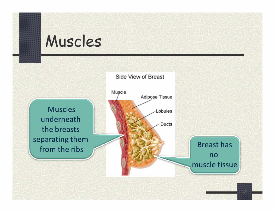
Breast hasno
muscle tissue
Musclesunderneath the breasts
separating them from the ribs
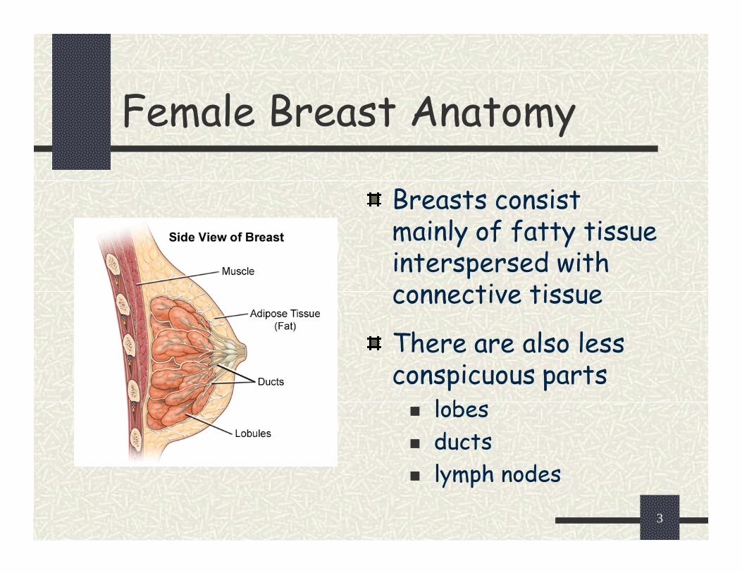
Female Breast AnatomyBreasts consist mainly of fatty tissue interspersed with connective tissueThere are also less conspicuous parts
� lobes� ducts� lymph nodes
3
Breast Gland
4
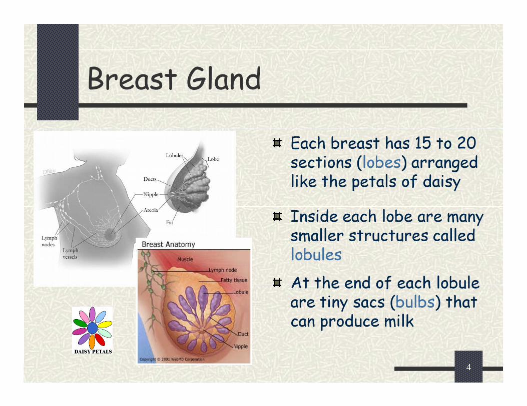
Each breast has 15 to 20 sections (lobes) arranged like the petals of daisyInside each lobe are many smaller structures called lobulesAt the end of each lobule are tiny sacs (bulbs) that can produce milk
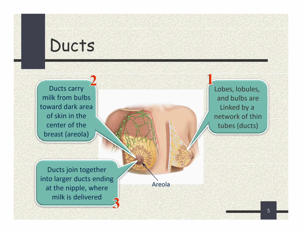
Ducts
5
Areola
Lobes, lobules, and bulbs areLinked by a
network of thintubes (ducts)
Ducts carrymilk from bulbs
toward dark areaof skin in thecenter of the
breast (areola)
Ducts join togetherinto larger ducts ending
at the nipple, wheremilk is delivered

Blood Supply
6

Venous Drainage
7

Lymphatic System
8
Lymph ducts: Drain fluid that carries white blood cells (that fight disease) from the breast tissues into lymph nodes under the armpit and behind the breastboneLymph nodes: Filter harmful bacteria and play a key role in fighting off infectionA network of vessels
Lymph ductLymph node

Lymphatic Drainage
9

Three Types of Vessels
10
BacteriaBlood
VesselsCell life
2
LymphNodes
LymphVessels
3
MilkLobules Ducts Nipple
1

Normal BreastBreast profile
A ducts
B lobules
C dilated section of duct to hold milk
D nipple
E fat
F pectoralis major muscle
G chest wall/rib cage
11
Enlargement
A normal duct cells
B basement membrane (duct wall)
C lumen (center of duct)
Illustration © Mary K. Bryson

Normal Histology
12
High power show inner cuboidalepithelia or low columbal layer & outer myoepithelial cell layer (contractile)

Normal Histology - Ducts
13
Intralobularexcretory ducts & alveoli lined by luminal epithelium & myoepithelialcells
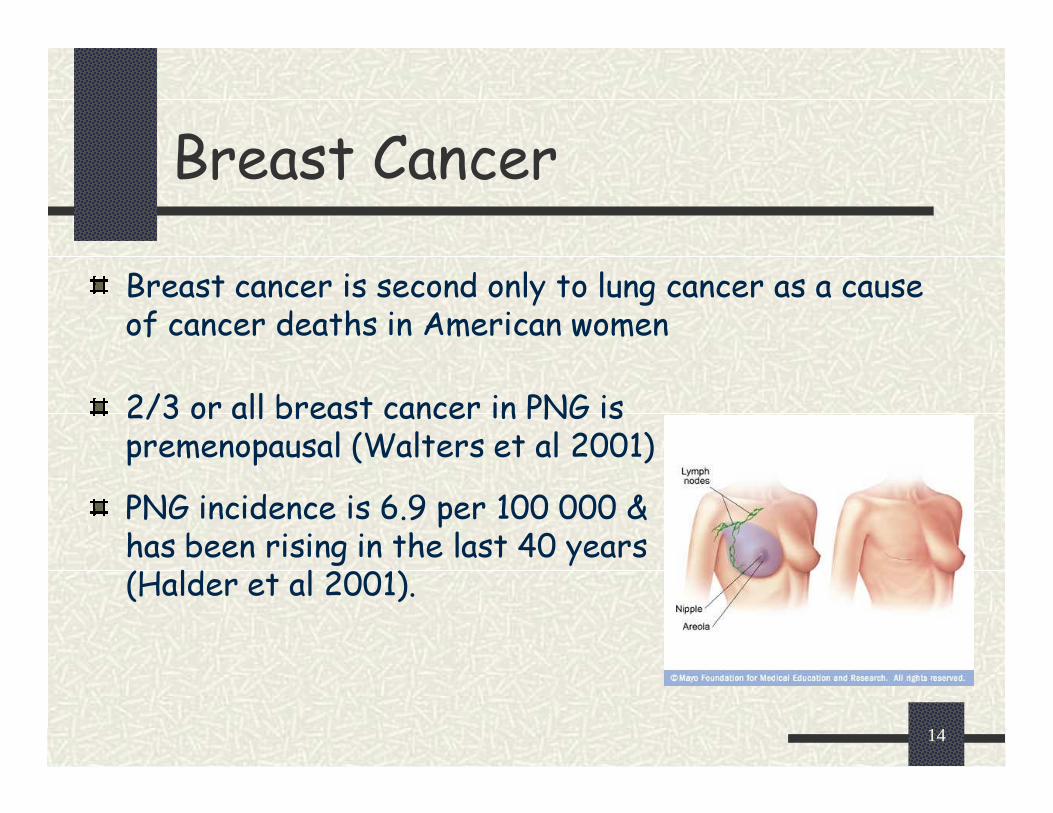
Breast Cancer
2/3 or all breast cancer in PNG is premenopausal (Walters et al 2001)PNG incidence is 6.9 per 100 000 & has been rising in the last 40 years (Halder et al 2001).
14
Breast cancer is second only to lung cancer as a cause of cancer deaths in American women

Epidemiology in PNG
Peak incidence: 45-54 age groups. But younger pts has been seen.
Highest incidence is 35-54 age groups.
15% of all breast cancers <35 years of age.
Most cases present late:
Common clinical signs on presentation:
� Ulceration of skin
� Peu d’ Orange
� Nipple retraction
� lymphoedema
15Halder et al 2001 ANZ J Surg.2001 Oct;71(10):590-3.

Risk Factors for Breast Cancer
Increasing age
Proliferative breast diseases
Carcinoma of the contralateral breast or endometrium
Radiation exposure
Geographical influences
Length of reproductive life –increases with early menarche and late menopause
Parity – increased risk in nulliparous
Exogenous Estrogens – small
Genetic factors – germ line mutations in BRAC1, BRAC2.
Hormonal influences
Environmental factors
Age at first child – increased in older primip
Obesity
16

BRAC 1 & BRAC 2
17
BRAC gene products involved in DNA repair

Classification
Invasive – 70-85%
� Ductal carcinoma – 79%
� Lobular carcinoma – 10%
� Tubular/Cribriform carcinoma – 6%
� Colloid (mucinous) carcinoma – 2%
� Medullary carcinoma – 2%
� Papillary carcinoma – 1%
Non-invasive (In Situ Carcinoma) – 15-30%
� Ducal carcinoma in situ – 80%
� Lobular carcinoma in situ – 20%18Robins Pathological Basis of Diseases, 6th Ed

Signs and Symptoms
19
Most common: lump or thickening in breast. Often painless
Change in color or appearance of areola
Redness or pitting of skin over the breast, like the skin of an orange
Discharge or bleeding
Change in size or contours of breast

Breast Cancer
20

Cancer Can also Invade Lymph or Blood Vessels
21Illustration © Mary K. Bryson
Cancer cells invade
lymph duct
Cancer cells invade blood vessel

NON-INVASIVE BREAST CARCINOMA
22

In Situ Carcinoma
Ductal carcinoma in situ (including Paget’s disease) most common
Lesions contain malignant cells that lack capacity to invade BM
However can spread throughout a ductal system
Divided into 4 types based on microscopic features
� Comedocarcinoma
� Noncomedocarcinoma DCIS
� Paget’s disease of the nipple
� DCIS with microinvasion
23

Ductal Carcinoma in situ (DCIS)
24Illustration © Mary K. Bryson
Ductal cancer cells
Normal ductalcellCarcinoma refers to any
cancer that begins in the skin or other tissues that cover internal organs
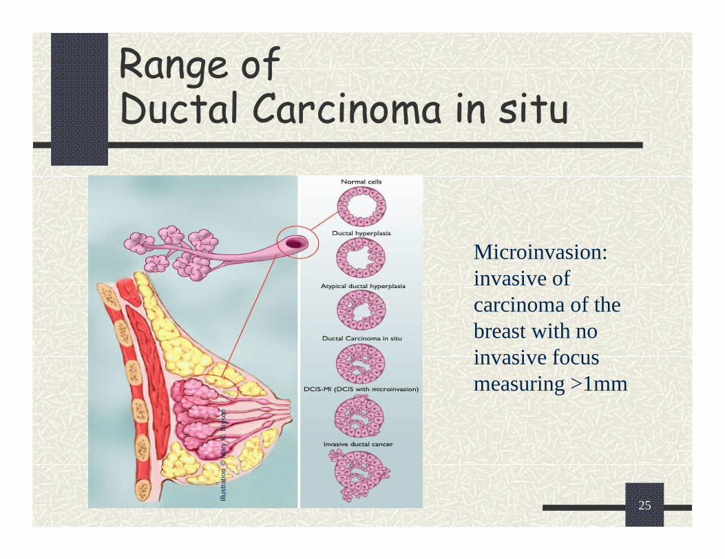
Range of Ductal Carcinoma in situ
25
Illus
trat
ion
© M
ary
K. B
ryso
n
Microinvasion: invasive of carcinoma of the breast with no invasive focus measuring >1mm

Microinvasion
26

Comedocarcinoma
Characterised by solid sheets of high-grade malignant cells and central necrosis.
27

Noncomedo DCISVery similar to comedocarcinoma
Cells appear monomorphic
3 variants – cribriform, papillary and micropapillary DCIS
28
CribriformDCIS: Lumen fills with secretions. Evenly spaced intraepithelial space
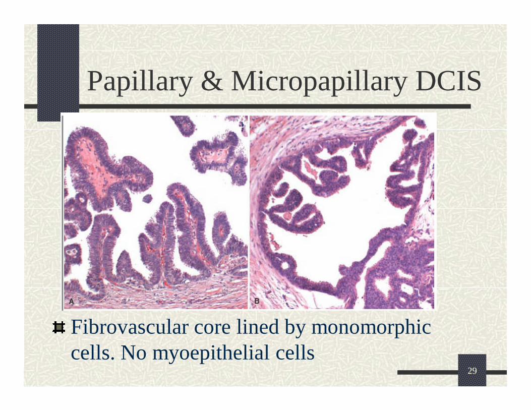
Papillary & Micropapillary DCIS
29
Fibrovascular core lined by monomorphiccells. No myoepithelial cells
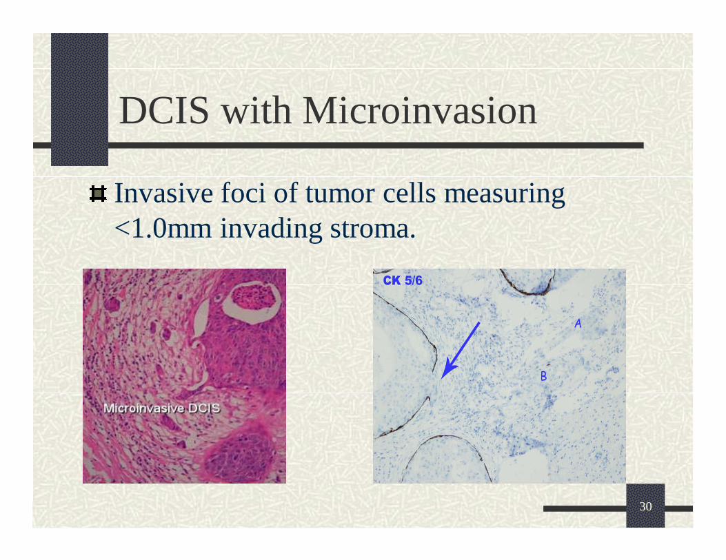
DCIS with Microinvasion
Invasive foci of tumor cells measuring <1.0mm invading stroma.
30
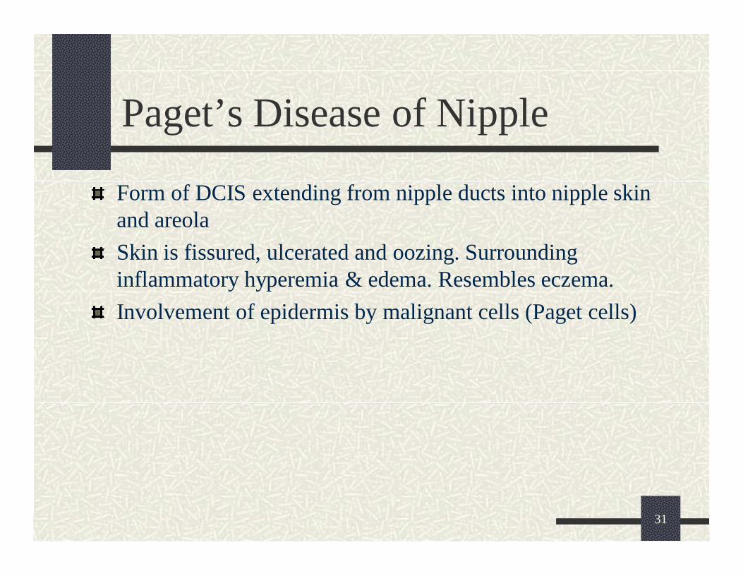
Paget’s Disease of Nipple
Form of DCIS extending from nipple ducts into nipple skin and areola
Skin is fissured, ulcerated and oozing. Surrounding inflammatory hyperemia & edema. Resembles eczema.
Involvement of epidermis by malignant cells (Paget cells)
31

Paget’s Disease of Nipple
32
Involvement of epidermis by malignant cells

Lobular Carcinoma In Situ
Proliferation in one or more terminal ducts or ductules (acini)
Monomorphic population of cells, loosely cohesive and larger than normal.
Oval to round nuclei with small nucleoli
Signet-ring cells containing mucincommonly present
33

Lobular Carcinoma In Situ
Commonly incidental finding on biopsies. Rarely forms mass
34

INVASIVE BREAST CARCINOMA
35
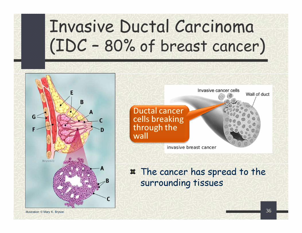
Invasive Ductal Carcinoma (IDC – 80% of breast cancer)
36
The cancer has spread to the surrounding tissues
Illustration © Mary K. Bryson
Ductal cancer cells breaking through the wall

Invasive Breast Cancer
Invasive ductal carcinoma accounts for 70-80% of invasive breast carcinoma.
Sharply demarcated nodules (1-2cm)
May attach to underlying structures
Macroscopic: Lesion is retracted below cut section and infiltrates surrounding tissue.
37

Invasive Ductal Carcinoma
Cells in cords, solid cell nests, tubules, anastomosing massess invading stroma.
Small to moderately hyperchromatic regular nuclei
Huge cells with large irregular hyperchromatic nuclei.
38Well differentiated Poorly differentiated
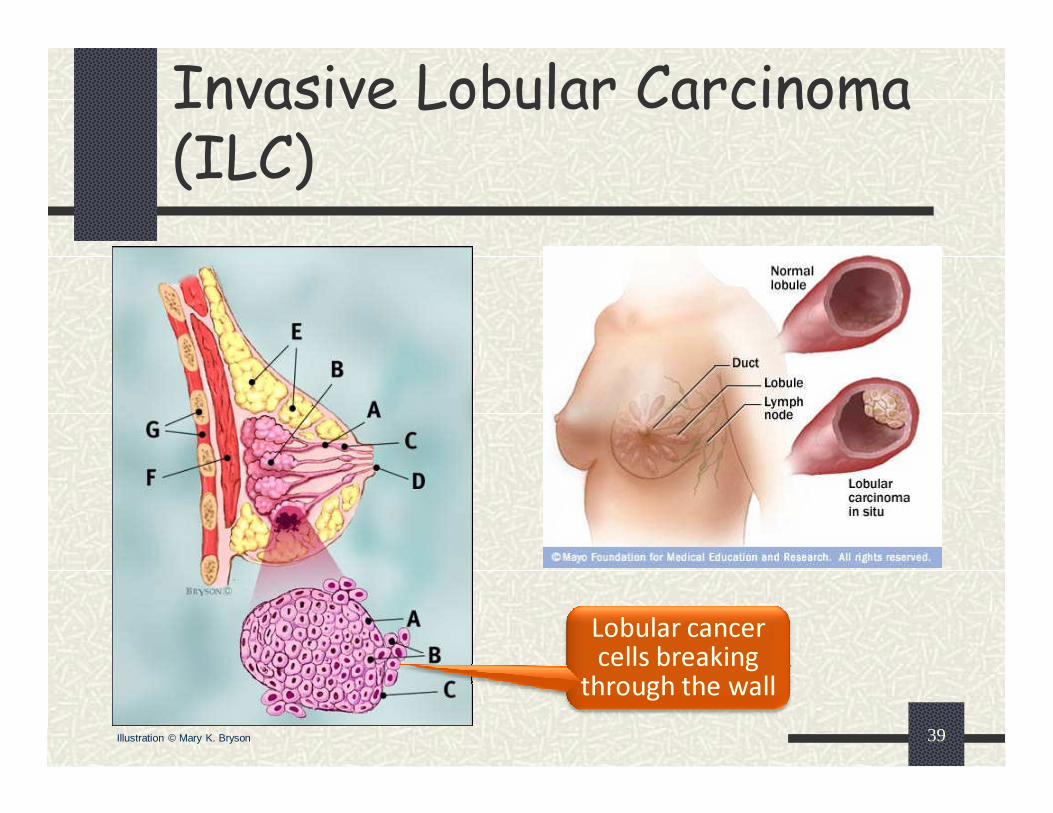
Invasive Lobular Carcinoma (ILC)
39Illustration © Mary K. Bryson
Lobular cancer cells breaking
through the wall

Invasive Lobular Carcinoma
Makes up 5-10% of cases
Bilateral (20% risk in contralateral breast) & multicentric within same breast
Frequent metastasis
Microscopy:� Strands of infiltrating tumor cells with no
formation of tubules or papillae
40
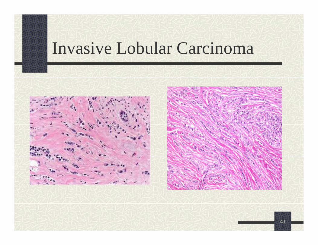
Invasive Lobular Carcinoma
41

Medullary Carcinoma
1-5% of invasive breast carcinoma
Large fleshy tumor (2-3cm)
Soft, fleshy consistency and well circumscribed.
Microscopy:
� Solid, syncytium-like sheets of large cells with pleomorphic nuclei.
� Moderate to marked lymphoplasmacytic infiltrate
� Noninfiltrating border (pushing border)
42

Medullary Carcinoma
43

Colloid (Mucinous) Carcinoma
Occurs in older women and grows slowly
1-6% of cases
Soft tumors and may mimic benign tumors
Microscopy shows large lakes of light staining mucin dissecting tissue spaces.
Floating within this mucin are small islands and isolated neoplastic cells
44

Colloid (Mucinous) Carcinoma
45
Balls of tumor cells floating in mucin

Tubular Carcinoma
2-10% of carcinomas less than 1cm on mammography.
Commonly picked up on mammogram
Present in late 35-40s.
Multifocal within one breast or can be bilateral.
Microscopy shows well formed tubules but no myoepithelial cells. Tumor cells in direct contact with stroma. Can be mistaken for a benign lesion.
DCIS (40%) & LCIS (10%) present46

Tubular Carcinoma
47
Tubular arrangement of cells. Single layer of infiltrating tumor cells. No myoepithelial cells

Features Common to Invasive Cancers
Adherent to underlying structures
Extension to skin show retraction & dimpling
Lymphedema
Nodal involvement
Distant sites: lung, bone, liver, adrenals, brain & meninges.
48
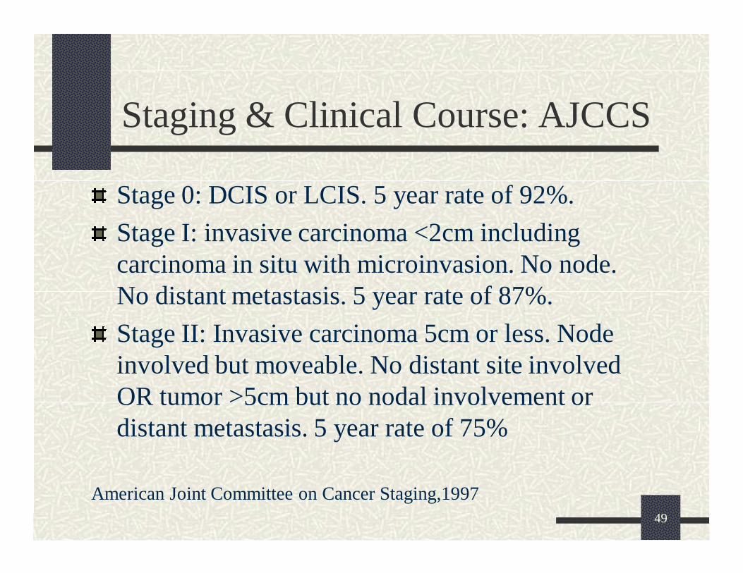
Staging & Clinical Course: AJCCS
Stage 0: DCIS or LCIS. 5 year rate of 92%.
Stage I: invasive carcinoma <2cm including carcinoma in situ with microinvasion. No node. No distant metastasis. 5 year rate of 87%.
Stage II: Invasive carcinoma 5cm or less. Node involved but moveable. No distant site involved OR tumor >5cm but no nodal involvement or distant metastasis. 5 year rate of 75%
49
American Joint Committee on Cancer Staging,1997

Staging& Clinical Course: AJCCS
Stage III: >5cm with nodal involvement OR cancer fixed to axillary node. OR any breast cancer with involvement of ipsilateralinternal mammary lymph nodes. OR Any breast cancer with skin involvement, pectoral or chest wall fixation, edema, clinical inflammatory carcinoma. No distant metastasis. 5 year rate of 46%
50
American Joint Committee on Cancer Staging,1997

Staging & Clinical Course: AJCCS
Stage IV: any form of breast cancer with distant metastasis, including ipsilateralsupraclavicular lymph node. 5 year rate of 13%.
Stage II & III are also subdivided according to number of axillary lymph node involvement.
51
American Joint Committee on Cancer Staging,1997

Prognostic Indicators
Lymph node metastasis
Locally advanced disease
Tumor size
Histological subtypes
Tumor grade
Oestrogen & progesterone receptor +ve or –ve.
� 54% of PNG tumors lack oestrogen or progesteronreceptors (Walters et al 1998)
� 4% positive for oesterogen & progesterone receptors (Walters et al 1998)
Lymphvascular invasion
DNA content
Expression of oncogenesor loss of expression of tumor-supressor genes
Angiogenesis
Presence of proteases
Proliferative rate
52

Diagnosis
History
Triple Test for breast lumps Inx: Clinical Exam, FNA, Imaging (ultrasound or mammogram)� Ultrasound - <35 y.o
� Mammography - >50 y.o
� 35-49 both can be used.
True cut biopsy
53

Management
Surgery
Chemotherapy
Radiotherapy
Monitoring
54

END
References Robins Pathological Basis of Diseases 6th Ed.
Various sources via Google images for images.
Download seminar notes at www.pathologyatsmhs.com
55