mechanisms involved in the homologous down-regulation of
TRANSCRIPT

Molecular and Cellular Endocrinology 173 (2001) 95–107
Mechanisms involved in the homologous down-regulation oftranscription of the follicle-stimulating hormone receptor gene in
Sertoli cells
Michael D. Griswold *, Jeong-Seon Kim, Walter A. TribleySchool of Molecular Biosciences, Washington State Uni6ersity, Pullman, WA 99164-4660, USA
Received 26 June 2000; accepted 25 September 2000
Abstract
The action of follicle-stimulating hormone (FSH) in spermatogenesis is regulated at a fundamental level by controlling thenumber of competent receptors present at the surface of Sertoli cells. By controlling the number of receptors, the cell is able tomodulate the timing and magnitude of subsequent signal transduction in response to FSH. One mechanism of control is thedown-regulation of the steady state levels of the FSH receptor gene after exposure to FSH or agents that stimulate or prolongthe cAMP signal transduction cascade (homologous down-regulation) in Sertoli cells. The goals of this study were to examinepossible mechanisms involved in the down-regulation of mRNA levels of this gene. Analysis of transcription and processing bya PCR-based assay showed that treatment of Sertoli cells with FSH caused at least a 50% reduction of hnRNA for the FSHreceptor gene. Reporter genes controlled by 5% flanking sequences of the FSH receptor gene that were transiently transfected intoSertoli cells were not down-regulated. In electrophoretic mobility shift assays (EMSA), cAMP-inducible nuclear protein complexcontaining c-Fos formed on the activator protein-1/cAMP responsive element-like site located at −216 to −210 in the promoterof the rat FSH receptor gene. We concluded from this study that there was no evidence for the putative role of ICER in thedown-regulation of the FSH receptor promoter. In addition, the FSH-induced down-regulation of the transcription of the FSHreceptor gene in Sertoli cells was prevented by the treatment of Sertoli cells with trichostatin A prior to the addition of FSH. Thisexperiment coupled with other observations suggested that the down-regulation may be mediated by changes in chromatinstructure. © 2001 Elsevier Science Ireland Ltd. All rights reserved.
Keywords: Follicle stimulating hormone receptor; Homologous down-regulation; Sertoli; Testis; Spermatogenesis; ICER
www.elsevier.com/locate/mce
1. Introduction
One of the most important biochemical signalingnetworks involved in controlling normal spermatogene-sis in mammals exists between the anterior lobe of thepituitary gland and Sertoli cells in the testis (Griswoldet al., 1975, 1976, 1977; Orth, 1984; Singh and Handels-man, 1996; Kumar et al., 1997; Tapanainen et al., 1997;Dierich et al., 1998). The biological events controlled bythis endocrine mechanism result from the interaction offollicle-stimulating hormone (FSH) with the FSH re-ceptor and the subsequent transduction of molecularinformation across the membrane leading to the pro-
duction of second messengers such as cAMP (Means etal., 1980) and Ca2+ (Chaudhary et al., 1996; Lalevee etal., 1999) and changes in gene expression.
Continuous stimulation of Sertoli cells with FSHleads to a desensitization of the cells to FSH(Gnanaprakasam et al., 1979). Desensitization of theFSH response in Sertoli cells involves multiple iden-tified steps in the FSH/cAMP signal transduction path-way including the following: (1) the rapidinternalization and sequestration of the ligand-boundreceptor (Fletcher and Reichert, 1984; Saez and Jail-lard, 1986; Shimizu et al., 1987); (2) post-translationalmodification of the receptor (Quintana et al., 1994;Hipkin et al., 1995); (3) reduction in adenylate cyclaseactivity (Le Gac et al., 1985); (4) increased phosphodi-esterase activity (Conti et al., 1983, 1986); (5) direct
* Corresponding author. Fax: +1-509-3359688.E-mail address: [email protected] (M.D. Griswold).
0303-7207/01/$ - see front matter © 2001 Elsevier Science Ireland Ltd. All rights reserved.PII: S 0 3 0 3 -7207 (00 )00412 -3
M.D. Griswold et al. / Molecular and Cellular Endocrinology 173 (2001) 95–10796
inhibition of protein kinase A by protein kinase Ainhibitor (Tash et al., 1979, 1981); and (6) the down-regulation of the transcription of the FSH receptorgene (Themmen et al., 1991; Monaco et al., 1995;Maguire et al., 1997). Given that the concentration ofFSH in the serum of non-seasonal-breeding males isrelatively constant, the regulation of the number ofFSH receptors and their competency to bind FSH andtransduce signal may be an important level of controlon the action of FSH in males (McGuiness and Gris-wold, 1995).
Homologous down-regulation of transcription of theFSH receptor gene in cultured rat Sertoli cells was firstreported by Themmen et al. (1991). The loss of approx-imately 90% of the steady-state level of FSH receptormRNA and a parallel loss of 125I FSH binding wasmeasured 4 h after the addition of 500 ng/ml OvineFSH (oFSH) to Sertoli cells in primary culture(Themmen et al., 1991). Themmen and coworkers sug-gested that down-regulation was stimulated by(Bu)2cAMP, did not require de novo transcription ortranslation and concluded that RNA degradation waslikely responsible for the homologous down-regulationof FSH receptor mRNA in Sertoli cells. Maguire andcoworkers measured the relative contribution of FSHreceptor mRNA decay to the process of homologousdown-regulation by uncoupling transcription andmRNA decay with the transcription inhibitor actino-mycin D (Maguire et al., 1997). Maguire and coworkersalso demonstrated homologous down-regulation of theFSH receptor gene in vivo. The results of their workdid not support the hypothesis that homologous down-regulation occurred via a cAMP-inducible mRNA de-cay pathway but rather suggested a role fortranscription in homologous down-regulation is likely.
A model for the mechanism of homologous down-regulation of the FSH receptor in Sertoli cells thatinvolved the inputs of de novo transcription and trans-lation was presented by Monaco et al. (1995). Theypresented evidence that the inducible cAMP early re-pressor (ICER) bound to the rat FSH receptor pro-moter at a presumptive CRE-like site in vitro andrepressed expression of a FSH promoter driven reportergene. This was observed when ICER was overexpressedfrom the SV40 promoter on a co-transfected templatein transient transfection assays (Monaco et al., 1995).In this study we have re-examined this putative CRE-like site and demonstrate that it functions as an AP-1site. The transcription factor, c-Fos, induced by cAMPor FSH, can bind to the AP1 site in the promoterregion of the FSH receptor gene in vitro and in vivo.
The restricted pattern of expression of the FSHreceptor gene to Sertoli cells in males and homologousdown-regulation of the gene are relaxed when non-chromatin templates are used to study the regulation ofthis important gene (Linder et al., 1994; Monaco et al.,
1995). We were able to demonstrate that treatment ofimmature rat Sertoli cells in primary culture with tri-chostatin A, which inhibits histone deacetylase activitycompletely prevents the homologous down-regulationof the transcription of the FSH receptor gene.
2. Materials and methods
2.1. DNA sequencing and synthesis
Automated analysis of DNA sequencing reactionsderived from PCR-based cycle sequencing was per-formed by the Laboratory for Bioanalysis and Biotech-nology (LBB) at Washington State University.Oligonucleotides were synthesized by the LBB usingphosphoramidite chemistry. All oligonucleotides weregel-purified on 12% polyacrylamide gels containing 7 Murea, eluted in TEN buffer (10 mM Tris–HCl, pH 8.0,1 mM EDTA, 0.25 M NaCl), ethanol precipitated, andwashed twice with 70% EtOH.
2.2. Cell culture
Sertoli cells were removed from 18- to 20-day-oldmale Sprague–Dawley rats as previously described(Karl and Griswold, 1990). The cells were plated inHam’s F-12 medium (Gibco BRL, Grand Island, NY)on 60-mm culture dishes for transient transfection ex-periments and 150-mm culture dishes for RNA extrac-tion. To reduce the amount of contaminating germ cellnuclear protein and RNA, the number of germ cellswas reduced by hypotonic shock using dilute Ham’sF-12 (1:10 dilution of Ham’s F-12 in dH2O) for 2 minon the third day in culture (Toebosch et al., 1989).Fresh Ham’s F-12 was added to the cultures for anadditional 24 h prior to the start of all experiments.
2.3. Quantitati6e RT-PCR analysis of FSH receptorhnRNA
A quantitative RT-PCR-based method of measuringthe level of FSH receptor hnRNA in oFSH-treated anduntreated cells was performed (Elferink and Reiners,1996). This method is based on the inclusion of aknown amount of in vitro-transcribed internal standardRNA that is identical to the region of the targethnRNA that will be amplified except that a restrictionendonuclease cleavage site is introduced in the standardby recombinant techniques. The internal standardRNA is reverse-transcribed and amplified with the sameefficiency as the target RNA. After PCR, the internalstandard is cleaved at the novel restriction endonucle-ase cleavage site allowing for the separation of theinternal standard from the target by electrophoresis.The signal from the internal standard is then used tonormalize the signal for the target.
M.D. Griswold et al. / Molecular and Cellular Endocrinology 173 (2001) 95–107 97
The internal standard plasmid construct was gener-ated by subcloning the first exon and part of the 5% endof the first intron of the FSH receptor gene intopGEM-T (Promega). The insert was generated by PCRusing subclone 54.111 of the rat FSH receptor gene asthe template (Heckert et al., 1992) and the followingprimers: sense primer c1 (+ −80) CAG TGT GTGGAG GAG CCT (−63) and antisense primer c1(dT)20 G6 AT ATC AGT CTA ATA GGA (this primer iscomplementary to the FSHR hnRNA in the first in-tron). Antisense primer c1 adds a poly (dT)20 tail tothe antisense strand of the plasmid insert that encodesa short poly (A) tail on the in vitro-transcribed internalRNA. This poly(A) tail facilitates the purification offull-length internal standard RNA molecules from thein vitro transcription reaction by allowing oligo(dT)cellulose affinity chromatography (Elferink and Rein-ers, 1996). A NcoI cleavage site was generated in thesubclone by introducing a T to G base substitution byPCR-based site-directed mutagenesis (Fig. 1A)(Higuchi, 1990). The final construct was verified byDNA sequencing and named pMDG1.
Internal standard RNA was generated by in vitrotranscription using Ambion’s Maxi Script protocol(Ambion). Sense FSH receptor internal standard hn-RNA was generated by transcription from the SP6promoter in SphI-linearized pMDG1 DNA by SP6
RNA polymerase (Ambion) and purification byoligo(dT) cellulose affinity chromatography. Thepurified internal standard RNA was quantified by mea-suring the absorbance at 260 nm.
Prior to the reverse transcription reactions, rat Sertolicells in primary culture were treated for 5 or 6 h withthe following reagents depending on the experiment: 5mg/ml actinomycin D (Sigma); 5 mg/ml actinomycin Dand 2 U/ml oFSH (NIDDK-oFSH-20-SIAFP); 10 mg/ml a-amanitin (Boehringer Mannheim, Mannheim,Germany); 10 mg/ml a-amanitin and 2 U/ml oFSH; 2U/ml oFSH only; or untreated. Total RNA, extractedfrom Sertoli cells as described previously (Chomczynskiand Sacchi, 1987; Maguire et al., 1997), was treatedwith DNase I (Boehringer Mannheim) in the presenceof 1 mM manganese as previously described except thatreactions were scaled-up to accommodate �30 mg oftotal RNA (Bauer et al., 1997).
Reverse transcription reactions were performed inreactions containing a heat-denatured mixture of 2 mgof DNase I-treated total Sertoli cell RNA, 5×10³
molecules of purified FSH receptor internal standardhnRNA, 1 nmol gene-specific RT primer (GAT ATCAGT CTA ATA GGA), and diethyl pyrocarbonate-treated dH2O with 10 U of RNasin (Promega), AMVreverse transcriptase buffer (50 mM Tris–HCl, pH 8.3,5 mM MgCl2, 5 mM DTT, 25 mM KCl), 500 mM eachdNTP, and 12.5 U of AMV reverse transcriptase(Gibco BRL) in a total of 50 ml. RT reactions wereincubated for 1 h at 37°C and then stopped by heatingat 94°C for 5 min. The cDNA in these reactions wasdiluted by adding 200 ml of dH2O and stored at −80°C.
PCR was carried out using 5 ml of the diluted cDNAas a template in a final volume of 50 ml containing 1×AmpliTaq DNA polymerase buffer (Perkin-Elmer), 100mM each dNTP, 0.3 mM each primer (sense primer c1CAG TGT GTG GAG GAG CCT; antisense primerc2 GCA TTC TAG ATG TGC GGT TTT GCT TTGT: see Fig. 1A), 5 mCi [a-32P]ATP (3000 Ci/mmol, NENLifeProducts), and 2.5 U of AmpliTaq DNA poly-merase (Perkin-Elmer). A master mix of all componentsexcept the cDNA templates and primers was added toindividual 0.6-ml thin-walled tubes containing the ap-propriate cDNA template. A hot start was performedby adding the appropriate volume of primer mini-mix.Thermocycling parameters were as follows: initial hotstart at 94°C; 30 cycles of 94°C for 60 s, 55°C for 10 s,72°C for 30 s; final extension at 72°C for 10 min. Afterthe PCR, the internal standard product was digested byadding 10 U of NcoI (Gibco) to the PCR mixture andincubating for 4 h at 37°C.
Radiolabeled RT-PCR products were resolved byelectrophoresis on a nondenaturing 7% polyacrylamidegel in 0.5× TBE. The gels were run at 200 V for 1.25h and then dried by standard techniques. The data were
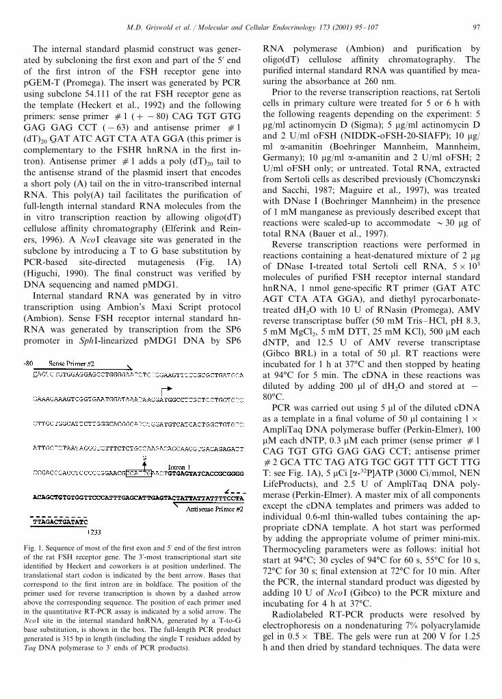
Fig. 1. Sequence of most of the first exon and 5% end of the first intronof the rat FSH receptor gene. The 3%-most transcriptional start siteidentified by Heckert and coworkers is at position underlined. Thetranslational start codon is indicated by the bent arrow. Bases thatcorrespond to the first intron are in boldface. The position of theprimer used for reverse transcription is shown by a dashed arrowabove the corresponding sequence. The position of each primer usedin the quantitative RT-PCR assay is indicated by a solid arrow. TheNcoI site in the internal standard hnRNA, generated by a T-to-Gbase substitution, is shown in the box. The full-length PCR productgenerated is 315 bp in length (including the single T residues added byTaq DNA polymerase to 3% ends of PCR products).
M.D. Griswold et al. / Molecular and Cellular Endocrinology 173 (2001) 95–10798
analyzed using a Molecular Dynamics PhosphorImager445 SI and ImageQuant software. The integrated opti-cal density of the target was normalized by dividing itby the integrated optical density of the signal for thelarge NcoI-digested fragment of the corresponding in-ternal standard.
2.4. Plasmid constructs for transient transfection assays
p383/−1/Luc contains 383 bp of the 5% flankingregion (including the 5% untranslated region) of the FSHreceptor gene upstream of the luciferase reporter genein pGL2 (Promega, Madison, WI) (Goetz et al., 1996).p383/−1/Luc was used as a template for PCR-basedsite-directed mutagenesis to generate mutants that con-tain changes in the AP1/CRE-like site (TTAGTCA) atposition −216 to −210 (relative to the translationstart site) in the rat FSH receptor promoter (Higuchi,1990; Heckert et al., 1992; Goetz et al., 1996). pCRE-F-Mut/Luc (CRE in F6 SHR M6 utant) contains the follow-ing mutation: TTAGTCA to TTT6 GTCA. pCRE-Pal/Luc contains a palindromic CRE promoter (Brindleand Montminy, 1992) in context with the AP1/CRE-like site within the FSH receptor promoter as follows:TTAGTCA to TG6 AC6 GTCA. A mutant of the palin-dromic CRE (pCRE-Pal-Mut/Luc) was generated andhas the following sequence at this site: TGTGGTCA.The plasmids p383/−1/Luc, pCRE-F-Mut/Luc,pCRE-Pal/Luc, and pCRE-Pal-Mut/Luc were clonedinto the NheI–HindIII site of pGL2 and verified byDNA sequencing.
p2700/Luc contains approximately 2700 bp of the 5%flanking region of the FSH receptor gene including the5% untranslated region (5%UTR). p2700/Luc is composedof the PstI–EcoRV fragment of the 5% end of the ratFSH receptor gene from l-genomic clone 54.111 (Heck-ert et al., 1992) ligated to the EcoRV-HindIII fragmentof the FSH receptor promoter in p383/−1/Luc. This�2700 bp fragment of the FSH receptor 5% flankingregion was cloned into the SacI–HindIII site in pGL2(Promega) after the PstI and SacI sticky ends wereconverted to blunt ends by the exonuclease activity ofT4 DNA polymerase using standard techniques. Thisconstruct was verified by restriction analysis and byDNA sequencing across all junctions generated byligation.
2.5. Transient transfection assays
Transient transfections of primary cultures of ratSertoli cells were performed using the CaPO4 coprecipi-tation method in 60-mm plastic culture dishes as de-scribed previously with minor modifications (Linder etal., 1994; Goetz et al., 1996). Cells were transfected onthe 4th day of culture with 6 mg of reporter plasmid and0.3 mg of pRL (Promega) coreporter plasmid. The
activity of the Renilla luciferase encoded by pRL wasused to normalize the firefly luciferase signal frompGL2- and pGL3-based constructs and control fortransfection efficiency. The data from the transfectionexperiments reported in this manuscript were derivedfrom at least three different experiments and two dis-tinct preparations of plasmid DNA using the QiagenEndotoxin-Free large-scale plasmid preparation proto-col. Transfected cells were treated with 2 U/ml of oFSHor vehicle only (1× phosphate-buffered saline, 1 mg/mlbovine serum albumen) such that all the cells in allgroups were harvested at the same time and assayed forluciferase activity.
Luciferase assays were performed using the DualLuciferase Reporter Assay System (Promega). Celllysates were analyzed for luciferase activity in a lumi-nometer (MicroLumat LB 96p, EG&G Berthold, Ger-many). Lysates of corresponding samples (e.g.p383/−1/Luc at 4 h with oFSH and without oFSH)were placed in the same location on two different96-well plates.
2.6. Northern blot analysis
To determine if the conditions of CaPO4-mediatedtransfection alter homologous down-regulation of theFSH receptor mRNA, Northern analysis was per-formed. Sertoli cells were transfected as describedabove and total RNA was extracted by the method ofChomczynski and Sacchi (1987). The relative amountof FSH receptor mRNA in each sample was analyzedby Northern blot analyses as described previously(Maguire et al., 1997).
2.7. Electrophoretic mobility shift assays (EMSA)
Extraction of nuclear proteins from primary culturesof Sertoli cells was performed as described by Andrewsand Faller and is the same procedure used by Monacoand coworkers (Andrews and Faller, 1991; Monaco etal., 1995). Nuclear proteins were extracted from Sertolicells that were treated with 1 mM (Bu)2cAMP or 2U/ml oFSH for 1 or 4 h and cells that were treated withvehicle only. Purified recombinant human c-Fos andJun-B were kindly provided by Mark Nissen and Ray-mond Reeves at Washington State University. Thec-Fos and Jun-B expression constructs were generouslyprovided by David Tremethick at The Australian Na-tional University (Ng et al., 1997).
The following is a list of the names and sequences ofthe sense strand of each double-stranded oligonucle-otide used in this study:
wtAP1: −225 TGACACACATTAGTCACATAT-TAAT −201mutAP1: −225 TGACACACATTAGTTGCATAT-TAAT −201

M.D. Griswold et al. / Molecular and Cellular Endocrinology 173 (2001) 95–107 99
CREpal: TGACACACATGACGTCACATATTAA-T.The numbering is based on the translational start
site. The location of the AP1/CRE-like site is under-lined and base substitutions are indicated in boldface.
Single-stranded oligonucleotides were end-labeledwith [a-32P]ATP (3000 Ci/mmol, NEN LifeProducts)using T4 polynucleotide kinase (Gibco BRL) prior toannealing. Labeled double-stranded oligonucleotideprobes were gel-purified in nondenaturing, 6% poly-acrylamide gels in 0.5× Tris Borate EDTA (TBE) andeluted in dH2O.
Each binding reaction was performed in a total vol-ume of 15 ml with �5.0×10⁴ cpm of labeled probe(0.5 ng DNA), 3 mg Sertoli cell nuclear protein extract,0.2 mg poly(dI)-poly(dC) (Pharmacia Biotech. Inc.,Uppsala, Sweden), in a buffer composed of 12.5 mMHepes, pH 7.9 (at 0°C), 25 mM KCl, 10 mM MgCl2, 5mM DTT, 0.05% Triton X-100, and 10% glycerol.Unlabeled competitor DNA was included in some reac-tions at 100-fold molar excess over the labeled oligonu-cleotide probe.
The identity of c-Fos in the Sertoli cell nuclearprotein–oligonucleotide probe complex was determinedby supershift analysis. Supershift reactions were per-formed with rabbit, anti-human c-Fos polyclonal anti-body (Ab) SC-253, rabbit, anti-human CREB-1polyclonal Ab SC-186, or rabbit, anti-human CREM-1polyclonal Ab SC-440 (Santa Cruz Biotechnology,Santa Cruz, CA) by adding 1 mg of the antibody to thebinding reaction after the 10-min incubation with la-beled probe and incubating for one additional hour onice. A control for nonspecific interactions between theDNA–protein complexes and antibodies was per-formed by adding 1 mg of rabbit, anti-human TFIIDpolyclonal antibody SC-273X (Santa Cruz Biotechnol-ogy) to control binding reactions and incubated on icefor 1 h.
Protein–DNA complexes were resolved by elec-trophoresis through a nondenaturing 4% polyacry-lamide gel in 0.5× TBE. The gels were run for 1.25 hat 200 V. After electrophoresis, the gels were driedusing standard techniques. The signals were detectedusing a Molecular Dynamics PhosphorImager 445 SIand ImageQuant software (Molecular Dynamics, Sun-nyvale, CA).
2.8. Treatment of Sertoli cells with trichostatin A
To determine if the inhibition of histone deacetylasesderepressed the homologous down-regulation of theFSH receptor gene, primary cultures of Sertoli cellswere prepared as described above and treated with 0.3mM trichostatin A (TSA) (Sigma). Cells were treatedwith trichostatin A for 1 h prior to the addition of 2U/ml oFSH for four additional hours. The level of
FSH receptor mRNA was determined by Northern blotas described previously (Maguire et al., 1997). TheNorthern blot was stripped and reprobed with a frag-ment of the cDNA for rat clusterin (Clark and Gris-wold, 1997) to control for loading and transfer.
2.9. Experimental animals
All research done with animals for the work de-scribed in this paper followed university and NIHguidelines and policies.
3. Results
3.1. Determination of le6els of FSH receptor hnRNA
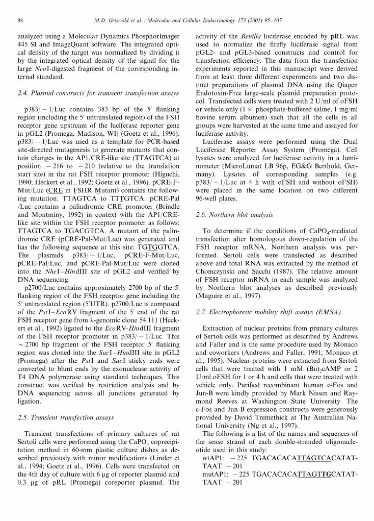
A strategy was designed to amplify the specificcDNA that corresponds to the first exon and 5% end ofthe first intron of the FSH receptor hnRNA (Fig. 1).Sample-to-sample variation in the efficiency of reversetranscription and PCR were controlled for by the inclu-sion of a known amount of internal standard RNA inthe RT reactions. The internal standard was identical tothe target in length and sequence with the exception ofa single base difference that generated a NcoI site in theinternal standard (Fig. 1). The signals generated by thetarget and internal standard cDNAs were in the expo-nential range after 30 cycles (data not shown). Giventhat the RT primer (antisense primer 1) was comple-mentary to bases in the first intron (Fig. 1), rigorouscontrols were performed to limit the signal generated bycontaminating genomic DNA. Total RNA was treatedwith DNase I prior to performing reverse transcriptionreactions and PCR was performed on all RNA samplesthat were placed in reverse transcription reactions de-void of reverse transcriptase (data not shown). We didnot collect data for the target or internal standard if weobserved significant background from genomic DNA.The identity of the target band (315 bp in Fig. 2A) andthe internal standard band after cleavage with a restric-tion enzyme (�220 bp in Fig. 2A) was confirmed byexcising these fragments from a gel, reamplifying byPCR, cloning into pGEM T (Promega) and sequencing(data not shown). FSH induced a reduction in thesteady-state level of FSH receptor hnRNA by at least50% after 5 h (Fig. 2A,B). A similar FSH-inducedreduction of FSHR hnRNA was seen if the cells werealso treated with either a-amanitin or actinomycin D(data not shown).
3.2. Transient transfection analysis of the 2.7 kbpromoter
The possibility that the cis-element(s) required forhomologous down-regulation are localized in the pro-
M.D. Griswold et al. / Molecular and Cellular Endocrinology 173 (2001) 95–107100
Fig. 2. Quantitative RT-PCR analysis of FSH receptor hnRNA in Sertoli cells. (A) PCR products from Sertoli cells that were not treated (lanes1–3), or treated with FSH (lanes 3–6) were digested with NcoI and resolved by polyacrylamide gel electrophoresis. The low molecular weightDNA mass ladder (Gibco) was radiolabeled and used as a marker (M). The identity of each band is given on the right as follows, T (315 bptarget), IS (internal standard NcoI-digestion products). (B) The integrated optical densities of the target bands were divided by the integratedoptical densities of the large (�220 bp) internal standard NcoI-digestion products and normalized to control samples. Data are the means fromthree separate experiments9S.E.
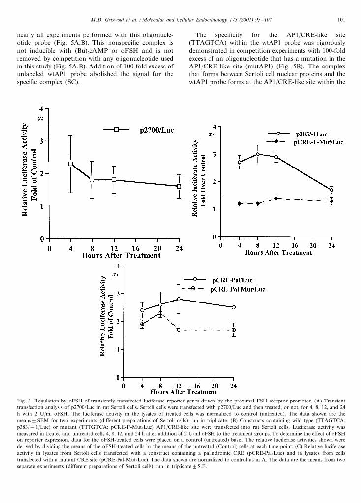
moter at a site in the extended (�2700 bp) 5% flankingregion was tested (Fig. 3A). Lysates from Sertoli cellsthat were transiently transfected with p2700/Luc andtreated with 2 U/ml oFSH for 4, 8, 12, and 24 h wereassayed for luciferase activity. The 2700 bp promoterwas not sufficient to direct homologous down-regula-tion in transient transfection assays (Fig. 3A).
3.3. Transient transfection analysis of theAP1/CRE-like site
A two- to three–fold increase in luciferase activitywas observed in Sertoli cells transiently transfected withthe luciferase reporter gene driven by the proximal383-bp fragment of the wild-type FSH receptor pro-moter (p383/−1/Luc) 8 h after the addition of oFSH(Fig. 3B). The increase in luciferase activity is abolishedwhen a single base mutation (−214 A to T) is made inthe AP1/CRE-like site contained within the proximal383 bp of the FSH receptor promoter (pCRE-F-Mut/Luc in Fig. 3B). The −116 A to T base substitutionmutates this site such that it is less like either the AP1or CRE consensus sequences (Faisst and Meyer, 1992).Altering the AP1/CRE-like site in the FSH receptorgene promoter to match the palindromic CRE consen-sus sequence (pCRE-Pal/Luc) did not alter the magni-tude or kinetics of the FSH-induced signal compared top383/−1/Luc (Fig. 3B,C). None of the constructs usedin Fig. 3B,C directed down-regulation in this transienttransfection system even when transfected cells werelysed and assayed 48 h after the addition of oFSH (data
not shown). The decrease in signal at the longer timepoints was probably due to the instability of the luci-ferase or the induction signal.
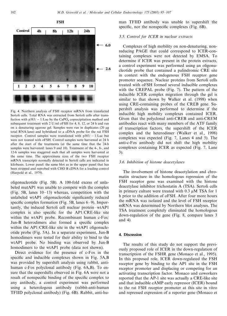
Northern blot analysis was performed to determine ifthe FSH-induced signal transduction pathway responsi-ble for the down-regulation of the FSH receptor genewas functioning under conditions of transient transfec-tion. The steady-state amount of FSH receptor mRNAwas measured by Northern blot in total RNA fromcells that were transfected with p383/−1/Luc and leftuntreated (Control) or treated with oFSH (Fig. 4).Homologous down-regulation occurred with the samekinetics as reported by Themmen et al. (1991). There-fore, the FSH-induced signal transduction pathway re-sponsible for down-regulation of the endogenous geneis apparently not disrupted by the experimental condi-tions of transient transfection used in this study.
3.4. Inducible c-Fos complex formation on theAP1/CRE-like site
A specific complex forms between Sertoli cell nuclearproteins and an oligonucleotide probe (wtAP1) thatcontains the wild-type AP1/CRE-like site (Fig. 5A).This result was expected because of the contribution ofthis site to the modest enhancement of expression fromtransiently transfected templates in FSH-treated Sertolicells as shown in Fig. 5A,B. There was a markedincrease in the specific complex (SC) after 4 h oftreatment with (Bu)2cAMP and oFSH (Fig. 5A, lanes 9and 10). A nonspecific complex (NS) is present in
M.D. Griswold et al. / Molecular and Cellular Endocrinology 173 (2001) 95–107 101
nearly all experiments performed with this oligonucle-otide probe (Fig. 5A,B). This nonspecific complex isnot inducible with (Bu)2cAMP or oFSH and is notremoved by competition with any oligonucleotide usedin this study (Fig. 5A,B). Addition of 100-fold excess ofunlabeled wtAP1 probe abolished the signal for thespecific complex (SC).
The specificity for the AP1/CRE-like site(TTAGTCA) within the wtAP1 probe was rigorouslydemonstrated in competition experiments with 100-foldexcess of an oligonucleotide that has a mutation in theAP1/CRE-like site (mutAP1) (Fig. 5B). The complexthat forms between Sertoli cell nuclear proteins and thewtAP1 probe forms at the AP1/CRE-like site within the
Fig. 3. Regulation by oFSH of transiently transfected luciferase reporter genes driven by the proximal FSH receptor promoter. (A) Transienttransfection analysis of p2700/Luc in rat Sertoli cells. Sertoli cells were transfected with p2700/Luc and then treated, or not, for 4, 8, 12, and 24h with 2 U/ml oFSH. The luciferase activity in the lysates of treated cells was normalized to control (untreated). The data shown are themeans9SEM for two experiments (different preparations of Sertoli cells) run in triplicate. (B) Constructs containing wild type (TTAGTCA:p383/−1/Luc) or mutant (TTTGTCA: pCRE-F-Mut/Luc) AP1/CRE-like site were transfected into rat Sertoli cells. Luciferase activity wasmeasured in treated and untreated cells 4, 8, 12, and 24 h after addition of 2 U/ml oFSH to the treatment groups. To determine the effect of oFSHon reporter expression, data for the oFSH-treated cells were placed on a control (untreated) basis. The relative luciferase activities shown werederived by dividing the means of the oFSH-treated cells by the means of the untreated (Control) cells at each time point. (C) Relative luciferaseactivity in lysates from Sertoli cells transfected with a construct containing a palindromic CRE (pCRE-Pal/Luc) and in lysates from cellstransfected with a mutant CRE site (pCRE-Pal-Mut/Luc). The data shown are normalized to control as in A. The data are the means from twoseparate experiments (different preparations of Sertoli cells) run in triplicate9S.E.
M.D. Griswold et al. / Molecular and Cellular Endocrinology 173 (2001) 95–107102
Fig. 4. Northern analysis of FSH receptor mRNA from transfectedSertoli cells. Total RNA was extracted from Sertoli cells after trans-fection with p383/−1/Luc by the CaPO4 coprecipitation method andsubsequent treatment with 2 U/ml oFSH for 4, 8, 12, or 24 h and runon a denaturing agarose gel. Samples were run in duplicates (20 mgtotal RNA/lane) and hybridized to a cDNA probe for the rat FSHreceptor. Control samples were transfected with p383/−1/Luc butwere not treated with oFSH. Control samples were harvested at 24 hafter the start of the treatments (at the same time that the 24-hsamples were harvested: lanes 9 and 10). Treatment of the 4-, 8-, and12-h samples was staggered such that all samples were harvested atthe same time. The approximate sizes of the two FSH receptormRNA transcripts normally detected in Sertoli cells are indicated inkilobases. Lower panel: the same blot as in the upper panel that hasbeen stripped and reprobed with CHO B cDNA for a loading control(Harpold et al., 1979).
man TFIID antibody was unable to supershift thespecific, nor the nonspecific complexes (Fig. 6B).
3.5. Control for ICER in nuclear extracts
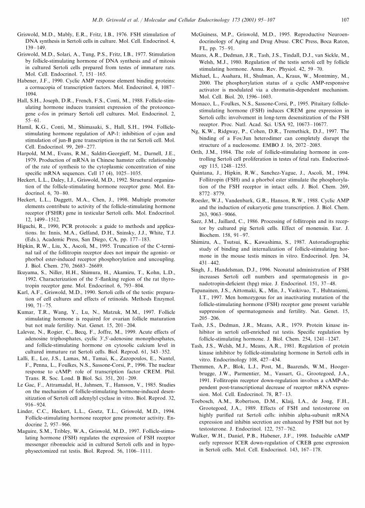
Complexes of high mobility on non-denaturing, non-reducing PAGE that could correspond to ICER-con-taining complexes were not detected by EMSA. Todetermine if ICER was present in the protein extracts,a control experiment was performed using an oligonu-cleotide probe that contained a palindromic CRE sitein context with the endogenous FSH receptor genepromoter sequence. Nuclear proteins from Sertoli cellstreated with oFSH formed several inducible complexeswith the CREPAL probe (Fig. 7). The pattern of theinducible ICER complex migration through the gel issimilar to that shown by Walker et al. (1998) whenusing CRE-containing probes of the CREB gene. Su-pershift analysis was performed to determine if theinducible high mobility complexes contained ICER.Given that the polyclonal anti-CREB and anti-CREMantibodies react with many members of the ATF familyof transcription factors, the supershift of the ICERcomplex and the heterodimer (Walker et al., 1998)complexes was expected (Fig. 7, Lanes 4 and 5). Theanti-c-Fos antibody did not shift the high mobilitycomplexes containing ICER as expected (Fig. 7, Lane3).
3.6. Inhibition of histone deacetylases
The involvement of histone deacetylation and chro-matin structure in the homologous repression of theFSH receptor gene was examined with the histonedeacetylase inhibitor trichostatin A (TSA). Sertoli cellsin primary culture were treated with 0.3 mM TSA for 1h prior to the addition of oFSH. After four more hoursthe mRNA was isolated and the level of FSH receptormRNA was determined by Northern blot analyses. TheTSA treatment completely eliminated the homologousdown-regulation of the gene (Fig. 8, compare lanes 3and 4).
4. Discussion
The results of this study do not support the previ-ously proposed role of ICER in the down-regulation oftranscription of the FSHR gene (Monaco et al., 1995).In this proposed role, ICER down-regulated the FSHreceptor gene by binding to the AP1 site in the FSHreceptor promoter and displacing or competing for anactivating transcription factor. Monaco and coworkersreported that the AP-1 site was actually a CRE-like siteand that inducible cAMP early repressor (ICER) boundto the rat FSH receptor promoter at this site in vitroand repressed expression of a reporter gene (Monaco et
oligonucleotide (Fig. 5B). A 100-fold excess of unla-beled mutAP1 was unable to compete with the complex(Fig. 5B, lanes 10–13) whereas, competition with theunlabeled wtAP1 oligonucleotide significantly reducedspecific complex formation (Fig. 5B, lanes 6–9). Impor-tantly, the induced Sertoli cell nuclear protein–wtAP1complex is also specific for the AP1/CRE-like sitewithin the wtAP1 probe. Recombinant human c-Fos/Jun-B heterodimers also formed a specific complexwithin the AP1/CRE-like site in the wtAP1 oligonucle-otide probe (Fig. 5A). In a separate experiment, Jun-Bhomodimers were tested for their ability to bind to thewtAP1 probe. No binding was observed by Jun-Bhomodimers to the wtAP1 probe (data not shown).
Direct evidence for the presence of c-Fos in thespecific and inducible complexes shown in Fig. 5A,Bwas provided by supershift analysis using rabbit, anti-human c-Fos polyclonal antibody (Fig. 6A,B). To en-sure that the supershifts observed in Fig. 6A were not aresult of nonspecific binding of the specific complex toany antibody, a control experiment was performedusing a heterologous antibody (rabbit-anti-humanTFIID polyclonal antibody) (Fig. 6B). Rabbit, anti-hu-

M.D. Griswold et al. / Molecular and Cellular Endocrinology 173 (2001) 95–107 103
Fig. 5. (A) Demonstration by EMSA that an inducible Sertoli cell nuclear protein–DNA complex forms on the wtAP1 oligonucleotide probe.Nuclear extracts from rat Sertoli cells that were untreated, treated with 1 mM (Bu)2cAMP (dbc), or treated with 2 U/ml oFSH (F) for 1 h (lanes2–7) or 4 h (lanes 8–13) were used in binding reactions with radiolabeled wtAP1 oligonucleotide probe. To determine if the interaction betweenthe nuclear proteins and wtAP1 was specific, a 100-fold excess of unlabeled wtAP1 competitor was added to some reactions. A nonspecific (NS)complex that was not removed upon the addition of unlabeled competitor was formed in all lanes with nuclear protein. Lane 1 is a probe-only(P) control and the migration of unbound, free probe is indicated (FP). (B) EMSA analysis of the AP1/CRE-like site within the wtAP1oligonucleotide probe. Sertoli cell nuclear proteins from cells that were untreated, treated with 1 mM (Bu)2cAMP (dbc), or treated with 2 U/mloFSH (F) for 4 h were added to labeled wtAP1 oligonucleotide probe (lanes 2, 3, 4, 6, 7, 8, 10, 11, and 12). Recombinant human cFos/Jun-Bheterodimer was added to labeled wtAP1 in some reactions (lanes 5, 9, and 13). A probe-only control is shown in lane 1. To determine specificityfor the wtAP1 probe, a 100-fold excess of unlabeled wtAP1 was added as a competitor to the reactions shown in lanes 6–9. Specificity for theAP1/CRE-like site within the wtAP1 probe was determined by including a 100-fold excess of unlabeled competitor oligonucleotide that containsa mutation in the AP1/CRE-like site (mutAP1). Positions of the specific complex (SC), nonspecific complex (NS), and free probe (FP) are shown.

M.D. Griswold et al. / Molecular and Cellular Endocrinology 173 (2001) 95–107104
al., 1995). The results of EMSA analyses in our studyusing the wtAP1 oligonucleotide probe clearly demon-strate the preferential binding of c-Fos, and likely Jun-B, to this site relative to any other protein in theextracts. The c-Fos and Jun-B transcription factors aremembers of the AP1 family of transcription factors, canfunction as transcriptional activators or repressors andare rapidly induced by cAMP in rat Sertoli cells (Hall etal., 1988; Hamil et al., 1994). Members of the ATF andAP1 families of transcription factors can bind to a widevariety of CRE and AP1 sites, including non-consensussites, and in a variety of homodimer and heterodimercombinations (Habener, 1990; Drust et al., 1991).
FSH stimulated the luciferase activity in Sertoli cellstransfected with p383/−1/Luc (Fig. 3B). This increasewas apparently due to the AP1 site since an inactivatingmutation suppressed the stimulation. The AP1 site orputative ICER binding site is not a consensus CRE andis not conserved in the mouse and human promoters inthe FSH receptor gene (Roesler et al., 1988; Brindleand Montminy, 1992; Faisst and Meyer, 1992; Maguireet al., 1997). Conversion of the site to an authentic
palindromic CRE site still resulted in a FSH-stimulatedincreased luciferase activity. None of the transfectedconstructs were down-regulated as a result of treatmentof the cells with FSH. Clearly, ICER was not active onthese transfected transcripts. It is possible that thenumber of templates present in a transfected cell over-comes the down-regulation machinery but the observa-tion that reporter constructs driven by the TSHreceptor promoter are down-regulated by the endoge-nous machinery in FRTL-5 cells counters that possibil-ity (Ikuyama et al., 1992). Also, ICER has beenreported to be a powerful repressor, able to represstranscription at substoichiometric concentrations (Lalliet al., 1996). In the studies reported by Monaco et al.(1995), endogenous ICER was not able to down-regu-late the expression of the CAT reporter gene driven bythe rat FSH receptor promoter (Monaco et al., 1995)Co-transfection with an ICER expression constructdriven by the SV-40 promoter was used to show cAMP-induced, ICER-dependent down-regulation of reporterconstructs in Sertoli cells (Monaco et al., 1995). Suchoverexpression of ICER may down-regulate the trans-gene by a different mechanism.
Fig. 6. The specific and inducible complex that forms on the AP1/CRE-like site in the wtAP1 oligonucleotide probe contains c-Fos. (A) EMSASupershift analysis using anti-hcFos antibody (lanes 8–11). Sertoli cell nuclear extracts from untreated cells, cells treated with 1 mM (Bu)2cAMP(dbc), cells treated with 2 U/ml oFSH or purified recombinant hcFos/Jun-B (F/J) were incubated with labeled wtAP1 oligonucleotide probe.Positions of the supershifted complexes (SS), specific complexes (SC), nonspecific complexes (NS), and free probe (FP) are indicated. Lane 1contains a probe-only control. Lanes 2 and 3 are blank. (B) A supershifted complex does not form with a heterologous antibody. Sertoli cellnuclear extracts (NE) from untreated cells or recombinant human c-Fos/Jun-B heterodimers (F/J) were included in binding reactions with labeledwtAP1 oligonucleotide probe. Supershift EMSA with anti-hcFos antibody (lanes 4 and 5) or anti-hTFIID (transcription factor II-D) antibody(lanes 6 and 7). Control samples did not receive antibody (lanes 2 and 3).

M.D. Griswold et al. / Molecular and Cellular Endocrinology 173 (2001) 95–107 105
Fig. 7. Electrophoretic mobility shift and supershift assay with palin-dromic CRE oligonucleotide probe and Sertoli cell nuclear proteinsshowing the presence of ICER in the protein extracts. Sertoli cellnuclear extracts from untreated cells, or cells treated with 2 U/mloFSH (F) were used in binding reactions with radiolabeled CREpaloligonucleotide probe. The presence of an antibody in a bindingreaction is indicated (+ ). The identity of the complexes is shown onthe left as follows: ATF (activating transcription factor family), cFos,heterodimers (complexes that contain ICER and some unknownprotein(s), ICER, NS (non-specific complex). The band on the bot-tom corresponds to unbound, free probe (FP).
In previous studies we presented evidence that thedown-regulation of the FSH receptor gene did notresult from changes in the stability of the FSHRmRNA (Maguire et al., 1997). We were able to showthat the kinetics of mRNA decay was the same in thepresence or absence of actinomycin D. Also, the addi-tion of FSH or actinomycin D to cultured Sertoli cellsappeared to produce similar kinetics suggesting thatFSH inhibited transcription. While it is still possiblethat mRNA stability plays a role we think it is unlikelyto be the primary mechanism. The FSH receptor genehas a relatively low transcription rate and direct mea-surement of transcription with nuclear run-on assayswas not possible. Therefore we utilized a quantitativePCR-based assay designed to measure the levels ofFSH receptor hnRNA. It has been demonstrated thatthis hnRNA RT-PCR assay is an acceptable substitutefor the nuclear run-on assay (Elferink and Reiners,1996). Both the hnRNA RT-PCR assay and the nuclearrun-on assay detected an equivalent increase in tran-scription of Cyp1a-1 in cultured murine Hepa 1c1c7cells following exposure to 2,3,7,8-tetrachlorodibenzo-p-dioxin (TCDD). The RT-PCR assay also revealedTCDD-dependent transcriptional activation of theCyp1a-1 gene in murine skin, a tissue unsuited to thenuclear run-on assay because of inherent difficultiesassociated with the isolation of nuclei (Elferink andReiners, 1996). The results presented here showed thatthe steady state level of hnRNA transcribed from theFSHR gene was decreased 5 h after the addition ofFSH. Since the levels of FSHR mRNA also decreaseand mRNA turnover does not change, it follows thatthe decreased amount of hnRNA must result fromdecreased transcription and not from decreased hn-RNA processing.
Two observations originally suggested that endoge-nous chromatin structure might play a role in theFSH-induced repression of the gene. First, none of thetransiently transfected constructs used in this study, orby Monaco et al. (1995), directed homologous down-
The role, if any, of c-Fos in the regulation of theFSH receptor gene is poorly understood. Evidence pre-sented in this paper that c-Fos/Jun-B heterodimers canbind to the AP1/CRE-like in EMSA experiments is thefirst reported involvement of the AP-1 family of tran-scription factors in the regulation of the FSH receptorgene. These studies do not support the previously de-scribed role of ICER and a putative AP-1/CRE site inthe down-regulation of the FSHR gene. The site isclearly shown to be a functional AP-1 site. When therat FSHR promoter was reconstituted into nucleosomesand mapped by hydroxyl radical footprinting we foundthat the Ap-1 site was not accessible to transcriptionfactors (unpublished data). The AP-1 site mapped closeto the surface on a very stable nucleosome. Thus it ispossible that the Ap-1 site plays no role in eithertranscription or down-regulation of the FSHR gene invivo.
Fig. 8. Upper panel: Northern blot analysis of FSH receptor mRNAin total RNA from Sertoli cells that were untreated (Lane 1), treatedwith TSA alone (Lane 2), TSA and oFSH (Lane 3), or oFSH alone(Lane 4) as described in Section 2. Lower panel: the same blot as inthe upper panel that has been stripped and reprobed with rat clusterincDNA.

M.D. Griswold et al. / Molecular and Cellular Endocrinology 173 (2001) 95–107106
regulation of the FSH receptor gene. The down-regula-tion of the mRNA level was not seen when 2.7 kb or383 base pairs of the promoter driving a reporter genewere transfected into Sertoli cells. In fact, in each ofthese constructs the luciferase reporter gene activity wasincreased in the presence of FSH. Second, in previousstudies from this laboratory we showed that the tissue-specific pattern of expression of the FSH receptor genewas also not recreated in transient transfection assays(Linder et al., 1994). Indeed, reporter genes driven byFSH receptor gene promoters of various lengths werepromiscuously expressed in a number of cell types intransient transfection assays whereas the expression ofsuch genes in transgenic mice was restricted to thegonads in one study (Linder et al., 1994).
The hypothesis that chromatin structure is involvedin the FSH-induced repression of the FSH receptorgene in Sertoli cells was tested in this study by addingTSA to Sertoli cells 1 h prior to the addition of FSH.TSA inhibits histone deacetylase activity and blocks theformation of repressive chromatin structure that iscaused by the recruitment of histone deacetylase activ-ity. Treatment of Sertoli cells with TSA prior to theaddition of FSH completely prevented the down-regu-lation of the gene (Fig. 8). Taken together, these obser-vations are consistent with, but certainly do not prove,the hypothesis that changes in chromatin structure areinvolved in the homologous down-regulation of theFSH receptor gene in Sertoli cells. The E-box and theE-box binding proteins USF-1 and USF-2 play criticalroles in the regulation of the FSH receptor gene inSertoli cells (Goetz et al., 1996; Heckert et al., 1998). Ina recent publication, the phosphorylation of a cAMP-responsive activator was shown to be modulated bymeans of a chromatin-dependent mechanism (Michaelet al., 2000). Similarly, to our studies it was shown thathistone deacetylase inhibitors influenced CREB activitybut only on chromatin templates. The FSHR genecould also utilize a mode of regulation that links thecontrol of gene expression by signal transduction andchromatin structure. Additional studies will be neededto establish the exact mechanism of transcriptionaldown-regulation.
Acknowledgements
We thank Alice Karl for preparing Sertoli cell cul-tures. Elena Lymar provided expert assistance with theEMSA work. Mark Nissen and Raymond Reeves atWashington State University generously provided therecombinant c-Fos and Jun-B proteins. David Treme-thick at The Australian National University providedthe c-Fos and Jun-B constructs. We are grateful to theNIDDK for supplying the oFSH and to GerhardMunske and Derek Pouchnik of the Laboratory for
Biotechnology and Bioanalysis at Washington StateUniversity for preparation of oligonucleotides andDNA sequencing. This work was supported by NIHgrant HD 10808.
References
Andrews, N.C., Faller, D.V., 1991. A rapid micropreparation tech-nique for extraction of DNA-binding proteins from limiting num-bers of mammalian cells. Nucleic Acids Res. 19, 2499–2503.
Bauer, P., Rolfs, A., Regitz-Zagrosek, V., Hildebrandt, A., Fleck, E.,1997. Use of manganese in RT-PCR eliminates PCR artifactsresulting from DNase I digestion. Biotechniques 22, 1128–1132.
Brindle, P.K., Montminy, M.R., 1992. The CREB family of tran-scription activators. Curr. Opin. Genet. Dev. 2, 199–204.
Chaudhary, J., Whaley, P.D., Cupp, A., Skinner, M.K., 1996. Tran-scriptional regulation of sertoli cell differentiation by follicle-stim-ulating hormone at the level of the c-fos and transferrinpromoters. Biol. Reprod. 54, 692–699.
Chomczynski, P., Sacchi, N., 1987. Single-step method of RNAisolation by acid guanidinium thiocyanate-phenol-chloroform ex-traction. Anal. Biochem. 162, 156–159.
Clark, A.M., Griswold, M.D., 1997. Expression of clusterin/sulfatedglycoprotein-2 under conditions of heat stress in rat Sertoli cellsand a mouse Sertoli cell line. J. Androl. 18, 257–263.
Conti, M., Toscano, M.V., Petrelli, L., Geremia, R., Stefanini, M.,1983. Involvement of phosphodiesterase in the refractoriness ofthe Sertoli cell. Endocrinology 113, 1845–1853.
Conti, M., Monaco, L., Geremia, R., Stefanini, M., 1986. Effect ofphosphodiesterase inhibitors on Sertoli cell refractoriness: reversalof the impaired androgen aromatization. Endocrinology 118,901–908.
Dierich, A., Sairam, M.R., Monaco, L., Fimia, G.M., Gansmuller,A., LeMeur, M., Sassone-Corsi, P., 1998. Impairing follicle-stimu-lating hormone (FSH) signaling in vivo: targeted disruption of theFSH receptor leads to aberrant gametogenesis and hormonalimbalance. Proc. Natl. Acad. Sci. USA 95, 13612–13617.
Drust, D.S., Troccoli, N.M., Jameson, J.L., 1991. Binding specificityof cyclic adenosine 3%,5%-monophosphate-responsive element(CRE)-binding proteins and activating transcription factors tonaturally occurring CRE sequence variants. Mol. Endocrinol. 5,1541–1551.
Elferink, C.J., Reiners, J.J., Jr, 1996. Quantitative RT-PCR onCYP1A1 heterogeneous nuclear RNA: a surrogate for the in vitrotranscription run-on assay. Biotechniques 20, 470–477.
Faisst, S., Meyer, S., 1992. Compilation of vertebrate-encoded tran-scription factors. Nucleic Acids Res. 20, 3–26.
Fletcher, P.W., Reichert, L.E., Jr, 1984. Cellular processing of folli-cle-stimulating hormone by Sertoli cells in serum-free culture.Mol. Cell. Endocrinol. 34, 39–49.
Gnanaprakasam, M.S., Chen, C.J., Sutherland, J.G., Bhalla, V.K.,1979. Receptor depletion and replenishment processes: in vivoregulation of gonadotropin receptors by luteinizing hormone,follicle stimulating hormone and ethanol in rat testis. Biol. Re-prod. 20, 991–1000.
Goetz, T.L., Lloyd, T.L., Griswold, M.D., 1996. Role of E box andinitiator region in the expression of the rat follicle-stimulatinghormone receptor. J. Biol. Chem. 271, 33317–33324.
Griswold, M., Mably, E., Fritz, I.B., 1975. Stimulation by folliclestimulating hormone and dibutyryl cyclic AMP of incorporationof 3H-thymidine into nuclear DNA of cultured Sertoli cell-en-riched preparations from immature rats. Curr. Top. Mol. En-docrinol. 2, 413–420.
M.D. Griswold et al. / Molecular and Cellular Endocrinology 173 (2001) 95–107 107
Griswold, M.D., Mably, E.R., Fritz, I.B., 1976. FSH stimulation ofDNA synthesis in Sertoli cells in culture. Mol. Cell. Endocrinol. 4,139–149.
Griswold, M.D., Solari, A., Tung, P.S., Fritz, I.B., 1977. Stimulationby follicle-stimulating hormone of DNA synthesis and of mitosisin cultured Sertoli cells prepared from testes of immature rats.Mol. Cell. Endocrinol. 7, 151–165.
Habener, J.F., 1990. Cyclic AMP response element binding proteins:a cornucopia of transcription factors. Mol. Endocrinol. 4, 1087–1094.
Hall, S.H., Joseph, D.R., French, F.S., Conti, M., 1988. Follicle-stim-ulating hormone induces transient expression of the protoonco-gene c-fos in primary Sertoli cell cultures. Mol. Endocrinol. 2,55–61.
Hamil, K.G., Conti, M., Shimasaki, S., Hall, S.H., 1994. Follicle-stimulating hormone regulation of AP-1: inhibition of c-jun andstimulation of jun-B gene transcription in the rat Sertoli cell. Mol.Cell. Endocrinol. 99, 269–277.
Harpold, M.M., Evans, R.M., Salditt-Georgieff, M., Darnell, J.E.,1979. Production of mRNA in Chinese hamster cells: relationshipof the rate of synthesis to the cytoplasmic concentration of ninespecific mRNA sequences. Cell 17 (4), 1025–1035.
Heckert, L.L., Daley, I.J., Griswold, M.D., 1992. Structural organiza-tion of the follicle-stimulating hormone receptor gene. Mol. En-docrinol. 6, 70–80.
Heckert, L.L., Daggett, M.A., Chen, J., 1998. Multiple promoterelements contribute to activity of the follicle-stimulating hormonereceptor (FSHR) gene in testicular Sertoli cells. Mol. Endocrinol.12, 1499–1512.
Higuchi, R., 1990, PCR protocols: a guide to methods and applica-tions. In: Innis, M.A., Gelfand, D.H., Sninsky, J.J., White, T.J.(Eds.), Academic Press, San Diego, CA, pp. 177–183.
Hipkin, R.W., Liu, X., Ascoli, M., 1995. Truncation of the C-termi-nal tail of the follitropin receptor does not impair the agonist- orphorbol ester-induced receptor phosphorylation and uncoupling.J. Biol. Chem. 270, 26683–26689.
Ikuyama, S., Niller, H.H., Shimura, H., Akamizu, T., Kohn, L.D.,1992. Characterization of the 5%-flanking region of the rat thyro-tropin receptor gene. Mol. Endocrinol. 6, 793–804.
Karl, A.F., Griswold, M.D., 1990. Sertoli cells of the testis: prepara-tion of cell cultures and effects of retinoids. Methods Enzymol.190, 71–75.
Kumar, T.R., Wang, Y., Lu, N., Matzuk, M.M., 1997. Folliclestimulating hormone is required for ovarian follicle maturationbut not male fertility. Nat. Genet. 15, 201–204.
Lalevee, N., Rogier, C., Becq, F., Joffre, M., 1999. Acute effects ofadenosine triphosphates, cyclic 3%,5%-adenosine monophosphates,and follicle-stimulating hormone on cytosolic calcium level incultured immature rat Sertoli cells. Biol. Reprod. 61, 343–352.
Lalli, E., Lee, J.S., Lamas, M., Tamai, K., Zazopoulos, E., Nantel,F., Penna, L., Foulkes, N.S., Sassone-Corsi, P., 1996. The nuclearresponse to cAMP: role of transcription factor CREM. Phil.Trans. R. Soc. Lond. B Biol. Sci. 351, 201–209.
Le Gac, F., Attramadal, H., Jahnsen, T., Hansson, V., 1985. Studieson the mechanism of follicle-stimulating hormone-induced desen-sitization of Sertoli cell adenylyl cyclase in vitro. Biol. Reprod. 32,916–924.
Linder, C.C., Heckert, L.L., Goetz, T.L., Griswold, M.D., 1994.Follicle-stimulating hormone receptor gene promoter activity. En-docrine 2, 957–966.
Maguire, S.M., Tribley, W.A., Griswold, M.D., 1997. Follicle-stimu-lating hormone (FSH) regulates the expression of FSH receptormessenger ribonucleic acid in cultured Sertoli cells and in hypo-physectomized rat testis. Biol. Reprod. 56, 1106–1111.
McGuiness, M.P., Griswold, M.D., 1995. Reproductive Neuroen-docrinology of Aging and Drug Abuse. CRC Press, Boca Raton,FL, pp. 75–91.
Means, A.R., Dedman, J.R., Tash, J.S., Tindall, D.J., van Sickle, M.,Welsh, M.J., 1980. Regulation of the testis sertoli cell by folliclestimulating hormone. Annu. Rev. Physiol. 42, 59–70.
Michael, L., Asahara, H., Shulman, A., Kraus, W., Montminy, M.,2000. The phosphorylation status of a cyclic AMP-responsiveactivator is modulated via a chromatin-dependent mechanism.Mol. Cell. Biol. 20, 1596–1603.
Monaco, L., Foulkes, N.S., Sassone-Corsi, P., 1995. Pituitary follicle-stimulating hormone (FSH) induces CREM gene expression inSertoli cells: involvement in long-term desensitization of the FSHreceptor. Proc. Natl. Acad. Sci. USA 92, 10673–10677.
Ng, K.W., Ridgway, P., Cohen, D.R., Tremethick, D.J., 1997. Thebinding of a Fos/Jun heterodimer can completely disrupt thestructure of a nucleosome. EMBO J. 16, 2072–2085.
Orth, J.M., 1984. The role of follicle-stimulating hormone in con-trolling Sertoli cell proliferation in testes of fetal rats. Endocrinol-ogy 115, 1248–1255.
Quintana, J., Hipkin, R.W., Sanchez-Yague, J., Ascoli, M., 1994.Follitropin (FSH) and a phorbol ester stimulate the phosphoryla-tion of the FSH receptor in intact cells. J. Biol. Chem. 269,8772–8779.
Roesler, W.J., Vandenbark, G.R., Hanson, R.W., 1988. Cyclic AMPand the induction of eukaryotic gene transcription. J. Biol. Chem.263, 9063–9066.
Saez, J.M., Jaillard, C., 1986. Processing of follitropin and its recep-tor by cultured pig Sertoli cells. Effect of monensin. Eur. J.Biochem. 158, 91–97.
Shimizu, A., Tsutsui, K., Kawashima, S., 1987. Autoradiographicstudy of binding and internalization of follicle-stimulating hor-mone in the mouse testis minces in vitro. Endocrinol. Jpn. 34,431–442.
Singh, J., Handelsman, D.J., 1996. Neonatal administration of FSHincreases Sertoli cell numbers and spermatogenesis in go-nadotropin-deficient (hpg) mice. J. Endocrinol. 151, 37–48.
Tapanainen, J.S., Aittomaki, K., Min, J., Vaskivuo, T., Huhtaniemi,I.T., 1997. Men homozygous for an inactivating mutation of thefollicle-stimulating hormone (FSH) receptor gene present variablesuppression of spermatogenesis and fertility. Nat. Genet. 15,205–206.
Tash, J.S., Dedman, J.R., Means, A.R., 1979. Protein kinase in-hibitor in sertoli cell-enriched rat testis. Specific regulation byfollicle-stimulating hormone. J. Biol. Chem. 254, 1241–1247.
Tash, J.S., Welsh, M.J., Means, A.R., 1981. Regulation of proteinkinase inhibitor by follicle-stimulating hormone in Sertoli cells invitro. Endocrinology 108, 427–434.
Themmen, A.P., Blok, L.J., Post, M., Baarends, W.M., Hooger-brugge, J.W., Parmentier, M., Vassart, G., Grootegoed, J.A.,1991. Follitropin receptor down-regulation involves a cAMP-de-pendent post-transcriptional decrease of receptor mRNA expres-sion. Mol. Cell. Endocrinol. 78, R7–13.
Toebosch, A.M., Robertson, D.M., Klaij, I.A., de Jong, F.H.,Grootegoed, J.A., 1989. Effects of FSH and testosterone onhighly purified rat Sertoli cells: inhibin alpha-subunit mRNAexpression and inhibin secretion are enhanced by FSH but not bytestosterone. J. Endocrinol. 122, 757–762.
Walker, W.H., Daniel, P.B., Habener, J.F., 1998. Inducible cAMPearly repressor ICER down-regulation of CREB gene expressionin Sertoli cells. Mol. Cell. Endocrinol. 143, 167–178.
.