medical imaging - spiespie.org/documents/conferencesexhibitions/mi16-call-lr.pdf · the journal of...
TRANSCRIPT
MEDICALIMAGING•
CONNECTING MINDS. ADVANCING LIGHT.
Call for Papers Submit Abstracts by 17 August 2015 www.spie.org/mi16call
WEST COAST LOCATION IN 2016 Town & Country Resort and Convention Center San Diego, California, USA
Conferences & Courses 27 February–3 March 2016 C2016
Call for Papers

2016Medical Imaging.Where the science of medical imaging is explored and presented.
CONTENTSMI101 Physics of Medical Imaging . . . . 2
MI102 Image Processing . . . . . . . . . . . . 4
MI103 Computer-Aided Diagnosis . . . . 5
MI104 Image-Guided Procedures, Robotic Interventions, and Modeling . . . . . . . . . . . . . . . . . . . . 6
MI105 Image Perception, Observer Performance, and Technology Assessment . . . . . . . . . . . . . . . . . 7
MI106 Biomedical Applications in Molecular, Structural, and Functional Imaging . . . . . . . . . . . 8
C.Call for Papers.
DATESConferences & Courses: 27 February – 3 March 2016
WEST COAST LOCATIONTown & Country Resort and Convention Center San Diego, California, USA
MI107 PACS and Imaging Informatics: Next Generation and Innovations . . . . . . . . . . . . . . . . . . 9
MI108 Ultrasonic Imaging and Tomography . . . . . . . . . . . . . . . . . 11
MI109 Digital Pathology . . . . . . . . . . . . 12
General Information . . . . . . . . . . . . . . . . . . . 13Awards . . . . . . . . . . . . . . . . . . . . . . . . . . . 14–15Submission of Abstracts . . . . . . . . . . . . . . .16
NEWFocus on Precision MedicineSee page 16 for details.
+1 360 676 3290 • [email protected] • twitter (#MedicalImaging) 1
2016 SYMPOSIUM CHAIRS:
Plan to Participate.A targeted program for medical physicists, researchers, scientists, developers, and practitioners in the field of imaging.
Steven C. Horii The Univ. of Pennsylvania Health System (USA)
Berkman SahinerU.S. Food and Drug Administration (USA)
The SPIE Medical Imaging meeting is the internationally recognized premier forum for reporting state-of-the-art research and development in medical imaging. We invite you to join your peers as they address topics ranging from underlying fundamental scientific principles, to technology developments, scientific evaluation, and clinical application. The symposium covers the full range of medical imaging modalities including medical image acquisition, display, processing, analysis, perception, decision support, and informatics. Broad topics of interest include the following:
- imaging physics, systems analysis, and modeling
- X-ray imaging and computed tomography
- ultrasonic acquisition and processing
- magnetic resonance imaging (MRI)
- molecular imaging
- digital pathology
- emerging image acquisition technologies
- tomographic image reconstruction
- quantitative imaging
- image processing and analysis
- computer-aided detection and diagnosis
- computational models
- image-guided therapies
- visual rendering of complex datasets
- visual perception and observer performance
- physiological and functional interpretation of image data
- clinical and technical evaluation of new technologies
- image data management (storage, retrieval, transmission)
- medical informatics.We encourage your contributions to Medical Imaging, where your work will be heard and read by colleagues from around the world. For those authors wishing to publish their work in a journal after the conference, SPIE copyright policy grants authors the right to include material from their Medical Imaging Proceedings papers in a peer-reviewed journal of their choice. Submissions to the SPIE Journal of Medical Imaging would be particularly welcome.
COOPERATING ORGANIZATIONS: AAPM—American Association of Physicists
in MedicineAPS—American Physiological SocietyCARS—Computer Assisted Radiology and
SurgeryMIPS—Medical Image Perception Society
RSNA—Radiological Society of North America
SIIM—Society for Imaging Informatics in Medicine
WMIS—World Molecular Imaging SocietyThe DICOM Standards Committee

2 SPIE MEDICAL IMAGING 2016 • www.spie.org/mi16call
MEDICAL IMAGING 2016
Physics of Medical Imaging (MI101)
Conference Chairs: Despina Kontos, The Univ. of Pennsylvania Health System (USA); Thomas G. Flohr, Siemens AG (Germany)
Conference Co-Chair: Joseph Y. Lo, Duke Univ. Medical Ctr. (USA)
Program Committee: Andreu Badal, U.S. Food and Drug Administration (USA); Kirsten Boedeker, Toshiba Medical Research Institute USA (USA); Hilde Bosmans, Katholieke Univ. Leuven (Belgium); Guang-Hong Chen, Univ. of Wisconsin-Madison (USA); Mini Das, Univ. of Houston (USA); Mats E. Danielsson, KTH Royal Institute of Technology (Sweden); Maria Drangova, Robarts Research Institute (Canada); Rebecca Fahrig, Stanford Univ. School of Medicine (USA); Taly Gilat-Schmidt, Marquette Univ. (USA); Stephen J. Glick, Univ. of Massachusetts Medical School (USA); Michael Grass, Philips Research (Germany); Marc Kachelriess, Deutsches Krebsforschungszentrum (Germany); Karim S. Karim, Univ. of Waterloo (Canada); Hee-Joung Kim, Yonsei Univ. (Korea, Republic of); Jinyi Qi, Univ. of California, Davis (USA); Magdalena Rafecas, Instituto de Física Corpuscular (Spain); John A. Rowlands, Thunder Bay Regional Research Institute (Canada); John M. Sabol, GE Healthcare (USA); Joseph W. Stayman, Johns Hopkins Univ. (USA); Anders Tingberg, Lund Univ. (Sweden); John Yorkston, Carestream Health, Inc. (USA); Wei Zhao, Stony Brook Medicine (USA)
This conference will cover all aspects of image formation in medical imaging, including systems using ionizing radiation (x-rays, gamma rays) or non-ionizing techniques (ultrasound, optical, thermal, magnetic resonance, or magnetic particle imaging). Systems of interest include those producing projec-tion, tomographic, volumetric, dynamic, or time re-solved studies, along with systems using specialized approaches for depth or tissue discrimination. Papers of a theoretical nature or papers reporting new ex-perimental results are invited. Topics of particular interest include experimental methods and results regarding image performance, image reconstruction, detector materials and electronic design, analytical and computer modeling of imaging systems, and novel methods for image formation including the physics of contrast media. The conference will cover predicted and measured system performance, includ-ing image noise and contrast, spatial and temporal resolution, and inherent artifacts. Work directed toward the imaging of human subjects, small animals, or tissue specimens are welcome. The conference will also cover dedicated approaches for various imaging tasks resulting from the above mentioned general imaging framework, like cardiovascular or neuroimaging tasks.
Original papers are especially requested in the fol-lowing areas:
IMAGING SCIENCE • Physics of signal detection, image formation and
signal degradation • Object characterization and contrast
mechanisms • Characterization of detector and system
performance (MTF, NPS, DQE, task- and observer-based)
TECHNOLOGY • Novel medical imaging systems and methods
including contrast media / nanoparticles • Properties of scintillating, photoconductive, or
other sensor materials • Novel sources of radiation • Image reconstruction methods (e.g., for CT,
tomosynthesis, SPECT and PET, optical imaging, MRI, etc.)
• Multi-energy (spectral) x-ray and CT imaging • Computer simulation of imaging systems
including models for radiation sources, imaged objects, physical interactions, and detectors
• Phantoms (physical and numerical) • Photon counting • Proton based imaging • Radiation (e.g., optical) and signal transport • Radiation dose, dosimetry, and dose effects
(risk), as well as possible stratification
DEVICES • Advanced multi-slice or cone beam CT systems • Advanced radiographic, fluoroscopic, or
angiographic systems (including phase contrast and diffraction)
• Non-ionizing radiation systems (ultrasound, MRI, optical, thermal, magnetic particle imaging)
• Small animal imaging systems • Nuclear medical imaging methods • Multi-modality imaging devices • Low-cost imaging devices with global health
applications
APPLICATIONS • Cardiovascular imaging • Neuroimaging • Mammographic imaging • Interventional imaging • Imaging applications in therapy (e.g., radiation
therapy, surgery, in-vivo verification) • Advanced applications (clinical, translational,
preclinical, basic science, biomarkers) • Novel medical imaging for precision medicine
applications
ABSTRACT DUE DATE: 17 AUGUST 2015
All submissions must include: a 250-word abstract and a two to four page supplemental file (see Submission Guidelines)
IMPORTANT DATES
Author Notification: 30 OCTOBER 2015Manuscripts Due: 1 FEBRUARY 2016PLEASE NOTE: Submissions imply the intent of at least one author to register, attend the conference, present the paper as scheduled, and submit a full-length manuscript for publication in the conference proceedings.

+1 360 676 3290 • [email protected] • twitter (#MedicalImaging) 3
CALL FOR PAPERS
Published by SPIE
AIMS AND SCOPEThe Journal of Medical Imaging covers fundamental and translational research and applications focused on photonics in medical imaging, which continue to yield physical and biomedical advancements in the early detection, diagnostics, and therapy of disease, as well as in the understanding of normal.
JMI provides a home for the peer-reviewed communication and archiving of scientific developments, translational and clinical applications, reviews, and recommendations for the field.
Maryellen Giger, Editor-in-Chief, is the A. N. Pritzker Professor of Radiology/Medical Physics at The University of Chicago. She received her PhD in medical physics at The University of Chicago.
www.spie.org/JMI
TOPIC AREAS: FOR THIS CONFERENCE ONLY During the submission process, you will be asked to choose three different categories to assist in the review process.
Choose one from each category: methodology, (e.g., image quality metrics, detector development, etc.), system (phase contrast, CT, CBCT, ultrasound MRI etc.), and application (cardiovascular, biomarkers, neuroimaging, etc.).
CATEGORY 1 (METHODOLOGY): • ALG - Algorithmic developments, simulations,
calibration, classification, etc. (for reconstruction use dedicated categories)
• CLIN - Clinical evaluation • CON - Physics of contrast enhancement using
contrast media / nanoparticles • DET - Detector technology; scintillators,
photoconductors, diodes, TFT • DOSE - Radiation dose, dosimetry, and dose
effects • METR - Measurement methods (MTF, NPS, DQE,
eDQE, gDQE, Spectra, ...) • PER - Observer or perception-based
performance evaluations of systems • PHT - Work involving development of phantoms
or anatomical simulation models • RECON - Image reconstruction including CT,
SPECT, PET, OCT and tomosynthesis • XIM - X-ray imaging, x-ray sources, scatter
reduction techniques • XME - Multi-energy radiography or
mammography • OTHER - Other methodology
CATEGORY 2 (SYSTEMS): • CT - All conventional and multi-energy CT topics
(for cone beam use dedicated category) • CTCB - Cone beam CT • IMG - Imaging methods including optical, MR,
ultrasound, etc. (for x-ray or nuclear based methods use dedicated categories)
• NUC - Nuclear medical imaging innovations (for reconstruction use dedicated category)
• PCI - Photon counting imaging • PHS - Phase contrast imaging • PRI - Proton based imaging • TSY - Tomosynthesis • OSY - Other complete systems
CATEGORY 3 (APPLICATIONS): • CARD - Cardiovascular imaging • DIAG - Diagnostic imaging • IGI - Image guided interventions • MAM - Imaging of the breast (any device) • NEURO - Neuroimaging • ONC - Oncology • SMAX - Small animal or microscopic imaging • VER - In-vivo verification • OAPPS - Other applications (including
translational preclinical imaging)

4 SPIE MEDICAL IMAGING 2016 • www.spie.org/mi16call
MEDICAL IMAGING 2016
Image Processing (MI102)Conference Chairs: Martin A. Styner, The Univ. of North Carolina at Chapel Hill (USA); Elsa D. Angelini, Télécom ParisTech (France)
Program Committee: Rafeef Abugharbieh, The Univ. of British Columbia (Canada); Paul Aljabar, King’s College London (United Kingdom); Brian B. Avants, Univ. of Pennsylvania (USA); Meritxell Bach-Cuadra, Univ. de Lausanne (Switzerland); Kyongtae Ty Bae, Univ. of Pittsburgh Medical Ctr. (USA); Christian Barillot, IRISA / INRIA Rennes (France); Benoit M. Dawant, Vanderbilt Univ. (USA); Marleen de Bruijne, Erasmus MC (Netherlands); Alexandre X. Falcão, Univ. Estadual de Campinas (Brazil); Baowei Fei, Emory Univ. (USA); Aaron Fenster, Robarts Research Institute (Canada); Alejandro F. Frangi, The Univ. of Sheffield (United Kingdom); Mona K. Garvin, The Univ. of Iowa (USA); James C. Gee, Univ. of Pennsylvania (USA); Benjamin Glocker, Imperial College London (United Kingdom); Guido Gerig, The Univ. of Utah (USA); Miguel Angel González Ballester, Univ. Pompeu; Fabra (Spain); Hayit Greenspan, Tel Aviv Univ. (Israel); Ghassan Hamarneh, Simon Fraser Univ. (Canada); David R. Haynor, Univ. of Washington (USA); Tobias Heimann, Siemens AG (Germany); Ivana Išgum, Univ. Medical Ctr. Utrecht (Netherlands); Stefan Klein, Erasmus MC (Netherlands); Bennett A. Landman, Vanderbilt Univ. (USA); Tianhu Lei, MD Imaging Research (USA); Boudewijn P. F. Lelieveldt, Leiden Univ. Medical Ctr. (Netherlands); Marius George Linguraru, Children’s National Medical Ctr. (USA); Murray H. Loew, The George Washington Univ. (USA); Cristian Lorenz, Philips Research (Germany); Frederik Maes, Katholieke Univ. Leuven (Belgium); Vincent A. Magnotta, The Univ. of Iowa Hospitals and Clinics (USA); Diana Mateus, Technische Univ. München (Germany); Sunanda D. Mitra, Texas Tech Univ. (USA); Kensaku Mori, Nagoya Univ. (Japan); Nassir Navab, Technische Univ. München (Germany), Johns Hopkins Univ. (USA); Mads Nielsen, Niels Bohr Institute (Denmark); Wiro J. Niessen, Erasmus MC (Netherlands); Brian Nutter, Texas Tech Univ. (USA); Sébastien Ourselin, Univ. College London (United Kingdom); Dzung L. Pham, Henry Jackson Foundation/USU (USA), National Institutes of Health (USA), Johns Hopkins Univ. (USA); Jerry L. Prince, Johns Hopkins Univ. (USA); Sonia Pujol, Brigham and Women’s Hospital (USA); Punam K. Saha, The Univ. of Iowa (USA); Olivier Salvado, Commonwealth Scientific and Industrial Research Organisation (Australia); Marius Staring, Leiden Univ. Medical Ctr. (Netherlands); Lin Shi, The Chinese Univ. of Hong Kong (China). Philippe Thevenaz, Ecole Polytechnique Fédérale de Lausanne (Switzerland); Jayaram K. Udupa, Univ. of Pennsylvania (USA); Koen Van Leemput, Harvard Medical School (USA), Massachusetts General Hospital (USA); Tom K. Vercauteren, Univ. College London (United Kingdom); Tomaz Vrtovec, Univ. of Ljubljana (Slovenia); Wolfgang Wein, ImFusion GmbH (Germany)
Original papers are invited on all aspects of the processing and analysis of medical, small animal, or cellular images, with applications in medicine, biological, and pharmaceutical research. Of interest are algorithms applied to all imaging modalities, including x-ray, DSA, CT, MRI, nuclear medicine, optical, ultrasound, macroscopic, and microscopic imaging. Papers dealing with the challenges of bring-
ing advances in research laboratories into clinical application are particularly welcomed.
Papers typically involve research that includes one or more of the following categories (in alphabetical order). • Augmented/virtual reality • Classification • Compressed sensing, sparse reconstruction methods • Computational anatomy and atlases • Computer vision • Deformable geometry • Diffusion MRI analysis • Functional imaging and connectivity analysis • Image representation and compression • Image restoration and enhancement • Image synthesis • Imaging genetics and precision medicine• Machine learning and pattern recognition • Model-based image analysis • Motion/time series analysis • Open software for medical image processing
and translational research • Population/clinical studies • Quantitative image analysis/quantitative
imaging biomarkers • Registration methodologies • Segmentation methodologies • Shape representation and analysis • Statistical methodology • Texture representation and analysis • Validation, including creation of ‘ground truth’
image repositories • Visualization methods • Voxel/deformation/tensor-based morphometry
TOPIC AREAS: FOR THIS CONFERENCE ONLY To assist the reviewers, choose up to three keywords in order of relevance from the following list. • Augmented/virtual reality • Classification • Compressed sensing, sparse reconstruction methods • Computational anatomy and atlases • Computer vision • Deformable geometry • Diffusion MRI analysis • Functional imaging and connectivity analysis • Image representation and compression • Image restoration and enhancement • Image synthesis • Imaging genetics • Machine learning and pattern recognition • Model-based image analysis • Motion/time series analysis • Open software for medical image processing
and translational research • Population/clinical studies • Quantitative image analysis/quantitative
imaging biomarkers • Registration methodologies • Segmentation methodologies • Shape representation and analysis • Statistical methodology • Texture representation and analysis • Validation, including creation of ‘ground truth’
image repositories • Visualization methods • Voxel/deformation/tensor-based morphometry

+1 360 676 3290 • [email protected] • twitter (#MedicalImaging) 5
CALL FOR PAPERS
Computer-Aided Diagnosis (MI103)
Conference Chairs: Georgia D. Tourassi, Oak Ridge National Lab. (USA); Samuel G. Armato III, The Univ. of Chicago (USA)
Program Committee: Susan M. Astley, The Univ. of Manchester (United Kingdom); Kyongtae Ty Bae, Univ. of Pittsburgh Medical Ctr. (USA); Matthew S. Brown, Univ. of California, Los Angeles (USA); Heang-Ping Chan, Univ. of Michigan Health System (USA); Marleen de Bruijne, Erasmus MC (Netherlands); Thomas M. Deserno, RWTH Aachen (Germany); Karen Drukker, The Univ. of Chicago (USA); Catalin Fetita, Télécom SudParis (France); Hiroshi Fujita, Gifu Univ. School of Medicine (Japan); Maryellen L. Giger, The Univ. of Chicago (USA); Hayit Greenspan, Tel Aviv Univ. (Israel); Lubomir M. Hadjiiski, Univ. of Michigan Health System (USA); Horst Karl Hahn, Fraunhofer MEVIS (Germany); Khan M. Iftekharuddin, Old Dominion Univ. (USA); Nico Karssemeijer, Radboud Univ. Nijmegen Medical Ctr. (Netherlands); JongHyo Kim, Seoul National Univ. Hospital (Korea, Republic of); Marius George Linguraru, Children’s National Medical Ctr. (USA); Fabrice Meriaudeau, Univ. de Bourgogne (France); Kensaku Mori, Nagoya Univ. (Japan); Janne J. Näppi, Massachusetts General Hospital (USA); Noboru Niki, Univ. of Tokushima (Japan); Carol L. Novak, Siemens Corp., Corporate Technology (USA); Nicholas A. Petrick, U.S. Food and Drug Administration (USA); Clarisa I. Sánchez, Radboud Univ. Nijmegen Medical Ctr. (Netherlands); Ronald M. Summers, National Institutes of Health (USA); Kenji Suzuki, Illinois Institute of Technology (USA); Bram van Ginneken, Radboud Univ. Nijmegen Medical Ctr. (Netherlands); Eva M. van Rikxoort, Radboud Univ. Nijmegen Medical Ctr. (Netherlands); Rafael Wiemker, Philips Research (Germany); Axel Wismüller, Univ. of Rochester Medical Ctr. (USA); Xiaofeng Yang, Emory Univ. (USA); Hiroyuki Yoshida, Massachusetts General Hospital (USA)
This conference will provide a forum for research-ers involved in development and application of computer-aided detection and diagnosis systems. Original papers are requested on all aspects of CAD, including segmentation, pattern recognition, feature extraction, classifier design, workstation design, human interaction, database construction, and evaluation. CAD methods involving any medical imaging modality are welcome, including x-ray, CT, MRI, nuclear medicine, molecular imaging, optical, ultrasound, endoscopy, macroscopic and microscopic imaging, and multi-modality technologies.
GRAND CHALLENGEA CAD grand challenge will be organized as part of the conference. This event will extend the suc-cessful LUNGx Challenge conducted during the CAD Conference of SPIE Medical Imaging 2015. The 2016 LUNGx Challenge will focus again on the diagnostic classification of malignant and benign lung nodules. An extended training dataset will be released in the summer for interested participants to start developing and refining their algorithms. The test dataset without truth will be released later in the year. More information will be provided at a later date.
LIVE DEMONSTRATIONS WORKSHOPA workshop featuring real-time demonstrations of algorithms and systems will be held during the conference. This workshop is intended to be a forum for developers to exhibit their software, find new collaborators, and inspire the attend-ees. All participants of the SPIE Medical Imaging Symposium are invited to submit a proposal for a demonstration. More information will be provided at a later date.
TOPIC AREAS: FOR THIS CONFERENCE ONLY During the submission process, you will be asked to choose no more than three topics (one Applications and up to two others) from the following list to assist in the review process.
Choose one topic from the following applications list: • Applications: Breast • Applications: (Cardio-)Vascular • Applications: Colon and other Gastrointestinal
Tract • Applications: Eye (including retina) • Applications: Head and Neck • Applications: Liver • Applications: Lung • Applications: Prostate • Applications: Musculoskeletal • Applications: Multiple Organ Systems • Applications: Microscopy and Histopathology • Applications: Oncology • Applications: Precision Medicine • Applications: Novel Applications • Applications: Other Organ Systems
Choose up to two topics from the following list: • Detection • Characterization and staging • Risk assessment • Segmentation • Feature extraction • False-positive reduction • Classification and machine learning • Deep Learning • Content-based image retrieval and/or reference
libraries • Visualization and human factors in CAD • Decision support systems • Radiomics • CAD system quality and validation • Observer studies • Comparative evaluation and/or fusing CAD
systems • Other (please specify)
6 SPIE MEDICAL IMAGING 2016 • www.spie.org/mi16call
MEDICAL IMAGING 2016
Image-Guided Procedures, Robotic Interventions, and Modeling (MI104)
Conference Chairs: Ziv R. Yaniv, National Library of Medicine (USA); Robert J. Webster III, Vanderbilt Univ. (USA)
Program Committee: Purang Abolmaesumi, The Univ. of British Columbia (Canada); Wolfgang Birkfellner, Medizinische Univ. Wien (Austria); Sandrine de Ribaupierre, Western Univ. (Canada); Baowei Fei, Emory Univ. (USA); Gabor Fichtinger, Queen’s Univ. (Canada); George J. Grevera, Saint Joseph’s Univ. (USA); David Hawkes, Univ. College London (United Kingdom); David R. Haynor, Univ. of Washington (USA); William E. Higgins, The Pennsylvania State Univ. (USA); David R. Holmes III, Mayo Clinic (USA); Pierre Jannin, Univ. de Rennes 1 (France); David M. Kwartowitz, Clemson Univ. (USA); Cristian A. Linte, Rochester Institute of Technology (USA); Lena Maier-Hein, Deutsches Krebsforschungszentrum (Germany); Michael I. Miga, Vanderbilt Univ. (USA); Kensaku Mori, Nagoya Univ. (Japan); Parvin Mousavi, Queen’s Univ. (Canada); Maryam E. Rettmann, Mayo Clinic (USA); Frank Sauer, Siemens Corp., Corporate Technology (USA); Eric J. Seibel, Univ. of Washington (USA); Guy Shechter, Philips Healthcare (USA); Amber L. Simpson, Memorial Sloan-Kettering Cancer Ctr. (USA); Stefanie Speidel, Karlsruher Institut für Technologie (Germany); Andrew D. Wiles, Northern Digital Inc. (Canada); Ivo Wolf, Hochschule Mannheim (Germany); Kenneth H. Wong, Virginia Polytechnic Institute and State Univ. (USA)
This conference is primarily concerned with applica-tions of medical imaging data in the engineering of therapeutic systems. Original papers are requested in the following topic areas: • Image-guided procedures • Minimally invasive surgery • Computer-assisted therapy and therapy
planning • Robotic interventions and surgical tools • Localization technologies and navigation
systems • Tracking and calibration • Intraoperative imaging • Intraoperative patient-to-image/-model
registration • Mathematical modeling to guide and understand
therapy • Modeling of intraprocedural changes • Modeling and analysis of procedures and
procedure workflows • Techniques in population-specific and patient-
specific model generation • Image-based models for characterization of
tissue and disease properties • Medical image-based simulation and training • Validation/evaluation • 3D visualization • Novel interfaces for therapy and visualization of
data • Augmented, virtual, and enhanced reality • Clinical applications and technology integration • High performance computing for real-time
modeling and/or large dataset visualization
• Safety and standards for image-guided and robotic procedures
• Other related areas.
Submissions that cross over between this confer-ence and others at SPIE Medical Imaging, and which would be appropriate for combined sessions, are also welcomed.
AWARDS:Papers from student authors are particularly encouraged; there is a competition for the best student paper, and limited student travel awards are also available. In addition, there is a conference-specific competition, the young scientist award. This is a prize awarded to first authors of high quality papers where the appli-cant is the first author of a paper and an early ca-reer scientist (students or postdoctoral fellow).
TOPIC AREAS: FOR THIS CONFERENCE ONLY During the submission process, you will be asked to choose no more than three topics from the following list to assist in the review process. • Abdominal procedures • Calibration • Cardiac procedures • Pelvic procedures • Diagnosis • Disease characterization • Localization and tracking technologies • Endoscopic procedures • Enhanced reality • Human factors • Image-guided therapy • Data integration for the clinic/OR • Intraoperative imaging • Medical robotics • Modeling • Monitoring and feedback • Multimodality display • Neurosurgical procedures • Registration • Segmentation • Stereoscopic display • Surgical simulation • Therapy planning • Treatment planning • Ultrasound guidance • Validation/evaluation • Visualization

+1 360 676 3290 • [email protected] • twitter (#MedicalImaging) 7
CALL FOR PAPERS
The paper you present will live far beyond the conference roomAll proceedings from this event will be published in the SPIE Digital Library, promoting breakthrough results, ideas, and organizations to millions of key researchers from around the world.
www.SPIEDigitalLibrary.org
Helping engineers and scientists stay current and competitive
Image Perception, Observer Performance, and Technology Assessment (MI105)
Conference Chairs: Craig K. Abbey, Univ. of California, Santa Barbara (USA); Matthew A. Kupinski, College of Optical Sciences, The Univ. of Arizona (USA)
Program Committee: François O. Bochud, Ctr. Hospitalier Univ. Vaudois (Switzerland); Jovan G. Brankov, Illinois Institute of Technology (USA); Alastair G. Gale, Loughborough Univ. (United Kingdom); Howard C. Gifford, Univ. of Houston (USA); Stephen L. Hillis, The Univ. of Iowa (USA); Elizabeth A. Krupinski, The Univ. of Arizona (USA); Maciej A. Mazurowski, Duke Univ. (USA); Anthony J. Maeder, The Univ. of Western Australia (Australia); Mark F. McEntee, The Univ. of Sydney (Australia); Claudia R. Mello-Thoms, The Univ. of Sydney (Australia), Univ. of Pittsburgh (USA); Robert M. Nishikawa, Univ. of Pittsburgh (USA); Subok Park, U.S. Food and Drug Administration (USA); Ljiljana Platiša, Univ. Gent (Belgium); Frank W. Samuelson, U.S. Food and Drug Administration (USA); Sian Taylor-Phillips, The Univ. of Warwick (United Kingdom); Pontus A. Timberg, Scanias Univ. Hospital (Sweden); David L. Wilson, Case Western Reserve Univ. (USA); Federica Zanca, Katholieke Univ. Leuven (Belgium)
This conference focuses on a broad understanding of medical image perception, observer-performance assessment, and the application of these methods to evaluation of medical technology. Areas of traditional interest include, but are not limited to, optimizing image acquisition, display and workstations; psy-chophysical and vision-science based models of human observer performance; perceptual factors that affect the diagnostic process; eye-movement studies; observer performance methodologies; human-computer interaction; medical decision-mak-
ing strategies; statistical models for evaluation of observer performance; and observer variability assessment. The conference welcomes new areas of research as well.
Original papers and posters are requested in the following areas: • Technology assessment • Diagnostic-performance evaluation
methodologies (ROC, FROC and alternatives) • Observer performance evaluation of new
technologies (acquisition devices, CAD, display devices etc.)
• Cognitive aspects of image interpretation • Visual search of medical images • Perceptual and performance factors in
diagnostic workstation and environmental design
• Perceptual and performance factors in new modalities (e.g., digital pathology and telemedicine)
• Models of detection, discrimination, and localization
• The nature of reader expertise • Sources of observer variance
TOPIC AREAS: FOR THIS CONFERENCE ONLY To assist the reviewers, choose up to three keywords in order of relevance from the following list. • Image Display • Image Perception • Observer Performance Evaluation • ROC Methodology • Model Observers • Technology Assessment • Technology Impact

8 SPIE MEDICAL IMAGING 2016 • www.spie.org/mi16call
MEDICAL IMAGING 2016
Biomedical Applications in Molecular, Structural, and Functional Imaging (MI106)
Conference Chairs: Barjor Gimi, Geisel School of Medicine at Dartmouth (USA); Andrzej Krol, SUNY Upstate Medical Univ. (USA)
Program Committee: Amir A. Amini, Univ. of Louisville (USA); Juan R. Cebral, George Mason Univ. (USA); Anne V. Clough, Marquette Univ. (USA); Alejandro F. Frangi, The Univ. of Sheffield (United Kingdom); Xavier Intes, Rensselaer Polytechnic Institute (USA); Vikram Kodibagkar, Arizona State Univ. (USA); Changqing Li, Univ. of California, Merced (USA) Armando Manduca, Mayo Clinic College of Medicine (USA); Robert C. Molthen, GE Healthcare (USA), Marquette Univ. (USA), Medical College of Wisconsin (USA); Nicholas J. Tustison, Univ. of Virginia (USA); John B. Weaver, Dartmouth Hitchcock Medical Ctr. (USA); Axel Wismüller, Univ. of Rochester Medical Ctr. (USA); Baohong Yuan, The Univ. of Texas at Arlington (USA)
This conference will cover all aspects of observing, measuring and quantifying molecular, structural and functional parameters from biomedical images. Descriptions of work based on any imaging technol-ogy, including multidimensional and multimodality, are invited. Techniques, methods, and systems for evaluation and interpretation of structure-function relationships and interrelationships from images of intact, living tissues, are of particular interest. Work in emerging areas such as novel contrast agents, small animal imaging, optical or electrical impedance tomography, and dual-modality imaging is also of specific interest.
Original papers are requested in, but not limited to, the following areas: • Imaging methods, processing, analysis,
registration • Preclinical imaging, small animal imaging,
molecular imaging • Multimodality imaging, hybrid imaging • Nanoparticle, biosensors and magnetic particle
imaging (MPI) • Optical, electrical impedance, terahertz or
microwave imaging • Pulmonary structure and function: perfusion,
ventilation, mechanics, and modeling • Vessel and airway imaging: detection, modeling,
trees, reactivity, blood flow, perfusion • Cardiac structure and function: perfusion,
modeling, electrophysiology • Functional neuro-imaging and brain mapping,
fMRI
• Magnetic resonance imaging (MRI) • MRI quantitation of fat, diffusion and CEST • Soft tissue imaging: deformation, quantification,
analysis • Breast imaging • Bone and skeletal imaging: micro-structure,
orthopedic, finite-element models • Biomechanical imaging and modeling • Nuclear medicine: PET, SPECT, molecular breast
imaging (MBI), scintigraphy • Novel physiological imaging agents/
probes: quantum dots, nanoparticles, radiopharmaceuticals
• Physiologic modeling: metabolism, receptor-ligand binding
• Pharmacokinetic models
TOPIC AREAS: FOR THIS CONFERENCE ONLY During the submission process, you will be asked to choose no more than three topics from the following list to assist in the review process. • Physiological modeling / computational
physiology • Novel imaging methods • Neuro-imaging, brain mapping, fMRI • Optical imaging • Vascular imaging • Breast imaging • Bone and skeletal imaging, biomechanics • Cardiac imaging and cardiomechanical modeling • Imaging agents/molecular probes: receptor-
ligand binding / pharmacokinetic models • Pulmonary structure and function: perfusion,
ventilation, mechanics, and modeling • Image processing, detection, segmentation,
registration, perception, analysis • Magnetic particle imaging (MPI) • Nanoparticle imaging: sensing/therapy
ABSTRACT DUE DATE: 17 AUGUST 2015
All submissions must include: a 250-word abstract and a two to four page supplemental file (see Submission Guidelines)
IMPORTANT DATES
Author Notification: 30 OCTOBER 2015Manuscripts Due: 1 FEBRUARY 2016PLEASE NOTE: Submissions imply the intent of at least one author to register, attend the conference, present the paper as scheduled, and submit a full-length manuscript for publication in the conference proceedings.
+1 360 676 3290 • [email protected] • twitter (#MedicalImaging) 9
CALL FOR PAPERS
(MI107) (continued next page)
PACS and Imaging Informatics: Next Generation and Innovations (MI107)
Conference Chairs: Jianguo Zhang, Shanghai Institute of Technical Physics (China); Tessa S. Cook, The Univ. of Pennsylvania Health System (USA)
Program Committee: Peter R. Bak, McMaster Univ. (Canada); William W. Boonn, The Univ. of Pennsylvania Health System (USA); Thomas M. Deserno, RWTH Aachen (Germany); Steven C. Horii, The Univ. of Pennsylvania Health System (USA); Maria Y. Law, Hong Kong Sanatorium and Hospital (Hong Kong, China); Heinz U. Lemke, Computer Assisted Radiology and Surgery (Germany); Brent J. Liu, The Univ. of Southern California (USA); Eliot L. Siegel, Univ. of Maryland Medical Ctr. (USA); Wyatt Tellis, Univ. of California, San Francisco (USA)
As more advanced medical imaging modalities and innovative emerging technologies are being used in patient care and medical research, the scope and volume of data and the complexity of associated analytics is increasing. As such, there is increasing need for new concepts, technologies and imaging informatics methods to aggregate, transfer, manip-ulate, analyze, manage, and visualize medical data for prediction, diagnosis, treatment, rehabilitation and research. There is a wealth of information within medical image data that is often difficult to mine effectively. One role of imaging informatics is bridging gaps between the scientific, diagnostic and therapeutic realms. This track focuses on methods for analyzing big data in medical imaging and infor-matics, emerging innovative imaging and informatics technologies, new research and applications of im-aging informatics, and the next generation of PACS that will accommodate other imaging-rich clinical specialties. The conference will include, but is not necessarily limited to, the following general themes:
THEME 1: BIG DATA IN MEDICAL IMAGING AND INFORMATICSMedical imaging practice and research activities generate big data, not only because of its sheer volume, but also due to the complexity, diversity, and the rich context of the data. Comparing to other kinds of big data such as text-based structure and unstructured data, big data in medical imaging has features that greatly impact its scalability, hetero-geneity, availability, storage, and processing, as well as the clinical utility and accessibility of the data in medical practice. New research, technical solutions, clinical challenges and experiences surrounding big data in medical imaging as they relate to image data and workflow management, business intelligence, systems integration and standards, quantitative anal-ysis, image content-based indexing and searching, data mining and image-based patient-specific data modeling, will be included in this theme.
THEME 2: NEW GENERATIONS OF PACS: CLINICAL APPLICATIONS IN RADIOLOGY AND BEYOND With its extended utilization in enterprise imaging, bioimaging, bioinformatics research, and clinical trials, new generations of PACS are emerging. Such approaches integrate PACS with electronic medical records in both patient care and research, optimize
PACS-based workflows, and apply PACS beyond radiology. Furthermore, clinical experiences, sys-tem performance, multimodality image display and navigation, intelligent display technologies, as well as sharing of images within a regional area define needs to revisit established PACS in the research area.
THEME 3: SURGICAL PACS AND THE DIGITAL OPERATING ROOM (DOR)Topics in the session on Surgical PACS and the Digital Operating Room will include: pre-operative image integration, intra-operative image acquisition, navigated control, intelligent cameras and surgical instruments, workflow management, DOR process redesign with EMR and signal integration, smart walls including n-dimensional visualization, model guided intervention, vendor independent integration of DOR technologies, interoperability, knowledge and decision management, clinical quantitative and statistical assessment of therapeutic outcomes, intelligent infrastructure and processes, surgical cockpit systems, surgical process repositories, full voice/gesture control, real-time CAD integration and intelligent (situation aware) robotic devices. DICOM in surgery, IHE integration profiles for surgery and pathology. (17th SPIE/IFCARS Joint Workshop titled “Information management, systems integra-tion, standards and approval issues for the Digital Operating Room” will be presented in conjunction with this track).
THEME 4: 3D PRINTING FOR MEDICAL APPLICATIONSAs technology for 3D printing has matured in recent years, new clinical applications are arising rapidly. Surgical planning for complex congenital heart dis-ease and scaffolds on which grafts and tissue-based replacements can be grown include just two of many recent applications. This topic welcomes novel ap-plications of 3D printing, including more efficient model generation, image processing to improve model production, dynamic models, and workflow development to support 3D printing.
THEME 5: IMAGING INFORMATICS FOR DIAGNOSTICS, THERAPEUTIC APPLICATIONS AND PRECISION MEDICINEDICOM provides a data-rich standard for image data that can be used for various diagnostic, therapeutic and rehabilitation applications. DICOM integration within radiation oncology, optical imaging and pa-thology in addition to research advancement in the utilization of DICOM-RT objects will be included; topics relating to research work performed based on DICOM WG26 are encouraged as well. Image-in-tensive diagnostic and therapeutic applications (e.g., surgery, radiation therapy, chemotherapy, and rehabilitation) will also be discussed. Presentations surrounding precision medicine (PM)—the tailoring of medical treatment to the individual characteristics of each patient based on molecular diagnostics, imaging and analytics—are welcome in this theme.
10 SPIE MEDICAL IMAGING 2016 • www.spie.org/mi16call
PACS and Imaging Informatics: Next Generation and Innovations (MI107) (continued)
MEDICAL IMAGING 2016
THEME 6: IMAGE SHARING IN REGIONAL AND NATIONAL HEALTH INFORMATION EXCHANGES (HIE) Imaging data are a core element of the patient’s lon-gitudinal health record and the sharing of these data among members of the care team enables higher quality medical practice. New technologies and stan-dards have emerged to support the discovery, access and display of imaging data in regional settings and over the public Internet. These technologies and standards present opportunities to change the way current imaging informatics is contemplated. Cloud computing and the power of mobile devices neces-sitates redefining RIS and PACS, and acknowledging the need for more complex security and access con-trol, as well as reliable, real-time performance over high latency networks. Any research, technology and clinical applications related to mobile viewing appli-cations, streaming protocols for diagnostic images, network security, and RIS/PACS architecture for the cloud are welcome in this theme.
THEME 7: COLLABORATION BETWEEN RADIOLOGISTS, PATIENTS AND REFERRING PHYSICIANSPatients are increasingly more engaged in their care and medical decision-making This places a greater demand on physicians to collaborate with patients. Radiologists contribute heavily to diagnosis, treat-ment planning, and patient education. With the evo-lution of social media technology, powerful unified communications infrastructure and the ubiquitous web, collaborating is commonplace in the consumer market. However, the culture and technology of col-laboration has been slower to permeate healthcare. There is a demand for new applications and a change in workflow to better integrate radiology into shared decision-making between patients and providers. Any research, technology and clinical applications related to collaboration, unified communication and social media for medical imaging are welcome in this theme.
THEME 8: IMAGING INFORMATICS FOR NON-RADIOLOGICAL IMAGES AND TRANSLATIONAL RESEARCH Image data generated in cardiology, digital pathol-ogy, endoscopy, ophthalmology, dermatology, and surgery has been widely used in screening, diagnosis and treatment, and often becomes part of the elec-tronic medical record. Compared to radiology-centric imaging practices, the data acquisition methods, workflow operations and management of these non-radiological images are quite different. As such, research within image data acquisition, management, systems Integration and standards, image processing and quantitative analysis, and imaging informatics for translational research specific to non-radiological imaging will be included in this theme.
TOPIC AREAS (FOR THIS CONFERENCE ONLY): During the submission process, you will be asked to choose no more than three topics from the following list to assist in the review process: • Big data technologies in medical imaging and
informatics • New generations of PACS • 3D printing for medical applications • Surgical PACS and the Digital Operating Room
(DOR) • Imaging informatics for diagnostics and
therapeutic applications • Medical and imaging informatics for precision
medicine • Image sharing for healthcare information
exchange and biomedical research • Cloud computing technology for medical
imaging • Mobile devices for imaging services and
applications • Collaboration for medical imaging and
applications • Imaging informatics for non-radiological images
and translational research
+1 360 676 3290 • [email protected] • twitter (#MedicalImaging) 11
CALL FOR PAPERS
Ultrasonic Imaging and Tomography (MI108)
Conference Chairs: Neb Duric, Delphinus Medical Technologies (USA), Barabara Ann Karmanos Cancer Institute (USA); Brecht Heyde, Katholieke Univ. Leuven (Belgium)
Program Committee: Mark A. Anastasio, Washington Univ. in St. Louis (USA); Jeffrey C. Bamber, The Royal Marsden NHS Foundation Trust (United Kingdom); Johan G. Bosch, Erasmus Univ. Rotterdam (Netherlands); Jan D’Hooge, Katholieke Univ. Leuven (Belgium); Marvin M. Doyley, Univ. of Rochester (USA); Stanislav Y. Emelianov, The Univ. of Texas at Austin (USA); Mostafa Fatemi, Mayo Clinic College of Medicine (USA); Aaron Fenster, Robarts Research Institute (Canada); Jérémie Fromageau, The Institute of Cancer Research (United Kingdom); James F. Greenleaf, Mayo Clinic (USA); Emma J. Harris, The Institute of Cancer Research (United Kingdom); Martin Christian Hemmsen, Technical Univ. of Denmark (Denmark); Michael Jaeger, Univ. Bern (Switzerland); Jørgen Arendt Jensen, Technical Univ. of Denmark (Denmark); Hyung Ham Kim, Univ. of Southern California (USA); Roman G. Maev, Univ. of Windsor (Canada); Stephen A. McAleavey, Univ. of Rochester (USA); Mohammad Mehrmohammadi, Wayne State Univ. (USA); Serge Mensah, Aix-Marseille Univ. (France); Svetoslav I. Nikolov, BK Medical (Denmark); Olivier Roy, Karmanos Cancer Institute (USA); Nicole V. Ruiter, Karlsruher Institut für Technologie (Germany); Kai E. Thomenius, General Electric Co. (USA); William F. Walker, Univ. of Virginia (USA)
This conference provides a forum for in-depth dis-cussion of all aspects related to medical ultrasound engineering, imaging and clinical applications. We are soliciting original contributions related to the following topics: physics of ultrasound wave propa-gation, image reconstruction techniques, hardware and system design, ultrasound image analysis strategies, ultrasound functional imaging, contrast agents, biological and biomedical applications of new ultrasound imaging modalities.
A joint session with the Image-Guided Procedures, Robotic Interventions, and Modeling conference will be held in order to have a high-level discussion on the state-of-the-art in ultrasound guidance of surgical interventions.
TOPIC AREAS: FOR THIS CONFERENCE ONLYDuring the submission process, you will be asked to choose no more than three topics from the following list to assist in the review process. • Physics and computer simulation • Transducers and beam forming • Novel imaging strategies • Ultrasound tomography and reconstruction • Elastography • Tissue characterization • Motion and deformation imaging • Ultrasound image analysis • Contrast imaging • Ultrafast imaging • Shear-wave imaging • High frequency imaging • Photoacoustic imaging • Acoustic microscopy • Ultrasound therapeutics • Ultrasound procedure guidance • New applications of ultrasound in medicine and
biology
ABSTRACT DUE DATE: 17 AUGUST 2015
All submissions must include: a 250-word abstract and a two to four page supplemental file (see Submission Guidelines)
IMPORTANT DATES
Author Notification: 30 OCTOBER 2015Manuscripts Due: 1 FEBRUARY 2016PLEASE NOTE: Submissions imply the intent of at least one author to register, attend the conference, present the paper as scheduled, and submit a full-length manuscript for publication in the conference proceedings.
12 SPIE MEDICAL IMAGING 2016 • www.spie.org/mi16call
MEDICAL IMAGING 2016
Digital Pathology (MI109)
Conference Chairs: Metin N. Gurcan, The Ohio State Univ. Wexner Medical Ctr. (USA); Anant Madabhushi, Case Western Reserve Univ. (USA)
Program Committee: Selim Aksoy, Bilkent Univ. (Turkey); Ulysses J. Balis, Univ. of Michigan Health System (USA); Rohit Bhargava, Univ. of Illinois at Urbana-Champaign (USA); Ulf-Dietrich Braumann, Hochschule für Technik, Wirtschaft und Kultur Leipzig (Germany); Eric Cosatto, NEC Labs. America, Inc. (USA); Scott Doyle, Rutgers, The State Univ. of New Jersey (USA); Michael D. Feldman, The Univ. of Pennsylvania Health System (USA); David J. Foran, Rutgers Cancer Institute of New Jersey (USA); Brandon D. Gallas, U.S. Food and Drug Administration (USA); Marios A. Gavrielides, U.S. Food and Drug Administration (USA); Tom R. L. Kimpe, Barco N.V. (Belgium); Elizabeth A. Krupinski, The Univ. of Arizona (USA); Richard M. Levenson, Univ. of California, Davis (USA); Olivier Lezoray, Univ. de Caen Basse-Normandie (France); Derek R. Magee, Univ. of Leeds (United Kingdom); Anne L. Martel, Sunnybrook Research Institute (Canada); Erik Meijering, Erasmus MC (Netherlands); James P. Monaco, VuCOMP, Inc. (USA); Bahram Parvin, Lawrence Berkeley National Lab. (USA); Josien P. W. Pluim, Image Sciences Institute (Netherlands); Nasir M. Rajpoot, Qatar Univ. (Qatar); Gustavo Kunde Rohde, Carnegie Mellon Univ. (USA); Berkman Sahiner, U.S. Food and Drug Administration (USA); Chukka Srinivas, Ventana Medical Systems, Inc. (USA); John E. Tomaszewski, Univ. at Buffalo (USA); Darren Treanor, Univ. of Leeds (United Kingdom); Jeroen van der Laak, Radboud Univ. Nijmegen Medical Ctr. (Netherlands); Aaron D. Ward, The Univ. of Western Ontario (Canada); Martin J. Yaffe, Sunnybrook Research Institute (Canada); Bülent Yener, Rensselaer Polytechnic Institute (USA)
This conference will address digital pathology, from acquisition of pathology data to its management, analysis, and interpretation by observers. The use of digital pathology data, by both the human and computer, is growing in importance with the recent advances in whole slide scanners and novel instru-mentation for multispectral, multiparametric tissue imaging. There is evidence that digital pathology can improve diagnosis and grading of cancer and other pathology tasks, but there are still limitations and challenges that must be addressed before it can be fully incorporated in the clinical workflow.
Although there has been great progress in the de-velopment and application of digital pathology over recent years, there are a number of significant com-putational challenges specific to pathology imaging that distinguish it from its radiological counterpart. There are also unique challenges in terms of how digitized pathology specimens and correlated data are presented to, modified and interpreted by clini-cians and computers.
We invite submissions that address specific problems related to image acquisition, display, interpretation, computer-aided diagnosis, and quantitative image analysis of pathology specimens. We particularly welcome contributions that identify and address challenges encountered in digital pathology imaging as well as in new approaches for image capture and analysis.
TOPIC AREAS: FOR THIS CONFERENCE ONLY During the submission process, you will be asked to choose no more than three topics from the following list to assist in the review process.
IMAGE ACQUISITION, STORAGE AND DISPLAY• Acquisition, storage, display and processing of
digital microscopy images • Image mosaicking of nontraditional near-real-
time microscopy (OCT, confocal) • Multispectral imaging • High-dimensional multiplexed staining and
imaging of tissues • Multi-focus volume imaging • Compression • Methodologies for the objective technical
assessment of digital pathology systems
QUANTITATIVE IMAGE ANALYSIS • Computer-aided diagnosis, prognosis and
predictive analysis • Automated quantification of tissue biomarkers • Grading and classification of pathology images • Segmentation of cellular and tissue structures • Shape analysis and morphology in pathology
imaging • Architectural feature extraction and
quantification • Multispectral- and volume-based segmentation • Content-based image retrieval • High-performance computing for whole-slide
tissue image analysis • Multiple marker co-expression analysis • Spectral unmixing and signal processing
methods • Multi-stain and multiplexed image analysis
INFORMATION FUSION • Radiology-pathology registration and fusion • Registration of multiple stained tissue
microscopy images • Integration of digital image features with ‘omics’
data for fused diagnostics
DIGITAL PATHOLOGY AND THE PATHOLOGIST • Observer performance, human factors and
diagnostic interpretation issues • Remote consultation • Metrics, variability and standardization issues
unique to digital pathology • Methodologies for the objective technical
assessment of digital pathology systems • Optical probe tracking and visualization tools • PACS and new DICOM standards for
histopathology • Color calibration, conversion

+1 360 676 3290 • [email protected] • twitter (#MedicalImaging) 13
GENERAL INFORMATION
VENUETown & Country Resort and Convention Center500 Hotel Circle North, San Diego, CA 92108 USAThe Town & Country Resort and Convention Center features 1,000 guest rooms spread over 40 lushly landscaped acres in San Diego’s Mission Valley. Three swimming pools, full service spa and health club, barber and beauty services, in-room movies, valet and room services, and a complimentary morning newspaper are available to each guest. Located in the heart of Mission Valley, the Town & Country Resort is ideally situated for attendees and their guests to enjoy the many adjacent and nearby attractions.
HOTEL INFORMATIONOpening of the hotel reservation process for SPIE Medical Imaging 2016 is scheduled for the beginning of October 2015. SPIE will arrange special discounted hotel rates for SPIE conference attendees.
The website will be kept current with any updates.
STUDENT TRAVEL GRANTSA limited number of SPIE student travel grants will be awarded based on need. Applications must be received no later than 25 November 2015. Eligible applicants must present an accepted paper at this meeting. Offer applies to undergraduate/graduate students who are enrolled full-time and have not yet received their PhD.
REGISTRATIONSPIE Medical Imaging registration will be available October 2015All participants, including invited speakers, con-tributed speakers, session chairs, co-chairs, and committee members, must pay a registration fee. Authors, coauthors, program committee members, and session chairs are accorded a reduced sympo-sium registration fee.
Fee information for conferences, courses, a registra-tion form, and technical and general information will be available on the SPIE website in October 2015.
SPIE SCHOLARSHIP PROGRAMInformation is available online at: www.spie.org/scholarships
CLEARANCE INFORMATIONIf government and/or company clearance is required to present and publish your presenta tion, start the process now to ensure that you receive clearance if your paper is accepted.
IMPORTANT NEWS FOR ALL VISITORS FROM OUTSIDE THE UNITED STATESFind important requirements for visiting the United States on the SPIE Medical Imaging website. There are new steps that ALL visitors to the United States need to follow. Online at: www.spie.org/visa
INTERESTED IN SPONSORING AN EVENT, ADVERTISING WITH SPIE OR TO LEARN MORE. Contact Al Ragan, +1 360 685 5539 · Fax: +1 360 647 1445 or [email protected]/mi16sponsor
S.SPONSORSHIPS
This year’s SPIE Medical Imaging conference sponsorships provide a unique opportunity to interact with the leading professionals in the field and gain high-profile visibility before, during, and after the event.
Conference and Career Sponsorship OpportunitiesSPONSOR PACKAGES: Includes table onsite in high-traffic areaGold Corporate Sponsor – $5,000 (2 Available)Silver Corporate Sponsor – $2,250Silver Career Recruitment Sponsor - $2,250
EVENT SPECIFIC SPONSORSHIPS: Includes your company logo and acknowledgement in printed programs, Medical Imaging website, and at the event.Poster Sessions $1,250 (Exclusive each day)Conference Sponsor $1,250 (Exclusive, one sponsor per conference)Student Networking Luncheon $2,500 (Exclusive) Workshop $1,250 (Exclusive)Conference Bags $500 (Exclusive)Conference Lanyards $500 (Exclusive)
ADVERTISING OPPORTUNITIESPrint and Web Ads
DIGITAL PATHOLOGY BIOMEDICAL APPLICATIONS IN MOLECULAR, STRUCTURAL, AND FUNCTIONAL IMAGING
ULTRASONIC IMAGING, TOMOGRAPHY, AND THERAPY
IMAGE-GUIDED PROCEDURES, ROBOTIC INTERVENTIONS, AND MODELING
An accurate method of extracting fat droplets in liver images for quantitative evaluation
Paper 9420-33
Author(s): Masahiro Ishikawa, Naoki Kobayashi, Hideki Komagata, Kazuma Shinoda, Saitama Medical Univ. (Japan); Masahiro Yamaguchi, Tokyo Institute of Technology (Japan); Tokiya Abe, Akinori Hashiguchi, Michiie Sakamoto, Keio Univ. (Japan)
Treatment planning for image-guided neuro-vascular interventions using patient-specific 3D printed phantoms
Paper 9417-79
Author(s): Megan K. Russ, Ryan P. O’Hara, Swetadri Vasan Setlur Nagesh, Maxim Mokin, Carlos Jimenez, Adnan H. Siddiqui, Daniel R. Bednarek, Stephen Rudin, Ciprian N. Ionita, Univ. at Buffalo (USA)
Ultrasound coherence imaging using hardware receive beamforming and broad transmit beams
Paper 9419-47
Author(s): Nick Bottenus, Gregg E. Trahey, Duke Univ. (USA); Kutay F. Ustuner, Siemens Medical Solutions USA, Inc. (USA)
SPONSORED BY
1ST PLACEB-Mode ultrasound pose recovery via surgical fiducial segmentation and trackingPaper 9415-84Author(s): Alessandro Asoni, Johns Hopkins Univ. (USA) Co-Author(s): Michael Ketcha, Nathanael Kuo, Devin Coon, Jerry L. Prince, Lei Chen, Emad M. Boctor, Johns Hopkins Univ. (USA)
HONORABLE MENTIONLine fiducial material and thickness considerations for ultrasound calibration Paper 9415-80Author(s): Golafsoun Ameri, Robarts Research Institute (Canada) Co-Author(s): A. Jonathan McLeod, John S. H. Baxter, Elvis C. S. Chen, Terry M. Peters, Robarts Research Institute (Canada)
HONORABLE MENTIONMethod for evaluation of predictive models of microwave ablation via post-procedural clinical imaging
Paper 9415-87Author(s): Jarrod A. Collins, Vanderbilt Univ. (USA);Co-Author(s): Dan Brown, Vanderbilt Univ. Medical Ctr. (USA); T. Peter Kingham, William R. Jarnagin, Memorial Sloan-Kettering Cancer Ctr. (USA); Michael I. Miga, Logan W. Clements, Vanderbilt Univ. (USA)
IMAGE PROCESSING — NOTE: Only student papers are considered for the award in this conference.
IMAGE PERCEPTION, OBSERVER PERFORMANCE, AND TECHNOLOGY ASSESSMENT PHYSICS OF MEDICAL IMAGING
Semi-automatic 3D segmentation of costal cartilage in CT data from Pectus Excavatum patients
Paper 9413-130
Author(s): Daniel Barbosa, ICVS/3B’s - PT Government Associate Lab. (Portugal), DIGARC, Instituto Politécnico do Cávado e do Ave (Portugal), School of Health Sciences, Univ. do Minho (Portugal); Sandro Queirós, ICVS/3B’s - PT Government Associate Lab. (Portugal); Nuno F. Rodrigues, DIGARC, Instituto Politécnico do Cávado e do Ave (Portugal), ICVS/3B’s - PT Government Associate Lab. (Portugal); Jorge Correia-Pinto, João L. Vilaça, ICVS/3B’s - PT Government Associate Lab. (Portugal
Investigation of methods for calibration of classifier scores to probability of disease
Paper 9416-57
Author(s): Weijie Chen, Berkman Sahiner, Frank W. Samuelson, Aria X. Pezeshk, Nicholas A. Petrick, U.S. Food and Drug Administration (USA)
SPONSORED BY
1ST PLACENon-invasive thermal IR detection of breast tumor development in vivoPaper 9412-176Author(s): Jason R. Case, Madison A. Young, Didier Dreau, Susan R. Trammell, The Univ. of North Carolina at Charlotte (USA)
RUNNER UPPhysics of a novel magnetic resonance and electrical impedance combination for breast cancer diagnosisPaper 9412-208Author(s): Maria Kallergi, Technological Educational Institute of Athens (Greece); John J. Heine, H. Lee Moffitt Cancer Ctr. & Research Institute (USA); Ernest Wollin, Wollin Ventures, Inc. (USA)
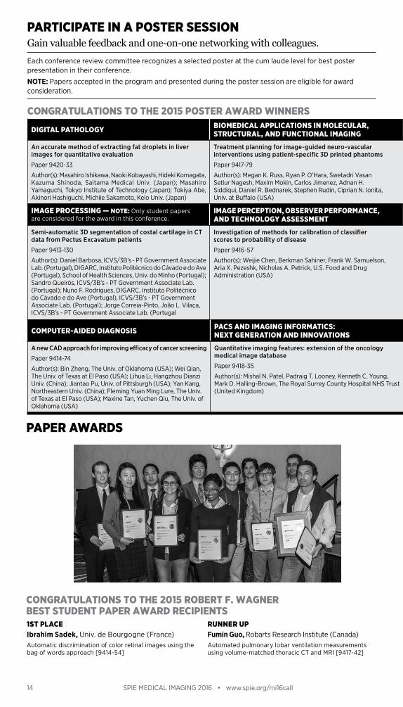
CONGRATULATIONS TO THE 2015 POSTER AWARD WINNERS
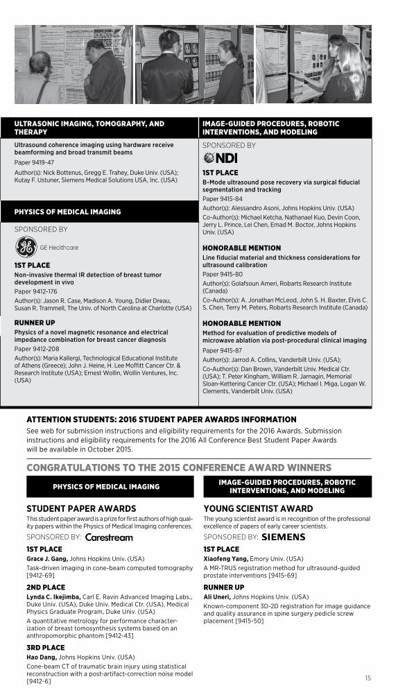
PARTICIPATE IN A POSTER SESSIONGain valuable feedback and one-on-one networking with colleagues.
PAPER AWARDS
1ST PLACEIbrahim Sadek, Univ. de Bourgogne (France) Automatic discrimination of color retinal images using the bag of words approach [9414-54]
RUNNER UPFumin Guo, Robarts Research Institute (Canada) Automated pulmonary lobar ventilation measurements using volume-matched thoracic CT and MRI [9417-42]
Each conference review committee recognizes a selected poster at the cum laude level for best poster presentation in their conference.
NOTE: Papers accepted in the program and presented during the poster session are eligible for award consideration.
CONGRATULATIONS TO THE 2015 ROBERT F. WAGNER BEST STUDENT PAPER AWARD RECIPIENTS
14 SPIE MEDICAL IMAGING 2016 • www.spie.org/mi16call
COMPUTER-AIDED DIAGNOSIS PACS AND IMAGING INFORMATICS: NEXT GENERATION AND INNOVATIONS
A new CAD approach for improving efficacy of cancer screening
Paper 9414-74
Author(s): Bin Zheng, The Univ. of Oklahoma (USA); Wei Qian, The Univ. of Texas at El Paso (USA); Lihua Li, Hangzhou Dianzi Univ. (China); Jiantao Pu, Univ. of Pittsburgh (USA); Yan Kang, Northeastern Univ. (China); Fleming Yuan Ming Lure, The Univ. of Texas at El Paso (USA); Maxine Tan, Yuchen Qiu, The Univ. of Oklahoma (USA)
Quantitative imaging features: extension of the oncology medical image database
Paper 9418-35
Author(s): Mishal N. Patel, Padraig T. Looney, Kenneth C. Young, Mark D. Halling-Brown, The Royal Surrey County Hospital NHS Trust (United Kingdom)
DIGITAL PATHOLOGY BIOMEDICAL APPLICATIONS IN MOLECULAR, STRUCTURAL, AND FUNCTIONAL IMAGING
ULTRASONIC IMAGING, TOMOGRAPHY, AND THERAPY
IMAGE-GUIDED PROCEDURES, ROBOTIC INTERVENTIONS, AND MODELING
An accurate method of extracting fat droplets in liver images for quantitative evaluation
Paper 9420-33
Author(s): Masahiro Ishikawa, Naoki Kobayashi, Hideki Komagata, Kazuma Shinoda, Saitama Medical Univ. (Japan); Masahiro Yamaguchi, Tokyo Institute of Technology (Japan); Tokiya Abe, Akinori Hashiguchi, Michiie Sakamoto, Keio Univ. (Japan)
Treatment planning for image-guided neuro-vascular interventions using patient-specific 3D printed phantoms
Paper 9417-79
Author(s): Megan K. Russ, Ryan P. O’Hara, Swetadri Vasan Setlur Nagesh, Maxim Mokin, Carlos Jimenez, Adnan H. Siddiqui, Daniel R. Bednarek, Stephen Rudin, Ciprian N. Ionita, Univ. at Buffalo (USA)
Ultrasound coherence imaging using hardware receive beamforming and broad transmit beams
Paper 9419-47
Author(s): Nick Bottenus, Gregg E. Trahey, Duke Univ. (USA); Kutay F. Ustuner, Siemens Medical Solutions USA, Inc. (USA)
SPONSORED BY
1ST PLACEB-Mode ultrasound pose recovery via surgical fiducial segmentation and trackingPaper 9415-84Author(s): Alessandro Asoni, Johns Hopkins Univ. (USA) Co-Author(s): Michael Ketcha, Nathanael Kuo, Devin Coon, Jerry L. Prince, Lei Chen, Emad M. Boctor, Johns Hopkins Univ. (USA)
HONORABLE MENTIONLine fiducial material and thickness considerations for ultrasound calibration Paper 9415-80Author(s): Golafsoun Ameri, Robarts Research Institute (Canada) Co-Author(s): A. Jonathan McLeod, John S. H. Baxter, Elvis C. S. Chen, Terry M. Peters, Robarts Research Institute (Canada)
HONORABLE MENTIONMethod for evaluation of predictive models of microwave ablation via post-procedural clinical imaging
Paper 9415-87Author(s): Jarrod A. Collins, Vanderbilt Univ. (USA);Co-Author(s): Dan Brown, Vanderbilt Univ. Medical Ctr. (USA); T. Peter Kingham, William R. Jarnagin, Memorial Sloan-Kettering Cancer Ctr. (USA); Michael I. Miga, Logan W. Clements, Vanderbilt Univ. (USA)
IMAGE PROCESSING — NOTE: Only student papers are considered for the award in this conference.
IMAGE PERCEPTION, OBSERVER PERFORMANCE, AND TECHNOLOGY ASSESSMENT PHYSICS OF MEDICAL IMAGING
Semi-automatic 3D segmentation of costal cartilage in CT data from Pectus Excavatum patients
Paper 9413-130
Author(s): Daniel Barbosa, ICVS/3B’s - PT Government Associate Lab. (Portugal), DIGARC, Instituto Politécnico do Cávado e do Ave (Portugal), School of Health Sciences, Univ. do Minho (Portugal); Sandro Queirós, ICVS/3B’s - PT Government Associate Lab. (Portugal); Nuno F. Rodrigues, DIGARC, Instituto Politécnico do Cávado e do Ave (Portugal), ICVS/3B’s - PT Government Associate Lab. (Portugal); Jorge Correia-Pinto, João L. Vilaça, ICVS/3B’s - PT Government Associate Lab. (Portugal
Investigation of methods for calibration of classifier scores to probability of disease
Paper 9416-57
Author(s): Weijie Chen, Berkman Sahiner, Frank W. Samuelson, Aria X. Pezeshk, Nicholas A. Petrick, U.S. Food and Drug Administration (USA)
SPONSORED BY
1ST PLACENon-invasive thermal IR detection of breast tumor development in vivoPaper 9412-176Author(s): Jason R. Case, Madison A. Young, Didier Dreau, Susan R. Trammell, The Univ. of North Carolina at Charlotte (USA)
RUNNER UPPhysics of a novel magnetic resonance and electrical impedance combination for breast cancer diagnosisPaper 9412-208Author(s): Maria Kallergi, Technological Educational Institute of Athens (Greece); John J. Heine, H. Lee Moffitt Cancer Ctr. & Research Institute (USA); Ernest Wollin, Wollin Ventures, Inc. (USA)
CONGRATULATIONS TO THE 2015 CONFERENCE AWARD WINNERS
STUDENT PAPER AWARDSThis student paper award is a prize for first authors of high qual-ity papers within the Physics of Medical Imaging conferences.
SPONSORED BY:
1ST PLACEGrace J. Gang, Johns Hopkins Univ. (USA) Task-driven imaging in cone-beam computed tomography [9412-69]
2ND PLACELynda C. Ikejimba, Carl E. Ravin Advanced Imaging Labs., Duke Univ. (USA), Duke Univ. Medical Ctr. (USA), Medical Physics Graduate Program, Duke Univ. (USA) A quantitative metrology for performance character-ization of breast tomosynthesis systems based on an anthropomorphic phantom [9412-43]
3RD PLACEHao Dang, Johns Hopkins Univ. (USA)Cone-beam CT of traumatic brain injury using statistical reconstruction with a post-artifact-correction noise model [9412-6]
YOUNG SCIENTIST AWARDThe young scientist award is in recognition of the professional excellence of papers of early career scientists.
SPONSORED BY:
1ST PLACEXiaofeng Yang, Emory Univ. (USA)A MR-TRUS registration method for ultrasound-guided prostate interventions [9415-69]
RUNNER UPAli Uneri, Johns Hopkins Univ. (USA) Known-component 3D-2D registration for image guidance and quality assurance in spine surgery pedicle screw placement [9415-50]
PHYSICS OF MEDICAL IMAGING IMAGE-GUIDED PROCEDURES, ROBOTIC INTERVENTIONS, AND MODELING
ATTENTION STUDENTS: 2016 STUDENT PAPER AWARDS INFORMATIONSee web for submission instructions and eligibility requirements for the 2016 Awards. Submission instructions and eligibility requirements for the 2016 All Conference Best Student Paper Awards will be available in October 2015.
15
COMPUTER-AIDED DIAGNOSIS PACS AND IMAGING INFORMATICS: NEXT GENERATION AND INNOVATIONS
A new CAD approach for improving efficacy of cancer screening
Paper 9414-74
Author(s): Bin Zheng, The Univ. of Oklahoma (USA); Wei Qian, The Univ. of Texas at El Paso (USA); Lihua Li, Hangzhou Dianzi Univ. (China); Jiantao Pu, Univ. of Pittsburgh (USA); Yan Kang, Northeastern Univ. (China); Fleming Yuan Ming Lure, The Univ. of Texas at El Paso (USA); Maxine Tan, Yuchen Qiu, The Univ. of Oklahoma (USA)
Quantitative imaging features: extension of the oncology medical image database
Paper 9418-35
Author(s): Mishal N. Patel, Padraig T. Looney, Kenneth C. Young, Mark D. Halling-Brown, The Royal Surrey County Hospital NHS Trust (United Kingdom)

16 SPIE MEDICAL IMAGING 2016 • www.spie.org/mi16call
By submitting an abstract, I agree to the following conditions:
ABSTRACT SUBMISSION
AN AUTHOR OR COAUTHOR (INCLUDING INVITED, ORAL, AND POSTER PRESENTERS) WILL:• Register at the reduced author registration rate
(current SPIE Members receive an additional discount on the registration fee).
• Attend the meeting.• Make the presentation as scheduled in the
program.• Submit a full-length manuscript (6 pages
minimum) for publication in the SPIE Digital Library, Proceedings of SPIE, and CD-ROM compilations.
• Obtain funding for their registration fees, travel, and accommodations, independent of SPIE, through their sponsoring organizations.
• Ensure that all clearances, including government and company clearance, have been obtained to present and publish. If you are a DoD contractor in the USA, allow at least 60 days for clearance.
Submit an abstract and summary online at: www.spie.org/mi16call • Please submit a 250-word text abstract for
technical review purposes that is suitable for publication. SPIE is authorized to circulate your abstract to conference committee members for review and selection purposes.
• Please also submit a 100-word text summary suitable for early release. If accepted, this summary text will be published prior to the meeting in the online or printed programs promoting the conference.
• Identify the topics appropriate to the specific conference. During the submission process you will be asked to choose no more than three topics from a predefined list and/or add a topic not included on the list. (See individual conference Call for Papers for topic categories.)
• Prepare your 2-4 page supplemental MS Word or PostScript file. Supplemental file instructions can be found online. For full consideration this file must include the paper title, authors, 250-word abstract text, and the following supplemental information: - Description of purpose - Method(s) - Results - New or breakthrough work to be presented - Conclusions - Whether the work is being, or has been,
submitted for publication or presentation elsewhere, and, if so, indicate how the submissions differ.
- This file may contain supporting images/tables/figures
- Failure to follow these guidelines may disqualify your submission.
• Only original material should be submitted.• Abstracts should contain enough detail to
clearly convey the approach and the results of the research.
• Commercial papers, papers with no new research/development content, and papers where supporting data or a technical description cannot be given for proprietary reasons will not be accepted for presentation in this conference.
• Please do not submit the same, or similar, abstracts to multiple conferences.
REVIEW, NOTIFICATION, AND PROGRAM PLACEMENT INFORMATION• To ensure a high-quality conference, all
submissions will be assessed by the Conference Chair/Editor for technical merit and suitability of content.
• Conference Chair/Editors reserve the right to reject for presentation any paper that does not meet content or presentation expectations.
• The contact author will receive notification of acceptance and presentation details by e-mail no later than 30 October 2015.
• Final placement in an oral or poster session is subject to the Chairs’ discretion.
PROCEEDINGS OF SPIE AND SPIE DIGITAL LIBRARY INFORMATION• Manuscript instructions are available from
the “For Authors/Presenters” link on the conference website.
• Conference Chair/Editors may require manuscript revision before approving publication and reserve the right to reject for publication any paper that does not meet acceptable standards for a scientific publication. Conference Chair/Editors’ decisions on whether to allow publication of a manuscript is final.
• Authors must be authorized to transfer copyright of the manuscript to SPIE, or provide a suitable publication license.
• Only papers presented at the conference and received according to publication guidelines and timelines will be published in the conference Proceedings of SPIE and SPIE Digital Library.
• Published papers are indexed in leading scientific databases including Astrophysical Data System (ADS), Chemical Abstracts (relevant content), Compendex, CrossRef, Current Contents, DeepDyve, Google Scholar, Inspec, Portico, Scopus, SPIN, and Web of Science Conference Proceedings Citation Index, and are searchable in the SPIE Digital Library. Full manuscripts are available to SPIE Digital Library subscribers worldwide.
NEWFOCUS ON PRECISION MEDICINEPrecision Medicine is an emerging approach for disease treatment and prevention that takes into account individual variability in genes, environment, and lifestyle for each person. If your research is related and you want to participate in this special focus, enter “PRECISION MEDICINE” when prompted during your abstract submission. Accepted papers will be highlighted in the technical program.
2014 SYMPOSIUM CHAIRS:
DATESConferences & Courses: 27 February – 3 March 2016
WEST COAST LOCATIONTown & Country Resort and Convention Center San Diego, California, USA
Gain visibility at the premier international forum on medical imaging.Join us at our California location and gain career insights as well as some relaxation time at the Town & Country Resort and Convention Center . Whether you are presenting to your peers, or are looking to gain further knowledge to enhance your research area, we welcome your participation .
SPIE and California are bringing you more of what you need for success
– Present a paper and participate in the conference
– Meet the top researchers and experts in the imaging field
– Receive feedback from your peers
– Hear the latest research
– Network with your colleagues
– Publish an accepted paper in the Journal of Medical Imaging
Plan to attend SPIE Medical Imaging 2016 in San Diego
BOOK HOTEL EARLY
SAVE $Reservation Process
Opening October 2015
Non-
Profi
t Org
.U.
S. P
osta
ge
Paid
SPIE
27 F
EBR
UA
RY–
3 M
AR
CH
20
16
SUB
MIT
YO
UR
A
BST
RA
CT
TOD
AY
•
WES
T CO
AST
LO
CA
TIO
N IN
20
16To
wn
& C
ount
ry R
esor
t an
d Co
nven
tion
Cen
ter
San
Die
go, C
alifo
rnia
, USA
ww
w.s
pie
.org
/mi1
6ca
ll
Plan
to p
arti
cipa
te in
the
conf
eren
ce w
here
th
e la
test
info
rmat
ion
is p
rese
nted
in
imag
e pr
oces
sing
, per
cept
ion,
reg
istr
atio
n,
info
rmat
ics,
and
seg
men
tati
on; c
ompu
ter-
aide
d di
agno
sis;
dig
ital
pat
holo
gy;
tom
ogra
phy;
and
ult
raso
nic
imag
ing.
P.O
. Box
10
Belli
ngha
m, W
A 98
227-
0010
USA