mod4lab - stream ecology - measuring periphyton biomass
TRANSCRIPT
Module 4 Stream Ecology Laboratory
Measuring Periphyton Biomass

Presentation NameUpdated March 16, 2003 – Author
Slide ID NumberPage 2
Periphyton Biomass Measurements
Ash-Free Dry Weight
Pigment Analysis
Biovolume Measurement
Presentation NameUpdated March 16, 2003 – Author
Slide ID NumberPage 3
Periphyton Biomass Measurements
Many methods have been developed to collect periphyton from natural substrates. All involve quanitatively removing material from a known surface area.
Different methods may be needed to collect periphyton from rocks, woody debris, aquatic macrophytes, or other substrates.
Substrate size will also determine how samples are collected. Large boulders require the use of a device that prevents the sample from being lost downstream. If the substrate is a fist-sized cobble the rock can be paced in a pan, scraped, then washed into a bottle with stream water.

Presentation NameUpdated March 16, 2003 – Author
Slide ID NumberPage 4
One way to scrape a known area is to lay a plastic 35 mm slide (film removed) over the rock and scrape off the material within the slide area (scrub area = 2.3cmX3.5cm=8cm2).
Presentation NameUpdated March 16, 2003 – Author
Slide ID NumberPage 5
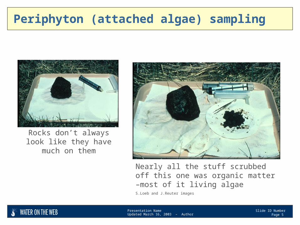
Periphyton (attached algae) sampling
Rocks don’t always look like they have much on them
Nearly all the stuff scrubbed off this one was organic matter –most of it living algaeS.Loeb and J.Reuter images
Presentation NameUpdated March 16, 2003 – Author
Slide ID NumberPage 6
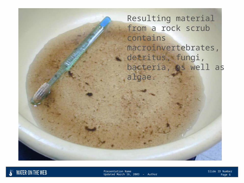
Resulting material from a rock scrub contains macroinvertebrates, detritus, fungi, bacteria, as well as algae.
Presentation NameUpdated March 16, 2003 – Author
Slide ID NumberPage 7
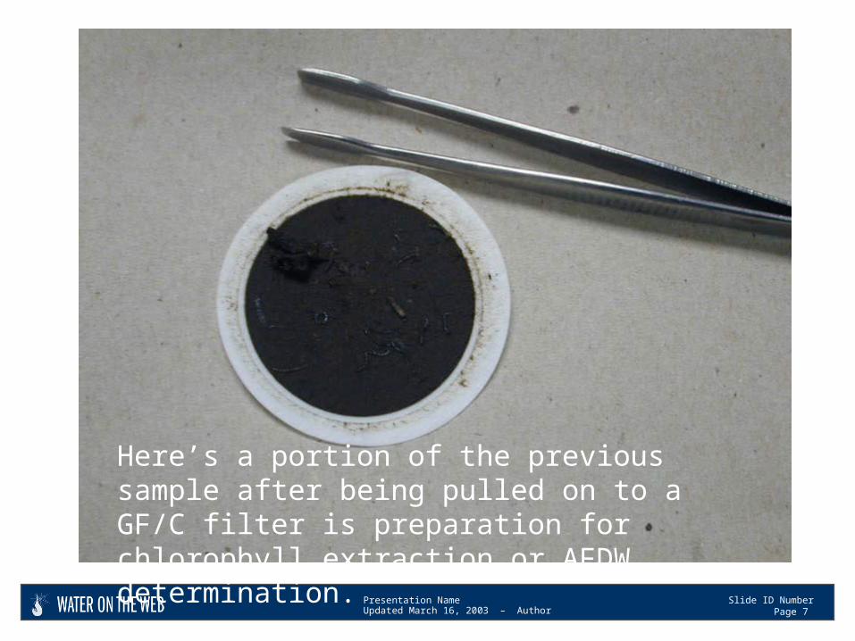
Here’s a portion of the previous sample after being pulled on to a GF/C filter is preparation for chlorophyll extraction or AFDW determination.
Presentation NameUpdated March 16, 2003 – Author
Slide ID NumberPage 8
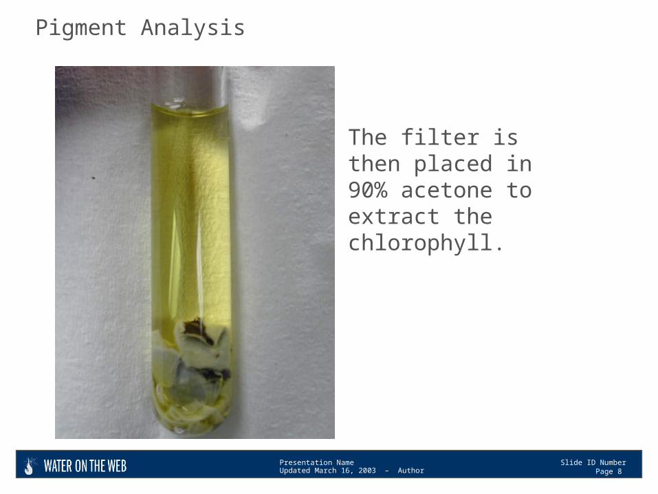
The filter is then placed in 90% acetone to extract the chlorophyll.
Pigment Analysis
Presentation NameUpdated March 16, 2003 – Author
Slide ID NumberPage 9
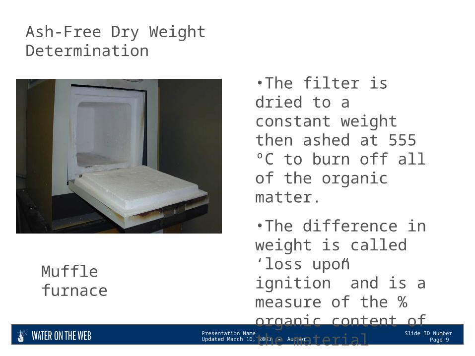
•The filter is dried to a constant weight then ashed at 555 ºC to burn off all of the organic matter.
•The difference in weight is called ‘loss upon ignition” and is a measure of the % organic content of the material
Ash-Free Dry Weight Determination
Muffle furnace

Presentation NameUpdated March 16, 2003 – Author
Slide ID NumberPage 10
Project NameWater Quality Samples scrub area = 2.3cmX3.5cm=8cm2
2002 8 cm2=0.0008 m2 X 3 scrubs = .0024 m2 total areaNRRI Central Analytical Labemr 12/4/02
Sample Run chlorophyll phaeophytin volume total chlorophyllPeriphyton Date Date ug/L ug/L filtered (mLs) volume (mLs) mg/m2Whatever Creek 5/6/2002 5/15/2002 130 60 45 45 2.4
Sample Run Dry Wt AFDW total volume AFDWDate Date mg/L mg/L volume (mLs) filtered (mLs) g/m2
Whatever Creek 5/6/2002 5/8/2002 156 117 319 122 6.0
chlorophyll
AFDW
Once you have a measure of chlorophyll or AFDW you’ll need to calculate per unit area.
Presentation NameUpdated March 16, 2003 – Author
Slide ID NumberPage 11
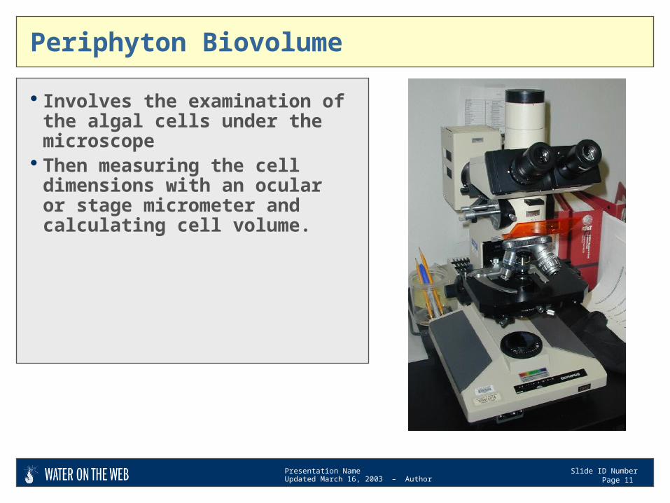
Periphyton Biovolume
Involves the examination of the algal cells under the microscope
Then measuring the cell dimensions with an ocular or stage micrometer and calculating cell volume.
Presentation NameUpdated March 16, 2003 – Author
Slide ID NumberPage 12
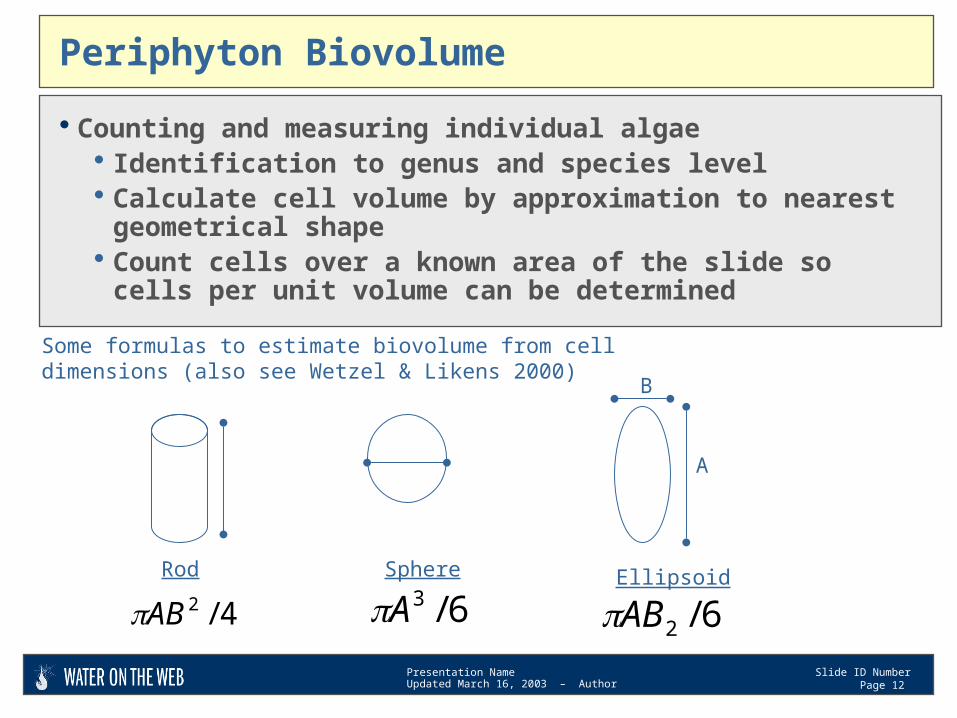
Periphyton Biovolume
Counting and measuring individual algae Identification to genus and species level Calculate cell volume by approximation to nearest geometrical
shape Count cells over a known area of the slide so cells per unit
volume can be determined
Rod
4/2AB
B
A
Some formulas to estimate biovolume from cell dimensions (also see Wetzel & Likens 2000)
6/3ASphere
A
Ellipsoid
6/2AB
B
A
Presentation NameUpdated March 16, 2003 – Author
Slide ID NumberPage 13
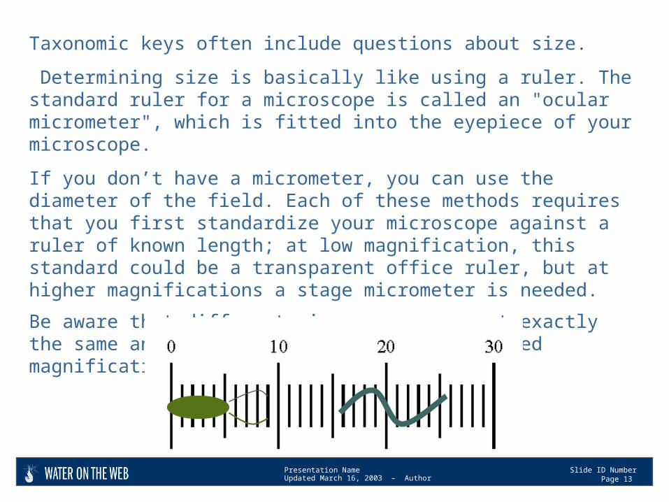
Taxonomic keys often include questions about size.
Determining size is basically like using a ruler. The standard ruler for a microscope is called an "ocular micrometer", which is fitted into the eyepiece of your microscope.
If you don’t have a micrometer, you can use the diameter of the field. Each of these methods requires that you first standardize your microscope against a ruler of known length; at low magnification, this standard could be a transparent office ruler, but at higher magnifications a stage micrometer is needed.
Be aware that different microscopes are not exactly the same and the size goes down with increased magnification.