molecular basis of human usher syndrome: deciphering the
TRANSCRIPT

Experimental Eye Research 83 (2006) 97e119www.elsevier.com/locate/yexer
Molecular basis of human Usher syndrome: Deciphering themeshes of the Usher protein network provides insights into
the pathomechanisms of the Usher disease
Jan Reiners1,2, Kerstin Nagel-Wolfrum1, Karin Jurgens, Tina Marker, Uwe Wolfrum*
Institute of Zoology, Department of Cell and Matrix Biology, Johannes Gutenberg University of Mainz, Mullerweg 6, D-55099 Mainz, Germany
Received 5 September 2005; accepted in revised form 21 November 2005
Available online 20 March 2006
Abstract
Usher syndrome (USH) is the most frequent cause of combined deaf-blindness in man. It is clinically and genetically heterogeneous and atleast 12 chromosomal loci are assigned to three clinical USH types, namely USH1A-G, USH2A-C, USH3A (Davenport, S.L.H., Omenn, G.S.,1977. The heterogeneity of Usher syndrome. Vth Int. Conf. Birth Defects, Montreal; Petit, C., 2001. Usher syndrome: from genetics to pathogen-esis. Annu. Rev. Genomics Hum. Genet. 2, 271e297). Mutations in USH type 1 genes cause the most severe form of USH. In USH1 patients,congenital deafness is combined with a pre-pubertal onset of retinitis pigmentosa (RP) and severe vestibular dysfunctions. Those with USH2have moderate to severe congenital hearing loss, non-vestibular dysfunction and a later onset of RP. USH3 is characterized by variable RPand vestibular dysfunction combined with progressive hearing loss. The gene products of eight identified USH genes belong to different proteinclasses and families. There are five known USH1 molecules: the molecular motor myosin VIIa (USH1B); the two cellecell adhesion cadherinproteins, cadherin 23 (USH1D) and protocadherin 15, (USH1F) and the scaffold proteins, harmonin (USH1C) and SANS (USH1G). In addition,two USH2 genes and one USH3A gene have been identified. The two USH2 genes code for the transmembrane protein USH2A, also termedUSH2A (‘‘usherin’’) and the G-protein-coupled 7-transmembrane receptor VLGR1b (USH2C), respectively, whereas the USH3A gene encodesclarin-1, a member of the clarin family which exhibits 4-transmembrane domains. Molecular analysis of USH1 protein function revealed that allfive USH1 proteins are integrated into a protein network via binding to PDZ domains in the USH1C protein harmonin. Furthermore, this scaffoldfunction of harmonin is supported by the USH1G protein SANS. Recently, we have shown that the USH2 proteins USH2A and VLGR1b as well asthe candidate for USH2B, the sodium bicarbonate co-transporter NBC3, are also integrated into this USH protein network. In the inner ear, theseinteractions are essential for the differentiation of hair cell stereocilia but may also participate in the mechano-electrical signal transduction andthe synaptic function of maturated hair cells. In the retina, the co-expression of all USH1 and USH2 proteins at the synapse of photoreceptor cellsindicates that they are organized in an USH protein network there. The identification of the USH protein network indicates a common pathophys-iological pathway in USH. Dysfunction or absence of any of the molecules in the mutual ‘‘interactome’’ related to the USH disease may lead todisruption of the network causing senso-neuronal degeneration in the inner ear and the retina, the clinical symptoms of USH.� 2006 Elsevier Ltd. All rights reserved.
Keywords: senso-neuronal disease; retinal degeneration; retinitis pigmentosa; deafness; supramolecular network; interactome
1. Introduction
Human communication and perception of the environmentare mainly formulated on information imported through the
* Corresponding author. Tel.: þ49 6131 39 25148; fax: þ49 6131 39 23815.
E-mail address: [email protected] (U. Wolfrum).1 Both authors contributed equally to this work.2 Present address: Abbott GmbH, Knollstr., 67061 Ludwigshafen, Germany.
0014-4835/$ - see front matter � 2006 Elsevier Ltd. All rights reserved.
doi:10.1016/j.exer.2005.11.010
ear and the eye. Chronic diseases affecting the inner ear andthe retina cause severe impairments of our communicationsystems. There are about 40 known human syndromes whichinclude the symptoms of blindness in combination with deaf-ness. In more than half of the cases, the Usher syndrome is theorigin of this defect (Gorlin, 1995; Vernon, 1969). The humanUsher syndrome (USH) is defined by congenital, bilateraldeafness and a later onset of the loss of the visual field, caused
98 J. Reiners et al. / Experimental Eye Research 83 (2006) 97e119
by retinitis pigmentosa (RP). In RP, retinal degeneration isbased on photoreceptor cell death which occurs from the pe-riphery to the macula of the retina. Night blindness is the firstsymptom of RP, followed by narrowing of the visual field(‘‘tunnel vision’’) and later to complete blindness (van Soestet al., 1999; Wang et al., 2005). These visual deficits are trig-gered by any one of over 130 mutated genes (Tschernutteret al., 2005). USH is the most common cause of combineddeaf-blindness (Vernon, 1969) and the most frequent form ofrecessive RP (Keats and Corey, 1999).
One of the earliest descriptions of USH was given by Al-brecht von Graefe, a pioneer of modern ophthalmology. He re-ported a case of a deaf and dumb male patient with retinaldegeneration who had two equally affected brothers (vonGraefe, 1858). Subsequently his student Richard Liebreich,screened the population of Berlin for syndromes includingRP and reported similar observations (Liebreich, 1861). Heemphasized the recessive nature of the disease by commentingon the combination of congenital deafness with RP in severalsiblings from either consanguineous marriages or familieswith several members affected in different generations. Thedisease was eventually named after Charles Usher, a Scottishophthalmologist who described the hereditary nature of thisdisorder in 19 cases out of 69 RP patients (Usher, 1914).
Based on the heterogenic clinical course of the disease de-scribed by Bell (1922) and Hallgren (1959) and co-workers,USH was subdivided into three clinical types, namely USH1,USH2 and USH3 (Davenport and Omenn, 1977). USH type1 is the most severe form of this disease. USH1 patients aredeaf at birth and the onset of RP is pre-pubertal. Most, butnot all USH1 patients exhibit severe dysfunction of the vestib-ular system which leads to a further subdivision of USH type 1(Otterstedde et al., 2001). USH type 2 is characterized by aconstant moderate to severe hearing impairment from birthon and RP can be diagnosed during puberty (Reisser et al.,2002). USH type 3 (USH3) is distinguished from USH1 andUSH2 by the later initiation of deafness combined with
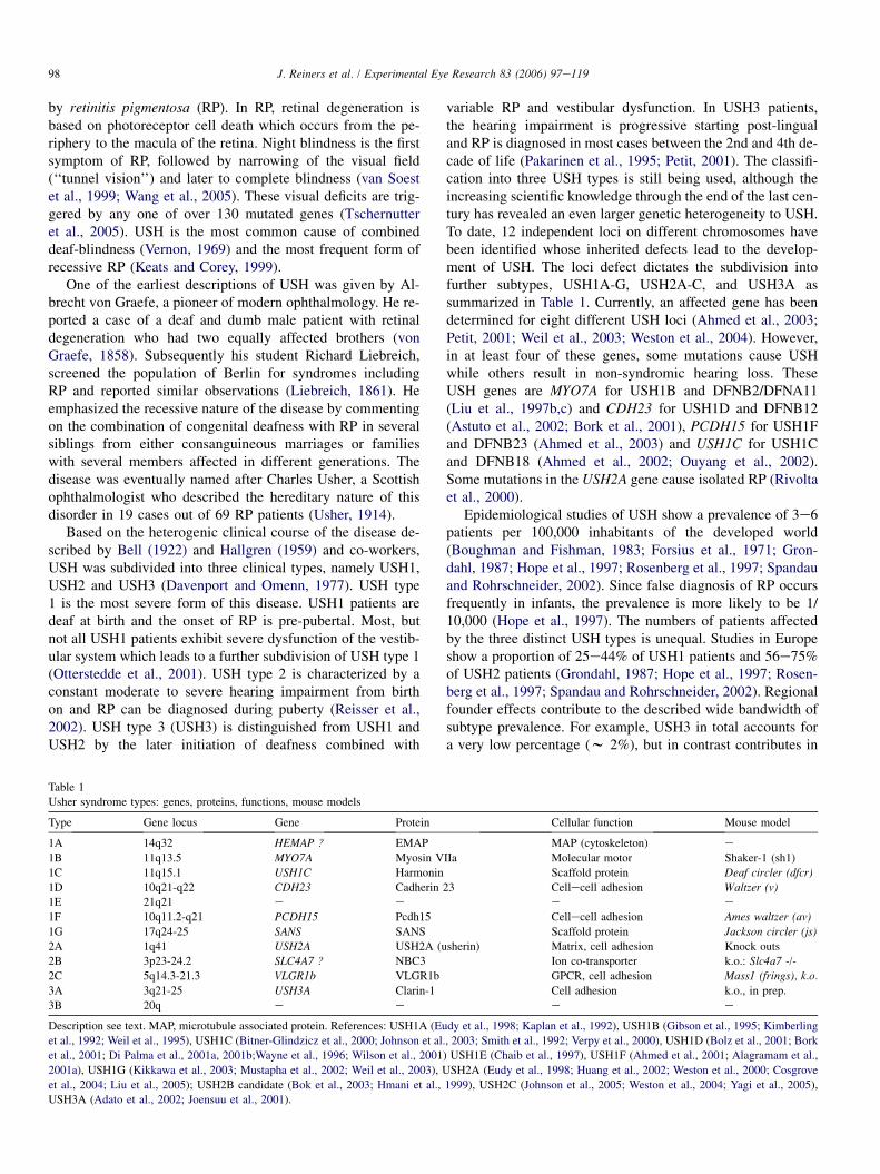
variable RP and vestibular dysfunction. In USH3 patients,the hearing impairment is progressive starting post-lingualand RP is diagnosed in most cases between the 2nd and 4th de-cade of life (Pakarinen et al., 1995; Petit, 2001). The classifi-cation into three USH types is still being used, although theincreasing scientific knowledge through the end of the last cen-tury has revealed an even larger genetic heterogeneity to USH.To date, 12 independent loci on different chromosomes havebeen identified whose inherited defects lead to the develop-ment of USH. The loci defect dictates the subdivision intofurther subtypes, USH1A-G, USH2A-C, and USH3A assummarized in Table 1. Currently, an affected gene has beendetermined for eight different USH loci (Ahmed et al., 2003;Petit, 2001; Weil et al., 2003; Weston et al., 2004). However,in at least four of these genes, some mutations cause USHwhile others result in non-syndromic hearing loss. TheseUSH genes are MYO7A for USH1B and DFNB2/DFNA11(Liu et al., 1997b,c) and CDH23 for USH1D and DFNB12(Astuto et al., 2002; Bork et al., 2001), PCDH15 for USH1Fand DFNB23 (Ahmed et al., 2003) and USH1C for USH1Cand DFNB18 (Ahmed et al., 2002; Ouyang et al., 2002).Some mutations in the USH2A gene cause isolated RP (Rivoltaet al., 2000).
Epidemiological studies of USH show a prevalence of 3e6patients per 100,000 inhabitants of the developed world(Boughman and Fishman, 1983; Forsius et al., 1971; Gron-dahl, 1987; Hope et al., 1997; Rosenberg et al., 1997; Spandauand Rohrschneider, 2002). Since false diagnosis of RP occursfrequently in infants, the prevalence is more likely to be 1/10,000 (Hope et al., 1997). The numbers of patients affectedby the three distinct USH types is unequal. Studies in Europeshow a proportion of 25e44% of USH1 patients and 56e75%of USH2 patients (Grondahl, 1987; Hope et al., 1997; Rosen-berg et al., 1997; Spandau and Rohrschneider, 2002). Regionalfounder effects contribute to the described wide bandwidth ofsubtype prevalence. For example, USH3 in total accounts fora very low percentage (w 2%), but in contrast contributes in
Table 1
Usher syndrome types: genes, proteins, functions, mouse models
Type Gene locus Gene Protein Cellular function Mouse model
1A 14q32 HEMAP ? EMAP MAP (cytoskeleton) e
1B 11q13.5 MYO7A Myosin VIIa Molecular motor Shaker-1 (sh1)
1C 11q15.1 USH1C Harmonin Scaffold protein Deaf circler (dfcr)
1D 10q21-q22 CDH23 Cadherin 23 Cellecell adhesion Waltzer (v)1E 21q21 e e e e
1F 10q11.2-q21 PCDH15 Pcdh15 Cellecell adhesion Ames waltzer (av)
1G 17q24-25 SANS SANS Scaffold protein Jackson circler (js)
2A 1q41 USH2A USH2A (usherin) Matrix, cell adhesion Knock outs
2B 3p23-24.2 SLC4A7 ? NBC3 Ion co-transporter k.o.: Slc4a7 -/-
2C 5q14.3-21.3 VLGR1b VLGR1b GPCR, cell adhesion Mass1 (frings), k.o.
3A 3q21-25 USH3A Clarin-1 Cell adhesion k.o., in prep.
3B 20q e e e e
Description see text. MAP, microtubule associated protein. References: USH1A (Eudy et al., 1998; Kaplan et al., 1992), USH1B (Gibson et al., 1995; Kimberling
et al., 1992; Weil et al., 1995), USH1C (Bitner-Glindzicz et al., 2000; Johnson et al., 2003; Smith et al., 1992; Verpy et al., 2000), USH1D (Bolz et al., 2001; Bork
et al., 2001; Di Palma et al., 2001a, 2001b;Wayne et al., 1996; Wilson et al., 2001) USH1E (Chaib et al., 1997), USH1F (Ahmed et al., 2001; Alagramam et al.,
2001a), USH1G (Kikkawa et al., 2003; Mustapha et al., 2002; Weil et al., 2003), USH2A (Eudy et al., 1998; Huang et al., 2002; Weston et al., 2000; Cosgrove
et al., 2004; Liu et al., 2005); USH2B candidate (Bok et al., 2003; Hmani et al., 1999), USH2C (Johnson et al., 2005; Weston et al., 2004; Yagi et al., 2005),
USH3A (Adato et al., 2002; Joensuu et al., 2001).

99J. Reiners et al. / Experimental Eye Research 83 (2006) 97e119
Birmingham (UK) for 20% of all USH cases and even higherpercentage, 42%, in Finland (Hope et al., 1997; Pakarinenet al., 1995).
In addition to the characteristic senso-neuronal degenera-tion in the eye and the inner ear of USH, several reports indi-cate that USH affects other tissues and organs. This isconfirmed by the rather wide expression profiles of all USHgene products obtained (see below). Functional studies onUSH1 and USH2 patients indicate lower odor identificationability (Zrada et al., 1996). However, a study by Seeligeret al. (1999) on olfaction in USH did not confirm differencesbetween USH patients and the control group. Nevertheless, re-cent expression analyses revealed that all analyzed USH mol-ecules are expressed in the olfactory epithelium (Mikosz,2005; Wolfrum et al., 1998 and Mikoz and Wolfrum, unpub-lished). Thus, the analysis of biopsies from the nasal epithe-lium of patients may be useful for USH diagnosis (Cohnet al., 2004). Furthermore, USH may also be related to braindysfunction. An increase of mental deficiencies, cerebral atro-phies, and ataxies are reported for USH patients (Drouet et al.,2003; Hess-Rover et al., 1999; Koizumi et al., 1988; Mango-tich and Misiaszek, 1983). Despite the expression of all knownUSH proteins in the brain (Wolfrum et al., unpublished), oftenUSH patients are highly educated and intelligent.
Sparse histopathological data from patients are often re-lated to undefined USH subtypes. In some cases, ciliary abnor-malities have been reported in patients of undefined USHsubtype in retinal photoreceptors (connecting cilium) and thenasal epithelium, the trachea, and sperm cells (e.g. Ardenand Fox, 1979; Baris et al., 1994; Barrong et al., 1992; Hunteret al., 1986; Tosi et al., 2003; Petrozza et al., 1991; van Aaremet al., 1999). Based on these observations, it has been sug-gested that USH is related to cilia dysfunction. When myosinVIIa, the product of the first identified USH gene, was local-ized in the connecting cilium of photoreceptor cells (Liuet al., 1997a), the latter suggestion warrants further attention.
The presence of cilia is one of the structural similarities be-tween retinal photoreceptor cells and the mechanosensitivehair cells of the inner ear (Fig. 1). In photoreceptor cells, theconnecting cilium, which is homologous to the transitionzone found at the base of every motile cilium, links the biosyn-thetic and metabolic active inner segment with the outer seg-ment, which is actually a modified cilium (Besharse andHorst, 1990). Each hair cell possesses one kinocilium whichis responsible for organizing the array of stereocilia duringhair cell differentiation (Kelley et al., 1992; Sobkowiczet al., 1995). In the mammalian cochlea, this ‘‘real’’ ciliumdisappears during hair cell maturation. The stereocilia, struc-tures that take up the mechanical stimulus in hair cells, arenot ‘‘real’’ cilia. On contrary, they are highly specialized mi-crovilli (‘‘stereovilli’’) characterized by a rigid actin filamentcore (Tilney et al., 1988). Microvillar-like, actin filament-supported structures are also found in photoreceptor cells in theform of calycal processes at the apical membrane of the innersegment (Nagle et al., 1986). These calyces sheath the base ofthe photoreceptor outer segment and thereby may stabilize it.Microvillar-like differentiations are more obvious on the
apical membrane of the cells of the retinal pigment epithelium.Furthermore, ribbon synapses are characteristic for both typesof sensory cells. Their synapses are a unique type of chemicalsynapses structurally and functionally specialized for massiveand sustained neurotransmitter release (Rao-Mirotznik et al.,1995; Wagner, 1997). Thus, the primary targets for defectscaused by USH are probably molecules which are present inthe subcellular compartments with the described similaritiesbetween both types of sensory cells. The molecular dissectionof these subcellular compartments could reveal candidategenes for USH, while the study of the identified molecules re-lated to USH should provide insights into novel processesshared by both types of sensory cells.
2. Characteristics and function of the proteins encodedby the identified USH genes
Of the three clinically characterized types of USH, eightgenes on 12 USH loci have been cloned. The protein productsof the eight USH genes, belong to different protein classes andtherefore possess various cellular functions (Table 1).
Fig. 1. Schematic representation of the sensory cells in the eye and the ear af-
fected by USH. (A) Scheme of a rod photoreceptor cell. The apical extensions
of cells of the retinal pigment epithelium (RPE) evolve the tips of light-sensi-
tive outer segments (OS) of photoreceptor cells. The OS are linked via a con-
necting cilium (CC) to an inner segment (IS). Calycal processes (CP) ensheath
the proximal outer segment. Nuclei (N) of photoreceptor cells are localized in
the outer nuclear layer (ONL). Synaptic terminals (S) link photoreceptor cells
and 2nd-order neurons, bipolar and horizontal cells. (B) Scheme of a mechano-
sensitive hair cell. The apical part of hair cells carries numerous rigid micro-
villi-like structures, improperly named stereocilia (SC, arrows), where the
mechanotransduction takes place. They are anchored in the actin filament-
rich cuticular plate (CP). Lateral to the longest stereocilum a kinocilium (black
arrowhead) is present. Its basal body is localized in the pericuticular region
(gray arrowheads). N, nucleus; S, synaptic junctions between hair cells and ef-
ferent and afferent neurons.

100 J. Reiners et al. / Experimental Eye Research 83 (2006) 97e119
2.1. USH1 proteins
Seven loci responsible for USH1 have been defined(USH1A-G) and five of the corresponding causative genesidentified (Table 1). The USH1A gene was mapped to the chro-mosomal locus of the gene encoding the human homologue ofa sea urchin microtubule-associated protein (EMAP) (Eudyet al., 1998), but had not yet been verified. In a search for aharmonin (USH1C) interacting partner, we identified a cellecell adhesion molecule encoded by a gene mapping to theUSH1E locus (Reiners, 2004). The candidate gene identifiedby this ‘‘reverse genetic’’ approach remains to be validated.
2.1.1. Myosin VIIa (USH1B), a membrane-associatedactin-based molecular motor
The myosin VIIa gene (MYO7A) was the first USH geneidentified (Weil et al., 1995) and at least half of the known
USH1 cases are caused by mutations in MYO7A (Astutoet al., 2000). Myosin VIIa is an unconventional myosin com-posed of several functional domains (Fig. 2A). The highly con-served N-terminal motor or head domain classifies myosin VIIaas an actin-based molecular motor. As in other myosins, thisdomain contains the actin-binding site and the ATP-bindingsite (Sellers, 2000) which allows the protein to move along ac-tin filaments towards their plus end in an ATP-dependent man-ner (Inoue and Ikebe, 2003; Udovichenko et al., 2002). In theneck region, five IQ (from the first two conserved isoleucineand glutamine residues (Bahler and Rhoads, 2002)) motifsare expected to bind myosin-light chains, e.g. the Ca2þ-bindingEF-hand protein calmodulin (Todorov et al., 2001; Udovi-chenko et al., 2002). The neck is followed by a long tail regionthat contains various functional domains and thereby deter-mines, as in other myosin classes, the functional specificityof myosin VIIa (Sellers, 2000). The tail domain begins with
Fig. 2. Schematic representations of USH1 proteins. (A) The unconventional myosin VIIa (USH 1B) is composed of a motor head, a neck containing five iso-
leucine-glutamine (IQ) motifs, and a tail region. The tail region consists of a coil-coiled (CC1) domain, followed by two large repeats separated by a poorly con-
served SH3 (src homology-3) domain. These repeats include a MyTH4 (myosin tail homology 4) and a FERM (4.1, ezrin, radixin, moesin) domain. (B) Harmonin
(USH1C) isoforms can be divided into three classes (aec). Common features of all isoforms are two PDZ (PSD95, discs large, ZO-1) domains (PDZ1ePDZ2) and
one coiled-coil (CC1) domain. The harmonin class a isoforms consist of an additional PDZ (PDZ3) domain. The longest class b isoforms contain also a third PDZ
domain (PDZ3), a second coiled-coil domain (CC2), and a proline, serine, threonine (PST)-rich region. Harmonin a1 and b4 possess a C-terminal class I PDZ-
binding motif (PBM). (C) Cdh23 (Cadherin 23, USH1D) is composed of 27 extracellular cadherin repeats (EC1eEC27), a transmembrane domain (TM) and a short
cytoplasmic domain with a class I PBM. (D) The non-classical cadherin Pcdh15 (Protocadherin 15, USH1F) comprises eleven ectodomains (EC1eEC11), one
transmembrane domain (TM) and a C-terminal class I PBM. (E) The ‘‘scaffold protein containing ankyrin repeats and SAM domain’’ (SANS, USH1G) contains
three N-terminal ankyrin repeats (ANK1eANK3), a central region (cen), a sterile alpha motif (SAM) and a C-terminal class I PBM. extra, extracellular; intra,
intracellular; asterisks indicate class I PBMs.

101J. Reiners et al. / Experimental Eye Research 83 (2006) 97e119
a coiled-coil domain which is predicted to mediate homodimerformation (Weil et al., 1995). Two large tandem repeats, sepa-rated by a poorly conserved SH3 (src homology-3), follow.These repeats consists of a MyTH4 (myosin tail homology 4)domain and a FERM domain (4.1, ezrin, readixin, moesin).The function of the MyTH4 domain of myosins is still un-known, however FERM domains are thought to be responsiblefor protein attachment to the plasma membrane (Chishti et al.,1998). Several myosin VIIa splice variants are mentioned in theliterature (Sahly et al., 1997) which may be tissue specific, butdetailed investigations of these variants are lacking. To datemore than 90 different mutations have been identified inUSH1B patients. These mutations are distributed along thefull length of the gene with some clustering in the head domain(Petit, 2001). Mutations in the MYO7A gene result in not onlyUSH1B but also USH3-like phenotypes, as well as in dominantor recessive isolated deafness with any degree of severity, con-genital or progressive, with or without vestibular dysfunction(Petit, 2001). So far, it is difficult to determine whether thetype of mutation accounts for the expression of USH or isolateddeafness. A functional test for activity of the various mutatedforms of myosin VIIa protein could help to link the individualeffect of a given mutation to a phenotype. However, the pheno-typic heterogeneity argues for a role of additional genetic and/or environmental factors.
Several ligands binding to the tail domain of myosin VIIahave been identified and specify myosin VIIa’s role in e.g. in-tracellular transport, endocytosis and cellecell adhesion (sum-marized in (Wolfrum, 2003)) (Table 2). The binding of myosinVIIa to the scaffold proteins harmonin (USH1C) and SANS(USH1G) unites the specific function of myosin VIIa to theUSH protein network discussed in Section 4.
Although myosin VIIa is almost ubiquitously expressed, inhuman USH1B patients, dysfunctions are restricted to the
Table 2
Myosin VIIa binding proteins
Interacting protein Myosin VIIa
target domain
References
Calmodulin (Ca2þ-binding
protein)
IQ motifs Todorov et al., 2001;
Udovichenko et al.,
2002
MAP2B (microtubule
association)
SH3, MyTH4 (2),
FERM (2)
Todorov et al., 2001
RIa of PKA (kinase
regulation)
MyTH4 (2),
FERM (2)
Kussel-Andermann
et al., 2000
Keap1 (actin-associated
protein)
SH3 Velichkova et al., 2002
Vezatin (transmembrane
protein)
FERM (2) Kussel-Andermann
et al., 2000
MyRIP (Rab-interacting
protein)
MyTH4 (2),
FERM (2)
El Amraoui et al., 2002
Harmonin (USH1C)
(scaffold protein)
FERM (2) Boeda et al., 2002
SANS (USH1G)
(scaffold protein)
MyTH4/FERM
(1,2)
Adato et al., 2005
PHR1 (transmembrane
protein)
MyTH4/FERM
(2)
Etournay et al., 2005
inner ear and the retina. Expression of this unconventional my-osin has been documented in all epithelial tissue containingciliary processes or microvillar structures (Hasson et al.,1997; Sahly et al., 1997; Wolfrum et al., 1998). In the innerear, myosin VIIa is expressed in the mechanosensory hair cellsof the vestibular organ and cochlea where it is predominantlylocalized in the stereocilia, but is also found along the lateralmembrane, in the cuticular plate, and in the synaptic region (ElAmraoui et al., 1996; Hasson et al., 1997; Weil et al., 1996;Wolfrum et al., 1998). Studies on myosin VIIa deficientShaker-1 mice indicate that myosin VIIa is essential for thedifferentiation and organization of hair cell stereocilia (Selfet al., 1998; Boeda et al., 2002; Adato et al., 2005). In addi-tion, there is evidence that myosin VIIa also participates insignal transduction in the hair cells (Kros et al., 2002; Siemenset al., 2004; Etournay et al., 2005). Interestingly, a recent studydemonstrates that the Drosophila homolog of myosin VIIa, thecrinkled gene product, is required for organization of theJohnston’s organ (Todi et al., 2005). In this insect auditoryorgan, myosin VIIa is not only localized in the sensory cellsof the mechanosensitive scolopidia, but is also necessary for theorganization of the actin filament bundles of their innermostsupport cell (Todi et al., 2005) which have previously beendescribed as arrangements analogous to the actin filamentcore of stereocilia (Wolfrum, 1990, 1991).
In the vertebrate eye, the cell types affected by myosin VIIadefects are less clear than in the auditory organs. Myosin VIIais expressed in the retinal pigment epithelium (RPE) as well asin rod and cone photoreceptor cells of the retina (Liu et al.,1997a). In RPE cells, myosin VIIa is highly concentrated inthe apical microvilli-like processes (Hasson et al., 1995; Liuet al., 1997a). The identification of specific interaction partners(El Amraoui et al., 2002) together with analyses of the RPE ofShaker-1 mice revealed an active role for myosin VIIA in themigration of RPE melanosomes and a contribution to thephagocytosis of photoreceptor cell outer segment tips byRPE cells (El Amraoui et al., 2002; Gibbs et al., 2003; Liuet al., 1998; Williams and Gibbs 2004b). A recent study alsosuggests that myosin VIIa is a common lysosome-associatedmotor in general (Soni et al., 2005).
In mammalian rod and cone photoreceptor cells, myosinVIIa is predominantly localized in the connecting cilium(Liu et al., 1997a; Wolfrum and Schmitt, 2000; Wolfrumet al., 1998), but it is also found at photoreceptor synapses(El Amraoui et al., 1996; Reiners et al., 2003, 2005a; Wolfrumet al., 2004). Several lines of evidence indicate that myosinVIIa participates in the molecular transport through the photo-receptor cilium (Liu et al., 1997a; Williams and Gibbs 2004b;Wolfrum and Schmitt, 2000). Immunoelectron microscopy re-vealed that myosin VIIa is associated with the ciliary mem-brane (probably via its tail) and uses actin filaments in thecilium as a track for its movement through the cilium. Theco-localization of myosin VIIa with opsin and the abnormalopsin accumulation in the membrane of the connecting ciliumin photoreceptor cells of myosinVIIa-deficient Shaker-1 miceindicate participation of myosin VIIa in the opsin transportthrough the cilium (Liu et al., 1999; Wolfrum and Schmitt,

102 J. Reiners et al. / Experimental Eye Research 83 (2006) 97e119
2000). In independent studies using different immunocyto-chemical and biochemical approaches, the localization ofmyosin VIIa has also been demonstrated in the synapses ofmammalian photoreceptor cells (El Amraoui et al., 1996;Reiners et al., 2003, 2005a; Wolfrum and Reiners, 2004).Our recent studies indicate that partner proteins of myosinVIIa’s organized in the USH protein network are also localizedat the photoreceptor synapse (see Section 4) which further sup-ports the synaptic expression of myosin VIIa. Nevertheless,this localization is still controversial (Williams and Gibbs,2004a; Wolfrum and Reiners, 2004). However, functionalroles of myosin VIIa at synapses are recently supportedby findings on the neuromuscular junctions of Drosophila(Marella et al., unpublished) and by the morphological disor-ganization of the photoreceptor synapses in the myosin VIIa-deficient mariner zebrafish mutant, presented at ARVO in2005 (Biehlmaier et al., 2005).
2.1.2. Harmonin (USH1C), a potent scaffold protein andkey organizer of the USH protein network
USH type 1C is caused by defects in the harmonin USH1Cgene (Bitner-Glindzicz et al., 2000; Verpy et al., 2000). TheUSH1C locus was first mapped in a small French-speaking,Acadian population of academics in southwest Louisianaand consequently named the academic USH (Kloepfer andLaguaite, 1966). The harmonin gene was originally identifiedas an autoimmune antigen upregulated in some forms of can-cer and in cases of autoimmune enteropathy (AIE) (Kobayashiet al., 1999; Scanlan et al., 1999). In these studies, two syno-nyms PDZ-73 and AIE-75 for the harmonin protein, in partic-ular the human isoform harmonin a1 (see below), wereintroduced and are occasionally still in use. Genome screensand sequence comparisons revealed that harmonin genes arenot present in invertebrates, e.g. Drosophila or C. elegans,but are highly conserved in genomes of vertebrates (Reiners,2004; Reiners et al., 2003).
The harmonin USH1C gene consists of 28 coding exons ofwhich eight are differentially spliced, generating a variety ofalternatively spliced transcripts (Verpy et al., 2000). All eluci-dated mutations of USH1C that cause USH1 are localized inconstitutively transcribed exons whereas mutations in alterna-tively transcribed exons are associated with non-syndromicdeafness (Bitner-Glindzicz et al., 2000; Ouyang et al., 2002;Verpy et al., 2000; Zwaenepoel et al., 2001). So far, nine mu-rine splice variants of harmonin have been described which aregrouped in three classes a, b and c (Figs. 2B, 3). These iso-forms differ in the composition of their domain structure,namely two or three PDZ domains, one or two coiled-coil do-mains and the presence or absence of a PST ( prolin, serin,threonine-rich) domain. These three functional domains par-ticipate in proteineprotein interactions, establishing harmoninas a potent scaffold protein (Fig. 4A). The PST domain ofharmonin b isoforms mediates binding to actin filaments andintroduces filament bundling (Boeda et al., 2002). Theinteraction between the 2nd coiled-coil domain (CC2) ofharmonin b isoforms with the PDZ1 and/or PDZ2 domainsof harmonin molecules provide the basis for homophilic
interactions (Adato et al., 2005). The most important domainsof the harmonin isoforms are the PDZ domains. PDZ domainsare short, w90 amino acid long, proteineprotein interactingdomains originally found in PSD95 ( post-synaptic densityprotein), Disk large (Dlg-A, tumor suppressor in Drosophila),and ZO-1 (zonula occludens-1, tight junction protein) and namedafter these proteins (Harris and Lim, 2001; Sheng and Sala,2001). Proteins commonly interact with PDZ domains viaa PDZ-binding motif (PBM) at their C-terminus. In some in-teracting proteins, internal b-hair pin loop structures mimica C-terminal PBM and allow unpredicted protein-PDZ bind-ing. Such loop structures are also found in PDZ domainswhich mediate PDZ-PDZ domain interactions (Nourry et al.,2003). In the cellular environment, PDZ domain proteins serveas organizers of supramolecular protein networks and com-plexes. They link, cluster, and coordinate the function of pro-teins at specific subcellular compartments, especially at theplasma membrane (Garner et al., 2000). The specific pro-teineprotein interactions mediated by harmonin within a net-work of other USH proteins are examined in Section 4 (Fig. 7).
Harmonin is expressed in nearly all tissues analyzed(Bitner-Glindzicz et al., 2000; Harf, 2003; Kobayashi et al.,1999; Reiners et al., 2003; Reiners, 2004; Scanlan et al.,1999; Verpy et al., 2000), but surprisingly none of the harmo-nin isoforms are found in the RPE or in cultured cells derivedfrom the RPE (see Fig. 6C,I) (Reiners et al., 2003; Reiners,2004). In most tissues harmonin is expressed as isoform classa and/or c. Transcripts encoding harmonin isoforms b werepreviously thought to be exclusive for the cochlea (Boedaet al., 2002; Verpy et al., 2000), but more recently, harmoninb isoforms were also found in the neuronal retina, brain, pan-creas, and testis (Harf, 2003; Johnston et al., 2004; Reinerset al., 2003; Reiners, 2004). Our analyses revealed the expres-sion of a novel harmonin isoform b4 which is specific for theretina (Reiners et al., 2003; Reiners, 2004).
In the inner ear, harmonin is expressed in the sensory haircells of the organ of Corti and in the vestibular organ. Harmo-nin b is detectable in differentiating hair cells and becomesconcentrated at the apex of the maturing stereocilia. Althoughthe expression of harmonin isoform b disappears in hair cellsof adult mice, proteins of the other harmonin isoform classesremain localized in the stereocilia and in the cuticular plateas well as at the lateral plasma membrane and at the synapsesthroughout the life of the mice (Boeda et al., 2002; Reinerset al., 2005b). Studies on the harmonin deficient Deaf circlermice further indicate that harmonin b is essential for theproper development of the hair cell stereocilia (Johnsonet al., 2003).
In the absence from the RPE, harmonin is localized in theganglion cell layer, the inner and the outer plexiform layer, butpredominantly in the photoreceptor layer of the neuronal ret-ina (Fig. 6C,I) (Reiners et al., 2003). In the subcellular com-partments of rod and cone photoreceptor cells, the scaffoldprotein harmonin is found in the outer and inner segments aswell as at the ribbon synapses in the outer plexiform layer,but is absent from the connecting cilium (Fig. 6C,I) (Reinerset al., 2003). Our studies further revealed that the harmonin

103J. Reiners et al. / Experimental Eye Research 83 (2006) 97e119
Fig. 3. The USH1C gene, USH1C transcripts and protein structures of harmonin isoforms. USH1C consists of 28 coding exons (white and gray squares) spaced by
several introns (lines). So far, nine alternatively spliced transcripts that result from the differential use of eight exons (gray squares) are described. They are grouped
into three subclasses according to their composition of functional domains. For description of the domain structures of harmonin isoforms see Fig. 2.
isoforms are differentially expressed in the subcellular com-partments of the photoreceptor cell. Whereas the localizationof harmonin b variants is restricted to the light-sensitive outersegment, isoforms a and c are detectable in all subcellularcompartments including the synaptic terminals (Reiners et al.,2003). At photoreceptor synapses, harmonin co-localizeswith all known USH molecules, actin filaments, and actin-associated cytoskeletal components of the USH protein net-work discussed in Section 4 (Figs. 6, 7).
In sensory pathways of cells, PDZ-containing scaffold pro-teins are commonly found to coordinate the organization ofsignaling molecules into macromolecular complexes (‘‘trans-ducisomes’’) providing specificity, sensitivity and speedin intracellular signaling (Montell, 1999; Tsunoda et al.,1997; Zuker and Ranganathan, 1999). It has been previouslyemphasized that harmonin may play an important role inthe mechano-signal transduction in the stereocilia ofmechanosensitive hair cells (Boeda et al., 2002; Gillespieand Walker, 2001; Montell, 2000). In the subcellular compart-ment of visual transduction of vertebrate photoreceptor cells,the outer segment, harmonin likely also participates in the
assembly of protein networks or complexes associated withthe actin cytoskeleton of the outer segment (Kajimura et al.,2000; Korschen et al., 1999; Reiners et al., 2003). Harmoninmay function as a vertebrate analogue of INAD which isknown to cluster the components of the visual signal transduc-tion cascade into a signal complex in the rhabdomeric photo-receptor cell of invertebrates (Montell, 1999).
2.1.3. USH cadherins, cadherin 23 (USH1D) andprotocadherin 15 (USH1F) mediate membraneemembraneadhesion
The USH cadherins, cadherin 23 (USH1D) and protocad-herin 15 (USH1F), are atypical members of the large cadherinsuperfamily of transmembrane proteins defined by the pres-ence of a variable number of extracellular cadherin domainstermed ‘‘EC’’ (Fig. 2C,D) (Bolz et al., 2002). These ECs,five in classical cadherins, mediate the Ca2þ-dependent dimer-ization of cadherin molecules and the trans-extracellular link-ages between cadherin dimers of two neighboring cells. Incellecell adhesion complexes, in general, these intercellularcontacts are indirectly coupled to the actin cytoskeleton by

104 J. Reiners et al. / Experimental Eye Research 83 (2006) 97e119
Fig. 4. Schematic representation illustrating proteineprotein interactions mediated by the two USH1 scaffold proteins harmonin (A) and SANS (B). (A) Harmonin
isoforms homophilically interact, bind to all USH proteins, and mediate links to the actin cytoskeleton. All harmonin isoforms can dimerize via their PDZ1 and/or
PDZ2 domains with the CC2 domain of harmonin b and/or with C-terminal PBMs (asterisk) of the isoforms a1 and b4. USH2 proteins, VLGR1b, USH2A, and
NBC3 bind via their C-terminal PBM to the harmonin’s PDZ1 which also interacts with the tails of myosin VIIa dimers and with the C-terminal PBM (asterisk)
and/or the SAM domain of SANS. SANS’s SAM domain also binds to PDZ3. The USH1 cadherins, Cdh23 and Pcdh15 bind via their C-terminal PBM to PDZ2.
The splice variant Cdh23 (þ68) isoform A also binds via an internal PBM to PDZ1. Actin filaments are linked to harmonin directly via the PST domain of har-
monin b and indirectly through myosin VIIa (see above) and the actin-associated protein filamin A and b-catenin which interact with PDZ3 or with PDZ1 and
PDZ3, respectively. (B) SANS (scaffold protein containing ankyrin repeats and SAM domain) homodimerizes via its central cen region. SANS’s cen region
also interact with the MyTH4-FERM of myosin VIIa. Molecular interactions of SANS with harmonin are described in (A). (For references please see text.)
the anchor proteins a-catenin and b-catenin via binding to thecytoplasmic tail of classical cadherins. The cytoplasmic do-main in cadherins also connects cellecell adhesion complexesto intracellular signaling pathways (Goodwin and Yap, 2004).In the cytoplasmic tails of the two USH cadherins, the consen-sus R1 and R2 binding sites for b-catenins (Imamura et al.,1999) are missing (Ahmed et al., 2001; Boeda et al., 2002;Bork et al., 2001; Siemens et al., 2002). But in contrast to clas-sical cadherins, they harbor class I PDZ-binding motifs(PBMs) in the C-terminus of their cytoplasmic tail
(Fig. 2C,D) (Ahmed et al., 2001; Boeda et al., 2002; Borket al., 2001; Siemens et al., 2002). Via these PBMs bothUSH cadherins are capable of being anchored to the actin cy-toskeleton via the USH1C PDZ-containing protein harmonin(Figs. 4A, 7) (Adato et al., 2005; Boeda et al., 2002; Reinerset al., 2005a).
2.1.4. Cadherin 23 (USH1D), the long-tailed USH cadherinMutations in the CDH23 gene are linked to USH1D and the
non-syndromic deafness DFN12, as well as deafness in Walzer

105J. Reiners et al. / Experimental Eye Research 83 (2006) 97e119
mice (Bolz et al., 2001; Bork et al., 2001; Di Palma et al.,2001a). Cadherin 23 (Cdh23), also known as Otocadherin(Di Palma et al., 2001a), is characterized by a short intracellu-lar domain and an extremely long extracellular domain of 27ECs. So far three isoforms of Cdh23, A, B, and C, havebeen described (Bork et al., 2001; Di Palma et al., 2001b;Michel et al., 2005; Siemens et al., 2002). Splice variantsCdh23 A and B differ in the presence (A, Cdh23 (þ68)) or ab-sence (B, Cdh23 (�68)) of exon 68 which encodes for an in-sert in the cytoplasmic domain (Bork et al., 2001; Di Palmaet al., 2001b; Siemens et al., 2002). The Cdh23 (þ68) A iso-form harbors, in addition to its C-terminal PBM, an internalPBM with homology to the internal PBM of the adaptor pro-tein Ril (Siemens et al., 2002). Both PBMs of Cdh23 (þ68)participate in interactions with the PDZ-containing scaffoldprotein harmonin (USH1C). As discussed in Section 4,Cdh23’s C-terminal PBM binds to the PDZ2 while the internalPBM of the Cdh23 (þ68) A isoform interacts with the PDZ1of harmonin (Figs. 4A, 7) (Boeda et al., 2002; Siemens et al.,2002). Whereas the Cdh23 (þ68) A isoform is expressed pref-erentially in the inner ear sensory epithelium (Di Palma et al.,2001a; Siemens et al., 2002), the Cdh23 (�68) B isoform ismore ubiquitously expressed in the heart, kidney and spleen,as well as the brain and the neuronal retina (Siemens et al.,2002). The more recently identified short Cdh23 isoformC consists of only the cytoplasmic domain, and a cellecelladhesion function can therefore be excluded. Competitionfor cytoplasmic binding partners between Cdh23 C and theother Cdh23 isoforms may play a role in the regulation in sig-naling pathways in the cytoplasm (Lagziel et al., 2005).
In the inner ear, Cdh23 expression was found in the sensoryhair cells and in the Reissner’s membrane (Boeda et al., 2002;Bolz et al., 2002; Wilson et al., 2001). Analyses of the Waltzermice revealed that Cdh23 is required for the normal develop-ment of the stereocilia of hair cells (Di Palma et al., 2001a).During the differentiation of hair cells, Cdh23 is localized attransient lateral links between the membranes of neighboringstereocilia, which are absent in mature cochlear hair cells(Boeda et al., 2002; Lagziel et al., 2005; Michel et al.,2005). In mature cochlear hair cells, Cdh23 is localized atthe centrosome, probably in the form of Cdh23 isoform Cand in the apical, vesicle-rich percuticular region (Lagzielet al., 2005; Michel et al., 2005). Nevertheless, Cdh23 wassuggested to be a component of the tip links (Siemens et al.,2004; Sollner et al., 2004), structures linking the tips of thehair bundles in mature hair cells which are proposed to servein gating the mechanosensory channel (Furness and Hackney,1985; Pickles et al., 1984, 1991). Since these findings couldnot be confirmed by independent studies (Boeda et al., 2002;Lagziel et al., 2005; Michel et al., 2005) they are stillcontroversial.
In the retina, Cdh23 is not expressed in the RPE, but is lo-calized in the inner segment, the connecting cilium, and thebasal body complex, as well as the ribbon synapses of rodand cone photoreceptor cells (Fig. 6D,I) (Reiners et al.,2003). Latter localizations were recently confirmed by immu-noelectron microscopy (Lillo et al., 2005). In the basal body
complex of the connecting cilium, the structure homolog tothe centrosome of none-ciliated cells, the short cytoplasmicCdh23 C isoform is the most likely expressed isoform. TheEC-domains of the transmembrane form of Cdh23 (þ68)expressed in the retina have been suggested to mediatemembraneemembrane adhesions between the inner segmentmembranes of neighboring photoreceptor cells, and betweenthe pre- and post-synaptic membranes of photoreceptor cellsand 2nd order retinal neurons. At synapses, it is assumed thatcadherins keep the synaptic cleft in close proximity, contributeto the organization of the pre- and post-synaptic cytomatricesof a synaptic junction, and play an important role in synapto-genesis (Bruses, 2000). Embedded in the USH protein networkof the photoreceptor synapse (see Section 4, Fig. 7), Cdh23 andthe 2nd USH cadherin, protocadherin 15 (see below) may playroles in membrane adhesion at the specialized ribbon synapseof photoreceptor cells (Reiners et al., 2003).
2.1.5. Protocadherin 15 (Pcdh15) (USH1F), the outersegment cadherin
Mutations in the PCDH15 gene encoding for protocadherin15 (Pcdh15) are responsible for USH1F (Ahmed et al., 2001).Pcdh15 consists of 11 EC motifs, a single transmembranedomain, and two prolin rich regions and a class I C-terminalPBM in the cytoplasmic domain (Fig. 2D). Via the latterPBM, Pcdh15 binds to harmonin’s PDZ2 domain (Fig. 4A)(see also Section 4, Fig. 7) (Adato et al., 2005; Reinerset al., 2005a). In adult mice and humans, Pcdh15 is expressedin a wide range of tissues including the liver, spleen, brain,inner ear, and retina (Ahmed et al., 2003; Alagramamet al., 2001a). In the fetal cochlea, Pcdh15 was detected insupporting cells, outer sulcus cells and the spiral ganglion(Alagramam et al., 2001b) while in the mature inner ear,Pcdh15 is also localized in stereocilia of sensory hair cellsof both the cochlea and the vestibular organ (Ahmed et al.,2003; Wolfrum, unpublished observations by immunoelectronmicroscopy). Studies in Ames waltzer (av) mice bearingmutations in the Pcdh15 gene indicate an important role ofPcdh15 in morphogenesis and/or maintenance of themicrovilli-like stereocilia of hair cells (Ahmed et al., 2003;Alagramam et al., 2001a; El Amraoui and Petit, 2005). Anorthologue of human Usher cadherin PCDH15, Cad99C hasrecently been identified and characterized in Drosophilamelanogaster (D’Alterio et al., 2005). Interestingly, in the fruit-fly, the Cad99C protein also participates in the morphogenesisof stereocilia-like structures, regular microvilli.
In the mammalian eye, Pcdh15 expression has been de-scribed in the photoreceptor layer, the outer plexiform layer,and the ganglion cell layer of the neuronal retina, but not inthe RPE layer (Fig. 6E,I) (Ahmed et al., 2003; Reinerset al., 2005a). In rod and cone photoreceptor cells, Pcdh15is localized in the synaptic region, in the cellecell adhesionsof the outer limiting membrane, and in the outer segment(Ahmed et al., 2003; Reiners et al., 2005a). Pcdh15 is associ-ated with the membranes of the entire outer segment, and isparticularly concentrated in the proximal outer segment ofcone cells, distinct from the connecting cilium (Reiners

106 J. Reiners et al. / Experimental Eye Research 83 (2006) 97e119
et al., 2005a,b). The latter localization of Pcdh15 correspondswith the localization of prCAD, a photoreceptor specific cad-herin described previously (Rattner et al., 2001; Reiners et al.,2005a). Both cadherins are suggested to contribute to the denovo formation of membrane disks, which occurs at the baseof the photoreceptor outer segment. The parallel localizationof Pcdh15 and harmonin in the outer segment (Fig. 6C,E,I)(Reiners et al., 2003, 2005a) makes their interaction obviousand suggests that harmonin coordinates the outer segmentfunction of Pcdh15. Moreover, both interacting partners arealso present at the photoreceptor synapses (Fig. 6C,E,I) wherethey are integral components of the USH protein network(Section 4, Fig. 7).
2.1.6. SANS (USH1G), a scaffold protein associatedwith the microtubule system
The gene product underlying USH1G was identified as thescaffold protein SANS (scaffold protein containing ankyrin re-peats and SAM domain) (Weil et al., 2003) (Table 1). SANSconsists of several putative proteineprotein interaction do-mains, three ankyrin domains (ANK1-3) at the N-terminus,a central region followed by a sterile alpha motif (SAM),and a class I PBM at its C-terminus (Fig. 2E) (Nourry et al.,2003; Sedgwick and Smerdon, 1999; Stapleton et al., 1999).Although ankyrin repeats mediate protein-binding in otherproteins, they do not appear to participate in the identifiedSANS interactions (Adato et al., 2005). The central domainis responsible for SANS homodimerization and its interactionwith myosin VIIa’s FERM domain while the SAM domainmediates binding to the PDZ1 and PDZ3 domain of harmonin(Adato et al., 2005). In contrast, previous analysis demon-strates the binding of harmonin’s PDZ1 to the C-terminalPBM of SANS (Weil et al., 2003) and from present experi-mental data in Adato et al. (2005) this form of interaction can-not be excluded. The known proteineprotein interactions ofSANS are summarized in Fig. 4B.
SANS shares its domain structure with Harp (harmonin-interacting, ankyrin repeat-containing protein) (Johnstonet al., 2004; Weil et al., 2003). The similar proteins display41% sequence identity and 65% sequence similarity (Weilet al., 2003). The most similar parts are ANK repeats andSAM domains while the central region is more divergent(26% identity) than the terminal regions (Johnston et al.,2004; Weil et al., 2003). The C-terminus of Harp also harborsthe PBM consensus sequence for binding to harmonin’s PDZ1(Johnston et al., 2004; Reiners et al., 2005b). Expression anal-yses of both proteins indicate differential tissue expression(Johnston et al., 2004; Weil et al., 2003). While Harp is ex-pressed in several tissues, the expression of SANS is probablyrestricted to tissues in the inner ear and eye.
In the ear, SANS is expressed in the inner ear hair cells aswell as some supporting cells (Adato et al., 2005). During haircell differentiation, SANS is localized in the apical hair cellbodies underneath the cuticular plate of cochlear and vestibu-lar hair cells, but not in the stereocilia. In cochlear outer haircells, this apical labeling of SANS is more concentrated be-neath the kinocilium basal body, which becomes more
prominent in mature outer hair cells (Adato et al., 2005).SANS localization is also found in the kinocilium which isin contrast to the microvilli-like stereocilia a ‘‘real’’ ciliumcontaining the axonemal 9 � 2 þ 2 arrangement of microtu-bules. Finally, SANS is also present in the region of the haircell synapses (Adato et al., 2005) where it co-localizes withits binding partner harmonin and other components of anUSH protein network (Kikkawa et al., 2003; Reiners et al.,2005b; Weil et al., 2003).
Preliminary data on the subcellular expression of SANS inthe rodent eye indicate, that SANS is localized in retinal pho-toreceptor cells, but it is not expressed in the RPE (Fig. 6I)(Marker et al., unpublished). In rod and cone cells, SANS ex-pression is found in the inner segment, but is more concentratedin the ciliary basal apparatus (including the basal body), theconnecting cilium, and at the synapses. In conclusion, theSANS localization is present in principally the same subcellu-lar compartments of mechanosensitive hair cells and photore-ceptor cells. These cellular compartments are known tocontain mircotubules rather than actin filaments, indicatingan association of SANS with the microtubule cytoskeleton.
2.2. The USH2 and USH3 proteins
Two of the three genes responsible for USH2 have beendefinitely identified, namely USH2A and VLGR1b (USH2C)(Eudy et al., 1998; van Wijk et al., 2004; Weston et al.,2004). With the 3rd USH2 gene, USH2B, the SLC4A gene en-coding the ion-co-transporter NBC3 has been implicated (Boket al., 2003) but has been not yet verified (Table 1, Fig. 5).
2.2.1. USH2 proteins
2.2.1.1. USH2A (‘‘Usherin’’) is connected to the extracellularmatrix. Mutations in the USH2A gene are responsible forUSH2A the most common USH subtype (Eudy et al., 1998;van Wijk et al., 2004). The USH2A gene codes for two alter-natively spliced isoforms, a short w170 kDa USH2A isoforma, previously termed usherin, and a much longer w580 kDaUSH2A isoform b (Fig. 5A) (Bhattacharya et al., 2002;Eudy et al., 1998; van Wijk et al., 2004). The USH2A isoforma is thought to be an extracellular matrix protein as it is com-posed of protein domains that are commonly seen in extracel-lular proteins or in extracellular domains of proteins that areinvolved in proteineprotein or proteinematrix interactions(Eudy et al., 1998; Huang et al., 2002; Weston et al., 2000).The domain structure of USH2A isoform a molecules startwith a N-terminal signal peptide followed by one lamininG-like domain (LamGL), one laminin N-terminal (LamNT),10 laminin-type EGF-like modules, and two sets of fibronectintype III (FN3) repeats spaced by two laminin G domains(LamG) (Fig. 5A). More recently, while searching for addi-tional mutations, 51 novel exons at the 30 end of the USH2Agene were identified (van Wijk et al., 2004). These encodein addition to the known functional domains for two lamininG domains, 28 fibronectin type III repeats, a transmembraneregion, and a cytoplasmic domain (Fig. 5A) (van Wijk et al.,

107J. Reiners et al. / Experimental Eye Research 83 (2006) 97e119
Fig. 5. Schematic representation of USH2 and USH3 proteins. (A) The extracellular domain of the USH2A isoform b (USH2A) contains a laminin G-like domain
(LamGL), a N-terminal laminin domain (LamNT), 10 laminin-type EGF-like modules (EGF-Lam), 32 fibronectin type III (FN3) repeats (4 þ 28), spaced by two
laminin G domains (LamG) followed by a transmembrane region (TM) and the intracellular C-terminal domain containing a PBM. (B) The transmembrane protein
NBC3 (USH2B candidate) possesses 12 transmembrane regions and a C-terminal PBM. (C) VLGR1b (very large G-protein coupled receptor 1b, USH2C) consists
of extracellular N-terminal extension with a LamG/TspN/PTX-homologous domain, seven EAR/EPTP repeats, 35 Ca2þ-binding calcium exchanger b (CalX-b)
modules, one 7-transmembrane domain (TM) as well as a short intracellular domain containing a PBM. (D) Clarin-1 (USH3A) is built up by four transmembrane
domains and contains a glycosylation consensus site. A C-terminal ‘‘TNV’’ signature may serve as a PBM (asterisk). extra, extracellular; intra, intracellular; as-
terisks indicate class I PBMs.
2004). The cytoplasmic tail contains a C-terminal class I PBMwhich interacts with the PDZ1 domain of harmonin, (Fig. 4A)integrating USH2A into the USH protein network discussedbelow (Section 4, Fig. 7) (Reiners et al., 2005b; Reiners andWolfrum, 2006).
Previous expression analyses with tools which did not dis-criminate between both USH2A isoforms demonstrate USH2Aexpression in the basement membrane of several tissues in ad-dition to the cochlea and the retina (Bhattacharya et al., 2002;Pearsall et al., 2002). RT-PCR using isoform specific primersrevealed USH2A isoform b expression predominantly in theretina, but also in the heart and the kidney (van Wijk et al.,2004). Biochemical analysis identified the USH2A isoforma as a component of the extracellular basement membranewhere it interacts via its EGF-like modules with type IVcollagen and fibronectin (Bhattacharya et al., 2002, 2004;Bhattacharya and Cosgrove, 2005).
A more recent subcellular analysis confirmed the localiza-tion of USH2A in the basement membrane of the cochlea andthe retina (e.g. Bruch’s membrane), but revealed additional
expression sites in both sensory epithelia (Fig. 6F,I) (Liuet al., 2005; Reiners et al., 2005b). In cochlear hair cells,USH2A was additionally localized in their stereocilia and attheir synaptic region. Retinal USH2A localization was ob-served in the connecting cilium and at the synaptic terminalsof cone and rod photoreceptor cells (Fig. 6F,I). At the synapsesof both sensory cell types, USH2A is believed to participate inadhesion of pre- and post-synaptic membranes integrated intothe USH protein network (Reiners et al., 2005b; Reiners andWolfrum, 2006) (see Section 4). The synaptic localization ofUSH2A, together with the homology of USH2A’s laminin do-mains to the axonal attractant matrix molecule netrine-1, sug-gests a role for USH2A in nerve fiber guidance (Bhattacharyaet al., 2002).
2.2.1.2. USH2Bdthe candidate NBC3dan ion co-transporterinvolved in pH regulation. The USH2B gene was mapped tochromosome 3 in the region p23-24.2 in a single Tunisian con-sanguineous family (Hmani et al., 1999; Hmani-Aifa et al.,2002) (Table 1; Fig. 5B). Although its locus is slightly outside

108 J. Reiners et al. / Experimental Eye Research 83 (2006) 97e119
Fig. 6. Localization of USH1 and USH2 proteins in rod photoreceptor cells. (A) Scheme of a rod photoreceptor cell. The apical extensions of cells of the retinal
pigment epithelium (RPE) evolve the tips of the photoreceptors light-sensitive outer segments (OS). The OS are linked via a connecting cilium (CC) to an inner
segment (IS), which contains the biosynthetic machinery. Photoreceptor nuclei (N) are localized in the outer nuclear layer (ONL). The synaptic terminals (S) be-
tween photoreceptor cells and bipolar and horizontal cells are located in the outer plexiform layer (OPL) of the retina. The Bruch’s membrane (BM) is located
above the RPE. (BeH) Indirect immunofluorescence of antibodies against myosin VIIa (B), harmonin (C), Cdh23 (D), Pcdh15 (E), USH2A (F), NBC3 (G),
and VLGR1b (H) in parallel longitudinal cryosections through a mouse retina. (B) Myosin is found in RPE cells, at the CC, and in photoreceptor synapses in
the OPL. (C) Harmonin is stained in the OS, IS and OPL (D) Cdh23 is localized in the CC, the IS and OPL (E) Pcdh15 is present in dot-like structures of distinct
sizes in the OS concentrated at its base, as well as in the OPL. (F) USH2A is stained in the BM, in the CC, and in the OPL. (G) NBC3 is only found in the OPL (H)
VLGR1b is detectable in the CC and with punctuated structures in the OPL. (I) Schematic summary of the USH1 and USH2 protein localization in the different
subcellular compartments of a photoreceptor cell, represented by the color code in the legend. Note: all eight analyzed USH proteins are localized in the OPL
where the ribbon synapses of photoreceptor cells are localized. Scale bar: 5 mm.

109J. Reiners et al. / Experimental Eye Research 83 (2006) 97e119
this region, the gene for a sodium bicarbonate co-transporterNBC3, SLC4A, was suggested as a candidate gene forUSH2B (Bok et al., 2003; Pushkin et al., 1999). Mice lackingNBC3 (Slc4a7�/�) develop a combined deaf-blindness pheno-type similar to that observed in USH patients (Bok et al.,2003). Recently this hypothesis was supported by our findingthat NBC3, like other identified USH1 and USH2 proteins,interacts with harmonin (Fig. 4A) and is in turn integratedin the USH protein network (Section 4, Fig. 7) (Reinerset al., 2005b; Reiners and Wolfrum, 2006).
NBC3 is expressed in a variety of alternatively spliced tran-scripts which are often tissue specific (Choi et al., 2000; Ishi-kawa et al., 1998; Pushkin et al., 1999). In the inner ear, NBC3expression was detected in regions beneath the stria vascularis(Bok et al., 2003; Reiners et al., 2005b), but also in the stereo-cilia, the lateral membrane, and in the synapses of cochlearhair cells (Reiners et al., 2005b). An association of NBC3with synapses was also described for the retinal neurons, inparticular for photoreceptor synapses (Fig. 6G,I) (Bok et al.,2003; Reiners et al., 2005b). NBC3 plays an important rolein the pH regulation of cellular compartments, especially atsynapses (Bok et al., 2003; Krizaj et al., 2002). NBC3-medi-ated bicarbonate-flux is thought to be essential for an efficientbuffering of Hþ-loads, necessary for the maintenance of anormal rate of the plasma membrane Ca2þ-ATPase (PMCA)-mediated Ca2þ-efflux (Soleimani, 2003). Especially in photo-receptor synapses, spatial integration of NBC3 and PMCA1may enhance the efficiency of Hþ-buffering, possibly via bindingof the C-terminal PBM of the PMCA1 (Krizaj et al., 2002) toone of harmonin’s PDZ domains. Analogous to this, a func-tional association between NBC3 and PMCAs can be postu-lated to occur in the stereocilia, the synaptic region, andlateral membrane of hair cells where major Ca2þ-fluxes takeplace (Dumont et al., 2001).
2.2.1.3. VLGR1b (USH2C), the ‘‘Goliath’’ USH molecule andlargest cell surface receptor ever found. Mutations in the verylarge G-coupled receptor 1b (VLGR1b) gene are responsiblefor USH2C (Table 1) (Burgess, 2001; Staub et al., 2002;Weston et al., 2004), febrile and afebrile seizures in humans(Skradski et al., 2001), and is linked with susceptibility to au-diogenic seizures in mice (McMillan and White, 2004). Thepreviously identified Mass (monogenic audiogenic seizure sus-ceptibility 1) gene is thought to be a splice-variant of VLGR1b(McMillan and White, 2004). In mammals, five isoforms ofVLGR1 (VLGR1a to 1e) have been identified so far (Westonet al., 2004;Yagi et al., 2005). They belong to the 33-membersubgroup of the large N-terminal family B of seven transmem-brane receptors. Their motif architectures are similar to that ofthe cadherin superfamily of integral membrane proteins in-volved in membraneemembrane adhesion (Fig. 5C). The iso-form VLGR1b is the largest cell surface receptor known(McMillan et al., 2002).
The N-terminus of VLGR1b starts with a signal peptide fol-lowed by repeated units of CalX-b (calcium exchanger b) do-mains named for the homology shared between the regulatorydomain of Naþ/Ca2þ-exchanger proteins and the cytoplasmic
domain of integrin b4 (McMillan et al., 2002). These modulesbind Ca2þ and other cations, and therefore might function as anextracellular Ca2þ-sink, in Ca2þ-dependent cellecell adhesion,or as an extracellular Ca2þ-monitor sensitive to extra- and intra-cellular Ca2þ trafficking (Nikkila et al., 2000; Weston et al.,2004). These features of VLGR1b might be the molecular basisfor the failure of normal development of stereocilia and an earlyonset of hearing impairment in VLGR1b deficient mice carryingthe Mass1frings mutation. In addition to the CalX-b motifs, the ec-todomain of VLGR1b consists of one LamG/TspN/PTX and a re-peated set of EAR/EPTP domains. The pentraxin (PTX)homology domain shares high homology with USH2A, suggest-ing that VLGR1b and USH2A may share either common bindingpartners or may interact with each other. The seven EAR/EPTPrepeats form a putative seven-bladed-b-propeller folding domain(Scheel et al., 2002; Staub et al., 2002). From other proteins it isknown that this structure acts as highly specific receptor (Ponset al., 2003). Interestingly, the extracellular region of USH2Ahas been discussed as a possible ligand to the EAR/EPTP domainof VLGR1b (Weston et al., 2004). The C-terminal part ofVLGR1b bears a class I PBM with the consensus sequence re-sponsible for binding to harmonin’s PDZ1 domain (Fig. 4A)(Reiners et al., 2005b; Reiners and Wolfrum, 2006).
VLGR1 is expressed in high levels in the developing ner-vous system of mice and zebrafish and is suggested to playa conserved role in neural proliferation (Gibert et al., 2005;McMillan et al., 2002). In the inner ear, VLGR1 expressionis detected in the synaptic region and in the stereocilia ofthe sensory hair cells (Reiners et al., 2005b). This sterociliaexpression, together with its long N-terminus, suggest thatVLGR1b may serve as a platform for extracellular filamentswhich harbors numerous of Ca2þ-binding motifs (Westonet al., 2004). This feature of VLGR1b is shared with tiplinks, elastic structures between neighboring stereocilia whichare thought to be involved in gating the mechanosensitivechannels in the stereocilia membrane (Pickles et al., 1991).In the retina, VLGR1b is localized in the synaptic terminalsand in the connecting cilium of photoreceptor cells(Fig. 6H,I) (Reiners et al., 2005b). At synapses, the largeG-protein coupled receptor, VLGR1b may play a cadherin-like role in adhesion of synaptic membranes and may, inparticular, participate in cell adhesion mediated G-protein sig-naling which is known to regulate the organization of the syn-aptic cytomatrix especially during synaptogenesis (Bruses,2000; Neubig and Siderovski, 2002; Togashi et al., 2002).Although precursors of retinal pigment epithelium (RPE) cellsexpress VLGR1b (McMillan et al., 2002), no protein is foundin mature RPE cells (Fig. 6H,I) (Reiners et al., 2005b).
2.2.2. USH3-proteinsTo date, only a single USH3-gene has been identified. A
second locus on the long arm of chromosome 20 is not yetdefined (Table 1) (Petit, 2001).
2.2.2.1. USH3A (clarin-1), a synaptic protein not expressedin photoreceptor synaptic terminals. The USH3A gene wasshown to encode a novel transmembrane protein (Joensuu

110 J. Reiners et al. / Experimental Eye Research 83 (2006) 97e119
et al., 2001) which is expressed as alternatively spliced tran-scripts (Adato et al., 2002). The longest isoform clarin-1 isa member of the novel vertebrate-specific clarin protein fam-ily. Characteristics of all clarins are 4-transmembrane do-mains, a single glycosylation site between transmembranedomain 1 and 2, and several conserved sequence motifs(Fig. 5D). The C-termini of human and murine clarin-1 beara ‘‘TNV’’ signature that might serve as a class I PBM (Adatoet al., 2002) (Fig. 5D).
Clarin-1 is expressed in several tissues (e. g. heart, skeletonmuscle, testis), in the olfactory epithelium, and in the sensoryepithelia of auditory and visual system (Adato et al., 2002;Joensuu et al., 2001). In the inner ear, clarin-1 expression ispresent in spiral ganglion cells and cochlear hair cells whereit is associated with pre- and post-synaptic membranes (Adatoet al., 2002). Based on its sequence homology to the synapticprotein stargazin, a synaptic protein described in the cerebel-lum a role for clarin-1 in the ribbon synapses of the innerear and the retina has been suggested (Adato et al., 2002). Pre-liminary immunohistochemical localization of clarin-1 in theinner plexiform layer of the mouse retina (Geller et al.,2004) was recently confirmed by mRNA expression analysisin a rat model with degenerated photoreceptor cells (Gellerand Flannery, 2005). The photoreceptor cell loss did notchange the mRNA-level of clarin-1 in retina indicating the ab-sence of expression of clarin-1 in photoreceptor cells.
3. Animal models for USH
Mouse models that mimic the mutant phenotypes of humandiseases are important in the development of therapies. Atleast one mouse model exists or is currently under generationfor all USH types (Table 1).
Mouse mutants defective for USH1 proteins myosin VIIa(Shaker-1) (Gibson et al., 1995; Mburu et al., 1997), cadherin23 (Waltzer) (Di Palma et al., 2001a), protocadherin 15 (Ameswaltzer) (Alagramam et al., 2001a), harmonin (Deaf circler),harmonin isoform b (Deaf circler-2J) (Johnson et al., 2003),and SANS (Jackson waltzer) (Kikkawa et al., 2003) havebeen reported (Table 1). More recently, rat and mouse modelsfor myosin VIIa were generated by mutagenesis (Rhodes et al.,2004; Smits et al., 2005). All rodent USH1 models are deafand exhibit vestibular dysfunction. The mechanosensitivehair cells of the inner ear display anomalies in the develop-ment of their stereocilia, indicating the essential function forUSH1 proteins in stereocilia differentiation (see recent reviewby El Amraoui and Petit, 2005).
Although all identified USH1 proteins are also expressed inthe rodent retina, none of the current USH1 rodent models forUSH1 proteins undergo progressive retinal degenerationscharacteristic for USH1 patients. So far, only two cases,Deaf circler mice and Shaker-1/Waltzer double-mutant mice,display a slight retinal degeneration noticed in 9 or 12 monthold mice, respectively (Johnson et al., 2003; Lillo et al., 2003).Although Shaker-1 mouse alleles develop a phenotype,namely anomalies in outer segment phagcytosis and melano-some motility in the RPE as well as in the transport of opsin
molecules through the connecting cilium of photoreceptorcells, they do not lead to retinal degeneration. In addition,electrophysiologic studies on USH1 mouse models (e.g.Shaker-1 and Waltzer mice) reveal a slight reduction of elec-troretinograms that is consistent with the dysfunction of syn-apses in neuronal retina (Libby and Steel, 2001; Libby et al.,2003). It is still not clear why retinal degeneration in USH1occurs in humans but not in mice. There are several possibil-ities including differences in specific mutations between theUSH1 mouse models and human patients, differences in thegenetic background, differences between the species in the ex-tent of the molecular redundancy. Finally, for USH1 patients,RP is typically first diagnosed at 10 years of age and if theprogress of the development of the defects by the mutatedUSH1 proteins is as slow in mice as it is in humans, photore-ceptor cell death will never be evident during the maximal lifespan (w2 years) of mice.
The phenotypes observed in the photoreceptor cells and theneuronal retina of the USH1 mouse models are not suitable forevaluation of therapeutic strategies to cure retinal defects causedby the USH1. There is need for alternative animal models. Cur-rently, defects caused by homologue USH1 genes are studied inthe zebrafish (Danio) (Biehlmaier et al., 2005; Ernest et al.,2000). Interestingly, in contrast to human and mice, the zebrafishexpresses two closely related Pcdh15 genes with independentroles in hearing and vision (Seiler et al., 2005). Whereas muta-tions in Pcdh15a cause vestibular dysfunction and deafness, re-duction of Pcdh15b activity results in a visual defect.
The Frings mouse and a recently generated Vlgr1 knock outmouse are animal models for USH2C (Johnson et al., 2005;Weston et al., 2004). Based on its early onset of hearing loss,the Frings mouse was introduced as a model for the pathologyof USH2C. In contrast, Vlgr1 knock out mice respond to audi-tory stimuli even at 6 months of age. Nothing is known aboutvisual impairments in either animal model. USH2A knockout mice were independently generated by the laboratories ofD. Cosgrove (Boys Town National Research Hospital, Omaha,USA) and T. Li (Harvard Medical School, Boston, USA). Con-tradictory preliminary reports on both mice were independentlypresented at ARVO meetings. Of the previously characterizedUSH2A knock out mice, the first has no hearing impairmentbut undergoes retinal degeneration (Cosgrove et al., 2004)whereas the other knock out strain displays no phenotype inthe retina but seems to exhibit abnormal hearing (Liu et al.,2005). The laboratory of J. Flannery (University of California,Berkeley, USA) has developed a clarin-1 (USH3A) knock outwhich has been not yet characterized (John Flannery, personalcommunication). Finally, based on the combined deaf-blind-ness phenotype of NBC3 deficient Slc4a7�/� mice, the sodiumbicarbonate co-transporter gene SLC4A was suggested to bea candidate gene for USH2B (Bok et al., 2003).
4. USH protein network is co-ordinated by the keyorganizer harmonin, an interactome related to USH
From growing evidence emerges the picture that all pro-teins related to the currently identified USH genes are

111J. Reiners et al. / Experimental Eye Research 83 (2006) 97e119
organized in a protein network. Several studies describe pro-teineprotein interactions between USH1 protein suggestingan USH1 protein network (Boeda et al., 2002; Siemenset al., 2002; Adato et al., 2005; Reiners et al., 2005a). Morerecently, we have shown that USH2 proteins are also inte-grated within this USH1/2 protein network (Reiners et al.,2005b; Reiners and Wolfrum, 2006). Moreover, there are indi-cations that the USH3A protein clarin-1 may also participatein the USH protein network (Adato et al., 2002; Reinerset al., 2005b). Finally, there is evidence that this USH proteinnetwork is connected to the actin cytoskeleton and probablyalso to the microtubules (Boeda et al., 2002; Adato et al.,2005; Reiners and Wolfrum, 2006).
The USH protein network is coordinated and integrated bythe USH1 gene products harmonin and SANS. Both scaffoldproteins possess protein domains which are responsible forthe specific interactions between themselves and other proteins(Fig. 4). SANS forms homodimers, which interact with myo-sin VIIa and harmonin, and seems to be associated with micro-tubules (Adato et al., 2005). The three different isoforms ofharmonin are capable of generating a harmonin network byhomophilic interactions. Within this harmonin network thetwo or three PDZ domains of the harmonin molecules providea platform for further interactions with other USH proteins.The USH2 proteins, USH2A isoform b and VLGR1b, andthe USH2B candidate NBC3, as well as SANS and theSANS relative Harp, contain in their C-terminus a consensussequence class I PBM specific for binding to harmoninPDZ1 (Weil et al., 2003; Reiners and Wolfrum, 2006; Reinerset al., 2005b). Myosin VIIa, SANS and Cdh23 also interactthrough an internal PBM with PDZ1 (Boeda et al., 2002;Adato et al., 2005). The binding of Cdh23 and Pcdh15 to har-monin’s PDZ2 occurs through a C-terminal class I PBM pres-ent in both USH1 cadherins (Boeda et al., 2002; Adato et al.,2005; Reiners et al., 2005a). Finally, harmonin also connectsthe network to the actin cytoskeleton. Direct interaction of har-monin b isofoms with actin filaments is mediated by the PSTdomain (Boeda et al., 2002). Additional indirect linkages ofthe protein network to the actin cytoskeleton are providedby binding to harmonin’s PDZ domains of the actin-associatedprotein filamin A, the actin-based molecular motor myo-sin VIIa, and b-catenin, which is anchored via a-catenin toactin filaments (Boeda et al., 2002; Johnston, et al., 2004;Reiners, 2004; Reiners and Wolfrum, 2006; Jurgens et al.,unpublished).
The experimentally verified proteineprotein interactions inthe entire USH protein network, the ‘‘interactome’’ of proteinsrelated to USH, is schematically illustrated in Fig. 7. The USHprotein network integrates the function of cell adhesion mole-cules (the two USH1 cadherins and two USH2 proteins) andother USH molecules (e.g. myosin VIIa, NBC3) via theUSH scaffold molecules, SANS and predominantly harmonin.Furthermore, harmonin provides linkages to the cytoskeleton,especially to the actin cytoskeleton and the motile activitiesassociated with the latter.
Where do these proteineprotein interactions between theUSH molecules occur in the cellular environment? There are
several lines of evidence that in the inner ear, the USH proteinnetwork is essential for the coordinated differentiation of the‘‘actin filament-based’’ stereocilia during the hair cell develop-ment (e.g. Boeda et al., 2002; Adato et al., 2005; Reiners et al.,2005b; and recent review by El Amraoui and Petit, 2005). Onescenario might be that myosin VIIa conveys harmonin b to thestereocilia where it stabilizes the actin filament core of stereo-cilia and connects the actin filament bundles to the transmem-brane proteins of the network (e.g. USH1 cadherins and USH2molecules). The latter may cooperate in the formation tran-sient lateral links between adjacent differentiation stereocilia.In mature hair cells, the USH protein network may also partic-ipate in processes involved mechano-electrical transduction inthe stereocilia (Kros et al., 2002; Siemens et al., 2004; Etour-nay et al., 2005) and may additionally be important for thefunction of the ribbon synapses of hair cells (Adato et al.,2005; Reiners et al., 2005b).
Currently the picture of the USH protein in the eye is morediverse. In the retina, the proteins related to USH and compo-nents of the USH protein network are expressed in differentcell types or are localized within diverse subcellular compart-ments of the photoreceptor cells. Since myosin VIIa is the onlyUSH protein expressed in the cells of the RPE (Reiners andWolfrum, 2006; Reiners et al., 2003, 2005b), the existenceof an USH protein network in the RPE can be excluded. Ifthe network is responsible for the development of the diseasein the retina (see below), the origin of USH would not be ex-pected to be located in the RPE. To date, with the exception ofclarin-1 (USH3A) for which there are no data published, allidentified USH1 and USH2 proteins including the USH2Bcandidate NBC3 are expressed in the retinal photoreceptorcells. It is obvious that the USH protein network can onlybe generated where the expression sites of USH proteins over-lap. The USH proteins are concentrated in several differentsubcellular compartments, namely the outer segment, the con-necting cilium, and the synaptic region (see above). The as-sembly of a network of outer segment molecules, includingmolecules of the visual transduction cascade and USH1F-protein Pcdh15, may be mediated by harmonin (Reiners et al.,2003, 2005a). The co-localization of SANS with the molecularmotor myosin VIIa and the transmembrane proteins Cdh23,USH2A, and VLGR1b in the connecting cilium indicatesa specific role in the ciliary function of photoreceptor cells(Besharse and Horst, 1990). So far all identified USH proteins(except clarin-1) are localized in the inner plexiform layer atthe specialized synaptic junctions between photoreceptor cellsand bipolar cells as well as horizontal cells. Both USH net-work scaffold proteins, SANS and harmonin may target thenetwork components and their physiologic function to the spe-cialized ribbon synapses. It has previously been stressed thatmolecular motors like myosin VIIa play an important role inthe physiology of ribbon synapses (Prescott and Zenisek,2005). As in other cellular environments, myosin VIIa mayparticipate in synaptic molecule trafficking and/or endocytosis(see above). The transmembrane proteins related to USH havebeen suggested to be involved in synaptic adhesion and in reg-ulation of the ionic homeostasis of the synapse. In synaptic

112 J. Reiners et al. / Experimental Eye Research 83 (2006) 97e119
Fig. 7. Schematic diagram illustrating the deciphered interactions within the USH protein network, ‘‘interactome’’. The three USH2 related transmembrane pro-
teins USH2A, NBC3 (‘‘USH2B’’ candidate) and VLGR1b (USH2C) bind via their C-terminal PBM to PDZ1 of harmonin. The cadherins Cdh23 (USH1D) and
Pcdh15 (USH1F) interact via their C-terminal PBM with PDZ2 of harmonin whereas the splice variant Cdh23 (þ68) (isoform A) binds through an internal PBM to
PDZ1. The scaffold protein SANS (USH1G) is suitable to form homomers via its central domain. It interacts with its C-terminal part with PDZ1 and PDZ3 of
harmonin, whereas its central domain also binds to the MyTH-FERM domains in the tail of myosin VIIa (USH1B). Homo- and hetromeric interactions between the
harmonin isoforms can occur via binding of PDZ1 to the C-terminal PBM of some splice variants (e.g. harmonin a1 and b4) and/or through interactions of PDZ1
and PDZ2 with the second coiled-coil (CC2) domain restricted to harmonin b isoforms. The USH protein network is directly connected to the actin cytoskeleton
through the PST domain present in harmonin b isoforms and/or indirectly via the actin-based molecular motor myosin VIIa, the actin-associated protein filamin A.
b-Catenin which is anchored through a-catenin to actin filaments, provides a further indirect connection of the USH network to actin filaments. (For references
please see text.)
terminals of neuronal cells in general, cell adhesion moleculesin the pre- and post-synaptic membrane interact via their ex-tracellular domains and keep the synaptic cleft in close prox-imity (Garner et al., 2000). Therefore, in the specializedribbon synapses, the cadherins, Cadh23, and Pcdh15, as wellas the transmembrane proteins USH2A and VLGR1b couldfulfill comparable functions. Although, the localization ofclarin-1 in the retina remains elusive, the homology ofclarin-1 to stargazin suggests a role of clarin-1 in forming pro-teineprotein contacts across the synaptic cleft. An importantrole of the sodium bicarbonate co-transporter NBC3 in thepH regulation at synaptic terminals was previously discussed(see above (Bok et al., 2003)). The localization of the USHmolecules in the region of specialized ribbon synapses inboth the retinal photoreceptor cells and the inner ear hair cells
also indicates that the synapses in both cell types are not onlymorphologically similar, but also have a similar molecularcomposition and function.
Phylogenetic analysis of components of the USH networkindicates that the network is restricted to the vertebrate phylum.While myosin VIIa and protcadherin 15 genes already exist ingenomes of rather low eukaryotes, the orthologous genes tothe USH1 molecules harmonin, cadherin 23, SANS, USH2A,VLGR1b and clarin-1 are found only in higher vertebrates(e.g. Adato et al., 2002; Ahmed et al., 2001; Bhattacharyaet al., 2002; D’Alterio et al., 2005; Reiners, 2004; Reinerset al., 2003; Seiler et al., 2005; Weil et al., 2002).
The USH subtypes show clinical similarities which findtheir molecular basis in the integration of the gene productsof all identified members of the USH subtypes within
113J. Reiners et al. / Experimental Eye Research 83 (2006) 97e119
a USH protein network. Although, causes of the retinal de-generation include defects in the RPE or in the photoreceptorcells (Kalloniatis and Fletcher, 2004), the functional analysisof the USH proteins provides evidence that in the case ofUSH, the origin of the retinal degeneration is the photorecep-tor synapse. The elucidation of the USH protein network fur-ther suggests a common pathophysiological pathway for thesenso-neuronal degeneration in USH. Dysfunction or absenceof any of the proteins in the mutual ‘‘interactome’’ related tothe USH disease may lead to the disruption of the networkcausing degeneration of the sensory epithelium of the innerear and the neuronal retina, the clinical symptoms character-istic for USH patients. Further unraveling of this ‘‘inter-actome’’ in the future will provide candidate genes forsyndromic and non-syndromic forms of deafness and retinaldegeneration.
5. Perspectives in therapy of USH
USH patients suffer from congenital hearing loss and RPstarting in childhood or even later in the 2nd to 4th decadeof life. Gene therapy shows great promise for the treatmentof monogenic diseases. In most forms of USH, with exceptionof USH3, the hearing impairment of USH patients originatesin developmental failures of the inner ear hair cells (seeabove). Therefore prenatal inner ear treatments are necessary.However, gene therapy strategies based on prenatal treatmentare currently not practical. Nevertheless, cochlear implants aresuccessfully used to solve some of the problems of the hearingdeficiency in USH patients.
Although research on visual implants is improving(Zrenner, 2002), this technology is currently not applicablefor the cure of disabilities in vision. In the absence of any othereffective treatment for inherited retinal degenerations, strate-gies for gene therapy approaches have emerged over the lastdecade. Gene therapy strategies are attractive for the treatmentof retinal degeneration in general. Due to the longevity of ret-inal cells a permanent correction of the mutated gene is eligi-ble. Furthermore, the retina is easily accessible and allowslocal application of therapeutic vectors with the reduced riskof systemic effects (Rolling, 2004).
The most common and effective vectors for deliveringgenes into retinal cells in vitro and in vivo are adenovirus,adeno-associated viruses, and lentiviral vectors (Chenget al., 2005 and citations therein). Between these deliverysystems, the lentiviruses have several advantages: (i) lentivi-ruses can infect post-mitotic cells (Lewis and Emerman,1994), (ii) they stably integrate their reverse-transcribedDNA into the host cell genome, and (iii) they thereby allowa rapid onset as well as (iv) a high level of persistent trans-gene expression. Subretinal injection of lentivirus results inspecific and efficient infection of RPE cells and long termexpression of lentiviral mediated gene expression (Naldiniet al., 1996; Tschernutter et al., 2005). Moreover, usingcell-specific promoters, lentiviral vectors have been shownto mediate photoreceptor-specific gene expression (Ikawaet al., 2003; Miyoshi et al., 1997). Furthermore, they have
a respectable cloning capacity which is close to 10 kband induce little host immune response (Sinn et al., 2005;Tschernutter et al., 2005).
To our knowledge, data of only one gene therapeutic ap-proach for USH defects has been presented. In D.S. Williams’laboratory (University of California, San Diego, USA), thegene replacement of myosin VIIa mediated by lentivirus hasbeen validated and preliminary results were recently shownat the ARVO meeting 2005 (Gibbs et al., 2005; Yang et al.,2005). In these studies, the lentiviral mediated transductionof myosin VIIa in cultured RPE cells derived from Shaker-1mice revealed a high transduction efficiency and the correctionof the phenotype previously observed in the myosin VIIa de-ficient RPE (Gibbs et al., 2005). After transduction, degrada-tion of phagocyted outer segments and melanosometransport occurs as in wild type RPE cells. Furthermore, invivo experiments with Shaker-1 mice revealed myosin VIIaexpression after subretinal injection of these viruses in theRPE of the Shaker-1 mice (Yang et al., 2005), but so far noth-ing is known about the rescuing effect of the RPE functionin vivo.
Despite of all the advantages of lentiviral vectors over otherdelivery systems, one disadvantage of all viral transfer vectorsis the limitation the cloning capacity. Although genes up to10 kb can be cloned into lentiviral vectors, this is still not suf-ficient for the coding regions of some USH genes, e.g. theVLGR1b and CDH23 genes. Furthermore, a previous study in-dicated that lentiviruses preferentially transduce RPE cells andonly to a minor extent photoreceptor cells (Miyoshi et al.,1997). Successful gene transfer to photoreceptor cells is im-portant for effective gene therapy of USH because degenera-tion primarily affects photoreceptor cells and all identifiedUSH genes are expressed specifically in photoreceptor cells(see above). To enhance tissue-specific expression, at leastpart of a photoreceptor cell specific promoter would have tobe inserted into the vector. This in turn restricts the size of cod-ing sequence which can be integrated into the vector. There-fore, alternative therapeutic strategies have to be developed.One option would be the precise evaluation of the protein do-mains which are necessary for the protein function. The nucle-otide coding sequence of such a minimized peptide couldmore easily delivered by using one of the specific transfervectors.
There are still some problems concerning gene therapy,for example, randomized integration of viral DNA into thehost genome and precise regulation of gene expression orexpression of alternative splice-variants. The latter is animportant issue for gene therapy of USH. As describedabove, several USH genes are known to be alternativelyspliced and in particular, the key organizer of the USH pro-tein network harmonin is expressed as various splice formswhich are differentially localized in functional compartmentsof photoreceptor cells. A therapeutic approach which over-comes these problems is gene repair (Sangiuolo and Novelli,2004). In contrast to gene replacement, the alternative ther-apeutic nucleic acid repair approach corrects a mutation bytargeting oligonucleotides to the genomic DNA sequence
114 J. Reiners et al. / Experimental Eye Research 83 (2006) 97e119
where alteration is required. Consequently, the relationshipbetween the coding sequences and regulatory elementsremains intact, and therefore the corrected gene is expressedat physiological levels in the appropriate cell type (Liuet al., 2002; Sangiuolo and Novelli, 2004). However, untilgene repair strategies can be clinically applied, several stepsto improve the efficiency of oligonucleotide delivery will benecessary.
Whatever therapeutic approaches for USH will be chosen,it is currently necessary to validate them in an animal modelthat mimics the mutant phenotype of human USH disease.At present, no appropriate rodent model which provides a phe-notype comparable to the retinal dysfunction in USH patientsis available for USH1 and USH2, or USH3A (see Section 3).Therefore, there is a need for alternative animal models forUSH to proceed in the development of USH therapies. In con-clusion, although no curative therapy of the vision impairmentof USH currently exists, some promising approaches havebeen initiated.
After acceptation of our manuscript, the PDZ domain pro-tein whirlin was identified to be connected to the dynamicUSH protein interactom (van Wijk et al 2006).
Acknowledgements
Our work is supported by DFG GRK 1044, Forschung con-tra Blindheit - Initative Usher Syndrom e.V., ProRetinaDeutschland e.V., and the FAUN-Stiftung, Nurnberg, Germany.The authors thank Brenda K. Huntley for her helpful com-ments on the manuscript and attentive linguistic corrections.K.N.W. and U.W. also thank Lisa for her patience.
References
Adato, A., Michel, V., Kikkawa, Y., Reiners, J., Alagramam, K.N., Weil, D.,
Yonekawa, H., Wolfrum, U., El Amraoui, A., Petit, C., 2005. Interactions
in the network of Usher syndrome type 1 proteins. Hum. Mol. Genet. 14,
347e356.
Adato, A., Vreugde, S., Joensuu, T., Avidan, N., Hamalainen, R., Belenkiy, O.,
Olender, T., Bonne-Tamir, B., Ben Asher, E., Espinos, C., Millan, J.M.,
Lehesjoki, A.E., Flannery, J.G., Avraham, K.B., Pietrokovski, S.,
Sankila, E.M., Beckmann, J.S., Lancet, D., 2002. USH3A transcripts en-
code clarin-1, a four-transmembrane-domain protein with a possible role
in sensory synapses. Eur. J. Hum. Genet. 10, 339e350.
Ahmed, Z.M., Riazuddin, S., Bernstein, S.L., Ahmed, Z., Khan, S.,
Griffith, A.J., Morell, R.J., Friedman, T.B., Riazuddin, S., Wilcox, E.R.,
2001. Mutations of the protocadherin gene PCDH15 cause Usher syn-
drome type 1F. Am. J. Hum. Genet. 69, 25e34.
Ahmed, Z.M., Riazuddin, S., Riazuddin, S., Wilcox, E.R., 2003. The molecu-
lar genetics of Usher syndrome. Clin. Genet. 63, 431e444.
Ahmed, Z.M., Smith, T.N., Riazuddin, S., Makishima, T., Ghosh, M.,
Bokhari, S., Menon, P.S., Deshmukh, D., Griffith, A.J., Riazuddin, S.,
Friedman, T.B., Wilcox, E.R., 2002. Nonsyndromic recessive deafness
DFNB18 and Usher syndrome type IC are allelic mutations of USHIC.
Hum. Genet. 110, 527e531.
Alagramam, K.N., Murcia, C.L., Kwon, H.Y., Pawlowski, K.S., Wright, C.G.,
Woychik, R.P., 2001a. The mouse Ames waltzer hearing-loss mutant is
caused by mutation of Pcdh15, a novel protocadherin gene. Nat. Genet.
27, 99e102.
Alagramam, K.N., Yuan, H., Kuehn, M.H., Murcia, C.L., Wayne, S.,
Srisailpathy, C.R., Lowry, R.B., Knaus, R., van Laer, L., Bernier, F.P.,
Schwartz, S., Lee, C., Morton, C.C., Mullins, R.F., Ramesh, A., van
Camp, G., Hageman, G.S., Woychik, R.P., Smith, R.J., Hagemen, G.S.,
2001b. Mutations in the novel protocadherin PCDH15 cause Usher syn-
drome type 1F. Hum. Mol. Genet. 10, 1709e1718.
Arden, G.B., Fox, B., 1979. Increased incidence of abnormal nasal cilia in pa-
tients with retinitis pigmentosa. Nature 279, 536.
Astuto, L.M., Bork, J.M., Weston, M.D., Askew, J.W., Fields, R.R.,
Orten, D.J., Ohliger, S.J., Riazuddin, S., Morell, R.J., Khan, S.,
Riazuddin, S., Kremer, H., van Hauwe, P., Moller, C.G., Cremers, C.W.,
Ayuso, C., Heckenlively, J.R., Rohrschneider, K., Spandau, U.,
Greenberg, J., Ramesar, R., Reardon, W., Bitoun, P., Millan, J.,
Legge, R., Friedman, T.B., Kimberling, W.J., 2002. CDH23 mutation
and phenotype heterogeneity: a profile of 107 diverse families with
Usher syndrome and nonsyndromic deafness. Am. J. Hum. Genet. 71,
262e275.
Astuto, L.M., Weston, M.D., Carney, C.A., Hoover, D.M., Cremers, C.W.,
Wagenaar, M., Moller, C., Smith, R.J., Pieke-Dahl, S., Greenberg, J.,
Ramesar, R., Jacobson, S.G., Ayuso, C., Heckenlively, J.R., Tamayo, M.,
Gorin, M.B., Reardon, W., Kimberling, W.J., 2000. Genetic heterogeneity
of Usher syndrome: analysis of 151 families with Usher type I. Am. J.
Hum. Genet. 67, 1569e1574.
Bahler, M., Rhoads, A., 2002. Calmodulin signaling via the IQ motif. FEBS
Lett. 513, 107e113.
Baris, B., Ataman, M., Sener, C., Kalyoncu, F., 1994. Bronchial asthma in a pa-
tient with Usher syndrome: case report. J. Asthma 31, 487e490.
Barrong, S.D., Chaitin, M.H., Fliesler, S.J., Possin, D.E., Jacobson, S.G.,
Milam, A.H., 1992. Ultrastructure of connecting cilia in different forms
of retinitis pigmentosa. Arch. Ophthalmol. 110, 706e710.
Bell, J., 1922. Retinitis pigmentosa and allied diseases. In: Pearson, K. (Ed.),
Retinitis Pigmentosa and Allied Diseases., vol. 1. Cambridge Press, Lon-
don, pp. 1e29.
Besharse, J.C., Horst, C.J., 1990. The photoreceptor connecting cilium ea model for the transition tone. In: Bloodgood, R.A. (Ed.), Ciliary and Fla-
gellar Membranes, vol. 15. Plenum., New York, pp. 389e417.
Bhattacharya, G., Cosgrove, D., 2005. Evidence for functional importance of
usherin/fibronectin interactions in retinal basement membranes. Biochem-
istry 44, 11518e11524.
Bhattacharya, G., Kalluri, R., Orten, D.J., Kimberling, W.J., Cosgrove, D.,
2004. A domain-specific usherin/collagen IV interaction may be required
for stable integration into the basement membrane superstructure. J. Cell
Sci. 117, 233e242.
Bhattacharya, G., Miller, C., Kimberling, W.J., Jablonski, M.M., Cosgrove, D.,
2002. Localization and expression of usherin: a novel basement membrane pro-
tein defective in people with Usher’s syndrome type IIa. Hear. Res. 163, 1e11.
Biehlmaier, O., Hodel, C., Neuhauss, S.C.F., 2005. The visual mutant zebrafish
mariner: a model system for human Usher syndrome 1B. Meeting
Abstracts 46, 1673.
Bitner-Glindzicz, M., Lindley, K.J., Rutland, P., Blaydon, D., Smith, V.V.,
Milla, P.J., Hussain, K., Furth-Lavi, J., Cosgrove, K.E., Shepherd, R.M.,
Barnes, P.D., O’Brien, R.E., Farndon, P.A., Sowden, J., Liu, X.Z.,
Scanlan, M.J., Malcolm, S., Dunne, M.J., Aynsley-Green, A., Glaser, B.,
2000. A recessive contiguous gene deletion causing infantile hyperinsulin-
ism, enteropathy and deafness identifies the Usher type 1C gene. Nat.
Genet. 26, 56e60.
Boeda, B., El Amraoui, A., Bahloul, A., Goodyear, R., Daviet, L.,
Blanchard, S., Perfettini, I., Fath, K.R., Shorte, S., Reiners, J.,
Houdusse, A., Legrain, P., Wolfrum, U., Richardson, G., Petit, C., 2002.
Myosin VIIa, harmonin and cadherin 23, three Usher I gene products
that cooperate to shape the sensory hair cell bundle. EMBO J. 21,
6689e6699.
Bok, D., Galbraith, G., Lopez, I., Woodruff, M., Nusinowitz, S.,
BeltrandelRio, H., Huang, W., Zhao, S., Geske, R., Montgomery, C.,
van, S.I., Friddle, C., Platt, K., Sparks, M.J., Pushkin, A., Abuladze, N.,
Ishiyama, A., Dukkipati, R., Liu, W., Kurtz, I., 2003. Blindness and audi-
tory impairment caused by loss of the sodium bicarbonate cotransporter
NBC3. Nat. Genet. 34, 313e319.

115J. Reiners et al. / Experimental Eye Research 83 (2006) 97e119
Bolz, H., Reiners, J., Wolfrum, U., Gal, A., 2002. Role of cadherins in Ca2þ-
mediated cell adhesion and inherited photoreceptor degeneration. Adv.
Exp. Med. Biol. 514, 399e410.
Bolz, H., von Brederlow, B., Ramirez, A., Bryda, E.C., Kutsche, K.,
Nothwang, H.G., Seeliger, M., del, C.-S., Vila, M.C., Molina, O.P.,
Gal, A., Kubisch, C., 2001. Mutation of CDH23, encoding a new member
of the cadherin gene family, causes Usher syndrome type 1D. Nat. Genet.
27, 108e112.
Bork, J.M., Peters, L.M., Riazuddin, S., Bernstein, S.L., Ahmed, Z.M.,
Ness, S.L., Polomeno, R., Ramesh, A., Schloss, M., Srisailpathy, C.R.,
Wayne, S., Bellman, S., Desmukh, D., Ahmed, Z., Khan, S.N.,
Kaloustian, V.M., Li, X.C., Lalwani, A., Riazuddin, S., Bitner-
Glindzicz, M., Nance, W.E., Liu, X.Z., Wistow, G., Smith, R.J.,
Griffith, A.J., Wilcox, E.R., Friedman, T.B., Morell, R.J., 2001. Usher
syndrome 1D and nonsyndromic autosomal recessive deafness DFNB12
are caused by allelic mutations of the novel cadherin-like gene CDH23.
Am. J. Hum. Genet. 68, 26e37.
Boughman, J.A., Fishman, G.A., 1983. A genetic analysis of retinitis pigmen-
tosa. Br. J. Ophthalmol. 67, 449e454.
Bruses, J.L., 2000. Cadherin-mediated adhesion at the interneuronal synapse.
Curr. Opin. Cell Biol. 12, 593e597.
Burgess, D.L., 2001. Listen carefully: positional cloning of an audiogenic sei-
zure mutation may yield Frings benefits. Neuron 31, 507e508.
Chaib, H., Kaplan, J., Gerber, S., Vincent, C., Ayadi, H., Slim, R.,
Munnich, A., Weissenbach, J., Petit, C., 1997. A newly identified locus
for Usher syndrome type I, USH1E, maps to chromosome 21q21. Hum.
Mol. Genet. 6, 27e31.
Cheng, L., Toyoguchi, M., Looney, D.J., Lee, J., Davidson, M.C.,
Freeman, W.R., 2005. Efficient gene transfer to retinal pigment epithelium
cells with long-term expression. Retina 25, 193e201.
Chishti, A.H., Kim, A.C., Marfatia, S.M., Lutchman, M., Hanspal, M.,
Jindal, H., Liu, S.C., Low, P.S., Rouleau, G.A., Mohandas, N.,
Chasis, J.A., Conboy, J.G., Gascard, P., Takakuwa, Y., Huang, S.C.,
Benz Jr., E.J., Bretscher, A., Fehon, R.G., Gusella, J.F., Ramesh, V.,
Solomon, F., Marchesi, V.T., Tsukita, S., Tsukita, S., Hoover, K.B.,
Arpin, M., Louvard, D., Tonks, N.K., Anderson, J.M., Fanning, A.S.,
Bryant, P.J., Woods, D.F., 1998. The FERM domain: a unique module
involved in the linkage of cytoplasmic proteins to the membrane. Trends
Biochem. Sci. 23, 281e282.
Choi, I., Aalkjaer, C., Boulpaep, E.L., Boron, W.F., 2000. An electroneutral
sodium/bicarbonate cotransporter NBCn1 and associated sodium channel.
Nature 405, 571e575.
Cohn, E.S., Bhattacharya, G., Orten, D.J., Kimberling, W.J., Cosgrove, D.,
2004. Nasal cytology and Usher syndrome diagnosis. ARVO Meeting
Abstracts 45, 4686.
Cosgrove, D.E., Bhattacharya, G., Meehan, D., Delimont, D., Gratton, M.A.,
Kimberling, W., Birch, D., 2004. Usherin binds integrins on RPE cells
and may mediate adhesion and cellular homeostasis. ARVO Meeting
Abstracts 45, 2483.
Davenport, S.L.H., Omenn, G.S., 1977. The heterogeneity of Usher syndrome.
Vth Int. Conf. Birth Defects, Montreal.
D’Alterio, C., Tran, D.D.D., Au Yeung, M.W.Y., Hwang, M.S.H., Li, M.A.,
Arana, C.J., Mulligan, V.K., Kubesh, M., Sharma, P., Chase, M.,
Tepass, U., Godt, D., 2005. Drosophila melanogaster Cad99C, the ortho-
logue of human Usher cadherin PCDH15, regulates the length of micro-
villi. J. Cell Biol. 171, 549e558.
Di Palma, F., Holme, R.H., Bryda, E.C., Belyantseva, I.A., Pellegrino, R.,
Kachar, B., Steel, K.P., Noben-Trauth, K., 2001a. Mutations in Cdh23, en-
coding a new type of cadherin, cause stereocilia disorganization in waltzer,
the mouse model for Usher syndrome type 1D. Nat. Genet. 27, 103e107.
Di Palma, F., Pellegrino, R., Noben-Trauth, K., 2001b. Genomic structure, al-
ternative splice forms and normal and mutant alleles of cadherin 23
(Cdh23). Gene 281, 31e41.
Drouet, A., Swalduz, B., Guilloton, L., Faivre, A., Felten, D., 2003. Usher syn-
drome: a case report. Rev. Neurol. (Paris) 159, 323e325.
Dumont, R.A., Lins, U., Filoteo, A.G., Penniston, J.T., Kachar, B.,
Gillespie, P.G., 2001. Plasma membrane Ca2þ-ATPase isoform 2a is the
PMCA of hair bundles. J. Neurosci. 21, 5066e5078.
El Amraoui, A., Petit, C., 2005. Usher I syndrome: unravelling the mecha-
nisms that underlie the cohesion of the growing hair bundle in inner ear
sensory cells. J. Cell Sci. 118, 4593e4603.
El Amraoui, A., Sahly, I., Picaud, S., Sahel, J., Abitbol, M., Petit, C., 1996.
Human Usher 1B/mouse shaker-1: the retinal phenotype discrepancy ex-
plained by the presence/absence of myosin VIIA in the photoreceptor cells.
Hum. Mol. Genet. 5, 1171e1178.
El Amraoui, A., Schonn, J.S., Kussel-Andermann, P., Blanchard, S.,
Desnos, C., Henry, J.P., Wolfrum, U., Darchen, F., Petit, C., 2002. MyRIP,
a novel Rab effector, enables myosin VIIa recruitment to retinal melano-
somes. EMBO Rep. 3, 463e470.
Ernest, S., Rauch, G.J., Haffter, P., Geisler, R., Petit, C., Nicolson, T., 2000.
Mariner is defective in myosin VIIA: a zebrafish model for human hered-
itary deafness. Hum. Mol. Genet. 9, 2189e2196.
Etournay, R., El Amraoui, A., Bahloul, A., Blanchard, S., Roux, I.,
Pezeron, G., Michalski, N., Daviet, L., Hardelin, J.P., Legrain, P.,
Petit, C., 2005. PHR1, an integral membrane protein of the inner ear sen-
sory cells, directly interacts with myosin 1c and myosin VIIa. J. Cell Sci.
118, 2891e2899.
Eudy, J.D., Yao, S., Weston, M.D., Ma-Edmonds, M., Talmadge, C.B.,
Cheng, J.J., Kimberling, W.J., Sumegi, J., 1998. Isolation of a gene
encoding a novel member of the nuclear receptor superfamily from
the critical region of Usher syndrome type IIa at 1q41. Genomics 50,
382e384.
Forsius, H., Eriksson, A., Nuutila, A., Vainio-Mattila, B., Krause, U., 1971.
A genetic study of three rare retinal disorders: dystrophia retinae dysacusis
syndrome, x-chromosomal retinoschisis and grouped pigments of the ret-
ina. Birth Defects Orig. Artic. Ser. 7, 83e98.
Furness, D.N., Hackney, C.M., 1985. Cross-links between stereocilia in the
guinea pig cochlea. Hear. Res. 18, 177e188.
Garner, C.C., Nash, J., Huganir, R.L., 2000. PDZ domains in synapse assembly
and signalling. Trends Cell Biol. 10, 274e280.
Geller, S.F., Flannery, J.G., 2005. Ush3A mRNA is expressed at wild-type
levels in degenerating rat retina. ARVO Meeting Abstracts 46, 545.
Geller, S.F., Isosomppi, J., Makela, H., Sankila, E.M., Johnson, P.T.,
Flannery, J.G., 2004. Vision loss in Usher syndrome type III is caused by
mutations in clarin-1, an inner retinal protein. ARVO Meeting Abstracts
45, 5123.
Gibbs, D., Hashimoto, T., Azarian, S.M., Yang, X.J., Williams, D.S., 2005.
Lentiviral-mediated gene transfer of MYO7A null RPE cells. ARVO Meet-
ing Abstracts 46, 5209.
Gibbs, D., Kitamoto, J., Williams, D.S., 2003. Abnormal phagocytosis by ret-
inal pigmented epithelium that lacks myosin VIIa, the Usher syndrome 1B
protein. Proc. Natl. Acad. Sci. U.S.A. 100, 6481e6486.
Gibert, Y., McMillan, D.R., Kayes-Wandover, K., Meyer, A., Begemann, G.,
White, P.C., 2005. Analysis of the very large G-protein coupled receptor
gene (Vlgr1/Mass1/USH2C) in zebrafish. Gene 353, 200e206.
Gibson, F., Walsh, J., Mburu, P., Varela, A., Brown, K.A., Antonio, M.,
Beisel, K.W., Steel, K.P., Brown, S.D., 1995. A type VII myosin encoded
by the mouse deafness gene shaker-1. Nature 374, 62e64.
Gillespie, P.G., Walker, R.G., 2001. Molecular basis of mechanosensory trans-
duction. Nature 413, 194e202.
Goodwin, M., Yap, A.S., 2004. Classical cadherin adhesion molecules: coor-
dinating cell adhesion, signaling and the cytoskeleton. J. Mol. Histol. 35,
839e844.
Gorlin, R.J., 1995. Genetic hearing loss associated with eye disorders.
In: Gorlin, R.J., Toriello, H.V., Cohen, M.M. (Eds.), Hereditary Hear-
ing Loss and Its Syndromes. Oxford University Press, New York, pp.
105e140.
Grondahl, J., 1987. Estimation of prognosis and prevalence of retinitis pigmen-
tosa and Usher syndrome in Norway. Clin. Genet. 31, 255e264.
Hallgren, B.L., 1959. Retinitis pigmentosa combined with congenital deaf-
ness; with vestibulo-cerebellar ataxia and mental abnormality in a propor-
tion of cases: a clinical and genetico-statistical study. Acta Psychiatr.
Scand. 34, 1e101.
Harf, J.C., 2003. Untersuchungen zur Expression und subzellularen Lokalisa-
tion von Usher1-Genprodukten. Thesis/Dissertation Johannes-Gutenberg-
Universitat Mainz, Germany.
116 J. Reiners et al. / Experimental Eye Research 83 (2006) 97e119
Harris, B.Z., Lim, W.A., 2001. Mechanism and role of PDZ domains in signal-
ing complex assembly. J. Cell Sci. 114, 3219e3231.
Hasson, T., Heintzelman, M.B., Santos-Sacchi, J., Corey, D.P.,
Mooseker, M.S., 1995. Expression in cochlea and retina of myosin VIIa,
the gene product defective in Usher syndrome type 1B. Proc. Natl.
Acad. Sci. U.S.A. 92, 9815e9819.
Hasson, T., Walsh, J., Cable, J., Mooseker, M.S., Brown, S.D., Steel, K.P.,
1997. Effects of shaker-1 mutations on myosin-VIIa protein and mRNA
expression. Cell Motil. Cytoskeleton 37, 127e138.
Hess-Rover, J., Crichton, J., Byrne, K., Holland, A.J., 1999. Diagnosis and
treatment of a severe psychotic illness in a man with dual severe sensory
impairments caused by the presence of Usher syndrome. J. Intellect. Dis-
abil. Res. 43, 428e434.
Hmani, M., Ghorbel, A., Boulila-Elgaied, A., Ben Zina, Z., Kammoun, W.,
Drira, M., Chaabouni, M., Petit, C., Ayadi, H., 1999. A novel locus for
Usher syndrome type II, USH2B, maps to chromosome 3 at p23-24.2.
Eur. J. Hum. Genet. 7, 363e367.
Hmani-Aifa, M., Ben Arab, S., Kharrat, K., Orten, D.J., Boulila-Elgaied, A.,
Drira, M., Hachicha, S., Kimberling, W.J., Ayadi, H., 2002. Distinctive au-
diometric features between USH2A and USH2B subtypes of Usher syn-
drome. J. Med. Genet. 39, 281e283.
Hope, C.I., Bundey, S., Proops, D., Fielder, A.R., 1997. Usher syndrome in
the city of Birmingham e prevalence and clinical classification. Br. J.
Ophthalmol. 81, 46e53.
Huang, D., Eudy, J.D., Uzvolgyi, E., Davis, J.R., Talmadge, C.B., Pretto, D.,
Weston, M.D., Lehman, J.E., Zhou, M., Seemayer, T.A., Ahmad, I.,
Kimberling, W.J., Sumegi, J., 2002. Identification of the mouse and rat or-
thologs of the gene mutated in Usher syndrome type IIA and the cellular
source of USH2A mRNA in retina, a target tissue of the disease. Genomics
80, 195e203.
Hunter, D.G., Fishman, G.A., Mehta, R.S., Kretzer, F.L., 1986. Abnormal
sperm and photoreceptor axonemes in Usher’s syndrome. Arch. Ophthal-
mol. 104, 385e389.
Ikawa, M., Tanaka, N., Kao, W.W., Verma, I.M., 2003. Generation of trans-
genic mice using lentiviral vectors: a novel preclinical assessment of len-
tiviral vectors for gene therapy. Mol. Ther. 8, 666e673.
Imamura, Y., Itoh, M., Maeno, Y., Tsukita, S., Nagafuchi, A., 1999. Functional
domains of alpha-catenin required for the strong state of cadherin-based
cell adhesion. J. Cell Biol. 144, 1311e1322.
Inoue, A., Ikebe, M., 2003. Characterization of the motor activity of mamma-
lian myosin VIIA. J. Biol. Chem. 278, 5478e5487.
Ishikawa, K., Nagase, T., Suyama, M., Miyajima, N., Tanaka, A., Kotani, H.,
Nomura, N., Ohara, O., 1998. Prediction of the coding sequences of un-
identified human genes. X. The complete sequences of 100 new cDNA
clones from brain which can code for large proteins in vitro. DNA Res.
5, 169e176.
Joensuu, T., Hamalainen, R., Yuan, B., Johnson, C., Tegelberg, S., Gasparini, P.,
Zelante, L., Pirvola, U., Pakarinen, L., Lehesjoki, A.E., de la, C.A.,
Sankila, E.M., 2001. Mutations in a novel gene with transmembrane domains
underlie Usher syndrome type 3. Am. J. Hum. Genet. 69, 673e684.
Johnson, K.R., Gagnon, L.H., Webb, L.S., Peters, L.L., Hawes, N.L.,
Chang, B., Zheng, Q.Y., 2003. Mouse models of USH1C and DFNB18:
phenotypic and molecular analyses of two new spontaneous mutations of
the Ush1c gene. Hum. Mol. Genet. 12, 3075e3086.
Johnson, K.R., Zheng, Q.Y., Weston, M.D., Ptacek, L.J., Noben-Trauth, K.,
2005. The Mass1frings mutation underlies early onset hearing impairment
in BUB/BnJ mice, a model for the auditory pathology of Usher syndrome
IIC. Genomics 85, 582e590.
Johnston, A.M., Naselli, G., Niwa, H., Brodnicki, T., Harrison, L.C.,
Gonez, L.J., 2004. Harp (harmonin-interacting, ankyrin repeat-containing
protein), a novel protein that interacts with harmonin in epithelial tissues.
Genes Cells 9, 967e982.
Kajimura, N., Harada, Y., Usukura, J., 2000. High-resolution freeze-etching
replica images of the disk and the plasma membrane surfaces in purified
bovine rod outer segments. J. Electron Microsc. (Tokyo) 49, 691e697.
Kalloniatis, M., Fletcher, E.L., 2004. Retinitis pigmentosa: understanding
the clinical presentation, mechanisms and treatment options. Clin. Exp.
Optom. 87, 65e80.
Kaplan, J., Gerber, S., Bonneau, D., Rozet, J.M., Delrieu, O., Briard, M.L.,
Dollfus, H., Ghazi, I., Dufier, J.L., Frezal, J., Munnich, 1992. A gene for
Usher syndrome type I (USH1A) maps to chromosome 14q. Genomics
14, 979e987.
Keats, B.J., Corey, D.P., 1999. The usher syndromes. Am. J. Med. Genet. 89,
158e166.
Kelley, M.W., Ochiai, C.K., Corwin, J.T., 1992. Maturation of kinocilia in am-
phibian hair cells: growth and shortening related to kinociliary bulb forma-
tion. Hear. Res. 59, 108e115.
Kikkawa, Y., Shitara, H., Wakana, S., Kohara, Y., Takada, T., Okamoto, M.,
Taya, C., Kamiya, K., Yoshikawa, Y., Tokano, H., Kitamura, K.,
Shimizu, K., Wakabayashi, Y., Shiroishi, T., Kominami, R.,
Yonekawa, H., 2003. Mutations in a new scaffold protein Sans cause deaf-
ness in Jackson shaker mice. Hum. Mol. Genet. 12, 453e461.
Kimberling, W.J., Moller, C.G., Davenport, S., Priluck, I.A., Beighton, P.H.,
Greenberg, J., Reardon, W., Weston, M.D., Kenyon, J.B.,
Grunkemeyer, J.A., Pieke Dahl, S., Overbeck, L.D., Blackwood, D.J.,
Brower, A.M., Hoover, D.M., Rowland, P., Smith, R.J.H., 1992. Linkage
of Usher syndrome type I gene (USH1B) to the long arm of chromosome
11. Genomics 14, 988e994.
Kloepfer, H.W., Laguaite, J.K., 1966. The hereditary syndrome of congenital
deafness and retinitis pigmentosa (Usher’s syndrome). Laryngoscope 76,
850e862.
Kobayashi, I., Imamura, K., Kubota, M., Ishikawa, S., Yamada, M.,
Tonoki, H., Okano, M., Storch, W.B., Moriuchi, T., Sakiyama, Y.,
Kobayashi, K., 1999. Identification of an autoimmune enteropathy-related
75-kilodalton antigen. Gastroenterology 117, 823e830.
Koizumi, J., Ofuku, K., Sakuma, K., Shiraishi, H., Iio, M., Nawano, S., 1988.
CNS changes in Usher’s syndrome with mental disorder: CT, MRI and
PET findings. J. Neurol. Neurosurg. Psychiatry 51, 987e990.
Korschen, H.G., Beyermann, M., Muller, F., Heck, M., Vantler, M.,
Koch, K.W., Kellner, R., Wolfrum, U., Bode, C., Hofmann, K.P.,
Kaupp, U.B., 1999. Interaction of glutamic-acid-rich proteins with the
cGMP signalling pathway in rod photoreceptors. Nature 400, 761e766.
Krizaj, D., Demarco, S.J., Johnson, J., Strehler, E.E., Copenhagen, D.R., 2002.
Cell-specific expression of plasma membrane calcium ATPase isoforms in
retinal neurons. J. Comp. Neurol. 451, 1e21.
Kros, C.J., Marcotti, W., van Netten, S.M., Self, T.J., Libby, R.T., Brown, S.D.,
Richardson, G.P., Steel, K.P., 2002. Reduced climbing and increased
slipping adaptation in cochlear hair cells of mice with Myo7a mutations.
Nat. Neurosci. 5, 41e47.
Kussel-Andermann, P., El Amraoui, A., Safieddine, S., Nouaille, S.,
Perfettini, I., Lecuit, M., Cossart, P., Wolfrum, U., Petit, C., 2000. Vezatin,
a novel transmembrane protein, bridges myosin VIIA to the cadherin-
catenins complex. EMBO J. 19, 6020e6029.
Lagziel, A., Ahmed, Z.M., Schultz, J.M., Morell, R.J., Belyantseva, I.A.,
Friedman, T.B., 2005. Spatiotemporal pattern and isoforms of cadherin
23 in wild type and waltzer mice during inner ear hair cell development.
Dev. Biol. 280, 295e306.
Lewis, P.F., Emerman, M., 1994. Passage through mitosis is required for on-
coretroviruses but not for the human immunodeficiency virus. J. Virol.
68, 510e516.
Libby, R.T., Kitamoto, J., Holme, R.H., Williams, D.S., Steel, K.P., 2003.
Cdh23 mutations in the mouse are associated with retinal dysfunction
but not retinal degeneration. Exp. Eye Res. 77, 731e739.
Libby, R.T., Steel, K.P., 2001. Electroretinographic anomalies in mice with
mutations in Myo7a, the gene involved in human Usher syndrome type
1B. Invest. Ophthalmol. Vis. Sci. 42, 770e778.
Liebreich, R., 1861. Abkunft aus Ehen unter Blutsverwandten als Grund von
Retinitis pigmentosa. Dtsch. Klin. 13, 53.
Lillo, C., Kitamoto, J., Liu, X., Quint, E., Steel, K.P., Williams, D.S., 2003.
Mouse models for Usher syndrome 1B. Adv. Exp. Med. Biol. 533, 143e150.
Lillo, C., Siemens, J., Kazmierczak, P., Mueller, U., Williams, D.S., 2005.
Roles and interactions of three USH1 proteins in the retina and inner
ear. ARVO Meeting Abstracts 46, 5176.
Liu, X., Bulgakov, O.V., Pawlyk, B., Adamian, M., Li, T., 2005. Genetic and
biochemical analyses of the Ush2A protein (Usherin). ARVO Meeting
Abstracts 46, 5190.

117J. Reiners et al. / Experimental Eye Research 83 (2006) 97e119
Liu, X., Sarthy, V.P., Craft, C.M., Honda, Y., Ide, C., 2002. Developmental ex-
pression of beta-catenin in mouse retina. Anat. Sci. Int. 77, 182e188.
Liu, X., Udovichenko, I.P., Brown, S.D., Steel, K.P., Williams, D.S., 1999.
Myosin VIIa participates in opsin transport through the photoreceptor cil-
ium. J. Neurosci. 19, 6267e6274.
Liu, X., Vansant, G., Udovichenko, I.P., Wolfrum, U., Williams, D.S., 1997a. My-
osin VIIa, the product of the Usher 1B syndrome gene, is concentrated in the
connecting cilia of photoreceptor cells. Cell Motil. Cytoskeleton 37, 240e252.
Liu, X.Z., Hope, C., Walsh, J., Newton, V., Ke, X.M., Liang, C.Y., Xu, L.R.,
Zhou, J.M., Trump, D., Steel, K.P., Bundey, S., Brown, S.D., 1998. Muta-
tions in the myosin VIIA gene cause a wide phenotypic spectrum, includ-
ing atypical Usher syndrome. Am. J. Hum. Genet. 63, 909e912.
Liu, X.Z., Newton, V.E., Steel, K.P., Brown, S.D., 1997b. Identification of
a new mutation of the myosin VII head region in Usher syndrome type
1. Hum. Mutat. 10, 168e170.
Liu, X.Z., Walsh, J., Mburu, P., Kendrick-Jones, J., Cope, M.J., Steel, K.P.,
Brown, S.D., 1997c. Mutations in the myosin VIIA gene cause non-
syndromic recessive deafness. Nat. Genet. 16, 188e190.
Mangotich, M., Misiaszek, J., 1983. Atypical psychosis in Usher’s syndrome.
Psychosomatics 24, 674e675.
Mburu, P., Liu, X.Z., Walsh, J., Saw Jr., D., Cope, M.J., Gibson, F., Kendrick-
Jones, J., Steel, K.P., Brown, S.D., 1997. Mutation analysis of the mouse
myosin VIIA deafness gene. Genes Funct 1, 191e203.
McMillan, D.R., Kayes-Wandover, K.M., Richardson, J.A., White, P.C., 2002.
Very large G protein-coupled receptor-1, the largest known cell surface
protein, is highly expressed in the developing central nervous system.
J. Biol. Chem. 277, 785e792.
McMillan, D.R., White, P.C., 2004. Loss of the transmembrane and cytoplas-
mic domains of the very large G-protein-coupled receptor-1 (VLGR1 or
Mass1) causes audiogenic seizures in mice. Mol. Cell Neurosci. 26,
322e329.
Michel, V., Goodyear, R.J., Weil, D., Marcotti, W., Perfettini, I., Wolfrum, U.,
Kros, C.J., Richardson, G.P., Petit, C., 2005. Cadherin 23 is a component
of the transient lateral links in the developing hair bundles of cochlear sen-
sory cells. Dev. Biol. 280, 281e294.
Mikosz, M., 2005. Expression of Usher syndrome-related proteins in the olfac-
tory epithelium. Bachelor thesis, University of Silesia, Katowice, Poland.
Miyoshi, H., Takahashi, M., Gage, F.H., Verma, I.M., 1997. Stable and effi-
cient gene transfer into the retina using an HIV-based lentiviral vector.
Proc. Natl. Acad. Sci. U.S.A. 94, 10319e10323.
Montell, C., 1999. Visual transduction in Drosophila. Annu. Rev. Cell Dev.
Biol. 15, 231e268.
Montell, C., 2000. A PDZ protein ushers in new links. Nat. Genet. 26, 6e7.
Mustapha, M., Chouery, E., Torchard-Pagnez, D., Nouaille, S., Khrais, A.,
Sayegh, F.N., Megarbane, A., Loiselet, J., Lathrop, M., Petit, C.,
Weil, D., 2002. A novel locus for Usher syndrome type I, USH1G, maps
to chromosome 17q24-25. Hum. Genet. 110, 348e350.
Nagle, B.W., Okamoto, C., Taggart, B., Burnside, B., 1986. The teleost cone
cytoskeleton. Localization of actin, microtubules, and intermediate fila-
ments. Invest. Ophthalmol. Vis. Sci. 27, 689e701.
Naldini, L., Blomer, U., Gage, F.H., Trono, D., Verma, I.M., 1996. Efficient
transfer, integration, and sustained long-term expression of the transgene
in adult rat brains injected with a lentiviral vector. Proc. Natl. Acad. Sci.
U.S.A. 93, 11382e11388.
Neubig, R.R., Siderovski, D.P., 2002. Regulators of G-protein signalling as
new central nervous system drug targets. Nat. Rev. Drug Discov. 1,
187e197.
Nikkila, H., McMillan, D.R., Nunez, B.S., Pascoe, L., Curnow, K.M.,
White, P.C., 2000. Sequence similarities between a novel putative G pro-
tein-coupled receptor and Naþ/Ca2þ exchangers define a cation binding
domain. Mol. Endocrinol. 14, 1351e1364.
Nourry, C., Grant, S.G., Borg, J.P., 2003. PDZ domain proteins: plug and
play!. Sci. STKE. 2003, RE7.
Otterstedde, C.R., Spandau, U., Blankenagel, A., Kimberling, W.J.,
Reisser, C., 2001. A new clinical classification for Usher’s syndrome based
on a new subtype of Usher’s syndrome type I. Laryngoscope 111, 84e86.
Ouyang, X.M., Xia, X.J., Verpy, E., Du, L.L., Pandya, A., Petit, C.,
Balkany, T., Nance, W.E., Liu, X.Z., 2002. Mutations in the alternatively
spliced exons of USH1C cause non-syndromic recessive deafness. Hum.
Genet. 111, 26e30.
Pakarinen, L., Tuppurainen, K., Laippala, P., Mantyjarvi, M., Puhakka, H.,
1995. The ophthalmological course of Usher syndrome type III. Int.
Ophthalmol. 19, 307e311.
Pearsall, N., Bhattacharya, G., Wisecarver, J., Adams, J., Cosgrove, D.,
Kimberling, W., 2002. Usherin expression is highly conserved in mouse
and human tissues. Hear. Res. 174, 55e63.
Petit, C., 2001. Usher syndrome: from genetics to pathogenesis. Annu. Rev.
Genomics Hum. Genet. 2, 271e297.
Petrozza, V., Vingolo, E.M., Pacini Costa, J.L., de Ruggieri, G., Carpino, F.,
Melis, M., 1991. Scanning electron microscopy observations of nasal mu-
cosa in patients affected by retinitis pigmentosa. Histol. Histopathol. 6,
247e250.
Pickles, J.O., Comis, S.D., Osborne, M.P., 1984. Cross-links between stereo-
cilia in the guinea pig organ of Corti, and their possible relation to sensory
transduction. Hear. Res. 15, 103e112.
Pickles, J.O., Rouse, G.W., von Perger, M., 1991. Morphological correlates of
mechanotransduction in acousticolateral hair cells. Scanning Microsc. 5,
1115e1124.
Pons, T., Gomez, R., Chinea, G., Valencia, A., 2003. Beta-propellers: associated
functions and their role in human diseases. Curr. Med. Chem. 10, 505e524.
Prescott, E.D., Zenisek, D., 2005. Recent progress towards understanding the
synaptic ribbon. Curr. Opin. Neurobiol. 15, 431e436.
Pushkin, A., Yip, K.P., Clark, I., Abuladze, N., Kwon, T.H., Tsuruoka, S.,
Schwartz, G.J., Nielsen, S., Kurtz, I., 1999. NBC3 expression in rabbit col-
lecting duct: colocalization with vacuolar Hþ-ATPase. Am. J. Physiol.
277, F974eF981.
Rao-Mirotznik, R., Harkins, A.B., Buchsbaum, G., Sterling, P., 1995. Mamma-
lian rod terminal: architecture of a binary synapse. Neuron 14, 561e569.
Rattner, A., Smallwood, P.M., Williams, J., Cooke, C., Savchenko, A.,
Lyubarsky, A., Pugh, E.N., Nathans, J., 2001. A photoreceptor-specific
cadherin is essential for the structural integrity of the outer segment and
for photoreceptor survival. Neuron 32, 775e786.
Reiners, J., 2004. Molekulare Analyse des Gerustproteins Harmonin in der
Retina und seine zentrale Rolle im Usher Syndrom. Thesis/Dissertation
Johannes-Gutenberg-Universitat Mainz, Germany.
Reiners, J., Reidel, B., El Amraoui, A., Boeda, B., Huber, I., Petit, C.,
Wolfrum, U., 2003. Differential distribution of harmonin isoforms and
their possible role in Usher-1 protein complexes in mammalian photore-
ceptor cells. Invest. Ophthalmol. Vis. Sci. 44, 5006e5015.
Reiners, J., Marker, T., Jurgens, K., Reidel, B., Wolfrum, U., 2005a. Photore-
ceptor expression of the Usher syndrome type 1 protein protocadherin 15
(USH1F) and its interaction with the scaffold protein harmonin (USH1C).
Mol. Vis. 11, 347e355.
Reiners, J., van Wijk, E., Marker, T., Zimmermann, U., Jurgens, K., te
Brinke, H., Overlack, N., Roepman, R., Knipper, M., Kremer, H.,
Wolfrum, U., 2005b. The scaffold protein harmonin (USH1C) provides
molecular links between Usher syndrome type 1 and type 2. Hum. Mol.
Genet. 14, 3933e3943.
Reiners, J., Wolfrum, U., 2006. Molecular analysis of the supramolecular
Usher protein complex in the retina: harmonin as the key protein of the
Usher syndrome. Adv. Exp. Med. Biol. 572, 349e353.
Reisser, C.F., Kimberling, W.J., Otterstedde, C.R., 2002. Hearing loss in Usher
syndrome type II is nonprogressive. Ann. Otol. Rhinol. Laryngol. 111,
1108e1111.
Rhodes, C.R., Hertzano, R., Fuchs, H., Bell, R.E., de Angelis, M.H.,
Steel, K.P., Avraham, K.B., 2004. A Myo7a mutation cosegregates
with stereocilia defects and low-frequency hearing impairment. Mamm.
Genome 15, 686e697.
Rivolta, C., Sweklo, E.A., Berson, E.L., Dryja, T.P., 2000. Missense mutation
in the USH2A gene: association with recessive retinitis pigmentosa with-
out hearing loss. Am. J. Hum. Genet. 66, 1975e1978.
Rolling, F., 2004. Recombinant AAV-mediated gene transfer to the retina: gene
therapy perspectives. Gene Ther. 11 (Suppl. 1), S26eS32.
Rosenberg, T., Haim, M., Hauch, A.M., Parving, A., 1997. The prevalence of
Usher syndrome and other retinal dystrophy-hearing impairment associa-
tions. Clin. Genet. 51, 314e321.

118 J. Reiners et al. / Experimental Eye Research 83 (2006) 97e119
Sahly, I., El Amraoui, A., Abitbol, M., Petit, C., Dufier, J.L., 1997. Expression
of myosin VIIA during mouse embryogenesis. Anat. Embryol. (Berl.) 196,
159e170.
Sangiuolo, F., Novelli, G., 2004. Sequence-specific modification of mouse ge-
nomic DNA mediated by gene targeting techniques. Cytogenet. Genome
Res. 105, 435e441.
Scanlan, M.J., Williamson, B., Jungbluth, A., Stockert, E., Arden, K.C.,
Viars, C.S., Gure, A.O., Gordan, J.D., Chen, Y.T., Old, L.J., 1999. Isoforms
of the human PDZ-73 protein exhibit differential tissue expression. Bio-
chim. Biophys. Acta. 1445, 39e52.
Scheel, H., Tomiuk, S., Hofmann, K., 2002. A common protein interaction do-
main links two recently identified epilepsy genes. Hum. Mol. Genet. 11,
1757e1762.
Sedgwick, S.G., Smerdon, S.J., 1999. The ankyrin repeat: a diversity of interac-
tions on a common structural framework. Trends Biochem. Sci. 24, 311e316.
Seeliger, M., Pfister, M., Gendo, K., Paasch, S., Apfelstedt-Sylla, E.,
Plinkert, P., Zenner, H.P., Zrenner, E., 1999. Comparative study of visual,
auditory, and olfactory function in Usher syndrome. Graefes Arch. Clin.
Exp. Ophthalmol. 237, 301e307.
Seiler, C., Finger-Baier, K.C., Rinner, O., Makhankov, Y.V., Schwarz, H.,
Neuhauss, S.C., Nicolson, T., 2005. Duplicated genes with split functions:
independent roles of protocadherin15 orthologues in zebrafish hearing and
vision. Development 132, 615e623.
Self, T., Mahony, M., Fleming, J., Walsh, J., Brown, S.D., Steel, K.P., 1998.
Shaker-1 mutations reveal roles for myosin VIIA in both development
and function of cochlear hair cells. Development 125, 557e566.
Sellers, J.R., 2000. Myosins: a diverse superfamily. Biochim. Biophys. Acta.
1496, 3e22.
Sheng, M., Sala, C., 2001. PDZ domains and the organization of supramolec-
ular complexes. Annu. Rev. Neurosci. 24, 1e29.
Siemens, J., Kazmierczak, P., Reynolds, A., Sticker, M., Littlewood-Evans, A.,
Muller, U., 2002. The Usher syndrome proteins cadherin 23 and harmonin
form a complex by means of PDZ domain interactions. Proc. Natl. Acad.
Sci. U.S.A. 99, 14946e14951.
Siemens, J., Lillo, C., Dumont, R.A., Reynolds, A., Williams, D.S.,
Gillespie, P.G., Muller, U., 2004. Cadherin 23 is a component of the tip
link in hair-cell stereocilia. Nature 428, 950e955.
Sinn, P.L., Sauter, S.L., McCray Jr., P.B., 2005. Gene therapy progress and
prospects: development of improved lentiviral and retroviral vectorse
design, biosafety, and production. Gene Ther. 12, 1089e1098.
Skradski, S.L., Clark, A.M., Jiang, H., White, H.S., Fu, Y.H., Ptacek, L.J.,
2001. A novel gene causing a mendelian audiogenic mouse epilepsy.
Neuron 31, 537e544.
Smith, R.J., Lee, E.C., Kimberling, W.J., Daiger, S.P., Pelias, M.Z.,
Keats, B.J., Jay, M., Bird, A., Reardon, W., Guest, M., Ayyagari, R., Field-
ing Hejtmancik, J., 1992. Localization of two genes for Usher syndrome
type I to chromosome 11. Genomics 14, 995e1002.
Smits, B.M., Peters, T.A., Mul, J.D., Croes, H.J., Fransen, J.A., Beynon, A.J.,
Guryev, V., Plasterk, R.H., Cuppen, E., 2005. Identification of a rat model
for Usher syndrome type 1B by ENU mutagenesis-driven forward genetics.
Genetics.
Sobkowicz, H.M., Slapnick, S.M., August, B.K., 1995. The kinocilium of
auditory hair cells and evidence for its morphogenetic role during the
regeneration of stereocilia and cuticular plates. J. Neurocytol. 24, 633e653.
Soleimani, M., 2003. Functional and molecular properties of Naþ:HCO-3
cotransporters (NBC). Minerva Urol. Nefrol. 55, 131e140.
Sollner, C., Rauch, G.J., Siemens, J., Geisler, R., Schuster, S.C., Muller, U.,
Nicolson, T., 2004. Mutations in cadherin 23 affect tip links in zebrafish
sensory hair cells. Nature 428, 955e959.
Soni, L.E., Warren, C.M., Bucci, C., Orten, D.J., Hasson, T., 2005. The uncon-
ventional myosin-VIIa associates with lysosomes. Cell Motil. Cytoskele-
ton 62, 13e26.
Spandau, U.H., Rohrschneider, K., 2002. Prevalence and geographical distri-
bution of Usher syndrome in Germany. Graefes Arch. Clin. Exp. Ophthal-
mol. 240, 495e498.
Stapleton, D., Balan, I., Pawson, T., Sicheri, F., 1999. The crystal structure of
an Eph receptor SAM domain reveals a mechanism for modular dimeriza-
tion. Nat. Struct. Biol. 6, 44e49.
Staub, E., Perez-Tur, J., Siebert, R., Nobile, C., Moschonas, N.K.,
Deloukas, P., Hinzmann, B., 2002. The novel EPTP repeat defines a super-
family of proteins implicated in epileptic disorders. Trends Biochem.
Sci. 27, 441e444.
Tilney, L.G., Tilney, M.S., Cotanche, D.A., 1988. New observations on the
stereocilia of hair cells of the chick cochlea. Hear. Res. 37, 71e82.
Todi, S.V., Franke, J.D., Kiehart, D.P., Eberl, D.F., 2005. Myosin VIIA defects,
which underlie the Usher 1B syndrome in humans, lead to deafness in
Drosophila. Curr. Biol. 15, 862e868.
Todorov, P.T., Hardisty, R.E., Brown, S.D., 2001. Myosin VIIA is specifically
associated with calmodulin and microtubule-associated protein-2B (MAP-
2B). Biochem. J. 354, 267e274.
Togashi, H., Abe, K., Mizoguchi, A., Takaoka, K., Chisaka, O., Takeichi, M.,
2002. Cadherin regulates dendritic spine morphogenesis. Neuron 35, 77e89.
Tosi, G.M., de Santi, M.M., Pradal, U., Braggion, C., Luzi, P., 2003. Clinico-
pathologic reports, case reports, and small case series: usher syndrome
type 1 associated with primary ciliary aplasia. Arch. Ophthalmol. 121,
407e408.
Tschernutter, M., Schlichtenbrede, F.C., Howe, S., Balaggan, K.S.,
Munro, P.M., Bainbridge, J.W., Thrasher, A.J., Smith, A.J., Ali, R.R.,
2005. Long-term preservation of retinal function in the RCS rat model
of retinitis pigmentosa following lentivirus-mediated gene therapy. Gene
Ther. 12, 694e701.
Tsunoda, S., Sierralta, J., Sun, Y., Bodner, R., Suzuki, E., Becker, A.,
Socolich, M., Zuker, C.S., 1997. A multivalent PDZ domain protein as-
sembles signalling complexes in a G-protein-coupled cascade. Nature
388, 243e249.
Udovichenko, I.P., Gibbs, D., Williams, D.S., 2002. Actin-based motor prop-
erties of native myosin VIIa. J. Cell Sci. 115, 445e450.
Usher, C., 1914. On the inheritance of retinitis pigmentosa, with notes of
cases. R. Lond. Ophthalmol. Hosp. Rep. 19, 130e236.
van Aarem, A., Wagenaar, M., Tonnaer, E., Pieke, D.S., Bisseling, J.,
Janssen, H., Bastiaans, B., Kimberling, W., Cremers, C., 1999. Semen
analysis in the Usher syndrome type 2A. ORL J. Otorhinolaryngol. Relat
Spec. 61, 126e130.
van Soest, S., van Rossem, M.J., Heckenlively, J.R., van den Born, L.I.,
de Meulemeester, T.M., Vliex, S., de Jong, P.T., Bleeker-
Wagemakers, E.M., Westerveld, A., Bergen, A.A., 1999. Integrated genetic
and physical map of the 1q31/q32.1 region, encompassing the RP12
locus, the F13B and HF1 genes, and the EEF1AL11 and RPL30 pseu-
dogenes. Cytogenet. Cell Genet. 84, 22e27.
van Wijk, E., Pennings, R.J., Te, B.H., Claassen, A., Yntema, H.G.,
Hoefsloot, L.H., Cremers, F.P., Cremers, C.W., Kremer, H., 2004. Identifi-
cation of 51 novel exons of the Usher syndrome type 2A (USH2A) gene
that encode multiple conserved functional domains and that are mutated
in patients with Usher syndrome type II. Am. J. Hum. Genet. 74, 738e744.
van Wijk, E., van der Zwaag, B., Peters T., Zimmerman, U., Te Brinke, H.,
Kersten, F.F.J., Marker, T., Aller, E., Hoefsloot, L.H., Cremers, C.W.R.J.,
Cremers, F.P.M., Wolfrum, U., Knipper, M., Roepman, R., Kremer, H.,
2006. The DFNB31 gene product whirlin connects to the Usher protein
network in cochlea and retina by direct association with USH2A and
VLGR1. Hum. Mol. Genet. 2006 Jan 24; [Epub ahead of print].
Velichkova, M., Guttman, J., Warren, C., Eng, L., Kline, K., Vogl, A.W.,
Hasson, T., 2002. A human homologue of Drosophila kelch associates
with myosin-VIIa in specialized adhesion junctions. Cell Motil. Cytoskel-
eton 51, 147e164.
Vernon, M., 1969. Usher’s syndrome e deafness and progressive blindness.
Clinical cases, prevention, theory and literature survey. J. Chronic. Dis.
22, 133e151.
Verpy, E., Leibovici, M., Zwaenepoel, I., Liu, X.Z., Gal, A., Salem, N.,
Mansour, A., Blanchard, S., Kobayashi, I., Keats, B.J., Slim, R.,
Petit, C., 2000. A defect in harmonin, a PDZ domain-containing protein
expressed in the inner ear sensory hair cells, underlies Usher syndrome
type 1C. Nat. Genet. 26, 51e55.
von Graefe, A., 1858. Exceptionelles Verhalten des Gesichtsfeldes bei Pig-
mententartung der Netzhaut. Arch. Ophthalmol. 4, 250e253.
Wagner, H.J., 1997. Presynaptic bodies (‘‘ribbons’’): from ultrastructural ob-
servations to molecular perspectives. Cell Tissue Res. 287, 435e446.
119J. Reiners et al. / Experimental Eye Research 83 (2006) 97e119
Wang, D.Y., Chan, W.M., Tam, P.O., Baum, L., Lam, D.S., Chong, K.K.,
Fan, B.J., Pang, C.P., 2005. Gene mutations in retinitis pigmentosa and
their clinical implications. Clin. Chim. Acta. 351, 5e16.
Wayne, S., Der Kaloustian, V.M., Schloss, M., Polomeno, R., Scott, D.A.,
Hejtmancik, J.F., Sheffield, V.C., Smith, R.J., 1996. Localization of the
Usher syndrome type ID gene (Ush1D) to chromosome 10. Hum. Mol.
Genet. 5, 1689e1692.
Weil, D., Blanchard, S., Kaplan, J., Guilford, P., Gibson, F., Walsh, J.,
Mburu, P., Varela, A., Levilliers, J., Weston, M.D., Kelley, P.M.,
Kimberling, W.J., Wagenaar, M., Levi-Acobas, F., Larget-Piet, D.,
Munnich, A., Steel, K.P., Brown, S.D.M., Petit, C., 1995. Defective myosin
VIIA gene responsible for Usher syndrome type 1B. Nature 374, 60e61.
Weil, D., El Amraoui, A., Masmoudi, S., Mustapha, M., Kikkawa, Y., Laine, S.,
Delmaghani, S., Adato, A., Nadifi, S., Zina, Z.B., Hamel, C., Gal, A.,
Ayadi, H., Yonekawa, H., Petit, C., 2003. Usher syndrome type I G (USH1G)
is caused by mutations in the gene encoding SANS, a protein that associates
with the USH1C protein, harmonin. Hum. Mol. Genet. 12, 463e471.
Weil, D., Levy, G., Sahly, I., Levi-Acobas, F., Blanchard, S., El Amraoui, A.,
Crozet, F., Philippe, H., Abitbol, M., Petit, C., 1996. Human myosin VIIA
responsible for the Usher 1B syndrome: a predicted membrane-associated
motor protein expressed in developing sensory epithelia. Proc. Natl. Acad.
Sci. U.S.A. 93, 3232e3237.
Weston, M.D., Eudy, J.D., Fujita, S., Yao, S., Usami, S., Cremers, C.,
Greenberg, J., Ramesar, R., Martini, A., Moller, C., Smith, R.J.,
Sumegi, J., Kimberling, W.J., Greenburg, J., 2000. Genomic structure
and identification of novel mutations in usherin, the gene responsible for
Usher syndrome type IIa. Am. J. Hum. Genet. 66, 1199e1210.
Weston, M.D., Luijendijk, M.W., Humphrey, K.D., Moller, C., Kimberling, W.J.,
2004. Mutations in the VLGR1 gene implicate G-protein signaling in the
pathogenesis of Usher syndrome type II. Am. J. Hum. Genet. 74, 357e366.
Williams, D.S., Gibbs, D., 2004a. Usher 1 Protein Complexes in the Retina.
IOVS eLetters.
Williams, D.S., Gibbs, D., 2004b. Myosin VIIa in the retina. In: Williams, DS
(Ed.), Cell Biology and Related Disease of the Outer Retina. World Scien-
tific Publishing Company Pte Ltd, Singapore, pp. 397e436.
Wilson, S.M., Householder, D.B., Coppola, V., Tessarollo, L., Fritzsch, B.,
Lee, E.C., Goss, D., Carlson, G.A., Copeland, N.G., Jenkins, N.A.,
2001. Mutations in Cdh23 cause nonsyndromic hearing loss in waltzer
mice. Genomics 74, 228e233.
Wolfrum, U., 1990. Actin filaments: the main components of the scolopale in
insect sensilla. Cell Tissue Res. 261, 96.
Wolfrum, U., 1991. Centrin- and a-actinin-like immunoreactivity in the ciliary
rootlets of insect sensilla. Cell Tissue Res. 266, 31e38.
Wolfrum, U., 2003. The cellular function of the Usher gene product myosin
VIIa is specified by its ligands. Adv. Exp. Med. Biol. 533, 133e142.
Wolfrum, U., Reiners, J. 2004. Myosin VIIa in Photoreceptor Cell Synapses
May Contribute to an Usher 1 Protein Complex in the Retina. IOVS
eLetters 26-5-2004.
Wolfrum, U., Schmitt, A., 2000. Rhodopsin transport in the membrane of
the connecting cilium of mammalian photoreceptor cells. Cell Motil.
Cytoskeleton 46, 95e107.
Wolfrum, U., Liu, X., Schmitt, A., Udovichenko, I.P., Williams, D.S., 1998.
Myosin VIIa as a common component of cilia and microvilli. Cell Motil.
Cytoskeleton 40, 261e271.
Yagi, H., Takamura, Y., Yoneda, T., Konno, D., Akagi, Y., Yoshida, K.,
Sato, M., 2005. Vlgr1 knockout mice show audiogenic seizure susceptibil-
ity. J. Neurochem. 92, 191e202.
Yang, X.-J., Hashimoto, T., Zhang, X.-M., Azarian, S.M., Lillo, C., Gibbs, D.,
Williams, D.S., 2005. Lentiviral vectors as gene therapy vehicles of Usher
syndrome 1B. ARVO Meeting Abstracts 46, 5223.
Zrada, S.E., Braat, K., Doty, R.L., Laties, A.M., 1996. Olfactory loss in Usher
syndrome: another sensory deficit? Am. J. Med. Genet. 64, 602e603.
Zrenner, E., 2002. Will retinal implants restore vision? Science 295, 1022e
1025.
Zuker, C.S., Ranganathan, R., 1999. The path to specificity. Science 283,
650e651.
Zwaenepoel, I., Verpy, E., Blanchard, S., Meins, M., Apfelstedt-Sylla, E.,
Gal, A., Petit, C., 2001. Identification of three novel mutations in the
USH1C gene and detection of thirty-one polymorphisms used for haplo-
type analysis. Hum. Mutat. 17, 34e41.