morphological evolution of the feeding mechanism … · morphological evolution of the feeding ......
TRANSCRIPT

MORPHOLOGICAL EVOLUTION OF THE FEEDING MECHANISM IN STINGRAYS
by
Matthew Arthur Kolmann
A thesis submitted in conformity with the requirements for the degree of Doctor of Philosophy
Ecology and Evolutionary Biology University of Toronto
© Copyright by Matthew A. Kolmann 2016
ii
Morphological evolution of the feeding mechanism in stingrays
Matthew A. Kolmann
Doctor of Philosophy
Ecology and Evolutionary Biology
University of Toronto
2016
Abstract
Evolutionary transitions between ecosystems are rare, and yet in lineages that have made these
transitions we observe drastic changes in their diversity and richness as they adapt to novel
habitats and novel resources. The main goal of my dissertation is to examine how transitions
among diets and habitats have molded the evolution of predator traits. To test hypotheses
regarding how prey materials have shaped the evolution of predators, I examined predator
feeding biomechanics using experimental methods and deep-time approaches. Using 3D printing,
computational milling, and computed tomography scanning, I compared the biomechanical
performance of jaw shape for durophagous stingrays which prey on mollusks with shells of
drastically different material and structural properties. I found that all of these jaw morphologies
were equally well-suited to crushing the entire breadth of prey mollusk diversity, establishing
equifinality of anatomical form stemming from convergent feeding mechanics. Insectivory has
evolved only once within modern sharks and rays, found within the enigmatic freshwater
stingrays of South America. Using high-speed videography and computed tomography scanning
I found that these freshwater stingrays use chewing motions of the jaws when feeding on prey,
facilitated by a highly kinetic cranial skeleton, and that these motions are exaggerated for
tougher prey like insects. Loose jaw joints, transverse jaw kinesis, passive tooth reorientation,
and a hydrodynamic tongue allow freshwater Potamotrygon rays to chew in a manner similar to

iii
many mammals. I investigated how the evolution of novel feeding modes such as insectivory
and molluscivory in these freshwater rays has altered the tempo of their evolution by generating
a molecular phylogeny for the family and analyzing feeding trait adaptation across 40 million
years of evolution. The evolution of molluscivores and insectivores in this stingray clade are
relatively modern innovations, coincident with the changing nature of the Amazon basin and
repeated colonization of new riverine habitats. These dietary strategies, in addition to piscivory,
are representative of truly novel adaptive peaks and have contributed to shifts in the rate of both
lineage and morphological evolution in these remarkable freshwater stingrays.

iv
Acknowledgments
I could not have made it this far without the many people who have helped me succeed these past
few years, family, friends, colleagues and collaborators. My parents, Charles and Marcia, have
been particularly helpful in providing me with the emotional and more tangible (i.e., fiscal)
support I needed, through good times and bad. More immediately in this academic sphere of
influence, Nate Lovejoy provided me with unlimited access to his lab, threw me into tropical
field studies before I was even settled in Toronto, and I think most importantly as an advisor –
provided a buffer and an ally from that all too unpredictable foe – the university bureaucracy. I
thank Nate for bringing an unruly, outspoken American into the lab and supporting me
throughout my tenure. For a longer duration than even this PhD, Mason Dean has been a
determined advocate, insightful confidant, and encouraging colleague. Mason has been
indispensable through both my MSc and my PhD with providing advice and introducing me to
new collaborators, not in the least being James ‘Weaverito’ Weaver. James and Mason provided
for an incredible experience at Harvard University, and both have been unbelievably supportive
and helpful throughout my degree. James himself is a tireless, and overwhelmingly kind-hearted
person – introducing me to just how awesome that ivory tower can be. Adam Summers really
reinvigorated my scientific interest in what I was doing, allowed me to break all manners of
equipment in his lab, and has been a real pleasure to work with – particularly to write with these
last few years. Most importantly maybe, he seems to like me so much he secured me some
means of a future posting with him at Friday Harbor – now that’s true dedication! Thanks
Adam! Hernan Lopez-Fernandez was one of my first mentors and advocates at University of
Toronto. I thank Hernan for getting me started with fieldwork in South America, and also being
instrumental with putting my ideas and literal dataset together for this dissertation, as well as for
being around to discuss science and all things fish over fine brews and good food. Hernan also
put me on a collision course with the one person who has singlehandedly taught me more about
fish than most anyone else, Donald Taphorn. Taphorn has a laundry list of notable
ichthyologists he’s ‘raised’ over the years and although I might be a little fish in that pond, Don
has been a tireless promoter and teacher through all of it. Sometimes too tireless! Among all
these sordid ichthyologists falls Ken Welch, bird & bat physiologist, who adopted me into his lab
and really facilitated the growth and development of my second chapter. Ken was always full of
good-cheer and can-do attitude, something that is not prevalent in large quantities at universities
as a rule – thanks Ken for being such a great friend and great mentor. Two people were

v
academic big brothers to me at UToronto – Nathan Lujan and Devin Bloom. Nathan and
Bernadette Chung housed and fed me, wide-eyed and lost in this concrete jungle, and continued
to be supportive friends and colleagues since I moved here. Devin is a great teammate in the
field, even when we’re catching the snottiest rocket-fish (anchovies), but most importantly Devin
was always available for information, advice and sarcasm, in slightly unequal measure. I have a
great many people who helped in the field and in the lab, namely Derrick Groom, Brandy Velten,
Ahmed Elbassiouny, and Sarah Steele in particular. Derrick, Brandy, and Ahmed went way out
of their way to facilitate my success in the lab, going above and beyond, and commiserating thru
and thru. Sarah Steele pushed a boat for miles through the stinking, hot, parasite-filled mud of
South American estuaries not once, but twice (because I wanted to find more stingrays). Not to
mention all the rivers we’ve waded through, water we’ve measured, and fish we’ve collected
while being gotten stung, stabbed, bitten, and threatened (by the locals). The tireless staff at
many museums have helped me accumulate all the tissues, specimens, and paperwork together to
finish my research and they are largely the unsung heroes of all collections efforts – I thank
Erling Holm (ROM), Marg Zur (ROM), Mary Burridge (ROM), Mark Sabaj-Perez (ANSP),
Kyle Luckenbill (ANSP), Karsten Hartel (MCZ), Andrew Williston (MCZ), Sandra Raredon
(USNM), Kris Murphy (USNM), Jeff Williams (USNM), and Alessio Datovo (MZUSP). These
folks are the gears who keep museums rollin’ and they don’t get near enough credit for it – may
your formalin stay tart, your tissues frozen, may your jars be topped off with only the finest
ethanol, and most importantly - keep me the hell away from your databases. I thank my
landlords and upstairs neighbors, Ian Theaker and Karyn McCallum, for providing me not only
with a residence, but with a home for the past four years. They have taught me more about what
it means to survive being an expat, and flourish in the limbo between nations and cultures.
Special thanks to Samer Husseini for taking time out of his day to print a copy of this dissertation
for me at the very last minute. The Al-Dajani and the Husseini families have been particularly
sweet and kind to me, accepting me into their world this last year, especially Nadia, who makes
everything a little easier to bear – who makes the day shine a little brighter for an old, pessimistic
soul like mine own.

vi
Table of Contents
Acknowledgments.......................................................................................................................... iv
Table of Contents ........................................................................................................................... vi
List of Tables ................................................................................................................................. ix
List of Figures ..................................................................................................................................x
List of Appendices ....................................................................................................................... xiii
General Introduction ...................................................................................................................1
1.1 Thesis Objectives .................................................................................................................5
Morphology does not predict performance: jaw curvature and prey crushing in
durophagous stingrays .................................................................................................................9
2.1 Abstract ................................................................................................................................9
2.2 Introduction ..........................................................................................................................9
2.3 Methodology ......................................................................................................................11
2.3.1 Jaw replica construction and jaw metrics ..............................................................11
2.3.2 Prey sample collection & 3D-Printing ...................................................................12
2.3.3 Prey-crushing simulations ......................................................................................13
2.3.4 Statistical analyses and experimental design .........................................................14
2.4 Results ................................................................................................................................15
2.4.1 Differences in performance and morphology among stingray genera ...................15
2.4.2 Artificial vs Natural Prey Types ............................................................................15
2.4.3 Crushing live prey ..................................................................................................16
2.4.4 Fracture behavior of prey items .............................................................................17
2.5 Discussion ..........................................................................................................................18
2.6 Acknowledgements ............................................................................................................21
Feeding kinematics of a generalized insectivorous stingray, Potamotrygon motoro ...............38
3.1 Abstract ..............................................................................................................................38

vii
3.2 Introduction ........................................................................................................................38
3.3 Methods..............................................................................................................................40
3.3.1 High-Speed Videography.......................................................................................40
3.3.2 Feeding kinematics ................................................................................................41
3.3.3 Statistical analyses .................................................................................................42
3.3.4 Computed tomography scanning ...........................................................................43
3.4 Results ................................................................................................................................44
3.4.1 Prey processing kinematics ....................................................................................44
3.4.2 Prey capture kinematics .........................................................................................45
3.4.3 Tooth Reorientation ...............................................................................................46
3.5 Discussion ..........................................................................................................................46
3.6 Acknowledgements ............................................................................................................50
Evolution of feeding specialization in Neotropical freshwater stingrays .................................59
4.1 Abstract ..............................................................................................................................59
4.2 Introduction ........................................................................................................................60
4.3 Methods..............................................................................................................................63
4.3.1 Taxon Sampling, DNA extraction, amplification, and sequencing .......................63
4.3.2 Alignment and Analysis .........................................................................................65
4.3.3 Phylogenetic analysis of diet and feeding biomechanics .......................................66
4.3.4 Species accumulation and disparity-through-time .................................................68
4.4 Results ................................................................................................................................69
4.4.1 Molecular data, phylogenetic relationships, and ancestral reconstruction of
diet..........................................................................................................................69
4.4.2 Feeding morphospace ............................................................................................71
4.4.3 Patterns of lineage and morphological diversification ...........................................72
4.5 Discussion ..........................................................................................................................73

viii
4.5.1 Phylogenetic systematics of Potamotrygonidae.....................................................74
4.5.2 Morphological and functional diversity in the feeding mechanism of
freshwater stingrays ...............................................................................................76
4.5.3 The case for an adaptive radiation of potamotrygonids .........................................79
4.6 Conclusion .........................................................................................................................81
4.7 Acknowledgements ............................................................................................................82
Concluding Discussion and Synthesis ......................................................................................96
5.1 Conclusions ........................................................................................................................96
5.2 Synthesis ............................................................................................................................98
References ...............................................................................................................................103
Appendices ...................................................................................................................................120

ix
List of Tables
Table 2.1 Myliobatid Jaw Curvature, Forces, and Work to fracture for artificial snails. Bolded
and italicized values are upper and lower jaw radius of curvature, respectively……….………..22
Table 2.2 Myliobatid Jaw Curvature, Forces, and Work to fracture for ceramic tubes. Bolded and
italicized values are upper and lower jaw radius of curvature, respectively…………………….23
Table 2.3 Myliobatid Jaw Curvature, Forces, and Work to fracture for live snails (Nucella sp.).
Bolded and italicized values are upper and lower jaw radius of curvature, respectively………..24
Table 2.4 Myliobatid Jaw Curvature, Forces, and Work to fracture for live mussels (Mytilus sp.).
Bolded and italicized values are upper and lower jaw radius of curvature, respectively………..25
Table 2.5 Myliobatid Jaw Curvature, Forces, and Work to fracture for live clams (Nuttalia sp.).
Bolded and italicized values are upper and lower jaw radius of curvature, respectively………..26
Table 3.1 Linear Mixed Model (LMM) results of gross prey-processing behavior for prey type
and prey mass…………………………….…………………………………………...………… 51
Table 3.2 Growth Modeling results for angular deviations of the jaws during prey-processing
behavior for prey type and prey mass. …………………………………………...…………..… 52
Table 4.1 Summary of specimens used in this study, the matched molecular sequences data, and
collection locality………………………...…………………………………………...………… 83
Table 4.2 Summary of references for potamotrygonid dietary information. ‘Unk/’ designates
those sources which are ambiguous or personal communications………….………...………… 85
Table 4.3 Model fitting results (log likelihood, ΔAIC) for the comparison of constant-rate versus
variable rate models for rates of evolution…………………………………………...………… 86

x
List of Figures
Figure 2.1 External and internal jaw morphology of myliobatid rays. Computed tomography (ct)
scans of Aetobatus narinari, Myliobatis californica, Aetomylaeus bovinus, and Rhinoptera
bonasus (top to bottom) in labial, lingual, lateral, and sagittal views (left to right). Prey
contribution to dietary proportions based on % frequency or % index of relative importance of
decapods, bivalves, and gastropods. Diet data from: Schluessel et al., 2010; Gray et al., 1997;
Capape, 1977; Ajemian & Powers, 2012 (in same order as species)………………...………… 27
Figure 2.2 Example force-displacement trace during crushing of live shells. Peak load (N)
represented the max load (N) reached at shell fracture. Yield load (N) represented by the
characteristic slope change suggesting plastic deformation of shell material prior to actual
fracture. Work-to-fracture (Nm) calculated as the area beneath the curve leading to peak
loading. Figure inset features mounted jaw replicas on mechanical loading frame with a replica
shell placed between the occlusal surfaces…………………………………………...………… 29
Figure 2.3 Box whisker plots showing size-corrected crushing performance on artificial and
natural prey. 3D printed Nucella shells in light grey, live Nucella in medium grey, and ceramic
filter tubes in dark grey. Size-corrected by taking the residuals of the linear regression of shell
height on respective performance metric (peak and yield loading, work-to-fracture). Boxes
represent 50% quantiles, with bar representing median values, whiskers represent standard
errors, and outlying data are represented by black dots. Jaw morphologies of each taxon are
represented………………………………….………………………………………...………… 31
Figure 2.4 Box whisker plots showing actual (not corrected for size) crushing performance on
live mollusks. Dogwhinkles (Nucella lamellosa) in medium grey, varnish clams (Nuttalia
obscurata) in dark grey, and common mussels (Mytilus edulis) in light grey. Boxes represent
50% quantiles, with bar representing median values, whiskers represent standard errors, and
outlying data are represented by black dots. Jaw morphologies of each taxon are represented.
………………………………………………………………………………………...………… 33
Figure 2.5 Box whisker plots showing size-corrected crushing performance on live mollusks.
Dogwhinkles (Nucella lamellosa) in medium grey, varnish clams (Nuttalia obscurata) in dark
grey, and common mussels (Mytilus edulis) in light grey. Size-corrected by taking the residuals

xi
of the linear regression of shell height on respective performance metric (peak and yield loading,
work-to-fracture). Boxes represent 50% quantiles, with bar representing median values,
whiskers represent standard errors, and outlying data are represented by black dots. Jaw
morphologies of each taxon are represented.………………………………………...…………. 35
Figure 2.6 Evident fracture patterns in Nucella shell models. Printed and live gastropods
consistently showed crack formation at the base of the spire in almost all trials. Crack
propagation continued dorsally along the spire suture, paralleling the shell aperture. This pattern
was repeated across shells regardless of shell size…………………………………...………… 37
Figure 3.1 Functional morphology of asymmetrical jaw protrusion in Potamotrygon motoro. (A)
Resting jaw and hyomandibular articulations; (B) asymmetrical protrusion of jaws relative to
kinetics of angular cartilage and hyomandibular articulations, inset and pointer: photo of
asymmetrical protrusion of live Potamotrygon while feeding on insect larvae; (C) medial flexion
of mandibular (Meckelian) symphyses; (D) computed tomography scan of Potamotrygon motoro
crania, inset: articulation of jaws to hyomandibular cartilage via dual angular cartilages……....53
Figure 3.2 Growth model showing declines in asymmetrical jaw activity during prey-processing
across three prey types…………………….. ………………………………………...………… 55
Figure 3.3 Computed tomography scans of: (A) P. motoro teeth, the animal analyzed in this
study, an insect-feeding generalist predator. ………………………………………...………… 57
Figure 4.1 Time-calibrated Bayesian phylogeny estimated from a partitioned mixed-model
analysis of all nuclear and mitochondrial genes. Numbers above nodes represent posterior
probabilities (PP). Genera are denoted by color, Paratrygon + Heliotrygon in red, Potamotrygon
in green, Plesiotrygon in orange, marine outgroups in blue. Scale of x-axis is in millions of years
from the present. ‘Clades’ denote particular groups of interest discussed explicitly in the text.
……………………………………………………………………………………………..…..…87
Figure 4.2 Maximum likelihood ancestral state reconstruction of diet in Potamotrygonidae. Pink
are generalists, blue are piscivores, orange are molluscivores, red are insectivores, and green are
crustacean-specialist taxa.…………………………………………...…………….……..………89

xii
Figure 4.3a Scores of the first two phylogenetically-informed principal component (PC) axes for
feeding morphology of potamotrygonid stingrays. Points represent actual species scores, convex
hulls represent the area over which trophic modes dominate. Inset shows the connecting
branches of the phylomorphospace along the same PC axes. Text adjacent to the axes
generalizes the functional gradient indicated by the loadings of the phyPCA……….…….……91
Figure 4.3a Scores of the second two phylogenetically-informed principal component (PC2 and
PC3) axes for feeding morphology of potamotrygonid stingrays. Points represent actual species
scores, convex hulls represent the area over which trophic modes dominate. Inset shows the
connecting branches of the phylomorphospace along the same PC axes. Text adjacent to the
axes generalizes the functional gradient indicated by the loadings of the phyPCA………..……92
Figure 4.4 Lineage-through-time plots for Potamotrygonidae. First plot denotes the actual
relationship between lineage accumulation and time since the present. Second plot denotes log-
lineage accumulation through time relative to the split of potamotrygonids from their nearest
outgroup. The final plot denotes the frequency of branching times, which are biased towards
recent cladogenic events……………………………………………………………………...….93
Figure 4.5 Disparity-through-time (DTT) analyses for Potamotrygonidae. The clade in red has
undergone a significant shift in the rate of evolution, starting at the node denoted by the red
circle. The numbered grey boxes denote novel adaptive peaks. For the DTT plot, the dashed
lines represent the median simulated (Brownian motion) subclade disparity across 10 000
simulations. The solid line represents the observed subclade disparity for potamotrygonids. The
grey shade region represents the 95% range of simulated Brownian subclade disparity…….…94

xiii
List of Appendices
Supplemental Figure 1a. Gene tree for ATP6 from PhyML maximum likelihood reconstruction
using the GTR + gamma model………………………………………………………………120
Supplemental Figure 1b. Gene tree for COI from PhyML maximum likelihood reconstruction
using the GTR + gamma model………………………………………………………………121
Supplemental Figure 1c. Gene tree for cytb from PhyML maximum likelihood reconstruction
using the GTR + gamma model………………………………………………………………122
Supplemental Figure 1d. Gene tree for RAG1 from PhyML maximum likelihood reconstruction
using the GTR + gamma model………………………………………………………………123
Supplemental Figure 1e. Gene tree for ENC1 from PhyML maximum likelihood reconstruction
using the GTR + gamma model………………………………………………………………124
Supplemental Figure 1f. Gene tree for ITS1 from PhyML maximum likelihood reconstruction
using the GTR + gamma model………………………………………………………………125
Supplemental Figure 1g. Gene tree for SCFD2 from PhyML maximum likelihood reconstruction
using the GTR + gamma model………………………………………………………………126

1
General Introduction In asking why some groups of related animals are more diverse in appearance and ecology than
others, the nature of the resources these organisms use is frequently mentioned (Simpson, 1953;
Schluter, 2000; Losos, 2010). Evolution along a resource gradient is frequently associated with
notions of adaptive radiation, where related taxa specialize along resource axes in order to stave
off competitive interactions (Connell, 1980; Schluter, 1996). The impetuses for novel resource
use are thought to be catalyzed by several potential phenomena: (1) loss of a competitor or
predator, (2) key innovations, (3) habitat transitions, or some combination of these occurrences,
all providing some ecological opportunity (Schluter, 2000; Glor, 2010). These ideas into the
conditions permitting adaptive radiation (ecological opportunity; e.g. habitat shift, key
innovations), the substrate upon which selection occurs (morphology, physiology), potential
mechanisms (e.g. character displacement), and the corresponding ecological covariate (the
resource gradient, e.g. habitat, diet) form the majority of what we know about the ecological
theory of adaptive radiation (Brown and Wilson, 1956; Grant, 1972; Schluter, 1994; 1996).
One of the primary difficulties in discussing character evolution in the context of adaptation is
demonstrating the fitness advantage of the focal trait. Wainwright (1989, 1994) proposed a
research program that posited study of ecomorphological performance as the foundation for
critically analyzing trait evolution in an ecological context. Many studies have measured general
shape change or shifts in number of meristic characters, but the functional relevance of these
traits is often unfounded. In order to illustrate that traits are adaptive and play a significant role
in the ecology of an animal, these traits must first be demonstrated to advantageously affect
performance (and indirectly, overall fitness) at a given task (Arnold, 1983). Adaptive traits do
not develop in a vacuum and depend on ecological context. Ecological opportunity is one of the
many cited tenets of adaptive radiation, whether it can be clearly detected or not (Glor, 2010;

2
Yoder et al., 2010). From the perspective of adaptive traits, ecological opportunity can be
considered an expanded niche volume in which species are now freer to move across the
adaptive landscape. Ecological opportunity is presumed to reduce or weaken selection on
phenotypes within the adaptive landscape, either by reducing competition for resources through a
key innovation or movement into a new habitat, or the elimination of predators and competitors.
Ecological opportunity can then either be the catalyst or the removal of barriers which permit
species to range farther from ancestral adaptive peaks, allowing the ‘jump’ to new peaks or a
shift to a broader adaptive plateau (Simpson, 1953).
A critical aspect of ecological theories of trait evolution is the amenability of a taxon to
adaptation along a resource gradient (Schluter, 2000). This implies that traits are able to
overcome phylogenetic, developmental, and constructional constraints in order to adapt to a
changing fitness paradigm (Arnold, 1983; Barel et al., 1989; Wainwright, 1994). This critical
aspect of trait evolution is frequently discussed, and until recently, difficult to address due to
limits on knowledge of a clade’s phylogenetic history, particularly its fossil record. The
disparity (and sometimes notions of ecological “success” or species richness of a given clade)
has been attributed to the plasticity of the focal clade given ecological opportunity (Liem, 1973;
Seehausen and Bouton, 1997).
A confounding factor for measuring the performance-efficacy of a structure is the concept of
equifinality, popularly known as ‘many-to-one mapping.’ This concept maintains that multiple
anatomical configurations can and do lead to similar performance outcomes (Young et al., 2007;
Wainwright et al., 2005, 2007). This allows animals to circumvent phylogenetic constraints on
form to find novel means of generating performance outcomes which are convergent with
distantly-related, and typically very disparate, taxa. This notion is intertwined with the idea of
phylogenetic “baggage” of a clade, whereby organisms are constrained in their ability to change

3
because they start from a fixed ancestral template molded by even older ancestors and
paleoecological prerogatives (Felsenstein, 1978, 1985). Providing the cap on this hierarchical
complexity is the ghost of competition past (Connell, 1980) where recent, but non-observable
ecological pressure has structured community dynamics.
Difficulties for a research program investigating trait evolution and phenotypic diversity
primarily lie in the inability of modern evolutionary models to predict and extrapolate change in
complex, interacting anatomical structures (constructional morphology; Barel et al., 1989). In
terms of understanding the rules of evolutionary change, our knowledge of the tendencies of
molecular evolution far exceeds that of our understanding of morphological change – making it
difficult to formulate null hypotheses when testing, predicting, and confirming how evolution
gives rise to complex forms. Recent advances in molecular and anatomical investigations of
development may in the future allow us to have greater insight into how complex traits arise.
Only within the last twenty years, coincident with the maturation of modern comparative
phylogenetic methods, has our ability to tie phenotype to performance and these paradigms to
fitness, borne fruit. These challenges have been discussed in depth by Wainwright (1994),
Schluter (2000) and others (Losos, 2010; Ingram and Mahler, 2013) as critical to our
understanding of trait evolution. Finally, our ability to discuss diversification depends heavily
on our taxon sampling and understanding of the fossil record (Slater et al., 2010). In order to
thoroughly understand the evolution of traits across a group of animals, we must first understand
how these animals are related to each other, which requires dedicated research in both
systematics and natural history. Habitat transitions have featured prominently in discussions of
adaptive radiation and trait evolution. Marine to freshwater transitions are one such example
where researchers have documented considerable changes in the ecology of organisms that
managed to overcome the hurdles of a new habitat, new competitors and predators, and new

4
resources (Betancur et al., 2012; Davis et al., 2012, 2014a,b; Bloom and Lovejoy, 2012). Marine
to freshwater transitions have been associated with increased rates of speciation and extinction,
but perhaps not sheer diversification within some freshwater fish clades (Orti et al., 2012; Vega
and Wiens, 2013; Bloom et al., 2013). With the exception of Davis et al. (2012, 2014a,b), no
studies have documented changes in morphological diversification rates upon the transition from
marine to freshwaters while tying these traits to actual ecological function. If marine-derived
lineages (MDLs) like needlefishes, drum, ariid catfishes, and potamotrygonid stingrays were
engaged by ecological opportunity after the habitat transition to freshwater, we should observe
an early-burst pattern of subclade disparity commiserate with this transition. These transitions
have been well-documented from a phylogenetic context (Orti et al., 2012; Bloom and Lovejoy,
2012; Bloom et al., 2013; Bloom et al., 2014) and provide an apt model system for determining
whether or not this particular kind of habitat transition has provided the opportunity for these
clades to adaptively radiate in freshwaters.
Stingrays (Myliobatiformes) comprise over half of the morphological diversity of the batoid
fishes – the most ecologically diverse group of cartilaginous fishes (>600 species; Aschliman et
al., 2012). These fishes are found in all major ocean basins as well as in freshwater habitats
across the tropics. Numerous stingray clades have undergone substantial habitat transitions,
either between marine and freshwaters or from benthic habitats to pelagic ones. The dietary
breadth and diversity of stingrays in these habitats is considerable, ranging from molluscivory to
planktivory, repeated across several major lineages. The repeated instances of the evolution of
novel dietary strategies after habitat transitions make stingrays an ideal system for study of
adaptive radiation commiserate with these habitat transitions. In particular, the families
Potamotrygonidae and Myliobatidae have at some point in their evolutionary history undergone
habitat transitions, followed by drastic shifts in trophic ecology. In Potamotrygonidae, marine

5
dasyatoid rays invaded the Amazon basin during a period of marine incursions into continental
South America (Lovejoy et al., 2006). These rays were subsequently isolated within the interior
waterways by the volcanism and continental uplift by the Andes chain, after which
potamotrygonids diversified to fill a myriad of specialized trophic roles ranging from
molluscivory, insectivory, to piscivory (Charvet-Almeida, 2001; Moro et al., 2012). In the
Myliobatidae, there was an ancient shift from a demersal to a pelagic habitat, with subsequent
diversification in this clade along two divergent resource gradients, that of shelled prey (here:
crustaceans, bivalves, gastropods, and brachiopods) for myliobatids proper, and all manners of
plankton and nekton (schooling fish) in related mobulid rays (Aschliman, 2014; Adnet et al.,
2014).
1.1 Thesis Objectives
My dissertation is composed of three data chapters, each representing a manuscript intended for
submission (or already submitted) to an international scientific journal. The objectives of this
research are to examine broad trends in functional trait evolution in several groups of stingrays,
reconciling disparity in character state (morphology, behavior) evolution with patterns of
resource use. Ultimately I ask the question, ‘How do characteristics of prey mold the evolution
of the predator’ from several standpoints: material, structural, and biomechanical. Specifically, I
am interested in how animals adapt to novel diets and diets which pose unique challenges,
including prey that are tough, stiff, hard, or generally robust. The ultimate goal is to approach
these questions at the macroevolutionary level in order to understand how biological complexity
is generated and maintained in the light of fundamental physical and engineering principles, in
addition to natural selection.
In Chapter 1 (Kolmann et al., 2015b), I examined the role of differing jaw morphologies in hard-
prey crushing stingrays (Myliobatidae), with regards to their performance feeding on mollusks of

6
varying material and structural characteristics. ‘Hard’ prey, generally meant as prey made of
stiffened ceramic-like materials, are typically amalgamated into a single category in ecological
and evolutionary studies. Biomaterials researchers would predict that predators exploiting these
kinds of prey might have drastically disparate morphologies given variation in prey material.
Therefore, I expected that jaw morphology (curvature) would have a differing effect on crushing
performance either through conveying size-selective advantages (i.e. some curvatures crush
smaller or larger shells more easily than others) or prey-material advantages (i.e. some
curvatures crush nacreous over composite shell materials more easily than others).
This study had four goals: (1) compare jaw cross-sectional curvature among four genera of
durophagous stingrays, and evaluate metrics for this comparison; (2) use physical models (jaw
replicas) from the four durophagous stingrays to compare crushing performance; (3) quantify and
compare differences in performance for the crushing of live prey items, complex physical
models, and simple physical models; and (4) quantify the “crushability” of three different species
of mollusk (one gastropod and two bivalves). Differences in jaw curvatures may explain
differences in resource partitioning (i.e., dietary preferences in hard prey preference) between
durophagous stingray taxa, which feature drastically different jaw morphologies.
In Chapter 2 (accepted, Proceeding of the Royal Society: Part B), I investigated the feeding
behavior and performance of Potamotrygon motoro – a generalist feeder on fishes, insects, and
crustaceans (Lonardoni et al., 2006; Shibuya et al., 2012). Insect-feeding is an evolutionary
anomaly for elasmobranchs (sharks and rays), and our understanding of this behavior will
elucidate the manner in which complex prey processing behaviors have evolved across the
vertebrates. I expected that Potamotrygon motoro uses greater overall kinesis of the jaws while
feeding on insects than other prey items. Specifically I expected greater asymmetrical jaw
protrusion, higher incidence of symphyseal flexion, and more frequent and variable (duration) of

7
jaw protrusion. These measures are analogs for the sort of transverse jaw actions seen in
mammalian insectivores, which characterize feeding on tough prey through use of shearing
occlusal forces. In addition, I expected that prey-handling times will also be greater for insect
prey over other (more compliant) prey.
The primary objectives of this study were to (1) test whether P. motoro uses chewing to process
prey, as assessed by asymmetric motions of the jaws that shear and compress food between the
occluding dentition. I predicted that, across a range of prey types, chewing motions would be
more exaggerated for chitinous food items (insects and crustaceans) than other prey items (fish).
I also tested the hypothesis that (2) P. motoro dissociates prey capture and processing by using
the whole body (disk) to capture prey items, and the mouth and jaws for processing, as observed
in two other batoid species (Wilga et al., 2012). Finally, as chewing is typically associated with
heterodonty, I determined (3) whether P. motoro, a generalist insectivore, and P. orbignyi, a
specialist, are capable of reorienting their teeth, producing a functionally heterodont dentition.
In Chapter 3, I examined patterns of lineage accumulation and ecomorphological diversification
in Potamotrygonidae, and the relationship between these phenomena and dietary niche. From a
generalist marine ancestor, potamotrygonid freshwater rays diversified to fill a multitude of
specialist dietary niches, including piscivory, molluscivory, and insectivory. I expected that both
shifts in diversification and the generation of novel adaptive peaks will correlate with some, if
not all, these instances of dietary specialization. Finally, I expected that in general,
potamotrygonids will show an early-burst of morphological disparity and initial exponential
increases in lineage accumulation through time. Whether these findings are confirmed will lend
credence to an expansion of functional space relative to the generalist ancestor of
potamotrygonids, and lays the groundwork for determining if potamotrygonids have undergone
an adaptive radiation.

8
This study addressed the following objectives: (1) what are the phylogenetic relationships within
the potamotrygonid stingrays? (2) What taxa represent the nearest-related marine outgroup to
potamotrygonids? The marine sister taxa of this family has been controversial, as is the
conclusive monophyly of the potamotrygonid genera, and the geological age of the clade in
general. Finally, I used this phylogeny to ask (3) how patterns of ecomorphological
diversification have proceeded in potamotrygonids, and (4) whether patterns of lineage
accumulation and morphological disparity suggest these rays are adaptively radiating. I
predicted that potamotrygonids will show an early-burst pattern of ecomorphological
diversification, corresponding to dietary mode, the presumed resource gradient over which these
rays have partitioned. I expected that shifts in rates of lineage evolution in this clade correlate
with geography or dietary mode Finally, I expected that the generation of novel adaptive peaks in
this lineage are commensurate with distinct dietary modes such as insectivory, piscivory, and
molluscivory.

9
Morphology does not predict performance: jaw curvature and prey crushing in durophagous stingrays
2.1 Abstract
All stingrays in the family Myliobatidae are durophagous, consuming bivalves and gastropods,
as well as decapod crustaceans. Durophagous rays have rigid jaws, flat teeth that interlock to
form pavement-like tooth plates, and large muscles which generate bite forces capable of
fracturing stiff biological composites (e.g., mollusk shell). The relative proportion of different
prey types in the diet of durophagous rays varies between genera with some stingray species
specializing on particular mollusk taxa, while others are generalists. The tooth plate module
provides a curved occlusal surface on which prey is crushed, and this curvature differs
significantly among myliobatids. I measured the effect of jaw curvature on prey-crushing
success in durophagous stingrays. I milled aluminum replica jaws rendered from computed
tomography scans, and crushed live mollusks, 3D printed gastropod shells, and ceramic tubes
with these fabricated jaws. Our analysis of prey items indicate that gastropods were consistently
more difficult to crush than bivalves (i.e. were stiffer), but that mussels require the greatest work-
to-fracture. I found that replica shells can provide an important proxy for investigations of failure
mechanics. I also found little difference in crushing performance between jaw shapes, suggesting
that disparate jaws are equally suited for processing different types of shelled prey. Thus,
durophagous stingrays exhibit a many-to-one mapping of jaw morphology to mollusk crushing
performance.
Key words: Myliobatidae, biomaterials, rapid prototyping, toughness, bite force
2.2 Introduction
Batoids (rays, skates, sawfishes and guitarfishes) comprise over half of the cartilaginous fish
diversity and include several lineages that independently evolved hard prey crushing. The
myliobatid stingrays are a monophyletic group in which the members either eat shelled prey that
exhibit high toughness, stiffness, and/or strength (Myliobatinae, Rhinopterinae, Aetobatinae) or
have abandoned biting altogether and filter feed (Mobulinae) (Summers, 2000; Aschliman,
2014). Myliobatid stingrays arose approximately 65-70 mya, coincident with the rise of other
durophagous fishes as well as a shift in the ecomorphological structure of molluscan

10
communities (Vermeij, 1977; Aschliman et al., 2012). Compared to non-durophagous stingrays,
myliobatids have reduced cranial mobility (e.g. due to jaw symphyseal fusion), several instances
of duplicated or reoriented muscles, and increased skeletal reinforcement, all features convergent
with other durophagous vertebrates (Summers, 2000; Kolmann et al., 2014; Mulvany and Motta,
2014). Durophagous stingrays also feature robust teeth, interlocking at their bases to form
shallow-domed tooth plate arrays (Figure 2.1). Batoids and sharks have continuous dental
replacement; in durophagous ray tooth modules, younger teeth mineralize and are conveyed
labially to replace older, worn teeth.
Myliobatid rays have considerable inter-taxon variation in the morphology of the jaw complex,
with the jaws and teeth varying in overall shape, length, width, and cross-sectional curvature
(Figure 2.1). Some species, such as eagle rays (Aetobatus narinari), prey almost exclusively on
gastropods (Schluessel et al., 2010), while others, such as bat rays (Myliobatis), appear to prey
preferentially on decapods (Gray et al., 1997; Szczepanski & Bengston, 2014) (Figure
2.1). Finally, cownose rays (Rhinoptera) feed on a wide variety of hard and soft prey, depending
on geographic distribution (Collins et al., 2007; Ajemian et al., 2012). By examining how
performance differs among jaw shapes, I may be able to determine whether or not disparate jaw
shapes are optimized for crushing different types of hard prey.
The crushing of hard prey provides a simple, direct, and useful performance metric for
investigating the relationship between form and function. There is little ambiguity in deciding
whether a prey item has been crushed, so there is a clear relationship between morphology and
performance. The main determinant of predator success is the ability to exert high loads (Pfaller
et al., 2011). For this reason it is possible to explore the implications of different predator and
prey morphologies and to determine their interactions (Bertness and Cunningham, 1981;
Whitenack and Herbert, 2015). Not only is there variation in the crushing jaws of the predators,
but there are also material and structural differences in the shells of the prey. Mollusk taxa differ
in the microstructure of the material that comprises the shell (involving so-called
fibrous, prismatic, cross-lamellar, or nacreous mineral-organic composite layers or combinations
of these), and the incorporated polymorphs of calcium carbonate mineral (aragonite and/or
calcite). The relationship between taxon-specific structural differences and shell mechanics is yet
to be clarified, but it is clear that the organic component of the composite layers results in drastic
increases in shell toughness, relative to aragonite or calcite alone (Currey, 1980).

11
The simple metric of crushing allows us to ask whether the predator’s morphology is a strong
predictor of feeding performance or if crushing success is more contingent on morphological
(structural) and/or material composition of the shells of prey. Here, I investigate the effect that
variation of jaw shape in durophagous stingray taxa has on crushing success. Our study had four
goals: (1) compare jaw cross-sectional curvature among four species of durophagous stingrays,
and evaluate metrics for this comparison; (2) use physical models (jaw replicas) from the four
durophagous stingrays to compare crushing performance; (3) quantify and compare differences
in performance for the crushing of live prey items, complex physical models, and simple
physical models; and (4) quantify the “crushability” of three different species of mollusk (one
gastropod and two bivalves).
Shelled prey are not all created equal in terms of the mechanical properties of their shells; I
investigated three parameters of crushing performance (peak load, yield load, and work-to-
fracture/toughness; Figure 2.2, the combination of which characterize the ability of shelled prey
to absorb energy before fracture (toughness) and to withstand forces (stiffness) before total
failure, illuminating mechanical differences in prey exoskeletons and jaw performance. Yield
loading, designated here as the amount of force required to plastically deform the shell (indent) is
contrasted with peak loading, the amount of force required to cause the shell to fail outright (Fig
2.2).
2.3 Methodology
2.3.1 Jaw replica construction and jaw metrics
Whole specimens of Aetomylaeus nichofii (mottled eagle ray), Aetobatus narinari (spotted eagle
ray), Myliobatis tobijei (Japanese bat ray), and Rhinoptera bonasus (cownose ray) were obtained
from museum collections during a prior study (Dean et al., 2007). These species represent the
four extant genera of durophagous myliobatid rays, which cover the range of ecological
variability in this clade. These specimens were computed tomography (CT) scanned with a 16-
slice medical grade Siemens RS SOMATOM Sensation (MDCT-16, Siemens Medical Solutions,
Malvern, PA, USA) with 0.75 mm slice thickness and helical-spiral scans. Specimens were
wrapped in alcohol saturated cheesecloth and scanned in large Ziploc© bags. Scans were
reconstructed as 8 bit .TIFF stacks and rendered as three-dimensional visualizations using Amira
software (v. 5.2.2, Visage Imaging, Inc., Richmond, VIC, AUS).
12
The upper and lower jaws (palatoquadrate and Meckel’s cartilage, respectively) and tooth plates
were segmented (digitally dissected) from the rest of the body. A medial sagittal section of each
jaw complex (including jaws and teeth) was manually traced in Adobe Illustrator CS (Adobe
Systems, Inc., San Jose, CA, USA). These two-dimensional images were then extruded
(extended into the z-axis), resulting in four pairs of simplified three-dimensional jaw models
scaled to 40mm standard width and cropped to include only the relevant occlusal surface (in an
anterior-posterior direction) in 123D Design (v. 1.4.51, Autodesk, Inc., San Rafael, CA,
USA). This functional occlusal surface was determined by examining the pattern of wear on
specimen tooth plates (e.g. note the wear in the lingual and sagittal images in Figure 2.1). Jaw
models were exported as .stl files into SprutCAM7 Pro (v. 7.1.5, Sprut Technologies, Inc.,
Tormach Inc., Waunakee, WI, USA), to generate tool paths for CNC (computer numerical
controlled) milling. Models were fashioned from 6061T aluminum stock using a 4-axis mill
(Tormach PCNC1100, Tormach Inc., Waunakee, WI, USA), deburred with a belt sander, and
polished (Figure 2.2 inset image).
Radius of curvature (RoC) of the occlusal surface of each jaw complex was measured by fitting a
circle to the upper and lower jaw of each species using ImageJ. Larger curvatures correspond to
increasingly “flatter” or more broadly-curved jaw sets, while smaller curvatures indicate a more
peaked or domed morphology. I used two metrics to characterize the jaws of each species: 1) the
average curvature of the upper and lower jaws together, and 2) a measure of the disparity
between upper and lower jaw curvatures which I generated by dividing the upper jaw curvature
by the lower.
2.3.2 Prey sample collection & 3D-Printing
Several types of “prey” were subjected to materials testing: (1) live common blue mussels
(Mytilus edulis; shell height size range = 6.0 - 20.5 cm), (2) live varnish clams (Nuttalia
obscurata; shell height size range = 6.0 - 19.3 cm), (3) live frilled dogwinkles (Nucella
lamellosa; shell height size range = 6.0 - 23.3 cm), (4) 3D-printed replica shells (ZPrinter 310,
ZCorporation, Inc. Rock Hill, SC, USA) of the frilled dogwinkle Nucella lamellosa (1.0 - 2.5
cm, four size classes at 0.5 cm intervals), and (5) ceramic tubes (FluVal BioMax filter media
rings, Hagen, Inc., Montreal, QC, CAN; size ranges: height = 0.9 - 1.3 cm, length = 1.3 - 2.1 cm,
inner dia. = 0.3 - 0.45 cm, outer dia. = 0.9 - 1.3 cm). Replica shells were based on .stl files

13
generated from micro-CT scans from Crofts and Summers (2014). Replica shells were printed in
plaster, hardened using a solution of magnesium chloride and water, and then placed in a vacuum
heater for 12 hours to dry and harden. Ceramic tube dimensions were measured using ImageJ (v.
1.40, National Institute of Health, Bethesda, MD, USA) prior to crushing. Live prey were
measured with digital calipers. Replica shells and ceramic tubes represent our “complex” and
“simple” artificial prey types, respectively.
Although the live prey species used in these experiments have not been reported from the diets of
the rays in question, congeneric or confamilial taxa are known to be consumed by myliobatids
(Capape, 1977; Gray et al., 1997; Yamaguchi et al., 2005; Jardas et al., 2004; Collins et al., 2007;
Schluessel et al., 2010; Ajemian and Powers, 2012). Live shellfish size series were collected
from the region around Friday Harbor, San Juan Island, WA from intertidal tide-pool
communities. Shell length, height, and depth were recorded for each specimen. Shell length
(spire length) was measured from the tip of the spire to the tip of the siphon in dogwinkles and
from the umbo to the anterior-most edge of the valves in mussels and clams. Shell width was
measured from the maximum extent of lateral opercular gape in dogwinkles, and from the
lateral-most extent of the valves in mussels and clams. Shell height was measured with the
operculum lying flat, to the vertical-most extent of the spire in dogwinkles, with height being the
maximum distance from the upper and lower valves in mussels and clams. Shell height is
presumed to be the shape parameter of greatest relevance to compression resistance, as it is
orthogonal to the normal (compressive) loading scenario (Kolmann and Huber, 2009; Crofts and
Summers, 2014).
2.3.3 Prey-crushing simulations
Aluminum jaw replicas were threaded and attached to a mechanical loading frame (Synergie
100, MTS Systems Corp.) coupled to a 500 N load cell (Figure 2.2 inset image). To explore the
ability of artificial prey types to mimic the failure of natural specimens, I measured the
performance (peak load, yield load, and work-to-fracture) required by each set of jaws to crush
ceramic media (n = 20 per jaw), and live and printed Nucella shells (n = 40 per jaw), all of
approximately similar size. Shell spires were positioned facing lingually for gastropods (Figure
2.2 inset image). Bivalves were placed with the hinge facing labially, as seen in videos of prey-
handling events of some durophagous rays (Fisher et al., 2011). Shells were crushed using a

14
compressive loading regime of 1.27 mm/s (Crofts and Summers, 2014). Peak load (N) and yield
load (N) were determined from stress/strain curves generated by TestWorks4 software (v. 4.08,
MTS Systems Corp.) and recorded after each trial. Work (Nm) was calculated using a custom R
script which estimates the area under the load-displacement curve for each trial, with the maxima
of the given loading event being the point at which peak load was achieved (Figure .22).
2.3.4 Statistical analyses and experimental design
Wilks-Shapiro and Levene tests were used to test for normality and equal variances as a
prerequisite for determining if data should be transformed prior to further analyses. The
interaction between shell size, jaw morphology, and prey type (tubes versus live or printed
snails) were compared using ANCOVA, with prey type as a covariate. Because the size and
shape of the prey items varied, especially in the live Nucella specimens, I also used an ordinary
least-squares (OLS) regression of prey height on crushing performance to determine the size-
corrected residuals of loading or work-to-fracture, and tested these data against prey type using a
two-way ANOVA. By contrasting the crushing performance across jaw morphologies between
printed dogwhinkles (n = 40) and live Nucella (n = 30) using ANCOVA with live versus printed
as a covariate, I were also able to determine how material and structural properties of live shells
contribute to differences in overall crushing performance.
To determine the effect of different shell sizes on jaw crushing performance, I used a two-way
ANOVA to test four size classes (n = 10 per jaw) of printed Nucella shells. Finally, I tested
whether different jaw morphologies convey any inherent advantage to crushing live snail (n =
30), mussel (n = 15), and clam (n = 15) shells, which vary considerably in shape and presumably
material and structural properties. The interaction between jaw morphology and shell
dimensions were investigated using two-way ANOVA. I also used an ordinary least-squares
(OLS) regression of prey height on crushing performance to determine the size-corrected
residuals of loading or work-to-fracture, and tested these data against jaw morphology using
ANOVA.
Post-hoc Tukey Honest Significant Difference (HSD) tests were run on ANOVAs to determine
pairwise differences between variables. All analyses were run in R (v. 2.15.0,
www.theRproject.org).

15
2.4 Results
Data were found to be non-normally distributed and in some cases to show unequal variances
among variables and were subsequently transformed before further analyses (Supplemental
Table 2.1). Performance variables generally increased with shell height. The residuals of the
regression of shell height on each performance variable were used as our size-corrected dataset
(Supplemental Table 2.2).
2.4.1 Differences in performance and morphology among stingray genera
Myliobatis had the broadest (flattest) occlusal surfaces when averaging both upper and lower
jaws, followed by Rhinoptera and Aetomylaeus, while Aetobatus had the most curved jaw
overall. Rhinoptera jaws showed the least amount of disparity in curvature between upper and
lower and jaws, and Aetobatus had the largest disparity in curvature (Tables 2.1-2.5).
Comparing between the myliobatid taxa, Aetobatus generally displayed lower performance
values (i.e. lower peak and yield loads and work-to-fracture) when compared to Aetomylaeus,
Rhinoptera, and Myliobatis, which exhibited similar peak and yield loads in addition to work-to-
fracture. There were differences between taxa for peak load (F: 3.211; p = 0.0233), but not yield
load (F: 2.04; p = 0.108) for all prey items. Tukey HSD results showed differences in peak
loading performance between Aetobatus and most other taxa (Myliobatis; p = 0.036 and
Rhinoptera; p = 0.069). According to Tukey HSD comparisons, yield loads were different for all
prey types (p < 0.0001). Work-to-fracture did not differ between stingray taxa (F: 2.476; p =
0.0615), and post-hoc analyses show that work-to-fracture differed between Aetobatus and
Rhinoptera only (p = 0.048). However, mussels tended to have higher work-to-fracture than
gastropods.
Overall, Aetomylaeus, Rhinoptera and Myliobatis exhibited similar peak and yield loads and
work-to-fracture, whereas Aetobatus had the lowest values for all performance metrics. There
was an effect of predator jaw shape on peak loading across the three live prey categories (F:
3.091; p = 0.0279), and of prey type (F: 177.46; p < 2.0 x 10⁻¹⁶) on yield load.
2.4.2 Artificial vs Natural Prey Types
Performance metrics (peak load, yield load, and work-to-fracture) varied by live prey type
(Tables 2.1-2.3). Yield loads were different for all prey types (p < 0.001), and post-hoc analyses

16
showed differences between all pairwise comparisons of prey types (p = 0.014) and between live
and printed snail shells which behaved more similarly to each other than either did to ceramic
tubes (Figure 2.3). Overall, when size was taken into account, ceramic tubes required greater
loading forces (peak load and yield load) to initiate fracture than either live or replica Nucella
snails (Figure 2.3). Work-to-fracture did not differ among prey types (F: 2.399; p = 0.093), and
ceramic tubes were shown to have generally higher work-to-fracture values than live Nucella
snails, albeit not significantly different (p = 0.08).
Using a multiple regression framework to examine how much prey size affected crushing
performance, prey type was found to be the most informative variable (35.3% of variance),
followed by shell width (22.2%), shell height (21.5%), and shell length (19.2%) when explaining
trends in peak loading. Yield load showed a similar trend, with prey type explaining over half
(55.2%) of the model variance, followed by shell width (15.0%), shell height (14.4%), and shell
length (14.5%). Finally, for work-to-fracture, prey type was again the most explanatory variable,
explaining 33.5% of the variance, followed by shell width (22.6%), shell height (22.5%), and
shell length (18%).
2.4.3 Crushing live prey
Live Nucella snails generally required 1.5 to 3.0 times greater force to crush or indent (peak and
yield loading, respectively) than varnish clams or mussels, and mussels failed under noticeably
lower loads (1.8 to 3.2 times lesser) than the other prey items (Figure 2.4; Tables 2.3-2.5). After
correcting for size, differences between prey species were still significant for all performance
metrics. Nucella required more force (peak load and yield load) to fail than the bivalves, but
mussels required higher peak loadings to fracture than clams. When corrected for size, mussels
require the greatest work-to-fracture (generally 1.25 times greater), followed by gastropods, and
then clams (Figure 2.5).
There was also a notable effect of shell height on peak load (F: 163.25; p < 0.001) and yield load
(F: 234.97; p < 0.001), with yield and peak loads increasing as shell height increased. Correcting
for shell size, only prey type was significant for peak load (F: 91.24; p < 0.001), with post-hoc
comparisons showing that all prey taxa differed from one another (p < 0.001). Similarly, after
correcting for size, only prey type was predictive of yield load (F: 155.9; p < 0.001), with both

17
bivalve taxa virtually indistinguishable from one another, but conspicuously different from
Nucella (p < 0.001).
Both shell height and prey type were correlated with work-to-fracture (F: 339.94; p < 0.001; F:
7.256; p < 0.001 - respectively). However, as with the loading variables, once corrected for prey
size, only prey type (F: 42.28; p < 0.001) was predictive of work-to-fracture, and post-hoc
comparisons showed that all prey taxa differed from one another (p < 0.001) in terms of work-to-
fracture.
When examining the effect of prey size on fracture mechanics explicitly, size consistently
affected crushing performance across all trials, whereas predator species accounted for less than
2% of all variance. Not unsurprisingly, the larger the shell, the more difficult it was to crush in
terms of both loading and work. Multiple regression results show that when all variables were
included, shell size parameters, typically shell height, were the most explanatory variables for
predicting fracture. For peak loads on natural prey, shell height and prey species were found to
explain 33.7% and 31.5% of the variance. Yield load showed a similar trend, but with prey
species explaining over half (55.6%) of the model variance, followed by shell height (25.4%),
and shell length (11.0%). For work-to-fracture, only shell height (35.6%), shell length (26.9%),
and prey type (20.6%) were informative.
2.4.4 Fracture behavior of prey items
Printed and live snails consistently showed crack formation at the base of the spire in almost all
trials (Figure 2.6). Crack propagation continued dorsally along the spire suture, paralleling the
shell aperture. This pattern was repeated across shells regardless of shell size. Generally, live
Nucella differed from both simple and complex prey models in having greater variability in the
ranges of both loading and work required to fracture the shell, 2.2 to 3.0 times greater than those
of artificial prey. Fracture in live clams and mussels typically started along the dorsal surface,
beginning at the umbo and continuing along the right valve (dorsal, in this case)
anteriorly. There was periodic failure at the conjoining margins of the valves as thinner material
buckled outwards.

18
2.5 Discussion
There are many differences in the feeding apparatus of durophagous rays, including the size,
shape, insertion, and pennation of muscles, and the arrangement of connective tissue (Kolmann
et al., 2014), but I cannot ascribe any performance difference to one of the most obvious
differences in morphology - the shape of the jaws. With minor exception, the shape of
myliobatid jaws had little effect on the crushing performance of hard prey, regardless of prey
type. Aetobatus and Rhinoptera, at opposite ends of a curvature continuum (larger to smaller
curvature ratio), had significant but small differences in the peak load required to crush some
prey types. Rather than evidence for the superiority of Rhinoptera’s morphology I take this to be
indicative of the power of our test scheme, which revealed a difference of just 221 N (for
Rhinoptera) versus 188 N (for Aetobatus) as statistically significant. The use of metal models
isolated the effect of the morphology of the jaws from the any material differences in the jaws
and from any effect of the shape and interdigitating pattern of the teeth. In addition to the
musculoskeletal differences among these stingrays I might expect that the tooth interdigitation
pattern, long recognized as a taxonomic character (Claeson et al., 2010), has some effect on
crushing performance. Regardless, the forces necessary to crush any of the examined live prey
(from 22 to 486 N, peak loading) were well within the performance bounds (> 500 N) calculated
for Rhinoptera bonasus, the only myliobatid ray for which bite force has been examined to date
(Kolmann et al., 2015a). However, evidence by Fisher et al. (2011) has shown that Rhinoptera
can consume some large oysters requiring in excess of 800-1000 N to crush. These crushing
behaviors on the largest oysters took Rhinoptera in excess of 60 minutes, a duration which seems
at odds with the low energy expenditure/high energy gain strategies predicted by optimal
foraging theory. Perhaps the curvature of the jaws in these stingrays conveys some performance
advantage at prey size extremes which our experimental design could not replicate.
Artificial prey, either simple (tubes) or complex (3D printed shells), had less individual variation
in crushability than live prey, as previously proposed (Crofts and Summers, 2014). Although
artificial prey and live snails were found to be significantly different from one another in terms
of the magnitude of loading required to fracture the shell (2.0-3.0 times greater in printed prey),
printed shells approximated the general mechanical behavior of live snails. That is, both live and
3D printed gastropods showed consistent fracture patterns, with stress fractures occurring at the
base of shell spire and then continuing dorsally along the spire suture. Work-to-fracture was

19
indistinguishable between artificial prey and live Nucella, suggesting that this important
characteristic of shell material can be mimicked by a powder-based 3D printer. I confirm that
replica shells can provide an important proxy for investigations of failure mechanics, clarifying
that features other than shell shape (e.g. shell material and structural properties) could contribute
to inter-individual variation in failure properties.
Live prey species differed significantly from one another in their ability to absorb energy before
fracture (work) and to withstand high forces (loading) before total failure. Nucella and Nuttalia
were stiffer and required higher forces to crush, whereas Mytilus required greater energy
investment per unit size. This suggests that inherent species-specific differences in shell
properties (e.g. shell materials, gross morphologies, and microarchitectures) provide different
strategies for avoiding predation that, in turn, perhaps, demand suites of feeding behaviors from
predators with diverse diets (e.g. the species examined in this study). In this way, the predator
and prey communities are not only shaping each other’s ecologies, but also the material and
mechanical properties of their skeletal and dental structures. This is underlined by fossil data:
prior to the Jurassic, most mollusks were predominantly thin-shelled, non-ornamented,
stationary, and epifaunal (Vermeij, 1977), whereas modern molluscan morphology and ecology
are thought to have been precipitated by the rise of durophagous predators during the late
Mesozoic (75-65 mya).
Our results show little direct relationship between crushing performance and jaw shape in
durophagous stingrays, despite observed variation in diet among these taxa. This may indicate
that the jaws of durophagous stingrays are an example of "many-to-one mapping", where
multiple and varied morphologies meet the performance requirements for a certain ecological
role (e.g. hard prey crushing) (Wainwright et al., 2005). This pattern is common across the vast
diversity of vertebrate feeding morphologies in the context of dietary specialization (Wainwright
et al., 2005; Young et al., 2007). Although most previous studies of elasmobranch durophagy
have focused primarily on musculoskeletal specializations for eating hard prey (e.g., Kolmann
and Huber, 2009; Mara et al., 2010), anecdotal evidence suggest that the diversity of strategies
for durophagy in elasmobranchs have only begun to be characterized. For example, the
bonnethead (Sphyrna tiburo) and horn shark (Heterodontus francisci) both purportedly use rapid,
repeated jaw contractions to crush prey (Wilga and Motta, 2000; Huber et al., 2005; Mara et al.,
2010), a method of cyclical loading to fatigue stiff exoskeletal materials that has been

20
documented in durophagous crabs as well (Kosloski & Allmon 2015). In our study, I only tested
the effects of constant rates of compression, but observations of myliobatid prey capture suggest
that they may also use cyclical jaw movements in prey crushing (Sasko et al., 2006).
Additionally, Mara et al. (2010) suggested that bonnethead sharks may also use stomach acidity
to weaken or dissolve the shell, supplementing their comparatively low bite forces to further
reduce hard-shelled prey to something more easily digestible. Therefore, although high bite
forces are clearly paramount for processing hard prey, durophagous elasmobranch taxa may use
a suite of mechanical and non-mechanical methods to reduce prey, suggesting that in
elasmobranchs, the concept of “many-to-one mapping” need be expanded to include more than
just morphological variation.
Our results underline that durophagous vertebrates are more morphologically variable than
previously expected (Crofts and Summers, 2014), even among closely related taxa, highlighting
the potential for alternative functional strategies in high-performance systems. The requirement
for durophagous taxa to resist high loadings and accumulative fatigue is imperative to the
survival of these animals, which tend to have delayed maturity and be generally long-lived
(Schluessel et al., 2010; Fisher et al., 2013). As myliobatid jaws appear to represent a “many-to-
one” system in terms of prey crushing performance, further work is required to determine why
the jaws exhibit such disparate curvatures across species. Our study focused on shape
parameters; however, other yet-to-be-examined features, including hard anatomy (skeletal and/or
dental), soft anatomy (tendons and/or muscles) or physiology (e.g. gut chemistry), may dictate
performance differences among these stingrays. Finally, durophagous systems are frequently
highlighted for their mechanical performance or structural strength, but infrequently are both
paradigms considered simultaneously, especially in relation to prey structural or material
properties. Properties of prey are frequently overlooked in the typical reliance on just one aspect
of performance (e.g. bite force). The work and energy required to process prey may relate more
intimately to optimal foraging strategies than purely biophysical estimates (e.g. maximum bite
force), especially when feeding behaviors may be more complex than simply biting with as much
force as possible.

21
2.6 Acknowledgements
I dedicate this manuscript to Sonja Fordham, Jeremy Vaudo, Neil Aschliman, Chris Bedore,
Matt Ajemian, Julie Neer, and Dean Grubbs as both proponents and provocateurs in research on
durophagous stingrays. I thank Stacy Farina, Nick Gidmark, and Misty Paig-Tran for
troubleshooting issues regarding experimental design, software quirks, hardware malfunctions,
and theoretical considerations. Jeremy J Lomax in particular was invaluable when providing
assistance for aluminum milling, as well as the FHL maintenance and shop team. Ronald Seidel
helped with segmentation of CT scans for this project. Joe Bizzarro, Jeremy Vaudo, Janne
Pfeiffenberger, and Gregory Erickson provided immeasurable advice regarding ideas and general
discussions of durophagy regarding this manuscript and elsewhere. Cassandra Donnatelli,
Matthew Tietbohl, and Anna Conrades helped collect live mollusks and other materials for this
project. I also thank all my colleagues at Friday Harbor Labs for providing an enthusiastic,
titillating work environment throughout the duration of my (MK) tenure there. Finally, I thank
Sigma Xi, Florida State Coastal and Marine Laboratory, and the National Science Foundation for
funding which contributed to gathering of preliminary data for this project idea.

22
Table 2.1 Forces and Work to fracture for artificial snails
Species
Jaw
Morphology
r
Curvature
Avg
Curvature
Ratio
Curvature
Peak Load
(N)
Yield Load
(N) Work (Nm)
Rhinoptera more similar 595.9
547.7 0.84 90.0 ± 38.4 55.8 ± 28.9 29.9 ± 20.3
499.5 36.7-169.5 15.4-145.4 3.9-83.6
Aetomylaeus
304.9 580.2 2.81
90.3 ± 47.3 55.7 ± 36.2 27.8 ± 23.0
855.4 31.4-207.9 21.5-181.5 3.0-96.9
Myliobatis
231.1 535.5 3.63
100.9 ± 42.4 64.3 ± 34.1 27.5 ± 19.2
839.9 38.8-212.1 25.1-182.8 3.9-92.0
Aetobatus more disparate
152.4 457.6 5.00
101.1 ± 68.2 61.1 ± 41.1 34.1 ± 37.3
762.8 22.6-350.8 15.2-180.2 4.6-173.7
Values are the mean ± s.d.
Bolded and italicized values are upper and lower jaw radius of curvature (r Curvature),
respectively (from Kolmann et al., 2015b)

23
Table 2.2 Forces and Work to fracture for ceramic tubes
Species
Jaw
Morphology
r
Curvature
Avg
Curvature
Ratio
Curvature
Peak Load
(N)
Yield Load
(N) Work (Nm)
Rhinoptera more similar 595.9
547.7 0.84 183.8 ± 29.0 165.1 ± 31.7 23.6 ± 5.4
499.5 131.1-235.2 108.5-229.2 15.5-33.5
Aetomylaeus
304.9 580.2 2.81
170.8 ± 30.1 137.5 ± 29.9 25.5 ± 7.9
855.4 121.0-254.4 88.0-224.5 16.9-53.3
Myliobatis
231.1 535.5 3.63
183.8 ± 40.6 165.5 ± 37.9 23.1 ± 7.2
839.9 128.1-273.3 109.3-251.5 13.5-46.5
Aetobatus more disparate
152.4 457.6 5.00
173.4 ± 39.7 145.2 ± 36.1 21.9 ± 7.1
762.8 131.6-314.1 110.0-274.3 12.8-46.5
Values are the mean ± s.d.
Bolded and italicized values are upper and lower jaw radius of curvature (r Curvature),
respectively (from Kolmann et al., 2015b)

24
Table 2.3 Forces and Work to fracture for live snails
(Nucella sp.)
Species
Jaw
Morphology
r
Curvature
Avg
Curvature
Ratio
Curvature
Peak Load
(N)
Yield Load
(N) Work (Nm)
Rhinoptera
more similar 595.9
547.7 0.84
276.9 ±
104.4 200.4 ± 85.4 57.9 ± 28.8
499.5 96.6-486.4 65.3-384.0 11.6-126.6
Aetomylaeus
304.9 580.2 2.81
281.8 ±
121.4
206.0 ±
102.9 60.9 ± 44.2
855.4 69.6-483.7 46.1-448.6 5.5-171.5
Myliobatis
231.1 535.5 3.63
276.4 ±
133.9
207.0 ±
118.8 61.2 ± 36.9
839.9 66.5-483.0 52.7-444.0 7.3-153.9
Aetobatus
more disparate
152.4 457.6 5.00
219.4 ±
103.5 160.9 ± 81.4 45.9 ± 36.0
762.8 53.4-436.3 37.4-336.1 4.1-165.3
Values are the mean ± s.d.
Bolded and italicized values are upper and lower jaw radius of curvature (r Curvature),
respectively (from Kolmann et al., 2015b)

25
Table 2.4 Forces and Work to fracture for live mussels
(Mytilus sp.)
Species
Jaw
Morphology
r
Curvature
Avg
Curvature
Ratio
Curvature
Peak Load
(N)
Yield Load
(N) Work (Nm)
Rhinoptera
more similar 595.9
547.7 0.84
147.6 ±
65.58
60.16 ±
33.39
54.61 ±
32.8
499.5 57.2-314.3 15.6-145.8 11.6-217.2
Aetomylaeus
304.9 580.2 2.81
159.5 ±
60.85
58.16 ±
33.48
84.65 ±
41.9
855.4 58.5-285.8 23.8-140.8 5.5-171.5
Myliobatis
231.1 535.5 3.63
151.1 ±
69.11
57.62 ±
22.07
69.36 ±
43.1
839.9 35.6-292.1 15.3-92.2 2.3-157.6
Aetobatus
more disparate
152.4 457.6 5.00
142.3 ± 59.9 65.9 ± 32.41
68.36 ±
43.1
762.8 56.4-237.5 20.3-121.2 2.9-165.3
Values are the mean ± s.d.
Bolded and italicized values are upper and lower jaw radius of curvature (r Curvature),
respectively (from Kolmann et al., 2015b)

26
Table 2.5 Forces and Work to fracture for live clams
(Nuttalia sp.)
Species
Jaw
Morphology
r
Curvature
Avg
Curvature
Ratio
Curvature
Peak Load
(N)
Yield Load
(N) Work (Nm)
Rhinoptera more similar 595.9
547.7 0.84 182.9 ± 78.7 114.7 ± 51.2 69.9 ± 60.0
499.5 72.0-373.2 53.1-253.1 13.5-217.2
Aetomylaeus
304.9 580.2 2.81
195.2 ± 95.7 116.1 ± 69.1 57.8 ± 41.9
855.4 50.8-335.9 26.1-257.5 7.7-112.4
Myliobatis
231.1 535.5 3.63
167.6 ± 85.4 114.7 ± 65.3 45.5 ± 39.5
839.9 32.3-288.5 18.9-229.5 2.3-157.6
Aetobatus more disparate
152.4 457.6 5.00
166.7 ± 84.8 105.9 ± 65.9 53.3 ± 40.9
762.8 32.3-380.2 24.9-277.0 2.9-112.5
Values are the mean ± s.d.
Bolded and italicized values are upper and lower jaw radius of curvature (r Curvature),
respectively (from Kolmann et al., 2015b)

27

28
Figure 2.1 External and internal jaw morphology of myliobatid rays. Computed tomography (ct)
scans of Aetobatus narinari, Myliobatis californica, Aetomylaeus bovinus, and Rhinoptera
bonasus (top to bottom) in labial, lingual, lateral, and sagittal views (left to right). Prey
contribution to dietary proportions based on % frequency or % index of relative importance of
decapods, bivalves, and gastropods. Diet data from: Schluessel et al., 2010; Gray et al., 1997;
Capape, 1977; Ajemian & Powers, 2012 (in same order as species). (from Kolmann et al.,
2015b)

29

30
Figure 2.2 Example force-displacement trace during crushing of live shells. Peak load (N)
represented the max load (N) reached at shell fracture. Yield load (N) represented by the
characteristic slope change suggesting plastic deformation of shell material prior to actual
fracture. Work-to-fracture (Nm) calculated as the area beneath the curve leading to peak
loading. Figure inset features mounted jaw replicas on mechanical loading frame with a replica
shell placed between the occlusal surfaces. (from Kolmann et al., 2015b)

31

32
Figure 2.3 Box whisker plots showing size-corrected crushing performance on artificial and
natural prey. 3D printed Nucella shells in light grey, live Nucella in medium grey, and ceramic
filter tubes in dark grey. Size-corrected by taking the residuals of the linear regression of shell
height on respective performance metric (peak and yield loading, work-to-fracture). Boxes
represent 50% quantiles, with bar representing median values, whiskers represent standard
errors, and outlying data are represented by black dots. Jaw morphologies of each taxon are
represented. (from Kolmann et al., 2015b)

33

34
Figure 2.4 Box whisker plots showing actual (not corrected for size) crushing performance on
live mollusks. Dogwhinkles (Nucella lamellosa) in medium grey, varnish clams (Nuttalia
obscurata) in dark grey, and common mussels (Mytilus edulis) in light grey. Boxes represent
50% quantiles, with bar representing median values, whiskers represent standard errors, and
outlying data are represented by black dots. Jaw morphologies of each taxon are represented.
(from Kolmann et al., 2015b)

35

36
Figure 2.5 Box whisker plots showing size-corrected crushing performance on live mollusks.
Dogwhinkles (Nucella lamellosa) in medium grey, varnish clams (Nuttalia obscurata) in dark
grey, and common mussels (Mytilus edulis) in light grey. Size-corrected by taking the residuals
of the linear regression of shell height on respective performance metric (peak and yield loading,
work-to-fracture). Boxes represent 50% quantiles, with bar representing median values,
whiskers represent standard errors, and outlying data are represented by black dots. Jaw
morphologies of each taxon are represented. (from Kolmann et al., 2015b)

37
Figure 2.6 Evident fracture patterns in Nucella shell models. Printed and live gastropods
consistently showed crack formation at the base of the spire in almost all trials. Crack
propagation continued dorsally along the spire suture, paralleling the shell aperture. This pattern
was repeated across shells regardless of shell size. (from Kolmann et al., 2015b)

38
Feeding kinematics of a generalized insectivorous stingray, Potamotrygon motoro
3.1 Abstract
Chewing, characterized by shearing jaw motions and high-crowned molar teeth, is considered an
evolutionary innovation that spurred dietary diversification and evolutionary radiation of
mammals. Complex prey processing behaviors have been thought to be lacking in fishes and
other vertebrates, despite the fact that many of these animals feed on tough prey, like insects or
even grasses. I investigated prey capture and processing in the insect-feeding freshwater
stingray, Potamotrygon motoro, using high-speed videography. I find that Potamotrygon motoro
uses asymmetrical motion of the jaws, effectively chewing, to dismantle insect prey. However,
CT-scanning suggests that this species has simple teeth. This suggests that in contrast to
mammalian chewing, asymmetrical jaw action is sufficient for mastication in other vertebrates. I
also determined that prey capture in these rays occurs through rapid uplift of the pectoral fins,
sucking prey beneath the ray’s body and dissociates the jaws from a prey capture role. We
suggest that the decoupling of prey capture and processing facilitated the evolution of a highly
kinetic feeding apparatus in batoid fishes, giving these animals an ability to consume a wide
variety of prey, including mollusks, fishes, aquatic insect larvae, and crustaceans. We propose
Potamotrygon as a model system for understanding evolutionary convergence of prey processing
and chewing in vertebrates.
Key words: cranial kinesis, chitin, Odonata, toughness, insect cuticle
3.2 Introduction
For predators to effectively digest prey, considerable mechanical or chemical processing is often
required (Fänge et al., 1979; Moore and Sanson, 1995). Chewing is used to break down tissue,
expose digestible elements, and increase surface area for chemical digestion. Mastication varies
across diet, but typically involves multi-axis shearing by dental occlusal surfaces rather than a
uniaxial compression-only loading regime. Complex teeth or the possession of multiple types of
teeth (heterodonty) are frequently associated with chewing, enabling some teeth to be used for
capture (e.g., canines) while others are used for processing (molars) (Evans and Sanson, 2003).
In terrestrial taxa, mastication is generally considered to be restricted to mammals (the ‘definitive
39
chewers’ of Reilly et al., 2001). Chewing is considered an evolutionary innovation in mammals
that spurred dietary diversification, allowing exploitation of food sources ranging from insects to
grasses. Dietary flexibility and efficiency made possible by chewing is thought to have
contributed to the evolutionary radiation of mammals (Lumsden and Osborn, 1977).
Many mammals capture and process prey with the oral jaws and associated teeth, with chewing
and prey processing therefore occurring within the mouth. However, in many other vertebrates,
prey capture and processing are accomplished using anatomically distinct modules, meaning that
these functions have become dissociated. For example, birds and some other archosaurs use a
beak or jaws to seize and rend prey, while a muscular gizzard is used to grind prey further
(Reilly et al., 2001). Most fishes use expansion of the oral jaws for prey capture through suction
feeding, but in many cases use pharyngeal dentition (posterior jaws derived from gill arches) to
crush or grind prey (Gidmark et al., 2014, 2015). In some cartilaginous fishes, notably batoids
(skates, stingrays, etc.) prey capture and processing may be handled by two systems—prey
capture is achieved with the disk (the morphological structure derived from encircling pectoral
fins; Wilga et al., 2012), leaving the jaws and teeth for prey processing. In all these cases,
different anatomical modules handle different functions, allowing for independent modular
evolution (Collar et al., 2014) and increased evolutionary flexibility.
In fishes, considerable variation in tooth shape, muscle motor activity, and jaw kinematics
suggests that prey-processing is more diverse than previously expected (Korff and Wainwright,
2004; Konow et al., 2008). Stingrays (Myliobatiformes) have conspicuously ‘loose’ jaw joints
(Kolmann et al., 2015a), a trait in common with mammals. Loose jaw joints allow for transverse
(medio-lateral translation, as in bovids and cervids), as well as propalineal (longitudinal
translation, as in rodents) translation of the jaws against one another (Reilly et al., 2001). Unlike
mammals, stingrays have homodont dentitions; however, some species can reorient the teeth
during feeding to a cusped occlusal surface, making them functionally heterodont (Ramsay and
Wilga, 2007; Dean et al., 2008). Batoid fishes also exhibit unilateral, asymmetric muscle activity
and asymmetric jaw action during lengthy prey-processing behaviors which strip invertebrate
prey of their exoskeleton (Dean and Motta, 2004; Gerry et al., 2010). Loose jaw joints,
transverse or longitudinal translation of the lower jaw, asymmetrical (unilateral) jaw activity, and
heterodonty are all chewing-associated traits shared with mammals (Herring, 1993). Stingrays

40
then offer an interesting example of convergence in function with other masticating vertebrates,
and are an apt model system for comparisons with other vertebrates.
Here, I analyze both prey capture and processing in the Neotropical freshwater stingray
Potamotrygon motoro (Müller & Henle, 1841). This species is a member of a lineage of
stingrays (20+ species) that likely invaded South American freshwaters during the Miocene
(Lovejoy et al., 2006) and diversified across a range of dietary niches. I selected P. motoro as a
model because it represents one of a few chondrichthyan species whose diet includes insects.
Several Potamotrygonid stingray species, including P. motoro, feed on chironomid, orthopteran,
dipteran, and odonate larvae (Lonardoni et al., 2006; Silva and Uieda, 2007; Almeida et al.,
2010; Shibuya et al., 2009). Chitin, a main component of insect cuticle, provides considerable
toughness (fracture resistance) and strength (elastic energy storage) (Vincent and Wegst, 2004).
For these reasons, insect cuticle generally survives digestion through the gut of many
insectivores, albeit in pieces. It is possible that the challenges of insect-feeding in the freshwater
stingrays prompted the evolution of mastication in this clade, providing an interesting parallel
with early mammals, many of which also fed on insects (Mills, 1972).
I used high speed videography to investigate feeding behavior by Potamotrygon motoro on
different prey types. Our primary objective was to test whether this species uses chewing to
process prey, as assessed by asymmetric motions of the jaws that shear and compress food
between occlusal surfaces. I predicted that, across a range of prey types, chewing motions would
be more exaggerated for more chitinous food items (insects and crustaceans). I also tested the
hypothesis that P. motoro dissociates prey capture and processing by using the whole body (disk)
to capture prey items, and the mouth and jaws for processing, as observed in two other batoid
species (Wilga et al., 2012). Finally, as chewing is typically associated with heterodonty, I
determined whether P. motoro are capable of reorienting their teeth, to produce a functionally
heterodont condition.
3.3 Methods
3.3.1 High-Speed Videography
Potamotrygon motoro (n = 4) were purchased from aquarium wholesalers and kept in 284 L
aquaria with sand bottoms. All animals were fed a diet of processed seafood mixes and

41
commercial elasmobranch foods. Rays were kept at a 12/12 hr day/night cycle. Food was buried
in the sand to promote natural foraging. I chose three experimental prey items: whole silversides
(fishes), Palaemonetes shrimps, and Libellula and Aeshna genera odonate larvae. These three
prey types span the diversity, in taxonomic and material terms, of P. motoro prey in the wild. In
general, calcified chitin (crustaceans) is stiffer and less tough than insect chitin, which without
mineralization can deform more freely during compression or tension (Vincent and Wegst,
2004). For feeding trials, rays were introduced into the filming aquarium and allowed to
acclimate in water from their home aquarium for at minimum two hours, with food being
withheld for a prior 24 hr period. Prey items were weighed prior to being placed in the feeding
aquarium.
Stingrays were filmed in a custom 290 L clear acrylic aquarium which allowed viewing from all
sides and the bottom. Rays were filmed feeding either at 250 frames/sec for most prey-capture
behaviors or, if processing behaviors were particularly lengthy, 150 frames/sec in order to not
exceed the integrated memory of the camera system. High-speed cameras (model SPR-I, High
Speed Imaging, Inc. AOS Technologies AG, Baden Daettwil, Switzerland) were placed at
orthogonal angles to the tank in order capture ventral, lateral, and frontal views simultaneously.
High intensity lights mounted on a retractable gantry could be raised/lowered to provide lighting.
Video data were streamed via Ethernet to AOS Imaging Studio software (AOS Technologies
AG, Baden Daettwil, Switzerland), cropped to only the pertinent behaviors, and saved as .RAW
files. These files were later converted to .AVI formats and read into ImageJ (ImageJ version
1.40, National Institute of Health, Bethesda, MD) as an iterated .TIFF stack for kinematic
analysis. A prey capture event was considered successful if prey was captured, processed, and
then ingested.
3.3.2 Feeding kinematics
To quantify asymmetrical jaw action, I examined the angular deviation of the jaws during
feeding relative to a resting state (when the rays were not feeding) (Figure 3.1). This metric
specifically quantified to what degree jaw protrusion during biting was asymmetrical (i.e.
unilateral or restricted to only one side of the jaw). This method was also used to quantify the
degree to which the medial symphysis of the lower jaw was flexed and then extended during
feeding, which shears prey held in place by the opposing lateral rami of the jaw (Gerry et al.,

42
2010). In either case, prey is pinched and held in place by one side of the jaw, and either sheared
against the opposing upper or lower jaw or pulled in tension when the symphysis is extended.
Both cases allow for either propalineal translation of the upper and lower jaws against each other
as well as transverse shearing of opposing left and right rami of the jaws.
Prey processing is a complex routine and the distinction between capture and processing is
difficult to pinpoint, but processing typically occurs when prey is ingested (moves beyond the
gape into the pharynx). I observed that rays rarely swallowed insect prey immediately after
ingestion, and instead spat prey back out of the mouth, then re-ingested it before maceration with
the jaws. Thus, the second biting event was designated as the beginning of the prey-processing
sequence for analyses, the start of prey winnowing. To determine whether feeding on insect prey
required longer prey-handling times and more frequent biting, I tallied the number of bites as
well as the duration of prey handling for processing events for 15 feeding events per prey type,
and calculated the bite rate as the number of bites divided by the duration of prey-handling.
For determining whether asymmetrical kinetic action of the jaws was more frequent when rays
fed on chitinous prey, I analyzed the first fifteen bites from a processing cycle. I note that in
addition to obvious biting, many more rapid and non-stereotyped motions of the jaws were
observed, suggesting that prey-processing also occurs deeper in the pharyngeal cavity. During
these quick bites, the magnitude of jaw protrusion and gape size were limited, but considerable
asymmetrical jaw motion (symphyseal flexion and asymmetrical jaw protrusion; Figure 3.1) was
observed. These motions presumably reflect internal processing events that cannot be recorded
with these methods. Beginning with the onset of lower jaw depression and finishing at time of
upper jaw retraction (which proceeded after jaw closure, i.e. the actual “bite”), I measured the
maximum deviations for symphyseal flexion and angle of jaw protrusion from a resting state. For
prey capture, the onset of rostral lifting was chosen as time zero with the rest of the kinematic
variables examined occurring relative to this moment. Peak rostral lifting occurred when the
anterior edge of the disk, closest to the prey reached peak height.
3.3.3 Statistical analyses
Mixed models were used in order to account for multiple recordings from single individuals in
our dataset, essentially a One-Way ANOVA with Fixed and Random Effects. Linear mixed

43
models (LMM) account for individual variation as a random effect as long as data follow a
normal distribution. The R package lme4 was used to generate LMM on our capture dataset with
“Individual” as our random effect, and uses maximum likelihood to estimate the parameters of
our equation model. Prey type and prey mass were treated as co-varying explanatory variables
against separately-analyzed kinematic response variables. To determine which distribution best
fit our data, I visualized each variable using the MASS package in R while simulating a normal,
log-normal, Poisson, gamma, and negative binomial distribution. In the case of non-normal
variables, I used generalized mixed models (GLMM) with the corresponding coefficient modifier
to fit the data. Analysis of Deviance (Type II Wald chi-square tests) were used to generate p-
values for LMM and GLMM results, coupled with Tukey tests for finding significant differences
between specific variables, represented as the mean and standard error (s.e.m.) in the Results.
Since prey are comminuted over time and predator feeding behavior reflects this change in prey
integrity, analysis of how aspects of feeding kinematics change over time are essentially auto-
correlated, with measurements closer in time expected to resemble one another more than
measurements taken farther apart in time. In order to examine how asymmetrical jaw action
varied between prey types generally, as well as how patterns of asymmetrical kinesis change
over time when feeding on different prey, we used time-explicit growth models using a linear
mixed model framework. I contrasted increasingly complex models accounting for individual
variability, auto-correlated error structure, slope and intercept variability and used the Akaike
Information Criterion (AIC) to determine the models which best fit our data for measurements of
symphyseal flexion and asymmetrical protrusion, separately. As with regular LMMs, Analysis
of Deviance (Type II Wald chi-square tests) were used to generate p-values, coupled with Tukey
tests for finding significant differences between specific variables (Bliese, 2013). All statistical
analyses were performed in R (version 2.15.0, www.theRproject.org).
3.3.4 Computed tomography scanning
In order to examine tooth morphology, and whether teeth reoriented during jaw protrusion, I
used one of the experimental animals (P. motoro) from filming trials in computed tomography
(CT) scanning. For comparison, I also examined a specimen of Potamotrygon orbignyi
(Castelnau, 1855), a congeneric insectivorous freshwater stingray species. For both specimens,
the oral cavity was filled with flexible hobby foam until the jaws attained a protruded state.
44
Specimens were scanned with a Bruker Skyscan 1173 at the Karel F. Liem Bioimaging Center at
Friday Harbor Labs at 60 kV and 100 μA and a voxel resolution of 35.5 μm. Specimens had their
pectoral fins removed in order to fit the specimens within the CT scanner, and wrapped in
alcohol saturated cheesecloth in large Ziploc© bags. The images were reconstructed and
visualized with Amira (v. 5.0, Mercury Computer Systems, Inc., USA).
3.4 Results
3.4.1 Prey processing kinematics
I observed asymmetrical jaw action in all prey processing trials as evidenced by consistent
symphyseal flexion and asymmetrical jaw protrusion across all prey types (Tables 3.1 and 3.2).
During each bite, some asymmetrical jaw action (symphyseal flexion and/or asymmetrical
protrusion) is observed. Prey-handling, including degree of asymmetric protrusion and
symphyseal flexion differed between insect prey and less-tough prey like fish and shrimp.
Handling durations were, on average, at least twice as long for insects (18.6 ± 1.76; mean ±
s.e.m.) compared to other prey (shrimp, 9.19 ± 0.12 s.e.; fish, 5.84 ± 0.71 s.e.; Table 3.1). Linear
mixed model results show that the duration of prey-processing differed significantly among prey
types (p < 0.0001), regardless of prey mass (p = 0.241). LMM results also showed handling
duration times for insect prey to be significantly different from other prey types (Insects | Fish, p
< 0.0001; Insects | Shrimp, p < 0.0001), while fish and shrimp prey did not differ from one
another (p = 0.084) (Table 3.1). Insect prey incurred twice as many bites on average as shrimp
prey (insects, 47.13 ± 4.21 s.e.; shrimp, 22.40 ± 3.07 s.e.), and greater than three times the
number of bites compared to feeding on fish (fish, 13.70 ± 1.32 s.e.). The number of bites during
a prey-handling bout also significantly differed among prey types (p < 0.0001), but did not differ
significantly with prey mass (p = 0.241) according to LMM analyses. LMM results confirmed
that the number of bites during feeding on insects was significantly different from other prey
types (Insects | Fish, p < 0.0001; Insects | Shrimp, p < 0.0001), while fish and shrimp prey did
not differ from one another (p = 0.241) (Table 3.1). Finally, bite frequency did not differ
significantly among prey types (p = 0.46) or according to prey mass (p = 0.39) (Table 3.1) and
averaged about 2.5 Hz (± 0.08 s.e.) across prey types.
Time-explicit growth modeling revealed similar trends as general LMM results for the effect of
prey type on asymmetrical jaw protrusion during prey processing (Figure 3.2; Table 3.2). For
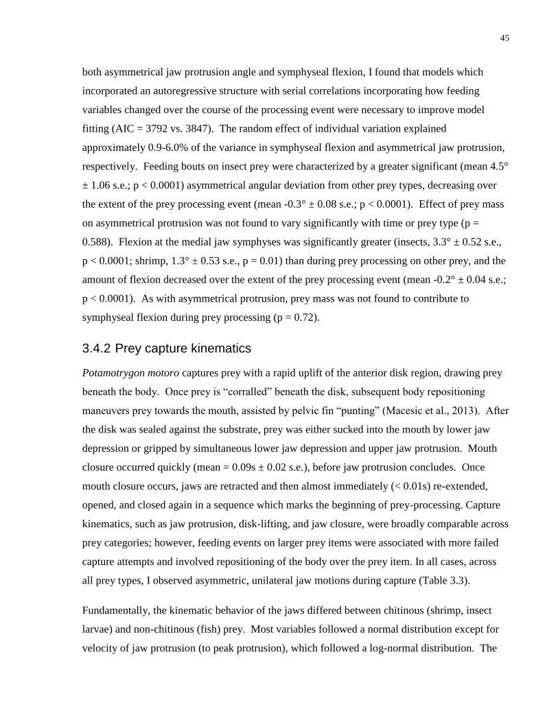
45
both asymmetrical jaw protrusion angle and symphyseal flexion, I found that models which
incorporated an autoregressive structure with serial correlations incorporating how feeding
variables changed over the course of the processing event were necessary to improve model
fitting (AIC = 3792 vs. 3847). The random effect of individual variation explained
approximately 0.9-6.0% of the variance in symphyseal flexion and asymmetrical jaw protrusion,
respectively. Feeding bouts on insect prey were characterized by a greater significant (mean 4.5°
± 1.06 s.e.; p < 0.0001) asymmetrical angular deviation from other prey types, decreasing over
the extent of the prey processing event (mean -0.3° ± 0.08 s.e.; p < 0.0001). Effect of prey mass
on asymmetrical protrusion was not found to vary significantly with time or prey type (p =
0.588). Flexion at the medial jaw symphyses was significantly greater (insects, 3.3° ± 0.52 s.e.,
p < 0.0001; shrimp, 1.3° ± 0.53 s.e., p = 0.01) than during prey processing on other prey, and the
amount of flexion decreased over the extent of the prey processing event (mean -0.2° ± 0.04 s.e.;
p < 0.0001). As with asymmetrical protrusion, prey mass was not found to contribute to
symphyseal flexion during prey processing (p = 0.72).
3.4.2 Prey capture kinematics
Potamotrygon motoro captures prey with a rapid uplift of the anterior disk region, drawing prey
beneath the body. Once prey is “corralled” beneath the disk, subsequent body repositioning
maneuvers prey towards the mouth, assisted by pelvic fin “punting” (Macesic et al., 2013). After
the disk was sealed against the substrate, prey was either sucked into the mouth by lower jaw
depression or gripped by simultaneous lower jaw depression and upper jaw protrusion. Mouth
closure occurred quickly (mean = 0.09s ± 0.02 s.e.), before jaw protrusion concludes. Once
mouth closure occurs, jaws are retracted and then almost immediately (< 0.01s) re-extended,
opened, and closed again in a sequence which marks the beginning of prey-processing. Capture
kinematics, such as jaw protrusion, disk-lifting, and jaw closure, were broadly comparable across
prey categories; however, feeding events on larger prey items were associated with more failed
capture attempts and involved repositioning of the body over the prey item. In all cases, across
all prey types, I observed asymmetric, unilateral jaw motions during capture (Table 3.3).
Fundamentally, the kinematic behavior of the jaws differed between chitinous (shrimp, insect
larvae) and non-chitinous (fish) prey. Most variables followed a normal distribution except for
velocity of jaw protrusion (to peak protrusion), which followed a log-normal distribution. The

46
results of the LMM of angular deviation during jaw protrusion (angular protrusion) were
significantly different among prey types (p = 5.96x10⁻¹²), with chitinous prey eliciting greater
jaw protrusion than fish (Insects | Fish, p = 1x10-04; Shrimp | Fish, p = 1x10-04), but not
differing significantly from one another (Insects | Shrimp, p = 0.0587). The effect of prey mass
on angular deviation during jaw protrusion was not significant (p = 0.055). The results of the
LMM of jaw protrusion distance (protrusion distance) were also significantly different between
prey types (p = 3.93x10⁻⁵), with chitinous prey eliciting greater protrusion distances (Insects |
Fish, p = 0.0031; Shrimp | Fish, p < 0.001), but not significantly from one another (Insects |
Shrimp, p = 0.760). The effect of prey mass on jaw protrusion distance was not significant (p =
0.4838). The best-fitting LM models suggest that jaw protrusion velocity was also significantly
different between prey types (p = 0.02805); however, Tukey results found only weak support for
this, with feeding events on shrimp eliciting faster velocities than fish, though differences were
not significant (Shrimp | Fish, p = 0.0525; Insects | Fish, p = 0.9968; Insects | Shrimp, p =
0.0659). The effect of prey mass on jaw protrusion velocity was not significant (p = 0.9968).
LM model-fitting demonstrated that mouth closure velocity was significantly different between
prey types (p = 0.01084), with insect prey eliciting faster closures than fish (Insects | Fish, p =
0.0102), but not significantly different than shrimp (Insects | Shrimp, p = 0.6145; Shrimp | Fish,
p = 0.0718). The effect of prey mass on mouth closure velocity was not significant (p =
0.11165).
3.4.3 Tooth Reorientation
I did not observe noticeable reorientation in the teeth of P. motoro when the jaws were protruded
and during symphyseal jaw flexion. However, in the related insectivore Potamotrygon orbignyi,
I observed noticeable tooth reorientation during jaw protrusion. The average angle of tooth cusp
reorientation in P. orbignyi varied from 8.5-28.1° (mean = 15.8°) from the resting angle (Figure
3.3).
3.5 Discussion
While several definitions of chewing have been proposed (Moore and Sanson, 1995; Reilly et al.,
2001; Evans and Sanson, 2005; Menegaz et al., 2015), I define chewing as an interaction of
upper and lower teeth which both compresses and shears food between occlusal surfaces during
asymmetric motion of the jaws. Although one of the most frequently cited hallmarks of

47
mammalian chewing is translational, opposing motion of the upper versus lower jaws, i.e. the
‘power stroke,’ some of the most successful mammalian chewers, rodents, lack a translational
action of the jaw, relying instead on propalineal, longitudinal shearing instead (Reilly et al.,
2001). When processing all types of prey, P. motoro stingrays show clear evidence of
asymmetrical, transverse motion of the jaws, owing to loose mandibular symphyses and jaw
joints, which allow the left and right rami of the jaws to move independently. Videos also appear
to show longitudinal translation of the upper versus lower jaws, although confirming these
kinematic sequences requires detailed imaging of internal anatomy, perhaps through x-ray
videography. P. motoro uses asymmetric, unilateral movements of the jaws to successfully
reduce tough insect cuticle during feeding, suggesting that stingrays and mammals have found a
similar kinematic solution for feeding on tough prey. Loose jaw and symphyseal joints coupled
with documented unilateral muscle activity (Gerry et al., 2010), allow for independent movement
of the upper and lower jaws relative to one another in batoid fishes, a hallmark of mammalian-
style chewing kinematics.
Another hallmark of mammal-like chewing is the use of high-crowned tribosphenic molars,
which raises the question: does chewing really require complex teeth? Our results indicate that
complex, asymmetrical jaw motions enable insect consumption by Potamotrygon motoro,
despite the homodont dentition of this species. This contrasts with mammals, which rely on
similar kinematic behaviors, but also have intricate tooth morphologies (Oron and Crompton,
1985; Freeman, 1972, 1992). However, P. motoro is a dietary generalist, and there is some
evidence suggesting that larger individuals include insects in their diet, while smaller individuals
eat more crustaceans and fishes (Almeida et al., 2010). I note that the congeneric and sympatric
Potamotrygon orbignyi, includes more insects in its diet than P. motoro (Filho, 2006), and can
reorient its teeth, making it facultatively heterodont. Thus, it may be that true or obligate
insectivores require both complex kinematic jaw function as well as teeth with complex tooth
shapes.
Our finding of chewing behaviour in freshwater stingrays adds this taxon to a list of other non-
mammalian animals that use complex jaw kinematics to process complex prey (Reilly et al.,
2001). Gerry et al. (2010) documented that both sharks and skates use asynchronous muscle
motor patterns during prey processing. Skates in particular use unilateral jaw muscle activation
and simple, piercing teeth to effectively ‘pinch’ prey on one side of the jaw, and use

48
hydrodynamic jetting to dissemble these prey. In fact, there are many examples of vertebrates
which feed on tough, stiff, or other manners of complex prey, which do so with relatively simple
teeth and complex jaw kinematics, including tuataras and Uromastix lizards. Some herbivorous
pleurodiran turtles and tortoises process food with an absence of teeth entirely (Gorniak et al.,
1982; Reilly et al., 2001; Schumacher, 1973; Bramble, 1974). Translational motion alone, I
suggest, is the key to shearing complex prey apart – perhaps mammalian jaws are simply too
integrated a system to allow for the complex jaw motions seen in other vertebrate ‘chewers,’
hence the necessity for tribosphenic molars.
The decoupled nature of the cranial skeleton (euhyostyly) in skates and rays allows these fishes
to independently position the jaws relative to one another, as well as the cranial skeleton (Gerry
et al., 2010). A similar effect of decoupled feeding structures is evident in some teleost fishes,
notably black carp and grass carp (Cyprinidae), which use the independent rami of the lower
pharyngeal jaws to shear prey against upper pharyngeal dentition, and show complex
asymmetrical muscle activation patterns (Gidmark et al., 2014, 2015). The anatomical traits and
behaviors characterizing ‘chewing’ as a prey processing behavior are found in a broader context
than just mammal, or even amniote systems. Stingrays have many of the hallmarks of chewing:
loose jaw joints, medio-lateral movement of occluding tooth surfaces, asymmetrical jaw and
muscle action, and at least in the insect-feeding specialist P. orbignyi, heterodont dentitions. But
in the case of large generalist insectivores like P. motoro, as well as many reptiles and
archosaurs, complex heterodont tooth morphologies are not required to process tough prey.
Batoid fishes represent much of the trophic ecological diversity within the elasmobranch fishes,
perhaps facilitated by behavioral plasticity of the feeding apparatus (Dean et al., 2007;
Aschliman et al., 2012; Jacobsen and Bennet, 2013). Stingrays spend a puzzling amount of time
processing prey, during which they are conspicuous to predators and competitors in the wild.
Gerry et al. (2010) documented that both sharks and skates behaviorally change prey processing
behavior to suit complex or tough prey, as documented here for Potamotrygon. Greater overall
jaw kinesis and longer prey-handling times occurred when Potamotrygon feed on chitinous prey,
markedly greater for insects over shrimp, and for both kinds of chitinous, arthropod prey over
fish. The rate of biting during processing remained constant regardless of prey, while
asymmetrical action of the jaws and frequency of biting decreased over the duration of the
feeding event. This suggests that P. motoro uses some stereotypy for feeding on complex prey,

49
biting at comparable rates regardless of prey, but changing the manner in which jaw action
occludes against prey, as well as how long processing cycles continue. The decreasing extent to
which these rays use abrupt, asymmetrical action of the jaws suggests that Potamotrygon
modifies its behavior to reflect the progressive dismantling of prey during processing. This
mechanical feedback allows predators to respond to nuances of prey material and structural
toughness during feeding. This ability to modulate prey processing has allowed stingrays to
access myriad trophic niches across numerous habitats, faced with novel prey like aquatic insect
larvae. This plasticity of feeding behavior may explain why, despite entrenched competitors in
novel habitats, freshwater rays were successful in making the transition from marine to aquatic
environments (Thorson and Watson, 1975).
I determined that Potamotrygon motoro uses its appendicular skeleton (its disk) for prey capture
and its jaws for prey processing, effectively dissociating the functional anatomy of these two
processes. In other fishes, decoupling of prey capture from prey processing is a function of
having two sets of jaws, oral and pharyngeal, of which the latter performs the majority of
processing (Liem and Sanderson, 1986). In some fish lineages, decoupling of anatomical
modules during feeding behavior has led to radical changes in the morphology of formerly
integrated cranial modules, correlated with increased diversification in these lineages (Collar et
al., 2014). Using the body as a method of prey restraint or to outright capture prey is prominent
in other vertebrates outside fishes, e.g. predatory birds use talons to hold and suffocate prey, bats
capture fish from rivers with their hindlimbs, and small and large mammals alike often grasp and
rend prey using their forelimbs (Csermely et al., 1998; Siemers et al., 2001; Rasa, 1973).
Although rare in bony fishes, prey capture using the pectoral fins occurs in other batoids, such as
guitarfishes and skates (Wilga et al., 1998, 2012; Mulvany and Motta, 2014). We suggest that
using the appendicular skeleton to trap prey is an innovation that was made possible by the
evolution of the pectoral fins to encircle the front of the head, forming a flexible, flattened disk,
and has evolved at least twice, independently in modern stingrays (Myliobatiformes) and skates
(Rajiformes) (Aschliman et al., 2012). In turn, this innovation may have facilitated the evolution
of extreme jaw kinesis and chewing behaviour in these stingrays. I suggest that asymmetrical jaw
kinematics are a frequent motif of stingray, and more generally, batoid feeding – a function of
their decoupled, loose jaws and cranial skeletal architecture. These properties establish

50
Potamotrygon as a model system for understanding evolutionary convergence of prey-processing
and chewing in vertebrates.
3.6 Acknowledgements
MAK dedicates this manuscript to Drs. Dean Grubbs and Neil Aschliman, mentors and friends
who convinced us of the incredible diversity and ecological significance of batoids way back
when. I thank S McCauley, A Elbassiouny, and R Castaneda for assistance with odonate
collection. A Elbassiouny, M Dobrovetsky, A Ahmad, E Lehmberg, and J Pearce were
instrumental in providing adequate care for our experimental animals. A Gristock and D Lauzon
solved critical aquatic facility issues in the best interests of the animals. M Dean, and in
particular, D Groom and N Al-Dajani provided statistical and methodological advice throughout
the duration of this project.

51
Table 3.1 Linear Mixed Model results of Prey-Processing Kinematics for Prey Type and Mass
(Kolmann et al. 2016)
Model Covariate Tukey Results
Estimate
s
Standard
Error t p
Number of Bites
Prey
Type - 17.48
4.41 3.96
1.53x10-
15
n = 15 / prey item Fish ǀ Insect 34.96 4.45 7.86 < 1x10⁻⁴
Shrimp ǀ Fish 10.55 4.60 2.29 0.057
Shrimp ǀ Insect
-24.41 4.25 -5.74 < 1x10⁻⁴
Prey
Mass - -16.75
14.30 -1.17 0.241
Duration of Prey-Handling
Prey
Type - 7.08
1.79 3.95
9.56x10-
14
n = 15 / prey item Fish ǀ Insect 13.31 1.81 7.36 < 1x10⁻⁴
Shrimp ǀ Fish 3.98 1.87 2.13 0.084
Shrimp ǀ Insect
-9.32 1.73 -5.40 < 1x10⁻⁴
Prey
Mass - -5.55
5.81 -0.96 0.339
Bite Rate
Prey
Type - 2.57
0.19
13.2
6 0.457
n = 15 / prey item Fish ǀ Insect 0.23 0.20 1.16 0.480
Shrimp ǀ Fish 0.05 0.20 0.24 0.968
Shrimp ǀ Insect
-0.18 0.19 -0.94 0.612
Prey
Mass - -0.55
0.63 -0.87 0.386
Significance level (α = 0.05).

52
Table 3.2 Growth Model results for angular deviations of the jaws during prey-processing (Kolmann et al., 2016)
Model Covariate Tukey Results Value Standard Error t p
Angular Jaw Protrusion Prey Type - 8.48 1.37 6.17 < 0.001
n = 15 / prey item Fish ǀ Insect 4.52 1.06 4.27 < 6.0x10⁻⁵
Shrimp ǀ Fish -0.09 1.08 -0.08 0.996
Shrimp ǀ Insect -4.61 1.02 -4.53 < 1.6x10⁻⁵
Prey Mass - 1.97 3.64 0.54 0.588
Time - -0.39 0.08 -5.17 < 0.001
Symphyseal Flexion Prey Type - 4.33 0.49 8.77 < 0.001
n = 15 / prey item Fish ǀ Insect 3.34 0.52 6.46 < 0.001
Shrimp ǀ Fish 1.35 0.53 2.54 0.030
Shrimp ǀ Insect -1.99 0.50 -4.02 < 0.001
Prey Mass - -0.61 0.53 2.54 0.721
Time - -0.21 0.04 -5.33 < 0.001
Significance level (α = 0.05).
53

54
Figure 3.1 Functional morphology of asymmetrical jaw protrusion in Potamotrygon motoro. (A)
Resting jaw and hyomandibular articulations; (B) asymmetrical protrusion of jaws relative to
kinetics of angular cartilage and hyomandibular articulations, inset and pointer: photo of
asymmetrical protrusion of live Potamotrygon while feeding on insect larvae; (C) medial flexion
of mandibular (Meckelian) symphyses; (D) computed tomography scan of Potamotrygon motoro
crania, inset: articulation of jaws to hyomandibular cartilage via dual angular cartilages. (from
Kolmann et al., 2016)

55

56
Figure 3.2 Growth model showing declines in asymmetrical jaw activity during prey-processing
across three prey types. Top panel is jaw protrusion angle, bottom panel is symphyseal flexion
angle. (from Kolmann et al., 2016)

57

58
Figure 3.3 Computed tomography scans of: (A) P. motoro teeth, the animal analyzed in this
study, an insect-feeding generalist predator and (B) P. orbignyi, an insect-feeding specialist.
(from Kolmann et al., 2016)

59
Evolution of feeding specialization in Neotropical freshwater stingrays
4.1 Abstract
The interplay between ecological and evolutionary forces drives morphological differentiation by
mitigating competition, promoting niche specialization, and ultimately structuring
macroecological communities. Although several studies have examined marine-freshwater
transitions in fishes, as well as the ecomorphological novelty present in many of these taxa, little
focus has been spent investigating whether marine-derived lineages are representative of
adaptive radiations, despite the fact that habitat transitions promoting ecological diversification
are a common theme in radiations. We investigated the macroevolutionary history of marine-
derived, South American river rays (Potamotrygonidae). The ancestral potamotrygonid lineage
invaded freshwater some 40 mya and subsequently diversified taxonomically and ecologically,
with several instances of piscivory, molluscivory, and insectivory (the only example within
elasmobranchs) evolving within this clade. I present a time-calibrated, multi-gene phylogeny for
Potamotrygonidae and examine evolutionary trends in diet specialization and ecomorphology.
Potamotrygonid morphological disparity exhibited two patterns, showing an early burst around
40 mya with the majority of morphological disparity partitioned among subclades. There is a
gradual shift to greater within-subclade disparity starting at 20.4 mya, with an abrupt spike in
morphological disparity peaking around 7.6 mya. These findings correspond with a rate shift in
lineage evolution for lower Amazon stingrays, and the occupation of novel adaptive peaks
corresponding to dietary specialization. The first of these peaks corresponds with the evolution
of fish-eating Paratrygon and Heliotrygon. More recent adaptive peaks correspond to the
evolution of mollusk-feeding, insect-feeding, and crustacean-feeding specialist stingrays within
Potamotrygon. These findings are not broadly consistent with an early-burst mode of
morphological disparity common to other adaptive radiations. Although potamotrygonids exhibit
an initial burst in morphological disparity, recent lineage and morphological diversification
suggests that new niche exploration has occurred relatively recently in potamotrygonids.
Key words: adaptive radiation, ecomorphology, insectivory, disparity, Pebas Mega-Wetland

60
4.2 Introduction
Some degree of habitat transition is considered common or even necessary for the onset of an
adaptive radiation (Schluter, 2000; Gavrilets & Losos, 2009). In Darwin’s finches, for example, a
mainland lineage arrived in the Galapagos and subsequently colonized and re-colonized various
isles (Grant & Grant, 2008). These finches diversified to feed on a variety of seeds, with
different species evolving more or less robust beaks according to the hardness of seeds they
preferentially feed on. A similar situation explains the repeated evolution of multiple Anolis
lizard “ecomorphs” on various Caribbean islands. Lizards living on different islands repeatedly
adopted similar limb morphologies according to their shared substrate preferences (plant trunks,
limbs, and branches) (Losos et al., 1997).
However, few studies have explicitly examined fishes that have diversified across the marine-
freshwater threshold in the context of adaptive radiation, despite well-documented
ecomorphological novelty in these clades (Weitzmann & Vari, 1988). Davis et al. (2012) tested
differences in lineage diversification rates in terapontid fishes across a marine-freshwater
transition, finding that freshwater clades had increased dietary breadth and faster rates of
diversification. However, Bloom et al. (2013) found that although silverside fishes have higher
speciation and extinction rates in freshwater, net lineage diversification was not significantly
different between marine and freshwater clades. These studies did not closely examine
morphological and ecological diversification in marine-derived lineages (MDLs).
Although habitat transitions can provide the ecological opportunity for diversification, the
ordering of species across some resource gradient (e.g., microhabitat and seed hardness for
Anolis and Geospiza, respectively) is mandatory for such a radiation to be termed “adaptive”
(Gavrilets & Losos, 2009), with phenotypically-variable species partitioning resources according
to their performance capacities. Also, adaptively radiating clades are expected to show both an
early-burst pattern of morphological disparity concomitant with similar patterns in lineage
accumulation (Simpson, 1953; Schluter, 2000; Pybus and Harvey, 2000). As niche space is
filled, both lineage and morphological diversification should slow, as competition limits
diversification (Simpson, 1953). This pattern has been seen recently in a number of radiations of
new world cichlids, several clades of which show tight relationships between foraging mode,
feeding morphology, and body shape changing commensurate with the generation of novel

61
adaptive peaks (Arbour and López-Fernández, 2013, 2014). In marine systems, patterns of
putative adaptive radiations are less obvious, with some clades seemingly to show an early-burst
pattern in their initial history, but with subsequent bursts in morphological diversification and
lineage accumulation occurring more in line with subsequent changes in habitat (Near et al.,
2012; Frédérich et al., 2013).
Evolutionary transitions of lineages from marine to freshwater habitats represent an interesting
opportunity for investigating adaptive radiation and evolution. Davis et al. (2013, 2014)
illustrated how terapontid fishes diversified across dietary niches upon the transition to
freshwater, these fishes diversifying into herbivorous, detritivorous, and benthic invertebrate
specialist niches from a generally carnivorous ancestor. Freshwater environments may offer
novel resources (e.g. aquatic insect larvae) that are unavailable to marine species or may offer
resources which are relatively free from competition. Indeed, Davis et al. (2013, 2014) found
signals of adaptive radiation: an early-burst in both lineage accumulation and morphological
diversity, followed by a gradual decline in lineage and ecological diversification. Similarly,
potamotrygonid stingrays (Potamotrygonidae, Myliobatiformes), a South American MDL,
appear to have diversified across an array of feeding niches, including piscivory, molluscivory,
insectivory, and generalist invertebrate feeding (Shibuya et al., 2009, 2012; Moro et al., 2012).
Potamotrygonids originated at least 12-38 mya (million years ago) when a marine ancestral
lineage invaded an ancient lowland brackish lagoon in central South America (Lovejoy et al.,
1998, 2006). The diversity of potamotrygonid feeding ecology is in stark contrast to the
hypothesized marine sister group of potamotrygonids, the two species of amphi-American
Himantura (Lovejoy 1996, Lovejoy et al., 1998), which are large, estuarine, generalist predators
with a limited geographic range spanning either side of the Panamanian Isthmus.
In general, elasmobranchs (sharks and rays) are conspicuously absent from most freshwater
ecosystems, which account for only 0.01% of the water on earth, yet harbor over 41% of all fish
species (Horn, 1972). Although the fossil record shows several incidences of elasmobranchs
diversifying in freshwater environments, only the potamotrygonid stingrays have diversified
entirely in freshwater, while other freshwater rays represent independent invasions of freshwater
by single species. Potamotrygonids currently number approximately 30 species in four genera
and although they are not the only instance of freshwater elasmobranchs, they display the highest
species richness of any extant, monophyletic lineage of freshwater elasmobranchs (Thorson &

62
Watson, 1975; Carvalho & Lovejoy, 2011). Stingrays account for over half the diversity of
elasmobranch fishes, but potamotrygonids may be unique in the amount of dietary,
morphological, and coloration diversity they exhibit relative to their species diversity. These
rays, when compared to their closest marine relatives, represent an abrupt ecological departure
from phylogenetic conservatism. For example, outside potamotrygonids there are very few if
any exclusively molluscivorous dasyatoid rays. There are also insectivorous potamotrygonids,
but this strategy does not appear in other elasmobranchs. These patterns are impressive,
considering that the species richness of the dasyatoid rays excluding Potamotrygonidae
(Urotrygonidae, Dasyatidae, Urolophidae, Plesiobatidae) is four times greater than that of
potamotrygonids. Potamotrygonids, given their dietary novelty, evidence of habitat transition,
and high species richness relative to their marine counterparts, offer a compelling system for the
study of adaptive evolution in elasmobranch fishes.
Here, I propose that transitions from marine to freshwater habitats may have spurred both lineage
and ecological diversification in potamotrygonids, commensurate with exposure of these
lineages to new prey options. Since foraging is time-consuming as well as costing resources in
terms of energy expenditure, study of feeding behavior in an adaptive context is particularly
relevant to ecological models of diversification (Wainwright et al., 2004). Most studies of
adaptive radiation deal with some aspect of feeding morphology matching a gradient in prey
resources, but do potamotrygonids follow a similar pattern? Critical to investigations of adaptive
radiation is establishing an ecomorphological, functional connection between traits and dietary
ecology. While there has been much research regarding the novelty and dynamism of
chondrichthyan feeding functional morphology, no studies have attempted to place this research
within a larger, explicit evolutionary framework.
To reconstruct the directionality and order of dietary mode and corresponding morphological
transitions in potamotrygonids, a robust phylogeny is required. Such a phylogeny would also
allow a test of whether or not potamotrygonids show an early-burst pattern of morphological and
lineage diversification, as predicted for adaptive radiations. To date, phylogenies of
potamotrygonids have been limited by reliance on single genes (Lovejoy et al., 2006; Carvalho
and Lovejoy, 2011) or limited sets of genes (mitochondria; Toffoli et al., 2008; Garcia et al.
2015), limited taxon sampling, or issues with alpha taxonomy. Also, the timescale of

63
potamotrygonid diversification has not been well-established using molecular phylogenetic
approaches.
Here, I use potamotrygonid stingrays as a model system for exploring evolutionary
diversification of morphology and diet, and attempt to determine whether or not this clade
exhibits some of the characteristics of an adaptive radiation. I address the following main
objectives: (1) I generate a robust, time-calibrated species-level molecular phylogeny for
Potamotrygonidae. (2) I use this tree to determine patterns of ecomorphological diversification in
potamotrygonids, and test whether potamotrygonids show characteristics of adaptive radiation
(early-burst pattern of ecomorphological diversification, high early lineage accumulation,
followed by a slowdown as niches are filled). (3) I determine whether shifts in rates of lineage
evolution correlate with shifts in diet, and whether adaptive peaks correspond to distinct dietary
types, such as insectivory, piscivory, and molluscivory.
4.3 Methods
4.3.1 Taxon Sampling, DNA extraction, amplification, and sequencing
Specimens were collected using gillnets, seines, dip-nets, and cast-nets, by various collectors
from multiple museums. Specimens obtained personally by the authors are done so under the
auspices of UACC protocol (# 2000). Muscle or fin tissue was stored in 95% ethanol. My
dataset is the most complete taxonomic sampling of potamotrygonid stingrays to date, including
21 of 28 described species from all four of the currently described genera, including multiple
representatives of some polymorphic and widely-distributed species (Paratrygon aiereba and
Potamotrygon motoro), as well as two undescribed species, Potamotrygon sp. ‘reticulata’ from
Colombia and Potamotrygon sp. ‘demerarae’ from Guyana. For outgroups, I included seven
species representing the immediate relatives of Potamotrygonidae (Pteroplatytrygon violacea,
Dasyatis geijskesi, D. guttata, Taeniura lymma, Neotrygon kuhlii, Himantura schmardae and H.
pacifica, Urotrygon simulatrix, and Urobatis halleri), including previously published sequence
data for more distantly-related taxa (see Table 4.1).
Whole genomic DNA was extracted using the DNeasy spin column tissue kit (Qiagen Inc.,
Valencia, CA, USA). We collected DNA sequence fragments from three mitochondrial loci (co1,
cytochrome oxidase I; cytb, cytochrome b; ATP6, ATPase subunit 6), using novel and published

64
primer sequences (cytb; Aschliman et al., 2012) primers. In addition, I sequenced four nuclear
loci (RAG1, Recombination activating gene 1; SCFD2, Sec1 Family Domain Containing 2;
ENC1, Ectodermal-Neural Cortex 1; ITS1, internal-transcribed space 1 and 2) using published
primers for RAG1 and SCFD2 (Aschliman et al., 2011) as well as novel primers for ENC1 and
for ribosomal ITS1. The PCR products for all genes were purified using USB ExoSAP-IT PCR
purification kit (Affymetrix Ltd., Santa Clara, CA). Primers for ENC1, ITS 1 and 2 and
sequencing primers for SCFD2, developed for this study were as follows: ENC1-FN1 (5’
GGAATGTCTGC TTTTGGAA 3’), ENC1-FN1 (5’ CAACCATCAGCAAGAGTGAAGA 3’),
ENC1-seqN1 (5’ CCCAAGGAGGAGCAAGGCATGTCCAG 3’), ITS1-F/ CAS18SF1 (5’
TACACACCGCCCGTCGCTACTA 3’), ITS1-R/ POT5.8SR1 (5’
CTAGCTGCGTTCTTAATCGACG 3’), ITS1-R/ POT5.8SR2 (5’
GTCGATGATCAATGTGTCCTGC 3’) and SCFD2-Seq1 (5’
CTGAGAGCTTACACTGGGTCG 3’). PCR for all genes were performed in 25 μL volumes,
which included a 2.5 μL of KCl/(NH4)2SO4 mixture PCR buffer, 2.5 μL MgCl2, 2.0 μL dNTPs
(10mM), 1.25 μL of each primer (10mM), 0.5 μL of Taq polymerase, 1-4 μL genomic DNA,
with the remaining volume of H2O. Touchdown PCR thermocycler conditions for ATP6 were 94
°C for 5 min, followed by 15 cycles of 94 °C for 30 s, 55 °C for 30 s, 72 °C for 1 min to a final
sustained annealing temperature of 55 °C for 30 s, for another 15 cycles, followed with a final
extension of 72 °C for 7 min. PCR thermocycler conditions for cytb & CO1 were 94 °C for 4
min, followed by 35 cycles of 94 °C for 30 s, 48 °C for 30 s, 72 °C for 1 min and a final
extension of 72 °C for 7 min. Touchdown PCR thermocycler conditions for RAG1 were 95 °C
for 5 min, followed by 20 cycles of 95 °C for 30 s, 54 °C for 1 min, 72 °C for 1.5 min to a final
sustained annealing temperature of 50 °C for 1 min, for another 20 cycles, followed with a final
extension of 72 °C for 7 min. PCR thermocycler conditions for ITS1 2 were 94 °C for 5 min,
followed by 35 cycles of 94 °C for 30 s, 55 °C for 30 s, 72 °C for 1.5 min and a final extension
of 72 °C for 7 min. PCR thermocycler conditions for ENC1 were 95 °C for 4 min, followed by
35 cycles of 95 °C for 30 s, 50 °C for 1 min, 72 °C for 1.5 min and a final extension of 72 °C for
5 min. PCR thermocycler conditions for SCFD2 were 95 °C for 2 min, followed by 35 cycles of
95 °C for 30 s, 50 °C for 1 min, 72 °C for 1.5 min and a final extension of 72 °C for 5 min.
Samples were sequenced at the SickKids Centre for Applied Genomics, Toronto, Canada.

65
4.3.2 Alignment and Analysis
Forward and reverse sequences were used to construct de novo consensus sequences, which were
then edited by trimming the distal ends of ambiguous base-pair (bp) calls in GENEIOUS v6
(Kearse et al. 2012). The resulting sequences were aligned in GENEIOUS using the MUSCLE
plugin and protein-coding genes were translated to amino acids to confirm an open reading
frame. Aligned sequences were then used to generate the following datasets: (1) the three
mitochondrial genes combined, (2) RAG1, (3) ENC1, (4) SCFD2, and (5) ITS1, concatenated to
form a single matrix of 8270 bp. This final matrix includes data for more than 75% of all
described potamotrygonid taxa and comprises data for 37 taxa in total. I used PartitionFinder
to determine the best-fit model of evolution and partition schema simultaneously for each gene
and possible reading frame. Models were selected using a Bayesian Information Criterion (BIC),
using a ‘greedy’ search scheme (Lanfear et al., 2012). Partitioned maximum likelihood (ML)
tree searches were performed with GTR+G models for each partition using the program RAXML
(Stamatakis, 2006) to create our gene trees. ML bootstrap estimates were based on 1000
replicates using the rapid boot-strapping algorithm in RAXML. For our consensus Bayesian
analysis, I used the gene partition recommendations from PartitionFinder: for ATP6, RAG1, and
cytb, the GTR+ gamma; for CO1, the GTR+ I+ gamma; for ENC1, the TrNef + I; for ITS1, the
HKY+ I+ gamma; and for SCFD2, the K80+ gamma.
For the consensus phylogeny used in our final analyses, I used BEAST (v. 1.8.3; Drummond and
Rambaut, 2007) to simultaneously estimate the phylogeny and diversification times of
potamotrygonids using a Bayesian framework. I partitioned my data according to gene, with
unlinked parameters and default priors. I used an uncorrelated lognormal tree prior and a pure
birth-death prior for our expectation of cladogenesis. I ran two separate BEAST analyses for 100
million generations, sampling every 5000 generations, and automatically discarding the first 10%
of trees as burn-in. I used Tracer 1.6 (Drummond and Rambaut, 2007) to assess convergence and
mixing of runs and to verify that effective sample sizes (ESS) were >200 for all parameters. An
additional first 20 million generations from each run were discarded as burn-in. To determine
calibrated, dated divergence times I used two fossil and two geological time calibrations (see
Appendix 1 for details), corresponding to: (1) the earliest dasyatoid fossil (~51.0 mya), (2) the
earliest potamotrygonid fossil (~41.0 mya), (3) the separation of modern potamotrygonid taxa
spanning the Rio Magdalena/Rio Atrato and Orinoco-Maracaibo basins (~12.0 mya), and (4) the

66
separation of amphi-American Himantura by the closure of the Isthmus of Panama (mean ~7.0
mya). Specifically:
†Dasyatis muricata (Carvalho & Maisey, 2012), from the Eocene of Italy – as the most recent
common ancestor (MRCA) for Dasyatoidea; with an exponential prior set to a mean of 1.9, st
dev = 1.0; offset = 50.0 mya, median of 71.7 mya; 95% quantile interval of (65.9, 112.5) mya.
†Potamotrygon ucayaliensis (Adnet et al., 2014), from the Miocene of Uruguay – as the most
recent common ancestor (MRCA) for the amphi-American Himantura + Potamotrygonidae; with
an exponential prior set to a mean of 1.9, st dev = 1.5; offset = 41.0 mya, median of 39.5 mya;
95% quantile interval of (38.0, 54.0) mya.
Separation between Potamotrygon yepezi from P. magdalenae and P. ‘reticulata’ by the
Cordillera Oriental Mountains of Colombia, Andean satellite ranges to the north. This node was
calibrated with an exponential prior set to a mean of 2.0, st dev = 1.0; offset = 10 mya, median of
13.0 mya; 95% quantile interval of (10.1, 15.0) mya.
Separation of the amphi-American Himantura species (H. pacifica and H. schmardae) by the
coalescence of the Isthmus of Panama and greater Central America. This node used a normal
prior to represent the ambiguity regarding the particular date for final separation of the Caribbean
from the Pacific, with a set mean of 2.0, st dev = 1.0; offset = 10.0, median of 5.0 mya; 95%
quantile interval of (3.3, 10.1) mya.
4.3.3 Phylogenetic analysis of diet and feeding biomechanics
Diet data was obtained from the literature and other sources (Table 4.2). Species were considered
specialists if >70% of the diet was reported as being a particular prey item, or if no other prey
items were listed as part of the diet. If diet data was not available, or if the grey literature or
personal communications were used to assess diet (17% of diet data), species were
conservatively labeled as dietary generalists. Diet categories were coded as a discrete multistate
character, and I used the Phytools package (Revell, 2012) to perform maximum likelihood
reconstructions of ancestral states in Potamotrygonidae + amphi-American Himantura.
Transitions between states were considered as being equally-likely.

67
To identify the major axes of morphological variation in biomechanical attributes across taxa and
to characterize species along ecomorphological axes (diet), I used a phylogenetically-informed
Principle Components Analysis (phyPCA; phyl.pca function in the phytools package; Revell,
2011). Morphological and functional traits were measured from computed tomography, or x-ray
scanning of museum specimens (ANSP, ROM, MCZ, USP, and CUMV; Table 4.1) spanning the
Potamotrygonidae as well as several dasyatoid outgroups (Dasyatidae, Urolophidae, and
Urotygonidae). Functional characters were chosen based on demonstrable covariation with
feeding performance or behavior (Huber et al., 2005; Dean et al., 2007; Anderson et al., 2009;
Arbour and Lopez-Fernandez, 2013; Balaban et al., 2015). The following morphological
variables were measured: (a) mechanical advantage (MA - leverage), (b) maximum jaw depth
(upper and lower), (c) jaw aspect ratio (upper and lower), (d) symphyseal height (lower jaw), (d)
occlusional offset (cm – measure of jaw closing tooth occlusion), (e) lower dental row length, (f)
hyomandibular offset (cm – linear measure of jaw protrusion), (e) jaw adductor fossa length
(cm), (f) gape width (cm), (g) jaw-hyomandibulae disarticulation (a function of joint range of
motion), (h) hyomandibular cartilage aspect ratio, and (i) propterygia aspect ratio (rigidity of the
skeletal elements which anchor the pectoral fins). Morphological measurements were scaled
relative to head length to standardize for different unit comparisons and thus adjusting for body
size.
Description of morphological measures as follows: mechanical advantage is the ratio of the in-
lever (muscle insertion to jaw joint) to out-lever (bite point to jaw joint) distances and measures
proportional force transmittance (leverage) across a structure. High MA represent high-
efficiency force propagation, low MA suggests higher effective velocities during rotation (mouth
closing). Maximum jaw depth, jaw aspect ratios, and symphyseal heights approximate the
second moment of area (I) for a beam-like structure; this measure represents the ability of a
structure to resist bending (Anderson et al., 2009). Taller jaws and symphyses represent those
which are less apt to deform during biting (Anderson et al., 2009). Occlusional offset and
hyomandibular offset are linear representations of angular rotations. Occlusional offset is
measured by measuring a line drawn tangent from the jaw joint to the plane in which the tooth
surface (occlusal plane) resides (for both jaws) (Anderson et al., 2009). Jaw occlusion either has
teeth occluding directly, or with a lateral, scissoring action – the former for applying
compressive forces, the latter for applying shear to prey. Hyomandibular offset ratio is a linear

68
representation of the angle between the occlusal plane and the hyomandibular at rest and at full
extension. The jaw-hyoid gap is a measure of the distance between the proximal ends of the
hyomandibular cartilage to the jaw joint. Both hyomandibular offset and jaw-hyoid gap indicate
a jaw suspensory apparatus kinesis potential, larger gaps and greater offsets allow the jaws to
have higher range of motion (Dean et al., 2007). This measure accounts for ease of comparison
between those species with and without an angular cartilage (Dean et al., 2007). Hyomandibular
dimensions and aspect ratio portray the relative gracility or robustness of the hyomandibular
cartilages; their resistance to bending. Thicker, stouter hyomandibulae are useful for generating
oral suction (Balaban et al., 2015). Longer, more gracile hyomandibulae allow for greater jaw
kinesis. Gape width is the proportion of the occlusal surface covered by teeth. Adductor fossa
length measures the length of muscle attachment area, of the two primary jaw adductors, the
adductor mandibulae lateralis and major. Max gape is the lateral distance between the jaw joints,
and is predictive of the size of prey consumed (particularly for piscivores). Finally, many batoid
fishes capture prey by rapidly raising the pectoral disk, which generates negative pressure under
the body and sucks prey beneath (Wilga et al., 2012). The pectoral propterygia support the
anterior pectoral fin region, their aspect ratio again gives an idea of the rigidity of the structure
and its resistance to bending. Stouter, more robust propterygia will allow for greater muscle
attachment area and therefore, greater suction generation.
4.3.4 Species accumulation and disparity-through-time
I expect that if potamotrygonids represent an adaptive radiation, their history should show rapid
initial lineage diversification, followed by a slowdown as lineage accumulation reaches
equilibrium, the result of niche space filling towards capacity. I used two methods to examine
lineage accumulation and diversification rates. To identify whether or not diversification rates
have shifted across the potamotrygonid tree, I used MEDUSA, which combines dated tips with
relative taxon richness values to account for missing data (i.e. incomplete taxon sampling) across
a diversity tree (Alfaro et al., 2009). This method fits various ‘breaks’ across which evolutionary
rates can vary across the diversity tree, comparing among increasingly complex, fitted models of
evolution. In order to determine whether lineage accumulation follows different evolutionary
patterns (niche-filling, exponential, or linear), I fit constant-rate pure-birth models, birth-death
models with fixed and variable rates, exponential (DDX) and logistic decline (DDL) models of
lineage accumulation and analyzed their fit to the data using corrected Akaike Information

69
Criterion (AICc) values in the LASER package (Rabosky, 2006). The gamma statistic was also
used to determine how cladogenesis might deviate from a constant-rate process (Pybus and
Harvey, 2000).
Adaptively radiating clades are also expected to show an initial burst of morphological disparity,
rather than a constant increase in overall disparity (Slater et al., 2010). I examined
ecomorphological disparity-through-time (DTT) plots generated from our cranial morphology
data. I compared cranial anatomical disparity across our time-calibrated tree to a simulated null
disparity dataset, using a Brownian motion model of morphological disparity iterated over
10,000 generations. I calculated the morphological disparity index (MDI) as the difference
between the simulated Brownian curve and our observed clade disparity and plotted this relative
to node age using the ‘dtt’ function in Geiger (Harmon and Slater). MDI captures the overall
difference in relative clade disparity (Harmon et al. 2003), with negative MDI values suggesting
lower subclade disparity, a frequently-cited component of early adaptive radiations. If lineage
accumulation and morphological disparity increase and then plateau, this would suggest that
ecological diversity within Potamotrygonidae has resulting from adaptive radiation.
Finally, I measured the extent of adaptive peak shifts and convergence following Ingram &
Mahler (2013), using the SURFACE package in R. SURFACE fits Hansen models with
alternative selective regimes in a stepwise fashion, comparing the fit of these models using AICc.
The procedure fits adaptive peaks iteratively to the phylogeny, i.e. the ‘forward’ run followed by
a ‘backyard’ phase which collapses weakly-supported peaks, according to whether the fit of
Hansen OU (Ornstein-Uhlenbeck) models was improved or not. Since computations in
SURFACE can stall on initially-favorable model conditions, the search function was extended to
randomly sample all peaks with delta AICc values < 2, with the ‘sample_shifts’ option set to
TRUE so the best-fitting peaks were compared at every step of both the forward and backward
iterative processes (Ingram and Mahler, 2013; Arbour and López-Fernández, 2014).
4.4 Results
4.4.1 Molecular data, phylogenetic relationships, and ancestral reconstruction of diet
The total molecular dataset resulted in a concatenated matrix of 8270 bp. This final matrix
includes data for more than 75% of all possible tip taxa, and comprises data from 36 species for

70
ATP6 (955 bp), 37 species for cytb (756 bp), 36 species for co1 (707 bp), 35 species for RAG1
(953 bp), 32 species for ENC1 (548 bp), 22 species for SCFD2 (663 bp), and 28 species for ITS
(1154 bp). BEAST was run twice with identical results recovered from each run; the resulting
tree is shown in Figure 4.1. Gene trees showed considerable incongruence between gene
histories, but generally reconstructed Potamotrygonidae as monophyletic and sister to the amphi-
American Himantura, as illustrated in the Appendix as Supplemental Figures 1a-1f. The
Paratrygon + Heliotrygon clade was typically recovered intact, while Potamotrygon +
Plesiotrygon relationships changed appreciably with each gene. Coalescent analyses supported
the monophyly of the family Potamotrygonidae with the amphi-American Himantura, H.
pacifica and H. schmardae, recovered as the family’s nearest sister taxon. I also recovered
separation of the family Potamotrygonidae into two major clades: Plesiotrygon + Potamotrygon
and Heliotrygon + Paratrygon, Potamotrygon is paraphyletic because of the nested position of
Plesiotrygon, with Potamotrygon brachyura diverging before the split of the remaining
Potamotrygon from Plesiotrygon. Potamotrygon from the Guiana Shield generally formed a
clade with Orinoco and Magdalena taxa. According to our ancestral state reconstruction, clade
inclusion was not predictive of dietary guild. Rather, novel dietary strategies have arisen
throughout the tree, typically from generalist (mostly crustacean-feeding) ancestors (Figure 4.2).
Insectivorous taxa entirely comprised this clade of northern potamotrygonids. A number of
endemic taxa restricted to the lower Amazon, as well as widespread taxa (P. motoro, P. orbignyi)
from the Xingu, Tocantins, and headwater drainages of Peru, form another clade.
According to our ancestral state reconstruction, novel dietary strategies have arisen
independently throughout the tree, typically from generalist (omnivorous) ancestors (Figure 4.2).
Insectivorous taxa have arisen at least four times, once in Plesiotrygon nana, although this is
supported by anecdotal evidence (Lucanus, pers comm; Table 4.2), again in the entire lineage
composed of P. magdalenae, P. yepezi, and P. ‘reticulata,’ again in Potamotrygon signata, and a
fourth time in Potamotrygon orbignyi. Other feeding modes, like piscivory, appear to have only
arisen once (the base of the Paratrygon + Heliotrygon clade), in molluscivores (Potamotrygon
leopoldi and P. henlei) as well as in crustacean-specialists (Potamotrygon scobina,
Potamotrygon motoro). Interestingly though, these last two transitions are notable in that while
other potamotrygonid dietary novelties arise from generalist ancestors, molluscivores arise

71
exclusively from crustacean-feeding specialists, as does the latest iteration of insectivory in P.
orbignyi.
4.4.2 Feeding morphospace
The first three axes of the phyPCA represent 37.6%, 18.0%, 16.5% of the variance in the data.
Species which loaded on the positive end of PC1 had larger gapes, robust jaws (thicker
symphyses and lower aspect ratios), high jaw mechanical advantages, and greater ability to lift
the pectoral disk (stouter pectoral propterygia) (Figure 4.3a). Species on the negative end of PC1
had increased jaw protrusion ability (long hyomandibulae) and greater jaw kinesis (longer
angular cartilages). Rays which loaded positively on PC2 had higher anterior jaw leverages,
larger gapes, scissoring jaw action, greater disk lifting ability, and increased jaw protrusion.
Species on the negative end of PC2 had higher posterior mechanical advantages, larger jaw
muscle attachment areas (adductor fossa length), more robust jaws, more robust hyomandibulae,
and more rigid jaw symphyses (Figure 4.3a). Rays which loaded positively on PC3 had higher
mechanical advantages and thus greater force efficiency, robust jaws, scissoring jaw action, and
greater jaw protrusion, while those species which loaded negatively on PC3 had larger gapes,
larger jaw muscles, and stouter pectoral propterygia (Figure 4.3b).
Figures 4.3a & b also highlight ecological trends in feeding morphology relative to diet. In
general most dietary guilds overlapped in the medial region of the morphospace. Overlap in
large part is driven by insectivores occupying the largest area of morphospace of all the dietary
modes, perhaps surprisingly. The two most outlying insectivores are Potamotrygon signata and
P. orbignyi, the former is a relatively older taxon, the latter relatively recent (but see discussion
of adaptive peaks, below) (Figure 4.3a). P. signata is characterized by generally higher
mechanical advantage jaws and wide gape. P. orbignyi has greater overall cranial kinesis, with
increased jaw protrusion (longer hyomandibulae and angular cartilages), flexible, gracile jaws
with much lower force-efficiency. Insectivores encompass crustacean specialist convex hulls
almost entirely, prey items which are presumably similar to insects although presumably stiffer
given mineralization of the chitinous cuticle in crustaceans. Molluscivores like P. leopoldi and P.
henlei ordinated low and negative on PC2 and positively on PC3, corresponding with more rigid
jaw skeletons, owing to a fusion of the medial symphyses of the jaw in these species (Figure
4.3a, b). Molluscivores were also characterized by high mechanical advantages and large jaw

72
adductor muscle attachment areas. The diet of Potamotrygon brachyura is largely uncertain, and
interestingly it falls out within the morphospace adjacent to both large crustacean specialists and
molluscivores, suggesting this species may in fact be specializing on stiff or tough prey. The
piscivorous taxa Paratrygon and Heliotrygon were characterized by large gapes, increased
ability to life the disk, as well as rounder, more gracile jaw cross-sectional shape (Figure 4.3a, b).
The middle of both phylomorphospaces is densely-packed, showing frequent shifts between
omnivorous, crustacean, and insect-feeding dietary modes (corroborated by the ancestral state
reconstructions), with PC2 and PC3 showing a greater divergence between piscivorous taxa and
other dietary guilds (Figure 4.3b).
4.4.3 Patterns of lineage and morphological diversification
The γ statistic provided no evidence for declining rates of lineage accumulation after an early
burst of diversification (γ = 0.196, p = 0.577), which is also evident in the lineage-through-time
plots (Figure 4.4). The fit of the constant-rate models versus the two density-dependent models
(DDX and DDL) did not clearly support one model over another, although a pure-birth Yule
model with multiple rate shifts (three) was slightly favored over other models according to ΔAIC
(Table 4.3). Although γ was recovered as positive, I cannot entirely rule out a density-dependent,
ecologically-driven process given almost equivocal support for one model over another. Even
the fitted Yule model showed a decline in rates of speciation through time (r1 = 0.061, r2 =
0.172, r3 = 0.035). MEDUSA, however, revealed strong support for a single rate shift at a node
uniting lower Amazon Potamotrygon (ΔAIC = 10.12) (Figure 4.5a). The net diversification rate
for this clade (r = 0.254 lineages Myr−1) was almost four times higher than the background rate
of diversification for most potamotrygonids (0.066 lineages Myr−1).
Adaptively radiating clades are expected to show an initial burst of morphological disparity,
rather than a constant increase in overall disparity (Harmon et al., 2003). This trend,
counterintuitively, is represented by high intitial morpho-disparity followed by negative subclade
disparity relative to simulated, random processes, i.e. accumulation of disparity-through-time
(Harmon et al., 2003; Slater et al., 2010). This means that subclades tend to overlap in their
overall disparity, with multiple convergent incursions into similar morphospace regions (Harmon
et al., 2003; Sidlauskas, 2008; Slater et al., 2010). The subclade disparity for potamotrygonids
starts as lower than expected given the null, i.e. was less than the simulated median for a

73
Brownian motion model of morphological evolution (Figure 4.5b). However, at approximately
20.4 mya, subclade disparity starts to exceed simulated levels with a sharp upturn occurring
approximately 9-10.0 mya – culminating in a major spike of disparity around 7-7.6 mya
(approximately 0.825–0.9 relative time). This spike is roughly coincident with the
diversification of potamotrygonids in the Lower Amazon, including species such as
Potamotrygon leopoldi and P. henlei, P. motoro, P. falkneri, and P. histrix (Figure 4.5). The
resultant, recent trend in MDI for the clade as a whole is therefore overwhelmingly positive
(0.0554, p = 0.93), starting at approximately 20 mya, due to recent increases in subclade
disparity starting at 20 mya, with a peak at approximately 8 mya.
SURFACE analyses revealed five adaptive peaks, with four local, as well as one ancestral peak
reconstructed at the base of Potamotrygonidae, with no convergent peaks indicated. These novel
adaptive peaks correspond to (1) the common ancestor of the piscivorous Heliotrygon +
Paratrygon clade, (2) the common ancestor of durophagous P. leopoldi + P. henlei, (3) the base
of the P. motoro lineage (from the Rio Ucayali) and finally, (4) the origin of insectivorous P.
orbignyi (Figure 4.5a). Model parameters for both forward and backward simulation runs
resulted in a single best-fit model, with evolutionary rates of σ2 for PC1 = 3.287, σ2 for PC2 =
130.26, and σ2 for PC3 = 2.77, and with selective constraint values of α for PC1 = 0.221, α for
PC2 = 9.221, and α for PC3 = 0.281.
4.5 Discussion
The combined mitochondrial and nuclear gene dataset resulted in a well-resolved phylogeny for
freshwater rays. Ancestral reconstruction of dietary traits on this tree indicates that generalist
lineages have given rise to several dietary specialist lineages, such as piscivores, insectivores,
and crustacean-feeding specialists. However, molluscivores appear to have arisen from
crustacean-feeding specialist ancestors. These different dietary strategies largely overlap in their
trait values according to phylomorphospace plots, with insectivores occupying a surprisingly
large and disparate region of potamotrygonid morphospace. This pattern may be a result of the
several independent derivations of insectivory across potamotrygonids, including P. orbignyi,
which appears to be a morphological outlier even within insect feeding specialists. SURFACE
analyses found that this species, as well as both species of molluscivorous rays, and all fish-
eating rays (Paratrygon and Heliotrygon) represent novel adaptive peaks relative to the ancestral

74
adaptive peak for the majority of potamotrygonids. Finally, patterns of lineage accumulation and
morphological diversification throughout the history of potamotrygonids are seemingly
decoupled; lineage accumulation appears mostly constant throughout time while morphological
diversification in potamotrygonids showed an early-burst in disparity, with recent shifts towards
positive subclade disparity. The case for a potamotrygonid radiation is more complex than
expected, since lineages do not show greater accumulation rates over log-linear expectations, nor
is there evidence of overall morphological diversity or lineage accumulation tapering off in more
recent time series. However, morpho-disparity does show an early-burst pattern, with positive
subclade trends the product of relatively recent phenomena.
4.5.1 Phylogenetic systematics of Potamotrygonidae
Our analyses show that Potamotrygonidae is monophyletic, and that marine, amphi-American
Himantura (H. schmardae and H. pacifica) are its sister lineage, relationships supported by
previous morphological and molecular studies (Lovejoy, 1996; Lovejoy et al. (1998); Aschliman
et al., 2012; Garcia et al., 2015). Within Potamotrygonidae, I find that Plesiotrygon is nested
within Potamotrygon, consistent with findings of previous studies (Toffoli et al., 2008; Carvalho
and Lovejoy, 2011; Figure 4.1), and making Potamotrygon, as currently composed, paraphyletic.
The sister taxon to Plesiotrygon and most species of Potamotrygon is Potamotrygon brachyura,
the largest species of Potamotrygon (greater than 90 cm disk width; Achenbach and Achenbach,
1976). P. brachyura is endemic to the Paraguay and Paraná basins, and has a poorly understood
life history and ecological role. I found that Paratrygon and Heliotrygon form a monophyletic
group, and together represent the sister group to all other potamotrygonids. These two genera are
specialized piscivores, and their close relationship indicates that their shared ancestor was
piscivorous. Paratrygon, currently considered a mono-specific genus, exhibits considerable
genetic diversity, perhaps unsurprising given its large range. Our results support the findings of
Frederico et al. (2012), who identified a lower Amazon clade (Xingu and Araguaia; in our
findings, Xingu and Iriri) and an upper Amazon clade (in our findings: Negro + Nanay), with the
latter having Guiana Shield (Rupununi River) Paratrygon as its nearest relative, a potential
signal of the Rupununi Portal, a seasonal ephermeral connection between the Branco/Negro
rivers basins and the rivers of the Guianas (de Souza et al., 2012).

75
Our dated tree suggests that the bulk of potamotrygonid morphological disparity was in place by
the early Miocene (see Lovejoy et al., 2006), starting with the piscivorous Paratrygon +
Heliotrygon clade diverging from other potamotrygonids at approximately 28.7 mya. By 20.1
mya all major lineages of potamotrygonids were present, including deepwater crustacean and
insect-feeding Plesiotrygon, fish-eating Paratrygon and Heliotrygon, and more omnivorous but
diverse Potamotrygon. Ancestral state reconstructions show that the ancestor of the amphi-
American Himantura and potamotrygonids was a generalist predator, with generalists being
strongly represented across the entire history of the freshwater clade (Figure 4.2). Piscivory was
presumably the first instance of dietary specialization in the potamotrygonids, with the
Heliotrygon + Paratrygon clade splitting from the rest of the family and being uniformly
piscivorous. Generalist lineages consistently gave rise to different kinds of dietary specialists,
with insectivory evolving in the northern Colombian-Venezuelan Cordillera clade and again in
both Potamotrygon signata and Potamotrygon orbignyi. Crustacea-feeding specialists also
evolved from lineages reconstructed as generalists, with the Potamotrygon motoro, as well as
Potamotrygon scobina, species complex feeding predominantly on crabs and prawns. Finally,
stemming from this crustacean-feeding specialist lineage, durophagous potamotrygonids like P.
henlei and P. leopoldi evolved. Interestingly, dietary specialists (excluding piscivorous
Paratrygon) are largely restricted to the lower Amazon, particularly the clear-water rivers of the
Xingu, Tocantins, and Tapajos.
The phylogeny reveals interesting biogeographical patterns within Potamotrygon. The topology
suggests a vicariant split between a northern cordillera clade, including Orinoco/Magdalena taxa
and rays from the Guiana Shield, from an Amazon clade dating to 17.8 mya. This latter clade
has several early-diverging taxa which are endemic to rivers in the Guianas (P. marinae –
Maroni River, P. boesemani – Corantyne River), and in the Negro River (P. wallacei),
suggesting a connection between the Guianas and the lower Amazon basin, with the Rupununi
savannah serving as a potential portal. I recovered a northern Colombian/Venezuelan clade
including [Potamotrygon yepezi + (P. magdalenae + P. ‘reticulata’), with P. yepezi an endemic
to the Maracaibo basin, and P. magdalenae and P. ‘reticulata’ found in both the Rio Atrato and
Rio Magdalena basins. The high incidence of insectivory in this Colombian/Venezuelan clade is
fascinating, and perhaps a function high-gradient river systems in these regions having high
aquatic insect larvae diversity, with relatively few consumers. The separation of northern
76
Orinoco-Magdalena rays at around 17 mya is coincident with the contraction and ultimate
fragmentation of the Pebas mega wetland, which during its zenith, united upper Amazon, proto-
Orinoco, and Guiana Shield drainages (references). Amazon basin potamotrygonids appear to
have experienced a noticeable radiation around 7-8 mya ago, giving rise to much of the extant
diversity of the genus Potamotrygon in particular. Geologically, these times correspond with the
Amazon River achieving its modern flow pattern, with Andean sediments being documented in
Atlantic facies at this time (Hoorn et al., 2010). During this global period of cooling and
declining sea-levels, the Acre-wetlands of middle South America would have disappeared,
suggesting that modern Potamotrygon diversity is associated less with wetlands and more with
increasing channelization of the Amazon basin.
4.5.2 Morphological and functional diversity in the feeding mechanism of freshwater stingrays
Piscivorous potamotrygonids (Paratrygon and Heliotrygon) generally have wide jaws with
dentition restricted to only a narrow margin at the medial symphysis of the jaws. This is in many
ways similar to the overall jaw morphology of other piscivorous rays, including marine butterfly
rays (Gymnura, Kolmann et al., 2014) and the pelagic stingray, Pteroplatytrygon (Kolmann, pers
obs.). Freshwater piscivorous stingrays also have reduced eyes, and tails, as well as a greatly
expanded pectoral disk, traits they again share in common with Gymnura. The thickness of the
pectoral propterygia in these species presumably serves as be a robust anchor for pectoral fin
musculature, which rapidly raise the anterior disk to capture elusive prey (Wilga et al., 2012;
Mulvany and Motta, 2013; Kolmann et al., 2016). Paratrygon and Heliotrygon go so far as to
hypertrophy and fuse the anterior-most radials of the disk, which may serve to facilitate faster
elevation of the disk during suction-capture. In other piscivorous fish lineages, strong sustained
selection for suction feeding performance has been posited as enforcing commensurately strong
morphological integration on the jaws, hyoid, and opercular apparatuses (Collar et al., 2009,
2014). In turn, piscivory for predators has generally been thought to be a highly lucrative niche
role, while simultaneously being an ultimate impediment for further morphological
diversification (Collar et al., 2009). The early divergence of these piscivorous rays from other
potamotrygonids, their drastically divergent morphology optimized for disk-driven suction
feeding, and their apparent lack of dietary diversification may indicate that a similar scenario
might be limiting further diversification in Paratrygon and Heliotrygon.

77
Insectivores occupy a large region of the feeding morphospace, with both omnivores and
crustacean-feeders subsumed within this variation. The presence of several independent
instances of insectivory in this clade can support the presence of such variation, especially since
some insectivores appear relatively unchanged from early stem-potamotrygonid adaptive peaks,
while more recent insect-feeders (P. orbignyi) exhibit truly novel phenotypes. P. orbignyi, as
stated previously, has more gracile jaws with much lower force-efficiency than other
potamotrygonids but has greater overall cranial kinesis conferred by flexible jaw symphyses
(Kolmann et al., 2016). This species feeds on a diet entirely composed of aquatic insect larvae,
which it presumably processes using considerable jaw kinesis, facilitated by both long
hyomandibulae and long angular cartilages (Kolmann et al., 2016). Angular cartilages, a
synapomorphy for potamotrygonids (Lovejoy, 1996) are an accessory skeletal element
connecting the paired hyomandibulae to the jaws, and may act as a novel joint within the feeding
apparatus of these rays. Similar novel joints in the jaw mechanisms of fishes have been
identified in many teleost lineages, including pomacanthids, acanthurids, and pomacentrids
(Konow et al., 2008; Gibb et al., 2008; Ferry-Graham and Konow, 2010). If specialization for
insectivory depends on increasing kinetic ability of the feeding apparatus, the angular cartilages
may facilitate such mobility, necessary for the masticatory behaviors of some Potamotrygon
species (Kolmann et al., 2016). This disambiguation between the jaws and hyomandibular
apparatus coupled with a dietary focus on complex prey seems to be common for both insect-
feeders and marine invertebrate-feeding batoids, although the presence of angular cartilages is
unique to potamotrygonids (Lovejoy, 1996; Dean et al., 2007).
Molluscivores like P. leopoldi and P. henlei and crustacean-feeding specialists like P.
motoro and P. scobina have rigid jaw skeletons and high mechanical advantages, owing to
compact, robust jaws, tall symphyses, and broad muscle attachment areas (Figure 4.3a). The
jaws of molluscivores like P. leopoldi and P. henlei, as well as the recently-described
Potamotrygon rex, exhibit some degree of fusion at the medial jaw symphyses as well as tightly-
interlocking, flattened teeth (Carvalho, 2016). The overall morphology of the jaws of these
species are remarkably similar to the feeding mechanism of marine durophagous rays such as
eagle, bat, and cownose rays (Summers, 2000). Both marine and freshwater molluscivorous rays
have hypertrophied jaw adductor muscles, fused jaw skeletons, and closely-interdigitating teeth
which effectively transfer force to shelled prey while maintaining structural resiliency (Summers,

78
2000; Kolmann et al., 2014, 2015a,b). The disparity between the jaws of an insect-feeder like P.
orbignyi relative to the jaws of P. leopoldi is shocking, with the former highly kinetic, and the
latter fused and highly mineralized. The jaws of Plesiotrygon iwamae are seemingly found
somewhere between these morphologies, with tall jaws forming a narrow gape, closely-
interlocking teeth, and large angular cartilages (Charvet-Almeida, 2001). This species seemingly
preys in large part on freshwater prawns, which are structurally intermediate between tough and
stiff prey (insects and mollusks, respectively), having exoskeletons made of lightly-mineralized
chitin. The diet of many potamotrygonid species is largely unknown, pending greater study into
the ecology of these animals. We suggest that the ecomorphological gradient observed above
may largely be a function of a dietary resource gradient: a prey biomaterials spectrum spanning
from more compliant, elusive prey (fishes) to tough prey like insect larvae, to progressively
stiffer, more mineralized prey such as decapod crustaceans, bivalves, and gastropods. This
scenario very broadly resembles the ecomorphological variation of one of the ‘poster-child’
clades for adaptive radiation, Geospiza spp., or Darwin’s finches (Van der Meij and Bout, 2004,
Van der Meij et al., 2004).
Similar patterns of morphological diversification reflecting evolution along a dietary
gradient have been documented in only one other lineage of marine-derived fishes (Davis et al.,
2012, 2013, 2014). Terapontid grunters invaded freshwaters of Australasia only once, and
subsequently diversified from a carnivorous ancestor to novel instances of carnivory, detritivory,
herbivory, and omnivory (Davis et al., 2012). Terapontids similarly experienced increased rates
of morphological evolution and the advent of novel adaptive peaks which correlated with new
dietary modes (Davis et al., 2013, 2014). However, these fishes invaded a freshwater system
with a comparably depauperate ecological community structure; almost all of Australasia’s
freshwater fishes are ‘secondary’ freshwater taxa, having originally evolved in the oceans.
Potamotrygonids in contrast invaded one of the most complex and presumably competitively-
intensive aquatic ecosystems on earth, the Amazon and adjacent river systems (Orti et al., 2012).
However, the presence of several lineages of marine-derived fishes in the Amazon would imply
that this competition is infrequently, maybe even occasionally, overcome. Although niche
conservatism has been suggested for some other MDLs (anchovies; Bloom and Lovejoy, 2012),
ecomorphological and dietary diversification is clearly evident in freshwater stingrays. Perhaps
the success of terapontids and potamotrygonids is due to the plastic ecological role of their

79
pioneering ancestors, with the invasion of novel ecosystems led by dietary generalist species,
whereas engraulids are overwhelmingly specialized for suspension-feeding and only diversified
into other niches (piscivory in Lycengraulis) after colonization of freshwater (Bloom and
Lovejoy, 2012).
4.5.3 The case for an adaptive radiation of potamotrygonids
Potamotrygonid morphological evolution shows patterns consistent with other examples of
adaptive radiation, including early, high overall disparity partitioned among subclades (i.e.
negative subclade diversity). Establishment of several disparate clades with non-overlapping
trait morphospaces early in the history of these rays suggests that partitioning of ecological
niches was rapid, consistent with other adaptively radiating lineages. However, this pattern
shifts midway through the history of potamotrygonids, with a gradual shift in subclade disparity
towards a positive trend starting around 18 mya. This pattern suggests that one or several clades,
notably within Potamotrygon, started exploring larger regions of morphospace; modern
potamotrygonid clade disparity is therefore accounted for by later diversification. This positive
upturn in disparity at 16.4 mya drastically changes from Brownian estimates at around 12 mya
and peaks at 5-6 mya, coincident with the drainage of the Pebas Mega Wetland and increased
channelization of the Amazon, respectively. MEDUSA analyses also resolved a shift in rates of
lineage accumulation within this interval (~ 8 mya), with a secondary radiation of Potamotrygon
species in the Amazon River proper as well as its lower tributaries. This secondary radiation
includes the majority of the novel adaptive peaks identified by SURFACE, corresponding to the
rise of stingrays specialized for insect-feeding, crustacean-feeding, and molluscivory.
As I predicted, potamotrygonids exhibit high subclade disparity, at least early during their
diversification throughout the Amazon basin (Figure 4.5). However, lineage accumulation is
essentially constant, rather than exponential and shows no indication of decreasing rates (Figure
4.4), contrary to our expectations (Harmon et al., 2003). The presence of at least one rate shift in
lineage diversification within this clade, constant accumulation of extant species, and initially
high subclade disparity also suggests that high background extinction, either sustained or
intermittent, could be masking signals of adaptive radiation (Pybus and Harvey, 2000; Rabosky
and Lovette, 2008). Slater et al. (2010) reported a similar phenomenon for modern cetaceans,
i.e. relatively constant, log-linear rates of lineage accumulation coupled with high levels of early

80
clade disparity, and ascribed this pattern to considerable background extinction in whales and
their allies. This suggestion is strengthened by an exemplar fossil record for stem and crown
cetaceans, which documents quite clearly the rise and fall of many lineages, now lost to
molecular methods of phylogenetic reconstruction (Uhen & Pyenson, 2007). Unfortunately
batoid fossils are typically restricted to isolated hyper-mineralized teeth, spines, or dermal
bucklers – which make confident taxonomic identification difficult. I have no way of knowing
how or if early extinct potamotrygonids were particularly morphologically diverse, given that
teeth in this clade are generally similar (Adnet et al., 2014).
I suggest that adaptive radiation in Potamotrygonidae is probable. However, the situation is
complicated by recent shifts in diversification; modern Potamotrygon lineages seem to have
undergone recent radiations in the channels of the Amazon, potentially overlapping in their
morphospace with confamilials and complicating the overall tempo of the potamotrygonid
radiation. More recent or secondary radiations into novel niche space, presumably with novel
disparity present in more recent taxa, like Potamotrygon leopoldi, P. henlei, and P. orbignyi may
have obscured the overall pattern of feeding diversification, which although originally
partitioned evenly among subclades, is now dominated by Potamotrygon, which is both
ecologically diverse as well as producing some novel ecological modes not explored by other
freshwater ray genera. Interestingly, Ingram et al. (2012) found that radiations which have high
proportions of omnivores, particularly if the clade is ancestrally omnivorous, are susceptible to
having the early divergence within phenotypic space masked by frequent transitions and
reversions back to omnivory. Early in the diversification of potamotrygonids, generalist
omnivore taxa gave rise to early specialists like Plesiotrygon and Paratrygon.
The pattern whereby generalists give rise repeatedly to specialists has been documented in other
lineages with later bursts of morpho-disparity, such as damselfishes and notothenioids (Frédérich
et al., 2012; Near et al., 2012). Concomitant with the end of the Pebas formation and the advent
of positive clade disparity in potamotrygonids a shift occurs whereby several lineages of
specialist taxa (molluscivores and insectivorous P. orbignyi) arise from crustacean-specializing
stingrays instead of generalists. This trend explains why I see the generation of novel adaptive
peaks in these clades, relative to the ancestral, global adaptive peak at the base of
Potamotrygonidae + Himantura. Potamotrygonid rays therefore offer a complicated history of
feeding adaptation, starting with ancestral rays diversifying within the Pebas wetland, with

81
distinct clades having distinct ecological roles as piscivores, insectivores, and generalists. Later
diversification within Potamotrygon involved a shift away from omnivory, with the advent of
crustacean-feeding specialists giving rise to dietary specialists like mollusk-feeders, and novel
experiments with insect feeding in the case of P. orbignyi. I maintain that signals of adaptive
radiation are retained in the recovered trends in morphological diversification, with recent
colonization of the lower Amazon potentially obscuring any pattern of gradual decrease in
lineage accumulation. I agree with Slater et al. (2010) that testing for patterns regarding both
lineage richness (accumulation) and diversity (morphological disparity) is imperative for
understanding adaptive evolutionary patterns.
4.6 Conclusion
The time frame of adaptive radiations is controversial and centers mostly on the observation that
diversification and lineage accumulation are in some way accelerated, particularly early on
within the diversifying clade (Gavrilets and Losos, 2009). Early negative subclade disparity, as
seen in early potamotrygonids, suggests rays partitioned the majority of their ecomorphological
diversity quickly. Later trends towards positive morphological disparity, starting around 16.4
mya, suggests that the ecological playing field for potamotrygonids shifted drastically. This
more recent accumulation of diversity suggests that modern potamotrygonid ecomorphology has
been shaped by more recent events, perhaps access to novel habitats and resources of the lower
Amazon basin. The magnitude of morphological disparity in clades undergoing radiation is
expected to become less disparate as lineages partition ecological niches, and potamotrygonids
show negative subclade disparity early in their history up until 20.5–16.4 mya. This era would
have been characterized by rays initially diversifying in the sub-Andean river systems which
drained northward into the Caribbean (Lovejoy, 1996; Lovejoy et al., 2006). The advent of the
Pebas system, which dominated western-central Amazonia 23-10 mya (Hoorn et al., 2010),
seems to have perturbed the slowdown of diversification in early potamotrygonids. Increases in
positive subclade disparity and the generation of new adaptive peaks occurs more recently,
coincident with increasing channelization of the Amazon basin and the dissolution of the Acre
system (Hoorn et al., 2010), which presumably exposed potamotrygonids to new habitats and
new resources.
82
Potamotrygonid rays diversified in the Eocene solely in freshwater, the only extant monophyletic
clade of elasmobranchs to do so. Modern freshwater ray diversity was established early on,
coincident with shallow interior wetlands of the Pebas and Acre phenomena, and exhibits a
secondary increase in ecomorphological disparity coincident with later channelization of the
Amazon basin. I propose that exposure to novel, clear-water habitats in the lower Amazon
promoted the advent of durophagy in this clade of fishes. Recent changes in the evolutionary
dynamics of freshwater rays, which I ascribe to geological processes extending ecological
opportunity through exposure to novel habitats, have complicated the tempo of diversification in
this clade.
4.7 Acknowledgements
MAK dedicates this manuscript to Karsten Hartel, Hernan Lopez-Fernandez, Andrew Williston,
Sarah Steele, Erling Holm, Marg Zur, Mary Burridge, and Mark Sabaj-Perez for helping us with
specimens, tissues, and facilitating our love of museums and their collections, both specimens
and curators. I thank Ahmed Elbassiouny in particular for advice and help regarding lab
procedures throughout the project. JP Fontanelle, Francesco Janzen, Emma Lehmberg, and
Devin Bloom assisted with lab procedures, theoretical discussions, and methodology. Research
was supported in part through a Rufford Foundation Small Grant to MAK.

83
Genus
Speci
es
Voucher
# Locality ATP6 CO1 cytb ENC1 ITS RAG1
SCF
D2
Heliotry
gon
gomes
i
CUMV
78485
Rio Nanay,
Peru
NL-
849
NL-
849
NL-
849
NL-
849
NL-
849
NL-
849 -
Paratryg
on
aiereb
a
ANSP1
98641
Iriri River,
Brazil
ROM-
15254
ROM-
15254
ROM-
15254
ROM-
15254
ROM-
15254
ROM-
15254 -
Paratryg
on
aiereb
a
CUMV
78489
Rio Nanay,
Peru
PU092
7
PU092
7
PU092
7
PU092
7
PU092
7
PU092
7
PU0
927
Paratryg
on
aiereb
a
CUMV
78490
Rio Negro,
Brazil
RN04
59
RN04
59
RN04
59
RN04
59
RN04
59
RN04
59
RN0
459
Paratryg
on
aiereb
a
ROM87
233
Rupununi
River, Guyana
ROM-
0966
ROM-
0966
ROM-
0966
ROM-
0966
ROM-
0966
ROM-
0966 -
Paratryg
on
aiereb
a
ANSP1
98642
Xingu River,
Brazil
TO05
79
TO05
79
TO05
79
TO05
79
TO05
79
TO05
79
TO0
579
Plesiotry
gon
iwam
ae
CUMV
78494
Rio Nanay,
Peru
PA031
04
PA031
04
PA031
04 -
PA031
04
PA031
04 -
Plesiotry
gon nana
CUMV
78813
Rio Nanay,
Peru
PU094
5
PU094
5
PU094
5
PU094
5
PU094
5
PU094
5
PU0
945
Potamot
rygon
boese
mani ROM91
269
Corantyne
River,
Suriname
ROM-
10800
ROM-
10800
ROM-
10800
ROM-
10800
ROM-
10800
ROM-
10800 -
Potamot
rygon
brach
yura
MZUSP
104224
Parana River,
Brazil
AC06
004
AC06
004
AC06
004
AC06
004
AC06
004
AC06
004
AC0
6004
Potamot
rygon
falkne
ri
ANSP1
42483
Parana River,
Brazil
MS04
32
MS04
32
MS04
32 -
MS04
32
MS04
32 -
Potamot
rygon henlei
CUMV
78491
Tocantins
River, Brazil
TO05
70
TO05
70
TO05
70
TO05
70
TO05
70
TO05
70
TO0
576
Potamot
rygon histrix
ANSP1
78098
Rio Nanay,
Peru
MS04
14
MS04
14
MS04
14
MS04
14
MS04
14
MS04
14 -
Potamot
rygon
leopol
di
ANSP1
98643
Xingu River,
Brazil
TO05
89
TO05
89
TO05
89
TO05
89
TO05
89
TO05
89
TO0
595
Potamot
rygon
magd
alena
e
ANSP7
9174
Rio Atrato,
Colombia NL-
3170
NL-
3170
NL-
3170
NL-
3170
NL-
3170
NL-
3170 -
Potamot
rygon
marin
ae
ROM97
978
Maroni River,
Suriname
ROM-
18145
ROM-
11827
ROM-
11827
ROM-
11827
ROM-
11827
ROM-
11827 -
Potamot
rygon
Orino
co
ROM88
355
Orinoco
River,
Venezuela
VZ13
08
VZ13
08
VZ13
08
VZ13
08
VZ13
08
VZ13
08
VZ1
308
Potamot
rygon
Ucaya
li
NL1011
8
Rio Ucayali,
Peru
NL-
10118
NL-
10118
NL-
10118
NL-
10118
NL-
10118
NL-
10118 -
Potamot
rygon Xingu
ANSP1
99758
Xingu River,
Brazil
ROM-
07146
ROM-
07146
ROM-
07146
ROM-
07146
ROM-
07146
ROM-
07146 -
Potamot
rygon
orbig
nyi
NL1011
4
Rio Ucayali,
Peru
TA06
18
TA06
18
TA06
18
TA06
18
TA06
18
TA06
18
TA0
618
Potamot
rygon
reticul
ata NL9800
Rio
Magdalena,
Colombia
NL-
9800
NL-
9800
NL-
9800
NL-
9800
NL-
9800
NL-
9800 -
Potamot
rygon
schro
ederi
ANSP1
61193
Rio Negro,
Brazil
RN04
94
RN04
94
RN04
94
RN04
94
RN04
94
RN04
94 -
Potamot
rygon
scobin
a
ANSP1
98639
Rio
Amazonas,
Brazil
PA038
0
PA038
0
PA038
0 -
PA038
0
PA038
0 -
Potamot
rygon
signat
a
ANSP6
9344
Rio Paranaiba,
Brazil
PA079
1
PA079
1
PA079
1
PA079
1
PA079
1
PA079
1 -
Potamot
rygon
tigrin
a
ANSP1
95978
Rio Nanay,
Peru
NL-
1230
NL-
1230
NL-
1230
NL-
1230 -
NL-
1235 -
Potamot
rygon
walla
cei
ROM25
798
Rio Negro,
Brazil
RN04
19
RN04
19
RN04
19 -
RN04
19 - -
Potamot
rygon yepezi
USNM1
21659
Maracaibo,
Venezuela
VZ11
02
VZ11
02
VZ11
02 -
VZ11
02
VZ11
02
VZ1
102

84
Table 4.1 Summary of specimens used in this study, the matched molecular sequences data, and
collection locality.
Potamot
rygon
demer
arae
ROM10
0073
Demerara
River, Guyana
NL-
10113 -
NL-
10113 -
NL-
10113
NL-
10113 -
Dasyatis
guttat
a
MCZ40
418
Rio de
Janeiro, Brazil
ROM-
20323
ROM-
20323
ROM-
20323
ROM-
20323 -
ROM-
20323 -
Dasyatis
geijsk
esi
MCZ40
425
Rio de
Janeiro, Brazil
ROM-
20697
ROM-
20697
ROM-
20697
ROM-
20697 -
ROM-
20697 -
Himantu
ra
schma
rdae
ROM10
0989
Demerara
River, Guyana
JN184
062
JN184
063
JN184
064
ROM-
20690
ROM-
20691
JN184
126
JN18
4169
Himantu
ra
pacifi
ca
ROM66
839
Boca del Toro,
Panama - -
NL-
1761 - - - -
Pteropla
tytrygon
violac
ea
ROM25
636
Georges Bank,
Canada
NC-
02457
0
NC-
02457
1
NC-
02457
2
NL-
11838 -
KT18
7542
NL-
1183
8
Taeniura
lymm
a
ROM40
480
Queensland,
Australia
JN184
079
JN184
080
JN184
081
NL-
1162 -
JN184
117
JN18
4160
Neotryg
on kuhlii
ANSP1
71541
Gulf of
Thailand,
Thailand
JN184
065
JN184
066
JN184
067
NL-
1159 -
JN184
115
JN18
4158
Urobatis
haller
i
MCZ-
S1419
Gulf of
California,
USA
JN184
083
JN184
084
JN184
085
NL-
11835 -
JN184
128
JN18
4171
Urotryg
on
simul
atrix
ROM66
841
Boca del Toro,
Panama
NL-
1758
NL-
1758
NL-
1758
NL-
1758 -
NL-
1758 -

85
Genus Species Diet Study
Heliotrygon gomesi Piscivore Lucanus, pers comm
Paratrygon aiereba Piscivore Shibuya et al., 2009
Plesiotrygon iwamae Crustaceans Charvet-Almeida, 2001
Plesiotrygon nana Insects Charvet-Almeida, 2001
Potamotrygon boesemani Unk/Omnivore Lucanus, pers comm
Potamotrygon brachyura Omnivore Achenbach and Achenbach, 1976
Potamotrygon falkneri Omnivore Lonardoni et al., 2006; Silva & Uieda, 2006
Potamotrygon henlei Molluscivore Pantano-Neto & Souza, 2002; Charvet-Almeida, 2006
Potamotrygon histrix Omnivore Achenbach and Achenbach, 1976; Lasso et al., 2013
Potamotrygon leopoldi Molluscivore Charvet-Almeida, 2006
Potamotrygon magdalenae Insectivore Ramos-Socha & Grijalba-Bendeck, 2011
Potamotrygon marinae Unk/Omnivore Lucanus, pers comm
Potamotrygon motoro Ucayali Crustaceans
Shibuya et al., 2007; Almeida et al., 2010; Vasconcelos &
Oliveira, 2011
Potamotrygon motoro Orinoco Omnivore Almeida et al., 2010; Vasconcelos & Oliveira, 2012
Potamotrygon motoro Xingu Omnivore
Melo et al., 2007; Almeida et al., 2010; Vasconcelos & Oliveira,
2011
Potamotrygon orbignyi Insectivore Shibuya et al., 2009; Moro et al., 2011; Gama & Rosa, 2015
Potamotrygon reticulata Insectivore Lasso et al., 2013
Potamotrygon schroederi Unk/Omnivore Góes de Araújo, 2009; Lasso et al., 2013
Potamotrygon scobina Crustaceans Braganca, 2004; Gama & Rosa, 2015
Potamotrygon signata Insectivore Moro et al., 2011
Potamotrygon tigrina Unk/Omnivore Lasso et al., 2013
Potamotrygon wallacei Omnivore Shibuya et al., 2009
Potamotrygon yepezi Insectivore Góes de Araújo, 2009; Lasso et al., 2013
Potamotrygon demerarae Omnivore Kolmann, pers obsv
Table 4.2 Summary of references for potamotrygonid dietary information. ‘Unk/’ designates
those sources which are ambiguous or personal communications.

86
Model Lk AIC dAIC
Yule
-
26.1303 54.260 2.311
birth-
death
-
26.0724 56.144 4.195
DDL
-
26.1217 56.243 4.294
DDX
-
26.0396 56.079 4.129
yule2rate
-
23.7171 53.434 1.484
yule3rate
-
20.9746 51.949 0.000
Table 4.3 Model fitting results (log likelihood, ΔAIC) for the comparison of constant-rate versus
variable rate models for rates of evolution.

87

88
Figure 4.1 Time-calibrated Bayesian phylogeny estimated from a partitioned mixed-model
analysis of all nuclear and mitochondrial genes. Numbers above nodes represent posterior
probabilities (PP). Marine outgroups in dark blue, marine in-group (amphi-American Himantura)
in light blue, and Potamotrygonidae sensu stricto in green. Scale of x-axis is in millions of years
from the present.

89

90
Figure 4.2 Maximum likelihood ancestral state reconstruction of diet in Potamotrygonidae. Pink
are generalists, blue are piscivores, orange are molluscivores, red are insectivores, and green are
crustacean-specialist taxa.

91
Figure 4.3a Scores of the first two phylogenetically-informed principal component (PC1 and
PC2) axes for feeding morphology of potamotrygonid stingrays. Points represent actual species
scores, convex hulls represent the area over which trophic modes dominate. Inset shows the
connecting branches of the phylomorphospace along the same PC axes. Text adjacent to the
axes generalizes the functional gradient indicated by the loadings of the phyPCA.

92
Figure 4.3b Scores of the second two phylogenetically-informed principal component (PC2 and
PC3) axes for feeding morphology of potamotrygonid stingrays. Points represent actual species
scores, convex hulls represent the area over which trophic modes dominate. Inset shows the
connecting branches of the phylomorphospace along the same PC axes. Text adjacent to the
axes generalizes the functional gradient indicated by the loadings of the phyPCA.

93
Figure 4.4 Lineage-through-time plots for Potamotrygonidae. First plot denotes the actual
relationship between lineage accumulation and time since the present. Second plot denotes log-
lineage accumulation through time relative to the split of potamotrygonids from their nearest
outgroup. The final plot denotes the frequency of branching times, which are biased towards
recent cladogenic events.

94

95
Figure 4.5 Disparity-through-time (DTT) analyses for Potamotrygonidae. The clade in red has
undergone a significant shift in the rate of evolution, starting at the node denoted by the red
circle. The numbered grey boxes denote novel adaptive peaks. For the DTT plot, the dashed
lines represent the median simulated (Brownian motion) subclade disparity across 10 000
simulations. The solid line represents the observed subclade disparity for potamotrygonids. The
grey shade region represents the 95% range of simulated Brownian subclade disparity.

96
Concluding Discussion and Synthesis
5.1 Conclusions
In this thesis I examined how predator morphology and behavior vary with regard to aspects of
prey material and structural properties, both within individuals and across multiple species. In
Chapter 2 I analyzed how morphology does or does not predict performance across the
myliobatid stingrays. In Chapter 4 I considered how ecomorphological diversity in New World
freshwater stingrays has changed the tempo and general evolutionary dynamics of the clade, by
generating a molecular phylogenetic tree for these stingrays. I also tested assumptions regarding
how predator feeding behavior might change in relation to the material challenges of prey, with a
focus on insectivory in freshwater stingrays (Chapter 3). Below, I summarize the findings of the
each chapter and finish with a synthesis of what this research has contributed to our
understanding of biomaterials and evolution from the perspective of predator-prey interactions.
In Chapter 2, I examined the performance of differing jaw morphologies in hard-prey crushing
stingrays, with regards to their ability to crush mollusks of varying material and structural
properties (Kolmann et al., 2015b). I found that these morphologies are functionally equivalent
despite prey varying in their response to compressive loading (along a toughness-stiffness
gradient) indicating that these durophagous rays represent an example of equifinality:
morphological variability does not predict functionally redundant outcomes. Many-to-one
mapping describes a property of organismal complexity that allows diversity; the optimizing
tendencies of evolution do not necessarily result in overtly-similar bauplans, as seen in studies of
convergence or parallelism (Stayton, 2008). Equifinality of form has been documented
elsewhere and is thought to at least partially override (or circumvent) phylogenetic conservatism
in morphological evolution, as many different morphological configurations can lead to mostly
equivalent functional consequences (Young et al., 2007; Wainwright et al., 2005). This study is
the first example of morpho-functional equifinality documented in elasmobranch fishes, and
underscores how durophagous vertebrates, despite often being used as case studies for
morphological convergence, are more morphologically diverse than expected.
In Chapter 3, I investigated the feeding behavior of Potamotrygon motoro, particularly with
respect to insect-feeding (Kolmann et al., 2016). Chewing, characterized by shearing jaw
motions and high-crowned molar teeth, is considered an evolutionary innovation that spurred

97
dietary diversification and evolutionary radiation of mammals. Complex prey processing
behaviors have been thought absent in fishes and other ‘lower’ vertebrates, despite many of these
animals consuming tough prey, like insects or even grasses (Reilly et al., 2001; Gidmark et al.,
2014, 2015). I find that Potamotrygon motoro uses asymmetrical motion of the jaws, effectively
chewing, to dismantle insect prey. Cranial kinesis, with respect to asymmetrical jaw action and
symphyseal flexion were evident when Potamotrygon feed on any kind of prey, but are
particularly exaggerated for tougher prey items like insect larvae or shrimp. Incidences of
asymmetric jaw action were elevated and maintained for longer when feeding on insects over
other prey, and these feeding bouts lasted twice as long as when feeding on other prey. These
findings were consistent regardless of prey size, meaning that ‘complex’ prey, i.e. prey that are
either mechanically difficult or particularly large, are approached in two different manners by
rays. Most interestingly, across a survey of the requisite anatomical and behavioral components
of chewing across vertebrates, stingrays share many of traits in common with other complex
prey-processors, including mammals. These traits include loose jaw joints, shearing (transverse)
jaw motions, cuspidate teeth (at least in insectivore specialists), ability to reposition the prey
bolus (e.g. hydrodynamic tongue), amongst others.
In Chapter 4, I determined the phylogenetic relationships within the potamotrygonid stingrays
and used this molecular tree to establish (A) the marine sister taxa for the family, (2) whether
potamotrygonid genera are monophyletic, and (3) what the geological age of the clade is in
general. I used this phylogeny to ask how patterns of ecomorphological diversification has
proceeded in potamotrygonids, and whether patterns of lineage accumulation and morphological
disparity suggest these rays are adaptively radiating. I predicted that potamotrygonids would
show an early-burst pattern of ecomorphological diversification, corresponding to dietary mode.
I also determined whether or not shifts in rates of lineage evolution in this clade correlate with
geography or dietary mode, and whether the generation of novel adaptive peaks correlate with
the evolution of insectivory, piscivory, and molluscivory. Piscivory, molluscivory, and a single
instance each of insectivory and crustacean-feeding indeed represent novel evolutionary adaptive
optima which evolved relatively recently, seemingly in parallel with the channelization of the
Amazon and the linking of the upper and lower regions of the basin. Potamotrygonids do show
an early-burst pattern of morphological disparity; however, this trend shifted toward positive
subclade disparity around the time of Pebas wetland formation and peaked during the final

98
channelization of the upper and lower Amazon. Conversely to expectations, lineage
accumulation was mostly linear throughout time. This early burst pattern of diversification
coupled with constant lineage accumulation suggests on-going, recent diversification. This may
suggest either substantial extinction for potamotrygonids midway through their occupation of
South American basins or recent expansion into regions which offered new resources and novel
opportunity. Modern potamotrygonids, particularly Potamotrygon, may be representative of a
relatively new trend in the evolution of these animals in riverine habitats, with earlier forms
adapted to Pantanal-style wetland habitats of the Pebas and Acre floodplains. With these more
lacustrine or swamp-adapted rays having gone extinct, modern potamotrygonids seem to have
adapted to the increasingly channelized and isolated basins of the Amazon, particularly in the
lower Amazon tributaries (Tocantins, Xingu, and Tapajos, in particular).
5.2 Synthesis
Considerable research effort has been spent investigating habitat transitions because they offer
replicated evolutionary experiments in which ecological opportunity for diversification is
presumably present. Transitions like these stand at the interface of drastic changes in clade
diversity and richness, suggesting that some radical change in the evolutionary dynamic of these
lineages occurred. For biomechanists, these systems are potent models for understanding how
physical and biomechanical constraints were overcome or have shaped modern biological form.
Some examples of classic transition studies include the transition from water to land by early
tetrapods and the advent of flight (at least, the second instance of powered flight in the
vertebrates) stemming from the transition between non-avian theropods to birds. Each of these
transitions is ultimately a physics discussion before it can be an evolutionary one – feathers alone
do not make a bird fly; lighter bones, powerful muscles, and an efficient respiratory apparatus
clearly contribute as well – in addition to myriad changes in brain morphology and physio-
chemistry. Transitioning terrestrial sarcopterygians faced musculoskeletal challenges in
supporting their mass given the higher gravity found on land, compared to water. All of these
transitions required animals to drastically change their behavioral and anatomical interface with
the primary forces of nature: gravity, momentum, and inertia, to name a few.
Combining experimental research in combination with historical, phylogenetic reconstructions
allows researchers to examine trait evolution within the malleable setting of the laboratory and

99
compare these observations to evolutionary pathways in deep-time, the relationships among
species and their communities. Within my dissertation I approach questions regarding the
evolution of form and function from the perspective of both biomechanics and evolution.
Recently three critical considerations on the evolution of form have arisen and my research bears
directly on some aspects of the questions raised by these discussions. Firstly, what is the role of
equifinality (or the trendier ‘many-to-one mapping’; Wainwright et al., 2005) in generating
morphological diversity (Young et al., 2007)? In my first chapter I found that despite
considerable differences in the shape and size of the jaws of myliobatid stingrays, their function
was equivalent despite clear differences in the material properties of their prey. Since these
species show demonstrable differences in diet, i.e. diet studies have shown these rays consume
more of one particular kind of mollusk over another, one might expect predator jaw
morphologies to reflect such a difference in ecology – this is the essence of functional
morphology. More and more studies have shown however, that quite variable morphologies can
result in equal or near-equal functional outcomes. For this reason, despite typically conserved
patterns of trait evolution due to phylogenetic constraints, animals can evolve similar functional
and ecological roles despite vastly different morphologies.
Our data demonstrate that while myliobatid jaw shape may not be predictive of feeding
performance, the jaws of these rays as well as other durophagous vertebrates, show remarkable
convergence in their form and function (Kolmann et al., 2015b). Many vertebrates which
consume particularly rigid or stiff prey like mollusks, bones, or nuts have reinforced skeletal
structures – robust teeth, thickened skeletons, and hypertrophied muscles (Summers, 2000).
Myliobatid rays share all these traits in common with animals as distinct as hyenas, Galapagos
finches, and modern carp (Binder and van Valkenburg, 2000, Soons et al., 2010; Kolmann et al.,
2014; Gidmark et al., 2015). Myliobatid rays as well as durophagous potamotrygonids (Chapter
3) have reinforced their jaws externally by thickening the mineralized perichondrium, as well as
reinforcing the inner matrix with beam-like struts of mineralized cartilage called trabeculae
(Summers, 2000; Dean et al., 2006). So although many-to-one mapping may be influential at
more immediate, local phylogenetic levels – the challenges of mechanically robust prey seem to
curve the evolution of durophagous animals towards robust, akinetic skeletons which can both
resist internal deformation while transmitting muscles forces to prey.

100
For mollusk-crushing predators, efficient force-transfer through the skeleton is essential in order
to fracture shelled prey, although the jaws must also be able to resist internal forces or stresses
incurred during biting. Both these performance prerogatives would be subsumed within a greater
sense of ‘constructional morphology’ (Barel et al., 1989), which sought to determine the
competing effects of interacting anatomical modules regarding overall performance. At the time
of Barel et al. (1989) however, the necessary sorts of computational modeling approaches (finite
element analysis, multibody dynamics, etc.) were not available and constructional morphology as
a subdiscipline of comparative anatomy stalled. The majority of the energy within the discipline
of functional morphology was redirected to reflect an emerging statistical renaissance in ecology,
the result being studies in ecomorphology. The natural next step in investigating notions of
equifinality is to examine phenotypic questions from a more holistic perspective. In the case of
my Chapter 2, the obvious extension of why these disparate morphologies exist despite
equivalent function would be: how do these jaw morphologies mitigate internal stresses incurred
during biting? These sorts of questions, involving how traits evolve at the behest of multiple,
oft-competing selective forces is still in its infancy – with only some recent papers regarding the
trade-offs between feeding performance and cranial strength across artificial or simulated
adaptive landscapes being some of the best examples of the field moving forward (Stayton,
2008; Dumont et al., 2014).
Many authors have expounded upon the necessity of using functional or ecomorphological
characters to examine the malleable nature of trait evolution within an adaptive radiation context.
If equifinality of form and function is as pervasive as some authors are suggesting, this begs the
question as to whether much of the patterns of trait diversity which researchers report is grossly
underestimated. If functional traits relating to ecological resource use (‘ecological traits’) are the
only relevant characters for understanding adaptive evolution, what relevance are other traits?
Do these traits ‘count’ to understanding the overall disparity of a group of organisms? This
paradigm might lend insight in how to account for traits which are under strong immediate
selection (functional characters, or ‘ecological traits’) in a current ecological context, versus
those morphological traits which exist under more ancient, conservative, yet stochastic
evolutionary flux – if not Connell’s ‘Ghost’ then it’s skeleton (the vast majority of
morphological traits, or ‘historical traits’). In fact, studies of adaptive radiation do not concern
themselves with the vast majority of the total morphological diversity of a clade, these ‘historical

101
traits.’ Rather, in a comparison to studies of molecular evolution, where differences in selection
are now based on frequencies of synonymous versus non-synonymous base changes – could the
difference between hypothetical curves documenting overall clade disparity versus functional
disparity give us a morpho-disparity deficit or sorts? Meaning, does the difference between
functionally-relevant trait disparity and overall trait disparity tell us something regarding a model
for morphological evolution? How many studies of adaptive radiation fail to reveal patterns of
adaptation simply because these studies do not choose adequate traits within a relevant
functional framework? How often are traits misinterpreted without first examining the
functional outcomes of a given set of morphological characters?
A perfect example of this is the presumed mechanical equivalency of many kinds of ‘hard prey’
in the literature. These mechanical and material considerations influence the evolution of dietary
transitions. For example, the success of early hominids is frequently predicated upon the
evolution of robust molars to masticate less-nutritious but abundant grasses, legumes, and nuts
(Lucas et al., 2013). Durophagy as a catch-all term in the ecological and evolutionary literature
has often amalgamated prey resources like insects, nuts, fruits, bone, mollusks, and even soft-
shell crab molts; essentially treating these prey as being of comparable structural and material
behavior (Gosler, 1986; Herrel et al., 2001; Mara et al., 2009). As a point of comparison, the
morphological structures comprising the feeding apparatus of insectivorous (Chapter 3) and
molluscivorous (Chapter 2) stingrays are found on opposing ends of an anatomical spectrum
which essentially equates to a gradient from kinetic to akinetic jaw skeletons, respectively.
Fusion of the upper and lower jaw rami (palatoquadrate and Meckel’s cartilages, respectively)
and increased imbrication of the teeth towards a singular, plate-like tooth surface is characteristic
of increasing rigidity and strength in molluscivorous durophagous stingray jaws (Summers,
2000; Kolmann et al., 2014). The opposing trend seen in insectivorous stingrays is towards
incredible jaw kinesis – unfused jaw symphyses, reoriented teeth on flexible dental ligaments,
and the evolution of an additional joint via the Meckelian-Angular articulation (Chapter 2).
Tough prey like insects requires shearing forces to effectively tear or shred the cuticle, while
composite ceramics like mollusk shell require considerable compressive force, either outright or
through successive winnowing. One can imagine that the morphological disparity between these
two ‘durophagous’ feeding strategies, insect- and mollusk-feeding, would be grossly

102
misrepresented in studies of adaptive evolution if insectivores and molluscivores were combined
into similar ecological categories.
Perhaps what is so remarkable regarding adaptive evolution is that such disparate morphologies
as those underlying dietary modes such as molluscivory and insectivory, can arise in such
closely-related animals over admittedly brief time periods. The divergence between insect
specialist Potamotrygon orbignyi and molluscivores such as P. leopoldi and P. henlei are on the
magnitude of 6 million years (Chapter 3). Potamotrygonids appear to be incredibly plastic in
their ability to shift feeding modes and drastically change their morphology to fit this shift in
ecology. Whereas other marine-derived fish lineages kept mostly to ancestral feeding strategies,
with some exceptions being large piscivorous freshwater anchovies like Lycengraulis, other
MDLs like needlefishes, drum, and toadfishes largely remained settled in ancestral niches
(Bloom and Lovejoy, 2012). Potamotrygonids represent an abrupt ecological departure from
their marine relatives, at least immediately in comparison to amphi-American Himantura, but
also in general – there are very few if any exclusively molluscivorous dasyatoid rays. As
mentioned earlier, there are no examples of insectivorous rays outside potamotrygonids, at least
that I am aware of. This pattern is impressive, considering that the species richness of the
dasyatoid rays excluding Potamotrygonidae (Urotrygonidae, Dasyatidae, Urolophidae,
Plesiobatidae) is four times greater than that of potamotrygonids. Potamotrygonids, given their
impressive and rapidly-evolved trophic and morphological diversity offer a potent study system
for understanding adaptive evolution.
The identification of model systems such as potamotrygonids and myliobatids, animals which
are morphological and ecological outliers within their immediate phylogenetic schema, offer
potent model systems for studying adaptive evolution. Not to mention that for evolutionary
theory to be tested at a truly holistic scale, as many different organisms as possible must be
brought into the fold, not simply model systems (of which elasmobranchs are definitely not).
With robust molecular phylogenies being present now for most vertebrate lineages, the
opportunity is there to test for repeated patterns of trait evolution across diverse clades.

103
References
Achenbach G.M. and Achenbach S.V.M. 1976. Notas acerca de algunas especies de raya fluvial
(Batoidei, Potamotrygonidae), que frecuentam el sistema hidrografico del Rio Parana medioen el
departamento La Capital. Com Mus Prov Cien Nat "Florentino Ameghino" 8:3-34.
Adnet, S., Gismondi, R.S. and Antoine, P.O. 2014. Comparisons of dental morphology in river
stingrays (Chondrichthyes: Potamotrygonidae) with new fossils from the middle Eocene of
Peruvian Amazonia rekindle debate on their evolution. Naturwissenschaften. 101: 33-45.
Ajemian, M.J., Powers, S.P. 2012. Habitat-specific feeding by cownose rays (Rhinoptera
bonasus) of the northern Gulf of Mexico. Environ Biol Fishes. 95, 79-97.
Alfaro, M.E., Santini, F., Brock, C., Alamillo, H., Dornburg, A., Rabosky, D.L., Carnevale, G.
and Harmon, L.J. 2009. Nine exceptional radiations plus high turnover explain species diversity
in jawed vertebrates. Proceedings of the National Academy of Sciences. 106: 13410-13414.
Almeida, M.P.D, Lins, P.M.D.O., Charvet-Almeida, P., and Barthem, R.B. 2010. Diet of the
freshwater stingray Potamotrygon motoro (Chondrichthyes: Potamotrygonidae) on Marajó Island
(Pará, Brazil). Brazilian Journal of Biology. 70: 155-162.
Anderson, P.S. 2009. Biomechanics, functional patterns, and disparity in Late Devonian
arthrodires. Paleobiology. 35: 321-342.
Arbour, J.H. and López-Fernández, H. 2013. Ecological variation in South American geophagine
cichlids arose during an early burst of adaptive morphological and functional evolution.
Proceedings of the Royal Society of London B: Biological Sciences. 280: 20130849.
Arbour, J.H. and López‐Fernández, H. 2014. Adaptive landscape and functional diversity of
Neotropical cichlids: implications for the ecology and evolution of Cichlinae (Cichlidae;
Cichliformes). Journal of Evolutionary Biology. 27: 2431-2442.
Arnold, S.J. 1983. Morphology, performance and fitness. American Zoologist. 23: 347-361.

104
Aschliman, N.C., Nishida, M., Inoue, J.G., Rosana, K.M., and Naylor, G.J.P. 2012. Body plan
convergence in the evolution of skates and rays (Chondrichthyes: Batoidea). Molecular
Phylogenetics and Evolution. 63: 28-42.
Aschliman, N.C. 2014. Interrelationships of the durophagous stingrays (Batoidea: Myliobatidae).
Environmental Biology of Fishes. 97: 967-979.
Balaban, J.P., Summers, A.P. and Wilga, C.A., 2015. Mechanical properties of the hyomandibula
in four shark species. Journal of Experimental Zoology Part A: Ecological Genetics and
Physiology, 323(1), pp.1-9.
Barel, C.D.N. 1984. Form-relations in the context of constructional morphology: The eye and
suspensorium of lacustrine Cichlidae (Pisces, Teleostei): With a discussion on the implications
for phylogenetic and allometric form-interactions. Netherlands Journal of Zoology. 34:439–502.
Bertness, M.D., and Cunningham, C. 1981. Crab shell-crushing predation and gastropod
architectural defense. Journal of Experimental Marine Biology and Ecology. 50: 213-230.
Binder, W.J. and Valkenburgh, B., 2000. Development of bite strength and feeding behaviour in
juvenile spotted hyenas (Crocuta crocuta). Journal of Zoology. 252: 273-283.
Bliese, Paul. "Multilevel modeling in R (2.5)." Retrieved September 3 (2013): 2013.
Bloom, D.D. and Lovejoy, N.R. 2012. Molecular phylogenetics reveals a pattern of biome
conservatism in New World anchovies (family Engraulidae). Journal of Evolutionary Biology.
25: 701-715.
Bloom, D.D., Weir, J.T., Piller, K.R. and Lovejoy, N.R. 2013. Do freshwater fishes diversify
faster than marine fishes? A test using state-dependent diversification analyses and molecular
phylogenetics of new world silversides (Atherinopsidae). Evolution. 67: 2040-2057.
Bloom, D.D. and Lovejoy, N.R. 2014. The evolutionary origins of diadromy inferred from a
time-calibrated phylogeny for Clupeiformes (herring and allies). Proceedings of the Royal
Society: Part B. Vol. 281: 20132081.

105
Bragança, A.J.M., Charvet-Almeida, P. and Barthem, R.B. 2004. Preliminary observations on the
feeding of the freshwater stingrays Potamotrygon orbignyi, Potamotrygon scobina and
Plesiotrygon iwamae (Chondrichthyes: Potamotrygonidae) in the Cotijuba Island region-Pará-
Brazil. In Proceedings of 6 International Congress on the Biology of Fish. 49-59.
Bramble, D.M. 1978. Origin of the mammalian feeding complex: models and mechanisms.
Paleobiology 4: 271-301.
Brown, W.L. and Wilson, E.O. 1956. Character displacement. Systematic Zoology. 5: 49-64.
Carvalho, M., Grande, L. and Maisey, J., 2003. The evolution of stingrays (Myliobatiformes),
with special reference to the freshwater stingrays of the Green River Formation of Wyoming
(Early Eocene). Journal of Vertebrate Paleontology. 23: 39.
De Carvalho, M.R. and Lovejoy, N.R. 2011. Morphology and phylogenetic relationships of a
remarkable new genus and two new species of Neotropical freshwater stingrays from the
Amazon basin (Chondrichthyes: Potamotrygonidae). Zootaxa. 2776: 48.
Collar, D.C., Wainwright, P.C., Alfaro, M.E., Revell, L.J., Mehta, R.S. 2014. Biting disrupts
integration to spur skull evolution in eels. Nature Communications. 5.
Csermely, D., Bertè, L., and Camoni, R. 1998. Prey killing by Eurasian kestrels: the role of the
foot and the significance of bill and talons. Journal of Avian Biology. 10-16.
Capape, C. 1977. Etude du régime alimentaire de la Mourine vachette, Pteromylaeus bovinus
(Geoffroy Saint-Hilaire, 1817) (Pisces, Myliobatidae) des côtes tunisiennes. Conseil
International pour l'Éxploration de la Mer. 37: 214-220.
Charvet-Almeida, P. 2001 Ocorrência, biologia e uso das raias de água doce na baía de Marajó
(Pará, Brasil), com ênfase na biologia de Plesiotrygon iwamae (Chondrichthyes,
Potamotrygonidae). MSc Thesis, Universidade Federal do Pará and Museu Paraense Emílio
Goeldi, Belém.
Charvet-Almeida, P. 2006. Historia natural e conservação das raias de água doce
(Chondrichthyes, Potamotrygonidae) no médio Rio Xingu, área de influência do projeto
hidrelétrico de Belo Monte (Pará, Brasil). PhD Thesis, Universidade Federal da Paraíba.

106
Claeson, K.M., O’Leary, M.A., Roberts, E.M., Sissoko, F., Bouaré, M., Tapanila, L., Goodwin,
D., and Gottfried, M.D. 2010. First Mesozoic record of the stingray Myliobatis wurnoensis from
Mali and a phylogenetic analysis of Myliobatidae incorporating dental characters. Acta
Palaeontologica Polonica. 55: 655-674.
Collins, A.B., M.R. Heupel, R.E. Hueter, and P.J. Motta. 2007. Hard prey specialists or
opportunistic generalists? An examination of the diet of the cownose ray, Rhinoptera bonasus.
Marine and Freshwater Research. 58, 135–144.
Connell, J.H. 1980. Diversity and the coevolution of competitors, or the ghost of competition
past. Oikos. 131-138.
Crofts, S.B., Summers, A.P. (2014). How to best smash a snail: the effect of tooth shape on
crushing load. Journal of the Royal Society Interface. 11: 20131053.
Currey, J.D. (1980). Mechanical properties of mollusk shell. In: Vincent, JF, Currey, JD, editors.
The Mechanical Properties of Biological Materials. Cambridge, UK: Press Syndicate of the
University of Cambridge. p 75-98.
Davis, A.M., Unmack, P.J., Pusey, B.J., Johnson, J.B. and Pearson, R.G. 2012. Marine–
freshwater transitions are associated with the evolution of dietary diversification in terapontid
grunters (Teleostei: Terapontidae). Journal of evolutionary biology, 25(6), pp.1163-1179.
Davis, A.M., Unmack, P.J., Pusey, B.J., Pearson, R.G. and Morgan, D.L. 2014a. Effects of an
adaptive zone shift on morphological and ecological diversification in terapontid fishes.
Evolutionary Ecology. 28: 205-227.
Davis, A.M., Unmack, P.J., Pusey, B.J., Pearson, R.G. and Morgan, D.L. 2014b. Evidence for a
multi‐peak adaptive landscape in the evolution of trophic morphology in terapontid fishes.
Biological Journal of the Linnean Society, 113(2), pp.623-634.
Dean, M.N., Motta, P.J. 2004. Feeding behavior and kinematics of the lesser electric ray,
Narcine brasiliensis (Elasmobranchii: Batoidea). Zoology. 107: 171-189.
107
Dean, M.N., Huber, D.R. and Nance, H.A., 2006. Functional morphology of jaw trabeculation in
the lesser electric ray Narcine brasiliensis, with comments on the evolution of structural support
in the Batoidea. Journal of Morphology. 267: 1137-1146.
Dean, M.N., Bizzarro, J.J., Summers, A.P. 2007. The evolution of cranial design, diet, and
feeding mechanisms in batoid fishes. Integrative and Comparative Biology. 47: 70-81.
Dean, M.N., Ramsay, J.B., Schaefer, J.T. 2008. Tooth reorientation affects tooth function during
prey processing and tooth ontogeny in the lesser electric ray, Narcine brasiliensis. Zoology. 111:
123–134.
Deynat, P. 2006. Potamotrygon marinae n. sp., a new species of freshwater stingrays from
French Guiana (Myliobatiformes, Potamotrygonidae). Comptes Rendus Biologies, 329: 483-493.
Drummond A.J., and Rambaut A. 2007 BEAST: Bayesian evolutionary analysis by sampling
trees. BMC Evol. Biol. 7, 214. (doi:10.1186/1471-2148-7-214).
Dumont, E.R., Samadevam, K., Grosse, I., Warsi, O.M., Baird, B. and Davalos, L.M. 2014.
Selection for mechanical advantage underlies multiple cranial optima in new world leaf-nosed
bats. Evolution. 68: 1436-1449.
Dunn, K.A., McEachran, J.D., and Honeycutt, R.L., 2003. Molecular phylogenetics of
myliobatiform fishes (Chondrichthyes: Myliobatiformes), with comments on the effects of
missing data on parsimony and likelihood. Molecular Phylogenetics and Evolution, 27: 259-270.
Evans, A.R., and Sanson, G.D. 2003. The tooth of perfection: functional and spatial constraints
on mammalian tooth shape. Biological Journal of the Linnean Society. 78: 173-191.
Evans, A.R., and Sanson, G.D. 2005. Biomechanical properties of insects in relation to
insectivory: cuticle thickness as an indicator of insect ‘hardness’ and ‘intractability.’ Aust J Zool.
53: 9-19.
Fänge, R., Lundblad, G., Lind, J., and Slettengren, K. 1979. Chitinolytic enzymes in the
digestive system of marine fishes. Marine Biology. 53: 317-321.

108
Felsenstein, J. 1978. Cases in which parsimony or compatibility methods will be positively
misleading. Systematic Biology. 27: 401-410.
Felsenstein, J. 1985. Phylogenies and the comparative method. American Naturalist. 125: 1-15.
Filho, G.R. 2006. Aspects of the taxonomy, diet, and reproduction of the freshwater ray
Potamotrygon orbignyi from the Rio Paranã-Tocantins. Thesis. University of São Paulo, Brazil.
Fisher, R. A., Call, G. C., and Grubbs, R. D. 2011. Cownose ray (Rhinoptera bonasus) predation
relative to bivalve ontogeny. Journal of Shellfish Research. 30: 187-196.
Fisher, R. A., Call, G. C., and Grubbs, R. D. 2013. Age, growth, and reproductive biology of
cownose rays in Chesapeake Bay. Marine and Coastal Fishes. 5: 224-235.
Frédérich, B., Sorenson, L., Santini, F., Slater, G.J. and Alfaro, M.E., 2013. Iterative ecological
radiation and convergence during the evolutionary history of damselfishes (Pomacentridae). The
American Naturalist. 181: 94-113.
Frederico, R.G., Farias, I.P., Araújo, M.L.G.D., Charvet-Almeida, P. and Alves-Gomes, J.A.,
2012. Phylogeography and conservation genetics of the Amazonian freshwater stingray
Paratrygon aiereba Müller & Henle, 1841 (Chondrichthyes: Potamotrygonidae). Neotropical
Ichthyology. 10: 71-80.
Freeman, P.W. 1972. Specialized insectivory: beetle-eating and moth-eating molossid bats.
Journal of the Mammalogy Society. 60: 467-479.
Freeman, P.W. 1992. Canine teeth of bats (Microchiroptera): size, shape and role in crack
propagation. Biological Journal of the Linnean Society. 45: 97-115.
de Souza Gama, C. and de Souza Rosa, R. 2015. Uso de recursos e dieta das raias de água doce
(Chondrichthyes, Potamotrygonidae) da Reserva Biológica do Parazinho, AP. Amazonian Biota.
5: 90-98.
Garcia, D.A., Lasso, C.A., Morales, M. and Caballero, S.J., 2015. Molecular systematics of the
freshwater stingrays (Myliobatiformes: Potamotrygonidae) of the Amazon, Orinoco, Magdalena,

109
Essequibo, Caribbean, and Maracaibo basins (Colombia–Venezuela): evidence from three
mitochondrial genes. Mitochondrial DNA. 1: 1-13.
Gavrilets, S. and Losos, J.B. 2009. Adaptive radiation: contrasting theory with data. Science,
323: 732-737.
Gerry, S.P., Summers, A.P., Wilga, C.D., Dean, M.N. 2010. Pairwise modulation of jaw muscle
activity in two species of elasmobranchs. Biological Journal of the Linnean Society. 281: 282-
292.
Gidmark, N.J., Tarrant, J.C., Brainerd, E.L. 2014. Convergence in morphology and masticatory
function between the pharyngeal jaws of grass carp, Ctenopharyngodon idella, and oral jaws of
amniote herbivores. Journal of Experimental Biology. 217: 1925-1932.
Gidmark, N.J., Taylor, C., LoPresti, E., Brainerd, E.L. 2015. Functional morphology of
durophagy in black carp, Mylopharyngodon piceus. Journal of Morphology. 276: 1422-1432.
Glor, R.E. 2010. Phylogenetic insights on adaptive radiation. Annual Review of Ecology,
Evolution, and Systematics. 41: 251-270.
Gorniak, G.C., Rosenberg, H.I., and Gans, C. 1982. Mastication in the tuatara, Sphenodon
punctatus (Reptilia: Rhynchocephalia): structure and activity of the motor system. Journal of
Morphology. 171: 321-353.
Gosler, A.G. 1987. Pattern and process in the bill morphology of the Great Tit Parus major. Ibis.
129: 451-476.
Grant, P.R. 1972. Convergent and divergent character displacement. Biological Journal of the
Linnean Society. 4: 39-68.
Grant, B.R. and Grant, P.R., 2008. Fission and fusion of Darwin's finches populations.
Philosophical Transactions of the Royal Society of London B: Biological Sciences. 363: 2821-
2829.
110
Gray, A.E., Mulligan, T.J., and Hannah, R.W. 1997. Food habits, occurrence and population
structure of the bat ray, Myliobatis californica, in Humboldt Bay, California. Environmental
Biology of Fishes. 49: 227–238.
Harmon, L.J., Schulte, J.A., Larson, A. and Losos, J.B. 2003. Tempo and mode of evolutionary
radiation in iguanian lizards. Science. 301: 961-964.
Harper, E.M., Peck, L.S., Hendry, K.R. 2009. Patterns of shell repair in articulate brachiopods
indicate size constitutes a refuge from predation. Marine Biology. 156, 1993–2000.
Herrel, A., Damme, R.V., Vanhooydonck, B. and Vree, F.D. 2001. The implications of bite
performance for diet in two species of lacertid lizards. Canadian Journal of Zoology. 79: 662-
670.
Herring, S.W. 1993. Functional morphology of mammalian mastication. American Zoologist. 33:
289-299.
Horn, M.H. 1972. The amount of space available for marine and freshwater fishes. US National
Marine Fisheries Service Fish Bulletin. 70: 1295-1297.
Hoorn, C., Wesselingh, F.P., Ter Steege, H., Bermudez, M.A., Mora, A., Sevink, J., Sanmartín,
I., Sanchez-Meseguer, A., Anderson, C.L., Figueiredo, J.P. and Jaramillo, C. 2010. Amazonia
through time: Andean uplift, climate change, landscape evolution, and biodiversity. Science.
330: 927-931.
Huber, D.R., Eason, T.G., Hueter, R.E, and Motta, P.J. 2005. Analysis of the bite force and
mechanical design of the feeding mechanism of the durophagous horn shark Heterodontus
francisci. Journal of Experimental Biology. 208: 3553-3571.
Ingram, T., Harmon, L.J. and Shurin, J.B. 2012. When should we expect early bursts of trait
evolution in comparative data? Predictions from an evolutionary food web model. Journal of
Evolutionary Biology. 25: 1902-1910.
Ingram, T. and Mahler, D.L., 2013. SURFACE: detecting convergent evolution from
comparative data by fitting Ornstein‐Uhlenbeck models with stepwise Akaike Information
Criterion. Methods in Ecology and Evolution. 4: 416-425.

111
Jacobsen, I.P., Bennett, M.B.. 2013. A comparative analysis of feeding and trophic level ecology
in stingrays (Rajiformes; Myliobatoidei) and electric rays (Rajiformes: Torpedinoidei). PloS one.
8: 71348.
Jardas, I., Santic, M., Pallaoro, A. 2004. Diet composition of the eagle ray, Myliobatis aquila
(Chondrichthyes: Myliobatidae), in the eastern Adriatic Sea. Cybium. 28: 372-374.
Kearse, M., Moir, R., Wilson, A., Stones-Havas, S., Cheung, M., Sturrock, S., Buxton, S.,
Cooper, A., Markowitz, S., Duran, C. and Thierer, T., 2012. Geneious Basic: an integrated and
extendable desktop software platform for the organization and analysis of sequence data.
Bioinformatics. 28: 1647-1649.
Kolmann, M.A., Huber, D.R. 2009. Scaling of feeding biomechanics in the horn shark
Heterodontus francisci: Ontogenetic constraints on durophagy. Zoology. 112:351–361.
Kolmann, M.A., Huber, D.R., Dean, M.N. and Grubbs, R.D. 2014. Myological variability in a
decoupled skeletal system: Batoid cranial anatomy. Journal of Morphology. 275: 862–881.
Kolmann, MA, Huber, DR, Motta, PJ, Grubbs, RD. 2015a. Feeding biomechanics of the
cownose ray, Rhinoptera bonasus, over ontogeny. Journal of Anatomy. 227: 341-351.
Kolmann, M.A., Crofts, S.B., Dean, M.N., Summers, A.P. and Lovejoy, N.R., 2015b.
Morphology does not predict performance: jaw curvature and prey crushing in durophagous
stingrays. Journal of Experimental Biology. 218: 3941-3949.
Kolmann, M.A., Welch, K.C., Summers, A.P. and Lovejoy, N.R., 2016. Always chew your food:
freshwater stingrays use mastication to process tough insect prey. Proceedings of the Royal
Society: Part B. 283: 1838.
Konow, N., Camp, A.L., and Sanford, C.P. 2008. Congruence between muscle activity and
kinematics in a convergently derived prey-processing behavior. Integrative and Comparative
Biology. 48: 246-260.
Korff, W.L., Wainwright, P.C. 2004. Motor pattern control for increasing crushing force in the
striped burrfish (Chilomycterus schoepfi). Zoology. 107:335-346.

112
Kosloski, M. E., and Allmon, W. D. (2015). Macroecology and evolution of a crab ‘super
predator’, Menippe mercenaria (Menippidae), and its gastropod prey. Biological Journal of the
Linnean Society.
Lanfear, R., Calcott, B., Ho, S.Y. and Guindon, S., 2012. PartitionFinder: combined selection of
partitioning schemes and substitution models for phylogenetic analyses. Molecular Biology and
Evolution. 29: 1695-1701.
Langenbach, G.E.J. and van Eijden, T.M.G.J. 2001. Mammalian feeding motor patterns.
American Zoologist. 41: 1338-1351.
Liem, K.F. 1973. Evolutionary strategies and morphological innovations: cichlid pharyngeal
jaws. Systematic Biology. 22: 425-441.
Liem, K.F. and Sanderson, S.L. 1986. The pharyngeal jaw apparatus of labrid fishes: a functional
morphological perspective. Journal of Morphology. 187: 143-158.
Lonardoni, AP., Goulart, E., de Oliveira, EF., Abelha, MCF. 2006. Hábitos alimentares e
sobreposição trófica das raias Potamotrygon falkneri e Potamotrygon motoro (Chondrichthyes,
Potamotrygonidae) na planície alagável do alto Rio Paraná, Brasil. Acta Scientiarum. 28: 195-
202.
Losos, J.B., Warheit, K.I. and Schoener, T.W. 1997. Adaptive differentiation following
experimental island colonization in Anolis lizards. Nature. 387: 70-73.
Losos, J.B. and Mahler, D.L. 2010. Adaptive radiation: the interaction of ecological opportunity,
adaptation, and speciation. Evolution since Darwin: the first 150 years. pp. 381-420.
Lovejoy, N.R. 1996. Systematics of myliobatoid elasmobranchs: with emphasis on the
phylogeny and historical biogeography of neotropical freshwater stingrays (Potamotrygonidae:
Rajiformes). Zoological Journal of the Linnean Society. 117: 207-257.
Lovejoy N.R., Bermingham, E., and Martin A.P. 1998. Marine incursion into South America.
Nature. 396:421-422.

113
Lovejoy, N.R., Albert, J.S., Crampton, W.G. 2006. Miocene marine incursions and
marine/freshwater transitions: Evidence from Neotropical fishes. Journal of South American
Earth Sciences. 21: 5-13.
Lucas, P.W., Omar, R., Al-Fadhalah, K., Almusallam, A.S., Henry, A.G., Michael, S., Thai,
L.A., Watzke, J., Strait, D.S. and Atkins, A.G. 2013. Mechanisms and causes of wear in tooth
enamel: implications for hominin diets. Journal of the Royal Society Interface. 10: p.20120923.
Lujan, N.K., Roach, K.A., Jacobsen, D., Winemiller, K.O., Vargas, V.M., Ching, V.R. and
Maestre, J.A. 2013. Aquatic community structure across an Andes‐to‐Amazon fluvial gradient.
Journal of Biogeography. 40: 1715-1728.
Lumsden, A.G.S, Osborn, J.W. 1977. The evolution of chewing: a dentist's view of
palaeontology. Journal of Dentistry. 5: 269-287.
Macesic, L.J., Mulvaney, D., Blevins, E.L. 2013. Synchronized swimming: coordination of
pelvic and pectoral fins during augmented punting by the freshwater stingray Potamotrygon
orbignyi. Journal of Zoology. 116: 144-150.
Maisey, J.G., 1980. An evaluation of jaw suspension in sharks. American Museum Novitates.
2706.
Mara, K.R., Motta, P.J., and Huber, D.R. 2010. Bite force and performance in the durophagous
bonnethead shark, Sphyrna tiburo. Journal of Experimental Biology. 313: 95-105.
McEachran, J.D., Aschliman, N. 2004. in Carrier, J.C., Musick, J.A. and Heithaus, M.R., eds.
Biology of Sharks and their Relatives. Biology of Sharks and Their Relatives. CRC Press. Boca
Raton, FL.
Melo, S.M.V., Lima, D.V.M., and Vieira, L.J.S. 2007. Aspectos da alimentação da família
Potamotrygonidae (Chondrichthyes: Elasmobranchii) na bacia do rio Juruá, Acre, Brasil. In
Proceedings of 17 Encontro Brasileiro de Ictiologia - EBI. Itajaí: UNIVALI, 2007. p. 153.
Menegaz, R.A., Baier, D.B., Metzger, K.A., Herring, S.W., Brainerd, E.L. 2015. XROMM
analysis of tooth occlusion and temporomandibular joint kinematics during feeding in juvenile
miniature pigs. Journal of Experimental Biology. 218: 2573-2584.

114
Mills, J.R. 1972. Evolution of mastication. Proceedings of the Royal Society of Medicine. 65:
392-396.
Moore, S.J. and Sanson, G.D. 1995. A comparison of the molar efficiency of two insect-eating
mammals. Journal of the Zoological Society of London. 235: 175-192.
Moro, G., Charvet, P. and Rosa, R.S. 2012. Insectivory in Potamotrygon signata
(Chondrichthyes: Potamotrygonidae), an endemic freshwater stingray from the Parnaíba River
basin, northeastern Brazil. Brazilian Journal of Biology. 72: 885-891.
Mulvany, S., Motta, P.J. 2014. Prey capture kinematics in batoids using different prey types:
Investigating the role of the cephalic lobes. Journal of Experimental Zoology. 9999: 1–16.
Near, T.J., Dornburg, A., Kuhn, K.L., Eastman, J.T., Pennington, J.N., Patarnello, T., Zane, L.,
Fernández, D.A. and Jones, C.D. 2012. Ancient climate change, antifreeze, and the evolutionary
diversification of Antarctic fishes. Proceedings of the National Academy of Sciences. 109: 3434-
3439.
Oron, U., and Crompton, A.W. 1985. A cineradiographic and electromyographic study of
mastication in Tenrec ecaudatus. Journal of Morphology. 185: 155-182.
Ortí, G., Stein, A.M., Marceniuk, A.P. and Alexander Pyron, R. 2012. Apparent signal of
competition limiting diversification after ecological transitions from marine to freshwater
habitats. Ecology Letters. 15: 822-830.
Pantano-Neto, J. and Souza, A.M. 2002. Anatomia da musculatura oro-braquial associada à
alimentação de duas espécies de raias de água doce (Potamotrygonidae; Elasmobranchii).
Publicações Avulsas do Instituto Pau Brasil de História Natural. 5: 53-65.
Pennell, M.W., Eastman, J.M., Slater, G.J., Brown, J.W., Uyeda, J.C., FitzJohn, R.G., Alfaro,
M.E. and Harmon, L.J. 2014. geiger v2. 0: an expanded suite of methods for fitting
macroevolutionary models to phylogenetic trees. Bioinformatics. 181.
Pfaller, J.B., Gignac, P.M., and Erickson, G.M. 2011. Ontogenetic changes in jaw-muscle
architecture facilitate durophagy in the turtle Sternotherus minor. Journal of Experimental
Biology. 214, 1655-1667.

115
Pybus, O.G. and Harvey, P.H. 2000. Testing macro–evolutionary models using incomplete
molecular phylogenies. Proceedings of the Royal Society of London B: Biological Sciences. 267:
2267-2272.
Rabosky, D.L. 2006. LASER: a maximum likelihood toolkit for detecting temporal shifts in
diversification rates from molecular phylogenies. Evolutionary Bioinformatics. 2.
Rabosky, D.L. and Lovette, I.J. 2008. Density-dependent diversification in North American
wood warblers. Proceedings of the Royal Society of London B: Biological Sciences. 275: 2363-
2371.
Ramos-Socha, H.B. and Grijalba-Bendeck, M., 2011. Bioecology of the freshwater stingrays,
Potamotrygon magdalenae (Duméril, 1865) (Myliobatiformes) from the Cia Naga de Sabayo,
Guiamaral, Colombia. Revista UDCA Actualidad & Divulgación Científica. 14: 109-118.
Ramsay, J.B. and Wilga, CD. 2007. Morphology and mechanics of the teeth and jaws of white‐
spotted bamboo sharks (Chiloscyllium plagiosum). Journal of Morphology. 268: 664-682.
Rasa, O.A.E. 1973. Prey capture, feeding techniques, and their ontogeny in the African dwarf
mongoose, Helogale undulata. Zeitschrift für Tierpsychologie, 32: 449-488.
Reilly, S.M., McBrayer, L.D., White, T.D. 2001. Prey processing in amniotes: biomechanical
and behavioral patterns of food reduction. Comparative Biochemistry and Physiology Part A:
Molecular & Integrative Physiology, 128: 397-415.
Revell, L.J. 2012. phytools: an R package for phylogenetic comparative biology (and other
things). Methods in Ecology and Evolution. 3: 217-223.
Rosa, R.S., Carvalho, M.R.D. and Wanderley, C.D.A. 2008. Potamotrygon boesemani
(Chondrichthyes: Myliobatiformes: Potamotrygonidae), a new species of Neotropical freshwater
stingray from Surinam. Neotropical Ichthyology. 6: 1-8.
Sasko, D.E., Dean, M.N., Motta, P.J., Hueter, R.E. 2006. Prey capture kinematics of the Atlantic
cownose ray, Rhinoptera bonasus. Journal of Zoology. 109: 171-181.

116
Schluessel, V., Bennett, M.B., Collin, S.P. 2010. Diet and reproduction in the white-spotted
eagle ray Aetobaus narinari from Queensland, Australia and the Penghu Islands, Taiwan. Marine
and Freshwater Research. 61: 1278-1289.
Schluter, D., 1994. Experimental evidence that competition promotes divergence in adaptive
radiation. Science. 266: 798-798.
Schluter, D., 1996. Ecological causes of adaptive radiation. American Naturalist. 148: 40-64.
Schluter, D. 2000. The ecology of adaptive radiation. Oxford University Press, Oxford,
Clarendon St, Oxford, UK OX2 6DP. pp. 288.
Schumacher, G.H. 1973. The head muscles and hyolaryngeal skeleton of turtles and crocodilians.
Biology of the Reptilia. 4: 101-200.
Seehausen, O. and Bouton, N., 1997. Microdistribution and fluctuations in niche overlap in a
rocky shore cichlid community in Lake Victoria. Ecology of Freshwater Fish. 6: 161-173.
Shibuya, A., Zuanon, J.A.S., Araujo, M.L.G. 2007. Composição da dieta de quatro raias da
família Potamotrygonidae da bacia do rio Negro, Amazonas, Brasil. In Proceedings of 17
Encontro Brasileiro de Ictiologia - EBI. Itajaí: UNIVALI, 2007. p. 201.
Shibuya, A., Araujo, M.L.G., Zuanon, J.A.S. 2009. Analysis of stomach contents of freshwater
stingrays (Elasmobranchii, Potamotrygonidae) from the middle Negro River, Amazonas, Brazil.
Pan-American Journal of Aquatic Sciences. 4: 466-475.
Sidlauskas, B. 2008. Continuous and arrested morphological diversification in sister clades of
characiform fishes: a phylomorphospace approach. Evolution. 62: 3135-3156.
Siemers, B.M., Dietz, C., Nill, D., Schnitzler, H.U. 2001. Myotis daubentonii is able to catch
small fish. Acta Chiropterologica. 3: 71-75.
Silva, T.B., and Uieda, V.S. 2007. Preliminary data on the feeding habits of the freshwater
stingrays Potamotrygon falkneri and Potamotrygon motoro (Potamotrygonidae) from the Upper
Parana River basin, Brazil. Biota Neotropica. 7: 221-226.
Simpson, G.G. 1953. The Baldwin effect. Evolution. 7: 110-117.

117
Slater, G.J., Price, S.A., Santini, F. and Alfaro, M.E. 2010. Diversity versus disparity and the
radiation of modern cetaceans. Proceedings of the Royal Society of London B: Biological
Sciences. 277: 3097-3104.
Soons, J., Herrel, A., Genbrugge, A., Aerts, P., Podos, J., Adriaens, D., De Witte, Y., Jacobs, P.
and Dirckx, J. 2010. Mechanical stress, fracture risk and beak evolution in Darwin's ground
finches (Geospiza). Philosophical Transactions of the Royal Society of London: Part B. 365:
1093-1098.
de Souza, L.S., Armbruster, J.W. and Werneke, D.C. 2012. The influence of the Rupununi portal
on distribution of freshwater fish in the Rupununi district, Guyana. Cybium. 36: 31-43.
Stamatakis, A. 2006. RAxML-VI-HPC: maximum likelihood-based phylogenetic analyses with
thousands of taxa and mixed models. Bioinformatics. 22: 2688-2690.
Stayton, C.T. 2008. Is convergence surprising? An examination of the frequency of convergence
in simulated datasets. Journal of Theoretical Biology. 252: 1-14.
Stayton, C.T. 2011. Biomechanics on the half shell: functional performance influences patterns
of morphological variation in the emydid turtle carapace. Zoology 114: 213-223.
Summers, A. P. 2000. Stiffening the stingray skeleton – an investigation of durophagy in
myliobatid stingrays (Chondrichthyes, Batoidea, Myliobatidae). Journal of Morphology. 243,
113–126.
Szczepanski, J.A., and Bengston, D.A. 2014. Quantitative food habits of the bullnose ray,
Myliobatis freminvillei, in Delaware Bay. Environmental Biology of Fishes. 97: 981-997.
Thorson, T.B., Watson, D.E. 1975. Reassignment of the African freshwater stingray,
Potamotrygon garouaensis, to the genus Dasyatis, on physiologic and morphologic grounds.
Copeia: 701-712.
Toffoli, D., Hrbek, T., Araújo, M.L.G.D., Almeida, M.P.D., Charvet-Almeida, P. and Farias, I.P.
2008. A test of the utility of DNA barcoding in the radiation of the freshwater stingray genus
Potamotrygon (Potamotrygonidae, Myliobatiformes). Genetics and Molecular Biology. 31: 324-
336.

118
Uhen, M.D. and Pyenson, N.D. 2007. Diversity estimates, biases, and historiographic effects:
resolving cetacean diversity in the Tertiary. Palaeontologia Electronica. 10: 11A-22.
Vasconcelos, H.C.G. and Sá-Oliveira, J.C. 2011. Alimentação de Potamotrygon motoro
(Chondrichthyes, Potamotrygonidae) na planície de inundação da APA do Rio Curiaú, Macapá-
Amapá-Brasil. Amazonian Biota. 1: 66-73.
Vega, G.C. and Wiens, J.J. 2012. Why are there so few fish in the sea? Proceedings of the Royal
Society of London: Part B. 279: 2323-2329.
Venkatesh, B., Lee, A.P., Ravi, V., Maurya, A.K., Lian, M.M., Swann, J.B., Ohta, Y., Flajnik,
M.F., Sutoh, Y., Kasahara, M. and Hoon, S. 2014. Elephant shark genome provides unique
insights into gnathostome evolution. Nature. 505: 174-179.
Vermeij, G.J. 1977. The Mesozoic marine revolution: evidence from snails, predators and
grazers. Paleobiology. 3: 245-258.
Vincent, J.F.V, Wegst, U.G.K. 2004. Design and mechanical properties of insect cuticle.
Arthopod Structure and Development. 33: 187-199.
Wainwright, P.C. 1989. Functional morphology of the pharyngeal jaw apparatus in perciform
fishes: an experimental analysis of the Haemulidae. Journal of Morphology. 200: 231-245.
Wainwright, P.C., Bellwood, D.R., Westneat, M.W., Grubich, J.R. and Hoey, A.S. 2004. A
functional morphospace for the skull of labrid fishes: patterns of diversity in a complex
biomechanical system. Biological Journal of the Linnean Society. 82: 1-25.
Wainwright, P.C, Alfaro, M.E., Bolnick, D.I., and Hulsey, C.D. 2005. Many-to-one mapping of
form to function: a general principle in organismal design? Integrative and Comparative
Biology. 45: 256-262.
Weitzman, S.H. and Vari, R.P. 1988. Miniaturization in South American freshwater fishes; an
overview and discussion. Proceedings of the Biological Society of Washington. 101: 444-465.

119
Whitenack, L. B., and Herbert, G. S. 2015. Did shell-crushing crabs trigger an escalatory arms
race in the aftermath of a Late Neogene regional mass extinction event? An experimental test.
Palaeogeography, Palaeoclimatology, Palaeoecology. 417, 57-65.
Wilga, C.D., and Motta, P.J. 1998. Feeding mechanism of the Atlantic guitarfish Rhinobatos
lentiginosus: modulation of kinematics and motor activity. Journal of Experimental Biology.
201: 3167-3184.
Wilga, C. D., Motta, P. J. 2000. Durophagy in sharks: feeding mechanics of the hammerhead
Sphyrna tiburo. Journal of Experimental Biology. 203: 2781-2796.
Wilga, C.D., Maia, A., Nauwelaerts, S., and Lauder, G.V. 2012. Prey handling using whole-body
fluid dynamics in batoids. Zoology, 115: 47-57.
Yamaguchi, A., Kawahara, I. and Ito, S. 2005. Occurrence, growth and food of longheaded eagle
ray, Aetobatus flagellum, in Ariake Sound, Kyushu, Japan. Environmental Biology of Fishes. 74:
229-238.
Yoder, J.B., Clancey, E., Des Roches, S., Eastman, J.M., Gentry, L., Godsoe, W., Hagey, T.J.,
Jochimsen, D., Oswald, B.P., Robertson, J. and Sarver, B.A.J. 2010. Ecological opportunity and
the origin of adaptive radiations. Journal of Evolutionary Biology. 23: 1581-1596.
Young, R.L., Haselkorn, T.S. and Badyaev, A.V. 2007. Functional equivalence of morphologies
enables morphological and ecological diversity. Evolution. 61, 2480-2492.

120
Appendices
Supplemental Figure 1a: Gene tree for ATPase Subunit 6 from PhyML maximum likelihood
reconstruction using the GTR + gamma model.

121
Supplemental Figure 1b: Gene tree for COI from PhyML maximum likelihood reconstruction
using the GTR + gamma model.

122
Supplemental Figure 1c: Gene tree for cytb from PhyML maximum likelihood reconstruction
using the GTR + gamma model.

123
Supplemental Figure 1d: Gene tree for RAG1 from PhyML maximum likelihood reconstruction
using the GTR + gamma model.

124
Supplemental Figure 1e: Gene tree for ENC1 from PhyML maximum likelihood reconstruction
using the GTR + gamma model.

125
Supplemental Figure 1f: Gene tree for ITS1 from PhyML maximum likelihood reconstruction
using the GTR + gamma model.

126
Supplemental Figure 1g Gene tree for SCFD2 from PhyML maximum likelihood reconstruction
using the GTR + gamma model.