morphology and surface properties of natural fiber … · · 2011-02-11morphology and surface...
TRANSCRIPT
Morphology and surface properties of natural fiber treated with electron
beam
Seong Ok Han1* and Hae Young Choi
2
1Energy Efficiency and Materials Convergence Research Division, Korea Institute of Energy Research, 71-2 Jang-dong,
Yuseong-gu, 305-343, Daejeon, Korea 2Department of clothing and textiles, University of Chungnam Natioanl University, Gung-dong 220, Yuseong-gu, 305-
764, Daejeon, Korea
*Corresponding author.
The surface morphological and topological properties and roughness of henequen fiber irradiated by electron beam (EB)
with different intensities were investigated with SEM and AFM. EB irradiation is being utilized to modify the surface of
natural fibers and the surface characteristics of henequen fiber were also changed by EB irradiation. The pectin and P layer
were removed, but S layer was maintained without any degradation at an EB dose of 10 kGy. When impurities were
removed, small pores of 1-10 µm were produced. Stronger EB irradiation results in striation having apparent height
differences between doses of 10 kGy to 100 kGy because of removal of P layer and exposal of S layers. The degradation
of S layer was observed with the higher EB doses over 50kGy. Morphological change was quantified by measuring
porosity characteristics using mercury porosimetry and nitrogen adsorption. EB irradiation of 10 kGy and 30 kGy was
effective to remove pectin, wax and P layer, and created many pores of 40 nm to 100 nm. Total surface area and pores
increased with low doses up to 30 kGy. On the other hand, degradation of S layers was observed at higher doses.
Keywords Natural fiber; Electron beam; Scanning electron microscopy; Atomic force microscopy
1. Introduction
Cellulose is the most abundant natural polymer and has been used as a renewable raw material in a wide range of
applications, such as paper, wood, and textile manufacturing [1]. With an increasing of environmental awareness,
cellulose fibers such as cotton, flax, hemp, jute and henequen have recently received increased attention both
industrially and scientifically, especially as reinforcements of polymer composites. Biocomposite consisting of a
polymeric matrix reinforced with natural fiber possess several advantages such as high modulus and strength, relative to
traditional materials such as glass fiber reinforced polymer composites. Also, these composites have the advantage of
low density, low cost, recyclability, and biodegradability. Their uses are growing in areas where lightweight and strong
structures are required, for example, in the aerospace, automobile, and building construction industries [2-6]. Despite
the advantages of cellulosic fibers, the polymer composite reinforced with natural fibers have a problem in the poor
bonding between the cellulose fiber and the polymer matrix. This is due to an opposite chemical nature between the
highly hydrophilic property of cellulose fibers and the hydrophobic property of polymer matrix, which is associated
with poor surface properties for association within the polymer matrix, and a degradation of mechanical properties
[7,8].
The adhesion between the reinforcing fibers and the polymer matrix in composites plays an important role in the final
mechanical properties of the material because the stress transfer between the matrix and fibers determines reinforcement
efficiency [9]. Therefore, chemical or mechanical processing on the natural fiber is being utilized to overcome these
problems. The chemical processes of mercerization [10], silane treatment [11], maleic acid [12], and acetylation [13]
were developed to clean the fiber surface, modify the surface chemically, restrain moisture absorption and make the
surface more hydrophobic, and remove the hydrophilic group of natural fiber. Also, physical treatments such as plasma
treatment [14], corona treatment [15] and electron beam irradiation have been conducted to create a hydrophobic group,
cause a cross-linking, and increase the interfacial surface area.
The electron beam (EB) irradiation technique is being increasingly utilized to modify the surfaces of various
polymer materials, such as fibers, textiles, and films. Cotton fabrics have been coated with pigment colors using EB to
improve color fastness, tensile mechanical, and crease resistance [16]. Takács et al. [17] studied the influence of gamma
irradiation and sodium hydroxide on the structure of cotton cellulose. Kim et al. [18] and Han et al. [19] irradiated
natural fibers with EB to improve adhesion between the fibers and thermoplastics. High dose of EB irradiation on
cellulose resulted in the dehydrogenation and destruction of anhydroglucose, while cross-linking occurred at low
irradiation dose [20,21]. Recently, the effects of EB irradiation on various types of cellulose fibers have been studied to
treat surface and improve the reactivity of natural fiber for biocomposites. Han et al. [19] reported that EB irradiation is
effective in both impurity removal and functional group development on the surface of natural fibers for better bonding
between natural fiber and polymer matrix. EB irradiation can modify the surface structure and preserve the inner
structure of natural fiber. EB irradiation on cellulose fibers has decreased alpha cellulose which has high DP, and
increased beta cellulose which has low DP. The advantages of modifying fibers by using EB irradiation are that no
Microscopy: Science, Technology, Applications and Education A. Méndez-Vilas and J. Díaz (Eds.)
1880 ©FORMATEX 2010
______________________________________________
chemicals are used; the process can be done dry, in a clean environment, and at room temperature. The EB irradiation
process saves energy, reduces processing time, does not require a catalyst, and is environmentally friendly [22].
Until now, research on interfacial adhesion between natural fiber and polymer matrix has been focused on opposite
chemical properties such as hydrophilic natural fiber and hydrophobic polymer matrix. Other factors including surface
area, surface structure, and porosity of fiber were relatively neglected in the consideration of interfacial adhesion
between fiber and matrix [23]. Recently, the surface morphology and porosity of natural fiber have been recognized as
significant factors for composite interfaces, and their effects on the performance of composites have been investigated.
S. Luo et al [24] studied the interfacial shear strength resulting from the fiber surface roughness using Scanning
Electron Microscope (SEM). Atomic Force Microscopy (AFM) [25,26] was used to investigate the morphology of the
surface of natural fibers treated by pulping [27], bleaching and cellulase [28]. Mercury porosimetry and nitrogen
porosimetry [29] are widely used to characterize the porous structure and surface area of fiber. The properties of natural
fibers generally vary more than those of commercially produced synthetic fibers in terms of geometry, morphology, and
surface characteristics. Understanding in details the structure of natural fibers has become increasingly important as the
need increases to use renewable resources in technological applications. The surface properties of natural fibers play
critical roles in high performance of composites, wicking, soil resistance, adhesion, and biocompatibility. In this study,
the effect of EB irradiation on henequen fiber was investigated to improve the interfacial property between natural fiber
and polymer matrix for biocomposites. We investigated the effect of EB irradiation on the surface morphology, surface
structure, surface area, and porosity of henequen fibers. SEM and AFM were utilized to analyze the surface morphology
and roughness. Also, surface area and porosity of henequen fiber were investigated with mercury porosimetry and
nitrogen absroption.
2. Experiment
2.1. Materials
Henequen (Agave fourcroydes) fibers from Yucatan, Mexico were used, with filament lengths in the range of 60–70 cm.
The average density was about 1.45 g/cm3. The diameter of filament was in the range of 150-200 µm. Henequen fiber is
composed of approximatery 77% cellulose, 4-8% hemicellulose, 13% lignin and 2-6% pectin and waxes by weight.
2.2. EB irradiation
Henequen placed in polyethylene bag was irradiated by an electron beam. The accelerator ELV-4 from eb-TECH Co.
Ltd. (Daejeon, Korea) was used for modification of natural fiber. The beam’s current was 4.95 mA. The voltages of
1.0Mev and transport velocity of 10 m/min were applied for EB irradiation doses of 10, 30, 50, 70, 100, 150, 200, and
500 kGy. A raw sample was used as the control for comparison.
2.3. Observation of surface morphology
2.3.1. Scanning electron microscopy (SEM)
The surface characteristics and cross-sectional structures of henequen fiber were all observed by scanning electron
microscopy (SEM), (S4700, HITACHI). The acceleration voltage was 10 kV. The samples were coated with Os using a
vacuum sputter coater.
2.3.2. Atomic force microscopy (AFM)
The commercial XE-Series Atomic force microscopy (AFM, XE-100, Park Systems, Suwon, Korea) equipped with a
12µm Z-scanner was used for the research. The henequen fibers were immobilized on stubs with adhesive tabs for
imaging using the AFM. All images were obtained using the True Non Contact Mode with silicon cantilever under
atmospheric conditions. Experiments were performed with a 0.3-0.5Hz. Vibrating frequencies and amplitude were 280-
330 kHz and 15-25nm, respectively. Root mean square (RMS) roughness data were obtained by analyzing topography
images using the size of 20 µm × 20 µm specimens.
2.4. Measurements of pore structures
2.4.1. Mercury porosimetry
In order to measure the total surface area, the pore size distribution and porosity, mercury porosimetry was carried out
with an Autopore Ⅳ 9500. In mercury porosimetry, gas is evacuated from the sample container and pressure is applied
to force mercury into the sample. Henequen fiber of 180-300 mg was placed into a glass dilatometer. Measurement was
Microscopy: Science, Technology, Applications and Education A. Méndez-Vilas and J. Díaz (Eds.)
©FORMATEX 2010 1881
______________________________________________
made in a pressure range to 60,000 psi. The pore distribution, porosity, and total surface area of henequen fiber were
calculated from the relationship between the pressure necessary for penetration and the volume of penetrated mercury
[30,31].
2.4.2. Nitrogen absorption
The specific surface area of henequen fibers was obtained from a BET analysis of nitrogen absorption isotherms using a
Micromeritics ASAP-2420. The total pore volume is obtained as the volume of adsorbed nitrogen at a relative pressure
approximating unity. Sample of 2.8-3.4 g were weighed, placed into sample tubes, and dried under vacuum at 100°C for
4 hours. Measurements were made of adsorption and desorption in a liquid nitrogen atmosphere and with liquid
nitrogen flushing. The pressures 0.05< p/p0 <0.30 was used to calculate the specific surface area for a monomolecular
covering [32].
3. Results and discussion
3.1 Observation of surface morphology
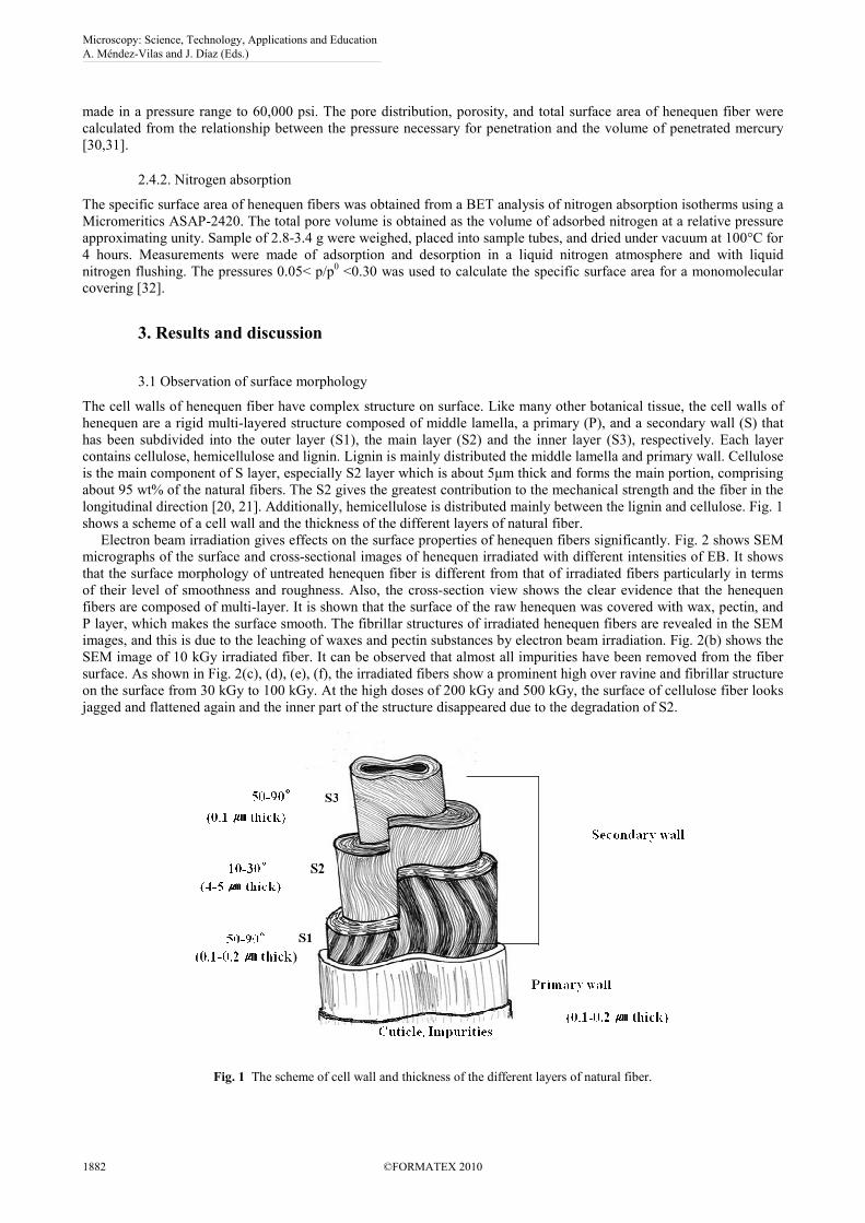
The cell walls of henequen fiber have complex structure on surface. Like many other botanical tissue, the cell walls of
henequen are a rigid multi-layered structure composed of middle lamella, a primary (P), and a secondary wall (S) that
has been subdivided into the outer layer (S1), the main layer (S2) and the inner layer (S3), respectively. Each layer
contains cellulose, hemicellulose and lignin. Lignin is mainly distributed the middle lamella and primary wall. Cellulose
is the main component of S layer, especially S2 layer which is about 5µm thick and forms the main portion, comprising
about 95 wt% of the natural fibers. The S2 gives the greatest contribution to the mechanical strength and the fiber in the
longitudinal direction [20, 21]. Additionally, hemicellulose is distributed mainly between the lignin and cellulose. Fig. 1
shows a scheme of a cell wall and the thickness of the different layers of natural fiber.
Electron beam irradiation gives effects on the surface properties of henequen fibers significantly. Fig. 2 shows SEM
micrographs of the surface and cross-sectional images of henequen irradiated with different intensities of EB. It shows
that the surface morphology of untreated henequen fiber is different from that of irradiated fibers particularly in terms
of their level of smoothness and roughness. Also, the cross-section view shows the clear evidence that the henequen
fibers are composed of multi-layer. It is shown that the surface of the raw henequen was covered with wax, pectin, and
P layer, which makes the surface smooth. The fibrillar structures of irradiated henequen fibers are revealed in the SEM
images, and this is due to the leaching of waxes and pectin substances by electron beam irradiation. Fig. 2(b) shows the
SEM image of 10 kGy irradiated fiber. It can be observed that almost all impurities have been removed from the fiber
surface. As shown in Fig. 2(c), (d), (e), (f), the irradiated fibers show a prominent high over ravine and fibrillar structure
on the surface from 30 kGy to 100 kGy. At the high doses of 200 kGy and 500 kGy, the surface of cellulose fiber looks
jagged and flattened again and the inner part of the structure disappeared due to the degradation of S2.
Fig. 1 The scheme of cell wall and thickness of the different layers of natural fiber.
Microscopy: Science, Technology, Applications and Education A. Méndez-Vilas and J. Díaz (Eds.)
1882 ©FORMATEX 2010
______________________________________________
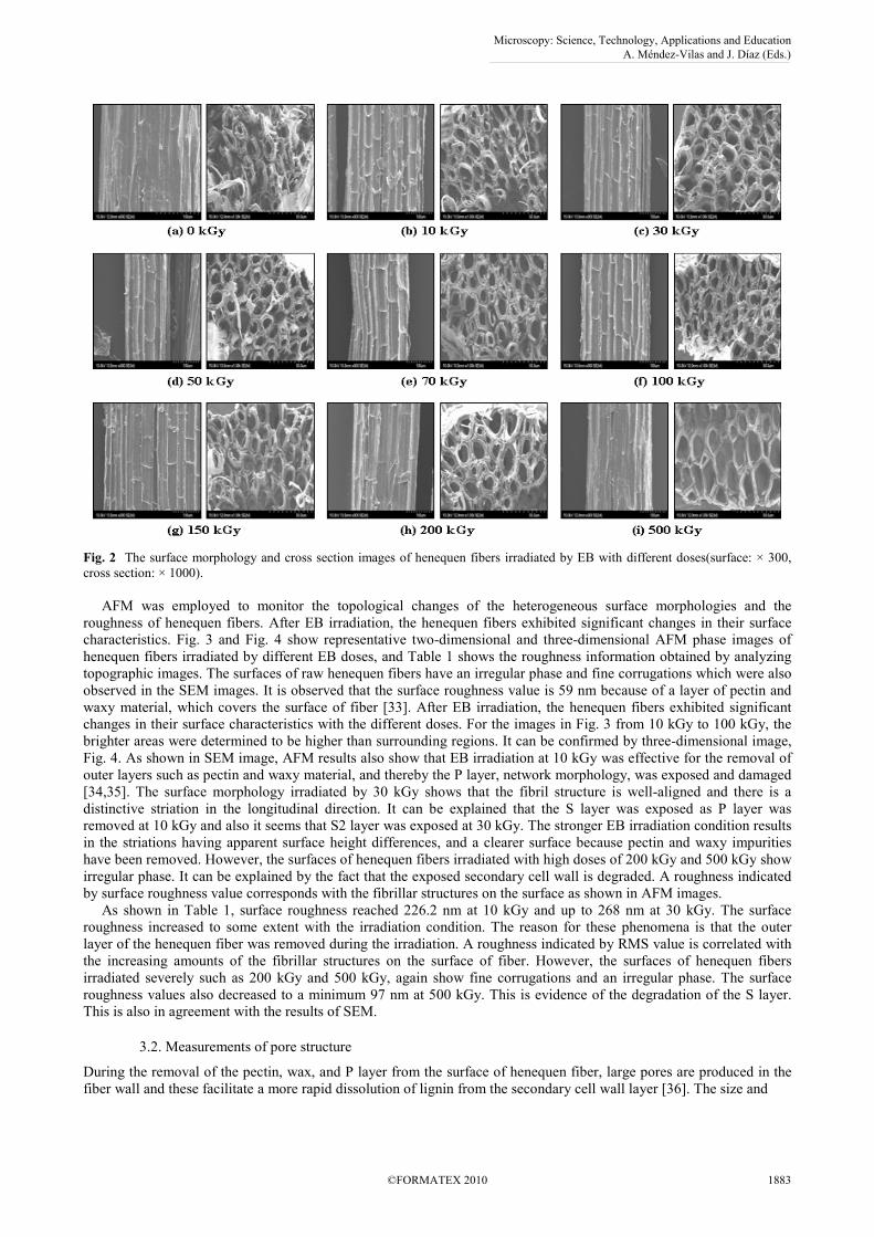
Fig. 2 The surface morphology and cross section images of henequen fibers irradiated by EB with different doses(surface: × 300,
cross section: × 1000).
AFM was employed to monitor the topological changes of the heterogeneous surface morphologies and the
roughness of henequen fibers. After EB irradiation, the henequen fibers exhibited significant changes in their surface
characteristics. Fig. 3 and Fig. 4 show representative two-dimensional and three-dimensional AFM phase images of
henequen fibers irradiated by different EB doses, and Table 1 shows the roughness information obtained by analyzing
topographic images. The surfaces of raw henequen fibers have an irregular phase and fine corrugations which were also
observed in the SEM images. It is observed that the surface roughness value is 59 nm because of a layer of pectin and
waxy material, which covers the surface of fiber [33]. After EB irradiation, the henequen fibers exhibited significant
changes in their surface characteristics with the different doses. For the images in Fig. 3 from 10 kGy to 100 kGy, the
brighter areas were determined to be higher than surrounding regions. It can be confirmed by three-dimensional image,
Fig. 4. As shown in SEM image, AFM results also show that EB irradiation at 10 kGy was effective for the removal of
outer layers such as pectin and waxy material, and thereby the P layer, network morphology, was exposed and damaged
[34,35]. The surface morphology irradiated by 30 kGy shows that the fibril structure is well-aligned and there is a
distinctive striation in the longitudinal direction. It can be explained that the S layer was exposed as P layer was
removed at 10 kGy and also it seems that S2 layer was exposed at 30 kGy. The stronger EB irradiation condition results
in the striations having apparent surface height differences, and a clearer surface because pectin and waxy impurities
have been removed. However, the surfaces of henequen fibers irradiated with high doses of 200 kGy and 500 kGy show
irregular phase. It can be explained by the fact that the exposed secondary cell wall is degraded. A roughness indicated
by surface roughness value corresponds with the fibrillar structures on the surface as shown in AFM images.
As shown in Table 1, surface roughness reached 226.2 nm at 10 kGy and up to 268 nm at 30 kGy. The surface
roughness increased to some extent with the irradiation condition. The reason for these phenomena is that the outer
layer of the henequen fiber was removed during the irradiation. A roughness indicated by RMS value is correlated with
the increasing amounts of the fibrillar structures on the surface of fiber. However, the surfaces of henequen fibers
irradiated severely such as 200 kGy and 500 kGy, again show fine corrugations and an irregular phase. The surface
roughness values also decreased to a minimum 97 nm at 500 kGy. This is evidence of the degradation of the S layer.
This is also in agreement with the results of SEM.
3.2. Measurements of pore structure
During the removal of the pectin, wax, and P layer from the surface of henequen fiber, large pores are produced in the
fiber wall and these facilitate a more rapid dissolution of lignin from the secondary cell wall layer [36]. The size and
Microscopy: Science, Technology, Applications and Education A. Méndez-Vilas and J. Díaz (Eds.)
©FORMATEX 2010 1883
______________________________________________

Fig. 3 2D Image (20 ㎛ × 20 ㎛) of henequen irradiated by EB with different doses.
Fig. 4 3D Image (20 ㎛ × 20 ㎛) of henequen irradiated by EB with different doses.
Microscopy: Science, Technology, Applications and Education A. Méndez-Vilas and J. Díaz (Eds.)
1884 ©FORMATEX 2010
______________________________________________
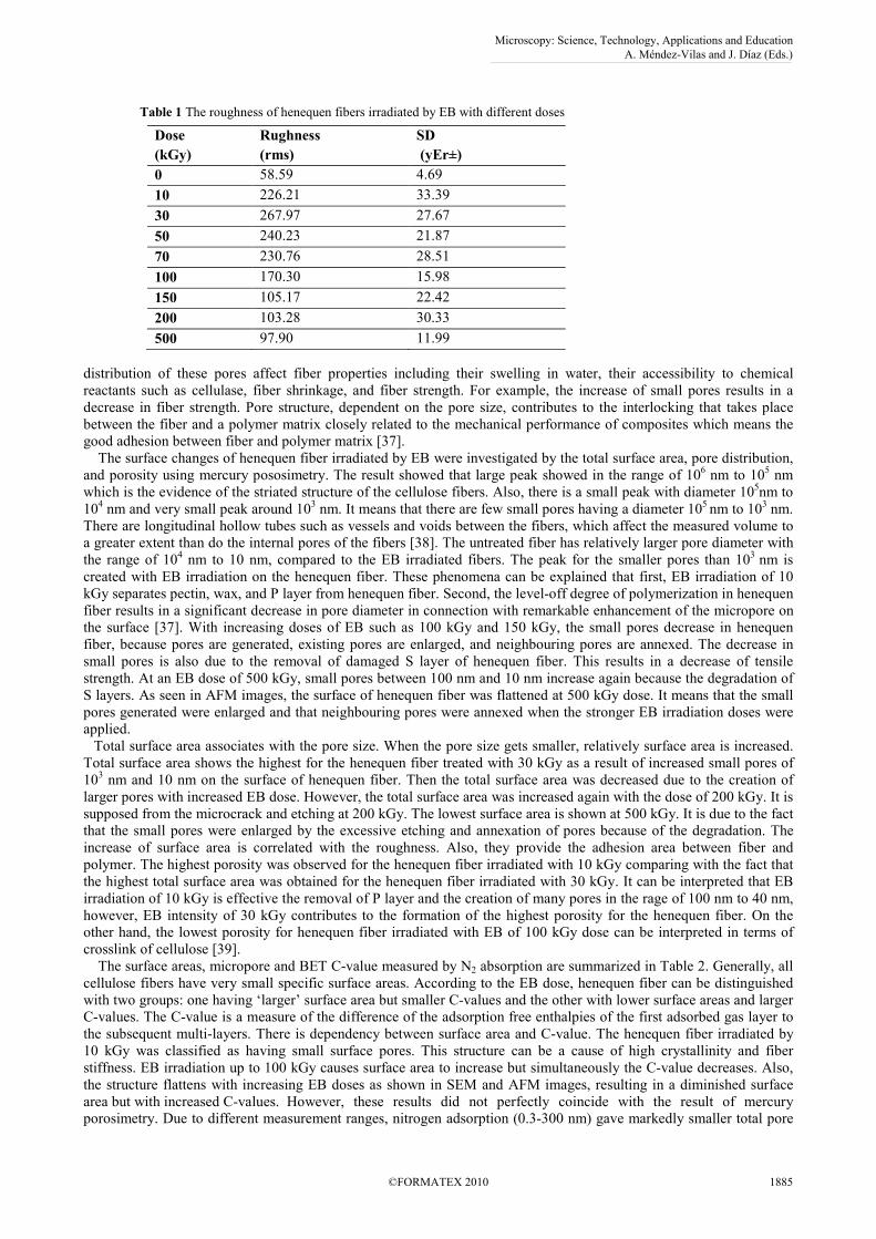
Table 1 The roughness of henequen fibers irradiated by EB with different doses
Dose
(kGy)
Rughness
(rms)
SD
(yEr±)
0 58.59 4.69
10 226.21 33.39
30 267.97 27.67
50 240.23 21.87
70 230.76 28.51
100 170.30 15.98
150 105.17 22.42
200 103.28 30.33
500 97.90 11.99
distribution of these pores affect fiber properties including their swelling in water, their accessibility to chemical
reactants such as cellulase, fiber shrinkage, and fiber strength. For example, the increase of small pores results in a
decrease in fiber strength. Pore structure, dependent on the pore size, contributes to the interlocking that takes place
between the fiber and a polymer matrix closely related to the mechanical performance of composites which means the
good adhesion between fiber and polymer matrix [37].
The surface changes of henequen fiber irradiated by EB were investigated by the total surface area, pore distribution,
and porosity using mercury pososimetry. The result showed that large peak showed in the range of 106 nm to 10
5 nm
which is the evidence of the striated structure of the cellulose fibers. Also, there is a small peak with diameter 105nm to
104 nm and very small peak around 10
3 nm. It means that there are few small pores having a diameter 10
5 nm to 10
3 nm.
There are longitudinal hollow tubes such as vessels and voids between the fibers, which affect the measured volume to
a greater extent than do the internal pores of the fibers [38]. The untreated fiber has relatively larger pore diameter with
the range of 104 nm to 10 nm, compared to the EB irradiated fibers. The peak for the smaller pores than 10
3 nm is
created with EB irradiation on the henequen fiber. These phenomena can be explained that first, EB irradiation of 10
kGy separates pectin, wax, and P layer from henequen fiber. Second, the level-off degree of polymerization in henequen
fiber results in a significant decrease in pore diameter in connection with remarkable enhancement of the micropore on
the surface [37]. With increasing doses of EB such as 100 kGy and 150 kGy, the small pores decrease in henequen
fiber, because pores are generated, existing pores are enlarged, and neighbouring pores are annexed. The decrease in
small pores is also due to the removal of damaged S layer of henequen fiber. This results in a decrease of tensile
strength. At an EB dose of 500 kGy, small pores between 100 nm and 10 nm increase again because the degradation of
S layers. As seen in AFM images, the surface of henequen fiber was flattened at 500 kGy dose. It means that the small
pores generated were enlarged and that neighbouring pores were annexed when the stronger EB irradiation doses were
applied.
Total surface area associates with the pore size. When the pore size gets smaller, relatively surface area is increased.
Total surface area shows the highest for the henequen fiber treated with 30 kGy as a result of increased small pores of
103 nm and 10 nm on the surface of henequen fiber. Then the total surface area was decreased due to the creation of
larger pores with increased EB dose. However, the total surface area was increased again with the dose of 200 kGy. It is
supposed from the microcrack and etching at 200 kGy. The lowest surface area is shown at 500 kGy. It is due to the fact
that the small pores were enlarged by the excessive etching and annexation of pores because of the degradation. The
increase of surface area is correlated with the roughness. Also, they provide the adhesion area between fiber and
polymer. The highest porosity was observed for the henequen fiber irradiated with 10 kGy comparing with the fact that
the highest total surface area was obtained for the henequen fiber irradiated with 30 kGy. It can be interpreted that EB
irradiation of 10 kGy is effective the removal of P layer and the creation of many pores in the rage of 100 nm to 40 nm,
however, EB intensity of 30 kGy contributes to the formation of the highest porosity for the henequen fiber. On the
other hand, the lowest porosity for henequen fiber irradiated with EB of 100 kGy dose can be interpreted in terms of
crosslink of cellulose [39].
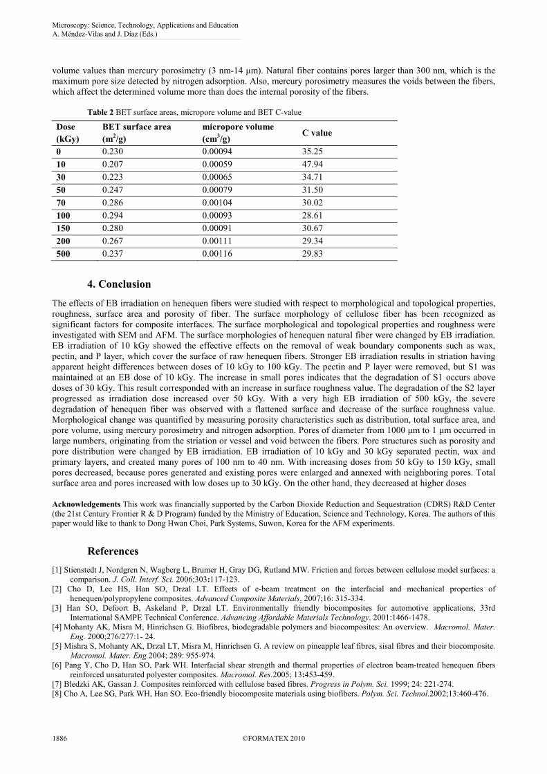
The surface areas, micropore and BET C-value measured by N2 absorption are summarized in Table 2. Generally, all
cellulose fibers have very small specific surface areas. According to the EB dose, henequen fiber can be distinguished
with two groups: one having ‘larger’ surface area but smaller C-values and the other with lower surface areas and larger
C-values. The C-value is a measure of the difference of the adsorption free enthalpies of the first adsorbed gas layer to
the subsequent multi-layers. There is dependency between surface area and C-value. The henequen fiber irradiated by
10 kGy was classified as having small surface pores. This structure can be a cause of high crystallinity and fiber
stiffness. EB irradiation up to 100 kGy causes surface area to increase but simultaneously the C-value decreases. Also,
the structure flattens with increasing EB doses as shown in SEM and AFM images, resulting in a diminished surface
area but with increased C-values. However, these results did not perfectly coincide with the result of mercury
porosimetry. Due to different measurement ranges, nitrogen adsorption (0.3-300 nm) gave markedly smaller total pore
Microscopy: Science, Technology, Applications and Education A. Méndez-Vilas and J. Díaz (Eds.)
©FORMATEX 2010 1885
______________________________________________
volume values than mercury porosimetry (3 nm-14 µm). Natural fiber contains pores larger than 300 nm, which is the
maximum pore size detected by nitrogen adsorption. Also, mercury porosimetry measures the voids between the fibers,
which affect the determined volume more than does the internal porosity of the fibers.
Table 2 BET surface areas, micropore volume and BET C-value
Dose
(kGy)
BET surface area
(m2/g)
micropore volume
(cm3/g)
C value
0 0.230 0.00094 35.25
10 0.207 0.00059 47.94
30 0.223 0.00065 34.71
50 0.247 0.00079 31.50
70 0.286 0.00104 30.02
100 0.294 0.00093 28.61
150 0.280 0.00091 30.67
200 0.267 0.00111 29.34
500 0.237 0.00116 29.83
4. Conclusion
The effects of EB irradiation on henequen fibers were studied with respect to morphological and topological properties,
roughness, surface area and porosity of fiber. The surface morphology of cellulose fiber has been recognized as
significant factors for composite interfaces. The surface morphological and topological properties and roughness were
investigated with SEM and AFM. The surface morphologies of henequen natural fiber were changed by EB irradiation.
EB irradiation of 10 kGy showed the effective effects on the removal of weak boundary components such as wax,
pectin, and P layer, which cover the surface of raw henequen fibers. Stronger EB irradiation results in striation having
apparent height differences between doses of 10 kGy to 100 kGy. The pectin and P layer were removed, but S1 was
maintained at an EB dose of 10 kGy. The increase in small pores indicates that the degradation of S1 occurs above
doses of 30 kGy. This result corresponded with an increase in surface roughness value. The degradation of the S2 layer
progressed as irradiation dose increased over 50 kGy. With a very high EB irradiation of 500 kGy, the severe
degradation of henequen fiber was observed with a flattened surface and decrease of the surface roughness value.
Morphological change was quantified by measuring porosity characteristics such as distribution, total surface area, and
pore volume, using mercury porosimetry and nitrogen adsorption. Pores of diameter from 1000 µm to 1 µm occurred in
large numbers, originating from the striation or vessel and void between the fibers. Pore structures such as porosity and
pore distribution were changed by EB irradiation. EB irradiation of 10 kGy and 30 kGy separated pectin, wax and
primary layers, and created many pores of 100 nm to 40 nm. With increasing doses from 50 kGy to 150 kGy, small
pores decreased, because pores generated and existing pores were enlarged and annexed with neighboring pores. Total
surface area and pores increased with low doses up to 30 kGy. On the other hand, they decreased at higher doses
Acknowledgements This work was financially supported by the Carbon Dioxide Reduction and Sequestration (CDRS) R&D Center
(the 21st Century Frontier R & D Program) funded by the Ministry of Education, Science and Technology, Korea. The authors of this
paper would like to thank to Dong Hwan Choi, Park Systems, Suwon, Korea for the AFM experiments.
References
[1] Stienstedt J, Nordgren N, Wagberg L, Brumer H, Gray DG, Rutland MW. Friction and forces between cellulose model surfaces: a
comparison. J. Coll. Interf. Sci. 2006;303:117-123.
[2] Cho D, Lee HS, Han SO, Drzal LT. Effects of e-beam treatment on the interfacial and mechanical properties of
henequen/polypropylene composites. Advanced Composite Materials. 2007;16: 315-334.
[3] Han SO, Defoort B, Askeland P, Drzal LT. Environmentally friendly biocomposites for automotive applications, 33rd
International SAMPE Technical Conference. Advancing Affordable Materials Technology. 2001:1466-1478.
[4] Mohanty AK, Misra M, Hinrichsen G. Biofibres, biodegradable polymers and biocomposites: An overview. Macromol. Mater.
Eng. 2000;276/277:1- 24.
[5] Mishra S, Mohanty AK, Drzal LT, Misra M, Hinrichsen G. A review on pineapple leaf fibres, sisal fibres and their biocomposite.
Macromol. Mater. Eng.2004; 289: 955-974.
[6] Pang Y, Cho D, Han SO, Park WH. Interfacial shear strength and thermal properties of electron beam-treated henequen fibers
reinforced unsaturated polyester composites. Macromol. Res.2005; 13:453-459.
[7] Bledzki AK, Gassan J. Composites reinforced with cellulose based fibres. Progress in Polym. Sci. 1999; 24: 221-274.
[8] Cho A, Lee SG, Park WH, Han SO. Eco-friendly biocomposite materials using biofibers. Polym. Sci. Technol.2002;13:460-476.
Microscopy: Science, Technology, Applications and Education A. Méndez-Vilas and J. Díaz (Eds.)
1886 ©FORMATEX 2010
______________________________________________

[9] Varez A, Mondragon I, Vazquez A. Influence of chemical treatments on the interfacial adhesion between sisal fibre and different
biodegradable polymer. Composite Interfaces. 2007;14:605-616.
[10] Mohd Edderozeya AM, Akil HM, Azhar AB, Zainal Ariffin MI. Chemical modification of kenaf fibers, Mater. Lett.
2007;61:2023-2025.
[11] Jacob M, Francis B, Varughese KT, Thoma S. The effects of silane coupling agents on the viscoelastic properties of rubber
biocomposites. Macromol. Mater. Eng. 2006;291:1119-1126
[12] Felix JM, Gatenholm P. The nature of adhesion in composites of modified cellulose fibers and polypropylene. J. Appl. Polym.
Sci. 1991;42: 609-620.
[13] You Y, Han SO, Cho D, Lee SG, Park WH. Acetiylation reaction methods for chemically modified flax fabrics. Journal of the
Korean Fiber Society. 2004;41:170-176.
[14] Lee SG, Choi SS, Park WH, Cho D. Characterization of surface modified flax fibers and their biocomposites with PHB.
Macromol. Symp.2003;197:89-99.
[15] Belgacem MN, Bataille P, Sapieha S. Effect of corona modification on the mechanical properties of polypropylene/cellulose
composites. J. Appl. Polym. Sci. 1994;53:379-385.
[16] El-Naggar AM, Zohdy MH, Said HM, El-Din MS, Noval DM. Pigment colors printing on cotton fabrics by surface coating
induced by electron beam and thermal curing. Applied Surface Science. 2005;241:420-430.
[17] Takács E, Wojnárovits L, Borsa J, Földvary Cs, Hargittai P, Zöld O. Effect of γ-irradiation on cotton-cellulose. Rad. Phy. Chem.
1999;55:663-666.
[18] Kim SW, Oh S, Lee K. Variation of mechanical and thermal properties of the thermoplastics reinforced with natural fibers by
electron beam processing. Radi. Physic. and Chem. 2007;76:1711-1714.
[19] Han SO, Cho DH, Park WH, Drzal LT. Henequen/poly(butylenes succinate) biocomposites: electron beam irradiation effects on
henequen fiber and the interfacial properties of biocomposites. Composite interfaces. 2006;13:231-247.
[20] Wood RJ, Pikayev AK. Applied radiation chemistry; Radiation processing. CELLULOSE. Wiley, New York;1994.
[21] Nevell TP, Zeronian SH. Cellulose chemistry and its applications. In: Cellulose chemistry fundamentals. John Wiley &
sons;1985.
[22] Dorschner H, Lappan U, Lunkewitz K. Electron beam facility in polymer research: radiation induced functionalization of
polytetrafluoroethylene. Nucl. Instr. and Meth. in Phys. Res. B. 1998;139:495-501.
[23] Zafeiropoulos EN, Baille CA, Hodgkinson JM. Engineering and characterisation of the interface in flax fibre/polypropylene
composite materials. Part II. The effect of surface treatments on the interface. Composites Part A.2002;33:1185-1190.
[24] Luo S, Netravali AN. Characterization of henequen fibers and the henequen fiber/poly(hydroxybutyrate-co-hydroxyvalerate)
interface. J. Adhesion Sci. Technol. 2001;15:423-437.
[25] Choi HY, Han SO, Lee JS. The effects of surface and pore characteristics of natural fiber on interfacial adhesion of henequen
fiber/PP biocomposites. Composite Interfaces. 2009;16:359-376.
[26] Chen N, Bhushan B. Morphological, nanomechanical and cellular structural characterization of human hair and conditioner
distribution using torsional resonance mode with an atomic force microscope. J. Microscopy. 2005;220:96-112.
[27] Poggi MA, Mancosky DG, Bottomley LA, Lucia LA. Atomic force microscopic analysis of hydrogen peroxide bleached kraft
northern black spruce fibres. J. Microsc. 2006;220:77-83.
[28] Bastidas JC, Venditti R, Pawlak J, Gilbert R, Zauscher S, Kadla JF. Characterization of fiber surfaces using chemical force
microscopy. Carbohydrate Polymers. 2005;62:369-378.
[29] Choi HY, Han SO, Lee JS. The effects of morphological properties of henequen fiber irradiated bt EB on the mechanical and
thermal properties of henequen fiber/PP composites. Composite Interfaces. 2009;16:751-768.
[30] Lee SG, Kin DC, Kang TJ. The Effect of Oxygen Plasma Treatment on the Physico-Chemical Properties and Interfacial Strength
of UHMWPE Fibers, Textile Science and Engineering. 1997;34: 459-465.
[31] Sing KSW, Everett DH, Haul RAW, Moscou L, Pierotti RA, Rouquérol J, Siemieniewska T. Reporting physisorption data for
gas/solid systems with special reference to the determination of surface area and porosity. Pure and Applied Chemistry. Pure
and Appl. Chem. 1985;57:603– 619.
[32] Sing KSW. Pure and Applied Chemistry. Reporting physisorption data for Gas/solid systems with special reference to the
determination of surface area and porosity. Pure and Applied Chemistry. 1986;57:603–619.
[33] Westermarck S, Juppo AM, Kervinen L, Yliruusi J. Pore structure and surface area of mannitol powder, granules and tablets
determined with mercury porosimetry and nitrogen adsorption. Eur. J. Pharm. Biopharm. 1998;46:61-68.
[34] Bismarck A, Aranberri-Askargorta I, Springer J. Surface Characterization of Flax, Hemp and Cellulose Fibres; Surface
Properties and the Water Uptake Behavior. Green chemistry. 2001;3:100-107.
[35] Fahlen J, Salmen L. Cross sectional structure of the secondary wall of wood fibers as affected by processing. J. mater. Sci.
2003;38:119-126.
[36] Zegota H. Some quantitative aspects of hydroxyl radical induced reactions in gamma irradiated aqueous solutions of pectins.
Food Hydrocolloids. 2000;16:353-361.
[37] Buschle-diller G, Fanter C, loth F. Effect of cellulase on the pore structure of bead cellulose. Cellulose. 1995;2:179-203
[38] Stone JE, Scallan AM. The effect of component removal upon the porous structure of the cell wall of wood.Ⅱ, Swelling in
water and the fiber saturation point. Tappi. 1967;50:496-501.
[39] Klemm D, Philipp B, Heinze T, Heinze U, Wagenknecht W. Comprehensive Cellulose Chemistry Volume1. Chapter2. General
considerations on structure and reactivity of cellulose. Wiley; 1998.
Microscopy: Science, Technology, Applications and Education A. Méndez-Vilas and J. Díaz (Eds.)
©FORMATEX 2010 1887
______________________________________________