morphometric alterations, steatosis, fibrosis and...
TRANSCRIPT

Morphometric Alterations, Steatosis, Fibrosisand Active Caspase-3 Detection in CarbamateBendiocarb Treated Rabbit Liver
Eva Petrovova,1 Halina Purzyc,2 David Mazensky,1 Lenka Luptakova,3 Norbert Torma,4
Igor Sopoliga,5 David Sedmera6
1Department of Anatomy, Histology and Physiology, University of Veterinary Medicine andPharmacy, Komenskeho 73, 04011 Kosice, Slovakia
2Department of Animal Physiology and Biostructure, University of Environmental and LifeSciences, Kozuchowska 5, 51-631 Wroclaw, Poland
3Department of Biology and Genetics, University of Veterinary Medicine and Pharmacy,Komenskeho 73, 04011 Kosice, Slovakia
4Eastern Slovakia Institute of Cardiovascular Disease, Kosice, Slovakia
5The Special Facility for Breeding and Diseases of Animals, Fish and Bees, University ofVeterinary Medicine and Pharmacy, Komenskeho 73, 04011 Kosice, Slovakia
6Institute of Anatomy, First Faculty of Medicine, Charles University in Prague, Unemocnice 3,12800 Prague 2, Czech Republic
Received 28 October 2012; revised 9 June 2013; accepted 9 June 2013
ABSTRACT: Increasing use of pesticides all over the world makes it necessary to reveal the toxic risk inpopulations of nontargeted organisms. Bendiocarb is one of the 12 insecticides recommended by theWorld Health Organization for use in malaria control in Africa, and is used against a variety of insects. Theliver has an important role in its process of detoxication and excretion. In our experiment 56 adult rabbitsof breed HY1, 28 males and 28 females were used. Animals were divided into groups (control, days 10,20, 30 of bendiocarb administration). The presence of many binucleated hepatocytes, the highest numberof liver cells and their decreased size at 10 day after bendiocarb administration was observed as an evi-dence of the hepatic regeneration. After the long-term treatment pronounced changes were presentedsuch as vacuolization and dilatation of hepatocytes, dilatation of sinusoids between hepatocytes, andfocal infiltration of inflammatory cells. Numerous cells with caspase-3 activity were present throughoutthe organ, most commonly around the portal tract and close to the central vein. Short and long-termbendiocarb treatment showed the central vein thickened rim with increased deposition of collagen,spreading of collagen fibers into the perisinusoidal, and pericellular space surrounding the central veins,
Correspondence to: E. Petrovova; e-mail: [email protected]
Contract grant sponsor: Slovak Ministry of Education.
Contract grant number: VEGA 1/0111/13.
Contract grant sponsor: Czech Ministry of Education.
Contract grant numbers: VZ 0021620806; PRVOUK-P35/LF1/5.
Contract grant sponsor: Czech Academy of Sciences.
Contract grant numbers: AV0Z50110509; RVO: 67985823.
Published online 9 July 2013 in Wiley Online Library (wileyonlinelibrary.com). DOI: 10.1002/tox.21887
212
VC 2013 Wiley Periodicals, Inc.
and septal fibrosis extended from the portal tract. Subsequently, presence of the lipid vacuoles both inthe liver parenchyma and inner of the hepatocytes were observed. These results suggest that bendiocarbtreatment leads to increased cell death, liver perisinusoidal fibrosis, and steatosis, especially during thelong-term administration. VC 2013 Wiley Periodicals, Inc. Environ Toxicol 30: 212–222, 2015.
Keywords: bendiocarb; caspase-3 activity; fibrosis; toxicity; rabbit; liver
INTRODUCTION
Pesticides are the significant contaminants of environment
that are used in agriculture and households. Their widely use
cause the question of toxicity pesticides to nontarget organ-
isms, their persistence, accumulation, and combined effect
with other chemicals. The worldwide annual consumption of
pesticides is about 2 million tons of which 24% is consumed
in the United States alone, 45% in Europe, and 25% in rest
of the world (Uggini et al., 2010). In the European Union the
wide using of pesticides are mainly organophosphates, car-
bamates, and pyrethroids. Further, over 25% of fruits, vege-
tables, and cereals grown in the European Union contain
detectable residues of at least two pesticides while their total
quantity, which Europe’s population is exposed to is
unknown (Bj�rling-Poulsen et al., 2008). In 2003, over
800 pesticide substances were used in European agriculture,
but only 380 of these are still authorized in March 2011
(Nougadere et al., 2011). At present, bendiocarb (2,2-
dimethyl-1,3-benzodioxol-4-yl methylcarbamate) is not
approved for using in the European Union, mainly through
Directive 91/414/EEC and Regulation (EC) No 1107/2009.
Although, it is one of 12 insecticides recommended by the
World Health Organization for use in malaria control in
Africa (Sadasivaiah et al., 2007). The widespread pyrethroid
resistance is becoming a major problem faced by several
National Malaria Control Programmes throughout Africa.
Field trials with the carbamates propoxur and bendiocarb for
indoor residual spraying treatment have been very effective
against pyrethroid resistant malaria vectors. Over the past
few years, there was an increasing interest in testing these
insecticides for public health purposes as alternatives to pyr-
ethroids (Akogbeto et al., 2010). Bendiocarb is a broad spec-
trum pesticide belonging to the N-methylcarbamate group
(Pacioni and Veglia, 2007). Using of bendiocarb can dramat-
ically reduce the risk of insect-borne diseases that are trans-
mitted by arthropod vectors such as mosquitoes (malaria,
dengue fever, yellow fever, encephalitis, filariasis, West Nile
fever and chikungunya), ticks (e.g., Lyme disease), and
sandflies (leishmaniasis; Nauen, 2007). Bendiocarb inhibits
acetylcholinesterase and elicits cholinergic hyperstimulation,
however causes only reversible inhibition of acetylcholines-
terase for a few minutes to a day (Kamel and Hoppin, 2004).
This lability tends to limit the duration of carbamate poison-
ings, accounts for the greater span between symptom-
producing and lethal doses, and it frequently invalidates the
measurement of blood cholinesterase activity as a diagnostic
index of poisoning (Reigart and Roberts, 1999). Further,
carbamate insecticides inhibit cellular metabolism including
energy, protein, and nucleic acid metabolism, thereby, caus-
ing cell regression, and death (Amanullah and Hari, 2011).
Symptoms of bendiocarb poisoning include weakness,
blurred vision, headache, nausea, abdominal cramps, chest
discomfort, miosis, sweating, muscle tremors and incoordi-
nation, decreased pulse, low blood pressure, heart irregular-
ities, giddiness, confusion, slurred speech, and loss of
reflexes. Respiratory failure resulting from inhibition of the
central respiratory drive and bronchospasms coupled with
depolarizing blockade at neuromuscular junctions-
diaphragm and intercostals (Krieger, 1991; Almasiova et al.,
2012). Many studies proved that bendiocarb increases inci-
dence of lymphoreticular tumours, such as lymphosarcomas,
reticulosarcomas, lymphoid, and myeloid leukaemia
(Dorko et al., 2011). Bendiocarb is degraded in the liver,
which has a major role in the biotransformation and excre-
tion of carbamate pesticides from the body. Reactions of
detoxication take place in hepatocytes by the enzymatic sys-
tem situated in the endoplasmic reticulum (Schenck and
Kolb, 1990), and bendiocarb is excreted as a sulphate
and b-glucuronide conjugates of the phenol derivatives
(National Pesticide Information Center, 2002). In a study
with rats, 90% of an oral bendiocarb dose was excreted in
the urine, 1–3% in expired air, and 3–8% in the feces. Excre-
tion was complete within 24 hours (Challis and Adcock,
1981).
In order to increase using of bendiocarb against vectors
of malaria, thus it is important to examine every potential
aspect of its toxicology. The lack of information at all levels
may be one of the most important causative factors of chem-
ical intoxication in developing countries (Forget, 1991). At
present, several studies of bendiocarb effect with rabbit as
an animal model have been published. We can continue with
results, particularly to organs which have a key role to bio-
transformation and excretion of bendiocarb. The aim of our
study was to observe the morphometric alterations, fibrotic
changes as well as the programmed cell death of the rabbit
liver tissue treated with bendiocarb, mostly in the area of the
central vein and portal tract.
MATERIAL AND METHODS
In our experiment were used 56 adult rabbits (age 5 84
days) of breed HY1, 28 males and 28 females, and with
average weight 2500 g from accredited animal farm (Nitra,
Slovakia). Animals were kept in cages (two per a cage) at
ALTERATIONS AFTER CARBAMATE BENDIOCARB TREATED RABBIT LIVER 213
Environmental Toxicology DOI 10.1002/tox

standard conditions (temperature 15–21�C, 12 hour light
period and relative humidity of 45%) and fed with granular
feed mixture (O-10 NORM TYP, Slovakia). Drinking water
was available for all animals ad libitum. Animals were
divided into groups (control, days 10, 20, 30 of administra-
tion), and each group comprised 14 animals. Rabbits in all
experimental groups received bendiocarb (96% Bendiocarb,
Bayer, Germany) per os in a dose 5 mg/kg per day (National
Pesticide Information Center, 2002) and after day 11 in a
same dose per 48 hours, with respect to the acute oral toxic-
ity of bendiocarb to rabbit, 35–40 mg/kg of the body weight
(Petrovova et al., 2011). Animals in control group were not
treated and they were killed at 30 day from the beginning of
the experimental work. Experimental animals were killed by
thiopental (Thiopental Valeant 1 g, ICN, Czech Republic;
100 mg/kg of body weight) intravenous administration at
days 10, 20, and 30 after bendiocarb treatment. Animal
weight was recorded at first and last day of bendiocarb treat-
ment, and calculation of the weight gain was made as the
difference between rabbit body weight in experimental
group and control group at days 10, 20, and 30 of the bend-
iocarb treatment. But the rabbit body weight of experimental
animals was unchanged in relation to control during the
whole experiment. The experimental work on rabbits was
performed with approval of the Ethic Committee of the
University of Veterinary Medicine and Pharmacy in Kosice
(No. 2647/07–221/5) and State Veterinary and Food Institute
in Bratislava (No. 1827/09–221/3) followed Slovakian
protocols for ethical standards for the use of laboratory
animals.
Histological Analysis
Liver samples from all animals were dissected immediately
after the animals were killed and fixed in 10% neutral forma-
lin. After the dehydration in 70–100% ethanol series they
were paraffin embedded. Paraffin sections (7 lm thick) were
stained for routine histological study using haematoxylin
and eosin (H&E) for histological evaluation under light
microscope (FL-800).
Morphometric Analysis
The morphometric model for liver characterization con-
sisted of many stereological variables. All morphometric
variables were obtained using a light microscope with
two objectives 20x and 40x, and photo-equipment (Olym-
pus Provis AX) to prepare images for morphometric anal-
ysis. More than 300 microscopic fields randomly selected
were examined.
A 16-points square lattice was used to quantitative evalu-
ation of hepatocytes (the total number of hepatocytes). The
grid was overlaid onto each microscopic field (87296 lm2)
displayed on the monitor (Vertemati et al., 2012). From each
animal of the control and experimental groups, we selected
randomly of 5–6 fields of the liver parenchyma which were
averaged. Subsequently, average for the group was meas-
ured. The morphometry was used to assess the diameter of
the central vein and size of the liver cell in H&E stained sec-
tions of the liver tissue. Structures of interest were analyzed
under 403 magnification, and the results of each sample
were averaged and evaluated. The morphometric analysis of
the vessel along the selected analysis path is based on the
quantification of the cross-sectional areas and diameters for
each cross-sectional section. For each in-plane direction, the
maximum, distance between two boundary points parallel to
that direction is determined. The minimum, average, and
maximum vessel diameters are then computed as the mini-
mum, average, and maximum values, respectively, of all of
these distances (Boskamp et al., 2004). For the evaluation of
the liver cell size were used of 120 measurements as well as
165 measurements for the diameter of the central vein. We
used to display of the microscopic fields the software (Adobe
Photoshop version 7.0), and for the measurement the Image
Acquisition Software getIT (Olympus Soft Imaging Solution
GmbH, Germany).
Oil red O Staining
Oil red staining was used for visualization of neutral lipids
on frozen sections. Samples of the liver were fixed in 4%
paraformaldehyde and sectioned on a cryomicrotome (Leica
CM 1850) for 5–10 lm thick. Liver sections were rinsed in
distilled water and 100% propylene glycol for 5 min, and
stained by Oil Red (0,5% Oil red in 100% propylene glycol;
Sigma-Aldrich, Germany) the preheated solution at 60�C,
8–10 min. Sections were differentiated in 85% propylene
glycol and rinsed twice in distilled water. Then the sections
were stained with hematoxylin for 30 seconds, differentiated
and rinsed in distilled water, and mounted in Kaiser
Glycerine-gelatine (Merck Millipore, Germany).
Caspase-3 Activity
The liver samples were dissected out and processed by the
histological approach. Samples of the liver were fixed in 4%
paraformaldehyde, sectioned on a cryomicrotome (Leica
CM 1850) for 5 lm thickness, and stained immunohisto-
chemically for observation of caspase activity, mainly close
to the central vein and portal tract. The programmed cell
death in the liver was observed by means of primary murine
monoclonal antibody IgG 1–Caspase-3/CPP32 (BD
Biosciences Pharmingen, California, USA) and secondary
antibody conjugated with Rhodamine Red dye (Jackson
ImmunoResearch Laboratories, Baltimore, USA). An apop-
tic signal such as granzyme B of cytotoxic T-cells or ICE-
like proteases induces the intracellular cleavage of Caspase-
3 from the inactive proform (32 kDa) to the active form,
which consists of the p17 subunit. To visualize the nuclei in
the liver, the respective sections were stained with Hoechst
33258 dye (Sigma-Aldrich, St. Louis, USA). The Rhoda-
mine Red-conjugated antibody was red while the Hoechst
214 PETROVOVA ET AL.
Environmental Toxicology DOI 10.1002/tox

33258 stain was blue, and replacement fibrosis was green
(WGA-wheat germ agglutinin staining). Image acquisition
was performed on a Leica SPE confocal microscope using
an oil immersed objective with 403 magnification. We used
the viewing field of size 100 lm3 for the counting the liver
cells with active caspase-3.
Fibrosis
Picro-sirius red staining was used for visualization of colla-
gen fibers and staging of hepatic fibrosis. Deparaffinized
liver sections were washed in water, and stained in Picro-
sirius red (0.1% Picro-sirius red in saturated aqueous picric
acid; Sigma-Aldrich) for 1 hour at room temperature. Sec-
tions were rinsed twice in 0.5% acetic acid and stained in
Hoechst (2 ll/200 ml 0.1% Triton/H2O). Subsequently, his-
tological sections were rinsed in distilled water, dehydrated
in absolute ethanol and xylene, and mounted in Depex
(VWR International GmbH, Austria). Microscopic evalua-
tion of fibrosis was analyzed using an Olympus BX46
microscope equipped with a camera DP70 (Olympus,
Hamburg, Germany) and objective with 20x magnification.
Five central veins and areas of portal tracts in each animal
were studied, for a total of 260 central veins and portal
tracts.
Statistics
The data were analyzed and expressed as means 6 SD
(standard deviation of the mean), the minimum, average,
and maximum values. Statistical analyses were performed
using the statistics guide (GraphPad Prism version 5.0 soft-
ware). Because the data are not normally distributed non-
parametric Mann–Whitney test was used to compare
between the data of the control and those of treatments. The
p-value; p� 0.05 was the level of statistical significance.
RESULTS
Liver Histology
Physiological structure of the liver parenchyma is presented
in the untreated samples. The polyedric liver cells are
arranged into the trabecules and blood sinusoids between of
them. Large, euchromatic nuclei were located in the centre
of the hepatocytes, and few binucleated liver cells were
observed as a physiological feature. The affected lobules of
the treated groups contained the focal infiltration of lympho-
cytes and granulocytes. Dilatation of blood sinusoids
between the hepatocytes was observed in each treated group,
but dilatation of hepatocytes was observed at 20 and 30 days
(Fig. 1). Mostly at 10 days the binucleated cells were
observed as well as the number of hepatocytes was increased
in comparison with the other treated groups (Table I). The
number of vacuoles in hepatocytes was observed at 10 and
30 days of bendiocarb administration, except at 20 day. But
the highest number of vacuoles was observed at 30 days,
and they changed the shape of the nuclei to semilunar shape
with pushing them away in the hepatocytes (Fig. 2). To fur-
ther clarify whether lipid accumulation was indeed induced
in the liver, the liver tissues were stained with Oil Red stain-
ing. The Oil Red staining clearly revealed moderate lipid
accumulation in hepatocytes of bendiocarb-treated animals
at 30 days. This result illustrates that the long-term bendio-
carb administration tested in this study could lead to the liver
steatosis (Fig. 3).
Significantly higher number of the liver cells was
observed at 10 day (26.34 6 6.46/100 lm3) and 30 day
(20.94 6 2.14/100 lm3) of bendiocarb treatment in compari-
son with the control group (19.55 6 1.95/100 lm3). The
number of hepatocytes of treatment group at 20 day was
unchanged to control (Table I). Changes of the size of hepa-
tocytes were observed at 10 and 20 day of the pesticide
Fig. 1. Representative alterations in the bendiocarb treated rabbit liver. A: Moderately dilatation of the blood sinusoids (arrow)between the hepatocytes was observed in each treated group. Mostly at 10 days the binucleated cells (headarrows) wereobserved as well as the number of hepatocytes was increased in comparison with the other treated groups. B: Dilatation ofhepatocytes was observed at 20 and 30 days. C: Affected lobules of the treated groups contained the focal infiltration ofinflammatory cells (arrow), and the number of vacuoles (headarrows) in hepatocytes was observed at 10 and 30 days ofbendiocarb administration, except at 20 day. Stained H&E, magnification 403, Scale bars: 50 lm. [Color figure can beviewed in the online issue, which is available at wileyonlinelibrary.com.]
ALTERATIONS AFTER CARBAMATE BENDIOCARB TREATED RABBIT LIVER 215
Environmental Toxicology DOI 10.1002/tox
treatment. The size of hepatocytes was significantly
decreased at 10 day (14.20 62.59 lm). But the hepatocyte
size was increased at 20 day (16.50 6 2.29 lm) against the
size of hepatocytes in the control group (15.50 6 2.12 lm;
Table I). The diameter of central veins ranged from 58.3 to
160.5 lm, and the mean diameter was lower at 10 day
(113.90 6 25.17 lm) compared with control (118.23 6
22.36 lm; Table I). Thus, they are representative central
veins as a general term (Danko et al., 2011), also called cen-
trolobular veins or terminal hepatic venules (Mak et al.,
2012).
Caspase Activity
Almost no positive cells were detected in the untreated sam-
ples. After 10 days of bendiocarb treatment, numerous
immune-reactive cells were present throughout the organ,
most commonly around the portal tract (PT). Positive cells
were still abundant at 20 days close to the central vein, with
a decline at 30 days at which point vacuoles were observed
in some cells together with replacement fibrosis.
After application of bendiocarb at 10 day, we observed
that in the viewing field of size 100 lm3 was the higher
number of liver cells with caspases activity around the PT
(13%). At this day only 7% of liver cells showed the caspase
activity near to the central vein in comparison with the con-
trol. After application of bendiocarb at 20 day in the same
viewing field of size the highest rate (14%) of caspase acti-
vated liver cells was observed close to the central vein in
comparison to 7% of rate around the PT. At 30 day the num-
ber of positive cells was decreased, mainly around the PT
(3%) in order to the space around the central vein (12%;
Fig. 4).
Fibrosis
We observed the histological features of central veins with
and without extension of collagen fibers into the perivenous
parenchyma, perisinusoidal, and pericellular space, particu-
larly at 10 and 20 days. The vein wall thickness ranged from
4 to 27 lm. Data of the vein wall thickness at 10
(14.9 6 4.1lm) and 30 days (16.8 6 4.38 lm) are higher
than control value (12.6 6 4.91 lm). At 20 day of bendio-
carb treatment the central veins show thinner rim with
decreased deposition of collagen fibers (9.9 6 1.65 lm). As
shown in Fig. 5, the vein wall thickness is increased at 10
and 30 days. Although Figure 6 shows that increase is with-
out significance.
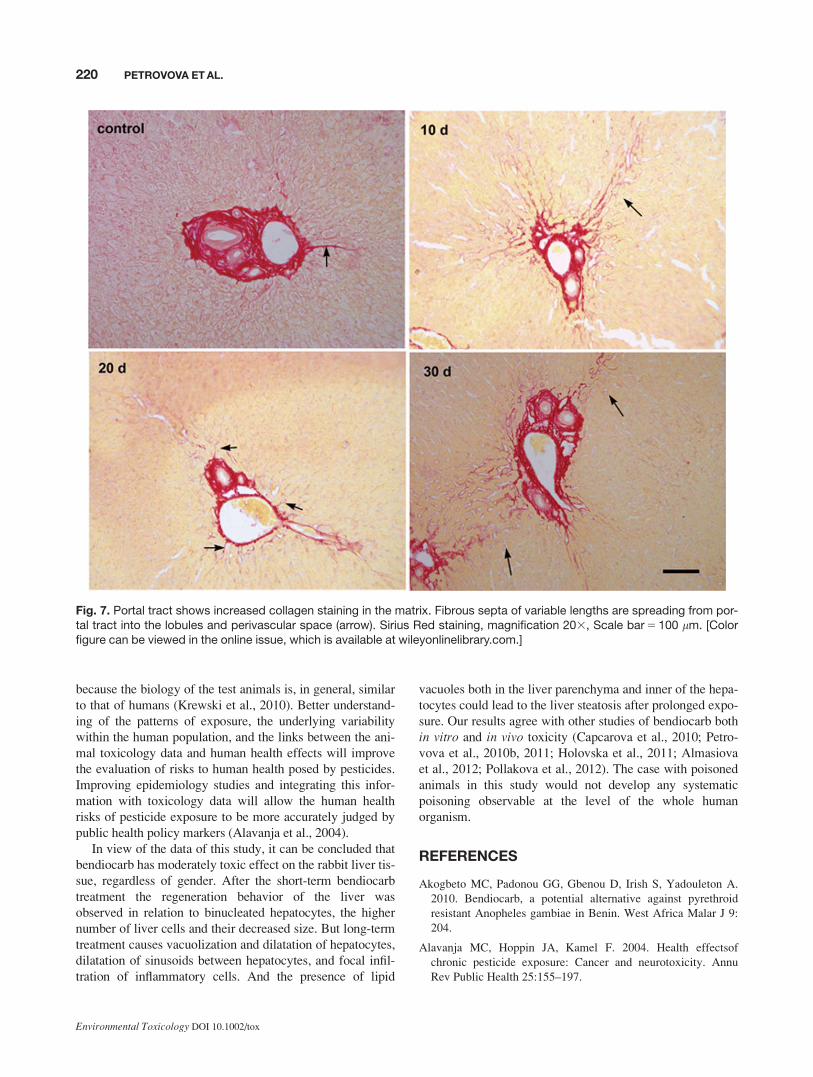
The control samples show a thin-walled central vein with
fine fibrous extension by Sirius red staining of collagens.
Fibrous extension was not detected in association with the
wall thickness of the central vein. Portal tract shows
increased collagen staining in the matrix. Further, at 10 and
TABLE I. Morphometric values of hepatocytes and venacentralis in the rabbit liver
Day x SDa Minimum Maximum
Number of
liver cells
(100 mm3)
Control 19.55 1.95 17.35 24.22
10 26.34b 6.46 16.69 43.69
20 20.67 2.61 14.57 24.55
30 20.94b 2.14 17.18 25.20
Size of liver
cells (mm)
Control 15.50 2.12 11.50 20.30
10 14.20b 2.59 9.00 19.33
20 16.50b 2.29 10.66 20.33
30 15.50 3.50 9.83 23.50
Diameter of
v. centralis(mm)
Control 118.23 22.36 58.34 158.78
10 113.90 25.17 62.15 147.95
20 118.71 33.21 63.19 155.87
30 121.08 35.74 67.93 160.46
aStandard deviation.bStatistical difference from the control; p� 0.05.
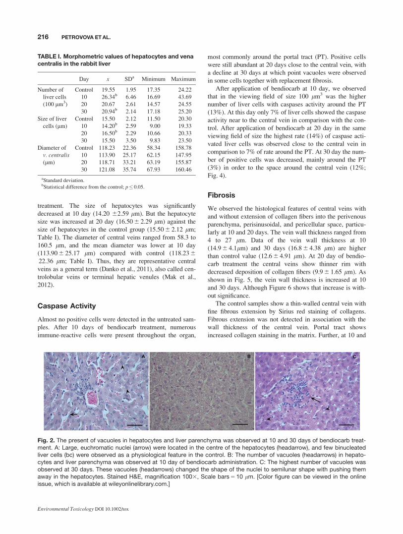
Fig. 2. The present of vacuoles in hepatocytes and liver parenchyma was observed at 10 and 30 days of bendiocarb treat-ment. A: Large, euchromatic nuclei (arrow) were located in the centre of the hepatocytes (headarrow), and few binucleatedliver cells (bc) were observed as a physiological feature in the control. B: The number of vacuoles (headarrows) in hepato-cytes and liver parenchyma was observed at 10 day of bendiocarb administration. C: The highest number of vacuoles wasobserved at 30 days. These vacuoles (headarrows) changed the shape of the nuclei to semilunar shape with pushing themaway in the hepatocytes. Stained H&E, magnification 1003, Scale bars 5 10 lm. [Color figure can be viewed in the onlineissue, which is available at wileyonlinelibrary.com.]
216 PETROVOVA ET AL.
Environmental Toxicology DOI 10.1002/tox

30 days the fibrous septa of variable lengths are spreading
from PT into the lobules and perisinusoidal space. The peri-
sinusoidal fibrosis is focal, which is marked by increased
collagen staining along the sinusoidal borders. Fine fibrous
strands extend from PT for a short distance into the lobules
at 20 day of bendiocarb treatment (Fig. 7).
DISCUSSION
It should be noted that many pesticides are transformed in
the environment through physical, chemical, and biological
processes that are intended to detoxify them, but often the
transformation process forms products that are more toxic
than the parent compound (Van Dyk and Pletschke, 2011). It
is well known that there is comparatively a low risk of carba-
mates for mammals, which is based on their biodegradation
(Sogorb and Vilanova, 2002). But on the contrary, our study
shows affected lobules of the treated groups contained the
focal infiltration of inflammatory cells and dilatation of sinu-
soids. The increased number of lipid vacuoles in hepatocytes
after long-term bendiocarb treatment we observed with Oil
Red O staining. Oil Red O staining is considered as a supe-
rior fat stain (therefore staining myelin), which has
extremely good depth of color and yet leaves cellular struc-
tures intact (Cholewiak et al., 1968). In the cholesterol fatty
rabbit liver, Nile red staining was comparable to that of Oil
Red O, a commonly used dye for tissue cholesteryl esters
and triacylglycerols. In contrast, Nile red staining of rabbit
aortic atheroma revealed ubiquitous lipid deposits not
observed with Oil Red O staining. These latter results sug-
gest that Nile red can detect neutral lipid deposits, presum-
ably unesterified cholesterol, not usually seen with Oil Red
O or other traditional fat stains (Fowler and Greenspan,
1985). A causal link between chronic exposure to pesticides
and their possible health effects is difficult to establish
because consequences appear years after a generally intense
exposure or after repeated low-intensity exposures over
many years (Nougadere et al., 2011). Regeneration process
was observed on 10 day after bendiocarb treatment. Histol-
ogy is long known to be an important tool for determining
the nature and extent of tissue damage (Kundu et al., 2011).
Histological examination of the liver of pregnant rat females
exposed to 400 ppm ditiocarbamate–propineb showed a dila-
tation of the wall of the central vein, as well as irregularity
and degeneration in hepatocytes around the vein. Moreover,
an increase in the number of vacuoles and hyalinization
were observed in hepatocytes. Also the study showed a very
clear dilatation of the sinusoids between the hepatocytes
(G€uven et al., 1999). Further, vacuolization of cell cyto-
plasm, infiltration of lymphocyte, and congestion in liver
were evaluated by Cengiz et al. (2001) after Thiodan (33.7%
endosulfan) treatment of mosquitofish, Gambusia affinis.Carbendazim caused changes in heterochromatin with pro-
nounced sporadic cell death of individual cells around the
central lobule area. Changes in heterochromatin caused by
imazalil were similar as in the carbendazim treated group,
and a sporadic cell death could be seen in areas close to cen-
tral vein (Dikic et al., 2011). The rabbit liver appeared the
most pronounced change on day 3 after bendiocarb adminis-
tration (5 mg/kg of body weight per day) with massive accu-
mulation of inflammatory cells within the portal spaces, and
regenerative features, such as binucleated hepatocytes, were
common (Holovska et al., 2011). We observed only weak
septal fibrosis (septa formation without bridging fibrosis)
around the portal tract, and perisinusoidal fibrosis close to
the central vein of the rabbit liver with the Picro-Sirius Red
staining. Previous studies of picro-dye reactions demon-
strated wide variations in the binding of different dyes.
Picro-Sirius Red was recommended because it colors all col-
lagens intensely and is suitable for polarization microscopy
(Puchtler et al., 1988). Sirius Red staining is presented as a
Fig. 3. Representative Oil Red staining of the rabbit liver tis-sue in control and experimental group after bendiocarbtreatment at 30 days (BK; arrows). Oil Red staining, magnifi-cation 403, Scale bar 5 100 lm. [Color figure can beviewed in the online issue, which is available atwileyonlinelibrary.com.]
ALTERATIONS AFTER CARBAMATE BENDIOCARB TREATED RABBIT LIVER 217
Environmental Toxicology DOI 10.1002/tox

method for collagen determination, enabling quantitative
morphometric measurements to be performed in locally
defined tissue areas (Malkusch et al., 1995). By means of
staining with Sirius Red F3BA in a saturated picric acid
solution, the collagen contents of rat livers with varying
degrees of fibrosis have been measured quantitatively in
Fig. 4. Active caspase-3 is detected in bendiocarb treated rabbit liver. Almost no positive cells were detected in the untreatedsample (0d). After 10 days of treatment, numerous immunoreactive cells (white arrows) were present throughout the organ,most commonly around the portal tract. Positive cells were still abundant at 20 days, with a decline at 30 days, at which pointvacuoles were observed in some cells (asterisks) together with replacement fibrosis (wheat germ agglutinin staining; WGA,green). Negative control (NC) showed only dim background autofluorescence under identical exposure conditions. Stainedimmunohistochemically, magnification 403, Scale bars 5 50 lm. [Color figure can be viewed in the online issue, which isavailable at wileyonlinelibrary.com.]
218 PETROVOVA ET AL.
Environmental Toxicology DOI 10.1002/tox

fixed and sectioned material. For analysis of collagen accu-
mulation in rat liver, section-based methods seem to be use-
ful and reliable, the extraction method giving the quickest
results for large-scale screening, and the histophotometric
method being more appropriate to take readings in selected
areas (James et al., 1990). Miranda et al., (2008) studied the
bioaccumulation of chlorinate pesticides and PCBs in the
tropical freshwater fish Hoplias malabaricus, and they found
the most important alterations in the liver were lesions such
as fibrosis, large necrosis area, and leukocyte infiltration.
The mammalian liver cells (WBF344) were the most sensi-
tive to insecticide bendiocarb, with significant suppression
of their proliferative activity (Pollakova et al., 2012). Atypi-
cal mitoses, cytologic alterations, cytomegaly, pigmentation,
necrosis, and neoplastic nodules of the liver in rats were
induced at 6 months after methyl carbamate treatment (0 or
400 mg/kg of body weight), 5 days per week (Chan et al.,
1992). The study of the other rabbit organs (thymus, spleen,
lymphoid tissue of the small intestine) detected changes in
the structure, after an experimental long-term bendiocarb
administration (Flesarova et al., 2007; Petrovova et al.,
2010a, 2011). Bendiocarb addition caused imbalance in
internal milieu of rabbits. Significant increase of creatinine
content, increase of aspartate aminotrasferase (AST) and
gamma glutamyl transferase (GGT), inform about possible
failure of liver caused by bendiocarb after 25 days of admin-
istration (5 mg/kg b.w. per day; Capcarova et al., 2010). Ani-
mals have served as models of human response for decades
Fig. 5. Control samples show a thin-walled central vein with fine fibrous extension by Sirius red staining of collagens. At 10and 30 days of bendiocarb treatment the wall of central vein increased in thickness. Furthermore, fibrous strands emergefrom the central vein into the parenchyma and perivascular space, particularly at 10 and 20 days (arrows). Sirius Red staining,magnification 203, Scale bar 5 100 lm. [Color figure can be viewed in the online issue, which is available atwileyonlinelibrary.com.]
Fig. 6. The central vein wall thickness in bendiocarb-treatedrabbit liver.
ALTERATIONS AFTER CARBAMATE BENDIOCARB TREATED RABBIT LIVER 219
Environmental Toxicology DOI 10.1002/tox
because the biology of the test animals is, in general, similar
to that of humans (Krewski et al., 2010). Better understand-
ing of the patterns of exposure, the underlying variability
within the human population, and the links between the ani-
mal toxicology data and human health effects will improve
the evaluation of risks to human health posed by pesticides.
Improving epidemiology studies and integrating this infor-
mation with toxicology data will allow the human health
risks of pesticide exposure to be more accurately judged by
public health policy markers (Alavanja et al., 2004).
In view of the data of this study, it can be concluded that
bendiocarb has moderately toxic effect on the rabbit liver tis-
sue, regardless of gender. After the short-term bendiocarb
treatment the regeneration behavior of the liver was
observed in relation to binucleated hepatocytes, the higher
number of liver cells and their decreased size. But long-term
treatment causes vacuolization and dilatation of hepatocytes,
dilatation of sinusoids between hepatocytes, and focal infil-
tration of inflammatory cells. And the presence of lipid
vacuoles both in the liver parenchyma and inner of the hepa-
tocytes could lead to the liver steatosis after prolonged expo-
sure. Our results agree with other studies of bendiocarb both
in vitro and in vivo toxicity (Capcarova et al., 2010; Petro-
vova et al., 2010b, 2011; Holovska et al., 2011; Almasiova
et al., 2012; Pollakova et al., 2012). The case with poisoned
animals in this study would not develop any systematic
poisoning observable at the level of the whole human
organism.
REFERENCES
Akogbeto MC, Padonou GG, Gbenou D, Irish S, Yadouleton A.
2010. Bendiocarb, a potential alternative against pyrethroid
resistant Anopheles gambiae in Benin. West Africa Malar J 9:
204.
Alavanja MC, Hoppin JA, Kamel F. 2004. Health effectsof
chronic pesticide exposure: Cancer and neurotoxicity. Annu
Rev Public Health 25:155–197.
Fig. 7. Portal tract shows increased collagen staining in the matrix. Fibrous septa of variable lengths are spreading from por-tal tract into the lobules and perivascular space (arrow). Sirius Red staining, magnification 203, Scale bar 5 100 lm. [Colorfigure can be viewed in the online issue, which is available at wileyonlinelibrary.com.]
220 PETROVOVA ET AL.
Environmental Toxicology DOI 10.1002/tox
Almasiova V, Holovska K, Tarabova L, Cigankova V,
Lukacinova V, Nistiar F. 2012. Structural and ultrastructural
study of the rabbit testes exposed to carbamate insecticide.
J Environ Sci Health A 47:1319–1328.
Amanullah M, Hari BY. 2011. Evaluation of carbamate insecti-
cides as chemotherapeutic agents for cancer. Ind J Cancer 48:
74–79.
Bj�rling-Poulsen M, Andersen H.R. Grandjean P. 2008. Potential
developmental neurotoxicity of pesticides used in Europe.
Environ Health 7:50.
Boskamp T, Rinck D, Link F, K€ummerlen B, Stamm G,
Mildenberger P. 2004. New vessel analysis tool for morphomet-
ric quantification and visualization of vessels in CT and MR
imaging data sets. Radiographics 24:287–297.
Capcarova M, Petrovova E, Flesarova S, Dankova M,
Massanyi P, Danko J. 2010. Bendiocarbamate induced altera-
tions in selected parameters of rabbit homeostasis after experi-
mental peroral administration. Pestic Biochem Physiol 98:
213–218.
Cengiz EI, Unlu E, Balci K. 2001. The histopathological effects
of Thiodan on the liver and gut of mosquitofish. Gambusia affi-
nis. J Environ Sci Health B 36:75–85.
Challis IR, Adcock JW. 1981. The Metabolism of the Carbamate
Insecticide Bendiocarb in the Rat and in Man. Pestic Sci 12:
638–644.
Chan PC, Huff J, Haseman JK, Quest JA, Hall W. 1992. Liver car-
cinogenesis by methyl carbamate in F344 rats and not in
B6C3F1 mice. Jpn J Cancer Res 83:258–263.
Cholewiak RW, Butcher L, Kettlewell NM. 1968. Oil Red O and
Hematoxylin: A rapid histologic technic. Physiol Behav 3:
585–586.
Danko J, Simon F, Artimova J. 2011. Nomina Anatomica Veteri-
naria. Kosice: University of Veterinary Medicine and Phar-
macy. 267 p.
Dikic D, Landeka I, Knezevic F, Mojsovic-Cuic A, Benkovic V,
Horvat-Knezevic A, Loncar G, Teparic R, Rogic D. 2011. Car-
bendazim impends hepatic necrosis when combined with ima-
zalil or cypermethrin. Basic Clin Pharmacol Toxicol 110:
433–440.
Dorko F, Danko J, Flesarova S, Boros E, Sobekova A. 2011.
Effect of pesticide bendiocarbamate on distribution of acetyl-
choline- and butyrylcholine-positive nerves in rabbit’s thymus.
Eur J Histochem 55:206–209.
Flesarova S, Lukac N, Danko J, Massanyi P. 2007. Bendiocarba-
mate induced structural alterations in rabbit thymus after exper-
imental peroral administration. J Environ Sci Health B 3:
329–334.
Forget G. 1991. Pesticides and the Third World. J Toxicol Envi-
ron Health 32:11–31.
Fowler SD, Greenspan P. 1985. Application of Nile red, a fluores-
cent hydrophobic probe, for the detection of neutral lipid depos-
its in tissue sections: Comparison with oil red O. J Histochem
Cytochem 33:833–836.
Guven K, Deveci E, Akba O, Onen A, de Pomerai D. 1998. The
accumulation and histological effects of organometallic fungi-
cides Propineb and Maneb in the kidneys of fetus and female
rats during pregnancy. Toxicol Lett 99:91–98.
Holovska K, Almasiova V, Tarabova L, Cigankova V. 2011.
Effect of xenobiotics on the structure of the rabbit’s liver. Folia
Veterinaria 55:69–72.
James J, Bosch KS, Aronson DC, Houtkooper JM. 1990. Sirius
red histophotometry and spectrophotometry of sections in the
assessment of the collagen content of liver tissue and its appli-
cation in growing rat liver. Liver 10:1–5.
Kamel F, Hoppin JA. 2004. Association of pesticide exposure
with neurologic dysfunction and disease. Environ Health Per-
spect 112:950–958.
Krewski D, Acosta D Jr, Andersen M, Anderson H, Bailar JC,
Boekelheide K, Brent R, Charnley G, Cheung VG, Green S. Jr,
Kelsey KT, Kerkvliet NI, Li AA, McCray L, Meyer O, Patterson
RD, Pennie W, Scala RA, Solomon GM, Stephens M, Yager J,
Zeise L. 2010. Toxicity testing in the 21st century: A vision and
a strategy. J Toxicol Environ Health B Crit Rev 13:51–138.
Krieger R. 1991. Handbook of Pesticide Toxicology, 2nd Ed. San
Diego: Academic Press. 2416 p.
Kundu CR, Roychoudhury S, Capcarova M. 2011. Malathion-
induced sublethal toxicity on the intestine of cricket frog
(Fejervarya limnocharis). J Environ Sci Health B 46:691–696.
Mak KM, Kwong AJ, Chu E, Hoo NM. 2012. Hepatic steatosis,
fibrosis, and cancer in elderly cadavers. Anat Rec (Hoboken)
295:40–50.
Malkusch W, Rhen B, Bruch J. 1995. Advantages of Sirius Red
staining for quantitative morphometric collagen measurements
in lungs. Exp Lung Res 21:67–77.
Miranda AL, Roche H, Randi MAF, Menezes ML, Oliveira
Ribeiro, CA. 2008. Bioaccumulation of chlorinate pesticides
and PCBs in the tropical freshwater fish Hoplias Malabaricus:
Histopathological, physiological, and immunological findings.
Environ Int 34:939–949.
National Pesticide Information Center. Bendiocarb (Technical
Fact Sheet). National Pesticide Information Center, Oregon
State University, Oregon, USA. Available at: http://npic.
orst.edu/factsheets/bendiotech.pdf, last accessed on 7 February
2008.
Nauen R. 2007. Perspective insecticide resistance in disease vec-
tors of public health importance. Pest Manag Sci 63:628–633.
Nougadere A, Reninger JC, Volatier JL, Leblanc JC. 2011.
Chronic dietary risk characterization for pesticide residues: A
ranking and scoring method integrating agricultural uses and
food contamination data. Food Chem Toxicol 49:1484–1510.
Pacioni NL, Veglia AV. 2007. Determination of poorly fluores-
cent carbamate pesticides in water, bendiocarb and promecarb,
using cyclodextrin nanocavities and related media. Anal Chim
Acta 583:63–71.
Petrovova E, Mazensky D, Luptakova L, Holovska K, Spalekova
E, Massanyi P, Haladova E, Toth T. 2010a. Alterations in the
rabbit lymphoid tissue after bendiocarb administration.
J Environ Sci Health B 45:719–728.
Petrovova E, Mazensky D, Vdoviakova K, Massanyi P,
Luptakova L, Smrco P. 2010b. Effect of bendiocarb develop-
ment of the chick embryo. J Appl Toxicol 30:397–401.
ALTERATIONS AFTER CARBAMATE BENDIOCARB TREATED RABBIT LIVER 221
Environmental Toxicology DOI 10.1002/tox
Petrovova E, Massanyi P, Capcarova M, Zivcak J, Stodola L.
2011. Structural alterations in rabbit spleen after bendiocarb
administration. J Environ Sci Health B 46:788–792.
Pollakova J, Kovalkovicova N, Csank T, Pistl J, Kocisova A,
Legath J. 2012. Evaluation of bendiocarb cytotoxicity in mam-
malian and insect cell cultures. J Environ Sci Health B 47:
538–543.
Puchtler H, Meloan SN, Waldrop FS. 1988. Are picro-dye reac-
tions for collagens quantitative? Chemical and histochemical
considerations. Histochemistry 88:243–256.
Reigart JR, Roberts JR. 1999. Recognition and management of
pesticide poisonings. Washington: Environmental Protection
Agency. 238 p.
Sadasivaiah S, Tozan Y, Breman JG. 2007. Dichlordiphenyltri-
chlorethane (DDT) for indoor residual spraying in Africa: How
can it be used for malaria control? Am J Trop Med Hyg 77:
249–263.
Schenck M, Kolb E. 1990. Basics of Physiological Chemistry.
Bratislava: Priroda. 666 p.
Van Dyk JS, Pletschke B. 2011. Review on the use of enzymes
for the detection of organochlorine, organophosphate and car-
bamate pesticides in the environment. Chemosphere 82:
291–307.
Sogorb MA, Vilanova E. 2002. Enzymes involved in the detoxifi-
cation of organophosphorus, carbamate and pyrethroid insecti-
cides through hydrolysis. Toxicol Lett 128:215–228.
Uggini GK, Patel PV, Balakrishnan S. 2012. Embryotoxic and ter-
atogenic effects of pesticides in chick embryos: A comparative
study using two commercial formulations. Environ Toxicol 27:
166–174.
Vertemati M, Moscheni C, Petrella D, Lamperti L, Cossa M,
Gambacort M, Goffredi M, Vizzotto L. 2012. Morphometric
analysis of hepatocellular nodular lesions in HCV cirrhosis.
Pathol Res Pract 208:240–244.
222 PETROVOVA ET AL.
Environmental Toxicology DOI 10.1002/tox