muscle senses reflex organization
TRANSCRIPT

Muscle senses & Reflex organization
Csilla Egri, KIN 306, Spring 2012
Ociffer, I’m not drunk, my proprioceptors are askew…

Outline
Proprioception Muscle spindles Golgi tendon organs
Spinal reflexes + clinical importance Stretch reflex Inverse myotatic reflex Flexion reflexes
Withdrawal reflex Crossed extensor reflex
2

Proprioception3
Sense the position of body parts in relation to each other and in space, as well as relative force applied to movements
Balance Vestibular system
Muscle length and force Muscle spindles Golgi tendon organs
B&L Figure 9-1

Muscle Spindles - intro4
Non-force generating intrafusal muscle fibers within a fluid filled capsule (spindle) Lie in parallel with extrafusal muscle
fibers Stretch or shorten along with extrafusal
fibers Innervated by both motor (efferent)
and sensory (afferent) axons Efferent innervation contracts intrafusal
fiber to match length of extrafusal fiber Afferent innervation sends info on relative
amount of muscle stretch Sense change in muscle length
Kandel Figure 36-3

Muscle spindles - structure5
Three types of intrafusal fibers Central regions are non-contractile
Mechanoreceptive sensory innervation Primary Ia afferents
all 3 fibers Secondary II afferents
static nuclear bag and nuclear chain fibers Motor innervation
Dynamic γ efferent Dynamic nuclear bag
Static γ efferent Combination of chain and static nuclear
bag
Kandel Figure 36-3
Muscle spindles – afferent function
6
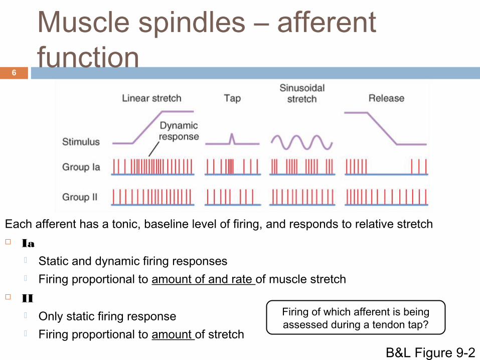
Each afferent has a tonic, baseline level of firing, and responds to relative stretch Ia
Static and dynamic firing responses Firing proportional to amount of and rate of muscle stretch
II Only static firing response Firing proportional to amount of stretch
Firing of which afferent is being assessed during a tendon tap?
B&L Figure 9-2

Muscle spindles – efferent function
7
γ motor neurons maintain sensitivity of spindle over a range of muscle lengths α-γ coactivation
Descending input can change relative dynamic vs. static γ activation to modulate spindle sensitivity
http://www.ncbi.nlm.nih.gov/books/NBK11119/bin/ch16f10.jpg
Activation of only dynamic γ motor neurons
increases responsiveness of _____
afferents
Golgi Tendon Organs (GTOs)8
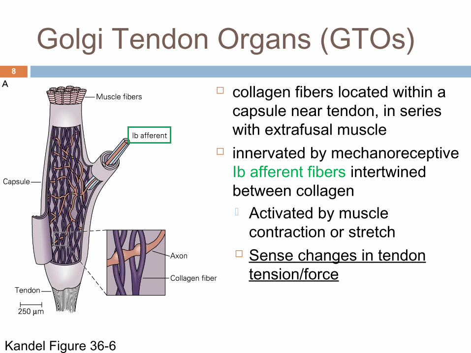
collagen fibers located within a capsule near tendon, in series with extrafusal muscle
innervated by mechanoreceptive Ib afferent fibers intertwined between collagen Activated by muscle
contraction or stretch Sense changes in tendon
tension/force
Kandel Figure 36-6

Reflex organization9
A reflex is a predictable, involuntary and stereotyped response to an eliciting stimulus Can be modulated by stimulus intensity and descending CNS input Testing reflexes is an important clinical tool in assessing neurological
and spinal function

Myotatic or stretch reflex10
Monosynaptic reflex mediated by muscle spindles Contraction in response to lengthening
Reflex arc:1. Muscle stretches2. Ia afferent of muscle spindle increase
firing3. Synapse on α motor neuron and
inhibitory interneuron in spinal cord4. α motor neuron of homonymous muscle
excited, and of antagonist muscle inhibited
5. Homonymous muscle contracts to oppose lengthening, antagonist muscle relaxes
B&L Figure 9-6

Myotatic or stretch reflex11
Stretch reflex has two phases:
Phasic (Ia) phase dynamic change in muscle length (ex. tendon tap) triggers a
transient phasic contraction Physiological importance: reflex contraction prevents
overstretch of extrafusal muscle fiber beyond physiological limits
Clinical importance: tendon tap used to determine integrity of spinal cord at different segmental levels
Myotatic or stretch reflex12
Tonic phase Static stretching of muscles produces a weaker, longer lasting,
tonic contraction Type II afferents also involved
Physiological importance: maintains muscle tone/posture via negative feedback Ex. Soldier standing at attention legs begin to fatigue and flex
quadriceps slowly begin lengthening tonic stretch reflex maintains tone and prevents collapse (to an extent)
Clinical importance: assessing presence of hypertonia Ex. Patients with cerebral palsy have very rigid, tight muscles
resistant to stretch overactive tonic stretch reflex due to upper motor neuron lesion

Motor neuron lesions13
Upper motor neuron lesion of the neural pathway inside the CNS (not including the ventral horn of the spinal cord or motor nuclei of the cranial nerves) stroke, traumatic brain injury or cerebral palsy
Lower motor neuron lesion affects nerve fibers within the ventral horn of the spinal cord travelling to the relevant muscle(s) Nerve trauma, polio
Upper motor neuron lesion
Lower motor neuron lesion
Reflexes Increased, may have pathological reflex signs (Babinski sign)
Decreased,
Muscle tone
Increased, contralateral Decreased, ipsilateral
Weakness Yes, contralateral Yes, ipsilateral

Inverse myotatic or Ib reflex14
Disynaptic reflex mediated by GTOs Relaxation in response to increased
tension Reflex arc:
1. Muscle contracts2. Ib afferent of GTO increase firing3. Synapse on one inhibitory and one
excitatory interneuron4. α motor neuron of homonymous
muscle inhibited, and of antagonist muscle excited
5. Homonymous muscle relaxes to oppose increased force in tendon, antagonist muscle contracts
B&L Figure 9-7
Ib

Inverse myotatic or Ib reflex15
Physiological importance: reflex relaxation thought to prevent excessive force from damaging
muscle tissue. Acts synonymously with the myotatic stretch reflex to maintain posture
and balance
Clinical importance: Clasp knife reflex: seen in patients with upper motor neuron lesions muscle has increased tone and resistance to stretch if sufficient force is applied, limb resistance suddenly decreases thought to be mediated by high threshold firing of GTO afferents (but other receptors may be involved as well)

Flexion withdrawalreflex
16
Polysynaptic reflex mediated by FRAs (flexion reflex afferents: nociceptors, mechanoreceptors etc.) flexion in response to painful stimuli
FRAs synapse on inhibitory and excitatory interneurons which excite ipsilateral flexor motorneurons & inhibit extensor motorneurons
Physiological importance: Rapid flexion away from painful
stimuli Clinical importance: upper motor
neuron lesion impairs flexion reflex pathalogical Babinski sign
B&L Figure 9-8

Upper motor neuron lesion: Babinski sign
17
(Type of flexion reflex) (pathological reflex)

Crossed extension reflex18
occurs in lower limbs as part of reflex arc for flexion reflex
FRAs synapse on interneurons which elicit contralateral limb extension to help maintain balance
Similar neuronal circuits involved in central pattern generators governing locomotion (next lecture)
B&L Figure 9-8

Summary of reflexes19
REFLEX
STIMULUS
(CLINICAL TEST)
RESPONSE
SENSORY RECEPTO
R
SYNAPSES
EFFECT ON
MUSCLEOTHER
EFFECTSFUNCTIO
N
Stretch (Myotatic) Reflex
Rapid Stretch of muscle (test: tap on muscle tendon)
Stretched muscle contracts rapidly (ex. knee jerk)
Muscle Spindle Primary (Ia) and Secondary (II) sensory neurons (tonic phase)
Ia: Mono-synapticII: (tonic phase) monosynaptic and polysnaptic
Excite Homonymous (same muscle)
Also Excite synergist muscles; Inhibit antagonist muscles (Reciprocal Inhibition)
Aid in maintaining posture, counter sudden stretch
Inverse Myotatic Reflex
Large force on tendon (pull on muscle when resisted)
Muscle tension decreases
Golgi Tendon Organ (Ib)
Disynaptic (via interneuron)
Inhibit Homonymous (same muscle)
Also Inhibit synergist muscles; Excite antagonist muscles
Protective, prevent damage to tendon
Flexor Reflex
Sharp, painful stimulus (as in stepping on nail)
Limb is rapidly withdrawn from stimulus
Cutaneous (skin) and pain receptors
Poly-synaptic (via interneuron)
Excite Flexor muscle
Also Inhibit extensor muscle of same limb; Excite extensor muscles and Inhibit flexors of opposite limb (Crossed Extensor Reflex)
Protective, withdraw from painful stimulus; Cross extension aids in maintaining posture when leg is lifted
http://musom.marshall.edu/anatomy/grosshom/spinalreflexes.html

Objectives
After this lecture you should be able to: Compare and contrast the structure and function of
muscle spindles with golgi tendon organs Describe the importance of αγ coactivation Describe the reflex pathway for the myotatic, inverse
myotatic, and flexion reflexes Give an example of a physiological and clinical importance for
each reflex Distinguish between upper and lower motor neuron lesions
20

21
1. Type __________ spindle afferents are responsive to the rate of change of muscle length.
2. A Babisnki sign is often associated with _____________ motor neuron lesions.
3. Relaxation of the quadriceps muscle increases/decreases firing of Ia afferents and increases/decreases firing of Ib afferents.
Test your knowledge