n-(2-hydroxypropyl) methacrylamide based cryogels ... suppl 1/64_s19.pdf · koetting et al. 2015),...
TRANSCRIPT
PHYSIOLOGICAL RESEARCH • ISSN 0862-8408 (print) • ISSN 1802-9973 (online) 2015 Institute of Physiology v.v.i., Academy of Sciences of the Czech Republic, Prague, Czech Republic Fax +420 241 062 164, e-mail: [email protected], www.biomed.cas.cz/physiolres
Physiol. Res. 64 (Suppl. 1): S19-S27, 2015
N-(2-Hydroxypropyl) Methacrylamide Based Cryogels – Synthesis and Biomimetic Modification for Stem Cell Applications A. GOLUNOVA1, J. JAROŠ2,3, V. JURTÍKOVÁ2,3, I. KOTELNIKOV1, J. KOTEK1, H. HLÍDKOVÁ1, L. STREIT4, A. HAMPL2, F. RYPÁČEK1, V. PROKS1 1Institute of Macromolecular Chemistry, Academy of Sciences of the Czech Republic, Prague, Czech Republic, 2Department of Histology and Embryology, Faculty of Medicine, Masaryk University, Brno, Czech Republic, 3International Clinical Research Center – Center of Biomolecular and Cellular Engineering, St. Anne's University Hospital, Brno, Czech Republic, 4Department of Plastic and Aesthetic Surgery, St. Anne University Hospital, Brno, Czech Republic Received June 14, 2015 Accepted July 16, 2015 Summary The design of favorable mechanical properties and suitable surface modifications of hydrogels in order to stimulate specific cell response is a great challenge. N-(2-Hydroxypropyl) methacryl-amide (HPMA) was utilized to form macroporous cryogel scaffolds for stem cell applications. Furthermore, one group of scaffolds was enhanced by copolymerization of HPMA with methacryloyl-GGGRGDS-OH peptide in an effort to integrate biomimetic adhesion sites. The cryogels were characterized by stiffness and equilibrium swelling measurements as well as by scanning electron microscopy. Cell culture experiments were performed with human adipose-derived stem cells and substrates were found completely non-toxic. Moreover, RGDS-enriched cryogels supported cell attachment, spreading and proliferation, so they can be considered suitable for designed aims. Key words Hydrogels • Cryogels • Stem cells • Scaffolds • Mechanical properties Corresponding author V. Proks, Institute of Macromolecular Chemistry, Academy of Sciences of the Czech Republic, Heyrovsky Sq. 2, 162 06 Prague 6, Czech Republic. E-mail: [email protected] Introduction
Hydrogels are three-dimensional hydrophilic polymer networks capable of absorbing high amounts of
water or biological fluids. Due to their swelling capacity and soft consistency, they more closely simulate natural living tissue than any other class of synthetic biomaterial (Ko et al. 2013, Hoffman 2012, Peppas et al. 2000). Since the pioneering soft contact lens application that dates back to 1960 (Wichterle and Lim 1960), these materials have been used as drug delivery systems (Bertz et al, 2013, Xinming et al. 2008), wound-healing matrices (Gong et al. 2013, Heilmann et al. 2013, Li et al. 2011) and for cell cultivation (Cavalcanti et al. 2013, Matricardi et al. 2013, Saha et al. 2008, Studenovska et al. 2008, Reis et al. 2012, Kubinová et al. 2013, Radhakrishnan et al. 2014).
A wide range of polymers and diverse compositions has been used to fabricate hydrogels (Buwalda et al. 2014, Drury and Mooney 2003, Hoffman 2012, Kirschner and Anseth 2013). They can be both natural and synthetic polymers such as acrylamide (Kuyukina et al. 2009, Plieva et al. 2007, Saha et al. 2008), poly(N-isopropylacrylamide) (Buwalda et al. 2014, Koetting et al. 2015), chitosan (Reis et al. 2012), polysaccharides (Li et al. 2011, Matricardi et al. 2013) or collagen (Lewis et al. 2012). A class of polymers based on monomer N-(2-Hydroxypropyl) methacrylamide (HPMA) was utilized as a smart polymer in many applications including tissue engineering, drug delivery and macromolecular cancer therapy due to its considerable advantages, such as non-immunogenicity and non-toxicity. Moreover, a racemic mixture of DL

S20 Golunova et al. Vol. 64 HPMA can be refined through crystallization to give the monomer higher purity (Ulbrich and Subr 2010, Kopecek and Kopeckova 2010). The fundamental requirements for biomaterials intended for stem cell applications consist in the design of favorable mechanical properties and suitable surface modifications to stimulate a specific cell response (Armentano et al. 2010). The greatest challenge is the tuning of mechanical properties of hydrogels in accordance with the desired type of tissue (Reilly and Engler 2010). The correlation between biomaterial stiffness and cell behavior has been demonstrated in in vitro studies of various engineered tissues, e.g. glial and neural tissues (E` ~ 100 Pa) (Saha et al. 2008), muscle (E` ~ 10 kPa) (Discher et al. 2005) and bone tissue (E` > 30 kPa) (Reilly and Engler 2010).
The ways of reaching these aims typically include tuning the material structure by introducing pores and cell binding ligands (Chen et al. 2011, He et al. 2007, Hsieh et al. 2007, Lozinsky et al. 2003, Noppe et al. 2007). In the case of chemically crosslinked hydrogels, a matrix architecture can be created by conducting polymerization in the presence of a porogen (Michalek et al. 2005, Sun et al. 2014). The additional incorporation of biomimetic motifs provides the possibility to stimulate various cell responses. Bioactive molecules derived from extracellular matrix proteins or other functional biomolecules are commonly used to enable e.g. the attachment of cells to material surfaces (Hacker et al. 2003, Kubinová et al. 2010, Rahmany and Van Dyke 2013, Yu et al. 2005). One of the commonly-used “golden standard” ligands is the fibronectin-derived RGD peptide (Adelöw et al. 2008, Ko et al. 2013).
Cryogels are a group of macroporous hydrogels that allow the user to adjust the before-mentioned parameters. Moreover, their preparation is usually low-cost and unpretentious since the material is produced under freezing conditions and crystals of the frozen solvent act as a porogen (He et al. 2007, Hsieh et al., 2007, Lozinsky et al. 2003, Lozinsky 2014, Noppe et al. 2007). The main advantage of this method is the production of gels with a high swelling capacity and a spongy structure while preserving good mechanical stability (Gun'ko et al. 2013, Fromageau et al. 2007, Topuz and Okay 2009).
The present work is focused on investigating the mechanical properties of HPMA cryogels during alteration of the initial reaction mixture composition. For these purposes, the N-(2-Hydroxypropyl) methacrylamide as a monomer and poly(ethylene glycol) dimethacrylate
as a crosslinker were used. To our current knowledge, the HPMA was never yet polymerized as cryogel. Furthermore, the methacryloylated RGDS-peptide was used as a comonomer to enrich materials with biomimetic sites. The functionality of the modified hydrogels was evaluated by seeding and growing adipose-derived stem cells within the gels.
Material and Methods Materials
Tetramethylethylenediamine (TEMED), ascorbic acid, poly(ethylene glycol) dimethacrylate (PEGDMA) and dichloromethane (DCM) were purchased from Sigma-Aldrich (Czech Republic), N-(2-hydroxypropyl) methacrylamide (HPMA) was prepared according to a method mentioned elsewhere (Kopecek and Bazilova 1974) and N-Methacryloyl-NH-GGGRGDSG-OH (RGDS) peptide was synthesized according to a previously-published procedure (Studenovska et al. 2010). The cryogelation was performed in a Ministat 2400 cryostat (Huber, Offenburg, Germany). The sample morphology was evaluated by scanning electron microscopy (SEM) on a Vega Plus TS 5135 (Tescan, Czech Republic). The stiffness of the hydrogels was characterized by measuring their compressive modulus using an Instron 6025R5800 (Norwood, MA, USA) universal tester.
Collagenase Type IV, DMEM (Gibco, Life Technologies, Paisley, UK), FBS, Penicillin/ Streptomycin (PAA Laboratories GmBH, Pasching, Austria) and gelatin were used to cultivate cells.
Acridine orange (A6014, Sigma-Aldrich) and ethidium bromide (46065, Sigma-Aldrich) were used for the biological live/dead analysis. Cell morphology was analysed by staining cytoskeletal actin and chromatin using Phalloidin Rhodamine (R415, Life technologies, Czech Republic) and 4,6-diamidino-2-phenylindole (DAPI, 32670, Sigma-Aldrich, Czech Republic). Microscopy observations were performed using Nikon Eclipse Ti-E system, equipped with an 20× objective (Nikon, Prague, Czech Republic) and a Cool Snap HQ2 CCD camera (Photometrics), controlled by NIS-Elements imaging software (Nikon, Prague, Czech Republic), Confocal Laser Scanning Microscopy system Fluoview500 (Olympus C&S Ltd., Prague, Czech Republic) equipped with an 40× objective for morphological analysis of cells by SEM on a Vega (Tescan Orsay Holding, Brno, Czech Republic).

2015 N-(2-Hydroxypropyl) Methacrylamide Based Cryogels S21
Formation of HPMA based cryogels Cryogel formation was carried out in 5-ml glass
vials. Two types of crygel based on N-(2-hydroxypropyl)methacrylamide (HPMA) were prepared. For the first type, appropriate amounts of HPMA and PEGDMA as a crosslinking were dissolved in Q-water to obtain 7.5 w/v% solutions with different a range between the monomer and the crosslinker. The second type of gel was modified with an RGD-peptide. In this case, methacrylated RGD peptide was used as a comonomer at 3 w/w% for all of the solids and the crosslinker concentration was 2.4 mol%.
A portion of the initial solution was added to each vial; the solution was then purged with nitrogen and brought to a temperature near zero followed by the TEMED and subsequent addition of APS (0.02 g/ml) for a final concentration 2 w/w% of all the solids. The vials were placed in a temperature of -15 °C for 3 h. Then, all vials with frozen solutions were kept at -18 °C for 12 h. After freezing, the samples were thawed at room temperature and rinsed with distilled water. The washed samples were dried in a freeze-dryer until a constant weight was reached. Water uptake measurement
The dried hydrogels were weighed and then swollen at room temperature in Q-water until equilibrium was reached. At least five measurements were performed for each hydrogel sample, and the average weight of the swollen hydrogels was calculated. The value of equilibrium water content was determined using equation 1.
S = ms−md
md
1𝜌𝜌, (1)
where ms - weight of the gel with equilibrium water
content, g; md - weight of the dry gel, g; ρ – solvent density, g/ml.
Scanning electron microscopy
A Vega TS 5135 scanning electron microscope (Tescan, Czech Republic) was employed to investigate the morphology of the hydrogels. Before observation with the electron microscope, the hydrogel samples were sputtered with a Pt layer (SCD 050 vacuum sputter coater; Leica). SEM micrographs were obtained using secondary electron imaging at 30 kV.
Material stiffness evaluation The studied hydrogels were subjected to
compressive testing. The stiffness of the hydrogels was characterized by the compressive modulus of elasticity. Cylindrical specimens having an h/D (height-to-diameter) ratio of ~ 1/3 were subjected to compressive loading using an Instron 6025R5800 universal testing machine. The specimens had a diameter of approximately 16.5 mm and were deformed at a constant test speed of 1.0 mm/min at ambient temperature. During the test, the specimens were immersed in distilled water. The modulus of elasticity was calculated from the linear portion of the stress-strain diagram by dividing the change in stress, σ2 - σ1, by the corresponding change in strain, ε2 = 0.10 minus ε1 = 0.05. Seeding of cells on materials
Adipose-derived stem cells were isolated from adipose tissue by centrifugation and collagenase extraction based on Zuk’s method (Zuk et al. 2002).
Briefly, adipose tissue was digested with 0.1 % Collagenase Type IV for 30 minutes at 37oC. Following enzyme activity neutralization by DMEM-F12 with 10 % FBS, the cells were separated by centrifugation at 600 g. The pellet was re-suspended in a cultivation medium (10 % FBS, 0.5 % Penicillin/Streptomycin in DMEM Glutamax) and propagated for 5 passages in a culture dish coated with 0.01 % gelatin. Thin layers of cryogels were cut into 3 x 3 mm working pieces and washed three times with a 0.1 M phosphate buffer. Adipose-derived stem cells were released from the cultivation plastic dish by trypsin digestion and seeded on cryogels in a 500 µl suspension with a concentration of 6.105 cells/ml and incubated in a 5 % CO2 atmosphere at 37 °C for 24 h. Cell adhesion evaluated by SEM
Hydrogel samples were fixed in 3 % glutaraldehyde (Polysciences, Inc., Warrington, USA) dissolved in a 0.1 M phosphate buffer for 1 h at room temperature. Subsequently, samples were washed three times for 10 min in a 0.1 M phosphate buffer, dehydrated in ascending ethanol grade (30, 50, 70, 80, 96, 100 %) and dried in a Critical Point Dryer (CPD 030, Balzers Union Limited, Balzers, Liechtenstein) using liquid carbon dioxide. Samples were sputtered with gold in a Sputter Coater (SCD 040, Balzers Union Ltd, Balzers, Liechtenstein) and examined by SEM (Vega, Tescan Orsay Holding, Brno). SEM micrographs were obtained using secondary electron imaging at 15 kV.

S22 Golunova et al. Vol. 64 Results and Discussion
All polymerization experiments were carried out at -15 °C. The ammonium persulfate / N,N,N,N-tetra-methylethylenediamine system was used for the initiation. The solvent uptake by the samples was measured in water. The inner structure (morphology) of the samples was analyzed by SEM and the stiffness of the hydrogels was measured by compression testing.
In our previous work, it was shown that mechanical properties, porosity and swelling were dependent on the method of hydrogel preparation. However, due to irradiation, additional crosslinks in the polymer network appeared and there was also a potential influence of the irradiation on the structure of the RGD peptide. In this work, we therefore used HPMA as a monomer, methacryloyl-GGGRGDS-OH peptide as a comonomer, and PEGDMA as a crosslinker. The
substantial initiation system was performed with TEMED/APS to overcome these limitations by using e-beam initiation conditions (Golunova et al. 2015).
The approach of hydrogel functionalization with biomimetic ligands, which was necessary for cell adhesion, was also changed. Previously-used click reactions are complicated in the case of methacrylic comonomer application due to the absence of a water soluble methacrylic derivative with alkyne functionality. Therefore, a water soluble methacrylated RGD-peptide was prepared. The next advantage in this approach was the elimination of click reaction catalyzer, Cu2+. Although we have proven the concentration level of applied Cu2+ nontoxic in contact with cells (Proks et al. 2012, Golunova et al. 2015), it still gives us better options for making biomimetic modifications. The peptide concentration for the specific cell response (e.g. adhesion) was showed to be sufficient by 3 % (w/w) (Studenovska et al. 2010), thus it was applied.
0 1 20
10
20
30
40
50
0 1 20
1
2 B
Equi
llibriu
m w
ater
con
tent
, g/g
Crosslinker concentration, mol%
HPMA HPMA-RGD
A
Mod
ulus
, kPa
Crosslinker concentration, mol%
HPMA HPMA-RGD
Fig. 1. Dependence of the equilibrium water content (A) and compressive modulus (B) on crosslinker concentration. The total monomer concentration was 7.5 wt%, the crosslinker (PEGDMA) concentration was 2.4 mol%, 1.4 mol%, 0.25 mol% respectively and the temperature was -15 °C. In the case of HPMA-RGD gels, the crosslinker concentration was 2.4 mol%. Elasticity and equilibrium water content
The equilibrium water content values decreased with an increasing concentration of crosslinker (Fig. 1A). It can be assumed that a lower crosslinker concentration would lead to fewer crosslinks and allow polymer chains to be more flexible and to be able to swell a higher amount of water. The swelling values of the RGD-modified gel were lower probably due to the extra bonds that could appear between the molecules of the peptides.
Contrary to this, the influence of the crosslinker concentration on stiffness was found to be very low, although the introduction of the peptide into the initial
composition of HPMA gels slightly increased hydrogel stiffness (Fig. 1B). That happened probably due to the extra hydrogel crosslinking between the peptide molecules through electrostatic interaction.
Previously reported poly(acrylamide) gels had a stiffness around 3 kPa (Golunova et al. 2015) and in comparison with the recent results, it can be assumed that the main effect on hydrogel stiffness was given by the main comonomer type. Therefore, we hypothesise, the stiffness regulation can be achieved by variation of the main monomer concentration, combination of different monomers and their ratio.

2015 N-(2-Hydroxypropyl) Methacrylamide Based Cryogels S23
Morphometry of materials by SEM SEM was used to evaluate the porosity of the
cryogels and to examine the effects of the peptide’s incorporation into the pore structure (Fig. 2). Both types of cryogels showed an interconnected macroporous
structure and the diameter of pores was measured in the range of 50-300 µm. It was also observed that the porous structure was not changed if the RGD peptide was used as a comonomer.
Fig. 2. Scanning electron micrographs of hydrogels obtained using different initial compositions. (A) An HPMA-based cryogel without an RGD-peptide and (B) with an RGD-peptide. The porous structure was comparable without any apparent morphological differences. The scale bar is 500 µm. Evaluation of material biocompatibility and cell morphological response
Evaluating the essential characteristics of these scaffolds include an analysis whether the materials and their degradation products are not potentially toxic to cells and subsequently how the cells interact with gels.
Firstly, it is necessary to assess the polymer functionalization with adhesion ligands and their physical contact with cells on the surface, and then we must determine the influence of the material’s stiffness and how this influences cell morphology and distribution.
Fig. 3. Live/dead assay of ADSCs seeded on hydrogels with or without surface modification by the RGD peptide. Viable cells were labeled with acridine orange (green) and dead cells were stained by ethidium bromide (red) and were rarely observed. (A) HPMA-based cryogel without an RGD-peptide and (B) with an RGD peptide. The scale bar is 200 μm.
S24 Golunova et al. Vol. 64
In this work, cells isolated from human adipose tissue were used, because they more closely approximate noncancerous human stem cells and are accessible for applications in clinical practice. Adipose-derived stem cells (ADSC) were homogeneously seeded within polymerized hydrogels with and without surface modification.
Microscopic observations of cells stained with live/dead assay displayed viable ADSCs (green) throughout both types of hydrogel scaffolds after 24 h cultivation, while there was only an occasionally appearance of dead (red) stained cells. Therefore, none of the materials induced cell death, which confirms their non-toxic nature (Fig. 3).
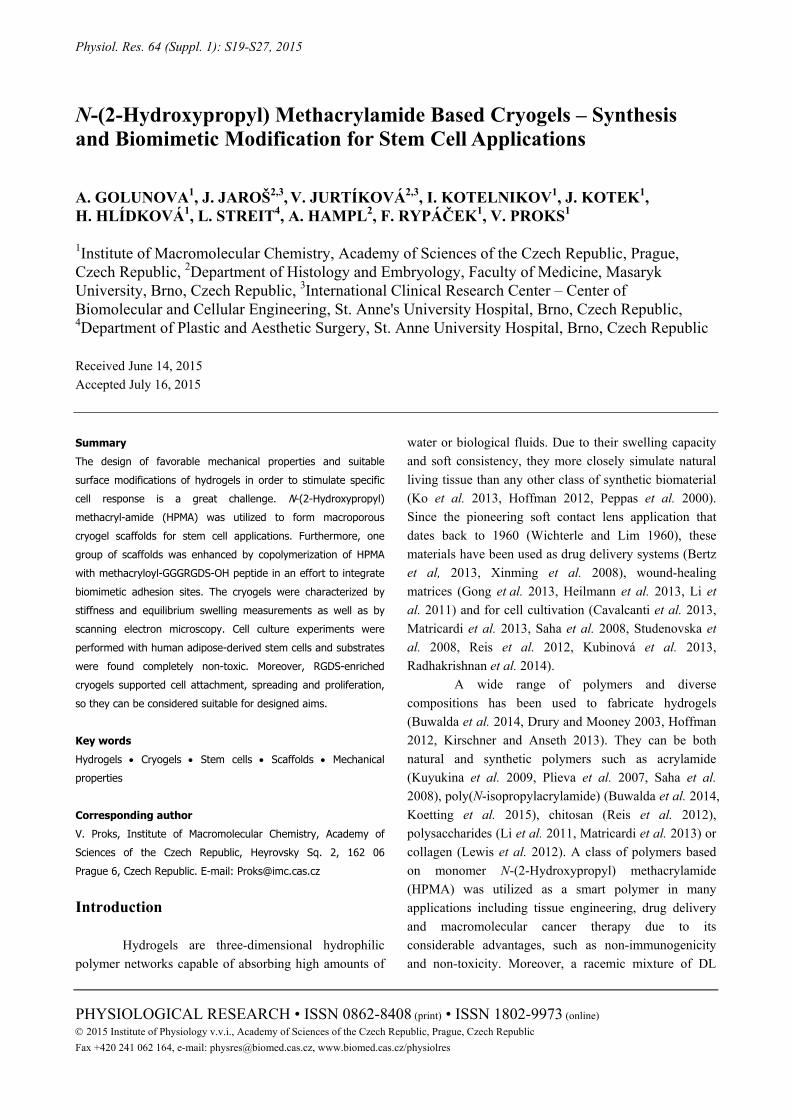
Fig. 4. Actin cytoskeleton (red) and nuclei (blue) staining of human adipose-derived stem cells. ADSCs attached and spread only to RGD modified hydrogels (B), ADSCs encapsulated in unmodified hydrogels (A) formed cell clusters without attaching to any material. The scale bar is 50 μm.
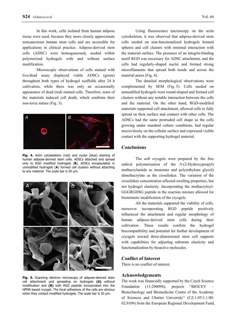
Fig. 5. Scanning electron microscopy of adipose-derived stem cell attachment and spreading on hydrogels (A) without modification and (B) with RGD peptide incorporated into the HPMA based cryogel. The focal adhesions of the cells are obvious when they contact modified hydrogels. The scale bar is 20 μm.
Using fluorescence microscopy on the actin cytoskeleton, it was observed that adipose-derived stem cells seeded on non-functionalized hydrogels formed spheres and cell clusters with minimal interaction with the material surface. The presence of an integrin-binding motif RGD was necessary for ADSC attachment, and the cells had regularly-shaped nuclei and formed strong microfilaments that spread both inside and across the material pores (Fig. 4).
The detailed morphological observations were complemented by SEM (Fig. 5). Cells seeded on unmodified hydrogels were round-shaped and formed cell clusters without any notable interaction between the cells and the material. On the other hand, RGD-modified materials supported cell attachment, allowed cells to fully spread on their surface and connect with other cells. The ADSCs had the same protruded cell shape as the cells growing under standard culture conditions, had regular microvilosity on the cellular surface and expressed visible contact with the supporting hydrogel material. Conclusions
The soft cryogels were prepared by the free radical polymerization of the N-(2-Hydroxypropyl) methacrylamide as monomer and poly(ethylene glycol) dimethacrylate as the crosslinker. The variation of the crosslinker concentration affected swelling properties, but not hydrogel elasticity. Incorporating the methacryloyl-GGGRGDSG peptide in the reaction mixture allowed for biomimetic modification of the cryogels.
All the materials supported the viability of cells, moreover incorporating RGD peptide positively influenced the attachment and regular morphology of human adipose-derived stem cells during their cultivation. These results confirm the hydrogel biocompatibility and potential for further development of cryogels toward three-dimensional stem cell supports with capabilities for adjusting substrate elasticity and functionalization by bioactive molecules. Conflict of Interest There is no conflict of interest. Acknowledgements The work was financially supported by the Czech Science Foundation (13-29009S), projects “BIOCEV – Biotechnology and Biomedicine Centre of the Academy of Sciences and Charles University” (CZ.1.05/1.1.00/ 02.0109) from the European Regional Development Fund,

2015 N-(2-Hydroxypropyl) Methacrylamide Based Cryogels S25
by the FNUSA-ICRC Project (No. CZ.1.05/1.1.00/ 02.0123) from the European Regional Development Fund, “HistoPARK − Centre for analysis and modeling of tissues and organs” (CZ.1.07/2.3.00/20.0185) from the European Social Fund in the Czech Republic and by
funds from the Faculty of Medicine of Masaryk University (MUNI/A/1558/2014). The authors also acknowledge the Charles University in Prague, Department of Physical and Macromolecular Chemistry for the opportunity for doctoral studies.
References ADELÖW C, SEGURA T, HUBBELL J, FREY P: The effect of enzymatically degradable poly(ethylene glycol)
hydrogels on smooth muscle cell phenotype. Biomaterials 29: 314-326, 2008. ARMENTANO I, DOTTORI M, FORTUNATI E, MATTIOLI S, KENNY JM: Biodegradable polymer matrix
nanocomposites for tissue engineering: A review. Polym Degrad Stab 95: 2126-2146, 2010. BERTZ A, WOEHL-BRUHN S, MIETHE S, TIERSCH B, KOETZ J, HUST M, BUNJES H, MENZEL H:
Encapsulation of proteins in hydrogel carrier systems for controlled drug delivery: Influence of network structure and drug size on release rate. J Biotechnol 163: 243-249, 2013.
BUWALDA SJ, BOERE KWM, DIJKSTRA PJ, FEIJEN J, VERMONDEN T, HENNINK W: Hydrogels in a historical perspective: From simple networks to smart materials. J Control Release 190: 254-273, 2014.
CAVALCANTI BN, ZEITLIN BD, NÖR JE: A hydrogel scaffold that maintains viability and supports differentiation of dental pulp stem cells. Dent Mater 29: 97-102, 2013.
DISCHER DE, JANMEY P, WANG YL: Tissue cells feel and respond to the stiffness of their substrate. Science 310: 1139-1143, 2005.
DRURY JL, MOONEY DJ: Hydrogels for tissue engineering: Scaffold design variables and applications. Biomaterials 24: 4337-4351, 2003.
FROMAGEAU J, GENNISSON JL, SCHMITT C, MAURICE RL, MONGRAIN R, CLOUTIER G: Estimation of polyvinyl alcohol cryogel mechanical properties with four ultrasound elastography methods and comparison with gold standard testings. IEEE Trans Ultrason Ferroelectr Freq Control 54: 498-509, 2007.
GOLUNOVA A, CHVÁTIL D, KRIST P, JAROŠ J, JURTÍKOVÁ V. POSPÍŠIL J, KOTELNIKOV I, ABELOVÁ L. KOTEK J, SEDLAČÍK T, KUČKA J, KOUBKOVÁ J, STUDENOVSKÁ H, STREIT L. HAMPL A, RYPÁČEK F, PROKS V: Toward structured macroporous hydrogel composites: Electron beam-initiated polymerization of layered cryogels. Biomacromolecules 16: 1146-1156, 2015.
GONG C, WU Q, WANG Y, ZHANG D, LUO F, ZHAO X, WEI Y, QIAN Z: A biodegradable hydrogel system containing curcumin encapsulated in micelles for cutaneous wound healing. Biomaterials 34: 6377-6387, 2013.
GUN’KO VM, SAVINA IN, MIKHALOVSKY SV: Cryogels: morphological, structural and adsorption characterisation. Adv Colloid Interface Sci 187-188: 1-46, 2013.
HACKER M, TESSMAR J, NEUBAUER M, BLAIMER A, BLUNK T, GOPFERICH A, SCHULZ M: Towards biomimetic scaffolds: Anhydrous scaffold fabrication from biodegradable amine-reactive diblock copolymers. Biomaterials 24: 4459-4473, 2003.
HE X, YAO K, SHEN S, YUN J: Freezing characteristics of acrylamide-based aqueous solution used for the preparation of supermacroporous cryogels via cryo-copolymerization. Chem Eng Sci 62: 1334-1342, 2007.
HEILMANN S, KUECHLER S, WISCHKE C, LENDLEIN A, STEIN C, SCHAEFER-KORTING M: A thermosensitive morphine-containing hydrogel for the treatment of large-scale skin wounds. Int J Pharm 444: 96-102, 2013.
HOFFMAN A.: Hydrogels for Biomedical Applications. Adv Drug Deliv Rev 64: 18-23, 2012. HSIEH CY, TSAI SP, HO MH, WANG DM, LIU CE, HSIEH CH, TSENG HC, HSIEH HJ: Analysis of freeze-
gelation and cross-linking processes for preparing porous chitosan scaffolds. Carbohydr Polym 67: 124-132, 2007.
KIRSCHNER CM, ANSETH KS: Hydrogels in healthcare: from static to dynamic material microenvironments. Acta Mater 61: 931-944, 2013.
KO DY, SHINDE UP, YEON B, JEONG B: Recent progress of in situ formed gels for biomedical applications. Prog Polym Sci 38: 672-701, 2013.

S26 Golunova et al. Vol. 64 KOETTING MC, PETERS JT, STEICHEN SD, PEPPAS N: Stimulus-responsive hydrogels: Theory, modern advances,
and applications. Mater Sci Eng R-Rep 93: 1-49, 2015. KOPECEK J, BAZILOVA H: Poly(N-(2-hydroxypropyl)methacrylamide) - iii. Eur Polym J 10: 465-470, 1974. KOPECEK J, KOPECKOVA P: HPMA copolymers: origins, early developments, present, and future. Adv Drug Deliv
Rev 62: 122-149, 2010. KUBINOVÁ S, HORAK D, HEJCL A, PLICHTA Z, KOTEK J, PROKS V, FOROSTYAK S, SYKOVA E:
SIKVAV-modified highly superporous PHEMA scaffolds with oriented pores for spinal cord injury repair. J Tissue Eng Regen Med 2013, DOI: 10.1002/term.1694.
KUBINOVÁ S, HORÁK D, KOZUBENKO N, VANECEK V, PROKS V, PRICE J, COCKS G, SYKOVÁ E: The use of superporous Ac-CGGASIKVAVS-OH-modified PHEMA scaffolds to promote cell adhesion and the differentiation of human fetal neural precursors. Biomaterials 31: 5966-5975, 2010.
KUYUKINA MS, RUBTSOVA EV, IVSHINA IB, IVANOV RV, LOZINSKY VI: Selective adsorption of hydrocarbon-oxidizing Rhodococcus cells in a column with hydrophobized poly(acrylamide) cryogel. J Microbiol Methods 79: 76-81, 2009.
LI H, YANG J, HU X, LIANG J, FAN Y, ZHANG X: Superabsorbent polysaccharide hydrogels based on pullulan derivate as antibacterial release wound dressing. J Biomed Mater Res Part A 98 A: 31-39, 2011.
LOZINSKY VI, GALAEV IY, PLIEVA FM, SAVINAL IN, JUNGVID H, MATTIASSON B: Polymeric cryogels as promising materials of biotechnological interest. Trends Biotechnol 21: 445-451, 2003.
LOZINSKY V: A brief history of polymeric cryogels. In: Polymeric Cryogels: Macroporous Gels with Remarkable Properties. OKAY O (ed), Springer, Berlin, 2014, pp 1-48.
MATRICARDI P, DI MEO C, COVIELLO T, HENNINK WE, ALHAIQUE F: Interpenetrating polymer networks polysaccharide hydrogels for drug delivery and tissue engineering. Adv Drug Deliv Rev 65: 1172-1187, 2013.
MICHALEK J, PRADNY M, ARTYUKHOV A, SLOUF M. SMETANA K: Macroporous hydrogels based on 2-hydroxyethyl methacrylate J Mater Sci Mater Med 16: 783-786, 2005.
NOPPE W, PLIEVA FM, VANHOORELBEKE K, DECKMYN H, TUNCEL M, TUNCEL A, GALAEV IY, MATTIASSON B: Macroporous monolithic gels, cryogels, with immobilized phages from phage-display library as a new platform for fast development of affinity adsorbent capable of target capture from crude feeds. J Biotechnol 131: 293-299, 2007.
PEPPAS NA, BURES P, LEOBANDUNG W, ICHIKAWA H: Hydrogels in pharmaceutical formulations. Eur J Pharm Biopharm 50: 27-46, 2000.
PLIEVA FM, GALAEV IY, MATTIASSON B: Macroporous gels prepared at subzero temperatures as novel materials for chromatography of particulate-containing fluids and cell culture applications. J Sep Sci 30: 1657-1671, 2007.
PROKS V, JAROŠ J, POP-GEORGIEVSKI O, KUČKA J, POPELKA Š, DVOŘÁK P, HAMPL A, RYPÁČEK F: “Click & seed” approach to the biomimetic modification of material surfaces. Macromol Biosci 12: 1232-1242, 2012.
RADHAKRISHNAN J, KRISHNAN UM, SETHURAMAN S: Hydrogel based injectable scaffolds for cardiac tissue regeneration. Biotechnol Adv 32: 449-461, 2014.
RAHMANY MB, VAN DYKE M: Biomimetic approaches to modulate cellular adhesion in biomaterials: A review. Acta Biomater 9: 5431-5437, 2013.
REILLY GC, ENGLER AJ: Intrinsic extracellular matrix properties regulate stem cell differentiation. J Biomech 43: 55-62, 2010.
REIS L, CHIULLY, LIANG Y, HYUNH K, MOMEN A, RADISIC M: A peptide-modified chitosan-collagen hydrogel for cardiac cell culture and delivery. Acta Biomater 8: 1022-1036, 2012.
SAHA K, KEUNG AJ, IRWIN EF, LI Y, LITTLE L, SCHAFFER DV, HEALY KE: Substrate modulus directs neural stem cell behavior. Biophys J 95: 4426-4438, 2008.
STUDENOVSKA H, SLOUF M, RYPACEK F: Poly(HEMA) hydrogels with controlled pore architecture for tissue regeneration applications J Mater Sci Mater Med 19: 615-621, 2008.

2015 N-(2-Hydroxypropyl) Methacrylamide Based Cryogels S27
STUDENOVSKA H, VODICKA P, PROKS V, HLUCILOVA J, MOTLIK J, RYPACEK F: Synthetic poly (amino acid) hydrogels with incorporated cell-adhesion peptides for tissue engineering. J Tissue Eng Regen Med 4: 454-463, 2010.
SUN J, WEI D, ZHU Y, ZHONG M, ZUO Y, FAN H, ZHANG X: A spatial patternable macroporous hydrogel with cell-affinity domains to enhance cell spreading and differentiation. Biomaterials 35: 4759-4768, 2014.
TOPUZ F, OKAY O: Macroporous hydrogel beads of high toughness and superfast responsivity. React Funct Polym 69: 273-280, 2009.
ULBRICH K, SUBR V: Structural and chemical aspects of HPMA copolymers as drug carriers. Adv Drug Deliv Rev 62: 150-166, 2010.
WICHTERLE O, LIM D: Hydrophilic gels for biological use. Nature 185: 117-118, 1960. XINMING L, YINGDE C, LLOYD AW, MIKHALOVSKY SV, SANDEMAN SR, HOWEL CA, LIEWEN L:
Polymeric hydrogels for novel contact lens-based ophthalmic drug delivery systems: A review. Contact Lens Anterior Eye 31: 57-64, 2008.
YU TT, SHOICHET MS: Guided cell adhesion and outgrowth in peptide-modified channels for neural tissue engineering. Biomaterials 26: 1507-1514, 2005.
ZUK PA, ZHU M, ASHJIAN P, DE UGARTE DA, HUANG JI, MIZUNO H, ALFONSO ZC, FRASER JK, BENHAIM P, HEDRICK MH: Human adipose tissue is a source of multipotent stem cells. Mol Biol Cell 13: 4279-4295, 2002.