nabeshima et al, role of at1 receptor in hepatic steatosis ... · sirna was assessed in an in vitro...
TRANSCRIPT

Nabeshima et al, Role of AT1 receptor in hepatic steatosis 1
Deletion of angiotensin II type I receptor reduces hepatic steatosis
Short title: Role of AT1 receptor in hepatic steatosis
Authors:
Yoshitaka Nabeshima1, Susumu Tazuma
2, Keishi Kanno
2, Hideyuki Hyogo
1, Kazuaki Chayama
1.
1Department of Medicine and Molecular Science, Graduate School of Biomedical Sciences, Hiroshima
University.
2Department of General Medicine, Hiroshima University Hospital.
Correspondence and reprint requests to:
Keishi Kanno, M.D., Ph.D.,
Department of General Medicine, Hiroshima University Hospital,
1-2-3, Kasumi, Minami-ku, Hiroshima, 734-8551, Japan.
Phone: 81-82-257-5461, Fax: 81-82-257-5461; Email: [email protected]
List of Abbreviations
angiotensin II type 1 receptor, AT1R; angiotensin II type 1 receptor blocker, ARB; peroxisome

Nabeshima et al, Role of AT1 receptor in hepatic steatosis 2
proliferator-activated receptor, PPAR; angiotensin, Ang; wild-type, WT; methionine-choline deficient,
MCD; thiobarbituric acid-reactive substances, TBARS; triglyceride, TG; nonalcoholic fatty liver
disease, NAFLD; nonalcoholic steatohepatitis, NASH; hepatic stellate cell, HSC; small interfering RNA,
siRNA; free fatty acid, FFA; phosphate buffered saline, PBS; aspartate aminotransferase, AST; alanine
aminotrasferase, ALT; apolipoprotein B, ApoB; acyl-CoA synthetase, ACSL.
Electronic word count: 3284 words for the main text and 197 words for the abstract
Number of figures/ tables: 6/1

Nabeshima et al, Role of AT1 receptor in hepatic steatosis 3
Abstract
Background/Aims: A distinct subgroup of angiotensin II type 1 receptor (AT1R) blockers
(ARBs) have been reported to suppress the development of hepatic steatosis. These effects were
generally explained by selective peroxisome proliferator-activated receptor (PPAR)γ modulating
properties of ARBs, independent of their AT1R blocking actions. Here, we provide genetic evidence of
the direct role for AT1R in hepatic steatosis.
Methods: The effect of AT1R deletion on steatohepatitis was investigated in AT1a-/- mice.
Furthermore, the influence of AT1R inhibition by telmisartan as well as gene silencing of AT1R by
siRNA was assessed in an in vitro experiment using HepG2 cells.
Results: Compared to wild-type (WT), AT1a-/- mice fed methionine-choline deficient (MCD)
diet resulted in negligible lipid accumulation in the liver with marked induction of PPARα mRNA. In
vitro experiments also demonstrated reduced cellular lipid accumulation by telmisartan and AT1R
knockdown following exposure of long chain fatty acids. This is presumably explained by the
observation that the expression of PPARα and its target genes were significantly up-regulated in specific
siRNA treated HepG2 cells.
Conclusions: Our data indicate, in addition to pharmacological effect of ARBs on PPARγ
activation, a key biological role for AT1R in the regulation of hepatic lipid metabolism.

Nabeshima et al, Role of AT1 receptor in hepatic steatosis 4
Keywords: lipid metabolism, renin-angiotensin system, nonalcoholic fatty liver disease, nonalcoholic
steatohepatitis, peroxisome proliferator-activated receptor α

Nabeshima et al, Role of AT1 receptor in hepatic steatosis 5
1. Introduction
Nonalcoholic fatty liver disease (NAFLD) encompasses a wide spectrum of liver pathology -
from simple steatosis alone, through necroinflammatory disorder referred to as nonalcoholic
steatohepatitis (NASH), to cirrhosis and liver cancer. Because of the high prevalence and the potential
mortality, NAFLD/NASH has emerged as a serious public concern and therefore therapeutic strategies
need to be established. Recent animal studies and clinical trials have demonstrated several
pharmacological treatments as potential therapeutic targets, which include insulin sensitizers (e.g.,
thiazolidinediones, metoformin), lipid lowering agents (e.g., statins, fibrates), antioxidants, angiotensin
(Ang) II type I receptor (AT1R) blockers (ARBs), etc [1-7]. Among these therapeutic options, ARBs
are initially expected to suppress the development of hepatic fibrosis in NASH. Hepatic stellate cell
(HSC), which is a major fibrogenic cell type in the liver and also contributes to hepatic inflammation
through induction of cytokines, expresses AT1R, and blockade of Ang II signaling markedly attenuates
hepatic inflammation and fibrosis in experimental models of chronic liver fibrosis [8,9]. In addition,
clinical report has demonstrated that treatment with losartan improved hepatic necroinflammation and
fibrosis in NASH patients [10].
In addition to anti-fibrotic/inflammatory effect, emerging data have suggested that ARBs
improve glucose and lipid metabolism. In a large clinical trial, losartan substantially lowered the risk

Nabeshima et al, Role of AT1 receptor in hepatic steatosis 6
for type 2 diabetes compared with other antihypertensive therapies [11]. Animal study using obese
Zucker rat has also demonstrated that high dose of irbesartan improved insulin sensitivity [12]. More
recently, it has been shown that telmisartan, unlike other ARBs improved the development of hepatic
steatosis in animal models [7,13,14]. These metabolic effects of ARBs might be explained in part by
the in vitro study, in which a distinct subtype of ARBs induced transcriptional activities of peroxisome
proliferator-activated receptor (PPAR) γ, independent of their AT1R blocking actions [15,16]. However,
it is noteworthy that PPAR γ-activating potency by ARBs appears rather modest as determined by
transcription reporter assays, with median effective concentration (EC50) of telmisartan, the most potent
PPARγ activator among ARBs, 5.02 µmol/L compared to 0.2 µmol/L of pioglitazone, a full agonist of
PPARγ [15,16]. Interestingly, treatment with olmesartan, which has no impact on PPARγ activation,
has been proved to attenuate the development of hepatic steatosis, suggesting the possibility that the
blockade of AT1R itself might contribute to hepatic lipid homeostasis [17]. To further investigate the
direct role for AT1R in hepatic steatosis, we applied to different approaches: 1) animal model of
steatohepatitis using mice lacking AT1aR (AT1a-/-), which is the only Ang II receptor subtype expressed
in rodent liver, and 2) in vitro cellular steatosis model in which gene silencing by RNA interference
targeting AT1R was performed [18]. Our data demonstrated reduced lipid accumulation in the absence
of AT1R with significant induction of PPARα. Apart from PPARγ modulating action of ARBs, these

Nabeshima et al, Role of AT1 receptor in hepatic steatosis 7
data support the potential efficacy of AT1R blockade in the treatment of NAFLD/NASH.

Nabeshima et al, Role of AT1 receptor in hepatic steatosis 8
2. Experimental Procedures
2.1. Animal
AT1a-/- mice were provided by Mitsubishi Tanabe Pharma (Osaka, Japan) and C57BL/6 mice
were obtained from Charles River Laboratories (Yokohama, Japan) [19]. Both strains of the mice have
the same genetic background. The mice were housed in a standard 12 h light/dark cycle facility, and
fed either standard chow diet or methionine-choline deficient (MCD) diet (Oriental Yeast Co., Tokyo,
Japan) for 8 weeks with free access to drinking water. Male mice at 6-8 weeks of age were used in this
study, and all animal procedures were done according to the guideline of Institute of Laboratory Animal
Science, Hiroshima University.
2.2. Histological examination
Liver samples were fixed in 4 % formaldehyde solution, embedded in paraffin, and cut into 5
µm-thick sections. Staining for hematoxylin and eosin (H-E) or Azan-Mallory was carried out with
standard techniques.
2.3. Analytical techniques
Serum triglyceride (TG) concentration was determined enzymatically using a Triglyceride

Nabeshima et al, Role of AT1 receptor in hepatic steatosis 9
E-test (Wako Chemicals, Osaka, Japan). To quantify hepatic TG content, hepatic lipid was extracted as
previously described by Bligh and Dyer and subjected to the same procedure as serum assay followed by
the standardization of protein concentrations [20]. Hepatic thiobarbituric acid-reactive substances
(TBARS) levels were quantified using an OXI-TEK TBARS Assay Kit (Zeprometrix Corporation, New
York) with protein standardization. Serum TBARS levels were assayed using the same kit.
Beta-hydroxybutyrate was assayed using an assay kit (BioVision, Mountain View, CA). The activities
of serum transaminases were determined enzymatically.
2.4. Cell culture and gene silencing by small interfering RNA (siRNA)
HepG2 cells (human hepatoma cell line) were cultured in Dulbecco’s modified Eagle’s
medium supplemented with 10% (v/v) fetal bovine serum. For gene silencing, two different sequences
of small interfering RNA (siRNA) targeting human AT1R were purchased from Sigma-Aldrich (siRNA
ID: SASI_Hs01_00206672, SASI_Hs02_00206672). Cells were transfected using Lipofectamine
RNAiMAX (Invitrogen) in 6-well plates containing 2.5×10⁵ cells in each well with 10 nM of siRNA
duplex. siPerfect Negative Controls (Sigma-Aldrich) was utilized as a negative control siRNA. In the
preliminary experiment, siRNA duplex of SASI_Hs02_00206672 effectively knocked down AT1R
expression compared to SASI_Hs01_00206672, and this was used for the following experiments.

Nabeshima et al, Role of AT1 receptor in hepatic steatosis 10
After 24 h and 48 h of transfection, cells were subjected to gene quantification by real-time PCR and in
vitro steatosis experiment, respectively.
2.5. In vitro model of cellular steatosis
Palmitic acid (C16:0) and oleic acid (C18:1) (Sigma, St. Louis, MO) were dissolved in
isopropanol to obtain 20 or 40 mM stock mixture solution (2:1 oleate: palmitate), and the concentration
of vehicle was 1 % in final incubations [21]. Telmisartan (provided by Boehringer Ingelheim,
Germany) was resolved in DMSO to obtain 10 mM stock solution. To investigate the effect of
telmisartan on cellular steatosis, cells were exposed to 200 or 400 µM of free fatty acids (FFAs) mixture
with or without 2 h preincubation of 10 µM telmisartan. To assess the influence of AT1R knockdown
on cellular steatosis, cells were treated with FFAs after 48 h of siRNA transfection. Following 24 h of
incubation with FFAs, cells were subjected to determination of cellular lipid content by Nile Red assay
and β-hydroxybutyrate levels in the media.
2.6. Nile Red assay
The lipid content in cultured cells was quantified fluorometrically using Nile Red, a vital
lipophilic dye as previously described [22]. Briefly, cell monolayers were washed twice with

Nabeshima et al, Role of AT1 receptor in hepatic steatosis 11
phosphate buffered saline (PBS) followed by fixation with 4 % formaldehyde solution for 15 min, and
washed with PBS twice again. Cells were stained for 30 min with Nile Red solution at a final
concentration of 200 µg/ml in PBS. Monolayers were washed thereafter with PBS and measured
fluorometorically (excitation; 488 nm. emission; 550 nm.) [23].
2.7. Quantitative real-time PCR.
Total RNA was isolated using RNeasy Mini Kit (Qiagen, Germany). cDNA was synthesized
from 1µg of total RNA with GeneAmp Gold RNA PCR Core Kit (Applied Biosystems, Foster City, CA).
To quantify AT1R mRNA expression in human heart and kidney, PCR Ready First Strand cDNA
(BioChain, Hayward, CA) was utilized. Specific primers except AT1R from PrimerBank, a public
resource for PCR primers (http://pga.mgh.harvard.edu/primerbank/, ID: 14043066) were designed using
Primer3 (http://fokker.wi.mit.edu/primer3/input.htm) with nucleotide sequences from GenBankTM as
listed in Table 1. Real-time PCR was carried out with Lightcycler 1.5 system using Lightcycler
FastStart DNA Master plus SYBR Green I (Roche Applied Science). The relative expression levels
were calculated with the formula 2-∆Ct
, where ∆Ct is the difference in threshold cycle (Ct) values
between target gene and ribosomal protein S18 as a control.
Nabeshima et al, Role of AT1 receptor in hepatic steatosis 12
2.8. Western blot analysis for AT1R
Fifty microgram of protein prepared from HepG2 cells using RIPA buffer supplemented with
protease inhibitors (Roche Diagnostics), as well as Total Protein-Human Adult Normal Tissues
(Biochain, Hayward, CA) were fractionated by SDS-PAGE and subjected to Western blot analysis using
rabbit polyclonal antibodies against human AT1R (N-10) (Santa Cruz Biotechnology, Santa Cruz, CA).
The blots were visualized by enhanced chemiluminescence.
2.9. Statistical analysis
The data are expressed as the means±S.E. The statistical analysis was performed using
Student’s t test, and differences were considered statistically significant for a two-tailed p<0.05.

Nabeshima et al, Role of AT1 receptor in hepatic steatosis 13
3. Results
3.1. Mice lacking AT1R are resistant to steatohepatitis
MCD diet has been reported to cause steatohepatitis which represents most of histological
feature of human NASH [24,25]. As shown in Figure 1A, histological analysis of the livers
demonstrated that MCD diet for 8 weeks resulted in apparent steatosis with inflammatory cell infiltration
mainly in portal area in wild-type (WT) mice. However, in contrast to WT mice, AT1a-/- mice displayed
no significant changes in the liver (Figure 1A, right panel). Azan-Mallory staining of the liver from
WT mice revealed mild pericellular fibrosis, which was absent in AT1a-/- mice (Figure 1A, lower panels).
In accordance with these histological observations, hepatic TG content increased by 3-fold in WT and
remained no significant 1.5-fold increase in AT1a-/- mice following 8 weeks of MCD diet (Figure 1B, left
panel). Serum TG level following MCD diet was reduced in both genetic groups with more drastic
change in WT mice (Figure 1B, right panel). The substantial changes in hepatic TG content suggested
the possibility that the expression of PPARα, a central player in hepatic lipid metabolism, might be
influenced by AT1aR expression. This was assessed by quantitative real-time PCR. While MCD diet
did not affect hepatic PPARα expression in both genetic strains, absence of AT1aR was associated with
significant 3-fold increase in PPARα mRNA in chow diet-fed mice (Figure 1C, right panel). Since
PPARα mediates hepatic expression of genes regulating lipid oxidation, we next assessed serum level of

Nabeshima et al, Role of AT1 receptor in hepatic steatosis 14
β-hydroxybutyrate, an end product of hepatic fatty acid oxidation. Figure 1D demonstrates 3-fold
increase in serum β-hydroxybutyrate in AT1a-/- mice compared to WT mice following MCD diet. This
might be a potential explanation for reduced hepatic lipid accumulation in AT1a-/- mice.
3.2. Lack of AT1R ameliorates liver injury
As shown in Figure 2, there were no differences in aspartate aminotransferase (AST) and
alanine aminotrasferase (ALT) levels between WT and AT1a-/- mice when fed chow diet. MCD diet
caused significant increase in AST and ALT levels in WT mice, whereas there was no significant
increase of these liver enzymes in AT1a-/- mice.
3.3. Influence of AT1R expression on oxidative stress
Since lipid peroxidation product plays an important role as a “2nd. hit” in the pathogenesis of
NASH, we next examined serum and hepatic levels of TBARS [26]. Figure 3 demonstrates marked
increase in TBARS levels in serum and liver of WT mice following MCD diet. In contrast, AT1a-/-
mice showed no significant changes in TBARS levels, which presumably reflected the reduction of liver
injury as demonstrated in AST and ALT levels.

Nabeshima et al, Role of AT1 receptor in hepatic steatosis 15
3.4. Expression of AT1R in the liver and HepG2 cells
To further explore the protective effect of AT1R blockade in hepatic steatosis observed in
animal model, we performed in vitro studies using HepG2 cells. We first ascertained AT1R expression
in HepG2 cells comparing that of heart, kidney, and liver tissues, which are known to express functional
AT1R. As displayed in Figure 4, Western blot analysis revealed AT1R protein expression in HepG2
cells, supporting the previous in vivo binding assay of Ang II that detected a homogeneous signal pattern
throughout the liver parenchyma [27]. Although the quantities of loaded protein were same among
samples, the band intensity in HepG2 appeared to be weaker compared to whole liver homogenate,
which comprises nonparenchymal liver cells such as hepatic stellate cells and Kupffer cells. However,
real-time PCR demonstrated similar level of AT1R in HepG2 as compared to kidney (Figure 4, right
panel).
3.5. AT1R blockade reduces lipid accumulation in hepatocellular in vitro model
To examine whether AT1R expression in HepG2 was functional, we utilized in vitro model of
steatosis in which HepG2 cells were treated with FFAs mixture with or without 10 µM of telmisartan.
As shown in Figure 5A, Nile Red assay quantified 4-5-fold increase in cellular lipid accumulation when
cells were treated with FFAs for 24 h in the absence of telmisartan. These changes were markedly
Nabeshima et al, Role of AT1 receptor in hepatic steatosis 16
attenuated by the presence of telmisartan by 60-70 %. To exclude the possibility that these
anti-steatotic effects might be contributable to pharmacological property of telmisartan, we investigated
the influence of AT1R knockdown using siRNA in the same experimental model. As shown in Figure
5B, transfection of HepG2 cells with siRNA targeting AT1R successfully depleted its mRNA expression
by 80% when compared to control siRNA. This siRNA knockdown of AT1R resulted in significant
15-30 % decrease in cellular lipid deposition following FFAs exposure (Figure 5B, right panel).
Compared to the treatment of telmisartan, the influence of AT1R knockdown appeared modest. This
might be because siRNA suppression of AT1R was not sufficient to elicit complete inhibition of the
AT1R signaling pathway. In connection with the observed ketogenesis in AT1a-/- mice, we examined
β-hydroxybutyrate levels in the culture media. Under the condition of 200 µM of FFAs overload, the
treatment of telmisartan as well as AT1R knockdown significantly increased β-hydroxybutyrate levels
(Figure 5C).
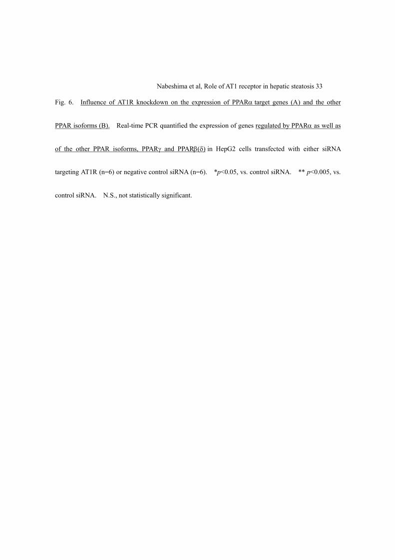
3.6. AT1R knockdown induces PPARa and its target genes
The observation that hepatic PPARα expression was up-regulated in AT1a-/- mice suggests the
possible link between AT1R signaling and PPARα expression. In this setting, our working hypothesis
is that AT1R down-regulation may in turn influence the hepatocellular expressions of PPARα and its

Nabeshima et al, Role of AT1 receptor in hepatic steatosis 17
target genes. As shown in Figure 6A, transient AT1R silencing by siRNA resulted in significant
up-regulation of PPARα by 41%. In addition, PPARα target genes, acyl-CoA synthetase 1 (ACSL1)
and apolipoprotein B (Apo B) 100 were also induced by 27 and 21 %, respectively. These data exclude
the possibility that PPARα up-regulation in AT1a-/- mice might be resulted from chronic adaptation to
the absence of AT1R. To exclude the possible contribution of the other PPAR isoforms in
hepatocellular steatosis, the expression of PPARγ and PPARβ(δ) were assessed by real-time PCR, which
resulted in no significant changes following AT1R depletion (Figure 5B).

Nabeshima et al, Role of AT1 receptor in hepatic steatosis 18
4. Discussion
The present study indicates a major role for AT1R in hepatic steatosis by providing a precise
genetic approach. Homozygous disruption of AT1R in mice resulted in marked reduction of hepatic
lipid accumulation as determined morphologically and by hepatic TG content. Recent studies have
demonstrated that ARBs improve insulin resistance and hyperinsulinemia. Because the improvement
of insulin resistance leads to reduction of FFAs flux to the liver together with increased uptake and
storage of FFAs in adipose tissue, ARBs are expected to reduce hepatic steatosis. However, the effect
of ARBs on hepatic steatosis is still controversial [7,14,17,28-30]. These conflicting findings might be
explained by differences in selective PPARγ modulating properties of ARBs [15,16], or attributed to
differences in the dose and length of ARBs supplementation as well as potential differences in the
mechanisms of steatosis in the different experimental models. Our data reinforces the theory that
blockade of AT1R, unrelated to pharmacological properties of ARBs, ameliorates the development of
hepatic steatosis.
The pathophysiology of NASH remains poorly understood, but is often described as “two-hit
process” consisting of hepatic TG accumulation (the 1st. step) and development of oxidative stress and
proinflammatory cytokines (the 2nd. step), which leads to hepatocyte injury, inflammation, and fibrosis

Nabeshima et al, Role of AT1 receptor in hepatic steatosis 19
[26]. Previous reports have demonstrated that pharmacological blockade or gene deletion of
renin-angiotensin system (RAS) significantly attenuates hepatic fibrosis in experimental animal models
[31,32]. Moreover, Ang II, the effecter peptide for AT1R, has been reported to stimulates an array of
fibrogenic actions in activated HSCs through the mediation of reactive oxygen species [33]. More
recent data has revealed that angiotensin converting enzyme (ACE) deficient (ACE-/-) mice which exhibit
reduced levels of Ang II by 70 % in plasma and by 85-97 % in tissues [34] showed pronounced increase
in hepatic gene expression related to lipolysis and fatty acid oxidation, suggesting close link between
RAS and hepatic lipid metabolism [35]. Taken together with our finding of the anti-steatotic effect, the
blockade of AT1R appears to contribute to wide range of steps and phases in the pathogenesis of NASH.
The up-regulation of PPARα in the liver of AT1a-/- mice is intriguing because PPARα plays a
central role in hepatic lipid homeostasis. It is well known that many of the genes encoding enzymes
involved in the mitochondrial and peroxisomal fatty acid β-oxidation pathways are regulated by PPARα.
To exclude the possibility that adaptation to persistent AT1R absence may induce PPARα expression in
mice, we performed in vitro siRNA experiments, which confirmed induction of PPARα and its target
genes by transient AT1R knockdown. In support of these observations, previous experiments have
demonstrated telmisartan-mediated induction of PPARα [36]. Whereas induction of PPARα was
observed in AT1a-/- mice, the present study lacks the direct evidence that suppressive effect of AT1R
Nabeshima et al, Role of AT1 receptor in hepatic steatosis 20
blockade on hepatic steatosis is mediated by PPARα. In this connection, we examined
β-hydroxybutyrate levels since it is documented that PPARα activation leads to stimulation of
ketogenesis [37,38]. This yielded marked increase in serum β-hydroxybutyrate in AT1a-/- mice. In
addition, our in vitro experiments demonstrated significant increase in β-hydroxybutyrate in the culture
media in response to AT1R blockade. These findings support the detected changes in gene expressions
of PPARα and ACSL1 following AT1R deletion. Although it remains controversial whether the
blockade of AT1R directly stimulates PPARα, up-regulation of PPARα likely increases the sensitivity to
agonist and thus clinical usage of ARBs together with PPARα ligand such as fibrates might have
synergistic effects on hepatic lipid metabolism.
A potential limitation of the present study is the use of MCD dietary animal model. Although
this model develops steatohepatitis morphologically similar to human NASH with increase in oxidative
stress and has been widely used for the study of NASH, absence of insulin resistance has been reported
[24,25,39,40]. Since insulin resistance is considered to be pivotal in the development of NASH, this
model might not entirely reflect the natural course and the etiological background of human NASH
[41,42]. Some other potential dietary models include ad libitum feeding of the high fat diet, which
develops obesity and insulin resistance but not noticeable steatohepatitis [43]. Additionally, intragastric
overfeeding of high fat diet induces NASH pathology, but some of the biological changes observed in

Nabeshima et al, Role of AT1 receptor in hepatic steatosis 21
the liver did not mimic human NASH [44]. Altogether, the study of pathophysiological process of
NASH is limited by the lack of suitable experimental animal models [45]. Interestingly, the effect of
olmesartan has been investigated in a genetically diabetic rat fed MCD diet [30]. In contrast to our
findings, olmesartan reduced hepatic steatosis only in diabetic but not in control rats. In the present
study, MCD diet caused increase in hepatic TG content associated with decrease in serum TG level,
suggesting that impaired TG secretion from liver might contribute to hepatic steatosis. These changes
were blunted in the absence of AT1R, implying improvement of TG secretion in AT1a-/- mice. This
might be in part explained by the observation of in vitro experiment that AT1 knockdown induced Apo B
which is requisite for the formation of very low density lipoprotein (VLDL).
In the present study, human hepatoma cell line HepG2 instead of rodent primary hepatocytes
was utilized for in vitro model of cellular steatosis. This was based on the previous report that human
primary hepatocytes and HepG2 cells reached similar levels of intracellular lipid accumulation close to
that determined in hepatocytes from human steatosis liver [23]. In addition, the observed up-regulation
of hepatic PPARα in AT1a-/- mice led to the speculation that PPARα might play a role in the
anti-steatotic effect of AT1 blockade. Because hepatic expression of PPARα and the sensitivity to it
notably differ among species [46], human-derived cell line was applied.
In conclusion, mice lacking AT1R are resistant to the development of hepatic steatosis with

Nabeshima et al, Role of AT1 receptor in hepatic steatosis 22
up-regulation of PPARα in MCD diet-induced steatohepatitis model. Accordingly, the levels of
TBARS, a marker of oxidative stress, as well as aminotransferases, indicators of liver damage were
significantly attenuated in AT1a-/- mice. In addition, in vitro experiment of hepatocellular steatosis
revealed that blockade of AT1R by telmisartan and specific siRNA knockdown markedly decreased
cellular lipid accumulation. Up-regulation of PPARα was also confirmed by transient AT1R
knockdown. These data provide strong evidence that, in addition to pharmacological effect of ARBs on
PPARγ activation, AT1R signaling is involved in hepatic lipid metabolism.

Nabeshima et al, Role of AT1 receptor in hepatic steatosis 23
References
[1] Belfort R, Harrison SA, Brown K, Darland C, Finch J, Hardies J, et al. A placebo-controlled trial
of pioglitazone in subjects with nonalcoholic steatohepatitis. N Engl J Med 2006;355:2297-2307.
[2] Promrat K, Lutchman G, Uwaifo GI, Freedman RJ, Soza A, Heller T, et al. A pilot study of
pioglitazone treatment for nonalcoholic steatohepatitis. Hepatology 2004;39:188-196.
[3] Marchesini G, Brizi M, Bianchi G, Tomassetti S, Zoli M, Melchionda N. Metformin in
non-alcoholic steatohepatitis. Lancet 2001;358:893-894.
[4] Antonopoulos S, Mikros S, Mylonopoulou M, Kokkoris S, Giannoulis G. Rosuvastatin as a novel
treatment of non-alcoholic fatty liver disease in hyperlipidemic patients. Atherosclerosis
2006;184:233-234.
[5] Basaranoglu M, Acbay O, Sonsuz A. A controlled trial of gemfibrozil in the treatment of patients
with nonalcoholic steatohepatitis. J Hepatol 1999;31:384.
[6] Jin H, Yamamoto N, Uchida K, Terai S, Sakaida I. Telmisartan prevents hepatic fibrosis and
enzyme-altered lesions in liver cirrhosis rat induced by a choline-deficient L-amino acid-defined
diet. Biochem Biophys Res Commun 2007;364:801-807.
[7] Fujita K, Yoneda M, Wada K, Mawatari H, Takahashi H, Kirikoshi H, et al. Telmisartan, an

Nabeshima et al, Role of AT1 receptor in hepatic steatosis 24
angiotensin II type 1 receptor blocker, controls progress of nonalcoholic steatohepatitis in rats. Dig
Dis Sci 2007;52:3455-3464.
[8] Bataller R, Gines P, Nicolas JM, Gorbig MN, Garcia-Ramallo E, Gasull X, et al. Angiotensin II
induces contraction and proliferation of human hepatic stellate cells. Gastroenterology
2000;118:1149-1156.
[9] Kanno K, Tazuma S, Nishioka T, Hyogo H, Chayama K. Angiotensin II participates in hepatic
inflammation and fibrosis through MCP-1 expression. Dig Dis Sci 2005;50:942-948.
[10] Yokohama S, Yoneda M, Haneda M, Okamoto S, Okada M, Aso K, et al. Therapeutic efficacy of
an angiotensin II receptor antagonist in patients with nonalcoholic steatohepatitis. Hepatology
2004;40:1222-1225.
[11] Dahlof B, Devereux RB, Kjeldsen SE, Julius S, Beevers G, de Faire U, et al. Cardiovascular
morbidity and mortality in the Losartan Intervention For Endpoint reduction in hypertension study
(LIFE): a randomised trial against atenolol. Lancet 2002;359:995-1003.
[12] Henriksen EJ, Jacob S, Kinnick TR, Teachey MK, Krekler M. Selective angiotensin II receptor
receptor antagonism reduces insulin resistance in obese Zucker rats. Hypertension
2001;38:884-890.
[13] Sugimoto K, Kazdova L, Qi NR, Hyakukoku M, Kren V, Simakova M, et al. Telmisartan increases

Nabeshima et al, Role of AT1 receptor in hepatic steatosis 25
fatty acid oxidation in skeletal muscle through a peroxisome proliferator-activated
receptor-gamma dependent pathway. J Hypertens 2008;26:1209-1215.
[14] Ibanez P, Solis N, Pizarro M, Aguayo G, Duarte I, Miquel JF, et al. Effect of losartan on early liver
fibrosis development in a rat model of nonalcoholic steatohepatitis. J Gastroenterol Hepatol
2007;22:846-851.
[15] Benson SC, Pershadsingh HA, Ho CI, Chittiboyina A, Desai P, Pravenec M, et al. Identification of
telmisartan as a unique angiotensin II receptor antagonist with selective PPARgamma-modulating
activity. Hypertension 2004;43:993-1002.
[16] Schupp M, Janke J, Clasen R, Unger T, Kintscher U. Angiotensin type 1 receptor blockers induce
peroxisome proliferator-activated receptor-gamma activity. Circulation 2004;109:2054-2057.
[17] Hirose A, Ono M, Saibara T, Nozaki Y, Masuda K, Yoshioka A, et al. Angiotensin II type 1
receptor blocker inhibits fibrosis in rat nonalcoholic steatohepatitis. Hepatology
2007;45:1375-1381.
[18] Burson JM, Aguilera G, Gross KW, Sigmund CD. Differential expression of angiotensin receptor
1A and 1B in mouse. Am J Physiol 1994;267:E260-267.
[19] Sugaya T, Nishimatsu S, Tanimoto K, Takimoto E, Yamagishi T, Imamura K, et al. Angiotensin II
type 1a receptor-deficient mice with hypotension and hyperreninemia. J Biol Chem

Nabeshima et al, Role of AT1 receptor in hepatic steatosis 26
1995;270:18719-18722.
[20] Bligh EG, Dyer WJ. A rapid method of total lipid extraction and purification. Can J Biochem
Physiol 1959;37:911-917.
[21] Malhi H, Bronk SF, Werneburg NW, Gores GJ. Free fatty acids induce JNK-dependent hepatocyte
lipoapoptosis. J Biol Chem 2006;281:12093-12101.
[22] Greenspan P, Fowler SD. Spectrofluorometric studies of the lipid probe, nile red. J Lipid Res
1985;26:781-789.
[23] Gomez-Lechon MJ, Donato MT, Martinez-Romero A, Jimenez N, Castell JV, O'Connor JE. A
human hepatocellular in vitro model to investigate steatosis. Chem Biol Interact
2007;165:106-116.
[24] Tomita K, Oike Y, Teratani T, Taguchi T, Noguchi M, Suzuki T, et al. Hepatic AdipoR2 signaling
plays a protective role against progression of nonalcoholic steatohepatitis in mice. Hepatology
2008;48:458-473.
[25] Igolnikov AC, Green RM. Mice heterozygous for the Mdr2 gene demonstrate decreased PEMT
activity and diminished steatohepatitis on the MCD diet. J Hepatol 2006;44:586-592.
[26] Day CP, James OF. Steatohepatitis: a tale of two "hits"? Gastroenterology 1998;114:842-845.
[27] Paizis G, Cooper ME, Schembri JM, Tikellis C, Burrell LM, Angus PW. Up-regulation of

Nabeshima et al, Role of AT1 receptor in hepatic steatosis 27
components of the renin-angiotensin system in the bile duct-ligated rat liver. Gastroenterology
2002;123:1667-1676.
[28] Ran J, Hirano T, Adachi M. Angiotensin II infusion increases hepatic triglyceride production via
its type 2 receptor in rats. J Hypertens 2005;23:1525-1530.
[29] Sugimoto K, Qi NR, Kazdova L, Pravenec M, Ogihara T, Kurtz TW. Telmisartan but not valsartan
increases caloric expenditure and protects against weight gain and hepatic steatosis. Hypertension
2006;47:1003-1009.
[30] Kurita S, Takamura T, Ota T, Matsuzawa-Nagata N, Kita Y, Uno M, et al. Olmesartan ameliorates
a dietary rat model of non-alcoholic steatohepatitis through its pleiotropic effects. Eur J Pharmacol
2008;588:316-324.
[31] Yoshiji H, Kuriyama S, Yoshii J, Ikenaka Y, Noguchi R, Nakatani T, et al. Angiotensin-II type 1
receptor interaction is a major regulator for liver fibrosis development in rats. Hepatology
2001;34:745-750.
[32] Kanno K, Tazuma S, Chayama K. AT1A-deficient mice show less severe progression of liver
fibrosis induced by CCl(4). Biochem Biophys Res Commun 2003;308:177-183.
[33] Bataller R, Schwabe RF, Choi YH, Yang L, Paik YH, Lindquist J, et al. NADPH oxidase signal
transduces angiotensin II in hepatic stellate cells and is critical in hepatic fibrosis. J Clin Invest

Nabeshima et al, Role of AT1 receptor in hepatic steatosis 28
2003;112:1383-1394.
[34] Campbell DJ, Alexiou T, Xiao HD, Fuchs S, McKinley MJ, Corvol P, et al. Effect of reduced
angiotensin-converting enzyme gene expression and angiotensin-converting enzyme inhibition on
angiotensin and bradykinin peptide levels in mice. Hypertension 2004;43:854-859.
[35] Jayasooriya AP, Mathai ML, Walker LL, Begg DP, Denton DA, Cameron-Smith D, et al. Mice
lacking angiotensin-converting enzyme have increased energy expenditure, with reduced fat mass
and improved glucose clearance. Proc Natl Acad Sci U S A 2008;105:6531-6536.
[36] Clemenz M, Frost N, Schupp M, Caron S, Foryst-Ludwig A, Bohm C, et al. Liver-specific
peroxisome proliferator-activated receptor alpha target gene regulation by the angiotensin type 1
receptor blocker telmisartan. Diabetes 2008;57:1405-1413.
[37] Mandard S, Müller M, Kersten S. Peroxisome proliferator-activated receptor alpha target genes.
Cell Mol Life Sci 2004;61:393-416.
[38] Luci S, Giemsa B, Kluge H, Eder K. Clofibrate causes an upregulation of PPAR-{alpha} target
genes but does not alter expression of SREBP target genes in liver and adipose tissue of pigs. Am
J Physiol Regul Integr Comp Physiol 2007;293:R70-7.
[39] Rinella ME, Green RM. The methionine-choline deficient dietary model of steatohepatitis does
not exhibit insulin resistance. J Hepatol 2004;40:47-51.

Nabeshima et al, Role of AT1 receptor in hepatic steatosis 29
[40] Vetelainen R, van Vliet A, van Gulik TM. Essential pathogenic and metabolic differences in
steatosis induced by choline or methione-choline deficient diets in a rat model. J Gastroenterol
Hepatol 2007;22:1526-1533.
[41] Pagano G, Pacini G, Musso G, Gambino R, Mecca F, Depetris N, et al. Nonalcoholic
steatohepatitis, insulin resistance, and metabolic syndrome: further evidence for an etiologic
association. Hepatology 2002;35:367-72.
[42] Sanyal AJ, Campbell-Sargent C, Mirshahi F, Rizzo WB, Contos MJ, Sterling RK, et al.
Nonalcoholic steatohepatitis: association of insulin resistance and mitochondrial abnormalities.
Gastroenterology 2001;120:1183-92.
[43] Collins S, Martin TL, Surwit RS, Robidoux J. Genetic vulnerability to diet-induced obesity in the
C57BL/6J mouse: physiological and molecular characteristics. Physiol Behav 2004;81:243-248.
[44] Deng QG, She H, Cheng JH, French SW, Koop DR, Xiong S, et al. Steatohepatitis induced by
intragastric overfeeding in mice. Hepatology 2005;42:905-914.
[45] Koteish A, Mae Diehl A. Animal models of steatohepatitis. Best Pract Res Clin Gastroenterol
2002;16:679-690.
[46] Gonzalez FJ, Peters JM, Cattley RC. Mechanism of action of the nongenotoxic peroxisome
proliferators: role of the peroxisome proliferator-activator receptor alpha. J Natl Cancer Inst

Nabeshima et al, Role of AT1 receptor in hepatic steatosis 30
1998;90:1702-9.
Nabeshima et al, Role of AT1 receptor in hepatic steatosis 31
Figure Legends
Fig. 1. The absence of AT1aR expression attenuates the development of MCD diet-induced hepatic
steatosis in mice. A. Liver sections from MCD diet-fed WT (A, C) and AT1a-/- mice (B, D) were
processed for Haematoxylin & Eosin (HE) (upper panels) or Azan-Mallory staining (lower panels).
Hepatic steatosis as well as fibrosis are evident only in WT mice (original magnification ×100). B.
TG concentrations in serum (left panel) and liver (right panel) obtained from WT (open bars) and AT1a-/-
(closed bars) mice (n=6/each group) were determined after feeding either normal chow or MCD diet. C.
Hepatic PPARα mRNA expression was quantified (n=5/each group) by quantitative real-time PCR. D.
The serum level of β-hydroxybutyrate in WT (n=5) and AT1a-/-
(n=6) mice fed MCD diet was
determined. *p<0.05, chow-fed vs. MCD diet-fed mice. †p<0.05, WT vs. AT1a-/- mice.
Fig. 2. Diminished hepatic injury following 8 weeks of MCD diet in the absence of AT1aR. AST and
ALT levels were determined in WT (open bars) and AT1a-/- (closed bars) mice fed either normal chow or
MCD diet for 8 weeks (n=6/group). *p<0.05, chow-fed vs. MCD diet-fed mice. †p<0.05, WT vs.
AT1a-/- mice.
Fig. 3. Influence of AT1aR expression on TBARS levels in serum and liver. Thiobarbituric

Nabeshima et al, Role of AT1 receptor in hepatic steatosis 32
acid-reactive substances (TBARS) levels were assayed in serum (left panel) and liver (right panel) from
WT (open bars) and AT1a-/- (closed bars) mice after feeding either normal chow or MCD diet (n=6/each
group). *p<0.05, chow-fed vs. MCD diet-fed mice. †p<0.05, WT vs. AT1a-/- mice.
Fig. 4. Expressions of AT1R in the liver and HepG2 cells. Western blots of AT1R protein expression
in homogenates from human liver and HepG2 cells by comparison with heart and kidney as positive
tissues for AT1R expression (50KDa) (left panel). Quantitative real-time PCR analysis for AT1R in
HepG2 (right panel). Results represent from at least 3 independent experiments.
Fig. 5. AT1R blockade attenuates cellular steatosis following fatty acids overload. A. Influence of
telmisartan on intracellular steatosis induced by free fatty acids (FFAs) overload (2:1 oleate: palmitate)
(n=6/each group). B. Effect of siRNA targeting AT1R on AT1 mRNA expression (left panel) and
influence of AT1 knockdown (KD) on cellular steatosis induced by FFAs overload (2:1 oleate: palmitate,
right panel) (n=6/each group). C. Influence of AT1R blockade on β-hydroxybutyrate levels in media
(n=6/each group). **p<0.005, vs. control siRNA. *p<0.05, vs. non-treated groups. †p<0.05, AT1R
blockade (either by telmisartan or AT1 knockdown) vs. control (either vehicle or control siRNA) HepG2.
Nabeshima et al, Role of AT1 receptor in hepatic steatosis 33
Fig. 6. Influence of AT1R knockdown on the expression of PPARα target genes (A) and the other
PPAR isoforms (B). Real-time PCR quantified the expression of genes regulated by PPARα as well as
of the other PPAR isoforms, PPARγ and PPARβ(δ) in HepG2 cells transfected with either siRNA
targeting AT1R (n=6) or negative control siRNA (n=6). *p<0.05, vs. control siRNA. ** p<0.005, vs.
control siRNA. N.S., not statistically significant.

Nabeshima et al, Role of AT1 receptor in hepatic steatosis 34
Acknowledgement
The authors thank Dr. Naseem Gaur for helpful discussion. This work was supported by a Grant-in-aid
from the Ministry of Health, Labor and Welfare of Japan to ST. This work was carried out in part at the
Analysis Center of Life Science, Hiroshima University.

Table 1. Primer used for quantitative real-time PCR
Gene Forward Reverse GenBank
accession num.
mPPARα tgcaaacttggacttgaacg tgatgtcacagaacggcttc BC016892
AT1 atccaagatgattgtcccaaagc gcccatagtggcaaagtcagtaa
PPARα cagtggagcattgaacatcg gttgtgtgacatcccgacag NM_001001928
ApoB100 agccttgctgaagaaaacca atgcccctcttgatgttcag M14162
ASCL1 ccagaagggcttcaagactg gccttctctggcttgtcaac NM_001995
RPS18 atagcctttgccatcactgc ggacctggctgtattttcca NM_022551
mPPARα, mouse peroxisome proliferator-activated receptor α; AT1, angiotensin II receptor type I; PPARα, human
PPARα; ApoB100, apolipoprotein B-100; ACSL1, acyl-CoA synthetase long-chain family member 1; RPS18,
ribosomal protein S18
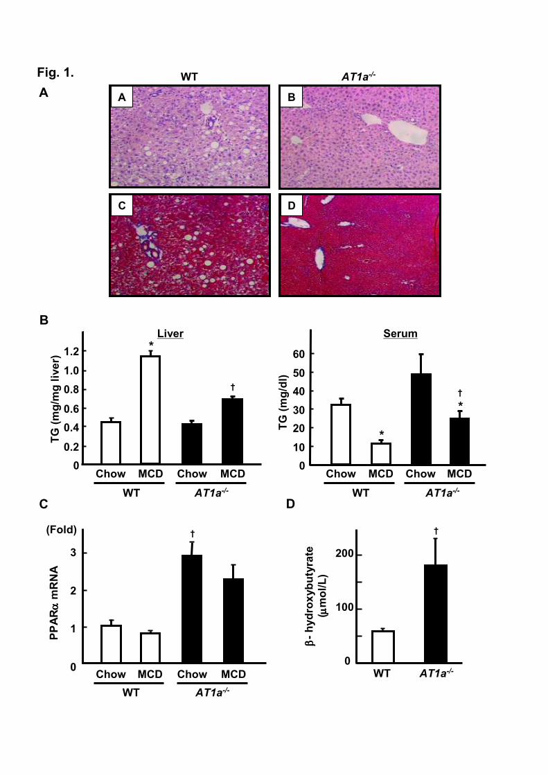
Fig. 1.
A B
C D
AT1a-/-WT
A
B
0
10
20
30
40
50
60
TG (mg/dl)
Serum
AT1a-/-WT
Chow MCDChow MCD
*
*
†
0
0.2
0.4
0.6
0.8
1.0
1.2
Liver
WT
Chow MCDChow MCD
AT1a-/-
*
†
C
AT1a-/-WT
0
1
2
3
Chow MCDChow MCD
PPARαα ααmRNA
(Fold)
0
100
200
AT1a-/-WT
ββ ββ-hydroxybutyrate
( µµ µµmol/L)
† †
D
TG (mg/mg liver)

Fig. 2.
0
100
200
300
400
500
600
AST (IU
/L)
AT1a-/-WT
Chow MCDChow MCD
*
0
10
20
30
40
50
60
ALT (IU
/L)
WT
Chow MCDChow MCD
*
AT1a-/-
†
†

Fig. 3.
0
0.5
1
1.5
2
2.5
Serum
AT1a-/-WT
Chow MCDChow MCD
TBARS (n
mol/m
l) *
†
0
1
2
3
4
5
6Liver
TBARS (n
mol/m
g p
rote
in)
WT
Chow MCDChow MCD
AT1a-/-
*
†

MW Heart Kidney LiverHepG2
50KDa
0
1
2
3
4
5
Heart Kidney HepG2
AT1 m
RNA (fold
)
Fig. 4.
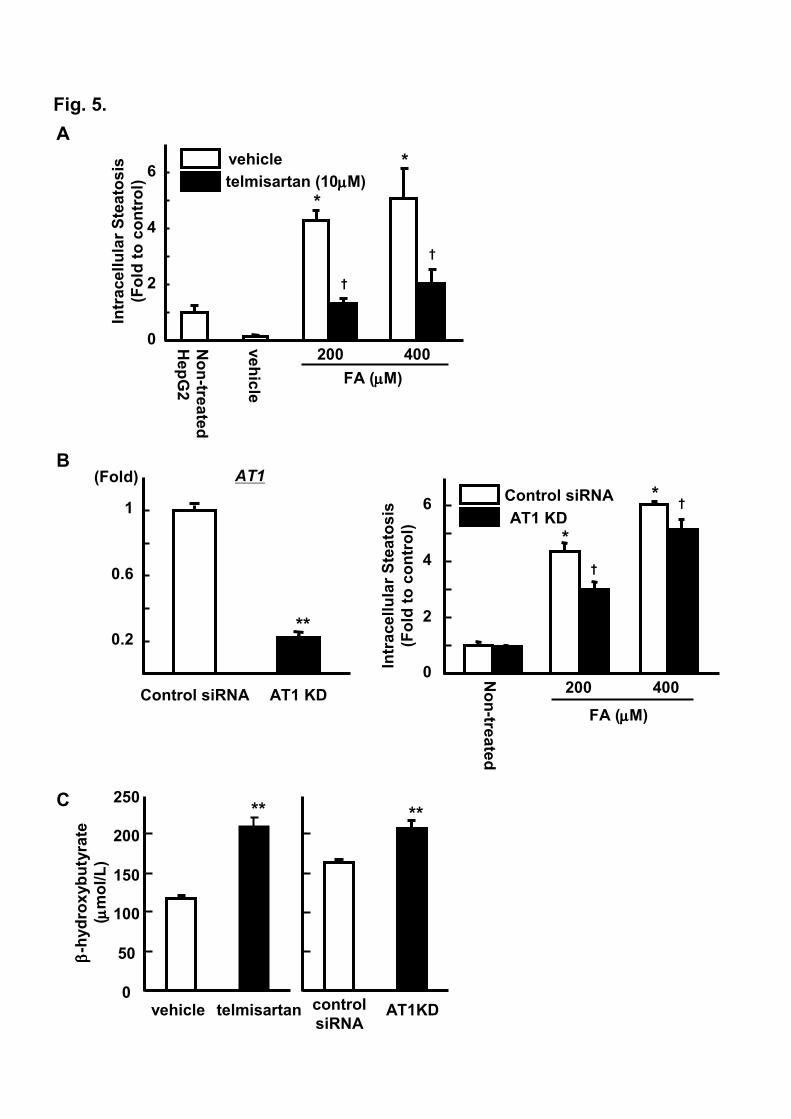
Fig. 5.
A
B
C
0
2
4
6
Intracellular Steatosis
(Fold to control)
Non-tre
ated
FA (µµµµM)
200 400
Control siRNA
AT1 KD
*
*†
†
Non-tre
ated
HepG2
0
2
4
6
Intracellular Steatosis
(Fold to control)
vehicle
FA (µµµµM)
200 400
*
*
†
†
telmisartan (10µµµµM)
vehicle
Control siRNA AT1 KD
0.2
0.6
1
(Fold) AT1
**
0
50
100
150
200
250
ββ ββ-hydroxybutyrate
( µµ µµmol/L)
vehicle telmisartan control
siRNAAT1KD
****

Fig. 6.
0.2
0.6
1
1.4
PPARαααα
Control siRNA AT1 KD
(Fold)**
0.2
0.6
1
(Fold) ApoB100
Control siRNA AT1 KD
*
0.2
0.6
1
1.4
ACSL1
Control siRNA AT1 KD
*
(Fold)
A
B
0.2
0.6
1
1.4
Control siRNA AT1 KD
(Fold) PPARββββ(δ)(δ)(δ)(δ) N.S.
p=0.183
0.2
0.6
1
(Fold) PPARγγγγ
N.S.
p=0.059
Control siRNA AT1 KD