neurophysiology - sinoe medical association tmsinoemedicalassociation.org/ap/neurophysiology.pdf ·...
TRANSCRIPT
Neurophysiology
Danil Danil HammoudiHammoudi.MD.MD

ACTION POTENTIAL

An action potential is a wave of electrical discharge that travels along the membrane of a cell.
Action potentials are an essential feature of animal life, rapidly carrying information within and between tissues.
Action potentials can be created by many types of cells, but are used most extensively by the nervous system for communication between neurons and to transmit information from neurons to other body tissues such as muscles and glands.Action potentials are an essential carrier of the neural code. They provide rapid centralized control and coordination of organs and tissues and may constrain the sizes of evolving anatomies.

GLOSSARY•A voltage, or difference in electrostatic potential.
• Electrophysiology is the study of the electrical properties of biological cells and tissues. It involves measurements of voltage change or electrical current flow on a wide variety of scales from single ion channel proteins, to whole tissues like the heart. In neuroscience, it includes measurements of the electrical activity of neurons, and particularly action potential activity.
•a threshold is a fixed location or value where an abrupt change is observed. an action potential is initiated if the membrane potential is depolarized to the threshold potential.
•The resting potential of a cell is the membrane potential that would be maintained if there were no action potentials, synaptic potentials, or other active changes in the membrane potential. In most cells the resting potential has a negative value, which by convention means that there is excess negative charge inside compared to outside. The resting potential is mostly determined by the concentrations of the ions in the fluids on both sides of the cell membrane and the ion transport proteins that are in the cell membrane. How the concentrations of ions and the membrane transport proteins influence the value of the resting potential is outlined below.

Electricity Definitions
Voltage (V) – measure of potential energy generated by separated charge
Potential difference – voltage measured between two points
Current (I) – the flow of electrical charge between two points
Resistance (R) – hindrance to charge flow
Insulator – substance with high electrical resistance
Conductor – substance with low electrical resistance

Electrical Current and the Body
Reflects the flow of ions rather than electrons
There is a potential on either side of membranes when:
The number of ions is different across the membrane
The membrane provides a resistance to ion flow

Types of plasma membrane ion channels:
Passive, or leakage, channels – always openChemically gated channels – open with binding of a specific neurotransmitter
Voltage-gated channels – open and close in response to membrane potential
Mechanically gated channels – open and close in response to physical deformation of receptors
Operation of a Gated Channel
Example: Na+-K+ gated channel
Closed when a neurotransmitter is not bound to the extracellular receptor
Na+ cannot enter the cell and K+ cannot exit the cell
Open when a neurotransmitter is attached to the receptor
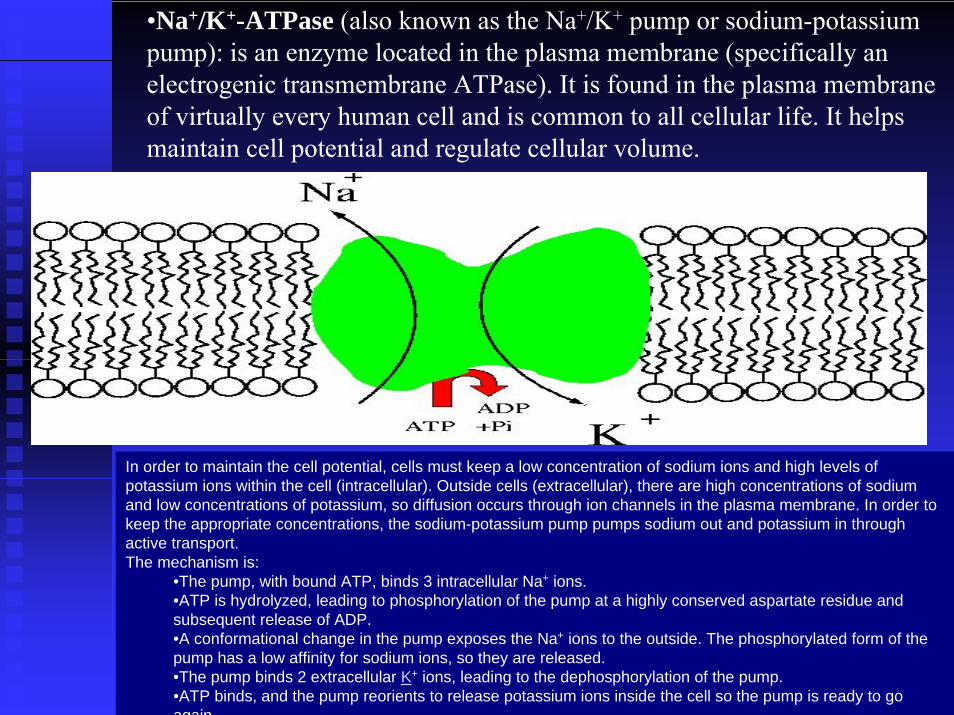
•Na+/K+-ATPase (also known as the Na+/K+ pump or sodium-potassium pump): is an enzyme located in the plasma membrane (specifically anelectrogenic transmembrane ATPase). It is found in the plasma membrane of virtually every human cell and is common to all cellular life. It helps maintain cell potential and regulate cellular volume.
In order to maintain the cell potential, cells must keep a low concentration of sodium ions and high levels of potassium ions within the cell (intracellular). Outside cells (extracellular), there are high concentrations of sodium and low concentrations of potassium, so diffusion occurs through ion channels in the plasma membrane. In order to keep the appropriate concentrations, the sodium-potassium pump pumps sodium out and potassium in through active transport.The mechanism is:
•The pump, with bound ATP, binds 3 intracellular Na+ ions.•ATP is hydrolyzed, leading to phosphorylation of the pump at a highly conserved aspartate residue and subsequent release of ADP.•A conformational change in the pump exposes the Na+ ions to the outside. The phosphorylated form of the pump has a low affinity for sodium ions, so they are released.•The pump binds 2 extracellular K+ ions, leading to the dephosphorylation of the pump.•ATP binds, and the pump reorients to release potassium ions inside the cell so the pump is ready to go again
The voltage of an inactive cell stays at a negative value (inside relative to outside the cell) and varies little.
When the membrane of an excitable cell is depolarized beyond a threshold, the cell will
undergo (or "fire") an action potential, often called a "spike"
An action potential is a rapid swing in the polarity of the voltage from negative to positive and back, the entire cycle lasting a few milliseconds.
Each cycle—and therefore each action potential—has a rising phase, a falling phase, and finally an undershoot [SEE NEXT SLIDE GRAPH]


Figure 11.6

Figure 11.7

Figure 11.8

Figure 11.9

Figure 11.10

Figure 11.11
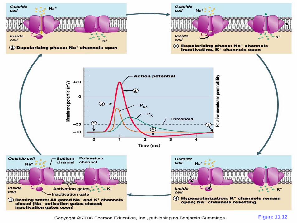
Figure 11.12

Figure 11.13

Figure 11.14

Figure 11.16

Figure 11.17a

The potential difference that exists across the membrane of all cells is usually negative inside the cell with respect to the outside. The membrane is said to be polarized. The potential difference across the membrane at rest is called the resting potential and is approximately -70 mV in neurons, with the negative sign indicating that the inside of the cell is negative with respect to the outside. The establishment of this potential difference involves several factors, most importantly the transport of ions across the cell membrane and the selective permeability of the membrane to these ions.The active transport of potassium and sodium ions into and out of the cell, respectively, is accomplished by a number of sodium-potassium pumps scattered across the cell membrane. Each pump transports two ions of potassium into the cell for every three ions of sodium pumped out. This establishes a particular distribution of positively charged ions across the cell membrane, with more sodium present outside the cell than inside, and more potassium inside the cell than outside. In some situations, the electrogenic sodium-potassium pumps make a significant contribution to the resting membrane potential, but in most cells there are potassium leakage channels that dominate the value of the resting potential.The natural tendency of sodium and potassium ions is to diffuse across their electrochemical gradients to attempt to reach their respective equilibrium potentials, with sodium diffusing into the cell and potassium diffusing out. However, the resting cell membrane is approximately 100 times more permeable to potassium than to sodium, so that more potassium diffuses out of the cell than sodium diffuses in. This permeability to potassium is due to potassium leak channels that are always open. As a result, the dominant outward leak of potassium ions produces a hyperpolarizing current that establishes the cell's resting potential of roughly -70 mV.Like the resting potential, action potentials depend upon the permeability of the cell membrane to sodium and potassium ions.

PhasesThe sequence of events that underly the action potential are outlined below:Resting potentialAt resting potential some potassium leak channels are open but the voltage-gated sodium channels are closed. Potassium diffusing down the potassium concentration gradient creates a negative-inside membrane potential.StimulationA local membrane depolarization caused by an excitatory stimulus causes some voltage-gated sodium channels in the neuron cell surface membrane to open and therefore sodium ions diffuse in through the channels along their electrochemical gradient. Being positively charged, they begin a reversal in the potential difference across the membrane from negative-inside to positive-inside. Initially, the inward movement of sodium ions is also favored by the negative-inside membrane potential.Rising phaseAs sodium ions enter and the membrane potential becomes less negative, more sodium channels open, causing an even greater influx of sodium ions. This is an example of positive feedback. As more sodium channels open, the sodium current dominates over the potassium leak current and the membrane potential becomes positive inside.PeakEstablishment of a membrane potential of around +40 mV closes the voltage-sensitive inactivation gates of the sodium channels, which are sensitive to the now-positive membrane potential gradient, preventing further influx of sodium. While this occurs, the voltage-sensitive activation gates on the voltage-gated potassium channels begin to open.Falling phaseAs voltage-gated potassium channels open, there is a large outward movement of potassium ions driven by the potassium concentration gradient and initially favored by the positive-inside electrical gradient. As potassium ions diffuse out, this movement of positive charge causes a reversal of the membrane potential to negative-inside and repolarization of the neuron back towards the large negative-inside resting potential.UndershootClosing of voltage-gated potassium channels is both voltage- and time-dependent. As potassium exits the cell, the resulting membrane repolarization initiates the closing of voltage-gated potassium channels. These channels do not close immediately in response to a change in membrane potential. Rather, voltage-gated potassium channels (also called delayed rectifier potassium channels) is delayed. As a result, potassium continues to flow out of the cell even after the membrane has fully repolarized. Thus the membrane potential dips below the normal resting membrane potential of the cell for a brief moment; this dip of hyperpolarization is known as the undershoot.

SYNAPSE
The junction between the axon terminals of a neuron and the receiving cell is called a synapse.
Synapses at muscle fibers are also called neuromuscular junctions or myoneural junctions.

•Action potentials travel down the axon of the neuron to its end(s), the axon terminal(s). •Each axon terminal is swollen forming a synaptic knob. •The synaptic knob is filled with membrane-bounded vesicles containing a neurotransmitter. •Arrival of an action potential at the synaptic knob opens Ca2+ channels in the plasma membrane. •The influx of Ca2+ triggers the exocytosis of some of the vesicles. •Their neurotransmitter is released into the synaptic cleft. •The neurotransmitter molecules bind to receptors on the postsynaptic membrane. •These receptors are ligand-gated ion channels.

The neurotransmitter at excitatory synapses depolarizes the postsynaptic membrane (of a neuron in this diagram).Example: acetylcholine (ACh)
•Binding of acetylcholine to its receptors on the postsynaptic cell opens up ligand-gated sodium channels. •These allow an influx of Na+ ions, reducing the membrane potential. •This reduced membrane potential is called an excitatory postsynaptic potential or EPSP. •If depolarization of the postsynaptic membrane reaches threshold, an action potential is generated in the postsynaptic cell.
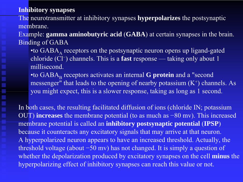
Inhibitory synapsesThe neurotransmitter at inhibitory synapses hyperpolarizes the postsynaptic membrane.Example: gamma aminobutyric acid (GABA) at certain synapses in the brain. Binding of GABA
•to GABAA receptors on the postsynaptic neuron opens up ligand-gated chloride (Cl−) channels. This is a fast response — taking only about 1 millisecond. •to GABAB receptors activates an internal G protein and a "second messenger" that leads to the opening of nearby potassium (K+) channels. As you might expect, this is a slower response, taking as long as 1 second.
In both cases, the resulting facilitated diffusion of ions (chloride IN; potassium OUT) increases the membrane potential (to as much as −80 mv). This increased membrane potential is called an inhibitory postsynaptic potential (IPSP) because it counteracts any excitatory signals that may arrive at that neuron. A hyperpolarized neuron appears to have an increased threshold. Actually, the threshold voltage (about −50 mv) has not changed. It is simply a question of whether the depolarization produced by excitatory synapses on the cell minus the hyperpolarizing effect of inhibitory synapses can reach this value or not.

•a presynaptic ending that contains neurotransmitters, mitochondria and other cell organelles,
•a postsynaptic ending that contains receptor sites for neurotransmitters
•a synaptic cleft or space between the presynapticand postsynaptic endings. It is about 20nm wide.
•Synapses consist of:•presynaptic ending (where neurotransmitters are made)•post synaptic ending (has neuroreceptors in the membrane)•synaptic cleft
•Action potentials cannot cross the synaptic cleft
•Nerve impulse is carried by neurotransmitters

1. At the end of the pre-synaptic neurone there are voltage-gated calcium channels. When an action potential reaches the synapse these channels open, causing calcium ions to flow into the cell.
2. These calcium ions cause the synaptic vesicles to fuse with the cell membrane, releasing their contents (the neurotransmitter chemicals) by exocytosis.
3. The neurotransmitters diffuse across the synaptic cleft.4. The neurotransmitter binds to the neuroreceptors in the post-
synaptic membrane, causing the channels to open. In the example shown these are sodium channels, so sodium ions flow in.
5. This causes a depolarisation of the post-synaptic cell membrane, which may initiate an action potential, if the threshold is reached.
6. The neurotransmitter is broken down by a specific enzyme in the synaptic cleft; for example the enzyme acetylcholinesterasebreaks down the neurotransmitter acetylcholine. The breakdown products are absorbed by the pre-synaptic neuroneby endocytosis and used to re-synthesise more neurotransmitter, using energy from the mitochondria. This stops the synapse being permanently on.
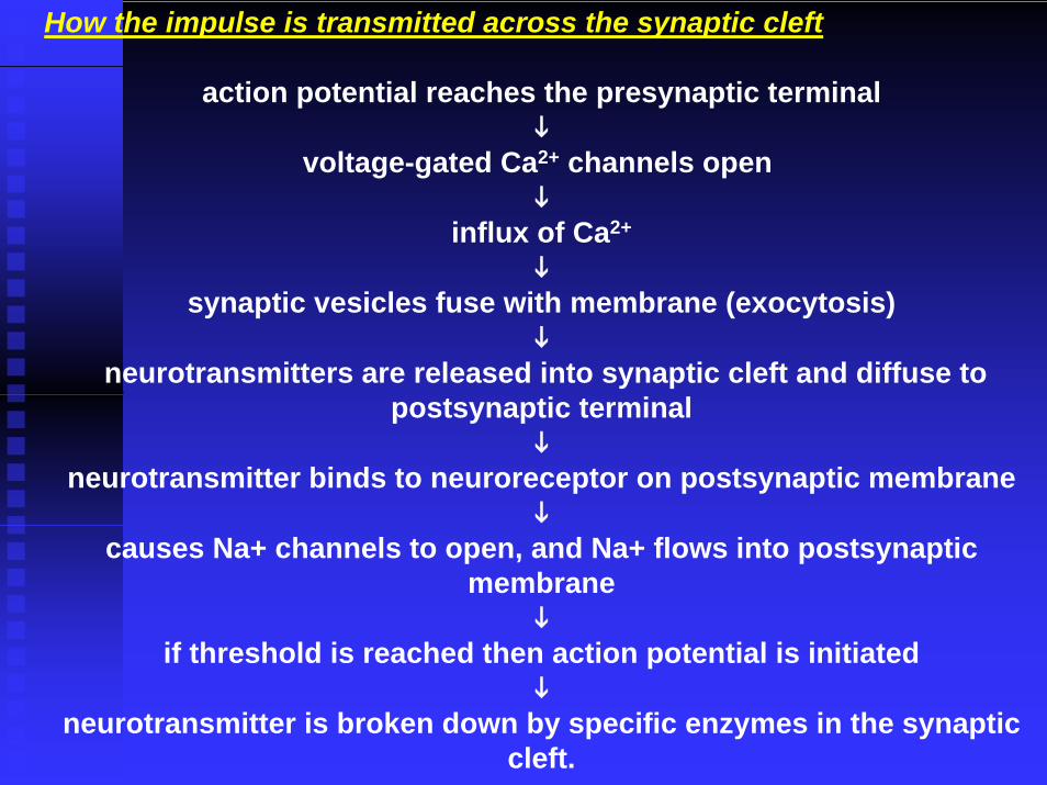
How the impulse is transmitted across the synaptic cleft
action potential reaches the presynaptic terminal
voltage-gated Ca2+ channels open
influx of Ca2+
synaptic vesicles fuse with membrane (exocytosis)
neurotransmitters are released into synaptic cleft and diffuse to postsynaptic terminal
neurotransmitter binds to neuroreceptor on postsynaptic membrane
causes Na+ channels to open, and Na+ flows into postsynaptic membrane
if threshold is reached then action potential is initiated
neurotransmitter is broken down by specific enzymes in the synaptic cleft.

Different types of synapsesDifferent types of synapses
Excitatory ion channel synapses - neuroreceptors are Na+ channels. When Na+ channels open, local depolarisaition occurs, if threshold is reached then action potential is initated
inhibitory ion channels - neuroreceptors are Cl- channels. When Cl-channels open, hyperpolarisation occurs, making action potential less likely
Non channel synapses - neuroreceptors are membrane-bound enzymes. When activated, they catalyse the 'messenger chemical', which in turn can affect the sensitivity of the ion channel receptors in the cell
Neuromuscular junctionsNeuromuscular junctions - synapses formed between motorneurones and muscle cells. Always use the neurotransmitteracetylchline, and are always excitatoryElectrical synapses - the membranes of the two cells actually touch and they chare proteins. The action potential can pass directly from one membrane to the next


Figure 11.16

Figure 11.17a

Figure 11.17b
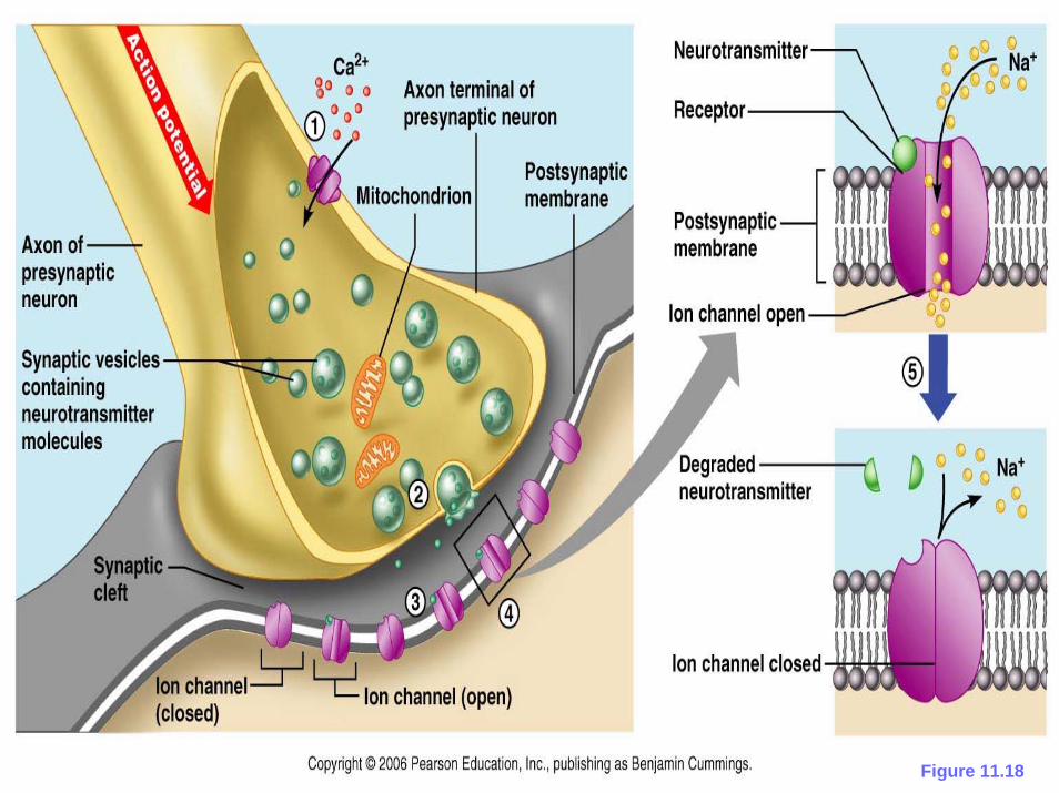
Figure 11.18