novel cross-talk within the ikk family controls innate
TRANSCRIPT
HAL Id: hal-00560693https://hal.archives-ouvertes.fr/hal-00560693
Submitted on 29 Jan 2011
HAL is a multi-disciplinary open accessarchive for the deposit and dissemination of sci-entific research documents, whether they are pub-lished or not. The documents may come fromteaching and research institutions in France orabroad, or from public or private research centers.
L’archive ouverte pluridisciplinaire HAL, estdestinée au dépôt et à la diffusion de documentsscientifiques de niveau recherche, publiés ou non,émanant des établissements d’enseignement et derecherche français ou étrangers, des laboratoirespublics ou privés.
Novel cross-talk within the IKK family controls innateimmunity
Kristopher Clark, Mark Peggie, Lorna Plater, Ronald J Sorcek, Erick RrYoung, Jeffrey B Madwed, Joanne Hough, Edward G Mciver, Philip Cohen
To cite this version:Kristopher Clark, Mark Peggie, Lorna Plater, Ronald J Sorcek, Erick Rr Young, et al.. Novel cross-talk within the IKK family controls innate immunity. Biochemical Journal, Portland Press, 2011, 434(1), pp.93-104. �10.1042/BJ20101701�. �hal-00560693�
Revised manuscript # BJ2010/1701
1
NOVEL CROSS-TALK WITHIN THE IKK FAMILY CONTROLS INNATE IMMUNITY
Kristopher Clark*, Mark Peggie*, Lorna Plater*, Ronald J. Sorcek†, Erick R. R. Young†, Jeffrey B. Madwed‡1, Joanne Hough§, Edward G. McIver§ and Philip Cohen*2
*MRC Protein Phosphorylation Unit, College of Life Sciences, Sir James Black Centre, University of Dundee, Dundee, DD1 5EH, Scotland, UK; †Department of Medicinal Chemistry, ‡Department of Cardiometabolic Disease Research, Boehringer Ingelheim Pharmaceuticals, Inc., 900 Ridgebury Rd./ P.O. Box 368, Ridgefield, 06877-0368 CT, USA §MRC Technology, 1-3 Burtonhole Lane, Mill Hill, London, NW7 1AD, UK. 1Present address: Merck Research Laboratories, 126 E. Lincoln Avenue/P.O. Box 2000, Rahway, 07065-0900, NJ, USA
2Address correspondence to Philip Cohen. Tel: +44-1382-384238; Fax: +44-1382-223778; E-mail: [email protected]
Running Title: Novel cross-talk between IKK family members
Abstract Members of the I�B kinase (IKK) family play a central role in innate immunity by inducing NF�B- and IRF-dependent gene transcription programmes required for the production of pro-inflammatory cytokines and interferons. However, the molecular mechanisms that activate these protein kinases and their complement of physiological substrates remain poorly defined. Using MRT67307, a novel inhibitor of IKK�/TBK1 and BI605906, a novel inhibitor of IKK�, we demonstrate that two different signalling pathways participate in the activation of the IKK-related protein kinases by ligands that activate the IL-1, TLR3 and TLR4 receptors. One signalling pathway is mediated by the canonical IKKs, which directly phosphorylate and activate IKK� and TBK1, whereas the second pathway appears to culminate in the autocatalytic activation of the IKK-related kinases. In contrast, the TNF�-induced activation of the IKK-related kinases is mediated solely by the canonical IKKs. In turn, the IKK-related kinases phosphorylate the catalytic subunits of the canonical IKKs and their regulatory subunit NEMO, which is associated with reduced IKK�/� activity and NF�B-dependent gene transcription. We also show that the canonical IKKs and the IKK-related kinases not only have unique physiological substrates, such as I�B�� p105 and RelA (IKK� and IKK�� and IRF3 (IKK� and TBK1), but also have several substrates in common, including the catalytic and regulatory (NEMO and TANK) subunits of the IKKs themselves. Taken together, our studies reveal that the canonical IKKs and the IKK-related kinases regulate each other by an intricate network involving phosphorylation of their catalytic and regulatory (NEMO, TANK) subunits to balance their activities during innate immunity.
Biochemical Journal Immediate Publication. Published on 07 Dec 2010 as manuscript BJ20101701T
HIS
IS N
OT
TH
E V
ER
SIO
N O
F R
EC
OR
D -
see
doi
:10.
1042
/BJ2
0101
701
Acce
pted
Man
uscr
ipt
Licenced copy. Copying is not permitted, except with prior permission and as allowed by law.
© 2010 The Authors Journal compilation © 2010 Portland Press Limited

Revised manuscript # BJ2010/1701
2
IntroductionThe production of inflammatory mediators in the innate immune system is triggered by the activation of a number of signalling pathways, including those that activate members of the I�B kinase (IKK) family of protein kinases. Four I�B kinases have been identified, which can be divided into the canonical IKKs, IKK� and IKK���and the IKK-related kinases, IKK� and TANK-Binding Kinase 1 (TBK1) [1]. The canonical IKKs activate the transcription factor NF�B, and hence NF�B-dependent gene transcription, by phosphorylating the inhibitor of NF�B � (I�B�� and other I�B isoforms. Phosphorylation of I�B�licences it for K48-linked polyubiquitylation by SCF�TRCP and subsequent destruction by the proteasome. This releases the NF�B subunits RelA and c-Rel, which translocate to the nucleus and stimulate the transcription of genes encoding many inflammatory mediators. In contrast, IKK� and TBK1 phosphorylate Interferon Regulatory Factor 3 (IRF3) leading to nuclear translocation and transcription of genes that include interferon (IFN) ���reviewed in [2]).
The Interleukin-1 Receptor (IL-1R) and every TLR, except for TLR3, signals via the adaptor protein MyD88 to activate the canonical IKKs. It is established that IL-1 cannot activate the canonical IKKs in mouse embryonic fibroblasts (MEFs) that do not express TRAF6 or do not express TGF�-activated protein kinase 1 (TAK1) [3-5]. The E3 ubiquitin ligase activity of TRAF6 is required for signalling [6], and is thought to be needed to generate K63-linked polyubiquitin (K63-pUb) chains, which bind to the TAB2 and TAB3 subunits of TAK1 [7, 8]. This interaction is reported to induce a conformational change that allows TAK1 to phosphorylate and activate itself in vitro [8, 9]. K63-pUb chains also interact with NEMO (also called IKK), the regulatory component of the canonical IKK complex [10, 11]. K63-pUb chains may therefore recruit and co-localise TAK1 and IKK����� in cells, facilitating the direct phosphorylation and activation of the canonical IKKs by TAK1. However, NEMO also binds linear pUb chains as well as K63-pUb chains [12, 13] and LUBAC, the E3 ligase that produces linear pUb chains specifically in vitro [14], appears to participate in the IL-1 stimulated activation of the canonical IKK complex, since activation by IL-1 was impaired in MEFs deficient in HOIL-1, a component of LUBAC [15]. Thus the relative importance of K63-pUb chains versus linear pUb chains in activating the canonical IKK complex requires further investigation.
The activation of the IKK-related protein kinases requires their phosphorylation at Ser172 [16] and these kinases are a point of convergence of TRIF-dependent signalling pathways that are activated by TLR3 and TLR4 agonists [2]. The IKK-related kinases then phosphorylate and activate IRF3. Recently, we showed that BX795, a potent inhibitor of the IKK-related kinases that suppresses the phosphorylation of IRF3 and the production of IFN-� in macrophages, enhances the phosphorylation of IKK� and TBK1 at Ser172 [17]. These findings indicated that a distinct protein kinase must be involved in the activation of the IKK-related kinases by poly(I:C) and LPS and that IKK�/TBK1 control a feedback loop that limits the extent to which they can be activated.
Although, the IKK-related kinases are widely believed to be activated solely by ligands that lead to the activation of IRF3 and the production of IFN�, we found that IL-1 and TNF� activated IKK� and TBK1 in MEFs without inducing the phosphorylation of IRF3 or IRF3-dependent gene transcription [17]. These findings raised the question of how these different agonists (IL-1, TNF�, LPS and poly(I:C)) activate the IKK-related kinases and the roles they play that are independent of IRF3. Since the specificity of signalling can frequently break down when components of signal transduction pathways are over-expressed, we decided to use a genetic and a pharmacological approach to study the activation of the endogenous IKK complexes. Our results demonstrate that MyD88-dependent (IL-1) and TRIF-dependent agonists (LPS, poly(I:C)), use two signalling pathways to activate the IKK-related kinases, one of which is mediated by the canonical IKKs. In contrast, TNF� only employs the canonical IKK-mediated pathway to activate the IKK-related kinases. We further show that the IKK-related kinases negatively regulate the canonical IKKs, and identify several proteins that are physiological substrates for all the IKK family members. Our results reveal a complex interlocking network by which the IKK family members regulate one another to ensure that their activities are tightly coupled.
Biochemical Journal Immediate Publication. Published on 07 Dec 2010 as manuscript BJ20101701T
HIS
IS N
OT
TH
E V
ER
SIO
N O
F R
EC
OR
D -
see
doi
:10.
1042
/BJ2
0101
701
Acce
pted
Man
uscr
ipt
Licenced copy. Copying is not permitted, except with prior permission and as allowed by law.
© 2010 The Authors Journal compilation © 2010 Portland Press Limited
Revised manuscript # BJ2010/1701
3
Materials and Methods
MaterialsThe chemical synthesis of MRT67307 and BI605906 will be described elsewhere. 5Z-7-oxozeaenol was purchased from BioAustralis Fine Chemicals. The Tpl2 inhibitor called Compound-1 (C-1) was synthesized by Dr Natalia Shpiro in the MRC Protein Phosphorylation Unit [18]. These pharmacological inhibitors were dissolved in DMSO and stored as 10 mM solutions at -20oC. Mouse IL-1� and TNF�were purchased from Sigma, Poly(I:C) was from Invivogen and LPS (E. coli strain O5:B55) from Alexis Biochemicals.
DNA constructsDNA vectors expressing GST-IRF3, FLAG-TBK1[K38A], GST-TBK1 and GST-IKK� have been described [17]. IKK� (NCBI O15111) was amplified from IMAGE EST 5275799 using KOD Hot Start DNA Polymerase (Novagen), cloned into pSC-b (Stratagene) and sequenced to completion. The insert was excised using NotI and inserted into pEBG6P to generate a DNA vectors expressing GST-IKK�. IKK�(NCBI AF080158) was cloned in a similar manner using IMAGE EST 5784717. TANK (NCBI AAH67779) and NEMO (NCBI AAH50612) were amplified from IMAGE EST 5296558 and IMAGE EST 2820134, respectively, and cloned into the BamHI and NotI sites of pCMVHA-1. Point mutations were created using the QuikChange mutagenesis kit (Stratagene), but using KOD Hot Start DNA Polymerase. PRDII (NF�B) elements from the IFN-� promoter cloned into the pLuc-MCS vector were a kind gift from Katherine Fitzgerald (University of Massachusetts, USA). pTK-RL was obtained from Stratagene.
AntibodiesAntibodies against human TBK1 (Sheep S041C, bleed 2) and the C-terminal peptide of mouse IKK�(NRLIERLHRVPSAPDV) (Sheep S277C, bleed 2), which were used to immunoprecipitate TBK1 and IKK�, respectively [17], and an antibody raised against the Ser172-phosphorylation site of IKK�(CEKFVS*VYGTE, where S* indicates phosphoserine) (Sheep S051C, bleed 2), which was used to immunoprecipitate the phosphorylated forms of both IKK� and TBK1 [17], anti-NEMO (S527C, bleed 2) and anti-GST were provided by the Division of Signal Transduction Therapy, University of Dundee) have been described, The following antibodies were used for immunoblotting:- HRP-conjugated secondary antibodies (Pierce), anti-FLAG, anti-IKK� (Sigma), anti-HA (Roche), anti-RelA, anti-TRAF3, anti-TRAF6, anti-NEMO (Santa Cruz), anti-IKK� (Upstate) anti-GAPDH, anti-TBK1, anti-IKK�, anti-p38�MAP kinase, anti-TANK and anti-I�B� (Cell Signaling Technology). Antibodies recognizing phosphorylated Ser933 (pSer933) of p105 (NF�B1), pSer176/180 of IKK� and pSer177/Ser181 of IKK�,pSer468 of RelA, pSer536 of RelA, pSer396 of IRF3 and the pThr-Gly-pTyr sequence of ERK1/2 and p38 MAP kinases were also from Cell Signaling Technology. The antibody recognizing the pThr-Pro-pTyr sequence of JNK1/2 was from Biosource, while that recognizing p-Ser172 of TBK1 was from Becton Dickinson.
Cell culture HEK293 cells stably expressing TLR3-FLAG (termed HEK293-TLR3 cells) were provided by Katherine Fitzgerald (University of Massachusetts, Worcester, USA). Immortalised MEFs from mice expressing a truncated, inactive form of TAK1 (Shizuo Akira, Osaka University, Japan), immortalised MEFs from IKK�-deficient and IKK�-deficient mice (Inder Verma, Salk Institute, La Jolla, USA), immortalised MEFs from TRAF6-deficient mice (Tak Mak, Toronto, Canada), Immortalised MEFs from TRAF3 knockout mice (Genhong Chen, UCLA, Los Angeles, USA) and immortalised MEFs from NEMO-deficient mice (Manolis Pasparakis, University of Cologne, Germany) were generous gifts from these investigators. HEK293-TLR3, RAW264.7 cells and MEFs were maintained in DMEM supplemented with 2 mM glutamine, 10% foetal calf serum and the antibiotics penicillin and streptomycin. HEK293 cells
Biochemical Journal Immediate Publication. Published on 07 Dec 2010 as manuscript BJ20101701T
HIS
IS N
OT
TH
E V
ER
SIO
N O
F R
EC
OR
D -
see
doi
:10.
1042
/BJ2
0101
701
Acce
pted
Man
uscr
ipt
Licenced copy. Copying is not permitted, except with prior permission and as allowed by law.
© 2010 The Authors Journal compilation © 2010 Portland Press Limited
Revised manuscript # BJ2010/1701
4
were grown in suspension in Pro-293S media supplemented with 2% foetal calf serum in a humidified incubator maintained at 37oC and 8% CO2. Bone-marrow derived macrophages (BMDM) were generated from mice as described [19]. HEK293 cells were transfected using polyethylenimine (PEI; Polysciences) [20], while immortalized MEFs were transfected using the Amaxa Nucleofection MEF2 kit using vectors described in supplemental information.
Immunoprecipitation and immunoblotting Pharmacological inhibitors dissolved in DMSO, or an equivalent volume of DMSO for control incubations, were added to the culture medium of cells grown as monolayers. After 1 h at 37oC, the cells were stimulated with LPS, poly(I:C), IL-1� or TNF� as described in the figure legends. Thereafter, the cells were rinsed in ice-cold PBS and extracted in lysis buffer (50 mM Tris/HCl pH 7.4, 1 mM EDTA, 1 mM EGTA, 50 mM NaF, 5 mM sodium pyrophosphate, 10 mM sodium �-glycerol 1-phosphate, 1 mM dithiothreitol, 1 mM sodium orthovanadate, 0.27 M sucrose, 1% (v/v) Triton X-100, 1 mg/ml aprotinin, 1 mg/ml leupeptin and 1 mM phenylmethylsulphonyl fluoride). Cell extracts were clarified by centrifugation at 14000 x g for 10 min at 4oC and protein concentrations determined using the Bradford assay. For immunoprecipitation of the phosphorylated forms of IKK� and TBK1, 1 mg of cell extract protein was incubated with 5 g of antibody for 90 min at 4oC followed by the addition of Protein G-Sepharose. After mixing for 15 min at 4oC and brief centrifugation, the immunocomplexes were washed three times in lysis buffer, denatured in SDS and subjected to SDS-PAGE. To detect proteins in cell lysates, 20 g of protein extract was separated by SDS-PAGE. After transfer to PVDF membranes, proteins were detected by immunoblotting and visualized by treating the blots with ECL (Amersham) followed by autoradiography. Antibodies used for immunoprecipitation and immunoblotting are described in supplemental information.
Purification of recombinant protein kinases Protein kinases were expressed as GST-fusion proteins for 48 h in HEK293 cells (0.5 L) grown in suspension. Cells were extracted in 25 ml lysis buffer, clarified by centrifugation (14000 x g for 10 min at 4oC) and protein concentrations determined using the Bradford assay. GST-fusion proteins were isolated from 200 mg of extract protein by affinity chromatography on glutathione-Sepharose beads (1 ml lacked volume) and after washing three times with lysis buffer containing 0.5M NaCl were released from GST and glutathione-Sepharose by cleavage with PreScission protease. Specific activities of the kinases were determined as described [17, 21].
Protein kinase assaysSubstrates and kinases were diluted in 50 mM Tris-HCl pH 7.5, 0.1% (v/v) 2-mercaptoethanol, 0.1 mM EGTA, 10 mM magnesium acetate. Reactions were initiated with [-32P]ATP (2500 cpm/pmol) to a final concentration of 0.1 mM and terminated after 15 min at 30oC by the addition of SDS and EDTA pH 7.0 to final concentrations of 1.0% (w/v) and 20 mM, respectively. After heating for 5 min at 100oC and separation by SDS-PAGE, the phosphorylated proteins were detected by autoradiography.
Phosphomapping by LC-MS/MS Colloidal Coomassie Blue-stained protein bands were excised and digested with trypsin. Protein phosphorylation site analysis was performed by precursor 79 scanning on a 4000 Q-Trap mass spectrometer system [22] and on an LTQ-orbitrap velos [23].
Statistical analysis Quantitative data are presented as the mean ± SEM. Statistical significance of differences between experimental groups was assessed using the Student’s t test. Differences in means were considered significant if p<0.05.
Biochemical Journal Immediate Publication. Published on 07 Dec 2010 as manuscript BJ20101701T
HIS
IS N
OT
TH
E V
ER
SIO
N O
F R
EC
OR
D -
see
doi
:10.
1042
/BJ2
0101
701
Acce
pted
Man
uscr
ipt
Licenced copy. Copying is not permitted, except with prior permission and as allowed by law.
© 2010 The Authors Journal compilation © 2010 Portland Press Limited
Revised manuscript # BJ2010/1701
5
Results
Characterisation of novel pharmacological inhibitors of the IKK family In earlier studies, we identified BX795 as a potent inhibitor of the IKK-related kinases (IKK� and TBK1) [17, 21] that suppressed the LPS or poly(I:C)-stimulated phosphorylation of IRF3 and production of IFN�,without inhibiting the activation of NF�B by the canonical IKKs [17]. However, BX795 did suppress the activation of JNK and p38� MAP kinase by inflammatory stimuli, an off target effect which limited its use as a probe for the functions of IKK� and TBK1. We therefore modified BX795 to generate the improved inhibitor MRT67307 (Fig. 1A), which inhibited IKK� and TBK1 with IC50 values of 19 nM and 160 nM when assayed at 0.1 mM ATP in vitro, but did not inhibit IKK� or IKK� even at 10 M.MRT67307 prevented the phosphorylation of IRF3 and the production of IFN� in macrophages (Figs. 1B and 1C) but no longer suppressed the activation of JNK or p38 MAPK by poly(I:C) (Fig. 1B) or other agonists (see later figures).
In the present study we also used BI605906 (Fig. 1D), an inhibitor of IKK� with improved selectivity. This compound inhibited IKK� in vitro with an IC50 value 380 nM when assayed at 0.1 mM ATP. The only other protein kinase that was inhibited of over 100 tested, which included IKK�, IKK� and TBK1, was the IGF1 receptor (IC50=7.6 M).
Identification of signaling pathways that mediate the IL-1-stimulated activation of IKK�����To investigate the signalling pathways by which IL-1 activates the different IKK subfamily members, we studied their activation in MEFs that do not express TRAF6 or TAK1, both of which are required for the IL-1-stimulated activation of the canonical IKKs. These studies showed that, like IKK� and IKK�, IKK�and TBK1 could not be activated by IL-1 in TRAF6-/- MEFs (Fig. 2A). In contrast, all the IKK family members were activated by IL-1 in TRAF3-/- MEFs (Fig. 2B). However, and in contrast to the canonical IKKs, IL-1 activated the IKK-related kinases similarly in TAK1-/- and wild type MEFs (Fig. 2C). Thus the IL-1-dependent pathways that activate the canonical IKKs and the IKK-related kinases diverge “downstream” of TRAF6. We therefore expected that the IKK-related kinases would be activated normally in MEFs that do not express NEMO, an essential regulatory component of the canonical IKKs. However, there was no activation of either the canonical IKKs or the IKK-related kinases in NEMO-/- MEFs, despite unimpaired activation of p38 MAP kinase and JNK (Fig. 2D). These observations led us to investigate the role of the canonical IKK complex in the activation of the IKK-related kinases.
Although IKK� plays a major role in the IL-1-stimulated phosphorylation of proteins, such as p105 (also called NF�B1) and the NF�B subunit RelA in MEFs, IKK��also contributes to the phosphorylation of these substrates (Fig. S1A). Therefore, as no specific inhibitors of IKK� are currently available, we studied the effects of the IKK� inhibitor BI605906 in IKK�-/- MEFs in the presence and absence of the IKK����� inhibitor MRT67307. These studies revealed that BI605906 partially inhibited the IL-1-stimulated activation of IKK�����, while MRT67307 had little effect. However, the combined addition of both inhibitors completely prevented activation of the IKK-related kinases by IL-1 (Fig. 3A). These results suggested that two different pathways contribute to the IL-1-induced activation of the IKK-related kinases in IKK�-/- MEFs, one dependent on IKK� activity and the other dependent on IKK������activity. Since IL-1 cannot activate the canonical IKKs in TAK1-deficient MEFs (Fig. 2C), we reasoned that the IL-1-stimulated activation of the IKK-related kinases should be completely blocked by the IKK����� inhibitor MRT67307 in these cells, which indeed proved to be the case (Fig. 3B).
The activation of the IKK-related kinases by the canonical IKKs could be mediated either directly or indirectly via another intervening protein kinase. Unequivocal evidence for a direct phosphorylation of IKK� and TBK1 by the canonical IKKs was obtained by reconstituting the kinase reaction in vitro using purified proteins. Incubation of purified IKK� and IKK� with recombinant catalytically inactive mutants of IKK� and TBK1 led to the phosphorylation of Ser172 within the activation loop of the IKK-related kinases (Fig. 3C). Similar results were obtained in co-transfection experiments (Fig. S1B).
Biochemical Journal Immediate Publication. Published on 07 Dec 2010 as manuscript BJ20101701T
HIS
IS N
OT
TH
E V
ER
SIO
N O
F R
EC
OR
D -
see
doi
:10.
1042
/BJ2
0101
701
Acce
pted
Man
uscr
ipt
Licenced copy. Copying is not permitted, except with prior permission and as allowed by law.
© 2010 The Authors Journal compilation © 2010 Portland Press Limited
Revised manuscript # BJ2010/1701
6
Inhibition of the canonical IKKs by IKK�����We noticed that MRT67307 not only failed to suppress the IL-1-stimulated phosphorylation of p105 at Ser933, but actually enhanced phosphorylation in IKK�-/- MEFs (Fig. 3A), suggesting that the IKK-related kinases might negatively regulate IKK�. To investigate this point, we studied the phosphorylation of RelA. The IL-1-stimulated phosphorylation of RelA at Ser468 was prevented by the IKK� inhibitor BI605906 in IKK�-/- MEFs, while MRT67307 enhanced phosphorylation (Fig. 3A). Similar results were obtained in wild type MEFs, where MRT67307 enhanced the IL-1-stimulated phosphorylation of p105 at Ser933 and RelA at both Ser468 and Ser536 (Fig. 3D). MRT67307 also enhanced IL-1-stimulated activation of NF�B-dependent gene transcription in wild type MEFs (Fig. 3E), again indicating that the IKK-related protein kinases function as negative regulators of IKK� in MEFs.
IL-1-stimulation decreased the electrophoretic mobility of IKK� in IKK�-/- MEFs as a result of phosphorylation (Fig. S1C), which was partially suppressed by BI605906, partially suppressed by MRT67307 and completely suppressed by a combination of both inhibitors (Fig. 3A). This experiment showed that the phosphorylation of IKK� was partially dependent on IKK�/TBK1 activity and partly dependent on autophosphorylation. However, in contrast to BI605906, MRT67307 did not inhibit the phosphorylation of IKK� at Ser177 and Ser181, the amino acid residues whose phosphorylation is required for activation (Fig. 3A), demonstrating that the IKK-related kinases phosphorylate IKK� at other sites. These conclusions were supported by further experiments in TAK1-/- MEFs. In these cells, IL-1 did not induce the phosphorylation of the activating serines on IKK��(Ser176/Ser180) and IKK�(Ser177/Ser181), but still decreased the electrophoretic mobility of the canonical IKKs, an effect that was completely suppressed by MRT67307 (Fig. 3B). Taken together, our data establish that the IKK-related kinases phosphorylate the canonical IKKs at sites outwith the activation loops.
To establish that IKK� and TBK1 can phosphorylate the canonical IKKs directly, we expressed mutants in which Ser176 and Ser180 of IKK� or Ser177 and Ser181 of IKK� were changed to Ala to prevent their phosphorylation and the activation of IKK� and IKK�. These mutants were phosphorylated by purified IKK� and TBK1 in vitro to ~1-2 mol /per mol protein, demonstrating that IKK� and TBK1 can phosphorylate the canonical IKKs at residues distinct from those that trigger their activation (Fig. 3F).
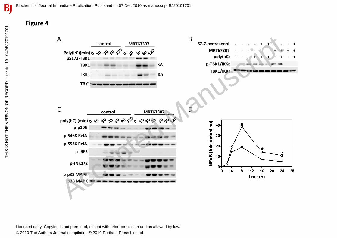
Cross-talk between canonical IKKs and IKK-related kinases during TLR3 and TLR4 signalling Since the IKK-related kinases play key roles in anti-viral immunity, we also investigated the regulation of IKK� and TBK1 by ligands that activate TLR3 and TLR4 in macrophages. Treatment of macrophages with MRT67307 enhanced the poly(I:C)-stimulated phosphorylation of TBK1 at Ser172 (Fig. 4A, top panel) and enhanced the catalytic activity of IKK� and TBK1 as judged by immunoprecipitation from extracts of poly(I:C)-stimulated cells followed by enzymatic assay in the absence of MRT67307 (Fig. 4A, middle two panels). Similar results were obtained in LPS-stimulated macrophages (Fig. S2B). This confirms our previous observations using the less specific inhibitor BX795 [17]. Thus, like the IL-1-stimulated, MyD88-dependent pathway, the TLR3/TLR4-TRIF dependent pathway also utilises a distinct upstream protein kinase for the activation of the IKK-related kinases.
We found that incubation of macrophages with the IKK� inhibitor BI605906 in the presence of MRT67307 only inhibited LPS- or poly(I:C)-stimulated phosphorylation of IKK� and TBK1 partially (results not shown), presumably because IKK� (which is not inhibited by BI605906) also contributes to the activation of the IKK-related kinases. Therefore, to investigate whether the TAK1-IKK�� signalling pathway was also involved in the activation of IKK� and TBK1 by the TRIF-dependent pathway, we treated macrophages with a potent inhibitor of TAK1 called 5Z-7-oxozeaenol to inhibit the activation of IKK� as well as IKK� [24]. At 1 M, 5Z-7-oxozeaenol inhibited the LPS-mediated activation of IKK��,the phosphorylation of p105 and the activation of p38 MAPK without affecting an upstream signalling event, namely the modification of IRAK1 by phosphorylation and polyubiquitination [25] (Fig. S2A). However, 5Z-7-oxozeaenol had little effect on poly(I:C) or LPS-induced activation of IKK� and TBK1
Biochemical Journal Immediate Publication. Published on 07 Dec 2010 as manuscript BJ20101701T
HIS
IS N
OT
TH
E V
ER
SIO
N O
F R
EC
OR
D -
see
doi
:10.
1042
/BJ2
0101
701
Acce
pted
Man
uscr
ipt
Licenced copy. Copying is not permitted, except with prior permission and as allowed by law.
© 2010 The Authors Journal compilation © 2010 Portland Press Limited
Revised manuscript # BJ2010/1701
7
(Fig. 4B and Fig. S2B). Complete blockade of the activation of IKK� and TBK1 by poly(I:C) or LPS required incubation of macrophages with a combination of 5Z-7-oxozeaenol and MRT67307 (Fig. 4B and Fig. S2B), demonstrating that TLR3 and TLR4-mediated activation of the IKK-related kinases involves the two signalling pathways described above for IL-1.
The phosphorylation of p105 by the canonical IKKs is required for activation of the protein kinase Tpl2 [26, 27], which lies at the head of the protein kinase cascade that activates the MAP kinases ERK1 and ERK2. It could therefore be argued that Tpl2, or a protein kinase activated by Tpl2, and not the canonical IKKs themselves, are the direct activators of the IKK-related kinases. However, Compound 1 (C1) a relatively specific inhibitor of Tpl2 [18] did not affect the LPS-stimulated activation of IKK� or TBK1 in the absence or presence of MRT67307 under conditions where it blocked the activation of ERK1 and ERK2 (Fig. S2C), Thus Tpl2 and/or the protein kinases that it activates are not required for the activation of the IKK-related kinases.
Treatment of macrophages with MRT67307 led to an increase in the poly(I:C) and LPS-stimulated phosphorylation of p105 and RelA (Fig. 4C and Fig. S2C) and enhanced NF�B transcriptional activity (Fig. 4D), indicating that the negative regulation of the canonical IKKs by the IKK-related kinases also operates in the TLR3/TLR4 signaling pathways.
Regulation of IKK� and TBK1 by TNF�We reported previously that, in contrast to the IL-1-stimulated activation of IKK�/TBK1, the TNF�-stimulated activation of these IKK-related kinases did not occur in TAK1-/- MEFs [17]. This result initially suggested that IL-1 and TNF� employ distinct signalling pathways to activate the IKK-related kinases. However, the present study suggested an alternative explanation, namely that TNF��may only be able to activate the IKK-related kinases using the TAK1-IKK�/IKK�-dependent arm of the signaling pathway. We therefore studied the effects of BI605906 and MRT67307 on the TNF�-stimulated phosphorylation of the IKK-related kinases. These experiments showed that the TNF�-induced phosphorylation of IKK-related kinases at Ser172 was suppressed by BI605906, in IKK�-/- MEFs, but not by MRT67307 (Fig. 5A), establishing that TNF� only activates IKK�/TBK1 via the canonical IKKs. Similar to the IL-1 signaling pathway, the IKK-related kinases inhibited the canonical IKKs in TNF��stimulated MEFs, since MRT67307 enhanced the TNF�-induced phosphorylation of p105 in either IKK�-deficient (Fig. 5A) or wild type MEFs (Fig. 5B). MRT67307 also enhanced TNF�-stimulated, NF�B-dependent gene transcription (Fig. 5C).
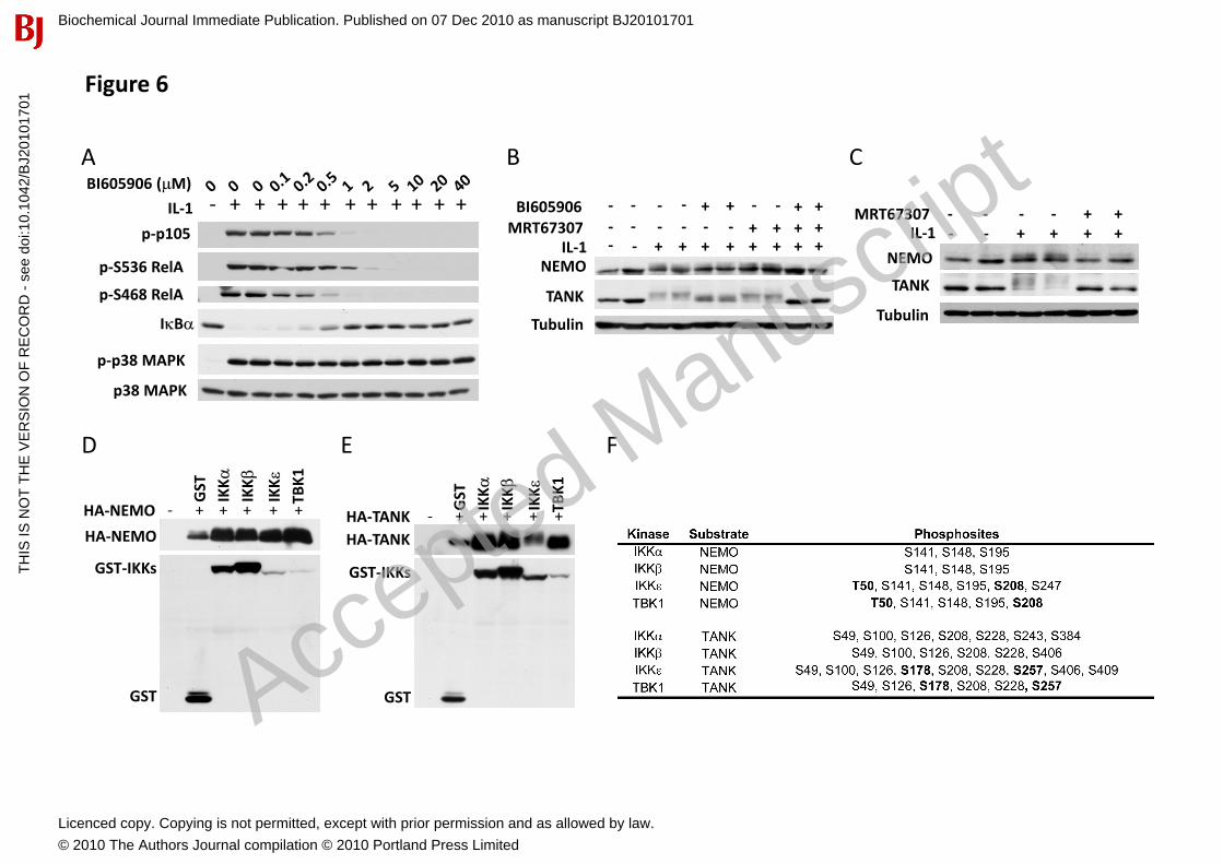
Identification of proteins targeted by both the canonical IKKs and the IKK-related kinases To date, the canonical IKKs and the IKK-related kinases have been widely believed to operate in separate pathways and to phosphorylate unique substrates. Thus the canonical IKKs, but not the IKK-related kinases phosphorylate p105, RelA and I�B� (Figs. 6A and 3D), while the IKK-related kinases phosphorylate IRF3 specifically (Fig. 1B). Here, we identify proteins that are substrates for both the canonical IKKs and the IKK-related kinases.
Pro-inflammatory stimuli induce the phosphorylation of NEMO [28, 29]� which can be monitored by a decrease in its electrophoretic mobility (Fig. 2D). Here we found that BI605906 or MRT67307 did not prevent the IL-1-stimulated phosphorylation of NEMO in IKK�-/- MEFs, but the combined addition of both compounds completely prevented phosphorylation (Fig. 6B). Thus IKK� and the IKK-related kinases are both involved in the IL-1-stimulated phosphorylation of NEMO. Consistent with this finding, MRT67307 completely suppressed the IL-1-stimulated decrease in the mobility of NEMO in TAK1-/- MEFs where the canonical IKKs are not activated (Fig. 6C).
LPS also stimulates the phosphorylation of TANK, a component of the TBK1 and IKK��complexes [30]. The IL-1-stimulated phosphorylation of TANK in IKK�-/- MEFs was unaffected by MRT67307, largely suppressed by BI605906 and completely prevented in the presence of both inhibitors (Fig. 6B). However in TAK1-/- MEFs, where the canonical IKKs are not activated, the IL-1-stimulated
Biochemical Journal Immediate Publication. Published on 07 Dec 2010 as manuscript BJ20101701T
HIS
IS N
OT
TH
E V
ER
SIO
N O
F R
EC
OR
D -
see
doi
:10.
1042
/BJ2
0101
701
Acce
pted
Man
uscr
ipt
Licenced copy. Copying is not permitted, except with prior permission and as allowed by law.
© 2010 The Authors Journal compilation © 2010 Portland Press Limited
Revised manuscript # BJ2010/1701
8
phosphorylation of TANK was completely suppressed by MRT67307 (Fig. 6C). Thus TANK, like NEMO, is phosphorylated in MEFs by both the canonical IKKs and the IKK-related kinases.
To identify the residues on NEMO and TANK that are phosphorylated by different members of the IKK subfamily, we co-transfected these proteins in HEK 293 cells, which decreased the electrophoretic mobilities of NEMO and TANK (Figs. 6D and 6E). Tryptic digestion followed by mass spectrometric analysis identified a number of Ser/Thr residues that became phosphorylated. All four members of the IKK subfamily phosphorylated NEMO at Ser141, Ser148, and Ser195, whereas Thr50 and Ser208 only became phosphorylated when NEMO was co-transfected with IKK� or TBK1. All the IKKs phosphorylated TANK at Ser49, Ser126, Ser208 and Ser228, but only IKK� and TBK1 phosphorylated Ser178 and Ser257 (Fig. 6F). Thus the different members of the IKK family members possess overlapping but not identical substrate specificities.
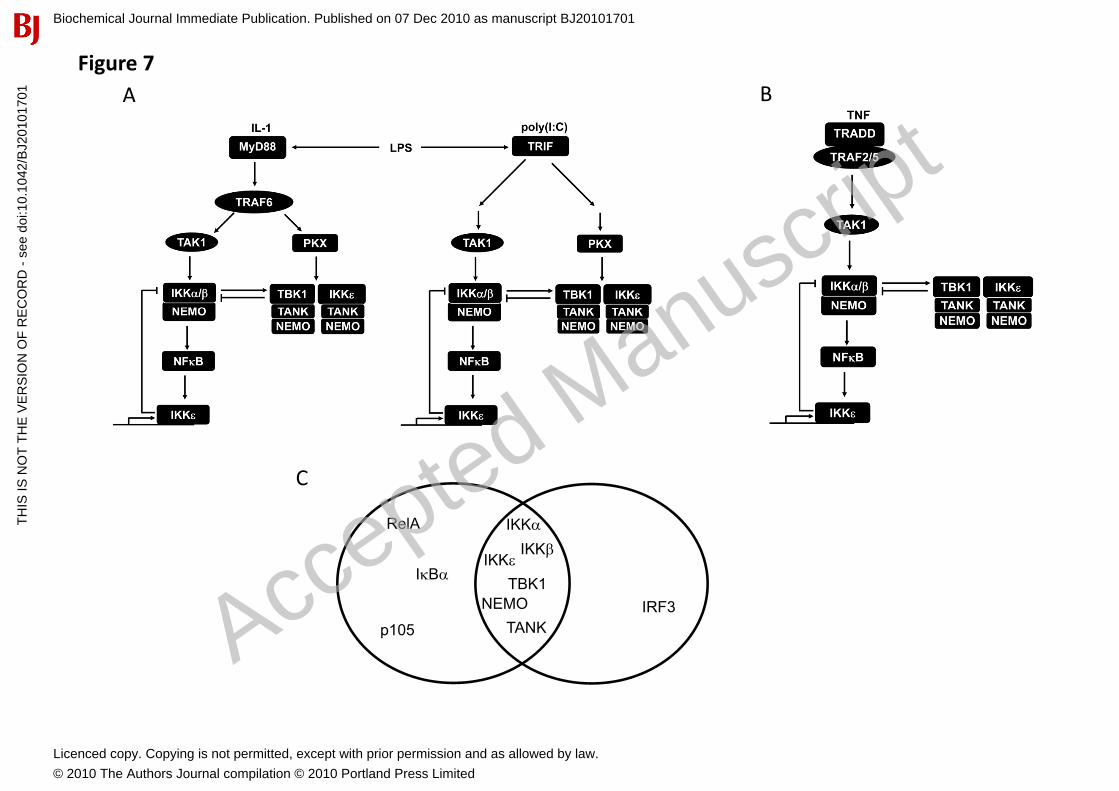
Discussion In an earlier study, we obtained evidence that the activation of the IKK-related kinases involves a separate “upstream” kinase(s) [17], which we identify here as the canonical IKKs, IKK� and IKK����We further show that two signalling pathways are involved in the IL-1-induced activation of the IKK-related protein kinases���They both require the E3 ubiquitin ligase TRAF6 but then diverge, one pathway requiring�����and the canonical IKKs, the second being independent of these protein kinases���The IKK��-independent pathway is suppressed by the IKK�/TBK1 inhibitor MRT67307, suggesting that it culminates in the autophosphorylation and autoactivation of the IKK-related kinases. However, it cannot be excluded that like IKK�, IKK� and TBK1 require another protein kinase (denoted as PKX in Fig. 7A) to initiate the autophosphorylation reaction. Similar to IL-1, the poly(I:C)-TLR3 and LPS-TLR4-stimulated activation of the IKK-related kinases also involves IKK��-dependent and -independent pathways (Fig. 7A). In contrast to IL-1, TNF� activates the IKK-related kinases solely through the canonical IKKs (Fig. 7B), explaining our earlier observation that the TNF�-stimulated activation of IKK�/TBK1 did not occur in TAK1-deficient MEFs [17].
The canonical IKKs phosphorylated IKK� and TBK1 at Ser172 in vitro, indicating that they activate the IKK-related kinases directly and not through an intervening kinase. However, we also found that the IKK-related kinases inhibit the canonical IKKs, since MRT67307 enhanced the phosphorylation of several physiological substrates of the canonical IKKs by all inflammatory stimuli tested. Thus the IKK-related kinases play an important role in limiting the extent of activation of the canonical IKKs and hence the activation of NF�B. To our knowledge this represents the first role for the IKK-related kinases in the MyD88-dependent signaling pathway to be identified, as well as the first general function of these protein kinases that is common to the IL-1, TNF�, poly(I:C) and LPS signalling pathways. In contrast, other known functions of IKK� and TBK1 are restricted to specific signalling events. For instance, the regulation of IRF3-dependent gene transcription by IKK� and TBK1 is associated with the TRIF-dependent production of IFN� [2]. Another example is the pro-survival function of TBK1, which protects foetal hepatocytes from TNF-induced apoptosis [31].
The negative regulation of the canonical IKKs by the IKK-related kinases is likely to be effected at multiple levels because the IKK-related kinases not only phosphorylate the catalytic subunits of IKK� and IKK� at sites distinct from those that trigger activation, but also phosphorylate NEMO. It has been reported that prolonged stimulation with TNF� leads to the deactivation of IKK� due to phosphorylation of a cluster of serine residues near its C-terminus�[32]. The phosphorylation of these sites was attributed to autophosphorylation, but the present study suggests that the IKK-related kinases contribute to the phosphorylation of these inhibitory sites. Moreover, the IKK�-catalysed phosphorylation of NEMO, on residues that were also identified in the current manuscript as sites phosphorylated by IKK�/TBK1, dampens IKK� activity and NF�B-dependent gene transcription [21]. The mechanism may involve the dissociation of NEMO from the IKK�� catalytic subunits leading to their inactivation [28, 33].
Biochemical Journal Immediate Publication. Published on 07 Dec 2010 as manuscript BJ20101701T
HIS
IS N
OT
TH
E V
ER
SIO
N O
F R
EC
OR
D -
see
doi
:10.
1042
/BJ2
0101
701
Acce
pted
Man
uscr
ipt
Licenced copy. Copying is not permitted, except with prior permission and as allowed by law.
© 2010 The Authors Journal compilation © 2010 Portland Press Limited

Revised manuscript # BJ2010/1701
9
The phosphorylation of TANK may also be important in the “cross-talk” between the IKK-related kinases and the canonical IKKs. Mice that do not express TANK show increased TLR7-dependent activation of NF�B and develop autoimmune disease due to enhanced signalling “downstream” of MyD88, but produce normal levels of Type 1 interferons during viral infection, presumably because the loss of TANK is compensated for by other TBK1/IKK� binding proteins, such as NAP1 and SINTBAD [34]. Since TANK interacts with TRAF6, and TRAF6 polyubiquitylation was slightly enhanced in TLR7-stimulated macrophages from TANK-/- mice, it was suggested that TANK suppresses the activation of NF�B by inhibiting TRAF6 polyubiquitylation [34]. This explanation may well be correct, but an additional possibility is that the NEMO-TANK interaction is critical for the negative regulation of the canonical IKKs by the IKK-related kinases, so that this negative regulation is lost in TANK-/- mice (Fig. 7). It would therefore clearly be of interest to study how the IKK-related kinases are regulated and how they control the canonical IKKs in TANK-/- cells. Dissecting the precise molecular mechanisms by which the IKK-related kinases negatively regulate the canonical IKKs remains a challenging task for future research.
It is well established that inflammatory stimuli not only activate IKK� but also increase the expression of the protein and, for this reason, it is also called IKKi (IKK inducible) [35]. It has been reported that IKK� expression is not induced in IKK�-/- or RelA-/- MEFs [36]. These observations, together with the presence of functional NF�B binding sites in the promoter of the gene encoding IKK��[37]� indicate that the canonical IKKs control the expression of IKK� as well as its activation. In the present study we confirmed these results by demonstrating that the IKK� inhibitor BI605906, but not MRT67307, suppresses the LPS-stimulated increase in IKK� expression (Fig. S2E). Thus, IKK���like I�B� and A20�represents an NF�B-dependent gene that is induced in response to pro-inflammatory stimuli to negatively control the IKK�-NF�B signalling axis (Fig. 7).
The IKK-related protein kinases are required for the phosphorylation and activation of IRF3 in a TRIF-dependent signalling pathway that is activated by agonists which engage TLR3 and TLR4 [2]. Inthese pathways, the phosphorylation of IRF3 requires the presence of TRAF3 and it has been inferred but, to our knowledge, never shown, that TRAF3 is required for the activation of the IKK-related kinases by these agonists [38, 39]. In the present study, we found that the IL-1-stimulated activation of the IKK-related kinases was unimpaired in TRAF3-/- MEFs (Fig. 2B). Moreover, the activation of the IKK-related kinases by IL-1 or TNF� [17], or by TLRs that signal via MyD88 (unpublished results), does not induce the phosphorylation of IRF3 or significant production of IFN�, respectively. However, TLRs that signal via MyD88 can induce IFN� production if they are colocalised with TRAF3 at the plasma membrane by expressing a fusion protein in which the phosphatidylinositol (4,5) bisphosphate-binding pleckstrin homology (PH) domain of PLC-�1 is linked to TRAF3 [40]. These findings suggest that TRAF3 may not be required for the activation of the IKK-related kinases, but instead may be needed to couple the IKK-related kinases to the phosphorylation of IRF3.
In IKK�-/- MEFs, we noticed that the IKK� inhibitor BI605906 not only prevented the phosphorylation of physiological substrates, such as p105 and RelA, but also the phosphorylation of the serine residues in IKK� (Ser177 and Ser181) that trigger its activation. Similar results were obtained with PS1145, a structurally unrelated inhibitor of IKK� (supplementary Fig. S1D,E). In contrast, BI605906 did not inhibit the phosphorylation of the equivalent residues of IKK� (Ser176 and Ser180) in IKK�-/- MEFs (results not shown), demonstrating that BI605906 exerts its effect by binding to IKK� and not by inhibiting a more upstream component of the pathway. These observations suggest that IKK� may phosphorylate and activate itself in MEFs. This would be consistent with the finding that the inactive canonical IKK complex can phosphorylate and activate itself in vitro upon incubation with MgATP and polyubiquitin chains of undefined composition [9]. However, the canonical IKKs are not activated by IL-1 or TNF�- in MEFs that do not express TAK1 or express a truncated, inactive mutant [4, 5]. It is therefore possible that TAK1 starts the activation of the canonical IKKs, which is then driven to completion autocatalytically by the canonical IKKs themselves.
Biochemical Journal Immediate Publication. Published on 07 Dec 2010 as manuscript BJ20101701T
HIS
IS N
OT
TH
E V
ER
SIO
N O
F R
EC
OR
D -
see
doi
:10.
1042
/BJ2
0101
701
Acce
pted
Man
uscr
ipt
Licenced copy. Copying is not permitted, except with prior permission and as allowed by law.
© 2010 The Authors Journal compilation © 2010 Portland Press Limited
Revised manuscript # BJ2010/1701
10
A report that NEMO is required for the production of IFN� “down-stream” of the cytoplasmic double-stranded RNA-receptor RIG-I in Sendai virus-infected cells [41], led us to study the activation of the IKK-related kinases by IL-1 in NEMO-/- MEFs. Since NEMO is required for the activity of the canonical IKKs, our expectation was that IL-1 would still activate the IKK-related kinases in NEMO-deficient MEFs via the pathway that is independent of IKK��and����� activity. However, the IL-1-stimulated activation of the IKK-related kinases was completely abolished in NEMO-/- MEFs, implying an essential role for NEMO in the activation of the IKK-related kinases independent of its role in regulating the canonical IKKs. NEMO does not bind directly to the catalytic subunits of IKK� and TBK1, but indirectly by binding to TANK [30, 41]. Since activation of the canonical IKKs by IL-1 may be initiated by the binding of K63-linked polyubiquitin chains to NEMO, the IKK-related kinases could be activated in a similar manner [10, 11]. For example, TRAF6-generated K63-pUb chains might bind to NEMO, inducing a conformational change in TANK that enables the IKK-related kinases to autophosphoryate and activate themselves. Consistent with this hypothesis, the NEMO[D311N] mutant that does not bind to K63-pUb chains, was unable to restore IRF3-dependent gene transcription induced by over-expression of the mitochondrial anti-viral-signaling protein (MAVS), a pathway that requires the activation of IKK� and TBK1 [30, 41]. Recent studies have begun to indicate that the TNF�-induced activation of the canonical IKKs may not be mediated by K63-pUb chains, as thought previously, but by other types of polyubiquitin chains [9, 15, 42]. The inability of TNF� to induce the formation of K63-pUb chains could explain why TNF� can only activate the IKK-related kinases through the canonical IKK-dependent pathway.
In the present study we identified several proteins that are phosphorylated by both the canonical IKKs and the IKK-related kinases, namely, NEMO, TANK, IKK�, IKK�, IKK� and TBK1, whereas I�B�,RelA and p105 are phosphorylated only by the canonical IKKs and IRF3 only by the IKK-related kinases (Fig. 7C). Interestingly, the substrates we found that are common to all four members of the IKK subfamily of protein kinases are their regulatory and catalytic subunits, whose function is to coordinate the activities of the canonical and IKK-related kinases. In contrast the substrates that are specific to either the canonical IKKs or the IKK-related kinases are components of transcription factors, which are dedicated to the expression of genes that encode particular inflammatory mediators. The finding that the canonical IKKs and the IKK-related kinases have overlapping substrate specificities in cells is consistent with recent studies on their preferences for synthetic peptide substrates [43, 44]. Since IL-1 and TNF� activate IKK��and TBK1 without inducing the phosphorylation of IRF3, the identification of novel substrates for the IKK-related kinases in the present study provides new cell-based readouts for these kinases.
In summary, we have identified a new role for the canonical IKKs in regulating the IKK-related kinases and established that the IKK-related kinases exert a negative regulation on the canonical IKKs that may be critical in preventing the overproduction of the inflammatory mediators that lead to inflammatory and autoimmune diseases. We have also identified new physiological substrates for the members of the IKK subfamily of protein kinases, through the introduction of improved pharmacological inhibitors of these enzymes. The wider exploitation of these compounds is likely to identify additional proteins that are targeted by both the canonical IKKs and IKK-related kinases.
Acknowledgements We thank Katherine Fitzgerald, Shizuo Akira, Inder Verma, Tak Mak, Genhong Chen, and Manolis Pasparakis for kindly providing the reagents and cell lines mentioned under Experimental Procedures. We also thank the National Centre for Protein Kinase Profiling, MRC Protein Phosphorylation Unit, University of Dundee (www.kinase-screen.mrc.ac.uk) for analysing the specificities of BI605906 and MRT67307, Nick Morrice for the identification of phosphorylation sites on NEMO and TANK, and the Protein and Antibody Production Teams of the Division of Signal Transduction Therapy at the University of Dundee (coordinated by Hilary McLauchlan and James Hastie) for proteins and antibodies. KC is a recipient of a Long-Term Fellowship from EMBO and PC is a Royal Society Research Professor. The work was supported by the UK Medical Research Council,and Boehringer Ingelheim. Authors declare no conflict of interests.
Biochemical Journal Immediate Publication. Published on 07 Dec 2010 as manuscript BJ20101701T
HIS
IS N
OT
TH
E V
ER
SIO
N O
F R
EC
OR
D -
see
doi
:10.
1042
/BJ2
0101
701
Acce
pted
Man
uscr
ipt
Licenced copy. Copying is not permitted, except with prior permission and as allowed by law.
© 2010 The Authors Journal compilation © 2010 Portland Press Limited
Revised manuscript # BJ2010/1701
11
References1 Hacker, H. and Karin, M. (2006) Regulation and function of IKK and IKK-related kinases. Sci
STKE. 2006, re13 2 Kawai, T. and Akira, S. (2010) The role of pattern-recognition receptors in innate immunity: update
on Toll-like receptors. Nat Immunol. 11, 373-384 3 Lomaga, M. A., Yeh, W. C., Sarosi, I., Duncan, G. S., Furlonger, C., Ho, A., Morony, S., Capparelli,
C., Van, G., Kaufman, S., van der Heiden, A., Itie, A., Wakeham, A., Khoo, W., Sasaki, T., Cao, Z., Penninger, J. M., Paige, C. J., Lacey, D. L., Dunstan, C. R., Boyle, W. J., Goeddel, D. V. and Mak, T. W. (1999) TRAF6 deficiency results in osteopetrosis and defective interleukin-1, CD40, and LPS signaling. Genes Dev. 13, 1015-1024
4 Sato, S., Sanjo, H., Takeda, K., Ninomiya-Tsuji, J., Yamamoto, M., Kawai, T., Matsumoto, K., Takeuchi, O. and Akira, S. (2005) Essential function for the kinase TAK1 in innate and adaptive immune responses. Nat Immunol. 6, 1087-1095
5 Shim, J. H., Xiao, C., Paschal, A. E., Bailey, S. T., Rao, P., Hayden, M. S., Lee, K. Y., Bussey, C., Steckel, M., Tanaka, N., Yamada, G., Akira, S., Matsumoto, K. and Ghosh, S. (2005) TAK1, but not TAB1 or TAB2, plays an essential role in multiple signaling pathways in vivo. Genes Dev. 19, 2668-2681
6 Walsh, M. C., Kim, G. K., Maurizio, P. L., Molnar, E. E. and Choi, Y. (2008) TRAF6 autoubiquitination-independent activation of the NFkappaB and MAPK pathways in response to IL-1 and RANKL. PLoS One. 3, e4064
7 Kanayama, A., Seth, R. B., Sun, L., Ea, C. K., Hong, M., Shaito, A., Chiu, Y. H., Deng, L. and Chen, Z. J. (2004) TAB2 and TAB3 activate the NF-kappaB pathway through binding to polyubiquitin chains. Mol Cell. 15, 535-548
8 Wang, C., Deng, L., Hong, M., Akkaraju, G. R., Inoue, J. and Chen, Z. J. (2001) TAK1 is a ubiquitin-dependent kinase of MKK and IKK. Nature. 412, 346-351
9 Xia, Z. P., Sun, L., Chen, X., Pineda, G., Jiang, X., Adhikari, A., Zeng, W. and Chen, Z. J. (2009) Direct activation of protein kinases by unanchored polyubiquitin chains. Nature. 461, 114-119
10 Ea, C. K., Deng, L., Xia, Z. P., Pineda, G. and Chen, Z. J. (2006) Activation of IKK by TNFalpha requires site-specific ubiquitination of RIP1 and polyubiquitin binding by NEMO. Mol Cell. 22, 245-257
11 Wu, C. J., Conze, D. B., Li, T., Srinivasula, S. M. and Ashwell, J. D. (2006) Sensing of Lys 63-linked polyubiquitination by NEMO is a key event in NF-kappaB activation [corrected]. Nat Cell Biol. 8,398-406
12 Rahighi, S., Ikeda, F., Kawasaki, M., Akutsu, M., Suzuki, N., Kato, R., Kensche, T., Uejima, T., Bloor, S., Komander, D., Randow, F., Wakatsuki, S. and Dikic, I. (2009) Specific recognition of linear ubiquitin chains by NEMO is important for NF-kappaB activation. Cell. 136, 1098-1109
13 Lo, Y. C., Lin, S. C., Rospigliosi, C. C., Conze, D. B., Wu, C. J., Ashwell, J. D., Eliezer, D. and Wu, H. (2009) Structural basis for recognition of diubiquitins by NEMO. Mol Cell. 33, 602-615
14 Kirisako, T., Kamei, K., Murata, S., Kato, M., Fukumoto, H., Kanie, M., Sano, S., Tokunaga, F., Tanaka, K. and Iwai, K. (2006) A ubiquitin ligase complex assembles linear polyubiquitin chains. EMBO J. 25, 4877-4887
15 Tokunaga, F., Sakata, S., Saeki, Y., Satomi, Y., Kirisako, T., Kamei, K., Nakagawa, T., Kato, M., Murata, S., Yamaoka, S., Yamamoto, M., Akira, S., Takao, T., Tanaka, K. and Iwai, K. (2009) Involvement of linear polyubiquitylation of NEMO in NF-kappaB activation. Nat Cell Biol. 11, 123-132
16 Kishore, N., Huynh, Q. K., Mathialagan, S., Hall, T., Rouw, S., Creely, D., Lange, G., Caroll, J., Reitz, B., Donnelly, A., Boddupalli, H., Combs, R. G., Kretzmer, K. and Tripp, C. S. (2002) IKK-i and TBK-1 are enzymatically distinct from the homologous enzyme IKK-2: comparative analysis of recombinant human IKK-i, TBK-1, and IKK-2. J Biol Chem. 277, 13840-13847
Biochemical Journal Immediate Publication. Published on 07 Dec 2010 as manuscript BJ20101701T
HIS
IS N
OT
TH
E V
ER
SIO
N O
F R
EC
OR
D -
see
doi
:10.
1042
/BJ2
0101
701
Acce
pted
Man
uscr
ipt
Licenced copy. Copying is not permitted, except with prior permission and as allowed by law.
© 2010 The Authors Journal compilation © 2010 Portland Press Limited
Revised manuscript # BJ2010/1701
12
17 Clark, K., Plater, L., Peggie, M. and Cohen, P. (2009) Use of the pharmacological inhibitor BX795 to study the regulation and physiological roles of TBK1 and IkappaB kinase epsilon: a distinct upstream kinase mediates Ser-172 phosphorylation and activation. J Biol Chem. 284, 14136-14146
18 Hall, J. P., Kurdi, Y., Hsu, S., Cuozzo, J., Liu, J., Telliez, J. B., Seidl, K. J., Winkler, A., Hu, Y., Green, N., Askew, G. R., Tam, S., Clark, J. D. and Lin, L. L. (2007) Pharmacologic inhibition of tpl2 blocks inflammatory responses in primary human monocytes, synoviocytes, and blood. J Biol Chem. 282, 33295-33304
19 Ananieva, O., Darragh, J., Johansen, C., Carr, J. M., McIlrath, J., Park, J. M., Wingate, A., Monk, C. E., Toth, R., Santos, S. G., Iversen, L. and Arthur, J. S. (2008) The kinases MSK1 and MSK2 act as negative regulators of Toll-like receptor signaling. Nat Immunol. 9, 1028-1036
20 Durocher, Y., Perret, S. and Kamen, A. (2002) High-level and high-throughput recombinant protein production by transient transfection of suspension-growing human 293-EBNA1 cells. Nucleic Acids Res. 30, E9
21 Bain, J., Plater, L., Elliott, M., Shpiro, N., Hastie, C. J., McLauchlan, H., Klevernic, I., Arthur, J. S., Alessi, D. R. and Cohen, P. (2007) The selectivity of protein kinase inhibitors: a further update. Biochem J. 408, 297-315
22 Williamson, B. L., Marchese, J. and Morrice, N. A. (2006) Automated identification and quantification of protein phosphorylation sites by LC/MS on a hybrid triple quadrupole linear ion trap mass spectrometer. Mol Cell Proteomics. 5, 337-346
23 Blomster, H. A., Imanishi, S. Y., Siimes, J., Kastu, J., Morrice, N. A., Eriksson, J. E. and Sistonen, L. (2010) In vivo identification of sumoylation sites by a signature tag and cysteine-targeted affinity purification. J Biol Chem, in press
24 Ninomiya-Tsuji, J., Kajino, T., Ono, K., Ohtomo, T., Matsumoto, M., Shiina, M., Mihara, M., Tsuchiya, M. and Matsumoto, K. (2003) A resorcylic acid lactone, 5Z-7-oxozeaenol, prevents inflammation by inhibiting the catalytic activity of TAK1 MAPK kinase kinase. J Biol Chem. 278,18485-18490
25 Windheim, M., Stafford, M., Peggie, M. and Cohen, P. (2008) Interleukin-1 (IL-1) induces the Lys63-linked polyubiquitination of IL-1 receptor-associated kinase 1 to facilitate NEMO binding and the activation of IkappaBalpha kinase. Mol Cell Biol. 28, 1783-1791
26 Waterfield, M., Jin, W., Reiley, W., Zhang, M. and Sun, S. C. (2004) IkappaB kinase is an essential component of the Tpl2 signaling pathway. Mol Cell Biol. 24, 6040-6048
27 Beinke, S., Robinson, M. J., Hugunin, M. and Ley, S. C. (2004) Lipopolysaccharide activation of the TPL-2/MEK/extracellular signal-regulated kinase mitogen-activated protein kinase cascade is regulated by IkappaB kinase-induced proteolysis of NF-kappaB1 p105. Mol Cell Biol. 24, 9658-9667
28 Prajapati, S. and Gaynor, R. B. (2002) Regulation of Ikappa B kinase (IKK)gamma /NEMO function by IKKbeta -mediated phosphorylation. J Biol Chem. 277, 24331-24339
29 Carter, R. S., Pennington, K. N., Ungurait, B. J. and Ballard, D. W. (2003) In vivo identification of inducible phosphoacceptors in the IKKgamma/NEMO subunit of human IkappaB kinase. J Biol Chem. 278, 19642-19648
30 Chariot, A., Leonardi, A., Muller, J., Bonif, M., Brown, K. and Siebenlist, U. (2002) Association of the adaptor TANK with the I kappa B kinase (IKK) regulator NEMO connects IKK complexes with IKK epsilon and TBK1 kinases. J Biol Chem. 277, 37029-37036
31 Bonnard, M., Mirtsos, C., Suzuki, S., Graham, K., Huang, J., Ng, M., Itie, A., Wakeham, A., Shahinian, A., Henzel, W. J., Elia, A. J., Shillinglaw, W., Mak, T. W., Cao, Z. and Yeh, W. C. (2000) Deficiency of T2K leads to apoptotic liver degeneration and impaired NF-kappaB-dependent gene transcription. Embo J. 19, 4976-4985
32 Delhase, M., Hayakawa, M., Chen, Y. and Karin, M. (1999) Positive and negative regulation of IkappaB kinase activity through IKKbeta subunit phosphorylation. Science. 284, 309-313
33 Palkowitsch, L., Leidner, J., Ghosh, S. and Marienfeld, R. B. (2008) Phosphorylation of serine 68 in the IkappaB kinase (IKK)-binding domain of NEMO interferes with the structure of the IKK complex and tumor necrosis factor-alpha-induced NF-kappaB activity. J Biol Chem. 283, 76-86
Biochemical Journal Immediate Publication. Published on 07 Dec 2010 as manuscript BJ20101701T
HIS
IS N
OT
TH
E V
ER
SIO
N O
F R
EC
OR
D -
see
doi
:10.
1042
/BJ2
0101
701
Acce
pted
Man
uscr
ipt
Licenced copy. Copying is not permitted, except with prior permission and as allowed by law.
© 2010 The Authors Journal compilation © 2010 Portland Press Limited
Revised manuscript # BJ2010/1701
13
34 Kawagoe, T., Takeuchi, O., Takabatake, Y., Kato, H., Isaka, Y., Tsujimura, T. and Akira, S. (2009) TANK is a negative regulator of Toll-like receptor signaling and is critical for the prevention of autoimmune nephritis. Nat Immunol. 10, 965-972
35 Shimada, T., Kawai, T., Takeda, K., Matsumoto, M., Inoue, J., Tatsumi, Y., Kanamaru, A. and Akira, S. (1999) IKK-i, a novel lipopolysaccharide-inducible kinase that is related to IkappaB kinases. Int Immunol. 11, 1357-1362
36 Kravchenko, V. V., Mathison, J. C., Schwamborn, K., Mercurio, F. and Ulevitch, R. J. (2003) IKKi/IKKepsilon plays a key role in integrating signals induced by pro-inflammatory stimuli. J Biol Chem. 278, 26612-26619
37 Wang, N., Ahmed, S. and Haqqi, T. M. (2005) Genomic structure and functional characterization of the promoter region of human IkappaB kinase-related kinase IKKi/IKKvarepsilon gene. Gene. 353,118-133
38 Hacker, H., Redecke, V., Blagoev, B., Kratchmarova, I., Hsu, L. C., Wang, G. G., Kamps, M. P., Raz, E., Wagner, H., Hacker, G., Mann, M. and Karin, M. (2006) Specificity in Toll-like receptor signalling through distinct effector functions of TRAF3 and TRAF6. Nature. 439, 204-207
39 Oganesyan, G., Saha, S. K., Guo, B., He, J. Q., Shahangian, A., Zarnegar, B., Perry, A. and Cheng, G. (2006) Critical role of TRAF3 in the Toll-like receptor-dependent and -independent antiviral response. Nature. 439, 208-211
40 Kagan, J. C., Su, T., Horng, T., Chow, A., Akira, S. and Medzhitov, R. (2008) TRAM couples endocytosis of Toll-like receptor 4 to the induction of interferon-beta. Nat Immunol. 9, 361-368
41 Zhao, T., Yang, L., Sun, Q., Arguello, M., Ballard, D. W., Hiscott, J. and Lin, R. (2007) The NEMO adaptor bridges the nuclear factor-kappaB and interferon regulatory factor signaling pathways. Nat Immunol. 8, 592-600
42 Haas, T. L., Emmerich, C. H., Gerlach, B., Schmukle, A. C., Cordier, S. M., Rieser, E., Feltham, R., Vince, J., Warnken, U., Wenger, T., Koschny, R., Komander, D., Silke, J. and Walczak, H. (2009) Recruitment of the linear ubiquitin chain assembly complex stabilizes the TNF-R1 signaling complex and is required for TNF-mediated gene induction. Mol Cell. 36, 831-844
43 Hutti, J. E., Shen, R. R., Abbott, D. W., Zhou, A. Y., Sprott, K. M., Asara, J. M., Hahn, W. C. and Cantley, L. C. (2009) Phosphorylation of the tumor suppressor CYLD by the breast cancer oncogene IKKepsilon promotes cell transformation. Mol Cell. 34, 461-472
44 Hutti, J. E., Turk, B. E., Asara, J. M., Ma, A., Cantley, L. C. and Abbott, D. W. (2007) IkappaB kinase beta phosphorylates the K63 deubiquitinase A20 to cause feedback inhibition of the NF-kappaB pathway. Mol Cell Biol. 27, 7451-7461
Biochemical Journal Immediate Publication. Published on 07 Dec 2010 as manuscript BJ20101701T
HIS
IS N
OT
TH
E V
ER
SIO
N O
F R
EC
OR
D -
see
doi
:10.
1042
/BJ2
0101
701
Acce
pted
Man
uscr
ipt
Licenced copy. Copying is not permitted, except with prior permission and as allowed by law.
© 2010 The Authors Journal compilation © 2010 Portland Press Limited
Revised manuscript # BJ2010/1701
14
FIGURE LEGENDS
Figure 1. MRT67307 and BI605906, novel pharmacological inhibitors of the IKK family. (A) Structure of MRT67307. (B) BMDM were incubated without or with 2 M MRT67307 for 1 h then stimulated with 10 g/ml poly(I:C) for the times indicated. Cell extracts (20 g protein) were immunoblotted with the antibodies indicated. (C) Inhibition of IFN� secretion in response to LPS. RAW264.7 macrophages were incubated for 1 h at the indicated concentrations of MRT67307 and then stimulated for 6 h with 100 ng/ml LPS. The concentration of IFN� released into the culture medium was measured using an ELISA kit from R&D Systems. (mean ± SEM, n=3). (D) Structure of BI605906.
Figure 2. Activation of IKK� and TBK1 in response to IL-1 requires TRAF6 and NEMO but not TAK1 or TRAF3. (A) TRAF6+/+ and TRAF6-/- MEFs or (B) TRAF3+/+ and TRAF3-/- MEFs or (C)TAK1+/+ and TAK1-/- or (D) NEMO +/+ and NEMO-/- MEFs were stimulated for 10 min with 5 ng/ml IL-1�. The Ser172-phosphorylated forms of IKK� and TBK1 were immunoprecipitated from cell extracts (1 mg) using the anti-pSer172 IKK� antibody (5 g) as described [17], and the presence of IKK��and TBK1 revealed by immunoblotting using antibodies recognising all forms of IKK� and TBK1 (top panel). In the other panels, Cell extracts were immunoblotted with the antibodies indicated.
Figure 3. Cross-talk between the canonical IKKs and IKK-related kinases during IL-1 signaling. (A) IKK�-/- MEFs were incubated for 1 h with 10 M BI605906, 2 M MRT67307 or both compounds, then stimulated for 10 min with 5 ng/ml IL-1�. Phosphorylation of IKK� and TBK1 at Ser172 was monitored as in Fig. 2. In the other panels, cell extracts were immunoblotted with the antibodies indicated. (B) MEFs expressing a truncated, inactive form of TAK1 were treated for 1 h with 2 M MRT67307, then stimulated for 10 min with 5 ng/ml IL-1�. Proteins were detected as described in (A). (C) Purified GST-IKK�[K38A] or GST-TBK1[K38A] proteins were incubated with 2 U/ml of recombinant IKK� and IKK�in the presence of MgATP for 30 min at 30oC. Phosphorylation of the activation loop was monitored by immunoblotting using anti-phosphoSer172 TBK1. Blots were stripped and reprobed with anti-GST as loading control. (D) WT MEFs were incubated for 1 h without (control) or with 2 M MRT67307 then stimulated for the times indicated with 5 ng/ml IL-1�. Cells extracts were immunoblotted with the antibodies indicated. (E) WT MEFs were co-transfected with DNA encoding an NF�B luciferase reporter construct and pTK-Renilla luciferase plasmid DNA. 24 h post-transfection, the cells were incubated for 1 h without (filled circles) or with (open circles) 2 M MRT67307 prior to stimulation with 5 ng/ml IL-1�for the times indicated. Luciferase activity was measured with a dual luciferase assay system (Promega) and normalized to Renilla luciferase activity (mean ± SEM, n=3). (F) Purified IKK�[S176A/S180A] or IKK�[S177A/S181A] were incubated with Mg[-32P]-ATP with or without 2 U/ml IKK� or TBK1. Reactions were terminated in SDS, the proteins resolved by SDS-PAGE, stained with Coomassie Blue (bottom panel) and the gel autoradiographed (top panel). See also Figure S1.
Figure 4. Cross-talk between IKK subfamily members also occurs during TLR3 signalling in macrophages. (A) BMDM were incubated without or with 2 M MRT67307 for 1 h then stimulated with 10 g/ml poly(I:C) for the times indicated. IKK� and TBK1 were immunoprecipitated from 50 g of cell extract protein and their catalytic activity measured in the absence of MRT67307 by incubation with GST-IRF3 and Mg[-32P]-ATP (KA= autoradiogram). Cell extracts were immunoblotted with the antibodies indicated. (B) RAW264.7 cells were treated with vehicle control, 1 M 5Z-7-oxozeaenol, 2 MMRT67307 or a combination of both for 1 h prior to stimulation with 10 g/ml poly(I:C) for 45 min. The phosphorylation of Ser172 of IKK� and TBK1 was monitored as in Fig. 2 (top panel). For other panels, cell extracts were immunoblotted with the indicated antibodies. (C) RAW264.7 cells were treated with vehicle control or 2 M MRT67307 for 1 h prior to stimulation with 10 g/ml poly(I:C) for the indicated times and extracts immunoblotted with the indicated antibodies. (D) The effect of MRT67307 on NF�B-
Biochemical Journal Immediate Publication. Published on 07 Dec 2010 as manuscript BJ20101701T
HIS
IS N
OT
TH
E V
ER
SIO
N O
F R
EC
OR
D -
see
doi
:10.
1042
/BJ2
0101
701
Acce
pted
Man
uscr
ipt
Licenced copy. Copying is not permitted, except with prior permission and as allowed by law.
© 2010 The Authors Journal compilation © 2010 Portland Press Limited
Revised manuscript # BJ2010/1701
15
dependent gene transcription was measured in HEK293-TLR3 cells in response to 25 g/ml poly(I:C) as in Fig 3E (mean ± SEM, n=3). See also Figure S2.
Figure 5. The TNF�-induced activation of IKK�TBK1 is controlled solely by the canonical IKK complex. (A) IKK�-deficient MEFs were treated with 10 M BI605906, 2 M MRT67307 or a combination of both inhibitors, then stimulated for 10 min with 10 ng/ml TNF�. Phosphorylation of IKK�and TBK1 was measured as in Fig 2. In all other panels, cell extract protein (20 g) was immunoblotted with the antibodies indicated. (B) WT MEFs were incubated for 1 h without (control) or with 2 MMRT67307 prior to stimulation with 10 ng/ml TNF� for the times indicated, and extracts immunoblotted with the antibodies indicated. (C) The effect of MRT67307 on NF�B-dependent gene transcription was measured in WT MEFs stimulated with 10 ng/ml TNF� as in Fig 3E (mean ± SEM, n=3).
Figure 6. Selective and shared substrates within the IKK family. (A) IKK�-/- MEFs were incubated for 1 h with the indicated concentrations of BI605906, stimulated for 10 min with 5 ng/ml IL-1� and cell extracts immunoblotted with the antibodies indicated. (B) IKK�-/- MEFs were incubated for 1 h with either 10 M BI605906 or 2 M MRT67307 or with a combination of both, stimulated for 10 min with 5 ng/ml IL-1� and cell extracts immunoblotted with the antibodies shown. (C) TAK1-deficient cells were treated for 1 h without (-) or with (+) 2 M MRT67307, stimulated for 10 min with 5 ng/ml IL-1� and immunoblotted as in B. (D) HEK293 cells were co-transfected with DNA vectors encoding HA-tagged NEMO and either GST, GST-IKK�, GST-IKK�, GST-IKK� or GST-TBK1. Cell extracts (20 g protein) were then immunoblotted with either anti-HA or anti-GST. (E) The experiment was performed as in (D) except that NEMO was replaced by TANK. (F) Sites on NEMO and TANK that became phosphorylated after co-transfection with IKK family members. Sites phosphorylated only after co-transfection with IKK�or TBK1 and not after co-transfection with IKK� or IKK� are shown in bold-face type.
Figure 7. Model for how different IKK family members are regulated in response to pro-inflammatory stimuli. (A) Agonists that engage the IL-1R, TLR3 or TLR4 activate IKK� and TBK1 by IKK�/�-dependent and -independent mechanisms. When the canonical IKK complex is inactive, IKK�and TBK1 are activated by a pathway that is suppressed by MRT67307. In this pathway, the binding of MRT67307 to IKK�TBK1 may prevent phosphorylation of the activation loops of IKK�/TBK1 by an as yet unidentified protein kinase(s), denoted as PKX. Alternatively, PKX may be identical to IKK�/TBK1 and the pathway may culminate in autoactivation of these protein kinases. Once activated, IKK� and TBK1 phosphorylate IKK�� decreasing the activity of the canonical IKK complex. In addition, IKK��induce the expression of IKK� through the activation of NF�B providing an extra level of control of the signalling pathway. (B) TNF� activates IKK�TBK1 solely by the IKK�/�-dependent pathway. (C) Venn diagram of the substrates phosphorylated in cells by the different members of the IKK family.
Biochemical Journal Immediate Publication. Published on 07 Dec 2010 as manuscript BJ20101701T
HIS
IS N
OT
TH
E V
ER
SIO
N O
F R
EC
OR
D -
see
doi
:10.
1042
/BJ2
0101
701
Acce
pted
Man
uscr
ipt
Licenced copy. Copying is not permitted, except with prior permission and as allowed by law.
© 2010 The Authors Journal compilation © 2010 Portland Press Limited

Figure 1
A BPoly(I:C)(min)
control MRT67307
p-p38 MAPK
TBK1
p-JNK1/2
pS396-IRF3
Poly(I:C)(min)
TBK1
MRT67307
C D
BI605906
Biochemical Journal Immediate Publication. Published on 07 Dec 2010 as manuscript BJ20101701T
HIS
IS N
OT
TH
E V
ER
SIO
N O
F R
EC
OR
D -
see
doi
:10.
1042
/BJ2
0101
701
Acce
pted
Man
uscr
ipt
Licenced copy. Copying is not permitted, except with prior permission and as allowed by law.
© 2010 The Authors Journal compilation © 2010 Portland Press Limited
Figure 2
A BTRAF6-/-TRAF6+/+ TRAF3-/-TRAF3+/+
p-TBK1/IKK�
TRAF6-/-� ++IL-1 ++ �
TRAF6+/+
TRAF3
p-TBK1/IKK�
TRAF3-/-� ++IL-1 ++ �
TRAF3+/+
p-IKK�� p-IKK��
p-JNK1/2
TRAF6
p-p38 MAPK
TBK1/IKK� p38 MAPK
TBK1
IKK�
p JNK1/2
DC
p-TBK1/IKK�
NEMO-/-� ++IL-1 ++ �NEMO+/+
p-IKK��
p-TBK1/IKK�
TAK1-/-� ++IL-1 ++ �TAK1+/+
p-p105p-IKK��
p-p38 MAPK
p-p105
p-JNK1/2TAK1
p-p38 MAPK
TBK1/IKK�
p p105
�TAK1
NEMOTBK1/IKK�
Biochemical Journal Immediate Publication. Published on 07 Dec 2010 as manuscript BJ20101701T
HIS
IS N
OT
TH
E V
ER
SIO
N O
F R
EC
OR
D -
see
doi
:10.
1042
/BJ2
0101
701
Acce
pted
Man
uscr
ipt
Licenced copy. Copying is not permitted, except with prior permission and as allowed by law.
© 2010 The Authors Journal compilation © 2010 Portland Press Limited
p�TBK1/IKK�� +� +IL�1
MRT67307++
++� � � �A BFigure�3
IL�1MRT67307BI605906
+++++++�+++� � � � �
� � � +�
�� +�
p TBK1/IKK�� +
+++
p /
p�IKK��
IKK�
IKK�IKK�
p�IKK�
p�p105
p�TBK1/IKK�
TBK1/IKK�
p�p38�MAPK
TBK1/IKK�
p�S468�RelA
GST�TBK1/IKK�[38A] ++++++IKK� � � � + +IKK� � � +
�+ � �
p�p105
IL�1�(min)
MRT67307controlC D
pSer172�TBK1
pSer172�IKK�
GST
GST
/ [ ]p�S468�RelA
p�S536�RelA
p�JNK1/2GST
p�p38�MAPK
p38�MAPK
p /
E FRecombinant�Kinase
+++IKK� [S177A/S181A]IKK� [S176A/S180A]
� � �+++ � � �++� ++�
+++� � �+++ � � �++� ++�
TBK1 IKK�E F
IKK��
32P�IKK��
Biochemical Journal Immediate Publication. Published on 07 Dec 2010 as manuscript BJ20101701T
HIS
IS N
OT
TH
E V
ER
SIO
N O
F R
EC
OR
D -
see
doi
:10.
1042
/BJ2
0101
701
Accepted M
anuscript
Licenced copy. Copying is not permitted, except with prior permission and as allowed by law.
© 2010 The Authors Journal compilation © 2010 Portland Press Limited
Figure�4
A BA B
pS172�TBK1Poly(I:C)(min)
control MRT67307
TBK1 KA p�TBK1/IKK�
poly(I:C)MRT67307
5Z�7�oxozeaenol
++++++�+++� � � �
+� � � � �
�
�
+
+++
� �++
TBK1
TBK1
IKK� KATBK1/IKK�
C D
( ) ( )
MRT67307control
p�p105
poly(I:C)�(min)
p�S536�RelA
p�S468�RelA
p S536 RelA
p�JNK1/2
p�IRF3
p�p38�MAPKp38�MAPK
Biochemical Journal Immediate Publication. Published on 07 Dec 2010 as manuscript BJ20101701T
HIS
IS N
OT
TH
E V
ER
SIO
N O
F R
EC
OR
D -
see
doi
:10.
1042
/BJ2
0101
701
Accepted M
anuscript
Licenced copy. Copying is not permitted, except with prior permission and as allowed by law.
© 2010 The Authors Journal compilation © 2010 Portland Press Limited
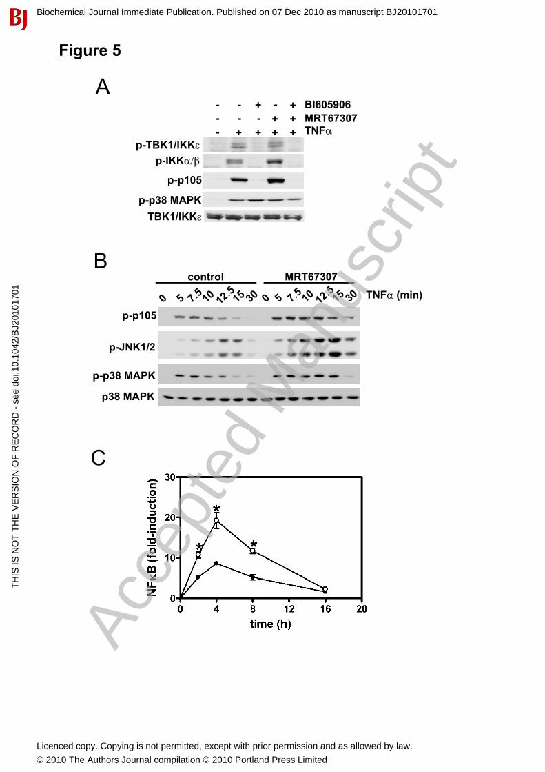
Figure 5
AMRT67307BI605906- - + - +
p-IKK��
p-p105
TNF�MRT67307
++++-++- - -
p-TBK1/IKK�
p-p38 MAPKTBK1/IKK�
B
p-p105
TNF� (min)MRT67307control
p JNK1/2
p-p38 MAPK
p38 MAPK
p-JNK1/2
C
Biochemical Journal Immediate Publication. Published on 07 Dec 2010 as manuscript BJ20101701T
HIS
IS N
OT
TH
E V
ER
SIO
N O
F R
EC
OR
D -
see
doi
:10.
1042
/BJ2
0101
701
Acce
pted
Man
uscr
ipt
Licenced copy. Copying is not permitted, except with prior permission and as allowed by law.
© 2010 The Authors Journal compilation © 2010 Portland Press Limited
Figure�6
A B C
MRT67307BI605906
+++� � � � �� � � +
��� +�
+++
� +� + ++++� � � �
IL�1MRT67307
p�p105
IL�1
BI605906�( M)� + + + + + + + + + ++
IL�1 +++++++� � +
Tubulin
TANK
NEMOTANK
Tubulin
NEMO
p p
p�S536�RelA
I�B�
p�S468�RelA
p�p38�MAPK
p38�MAPK
D E F
HA�NEMO
TBK1
IKK�
IKK�
IKK�
GST
� +++++ HA�TANK � +++++ TBK1
IKK�
IKK�
IKK�
GST
GST�IKKs
HA�NEMO
GST�IKKs
HA�TANK
GST GST
Biochemical Journal Immediate Publication. Published on 07 Dec 2010 as manuscript BJ20101701T
HIS
IS N
OT
TH
E V
ER
SIO
N O
F R
EC
OR
D -
see
doi
:10.
1042
/BJ2
0101
701
Accepted M
anuscript
Licenced copy. Copying is not permitted, except with prior permission and as allowed by law.
© 2010 The Authors Journal compilation © 2010 Portland Press Limited
A BFigure�7
CC
RelA
I B
IKK�IKK�
IKK�
IRF3p105
I�B� TBK1NEMO
TANK
Biochemical Journal Immediate Publication. Published on 07 Dec 2010 as manuscript BJ20101701T
HIS
IS N
OT
TH
E V
ER
SIO
N O
F R
EC
OR
D -
see
doi
:10.
1042
/BJ2
0101
701
Accepted M
anuscript
Licenced copy. Copying is not permitted, except with prior permission and as allowed by law.
© 2010 The Authors Journal compilation © 2010 Portland Press Limited