vasoactiveintestinalpeptideand … · vasoactiveintestinalpeptideand...
TRANSCRIPT
Vasoactive Intestinal Peptide andPituitary Adenylate Cyclase-
Activating Polypeptide InhibitChemokine Production in
Activated MicrogliaMARIO DELGADO,1,2 G. MILLER JONAKAIT,1,3 AND DOINA GANEA1*
1Department of Biological Sciences, Rutgers University, Newark, New Jersey2Departamento Biologia Celular, Facultad de Biologia, Universidad Complutense,
Madrid, Spain3New Jersey Institute of Technology, Newark, New Jersey
KEY WORDS inflammation; chemokine; activated microglia; neuropeptides; chemo-taxis
ABSTRACT Microglia react to even minor disturbances in CNS homeostasis and func-tion as critical regulators of CNS inflammation. Activated microglia secrete inflammatorymediators such as cytokines and chemokines, which contribute to the pathophysiologicalchanges associated with several neuroimmunologic disorders. Microglia-derived inflamma-tory chemokines recruit various populations of immune cells, which initiate and maintainthe inflammatory response against foreign antigens. Entry and retention of activatedimmune cells in the CNS is a common denominator in a variety of traumatic, ischemic, anddegenerative diseases. Vasoactive intestinal peptide (VIP) and pituitary adenylate cyclase-activating polypeptide (PACAP) are two structurally related neuropeptides that function aspotent anti-inflammatory factors in the periphery. Here we investigated the effects of VIPand PACAP on chemokine production by activated microglia. VIP and PACAP inhibit theexpression of the microglia-derived CXC chemokines MIP-2 and KC, and of the CC chemo-kines MIP-1�, -1�, MCP-1, and RANTES. The inhibition of chemokine gene expressioncorrelates with an inhibitory effect of VIP/PACAP on NFkB binding. The VIP/PACAPinhibition of both chemokine production and of NFkB binding is mediated through thespecific receptor VPAC1 and involves a cAMP-dependent intracellular pathway. Of biolog-ical significance is the fact that the inhibition of chemokine production by VIP/PACAP leadsto a significant reduction in the chemotactic activity generated by activated microglia forperipheral leukocytes, i.e., neutrophils, macrophages, and lymphocytes. Because reductionin the number and activation of infiltrating leukocytes represents an important factor in thecontrol of inflammation in the CNS, VIP and/or PACAP released by neurons during aninflammatory response could serve as neuronal survival factors by limiting the inflamma-tory process. GLIA 39:148–161, 2002. © 2002 Wiley-Liss, Inc.
INTRODUCTION
Entry and retention of activated immune cells in theCNS is a common denominator in a variety of trau-matic, ischemic, and degenerative diseases. These cel-lular events are determined and controlled by a net-work of chemokines, adhesion molecules, and cytokines.In the CNS, resident microglia represent one of themost potent sources for proinflammatory products. Mi-croglia are readily activated in most neuropathologicalconditions, and the activation process leads to changes
in morphology, cell surface receptors, expression of cy-tokines and chemokines (Streit et al., 2000). Chemo-kines, small molecular weight (MW) polypeptides, are
*Correspondence to: Doina Ganea, Rutgers University, Department of Biolog-ical Sciences, 101 Warren Street, Newark, NJ 07102.E-mail: [email protected]
Received 27 January 2002; Accepted 15 April 2002
DOI 10.1002/glia.10098
Published online 00 Month 2002. in Wiley InterScience (www.interscience.wiley.com).
GLIA 39:148–161 (2002)
© 2002 Wiley-Liss, Inc.
among the earliest secreted products, function as che-moattractants for cells expressing the appropriate che-mokine receptors, and are responsible for the direc-tional migration of blood-derived leukocytes to theCNS. Proinflammatory chemokines have been associ-ated with various neuropathological conditions such asAlzheimer’s disease, stroke, AIDS dementia, and par-ticularly multiple sclerosis (MS). In patients with MS,increases in the expression of monocyte chemoattrac-tant protein (MCP-1; CCL2), macrophage inflamma-tory protein (MIP-1�; CCL3), IFN�-inducible protein10 (IP-10; CXCL10), and RANTES (CCL5) are associ-ated with demyelinating lesions and clinical neurolog-ical dysfunctions (Zhang et al., 2000). In addition, inexperimental allergic encephalomyelitis (EAE), in-creases in MCP-1 and MIP-1� mRNA precede the onsetof the disease, and treatment with either anti–MIP-1�or anti–MCP-1 Abs leads to a reduction in clinicalsymptoms (Godiska et al., 1995; Karpus and Kennedy,1997; Karpus and Ransohoff, 1988).
The CNS consists of several cell types, which obvi-ously interact both directly and through secreted prod-ucts. In terms of consequences for the initiation andoutcome of an inflammatory process, the interactionsbetween these different cell types are not understood.Some of the neuronal-released neuropeptides were re-ported to affect glial cell activation. For example, so-matostatin inhibits microglia proliferation (Feindt etal., 1998), vasoactive intestinal peptide (VIP) inhibitsIFN�-induced MHC class II expression and synergizeswith IL-1� to stimulate IL-6 production in astrocytes(Frohman et al., 1988; Gottschall et al., 1994), sub-stance P increases, and �-melanocyte stimulating hor-mone (�-MSH), adrenocorticotropin (ACTH), VIP, andpituitary adenylate cyclase-activating polypeptide(PACAP) inhibit TNF� secretion from LPS-stimulatedmicroglia (Luber-Narod et al., 1994; Galimberti et al.,1999; Kim et al., 2000). However, with the exception ofmorphine, which was shown to inhibit RANTES(CCL5) production from LPS- and IL-1�–stimulatedmicroglia (Hu et al., 2000), there is no informationregarding the effects of neuropeptides on chemokineproduction by activated microglia. Based on our previ-ous report that the two structurally related neuropep-tides, VIP and PACAP, inhibit proinflammatory che-mokine production in macrophages (Delgado andGanea, 2001a), we investigated their potential role inregulating chemokine production by activated micro-glia.
MATERIALS AND METHODSReagents
Synthetic VIP and PACAP38 were purchased fromCalbiochem-Novabiochem (San Diego, CA). The PAC1/VPAC2 antagonist PACAP6–38 was obtained from Pen-insula Laboratories (Belmont, CA). The VPAC1 antag-onist [Ac-His1, D-Phe2, K15, R16, L27] VIP (3–7)-GRF(8–27) and the VPAC1 agonist [K15, R16, L27] VIP (1–
7)-GRF (8–27) were kindly donated by Dr. Patrick Rob-berecht (Universite Libre de Bruxelles, Belgium). TheVPAC2 agonist Ro 25–1553 Ac-[Glu8, Lys12, Nle17,Ala19, Asp25, Leu26, Lys27,28, Gly29,30, Thr31]-VIP cyclo(21–25) was a generous gift from Drs. Ann Welton andDavid R. Bolin (Hoffmann-La Roche, Nutley, NJ). Thesynthetic PAC1 agonist maxadilan was a generous giftfrom Dr. Ethan A. Lerner (Massachusetts GeneralHospital, Charlestown, MA). Murine recombinantTNF�, IFN�, and IL-1�, and capture and biotinylatedantibodies against JE/MCP-1 and MIP-1� murine werepurchased from Pharmingen (San Diego, CA). Captureand biotinylated antibodies against murine KC, RAN-TES, MIP-2, and MIP-1� were purchased from Pre-proTech (Rocky Hill, NJ). LPS (from E. coli 055:B5),protease inhibitors, db-cAMP, and forskolin werepurchased from Sigma Chemicals (St. Louis, MO),and N-[2-(p-bromocinnamyl-amino)ethyl]-5-iso-quino-linesulfonamide (H89) from ICN Pharmaceuticals(Costa Mesa, CA). Recombinant IkB� (1-317) and an-tibodies against p65, p50, IkB�, phosphorylated-IkB�,IkB-kinase (IKK�) and CREB were purchased fromSanta Cruz Biotechnology (Santa Cruz, CA).
Cell Cultures
Microglial cell cultures were prepared as previouslydescribed (Chao et al., 1993). Briefly, cerebral corticalcells from 1-day-old Balb/c mice were dissociated bytrypsinization (0.25% trypsin, 30 min) and were platedin 75 cm2 Falcon culture flasks in Dulbecco’s modifiedEagle’s medium (DMEM) high-glucose formula (LifeTechnologies) supplemented with 10% heat-inacti-vated fetal calf serum (FCS; Gibco-BRL), containing 10mM HEPES buffer, 1 mM pyruvate, 0.1 M nonessentialaminoacids, 2 mM glutamine, 50 mM 2-mercaptoetha-nol, 100 U/ml penicillin, and 10 �g/ml streptomycin(complete medium). The medium was replenished 1and 4 days after plating, and on day 8 the plates wereshaken for 20 min at a speed of 200 rpm in an orbitalshaker to remove oligodendrocytes. On day 12, theplates were shaken again for 2 h at a speed of 180–200rpm. The harvested cells were filtered through a 20 �mnylon mesh, plated in 60 mm Petri dishes, and incu-bated for 15 min at 37°C. After extensive washing withculture medium, adherent cells (microglia) were col-lected with a rubber policeman and centrifuged (1000rpm, 10 min). The purified microglial cells were � 98%MAC-1� and � 2% GFAP� specific. Two mouse micro-glial cell lines (EOC13 and BV2) were used in someexperiments. The EOC13 cell line was maintained aspreviously described (Walker et al., 1995) in DMEMcomplete medium containing 2 mM glutamine, 10%heat-inactivated FCS, and 20% LADMAC-conditionedmedium. The BV2 murine microglial cell line gener-ated by Blasi et al. (1990) was maintained in DMEMhigh glucose supplemented with 5% heat-inactivatedFCS, 4 mM L-glutamine, 0.2 mM penicillin, 0.05 mMstreptomycin, and 20 mM HEPES.
149VIP AND PACAP INHIBIT CHEMOKINES
Microglia (murine primary microglia, EOC13, andBV2 cells) were incubated with complete medium andstimulated with 100 ng/ml LPS in the presence orabsence of VIP or PACAP38 (from 10�12 to 10�6 M).Cell-free supernatants were harvested at designatedtime points and kept frozen (�20°C) until chemokinedetermination by ELISA.
Mouse peritoneal exudate cells (PECs) were obtainedby peritoneal lavage with ice-cold RPMI 1640 mediumfrom male Balb/c mice (aged 6–10 weeks). PECs, con-taining polymorphonuclear cells (PMN), lymphocytes,and macrophages, were washed twice and resuspendedin ice-cold RPMI 1640 medium supplemented with 2%heat-inactivated FCS, containing 10 mM HEPESbuffer, 1 mM pyruvate, 0.1 M nonessential aminoacids,2 mM glutamine, 50 mM 2-mercaptoethanol, 100 U/mlpenicillin, and 10 �g/ml streptomycin.
RT-PCR for Detection of VPAC1, VPAC2, andPAC1 mRNA Expression
Microglia cells were cultured at a concentration of2 106 cells/ml in 100 mm tissue culture dishes andstimulated with LPS (0.5 �g/ml) for up to 12 h. Cellswere collected at different time points (0 and 12 h) andtotal RNA was isolated using the Ultraspec RNA re-agent (Biotecx, Houston, TX) as recommended by themanufacturer; 2 �g of total RNA was reverse-tran-scribed in the presence of 200 units of MMLV-RT, 40units of RNasin, 1 �g of random primers, 0.5 mMdNTPs, 3 �g of BSA, and MMLV reaction buffer (50mM Tris-HCl, pH 8.3, 75 mM KCl, 3 mM MgCl2) in atotal volume of 30 �l at 37°C for 1 h.
The cDNA was then amplified with specific primers.Amplification with GADPH primers (Stratagene) wasused as a control. The primers for VPAC1, VPAC2, andPAC1 receptors have been described before and havethe following sequence: VPAC1 sense 5-CCTTCT-TCTCTGAGCGGAAGTACTT-3 and antisense 5-CCTGCACCTCGCCATTGAGGAAGCAG-3; VPAC2sense 5-GTCAAGGACAGCGTGCTCTACTCC-3 andantisense 5-CCCTGGAAGGAACCAACACATAAC-3;PAC1 sense 5-CAAGAAGGAGCAAGCCATGTGC-3and antisense 5-CATCGAAGTAATGGGGGAAGGG-3; GAPDH sense 5-TCCTGCACCACCAACTGCT-TAGCC-3 and antisense 5-GTTCAGCTCTGGGAT-GACCTTGCC-3. The expected sizes for the amplifiedfragments are 453 bp for VPAC1, 572 bp for VPAC2,317 bp for PAC1, and 225 bp for GAPDH; 5 �l ofreverse-transcribed cDNA was subjected to PCR in thepresence of 0.5 units of pyrostase, 1 �M sense andantisense primers, 0.2 mM dNTPs, and polymerasebuffer (50 mM Tris-HCl, pH 9.0, 1.5 mM MgCl2, 20 mM(NH4)2SO4, 50 �g/ml BSA). The PCR conditions weredenaturation at 94°C for 1 min, annealing at 60°C for 1min, primer extension at 72°C for 2 min for 38 cycles.The PCR products were size-separated in 2% agarosegel and visualized by UV light.
Electrophoretic Mobility Shift Assay (EMSA)
BV2 cells were cultured at a density of 107 cells insix-well plates, stimulated as described above, washedtwice with ice-cold PBS/0.1% BSA, and harvested. Nu-clear extracts were prepared by the mini-extractionprocedure of Schreiber et al. (1989). Double-strandedoligonucleotides (50 ng) corresponding to the NF-kBsites from murine MCP-1 (5-ACTGCCCTCAGAATGG-GAATTTCCACGCTCTTATC-3) (Freter et al., 1992),RANTES (5-TTTTGGAAACTCCCCTTAGGGGATGC-CCCTCA-3) (Nelson, 1993), MIP-2 (5-CCCTGAGCT-CAGGGAATTTCCCTGGTCCCCG-3) (Widmer et al.,1993), and KC (5-TACTCCGGGAATTTCCCTGGCC-3) (Ohmori et al., 1995) were end-labeled with �32P-ATP. The binding reaction mixtures (15 �l) were set upas follows: 0.5–1 ng DNA probe (20,000–50,000 cpm),nuclear extract (5 �g protein), 2 �g poly(dI-dC).poly(dI-dC), and binding buffer (50 mM NaCl, 0.2 mM EDTA,0.5 mM DTT, 5% glycerol, and 10 mM Tris-HCl, pH7.5). The mixtures were incubated on ice for 15 minbefore adding the probe, followed by 20 min at roomtemperature. Samples were loaded on 4% nondenatur-ing polyacrylamide gels and electrophoresed in TGEbuffer (50 mM Tris-HCl, pH 7.5, 0.38 M glycine, and 2mM EDTA) at 100 V, followed by transfer to Whatmanpaper, and autoradiography. In competition and anti-body supershift experiments, the nuclear extracts wereincubated for 15 min at room temperature with thespecific antibody (1 �g), or with competing cold oligo-nucleotide (50-fold excess) before the addition of thelabeled probe.
Chemokine ELISA
The content of chemokines in the culture superna-tants was determined by specific sandwich ELISAs aspreviously described (Delgado and Ganea, 2001a). Thecytokine amount in each supernatant was extrapolatedfrom standard curves. The lower limit of detection forJE/MCP-1, MIP-1�, MIP-1�, MIP-2, RANTES, and KCwas 16, 5, 7, 8, 10, and 15 pg/ml, respectively.
VIP Elisa
VIP concentrations were determined by using a spe-cific competitive ELISA as previously described (Mar-tinez et al., 1999).
Ribonuclease Protection Assay (RPA) forDetection of Chemokine mRNA Expression
Murine primary microglia or microglia cell lineswere cultured at a concentration of 2 106 cells/ml in100 mm tissue culture dishes and stimulated with LPS(100 ng/ml) in the presence or absence of VIP (10�8 M)or PACAP (10�8 M) for up to 12 h. Total RNA was
150 DELGADO ET AL.
isolated using the Ultraspec RNA reagent (Biotecx,Houston, TX) as recommended by the manufacturer.RNase protection assays (RPA) were performed with2.5–5 �g RNA using the Riboquant MultiProbe RNaseProtection Assay System (PharMingen) according tothe manufacturer’s instructions. Each commercial kitcontained a set of chemokine templates as well as atemplate for the housekeeping gene, glyceraldehyde-3-phosphate dehydrogenase (GAPDH). The samples wereelectrophoresed on 5% denaturing polyacrylamide gels.Signal quantitation was performed in a PhosphorIm-ager SI (Molecular Dynamics, Sunnyvale, CA).
Western Blotting
Whole cell lysates, cytoplasmic fractions, or nuclearextracts containing 20 to 30 �g of protein were sub-jected to reducing SDS-PAGE (12.5%). After electro-phoresis, the gel was electroblotted and the mem-branes were with primary antibodies (rabbitantimouse IgG against IkB�, IKK�, NF-kB p50, orNF-kB p65, or mouse IgG against phosphorylated-IkB�, at dilutions ranging from 1:500 to 1:2,000), fol-lowed by secondary antibodies, peroxidase-conjugatedgoat antirabbit IgG or rat antimouse IgG (1:5,000 di-lution). The membranes were developed by using theenhanced chemiluminiscence detection system (ECL,Amersham).
In Vitro Kinase Assay
The in vitro IKK� kinase assay was performed aspreviously described (Delgado and Ganea, 2001b).Whole cell lysates were prepared from 2 106 cells andcleared by centrifugation at 13,000 g for 3 min. Theendogenous IKK� was immunoprecipitated by incubat-ing the lysates (150–250 �g protein) with 0.5 �g ofanti-IKK� antibody for 2 h at 4°C. The immune com-plexes were collected by incubation with protein A/G-Sepharose beads for 45 min at 4°C. Following extensivewashing, the pelleted beads were resuspended in 30 �lkinase buffer containing 15 �M ATP, 10 �Ci [�32P]ATP(3,000 Ci/mmol), and 5 �g of recombinant IkB�. Thekinase reaction was performed at 30°C for 30 min andstopped by the addition of 15 �l of 2 SDS samplebuffer. Following boiling for 5 min, the samples weresubjected to SDS-PAGE (9%). Proteins were trans-ferred on nitrocellulose membranes, followed by auto-radiography and quantitation of radioactive IkB� byphosphoimaging. Presence of IKK� protein was veri-fied by immunoblotting.
Chemotactic Activity of MicroglialConditioned Medium
Cell migration of peritoneal cell populations wasevaluated using a 48-well chemotaxis microchamber,
as previously described (Meda et al., 1996). Briefly,dilutions of supernatants from microglia cultures wereadded to the lower wells of the chemotaxis chamber(Neuroprobe, Pleasanton, CA). Complete medium wasused as basal control. A polycarbonate filter (5 �m poresize, Neuroprobe) was layered onto the wells and cov-ered with a silicon gasket and the top plate. Freshlyisolated peritoneal cells (1.5 106 cells/ml) wereseeded in the upper chamber. Incubation was con-ducted at 37°C for 90 min. The filters were removed,stained with May-Grunwald and Giemsa, and thenumber of macrophages, lymphocytes, and PMN thathad migrated was evaluated by counting eight micro-scopic fields (oil immersion) and distinguished based onmorphology.
Statistics
Comparison between groups were made using thestudent’s t-test followed by Scheffe’s F-test, with P �0.001 as the minimum significant level.
RESULTSVIP and PACAP Inhibit LPS-Induced
Production of CXC and CC Chemokinesin Microglia
Primary microglia were activated with LPS in theabsence or presence of various doses of VIP or PACAP,and the amounts of chemokines released in culturesupernatants were assayed by ELISA. Unstimulatedmicroglia produce very low amounts of chemokines(Fig. 1A). LPS stimulation results in a time-dependentincrease in the production of the CXC chemokinesMIP-2 and KC (murine neutrophil attractants), and ofthe CC chemokines MIP-1� (CCL3), MIP-1� (CCL4),MCP-1 (CCL2), and RANTES (CCL5), with peak levelsat 8 to 24 h (Fig. 1A). Chemokine levels declined onlyslowly after 48 h. VIP and PACAP inhibit in a dose-and time-dependent manner the production of the CXCand CC chemokines tested (Fig. 1). Chemokine produc-tion was significantly inhibited as early as 2 h, withmaximum inhibitory effects at 24 to 48 h (Fig. 1A). Thereduction of chemokine production was maintainedthroughout 72 h (not shown), indicating that VIP andPACAP do not delay, but rather reduce chemokinerelease. The dose-response curves were similar for VIPand PACAP, with maximal effects at 10�8 M (Fig. 1B).
The inhibitory effects were not the result of a de-creased number of microglial cells, as neither VIP norPACAP affected cell numbers or the viability of stimu-lated microglia after 36 h of culture (viabilities deter-mined by Trypan blue exclusion were in the range of87%–92% with or without neuropeptides).
In addition, VIP and PACAP also inhibit chemokineproduction following activation with proinflammatorycytokines (i.e., TNF�, IL-1�, and IFN�; Fig. 2). Sincethe highest degree of inhibition was observed for mi-
151VIP AND PACAP INHIBIT CHEMOKINES

croglia stimulated with 100 ng/ml LPS at a neuropep-tide concentration of 10�8 M after 24 h of culture, weused these conditions for the rest of the experiments.
Inhibition of Chemokine Production by VIPand PACAP Is Mediated Through VPAC1
Next we investigated whether the inhibitory effect ofVIP/PACAP is mediated through specific receptors.
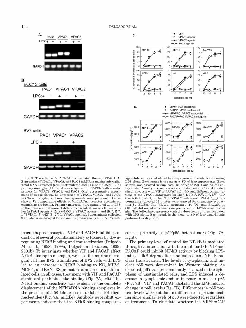
VIP and PACAP act through a family of receptors con-sisting of VPAC1, VPAC2, and PAC1 (Harmar et al.,1998). RT-PCR analysis indicates that murine primarymicroglia express both PAC1 and VPAC1 mRNA (Fig.3A). In contrast, VPAC2 mRNA was not expressedeven following LPS activation (Fig. 3A). A similar pat-tern of VIP/PACAP receptor expression was observedin rat primary microglia (Kim et al., 2000) and in thetwo murine microglial cell lines EOC13 and BV2 (Fig.
Fig. 1. VIP and PACAP inhibit LPS-induced chemokine production.A: Time-dependent inhibition. Primary microglia (2 105 cells/ml)were cultured in 48-well plates (1 ml/well) and stimulated with LPS(100 ng/ml), in the absence or presence of 10�8 M VIP or PACAP.Supernatants harvested at different time points were assayed for
chemokine content by ELISA. B: Dose-dependent inhibition. Primarymicroglia were stimulated with LPS and treated with various concen-trations of either VIP or PACAP for 24 h. The chemokine contents inthe culture supernatants were determined by ELISA. Each result isthe mean � SD of four separate experiments performed in duplicate.
152 DELGADO ET AL.

3B). In order to determine which of the VIP/PACAPreceptors is involved in the inhibition of chemokineproduction, we used specific receptor agonists and an-tagonists. The effects of the VPAC1 agonist [K15, R16,L27] VIP (1–7)-GRF (8–27) (Gourlet et al., 1997a), theVPAC2 agonist (Ro 25–1553) (Xia et al., 1997), and ofmaxadilan, a PAC1 agonist (Moro and Lerner, 1996),were tested. The VPAC1 agonist, but not the VPAC2 orthe PAC1 agonist, inhibits the release of chemokines,with a potency similar to that of VIP/PACAP (Fig. 3C).In addition, we investigated the ability of PACAP6–38, aPAC1 antagonist that also binds with less affinity toVPAC2 (Gourlet et al., 1995), and of the specific VPAC1antagonist [Ac-His1, D-Phe2, K15, R16, L27] VIP (3–7)-GRF (8–27) (Gourlet et al., 1997b), to reverse the ef-fects of VIP and PACAP. Increasing concentrations ofthe antagonists (10�8 to 10�5 M) were added simulta-neously with 10�8 M VIP or PACAP. The VPAC1 an-tagonist reversed the effects of VIP/PACAP in a dose-dependent manner (Fig. 3D). In contrast, PACAP6–38did not reverse the inhibitory effect (Fig. 3D). Together,these results indicate that both neuropeptides exerttheir action primarily through VPAC1.
Intracellular Signal Pathways Involved inInhibitory Effect of VIP and PACAP on
Chemokine Production
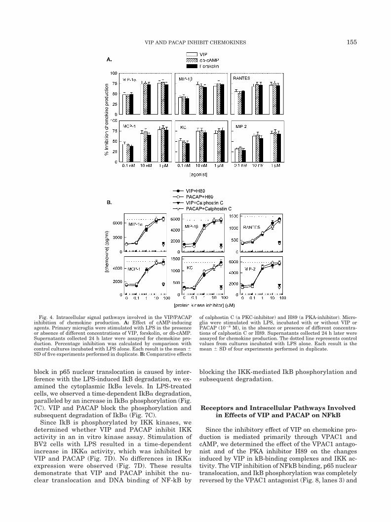
To determine whether intracellular cAMP acts as asecondary messenger, we determined the effects of H89(a PKA inhibitor), calphostin C (a PKC inhibitor), for-skolin (a cAMP-inducing agent), and db-cAMP (a cAMPanalog). Forskolin and db-cAMP inhibit chemokine re-lease similar to VIP and PACAP (Fig. 4A). In addition,
H89, but not calphostin C, completely reverses theinhibitory effect of VIP/PACAP (Fig. 4B). These resultssuggest that the inhibitory effect of VIP/PACAP is me-diated through increases in intracellular cAMP.
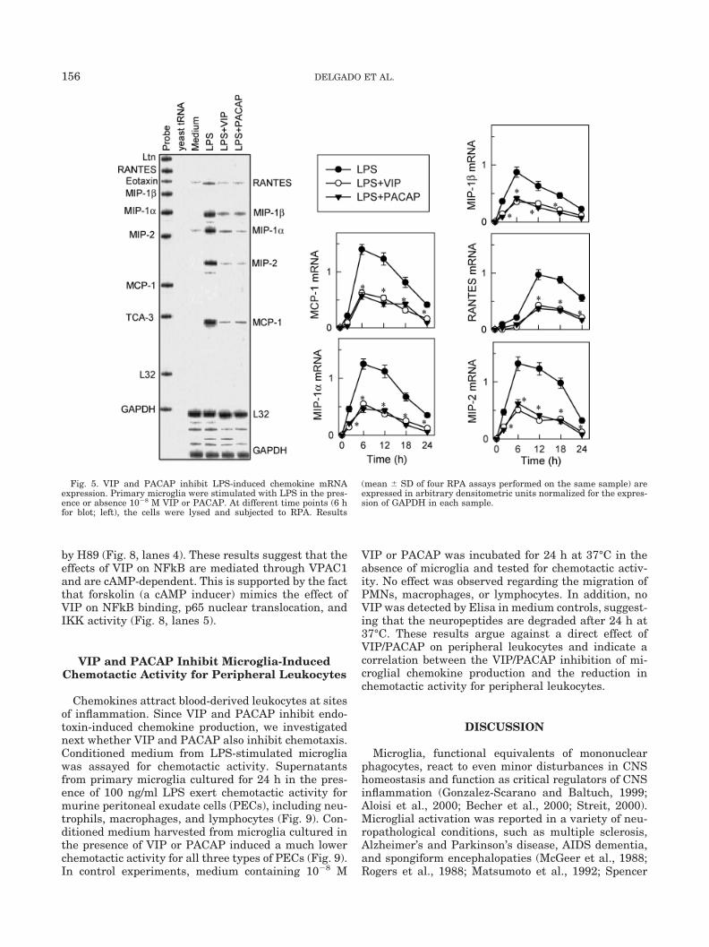
VIP and PACAP Inhibit ChemokineProduction at mRNA Level
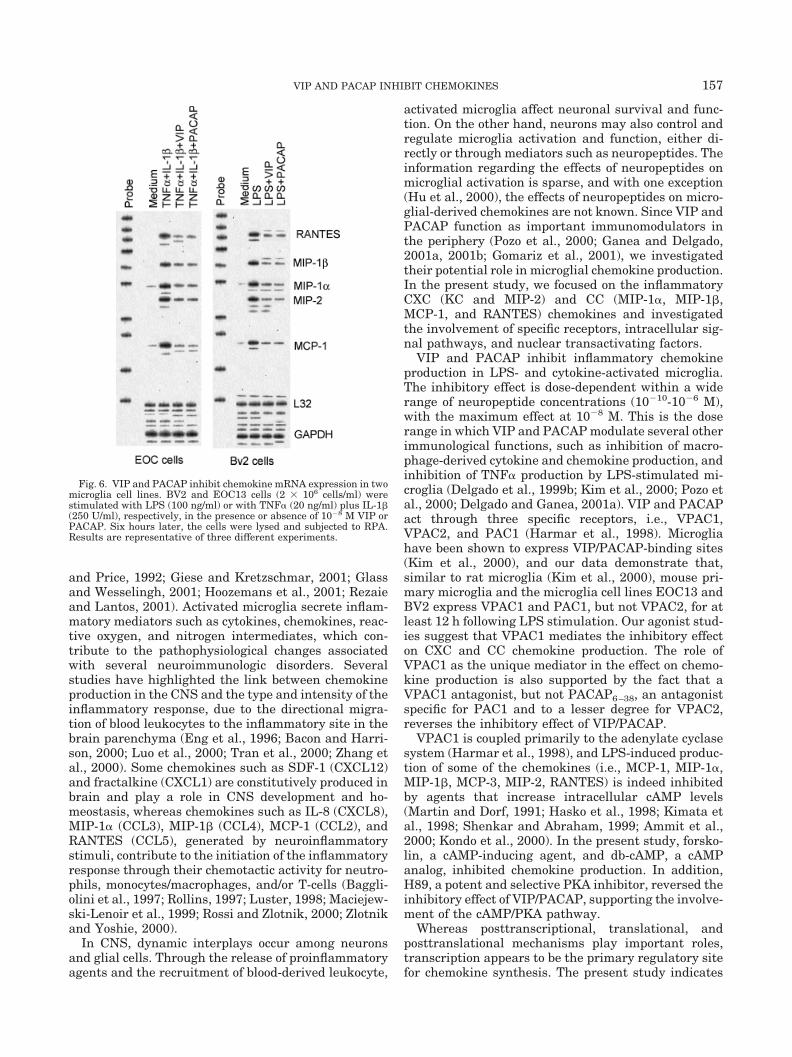
Primary microglia were treated with LPS in thepresence or absence of 10�8 M VIP or PACAP for 2, 6,12, 18, and 24 h; total RNA was prepared and subjectedto RPA analysis. Although no or very little chemokinemRNA is detectable in unstimulated cells (Fig. 5), pro-gressively increased levels of MIP-2, MIP-1�, MIP-1�,MCP-1, and RANTES mRNA are present in LPS-stim-ulated cells (maximal at 6–12 h; Fig. 5). At all timepoints, VIP and PACAP significantly inhibited the lev-els of chemokine mRNA (Fig. 5). These results indicatethat the two neuropeptides significantly reduce MIP-2,MIP-1�, MIP-1�, MCP-1, and RANTES steady-statemRNA levels. A similar pattern of chemokine mRNAinhibition was observed in EOC13 and BV2 cells (Fig. 6).
VIP and PACAP Prevent NFkB Binding toChemokine Promoters and Inhibit Subsequent
NFkB-Dependent Gene Activation
Although the promoters of most chemokines containcomplex arrays of transactivating binding sites, NFkBappears to be essential for maximal chemokine tran-scription following LPS stimulation (Freter et al., 1992,1995, 1996; Grover and Plumb, 1993; Nelson et al.,1993; Widmer et al., 1993; Ohmori et al., 1995). In
Fig. 2. VIP and PACAP inhibit cytokine-induced chemokine pro-duction. Murine primary microglia were stimulated with TNF� (20ng/ml), IFN� (250 U/ml), TNF� plus IFN�, or TNF� plus IL-1� (250U/ml), in the absence or presence of 10�8M VIP or PACAP, and
supernatants harvested at different time points were assayed forchemokine production by ELISA. Each result is the mean � SD ofthree separate experiments performed in duplicate.
153VIP AND PACAP INHIBIT CHEMOKINES
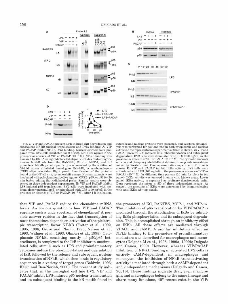
macrophages/monocytes, VIP and PACAP inhibit pro-duction of several proinflammatory cytokines by down-regulating NFkB binding and transactivation (DelgadoM et al., 1998, 1999a; Delgado and Ganea, 1999,2001b). To investigate whether VIP and PACAP affectNFkB binding in microglia, we used the murine micro-glial cell line BV2. Stimulation of BV2 cells with LPSled to an increase in NFkB binding to KC, MIP-2,MCP-1, and RANTES promoters compared to unstimu-lated cells; in all cases, treatment with VIP and PACAPsignificantly inhibited the binding (Fig. 7A, left). TheNFkB binding specificity was evident by the completedisplacement of the NFkB/DNA binding complexes inthe presence of a 50-fold excess of unlabeled kB oligo-nucleotides (Fig. 7A, middle). Antibody supershift ex-periments indicate that the NFkB-binding complexes
consist primarily of p50/p65 heterodimers (Fig. 7A,right).
The primary level of control for NF-kB is mediatedthrough its interaction with the inhibitor IkB. VIP andPACAP could inhibit NF-kB activity by blocking LPS-induced IkB degradation and subsequent NF-kB nu-clear translocation. The levels of cytoplasmic and nu-clear p65 were determined by Western blotting. Asexpected, p65 was predominantly localized in the cyto-plasm of unstimulated cells, and LPS induced a de-crease in cytoplasmic and an increase in nuclear p65(Fig. 7B). VIP and PACAP abolished the LPS-inducedchange in p65 levels (Fig. 7B). Differences in p65 pro-tein levels were not due to differences in protein load-ing since similar levels of p50 were detected regardlessof treatment. To elucidate whether the VIP/PACAP
Fig. 3. The effect of VIP/PACAP is mediated through VPAC1. A:Expression of VPAC1, VPAC2, and PAC1 mRNA in murine microglia.Total RNA extracted from unstimulated and LPS-stimulated (12 h)primary microglia (107 cells) was subjected to RT-PCR with specificprimers for VPAC1, VPAC2, and PAC1. One representative experi-ment of two is shown. B: Expression of VPAC1, VPAC2, and PAC1mRNA in microglia cell lines. One representative experiment of two isshown. C: Comparative effects of VIP/PACAP receptor agonists onchemokine production. Primary microglia were stimulated with LPSin the presence or absence of different concentrations of VIP, maxadi-lan (a PAC1 agonist), Ro 25–1553 (a VPAC2 agonist), and [K15, R16,L27] VIP (1–7)-GRF (8–27) (a VPAC1 agonist). Supernatants collected24 h later were assayed for chemokine production by ELISA. Percent-
age inhibition was calculated by comparison with controls containingLPS alone. Each result is the mean � SD of four experiments. Eachsample was assayed in duplicate. D: Effect of PAC1 and VPAC an-tagonists. Primary microglia were stimulated with LPS and treatedsimultaneously with VIP or PACAP (10�8M), and different concentra-tions of the VPAC1 antagonist [Ac-His1, D-Phe2, K15, R16, L27] VIP(3–7)-GRF (8–27), or the PAC1/VPAC2 antagonist (PACAP6–38). Su-pernatants collected 24 h later were assayed for chemokine produc-tion by ELISA. The VPAC1 antagonist (10�6M) and PACAP6–38(10�6M) did not affect chemokine production in LPS-treated micro-glia. The dotted line represents control values from cultures incubatedwith LPS alone. Each result is the mean � SD of four experimentsperformed in duplicate.
154 DELGADO ET AL.
block in p65 nuclear translocation is caused by inter-ference with the LPS-induced IkB degradation, we ex-amined the cytoplasmic IkB� levels. In LPS-treatedcells, we observed a time-dependent IkB� degradation,paralleled by an increase in IkB� phosphorylation (Fig.7C). VIP and PACAP block the phosphorylation andsubsequent degradation of IkB� (Fig. 7C).
Since IkB is phosphorylated by IKK kinases, wedetermined whether VIP and PACAP inhibit IKKactivity in an in vitro kinase assay. Stimulation ofBV2 cells with LPS resulted in a time-dependentincrease in IKK� activity, which was inhibited byVIP and PACAP (Fig. 7D). No differences in IKK�expression were observed (Fig. 7D). These resultsdemonstrate that VIP and PACAP inhibit the nu-clear translocation and DNA binding of NF-kB by
blocking the IKK-mediated IkB phosphorylation andsubsequent degradation.
Receptors and Intracellular Pathways Involvedin Effects of VIP and PACAP on NFkB
Since the inhibitory effect of VIP on chemokine pro-duction is mediated primarily through VPAC1 andcAMP, we determined the effect of the VPAC1 antago-nist and of the PKA inhibitor H89 on the changesinduced by VIP in kB-binding complexes and IKK ac-tivity. The VIP inhibition of NFkB binding, p65 nucleartranslocation, and IkB phosphorylation was completelyreversed by the VPAC1 antagonist (Fig. 8, lanes 3) and
Fig. 4. Intracellular signal pathways involved in the VIP/PACAPinhibition of chemokine production. A: Effect of cAMP-inducingagents. Primary microglia were stimulated with LPS in the presenceor absence of different concentrations of VIP, forskolin, or db-cAMP.Supernatants collected 24 h later were assayed for chemokine pro-duction. Percentage inhibition was calculated by comparison withcontrol cultures incubated with LPS alone. Each result is the mean �SD of five experiments performed in duplicate. B: Comparative effects
of calphostin C (a PKC-inhibitor) and H89 (a PKA-inhibitor). Micro-glia were stimulated with LPS, incubated with or without VIP orPACAP (10�8 M), in the absence or presence of different concentra-tions of calphostin C or H89. Supernatants collected 24 h later wereassayed for chemokine production. The dotted line represents controlvalues from cultures incubated with LPS alone. Each result is themean � SD of four experiments performed in duplicate.
155VIP AND PACAP INHIBIT CHEMOKINES
by H89 (Fig. 8, lanes 4). These results suggest that theeffects of VIP on NFkB are mediated through VPAC1and are cAMP-dependent. This is supported by the factthat forskolin (a cAMP inducer) mimics the effect ofVIP on NFkB binding, p65 nuclear translocation, andIKK activity (Fig. 8, lanes 5).
VIP and PACAP Inhibit Microglia-InducedChemotactic Activity for Peripheral Leukocytes
Chemokines attract blood-derived leukocytes at sitesof inflammation. Since VIP and PACAP inhibit endo-toxin-induced chemokine production, we investigatednext whether VIP and PACAP also inhibit chemotaxis.Conditioned medium from LPS-stimulated microgliawas assayed for chemotactic activity. Supernatantsfrom primary microglia cultured for 24 h in the pres-ence of 100 ng/ml LPS exert chemotactic activity formurine peritoneal exudate cells (PECs), including neu-trophils, macrophages, and lymphocytes (Fig. 9). Con-ditioned medium harvested from microglia cultured inthe presence of VIP or PACAP induced a much lowerchemotactic activity for all three types of PECs (Fig. 9).In control experiments, medium containing 10�8 M
VIP or PACAP was incubated for 24 h at 37°C in theabsence of microglia and tested for chemotactic activ-ity. No effect was observed regarding the migration ofPMNs, macrophages, or lymphocytes. In addition, noVIP was detected by Elisa in medium controls, suggest-ing that the neuropeptides are degraded after 24 h at37°C. These results argue against a direct effect ofVIP/PACAP on peripheral leukocytes and indicate acorrelation between the VIP/PACAP inhibition of mi-croglial chemokine production and the reduction inchemotactic activity for peripheral leukocytes.
DISCUSSION
Microglia, functional equivalents of mononuclearphagocytes, react to even minor disturbances in CNShomeostasis and function as critical regulators of CNSinflammation (Gonzalez-Scarano and Baltuch, 1999;Aloisi et al., 2000; Becher et al., 2000; Streit, 2000).Microglial activation was reported in a variety of neu-ropathological conditions, such as multiple sclerosis,Alzheimer’s and Parkinson’s disease, AIDS dementia,and spongiform encephalopaties (McGeer et al., 1988;Rogers et al., 1988; Matsumoto et al., 1992; Spencer
Fig. 5. VIP and PACAP inhibit LPS-induced chemokine mRNAexpression. Primary microglia were stimulated with LPS in the pres-ence or absence 10�8 M VIP or PACAP. At different time points (6 hfor blot; left), the cells were lysed and subjected to RPA. Results
(mean � SD of four RPA assays performed on the same sample) areexpressed in arbitrary densitometric units normalized for the expres-sion of GAPDH in each sample.
156 DELGADO ET AL.
and Price, 1992; Giese and Kretzschmar, 2001; Glassand Wesselingh, 2001; Hoozemans et al., 2001; Rezaieand Lantos, 2001). Activated microglia secrete inflam-matory mediators such as cytokines, chemokines, reac-tive oxygen, and nitrogen intermediates, which con-tribute to the pathophysiological changes associatedwith several neuroimmunologic disorders. Severalstudies have highlighted the link between chemokineproduction in the CNS and the type and intensity of theinflammatory response, due to the directional migra-tion of blood leukocytes to the inflammatory site in thebrain parenchyma (Eng et al., 1996; Bacon and Harri-son, 2000; Luo et al., 2000; Tran et al., 2000; Zhang etal., 2000). Some chemokines such as SDF-1 (CXCL12)and fractalkine (CXCL1) are constitutively produced inbrain and play a role in CNS development and ho-meostasis, whereas chemokines such as IL-8 (CXCL8),MIP-1� (CCL3), MIP-1� (CCL4), MCP-1 (CCL2), andRANTES (CCL5), generated by neuroinflammatorystimuli, contribute to the initiation of the inflammatoryresponse through their chemotactic activity for neutro-phils, monocytes/macrophages, and/or T-cells (Baggli-olini et al., 1997; Rollins, 1997; Luster, 1998; Maciejew-ski-Lenoir et al., 1999; Rossi and Zlotnik, 2000; Zlotnikand Yoshie, 2000).
In CNS, dynamic interplays occur among neuronsand glial cells. Through the release of proinflammatoryagents and the recruitment of blood-derived leukocyte,
activated microglia affect neuronal survival and func-tion. On the other hand, neurons may also control andregulate microglia activation and function, either di-rectly or through mediators such as neuropeptides. Theinformation regarding the effects of neuropeptides onmicroglial activation is sparse, and with one exception(Hu et al., 2000), the effects of neuropeptides on micro-glial-derived chemokines are not known. Since VIP andPACAP function as important immunomodulators inthe periphery (Pozo et al., 2000; Ganea and Delgado,2001a, 2001b; Gomariz et al., 2001), we investigatedtheir potential role in microglial chemokine production.In the present study, we focused on the inflammatoryCXC (KC and MIP-2) and CC (MIP-1�, MIP-1�,MCP-1, and RANTES) chemokines and investigatedthe involvement of specific receptors, intracellular sig-nal pathways, and nuclear transactivating factors.
VIP and PACAP inhibit inflammatory chemokineproduction in LPS- and cytokine-activated microglia.The inhibitory effect is dose-dependent within a widerange of neuropeptide concentrations (10�10-10�6 M),with the maximum effect at 10�8 M. This is the doserange in which VIP and PACAP modulate several otherimmunological functions, such as inhibition of macro-phage-derived cytokine and chemokine production, andinhibition of TNF� production by LPS-stimulated mi-croglia (Delgado et al., 1999b; Kim et al., 2000; Pozo etal., 2000; Delgado and Ganea, 2001a). VIP and PACAPact through three specific receptors, i.e., VPAC1,VPAC2, and PAC1 (Harmar et al., 1998). Microgliahave been shown to express VIP/PACAP-binding sites(Kim et al., 2000), and our data demonstrate that,similar to rat microglia (Kim et al., 2000), mouse pri-mary microglia and the microglia cell lines EOC13 andBV2 express VPAC1 and PAC1, but not VPAC2, for atleast 12 h following LPS stimulation. Our agonist stud-ies suggest that VPAC1 mediates the inhibitory effecton CXC and CC chemokine production. The role ofVPAC1 as the unique mediator in the effect on chemo-kine production is also supported by the fact that aVPAC1 antagonist, but not PACAP6–38, an antagonistspecific for PAC1 and to a lesser degree for VPAC2,reverses the inhibitory effect of VIP/PACAP.
VPAC1 is coupled primarily to the adenylate cyclasesystem (Harmar et al., 1998), and LPS-induced produc-tion of some of the chemokines (i.e., MCP-1, MIP-1�,MIP-1�, MCP-3, MIP-2, RANTES) is indeed inhibitedby agents that increase intracellular cAMP levels(Martin and Dorf, 1991; Hasko et al., 1998; Kimata etal., 1998; Shenkar and Abraham, 1999; Ammit et al.,2000; Kondo et al., 2000). In the present study, forsko-lin, a cAMP-inducing agent, and db-cAMP, a cAMPanalog, inhibited chemokine production. In addition,H89, a potent and selective PKA inhibitor, reversed theinhibitory effect of VIP/PACAP, supporting the involve-ment of the cAMP/PKA pathway.
Whereas posttranscriptional, translational, andposttranslational mechanisms play important roles,transcription appears to be the primary regulatory sitefor chemokine synthesis. The present study indicates
Fig. 6. VIP and PACAP inhibit chemokine mRNA expression in twomicroglia cell lines. BV2 and EOC13 cells (2 106 cells/ml) werestimulated with LPS (100 ng/ml) or with TNF� (20 ng/ml) plus IL-1�(250 U/ml), respectively, in the presence or absence of 10�8 M VIP orPACAP. Six hours later, the cells were lysed and subjected to RPA.Results are representative of three different experiments.
157VIP AND PACAP INHIBIT CHEMOKINES
that VIP and PACAP reduce the chemokine mRNAlevels. An obvious question is how VIP and PACAPregulate such a wide spectrum of chemokines? A pos-sible answer resides in the fact that transcription ofmost chemokines depends on activation of the pleiotro-pic transcription factor NF-kB (Freter et al., 1992,1995, 1996; Grove and Plumb, 1993; Nelson et al.,1993; Widmer et al., 1993; Ohmori et al., 1995). Cyto-plasmic NF-kB, consisting mostly of p50/p65 het-erodimers, is complexed to the IkB inhibitor in unstimu-lated cells; stimuli such as LPS and proinflammatorycytokines induce the phosphorylation and degradationof IkB, followed by the release and subsequent nucleartranslocation of NFkB, which then binds to regulatorysequences in a variety of target genes (Baldwin, 1996;Karin and Ben-Neriah, 2000). The present study indi-cates that, in the microglial cell line BV2, VIP andPACAP inhibit LPS-induced p65 nuclear translocationand its subsequent binding to the kB motifs found in
the promoters of KC, RANTES, MCP-1, and MIP-1�.The inhibition of p65 translocation by VIP/PACAP ismediated through the stabilization of IkB� by inhibit-ing IkB� phosphorylation and its subsequent degrada-tion. This is accomplished through an inhibitory effecton IKK�. All these effects are mediated throughVPAC1 and cAMP. A similar inhibitory effect onNFkB binding to the promoters of proinflammatorymediators was described for macrophages and mono-cytes (Delgado M et al., 1998, 1999a, 1999b; Delgadoand Ganea, 1999). However, whereas VIP/PACAPinhibition of NF-kB binding in activated BV2 cells isentirely cAMP-dependent, in macrophages andmonocytes, the inhibition of NFkB transactivatingactivity is mediated through both a cAMP-dependentand -independent mechanisms (Delgado and Ganea,2001b). These findings indicate that, even if micro-glia and macrophages belong to the same lineage andshare many functions, differences exist in the VIP/
Fig. 7. VIP and PACAP prevent LPS-induced IkB degradation andsubsequent NF-kB nuclear translocation and DNA binding. A: VIPand PACAP inhibit NF-kB DNA binding. Nuclear extracts were pre-pared from BV2 cells incubated for 2 h with LPS (100 ng/ml) in thepresence or absence of VIP or PACAP (10�8 M). NF-kB binding wasassessed by EMSA using radiolabeled oligonucleotides containing themurine NF-kB site from the RANTES, MIP-1�, MCP-1, and KCpromoters. Middle panel: Specificity was assessed by the addition of50-fold excess unlabeled homologous (NF-kB), or nonhomologous(CRE) oligonucleotides. Right panel: Identification of the proteinsbound to the NF-kB site, by supershift assays. Nuclear extracts wereincubated with polyclonal antibodies against CREB, p65, or p50 for 20min before adding the radiolabeled probe. Similar results were ob-served in three independent experiments. B: VIP and PACAP inhibitLPS-induced p65 translocation. BV2 cells were incubated with me-dium alone (unstimulated) or stimulated with LPS (100 ng/ml) in thepresence or absence of VIP or PACAP (10�8 M). After 1-h incubation,
cytosolic and nuclear proteins were extracted, and Western blot anal-ysis was performed for p50 and p65 in both cytoplasmic and nuclearextracts. One representative experiment of three is shown. C: VIP andPACAP prevent LPS-induced IkB� phosphorylation and subsequentdegradation. BV2 cells were stimulated with LPS (100 ng/ml) in thepresence or absence of VIP or PACAP (10�8 M). The cytosolic amountsof IkB� and phosphorylated-IkB� at different time points were deter-mined by Western blot. One representative experiment of three isshown. D: VIP and PACAP inhibit IKK� activity. BV2 cells werestimulated with LPS (100 ng/ml) in the presence or absence of VIP orPACAP (10�8 M) for different time periods (10 min for blots in toppanel). IKK� activity was assayed in an in vitro kinase assay. Lowerpanel: IKK� activity is expressed as arbitrary densitometric units.Data represent the mean � SD of three independent assays. Ascontrol, the amounts of IKK� were determined by immunoblottingwith anti-IKK� Ab (top panel).
158 DELGADO ET AL.

PACAP transduction pathways for the regulation ofproinflammatory factors.
Of obvious biological significance is the fact that VIPand PACAP reduce the chemotactic activity generatedby activated microglia for peripheral leukocytes, i.e.,neutrophils, macrophages, and lymphocytes. The re-duction in chemokine production is in good correlationwith the decrease in chemotactic activity, suggestingthat the effect of VIP/PACAP on microglia-derived che-mokines has significant physiological consequences.
Reduction in the number and activation of infiltratingleukocytes represents an important factor in the con-trol of inflammation in the CNS.
These data lead us to propose a model for the inter-actions between cells in the CNS during a normal re-sponse to infectious agents or to traumatic injury. Al-though the initial step of antigen uptake andpresentation is not entirely clear, microglia, and to acertain extent astrocytes, can function as antigen-pre-senting cells, upregulating the expression of MHC classII, CD40, and B7 molecules. Presentation of the anti-gen, possibly to patrolling T-cells, contributes to fur-ther activation of glial cells, characterized by secretionof chemokines, cytokines, nitric oxide, and oxygen rad-icals. All these proinflammatory factors attract andfurther activate immune cells in the CNS and contrib-ute to the direct destruction of the invading pathogen.However, an active proinflammatory response in theCNS can also damage and kill neighboring cells, in-cluding neurons. It would make sense that, faced witha nearby inflammatory response, neurons will try toprotect themselves by secreting anti-inflammatory fac-tors. It is interesting that neuronal injury leads to asignificant increase in neuropeptide message, and fur-ther that most neuropeptides are indeed anti-inflam-matory. In this context, VIP and/or PACAP released byneurons during an inflammatory response could serveas neuronal survival factors by limiting the inflamma-tory process.
ACKNOWLEDGMENTS
This work was supported by grants PHS AI041786-03 (to D.G.) and PM98-0081 (to M.D.). Theauthors thank Dr. Patrick Robberecht (Universite Li-bre de Bruxelles, Brussels, Belgium) for the VPAC1agonist and antagonist, Drs. David Bolin and Ann Wel-ton (Hoffmann-LaRoche, Nutley, NJ) for the VPAC2agonist Ro 25–1553, Dr. Ethan Lerner (MassachusettsGeneral Hospital, Charlestown, MA) for the PAC1 ag-
Fig. 8. Receptors and intracellular pathways involved in the VIPand PACAP regulation of NFkB nuclear translocation and IKK activ-ity. BV2 cells were activated with LPS (100 ng/ml) in the absence(lanes 1) or presence of VIP (10�8 M, lanes 2), or forskolin (10�6 M,lanes 5). VPAC1 antagonist (10�7 M, lanes 3) or H89 (100 ng/ml, lanes4) were added simultaneously with VIP (10�8 M). A: NF-kB bindingwas analyzed 1 h after stimulation by EMSA as described in Figure 7.
One representative experiment of three is shown. B: After 1-h incu-bation, nuclear proteins were extracted, and Western blot analysiswas performed for p50 and p65 in nuclear extracts. One representa-tive experiment of four is shown. C: IKK� activity (20 min afterstimulation) was analyzed with IkB� as substrate by using an in vitrokinase assay. One representative experiment of three is shown.
Fig. 9. VIP and PACAP inhibit microglia-induced chemotactic ac-tivity for peritoneal leukocytes. Primary microglia were stimulatedwith LPS in the presence or absence of VIP or PACAP (10�8 M).Supernatants collected 24 h later were seeded in lower wells of 48-well chemotaxis microchamber, and chemotaxis of freshly isolatedmurine peritoneal cells was assayed at 37°C for 90 min as described intext. At the end of the incubation period, the filters were stained, andthe number of the different peritoneal cell populations that migratedwere evaluated (chemotaxis index). Complete medium instead culturesupernatants was used as basal control (none). Results are expressedas mean � SD of triplicate determinations of one representativeexperiment of four.
159VIP AND PACAP INHIBIT CHEMOKINES

onist maxadilan, Dr. Etty N. Benveniste (University ofAlabama at Birmingham, Birmingham, AL) for EOC13cells, and Dr. Virginia Bocchini (Universita degli Studidi Perugia, Perugia, Italy) for the BV2 cells.
REFERENCES
Aloisi F, Serafini B, Adorini L. 2000. Glia-T cell dialogue. J Neuroim-munol 107:111–117.
Ammit AJ, Hoffman RK, Amrani Y, Lazaar AL, Hay DW, Torphy TJ,Penn RB, Panettieri RA. 2000. Tumor necrosis factor-alpha-inducedsecretion of RANTES and interleukin-6 from human airwaysmooth-muscle cells: modulation by cyclic adenosine monophos-phate. Am J Respir Cell Mol Biol 23:794–802.
Bacon KB, Harrison JK. 2000. Chemokines and their receptors inneurobiology: perspectives in physiology and homeostasis. J Neu-roimmunol 104:92–97.
Baggiolini M, Dewald B, Moser B. 1997. Human chemokines: anupdate. Ann Rev Immunol 15:675–705.
Baldwin AS Jr. 1996. The NFkappaB and IkappaB proteins: newdiscoveries and insights. Ann Rev Immunol 14:649–681.
Becher B, Prat A, Antel JP. 2000. Brain-immune connection: immuno-regulatory properties of CNS-resident cells. Glia 29:293–304.
Blasi E, Barluzzi R, Bocchini V, Mazzolla R, Bistoni F. 1990. Immor-talization of murine microglial cells by v-raf/v-myc carrying retro-virus. J Neuroimmunol 27:229–237.
Chao CC, Molitor TW, Shuxian H. 1993. Neuroprotective role of IL-4against activated microglia. J Immunol 151:1473–1481.
Delgado R, Carlin A, Airaghi L, Demitri MT, Meda L, Galimberti D,Baron P, Lipton JM, Catania A. 1998. Melanocortin peptides inhibitproduction of proinflammatory cytokines and nitric oxide by acti-vated microglia. J Leukoc Biol 63:740–745.
Delgado M, Munoz-Elias EJ, Kan Y, Gozes I, Fridkin M, BrennemanDE, Gomariz RP, Ganea D. 1998. Vasoactive intestinal peptide andpituitary adenylate cyclase activating polypeptide inhibit TNF�transcriptional activation by regulating NF-kB and CREB/c-Jun.J Biol Chem 273:31427–31436.
Delgado M, Ganea D. 1999. VIP and PACAP inhibit IL-12 transcrip-tion by regulating NFkB and Ets activation. J Biol Chem 274:31930–31940.
Delgado M, Munoz-Elias EJ, Gomariz RP, Ganea D. 1999a. VIP andPACAP prevent inducible nitric oxide synthase transcription inmacrophages by inhibiting NF-kB and interferon regulatory factor1 activation. J Immunol 162:4685–4696.
Delgado M, Munoz-Elias EJ, Martinez C, Gomariz RP, Ganea D.1999b. VIP and PACAP modulate cytokine and nitric oxide produc-tion in peritoneal macrophages and macrophage cell lines. Ann NYAcad Sci 897:401–414.
Delgado M, Ganea D. 2001a. Inhibition of endotoxin-induced macro-phage chemokine production by vasoactive intestinal peptide andpituitary adenylate cyclase-activating polypeptide in vitro and invivo. J Immunol 167:966–975.
Delgado M, Ganea D. 2001b. Vasoactive intestinal peptide and pitu-itary adenylate cyclase-activating polypeptide regulated NFkB atmultiple levels. J Biol Chem 276:369–380.
Eng LF, Ghirnikar RS, Lee Y. 1996. Inflammation in EAE: role ofchemokine/cytokine expression by resident and infiltrating cells.Neurochem Res 21:511–525.
Feindt J, Schmidt A, Mentlein R. 1998. Receptors and effects of theinhibitory neuropeptide somatostatin in microglial cells. Brain ResMol Brain Res 60:228–233.
Freter RR, Irminger JC, Porter JA, Jones SD, Stiles CD. 1992. A novel7-nucleotide motif located in 3 untranslated sequences of the im-mediate-early gene set mediates platelet-derived growth factor in-duction of the JE gene. Mol Cell Biol 12:5288–5300.
Freter RR, Alberta JA, Lam KK, Stiles CD. 1995. A new platelet-derived growth factor-regulated genomic element which binds aserine/threonine phosphoprotein mediates induction of the slowimmediate-early gene MCP-1. Mol Cell Biol 15:315–325.
Freter RR, Alberta JA, Hwang GY, Wrentmore AL, Stiles CD. 1996.Platelet-derived growth factor induction of the immediate-earlygene MCP-1 is mediated by NF-kappaB and a 90-kDa phosphopro-tein coactivator. J Biol Chem 271:17417–17424.
Frohman EM, Frohman TC, Vayuvegula B, Gupta S, van den Noort S.1988. Vasoactive intestinal peptide inhibits the expression of theMHC class II antigens on astrocytes. J Neurol Sci 88:339–346.
Galimberti D, Baron P, Meda L, Prat E, Scarpini E, Delgado R,Catania A, Lipton JM, Scarlato G. 1999. Alpha-MSH peptides in-
hibit production of nitric oxide and tumor necrosis factor-alpha bymicroglial cells activated with beta-amyloid and interferon gamma.Biochem Biophys Res Comm 263:251–256.
Ganea D, Delgado M. 2001a. Neuropeptides as modulators of macro-phage functions. Regulation of cytokine production and antigenpresentation by VIP and PACAP. Arch Immunol Therap Exp 49:101–110.
Ganea D, Delgado M. 2001b. Inhibitory neuropeptide receptors onmacrophages. Microbes Inf 3:141–147.
Giese A, Kretzschmar HA. 2001. Prion-induced neuronal damage: themechanisms of neuronal destruction in the subacute spongiformencephalopathies. Curr Top Microbiol Immunol 253:203–217.
Glass JD, Wesselingh SL. 2001. Microglia in HIV-associated neuro-logical diseases. Microsc Res Tech 54:95–105.
Godiska R, Chantry D, Dietsch GN, Gray PW. 1995. Chemokineexpression in murine experimental allergic encephalomyelitis.J Neuroimmunol 58:167–176.
Gomariz RP, Martinez C, Abad C, Leceta J, Delgado M. 2001. Immu-nology of VIP: a review and therapeutical perspectives. Curr Phar-macol Design 7:89–111.
Gonzalez-Scarano F, Baltuch G. 1999. Microglia as mediators of in-flammatory and degenerative diseases. Ann Rev Neurosci 22:219–240.
Gottschall PE, Tatsuno I, Arimura A. 1994. Regulation of IL-6 secre-tion in primary cultured rat astrocytes: synergism of IL-1 andPACAP. Brain Res 637:197–203.
Gourlet P, Vandermeers-Piret MC, Rathe J, De Neef P, Robberecht P.1995. Fragments of the pituitary adenylate cyclase-activatingpolypeptide discriminate between type I and II recombinant recep-tors. Eur J Pharmacol 287:7–11.
Gourlet PA, Vandermeers P, Vertongen J, Ratche P, De Neef J,Cnudde M, Waelbroeck M, Robberecht P. 1997a. Development ofhigh affinity selective VIP1 receptor agonists. Peptides 18:1539–1545.
Gourlet P, De Neef P, Cnudde J, Waelbroeck M, Robberecht P. 1997b.In vitro properties of a high affinity selective antagonist of the VIP1receptor. Peptides 18:1555–1560.
Grove M, Plumb M. 1993 C/EBP, NF-kappa B, and c-Ets familymembers and transcriptional regulation of the cell-specific and in-ducible macrophage inflammatory protein 1 alpha immediate-earlygene. Mol Cell Biol 13:5276–5289.
Harmar AJ, Arimura A, Gozes I, Journot L, Laburthe M, Pisegna JR,Rawlings SR, Robberecht P, Said SI, Sreedharan SP, Wank SA,Washeck JA. 1998. Nomenclature of receptors for vasoactive intes-tinal peptide (VIP) and pituitary adenylate cyclase activatingpolypeptide (PACAP). Pharmacol Rev 50:265–270.
Hasko G, Shanley TP, Egnaczyk G, Nemeth ZH, Salzman AL, Vizi ES,Szabo C. 1998. Exogenous and endogenous catecholamines inhibitthe production of macrophage inflammatory protein (MIP) 1 alphavia a beta adrenoreceptor mediated mechanism. Br J Pharmacol125:1297–1303.
Hoozemans JJ, Rozemuller AJ, Veerhuis R, Eikelenboom P. 2001.Immunological aspects of Alzheimer’s disease: therapeutic implica-tions. BioDrugs 15:325–337.
Hu S, Chao CC, Hegg CC, Thayer S, Peterson PK. 2000. Morphineinhibits human microglial cell production of, and migration to-wards, RANTES. J Psychophamrmacol 14:238–243.
Karin M, Ban-Neriah Y. 2000. Phosphorylation meets ubiquitination:the control of NF-[kappa]B activity. Ann Rev Immunol 18:621–664.
Karpus WJ, Kennedy KJ. 1997. MIP-1� and MCP-1 differentiallyregulate acute and relapsing autoimmune encephalomyelitis aswell as Th1/Th2 lymphocyte differentiation. J Leukocyte Biol 62:681–687.
Karpus WJ, Ransohoff RM. 1998. Chemokine regulation of experi-mental autoimmune encephalomyelitis: temporal and spatial ex-pression patterns govern disease pathogenesis. J Immunol 161:2667–2671.
Kim, W-K, Kan Y, Ganea D, Hart R, Gozes I, Jonakait GM. 2000.Vasoactive intestinal peptide and pituitary adenylyl cyclase-acti-vating polypeptide inhibit tumor necrosis factor-a production ininjured spinal cord and in activated microglia via a cAMP-depen-dent pathway. J Neurosci 20:3622–3630.
Kimata M, Shichijo M, Daikoku M, Inagaki N, Mori H, Nagai H. 1998.Pharmacological modulation of LPS-induced MIP-1 alpha produc-tion by peripheral blood mononuclear cells. Pharmacology 56:230–236.
Kondo A, Isaji S, Nishimura Y, Tanaka T. 2000. Transcriptional andpost-transcriptional regulation of monocyte chemoattractant pro-tein-3 gene expression in human endothelial cells by phorbol esterand cAMP signaling. Immunology 99:561–568.
160 DELGADO ET AL.
Luber-Narod J, Kage R, Leeman SE. 1994. Substance P enhances thesecretion of tumor necrosis factor-alpha from neuroglial cells stim-ulated with lipopolysaccharide. J Immunol 152:819–824.
Luo Y, Fischer FR, Hancock WW, Dorf ME. 2000. Macrophage inflam-matory protein-2 and KC induce chemokine production by mouseastrocytes. J Immunol 165:4015–4023.
Luster AD. 1998. Chemokines: chemotactic cytokines that mediateinflammation. N Engl J Med 338:436–445.
Maciejewski-Lenoir D, Chen S, Feng L, Maki R, Bacon KB. 1999.Characterization of fractalkine in rat brain cells: migratory andactivation signals for CX3CR-1-expressing microglia. J Immunol163:1628–1635.
Martin CA, Dorf ME. 1991. Differential regulation of interleukin-6,macrophage inflammatory protein-1, and JE/MCP-1 cytokine ex-pression in macrophage cell lines. Cell Immunol 135:245–258.
Martinez C, Delgado M, Abad C, Gomariz RP, Ganea D, Leceta J.1999. Regulation of VIP production and secretion by murine lym-phocytes. J Neuroimmunol 93:126–138.
Matsumoto Y, Ohmori K, Fujiwara M. 1992. Microglial and astroglialreactions to inflammatory lesions of experimental autoimmune en-cephalomyelitis in the rat central nervous system. J Neuroimmunol37:23–33.
McGeer PL, Itagaki S, Boyes BE, McGeer EG. 1988. Reactive micro-glia are positive for HLA-DR in the substantia nigra of Parkinson’sand Alzheimers disease brains. Neurology 38:1285–1291.
Meda L, Bernasconi S, Bonaiuto C, Sozzani S, Zhou D, Otvos L,Mantovani A, Rossi F, Cassatella MA. 1996. �-amyloid (25-35)peptide and IFN-� synergistically induce the production of thechemotactic cytokine MCP-1/JE in monocyte and microglial cells.J Immunol 157:1213–1218.
Moro O, Lerner EA. 1997. Maxadilan, the vasodilator peptide fromsand flies, is a specific pituitary adenylate cyclase activating pep-tide type I receptor agonist. J Biol Chem 271:966–970.
Nelson PJ, Kim HT, Manning WC, Goralski TJ, Kernsky AM. 1993.Genomic organization and transcriptional regulation of the RAN-TES chemokine gene. J Immunol 151:2601–2612.
Ohmori Y, Fukumoto S, Hamilton TA. 1995. Two structurally distinctkB sequence motifs cooperatively control LPS-induced KC genetranscription in mouse macrophages. J Immunol 155:3593–3600.
Pozo D, Delgado M, Martinez C, Guerrero JM, Leceta J, Gomariz RP,Calvo JR. 2000. Immunobiology of vasoactive intestinal peptide(VIP). Immunol Today 21:7–11.
Rezaie P, Lantos PL. 2001. Microglia and the pathogenesis of spon-giform encephalopathies. Brain Res Brain Res Rev 35:55–72.
Rollins BJ. 1997. Chemokines. Blood 90:909–928.Rogers J, Luber-Nardo J, Styren SD, Civin WH. 1988. Expression of
immune system-associated antigens by cells of the central nervoussystem: relationship to the pathology of Alzheimers disease. Neu-robiol Aging 9:339–449.
Rossi D, Zlotnik A. 2000. The biology of chemokines and their recep-tors. Ann Rev Immunol 18:217–242.
Schreiber E, Metthias P, Muller MM, Shaffner W. 1989. Rapid detec-tion of octamer binding proteins with “mini-extracts” prepared froma small number of cells. Nucl Acids Res 17:6419.
Shenkar R, Abraham E. 1999. Mechanisms of lung neutrophil activa-tion after hemorrhage or endotoxemia: roles of reactive oxygenintermediates, NF-kappa B, and cyclic AMP response element bind-ing protein. J Immunol 163:954–962.
Spencer DC, Price RW. 1992. Human immunodeficiency virus and thecentral nervous system. Ann Rev Microbiol 46:655–693.
Streit WJ, Semple-Rowland SL, Hurley SD, Miller RC, Popovich PG,Stokes BT. 1998. Cytokine mRNA profiles in contused spinal cordand axotomized facial nucleus suggest a beneficial role for inflam-mation and gliosis. Exp Neurol 15:74–87.
Streit WJ. 2000. Microglial response to brain injury: a brief synopsis.Toxicol Pathol 28:28–30.
Tran EH, Prince EN, Owens T. 2000. IFN-� shapes immune invasionof the central nervous system via regulation of chemokines. J Im-munol 164:2759–2768.
Walker WS, Gatewood J, Olivas E, Askew D, Havenith CEG. 1995.Mouse microglial cell lines differing in constitutive and interferon-g-inducible antigen-presenting activities for naı̈ve and memoryCD4� and CD8� T cells. J Neuroimmunol 63:163–174.
Widmer U, Manogue KR, Cerami A, Sherry B. 1993. Genomic cloningand promoter analysis of macrophage inflammatory protein(MIP)-2, MIP-1�, and MIP-1�, members of the chemokine super-family of proinflammatory cytokines. J Immunol 150:4996–5012.
Xia M, Sreedharan SP, Bolin DR, Gaufo GO, Goetzl EJ. 1997. Novelcyclic peptide agonist of high potency and selectivity for the type IIvasoactive intestinal peptide receptor. J Pharmacol Exp Ther 281:629–633.
Zhang GX, Baker CM, Kolson DL, Rostami AM. 2000. Chemokinesand chemokine receptors in the pathogenesis of multiple sclerosis.Mult Scler 6:3–13.
Zlotnik A, Yoshie O. 2000. Chemokines: a new classification systemand their role in immunity. Immunity 12:121–127.
161VIP AND PACAP INHIBIT CHEMOKINES