ikk is required for peripheral b cell survival and ... · ikk is required for peripheral b cell...
TRANSCRIPT

of December 1, 2018.This information is current as
Survival and Proliferation Is Required for Peripheral B CellβIKK
and Robert C. RickertZhi-Wei Li, Sidne A. Omori, Tord Labuda, Michael Karin
http://www.jimmunol.org/content/170/9/4630doi: 10.4049/jimmunol.170.9.4630
2003; 170:4630-4637; ;J Immunol
Referenceshttp://www.jimmunol.org/content/170/9/4630.full#ref-list-1
, 26 of which you can access for free at: cites 47 articlesThis article
average*
4 weeks from acceptance to publicationFast Publication! •
Every submission reviewed by practicing scientistsNo Triage! •
from submission to initial decisionRapid Reviews! 30 days* •
Submit online. ?The JIWhy
Subscriptionhttp://jimmunol.org/subscription
is online at: The Journal of ImmunologyInformation about subscribing to
Permissionshttp://www.aai.org/About/Publications/JI/copyright.htmlSubmit copyright permission requests at:
Email Alertshttp://jimmunol.org/alertsReceive free email-alerts when new articles cite this article. Sign up at:
Print ISSN: 0022-1767 Online ISSN: 1550-6606. Immunologists All rights reserved.Copyright © 2003 by The American Association of1451 Rockville Pike, Suite 650, Rockville, MD 20852The American Association of Immunologists, Inc.,
is published twice each month byThe Journal of Immunology
by guest on Decem
ber 1, 2018http://w
ww
.jimm
unol.org/D
ownloaded from
by guest on D
ecember 1, 2018
http://ww
w.jim
munol.org/
Dow
nloaded from

IKK � Is Required for Peripheral B Cell Survival and Proliferation 1
Zhi-Wei Li,* † Sidne A. Omori,†‡ Tord Labuda,2*† Michael Karin, 3*† and Robert C. Rickert3†‡
NF-�B activity in mammalian cells is regulated through the I�B kinase (IKK) complex, consisting of two catalytic subunits (IKK�and IKK �) and a regulatory subunit (IKK �). Targeted deletion ofIkk� results in early embryonic lethality, thus complicating theexamination of IKK � function in adult tissues. Here we describe the role of IKK� in B lymphocytes made possible by generationof a mouse strain that expresses a conditionalIkk� allele. We find that the loss of IKK� results in a dramatic reduction in allperipheral B cell subsets due to associated defects in cell survival. IKK�-deficient B cells are also impaired in mitogenic responsesto LPS, anti-CD40, and anti-IgM, indicating a general defect in the ability to activate the canonical NF-�B signaling pathway.These findings are consistent with a failure to mount effective Ab responses to T cell-dependent and independent Ags. Thus, IKK�provides a requisite role in B cell activation and maintenance and thus is a key determinant of humoral immunity. The Journalof Immunology, 2003, 170: 4630–4637.
N uclear factor-�B designates a family of transcription fac-tors activated by a diverse array of proinflammatory cyto-kines, pathogen-associated molecular patterns (PAMPs),4
cell-bound ligands, Ags, and physical stresses (1). Expression ofNF-�B target genes is essential for mounting innate immune re-sponses to infectious microorganisms (2), but is also important for theproper development and cellular compartmentalization of secondarylymphoid organs necessary to orchestrate an adaptive immune re-sponse (3–5). In mammals, the NF-�B family consists of five mem-bers, RelA(p65), RelB, c-Rel, p50(NF-�B1), and p52(NF-�B2), thatform hetero- and homodimeric sequence-specific transcriptional reg-ulators (2). Regulation of NF-�B activity occurs at multiple levels,primarily being dependent on inducible degradation of I�B inhibitoryproteins, which hold NF-�B dimers in the cytoplasm, and the proteo-lytic processing of the p105 and p100 precursor proteins to the maturep50 and p52 subunits, respectively (1). The processing of p105 isthought to occur constitutively (1), whereas p100 cleavage is an in-ducible process (6, 7). I�B degradation depends on site-specific serine
phosphorylation, which triggers their polyubiquitination and protea-some-mediated destruction (1). The release of NF-�B from I�Bs al-lows nuclear translocation and binding to specific DNA sequences(�B sites) at the regulatory regions of specific target genes (1, 2).While other mechanisms, some of which are based on post-transla-tional modifications of Rel/NF-�B proteins, do exist, the key regula-tory step in the canonical NF-�B activation pathway is phosphoryla-tion of I�B proteins by the I�B kinase (IKK) complex (2).
Three key components of the IKK complex were identified:IKK�, IKK�, and IKK� (2). IKK� and IKK� are catalytic sub-units showing significant sequence similarity, whereas IKK� (alsocalled NEMO) is unrelated in sequence and performs a regulatoryrole (8). Contrary to initial predictions, however, examination ofIKK�-, IKK�-, and IKK�-deficient mice has revealed that activa-tion of NF-�B by most proinflammatory stimuli and PAMPs doesnot require IKK�, but is dependent on IKK� and IKK� function(9–19). Ikk��/� mice die in midgestation due to extensive apo-ptosis of fetal hepatocytes culminating in liver failure (12, 14, 19).This phenotype is shared by Rela�/� mice and is attributed toTNF-�-induced programmed cell death, as compound mutants de-fective in either Ikk� or Rela and either Tnfr1 or Tnf� genes arerescued from embryonic lethality (14, 20, 21). Further examinationof Ikk��/�Tnfr1�/� double-mutant mice revealed reduced thymo-cyte cellularity that was attributed to impaired thymocyte prolif-eration (22). Notably, Rela�/�Tnfr1�/� mice can survive up to 6mo before they succumb to opportunistic infections (20), whereasmost Ikk��/�Tnfr1�/� mice die �1 wk after birth from rampantinfections (Z.-W. Li, unpublished observations). Thus, IKK� ac-tivity is necessary for activation of innate immune responses,which depend in part on RelA (p65)-containing dimers. Thesefindings also suggest that IKK� is responsible for activation ofseveral different forms of NF-�B besides those that contain RelA.Therefore, its elimination results in a larger decrease in totalNF-�B activity than the mere elimination of RelA, thereby causinga more dramatic increase in sensitivity to infections. Although thegeneral role of IKK� and RelA in activation of innate immuneresponse is fairly well established, there are reasons to believe thatthese proteins are also involved in adaptive immune responses,probably through effects on lymphocyte development and activa-tion (23, 24).
The large decrease in total NF-�B activity caused by the loss ofIKK� provides an opportunity to learn more about the functions ofNF-�B in lymphoid cells and a context for comparing previous
*Laboratory of Gene Regulation and Signal Transduction, Department of Pharma-cology, †Cancer Center, and ‡Division of Biology, University of California-San Di-ego, La Jolla, CA 92093
Received for publication November 25, 2002. Accepted for publication February24, 2003.
The costs of publication of this article were defrayed in part by the payment of pagecharges. This article must therefore be hereby marked advertisement in accordancewith 18 U.S.C. Section 1734 solely to indicate this fact.1 This work was supported by National Institutes of Health Grants AI43477 andES06376, the Superfund Basic Research Program (Grant ES10337), the Sandler Pro-gram for Asthma Research and the State of California Cancer Research Program(Grants 99-00529V and 10249) to M.K., who is an American Cancer Society Re-search Professor. R.C.R. is an awardee of the Hellman Fellows Program for juniorfaculty. Z.W.L. and T.L. were supported by postdoctoral fellowships from the CancerResearch Institute and the Swedish Research Council, respectively.2 Current address: Department of Medical Microbiology and Immunology, Universityof Copenhagen, Panum Institute Building 22.5.37, Blegdamsnej 3, 2200 Copenhagen,Denmark.3 Address correspondence and reprint requests to Dr. Robert C. Rickert, Division ofBiology, University of California-San Diego, 9500 Gilman Drive, La Jolla, CA92093-0322. E-mail address: [email protected]; or Dr. Michael Karin, Laboratory ofGene Regulation and Signal Transduction, Department of Pharmacology, Universityof California-San Diego, 9500 Gilman Drive, La Jolla, CA 92093-0636. E-mail ad-dress: [email protected] Abbreviations used in this paper: PAMPs, pathogen-associated molecular patterns;BrdU, 5-bromo-2�-deoxyuridine; Cre, Cre recombinase; ES, embryonic stem; F,floxed, loxP-flanked; HSA, heat-stable Ag; I�B, inhibitor of NF-�B; IKK�, I�B ki-nase; LT�R, lymphotoxin � receptor; MZ, marginal zone; PC, phosphocholine; PI,propidium iodide; TD, T cell dependent; TI, T cell independent; TNFR1, TNF re-ceptor 1; TNP, 2,4,6-trinitrophenyl; wt, wild-type.
The Journal of Immunology
Copyright © 2003 by The American Association of Immunologists, Inc. 0022-1767/03/$02.00
by guest on Decem
ber 1, 2018http://w
ww
.jimm
unol.org/D
ownloaded from

findings in mice deficient for specific members of the Rel/NF-�Bfamily. Previous results have shown that the loss of RelA blocksorganogenesis of Peyer’s patches and lymph nodes, and splenicmicroarchitecture is perturbed as well (4). This effect appears to beprimarily associated with stromal cell function and may involvesignaling through lymphotoxin � receptor (LT�R) and TNF re-ceptor 1 (TNFR1), as RelA/TNFR1 double-deficient hemopoieticcells can efficiently reconstitute lethally irradiated wild-type (wt)recipients (20). Targeted deletion of Nfkb1 results in selective im-pairment in marginal zone (MZ) B cell formation (25), whileNfkb2�/� mice have a dramatic, but B cell extrinsic, defect ingerminal center formation (3, 5). Similarly, RelB is required forhoming as well as proper formation of the splenic microarchitec-ture, germinal center formation, and MZ organization (26). B celldifferentiation in Nfkb1�/�Nfkb2�/� mice is blocked at the tran-sitional 1 (T1) stage (IgMhighIgDlow3 IgMhighIgDhigh) (27), sim-ilar to that in Rela�/�c-Rel�/� double-mutant mice, which isblocked at the transitional 2 (T2) stage (IgMhighIgDhigh) (24),whereas Nfkb1�/�c-Rel�/� mice exhibit blocks in B cell activa-tion and function (28). These findings support a role for Rel/NF-�B factors in B cell differentiation and survival, but outstand-ing questions of functional redundancy of NF-�B membersremain. We found that lethally irradiated mice reconstituted withIkk��/� fetal liver progenitor cells exhibit a complete absence ofB and T cells when analyzed 6–8 wk post-transplantation (22).However, further analysis indicated that the absence of T cells isdue to the increased sensitivity of IKK�-deficient thymocytes toTNF-�-induced apoptosis, as stem cells derived from Ikk��/�
Tnfr1�/� double-mutant embryos were capable of reconstitutingnormal T lymphopoiesis in lethally irradiated mice (22). Nonethe-less, IKK�-deficient T cells display a proliferation defect similar tothat exhibited by T cells lacking the protein kinase C � isozyme,which is required for NF-�B activation in response to ligation ofthe TCR (29). As the function of NF-�B subunits is perhaps bestcharacterized in B cells (30, 31), we sought to investigate the roleof IKK� in B cell development and function.
In general agreement with the results of deletion of individualNF-�B subunits, we found that B cell-specific depletion of IKK�results in an increased propensity for spontaneous apoptosis in theabsence of antigenic stimulation and decreased proliferative re-sponses to a variety of B cell mitogens. Thus, IKK�, through ac-tivation of the canonical NF-�B pathway, plays a key role in themaintenance and expansion of the B cell compartment. This func-tion is distinct from that of the IKK� catalytic subunit, which isspecifically required for late B cell maturation and formation ofsecondary lymphoid organs (6, 32).
Materials and MethodsGeneration of Ikk�F/� and CD19CreIkk�F/F mice
Ikk�F/� mice were generated according to the standard two-step procedure(33). A targeting vector was designed to insert loxP sites and the selectionmarker genes, Neor and TK, into the Ikk� genomic locus. The floxed1.65-kb HindIII/XbaI fragment of Ikk� contains exon 3, which codes forthe ATP binding site of the IKK� kinase domain. A 3.4-kb HindIII frag-ment and a 5.2-kb XbaI fragment were used as 5� and 3� homologous arms,respectively. After electroporation, the transfected embryonic stem (ES)cells (clone GS; Incyte Genomics, St. Louis, MO) were selected with G418(0.2 mg/ml), and the homologous recombinants were identified by South-ern blot analysis of KpnI-digested ES cell genomic DNA using a 1.7-kbXbaI/KpnI fragment downstream of the 3� homologous arm as the probe.One of seven homologous recombinants was further cultured and trans-fected with a Cre recombinase (Cre) expression vector to delete the selec-tion marker genes and to generate Ikk�F/� ES cells. Two Ikk�F/� ES clonesidentified by PCR were injected into E3.5 C57BL/6 blastocysts, and theprogeny with the highest degree of chimerism were crossed with C57BL/6mice to derive lines. PCR genotyping was performed using the primers
5�-GTC ATT TCC ACA GCC CTG TGA-3� and 5�-CCT TGT CCT ATAGAA GCA CAA C-3�, which amplify both the Ikk�� (220-bp) and Ikk�F
(310-bp) alleles. B cell-specific, IKK�-deficient (CD19CreIkk�F/F) mice weregenerated by breeding Ikk�F/F mice with CD19Cre�/� knockin mice express-ing Cre under control of the endogenous CD19 promoter (34).
Isolation of B cells
Six- to 12-wk-old mice were sacrificed, and bone marrow, spleen, lymphnodes, peripheral blood, and peritoneal fluid were collected. Restingsplenic B cells were isolated by depletion of CD43� cells with magneticbeads (MACS; Miltenyi Biotec, Auburn, CA) and were used for all ex-periments except Southern blot analysis. For Southern blot analysis, bonemarrow and spleen single-cell suspensions were purified with anti-B220beads. Alternatively, B cells were purified by complement lysis of T cellsusing anti-Thy1.2 Abs (FD75 and HO11.34) and rabbit complement (Ce-darlane, Hornby, Canada). Resting B cells isolated by all methods were atleast 90% B220� as verified by flow cytometry.
Flow cytometric analysis
Single-cell suspensions were prepared from bone marrow, spleen, lymphnode, peripheral blood, and peritoneal cavity of wt (�/�) andCD19CreIkk�F/F (F/F) mice. Bone marrow and spleen were depleted ofRBC by hypotonic lysis with ACK buffer, and PBMC were isolated byFicoll (Pharmacia Biotech, Piscataway, NJ) gradient centrifugation beforestaining. The Abs used for staining include anti-B220-PE, anti-B220-Cy-Chrome, anti-Thy1.2-CyChrome, anti-CD3-FITC, anti-CD5-biotin, anti-CD23-PE, anti-CD21-FITC, anti-CD43-biotin, anti-IgMa-PE, anti-IgMb-PE,anti-IgD-FITC, anti-CD24/heat-stable Ag (HSA)-biotin (M1/69), and strepta-vidin-PerCP (all purchased from BD PharMingen, San Diego, CA) and anti-B220-allophycocyanin and streptavidin-Tricolor (Caltag Laboratories, Burlin-game, CA). Flow cytometric analysis was performed using a FACScan or aFACSCalibur and CellQuest software (BD Biosciences, Mountain View, CA).
Immunohistochemistry
Cryostat sections (8–10 �m) prepared from OCT (Tissue-Tek; Sakura,Torrance, CA)-embedded spleen were dried briefly and then stored in hu-midified chambers overnight at 4°C. Sections were fixed in ice-cold ace-tone for 10 min, dried, then blocked for �1 h at room temperature withPBS supplemented with 5% FBS. Sections were incubated with anti-IgMa-PE and anti-IgD-FITC (BD PharMingen) in blocking buffer for 2 hat room temperature, followed by three washes with PBS supplementedwith 0.05% Tween 20. Slides were mounted in Gel/Mount (Biomeda, Hay-ward, CA) and viewed under a fluorescent microscope (E800; Nikon,Melville, NY).
Western blotting and kinase assay
Resting B cells were cultured at 2–5 � 106/ml in six-well plates for 2 h at37°C before being stimulated with 20 �g/ml LPS (Sigma-Aldrich, St.Louis, MO) for 1 h. Total cell lysates were used for Western blotting andkinase assays as previously described (12). The Abs used are anti-IKK�(BD PharMingen; clone 73-764) for immune precipitation; anti-IKK� (Im-genex, San Diego, CA; clone 14A231) and anti-IKK� (Upstate Biotech-nology, Lake Placid, NY; clone 10AG2) for Western blot analysis.
In vitro proliferation and apoptosis
Resting B cells (106/ml) at 105/well in triplicate in 96-well plates werecultured for 48 h in the presence of complete RPMI medium alone, IL-4 (5ng/ml; BD PharMingen), anti-IgM (10 �g/ml; Jackson ImmunoResearchLaboratories, West Grove, PA), LPS (20 �g/ml; Sigma-Aldrich), or anti-CD40 (5 �g/ml, clone 3/23; BD PharMingen), respectively. [3H]thymidine(5 �Ci/ml; Amersham Pharmacia Biotech, Arlington Heights, IL) wasadded for the final 16 h of the culture, and incorporation was measured byscintillation counting. For CFSE-labeled in vitro proliferation assay, Bcells (107/ml) were incubated in the dark with 5 �M CFSE (MolecularProbes, Eugene, OR) for 10 min in PBS at 37°C, washed with medium, andcultured with the additives as described above for 3 days before flow cy-tometric analysis. To measure apoptosis, cells stimulated for 2 days underthe aforementioned conditions were incubated for 10 min on ice with hy-poPI buffer (0.1% sodium citrate, 0.1% Triton X-100, 50 �g/ml RNase A,and 100 �g/ml propidium iodide (PI)), and the fraction of subdiploid cellswas measured by flow cytometry.
Determination of loss of the Ikk�F/F allele by real-time PCR
Genomic DNA was isolated (DNeasy Tissue Kit, Qiagen) from resting Bcells cultured for 0, 1, and 2 days. One microliter of serially diluted
4631The Journal of Immunology
by guest on Decem
ber 1, 2018http://w
ww
.jimm
unol.org/D
ownloaded from

genomic DNA (12.5, 25, 50, and 100 ng) was added to 24 �l of a mixtureof Sybr Green PCR Master Mix (12.5 �l), primers (1 �l at 25 �M each),and H2O (10.5 �l). Real-time PCR was performed for 40 cycles at 95°C for15 s and at 60°C for 1 min using an ABI PRISM 7700 Sequence Detector(PE Applied Biosystems, Foster City, CA). Primers 5�-TAG TCC AACTGG CAG CGA ATA C-3� and 5�-CGC CTA GGT AAG ATG GCT GTCT-3� were used to amplify the Ikk�� allele, primers 5�-AAG ATG GGCAAA CTG TGA TGT G-3� and 5�-CAT ACA GGC ATC CTG CAG AACA-3� were used to amplify the Ikk�F allele, and primers 5�-ATT CGC CAATGA CAA GAC GCT GG-3� and 5�-GGC TGC AGT CCA CGC ACTGG-3� were used to amplify the Tnfr1 gene as a control. The ratio of Ikk��
and Ikk�F was calculated after normalization to the Tnfr1 signal.
5-Bromo-2�-deoxyuridine (BrdU) incorporation assay
Mice were administered BrdU (Sigma-Aldrich; 1 mg/ml plus 2% glucose)in drinking water for 3 or 6 days before sacrifice. B220� B cells wereisolated from bone marrow and spleen by MACS, permeabilized, andstained with anti-BrdU-FITC or an FITC-conjugated isotype-matched con-trol Ab (BD PharMingen) in accordance with the company protocol.
In vivo lymphocyte survival assay
Splenocytes of CD19CreIkk�F/F and Ikk�F/F mice were labeled with CFSE(1 �M) and washed with PBS. Two hundred microliters of labeled cells at1–2 � 108/ml were injected i.v. into 8 wk old 129 � C57BL/6 F1 malemice (The Jackson Laboratory, Bar Harbor, ME). Labeled cells were also
stained with anti-Thy1.2-PE and anti-B220-CyChrome and analyzed byflow cytometry before transfer. Seven days later splenocytes from the re-cipients were stained with anti-Thy1.2-PE and anti-B220-CyChrome andanalyzed by flow cytometry. The ratio of B220� to Thy1.2� CFSE-positivecells was calculated and normalized to the ratio before transfer.
Immunization and ELISA
Mice were immunized with T-independent (nonencapsulated, type 2 Strep-tococcus pneumoniae (R36A), 108 CFU/mouse) or T-dependent (2,4,6-trinitrophenyl (TNP)-OVA, 100 �g/mouse) Ags by i.p. injection. Serialdilutions of preimmune and immune sera from immunized animals wereincubated on 96-well microtiter plates coated with goat anti-mouse IgM,goat anti-mouse IgG (both from Southern Biotechnology Associates, Bir-mingham, AL), TNP-BSA (Biosearch Technologies, Novato, CA), orphosphocholine-BSA (gift from Dr. G. Silverman, University of Califor-nia-San Diego). Plates were developed using alkaline phosphatase-conju-gated anti-mouse Ig�, anti-mouse IgM, and anti-mouse IgG and the sub-strate p-nitrophenylphosphate (all from Southern BiotechnologyAssociates) and were read at 405 nm.
ResultsConditional deletion of Ikk� in B cells
To study the role of IKK� in B cell development, function, andsurvival, mice were generated bearing a conditional Ikk� allele
FIGURE 1. Conditional deletion of Ikk� in B lymphocytes. A, A conditional Ikk� allele (Ikk�F) was generated by insertion of Cre recombinase bindingsites (loxP) into the intronic regions flanking exon 3 (ex3). Homologous recombinants were subjected to in vitro Cre-mediated deletion to remove theNEO-TK cassette and generate the loxP-flanked (floxed, F) exon 3. A nonfunctional, deleted Ikk� allele (�) is generated in vivo in cells that express theCre recombinase. Restriction enzymes sites are: H, HindIII; and X, XbaI. B, Genomic DNA was prepared from tail biopsies or B220/CD45R-positive Bcell populations from spleen (SP) and bone marrow (BM) of CD19Cre�/� mice bearing the indicated Ikk� allelic combinations. A Southern blot ofHindIII-digested mouse genomic DNA was probed with the indicated XbaI-HindIII fragment (probe B) to discriminate the three distinct alleles as shown.C, IKK kinase activity (KA) assay and expression were examined in splenic B (CD43�) cells cultured with LPS (20 �g/ml) for 1 h. The levels of IKK�and IKK� were determined by Western blot (WB) analysis. The results are representative of three separate experiments.
4632 IKK�-DEPENDENT B CELL SURVIVAL AND PROLIFERATION
by guest on Decem
ber 1, 2018http://w
ww
.jimm
unol.org/D
ownloaded from
(Ikk�F) subject to inactivation by Cre recombinase-mediated de-letion (33). This was accomplished by construction of a gene tar-geting vector that introduces loxP sites into regions flanking exon3 of the Ikk� gene following homologous recombination in EScells (Fig. 1A). Homologous recombinants were identified by PCRand confirmed by Southern blot analysis. Selected recombinantswere subjected to transient Cre expression and selected on thebasis of acquired G418 resistance. ES cells that underwent a partialdeletion to generate a loxP-flanked (floxed, F) exon 3 were chosenfor blastocyst injection and eventual germline transmission of theconditional Ikk�F allele. Mice homozygous for the Ikk�F allelewere viable and phenotypically normal, confirming that insertionof loxP sequences did not alter Ikk� gene function.
To obtain B cell-specific inactivation of Ikk�, Ikk�F/F mice werebred with CD19Cre (CD19�/�, Cre�) mice that express Cre in theB lineage by virtue of targeted insertion of Cre into the Cd19 locus(34). As shown in Fig. 1B, recombination of the Ikk�F allele ismade evident by the appearance of a deletion product (Ikk��) firstdetected in developing B cells in the bone marrow and more highlyrepresented in the mature population of splenic B cells. B cellsfrom homozygous (Ikk�F/F) mice are rendered IKK�-deficient inthe presence of Cre, which is evident from reduced IKK� proteinlevels and total IKK kinase activity in splenic B cells followingLPS stimulation (Fig. 1C). Nonetheless, a significant proportion ofB cells isolated from CD19CreIkk�F/F mice retain a functionalIkk�F allele. This finding does not appear to be due to poor dele-tion efficiency or specificity of Cre expression, since splenic Bcells from mice heterozygous for the conditional allele(CD19CreIkk��/F) show almost complete conversion of the Ikk�F
allele to the Ikk�� form (Fig. 1B). Rather, it suggests that the lossof IKK� poses a severe disadvantage to B cell survival, thus se-lecting for variants that have managed to avoid deletion. Supportfor this argument is provided by the assessment of deletion effi-ciency in splenic B cells from young, adult, and aged mice (Fig.
1B), showing a greater accumulation of cells bearing an intactIkk�F allele(s) and decreased representation of the Ikk�� allele inolder mice.
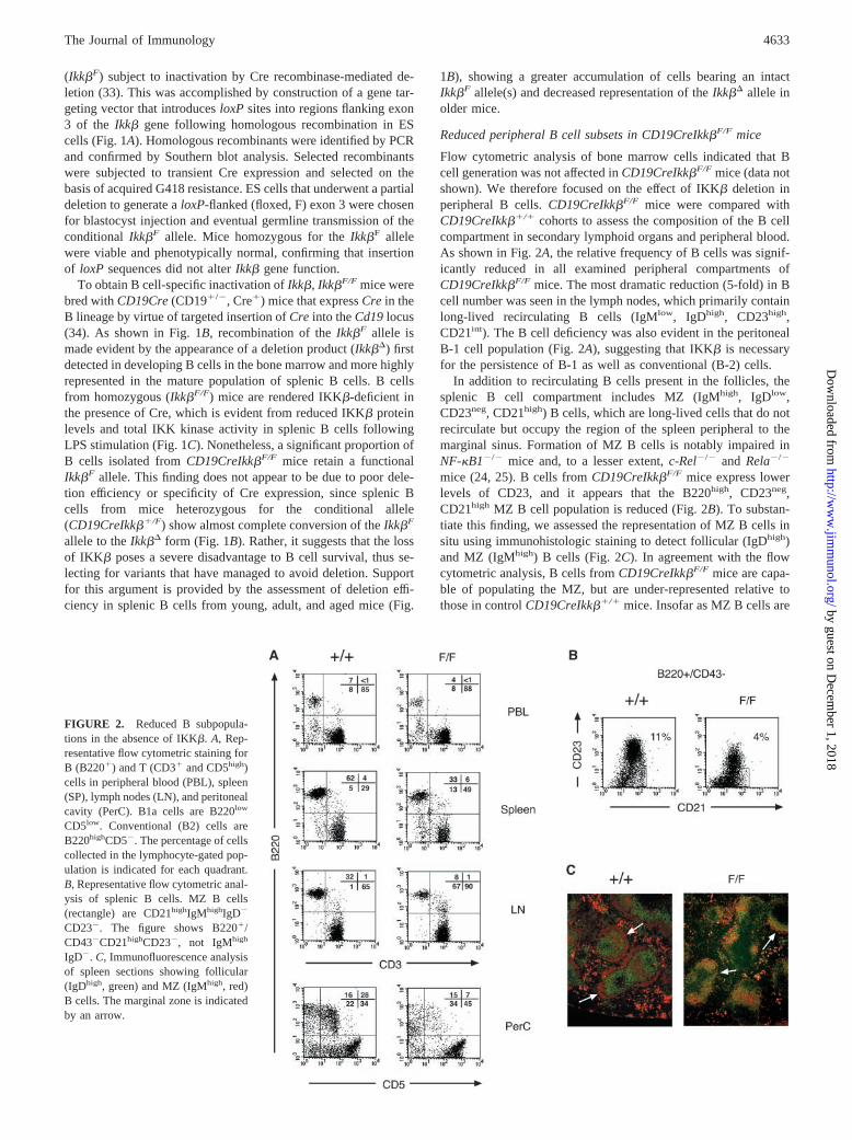
Reduced peripheral B cell subsets in CD19CreIkk�F/F mice
Flow cytometric analysis of bone marrow cells indicated that Bcell generation was not affected in CD19CreIkk�F/F mice (data notshown). We therefore focused on the effect of IKK� deletion inperipheral B cells. CD19CreIkk�F/F mice were compared withCD19CreIkk��/� cohorts to assess the composition of the B cellcompartment in secondary lymphoid organs and peripheral blood.As shown in Fig. 2A, the relative frequency of B cells was signif-icantly reduced in all examined peripheral compartments ofCD19CreIkk�F/F mice. The most dramatic reduction (5-fold) in Bcell number was seen in the lymph nodes, which primarily containlong-lived recirculating B cells (IgMlow, IgDhigh, CD23high,CD21int). The B cell deficiency was also evident in the peritonealB-1 cell population (Fig. 2A), suggesting that IKK� is necessaryfor the persistence of B-1 as well as conventional (B-2) cells.
In addition to recirculating B cells present in the follicles, thesplenic B cell compartment includes MZ (IgMhigh, IgDlow,CD23neg, CD21high) B cells, which are long-lived cells that do notrecirculate but occupy the region of the spleen peripheral to themarginal sinus. Formation of MZ B cells is notably impaired inNF-�B1�/� mice and, to a lesser extent, c-Rel�/� and Rela�/�
mice (24, 25). B cells from CD19CreIkk�F/F mice express lowerlevels of CD23, and it appears that the B220high, CD23neg,CD21high MZ B cell population is reduced (Fig. 2B). To substan-tiate this finding, we assessed the representation of MZ B cells insitu using immunohistologic staining to detect follicular (IgDhigh)and MZ (IgMhigh) B cells (Fig. 2C). In agreement with the flowcytometric analysis, B cells from CD19CreIkk�F/F mice are capa-ble of populating the MZ, but are under-represented relative tothose in control CD19CreIkk��/� mice. Insofar as MZ B cells are
FIGURE 2. Reduced B subpopula-tions in the absence of IKK�. A, Rep-resentative flow cytometric staining forB (B220�) and T (CD3� and CD5high)cells in peripheral blood (PBL), spleen(SP), lymph nodes (LN), and peritonealcavity (PerC). B1a cells are B220low
CD5low. Conventional (B2) cells areB220highCD5�. The percentage of cellscollected in the lymphocyte-gated pop-ulation is indicated for each quadrant.B, Representative flow cytometric anal-ysis of splenic B cells. MZ B cells(rectangle) are CD21highIgMhighIgD�
CD23�. The figure shows B220�/CD43�CD21highCD23�, not IgMhigh
IgD�. C, Immunofluorescence analysisof spleen sections showing follicular(IgDhigh, green) and MZ (IgMhigh, red)B cells. The marginal zone is indicatedby an arrow.
4633The Journal of Immunology
by guest on Decem
ber 1, 2018http://w
ww
.jimm
unol.org/D
ownloaded from
thought to be an activated subset primed by internal Ags, the ob-served reduction may be due to impaired activation or survival inthe absence of IKK�.
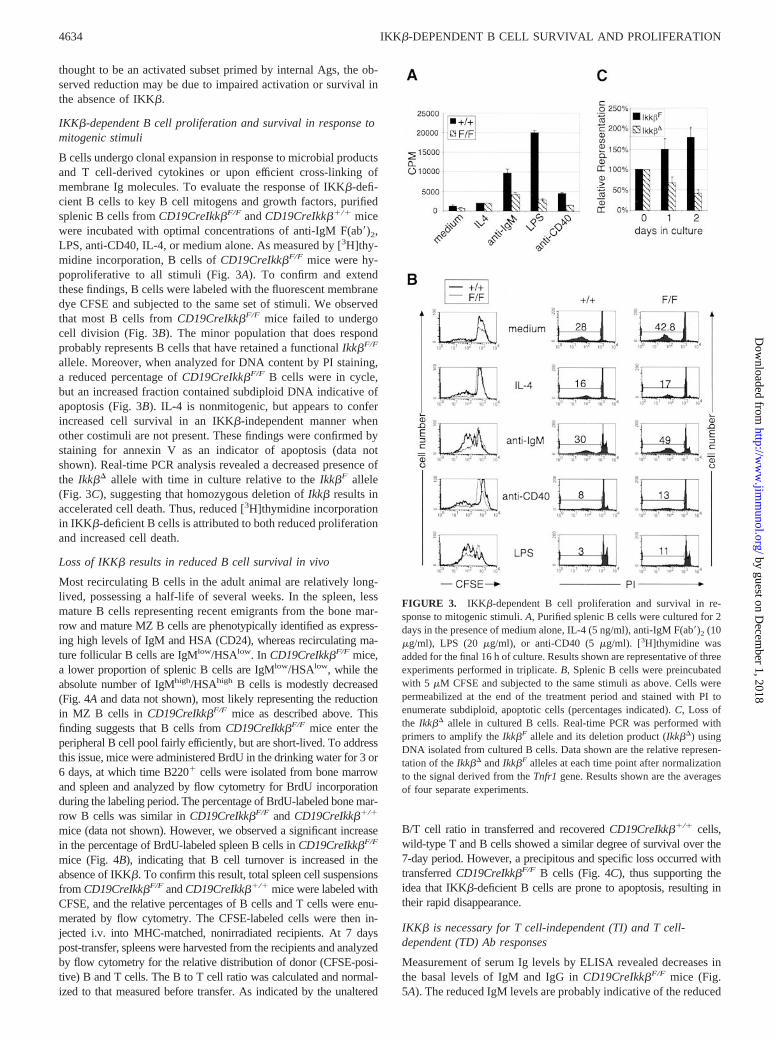
IKK�-dependent B cell proliferation and survival in response tomitogenic stimuli
B cells undergo clonal expansion in response to microbial productsand T cell-derived cytokines or upon efficient cross-linking ofmembrane Ig molecules. To evaluate the response of IKK�-defi-cient B cells to key B cell mitogens and growth factors, purifiedsplenic B cells from CD19CreIkk�F/F and CD19CreIkk��/� micewere incubated with optimal concentrations of anti-IgM F(ab�)2,LPS, anti-CD40, IL-4, or medium alone. As measured by [3H]thy-midine incorporation, B cells of CD19CreIkk�F/F mice were hy-poproliferative to all stimuli (Fig. 3A). To confirm and extendthese findings, B cells were labeled with the fluorescent membranedye CFSE and subjected to the same set of stimuli. We observedthat most B cells from CD19CreIkk�F/F mice failed to undergocell division (Fig. 3B). The minor population that does respondprobably represents B cells that have retained a functional Ikk�F/F
allele. Moreover, when analyzed for DNA content by PI staining,a reduced percentage of CD19CreIkk�F/F B cells were in cycle,but an increased fraction contained subdiploid DNA indicative ofapoptosis (Fig. 3B). IL-4 is nonmitogenic, but appears to conferincreased cell survival in an IKK�-independent manner whenother costimuli are not present. These findings were confirmed bystaining for annexin V as an indicator of apoptosis (data notshown). Real-time PCR analysis revealed a decreased presence ofthe Ikk�� allele with time in culture relative to the Ikk�F allele(Fig. 3C), suggesting that homozygous deletion of Ikk� results inaccelerated cell death. Thus, reduced [3H]thymidine incorporationin IKK�-deficient B cells is attributed to both reduced proliferationand increased cell death.
Loss of IKK� results in reduced B cell survival in vivo
Most recirculating B cells in the adult animal are relatively long-lived, possessing a half-life of several weeks. In the spleen, lessmature B cells representing recent emigrants from the bone mar-row and mature MZ B cells are phenotypically identified as express-ing high levels of IgM and HSA (CD24), whereas recirculating ma-ture follicular B cells are IgMlow/HSAlow. In CD19CreIkk�F/F mice,a lower proportion of splenic B cells are IgMlow/HSAlow, while theabsolute number of IgMhigh/HSAhigh B cells is modestly decreased(Fig. 4A and data not shown), most likely representing the reductionin MZ B cells in CD19CreIkk�F/F mice as described above. Thisfinding suggests that B cells from CD19CreIkk�F/F mice enter theperipheral B cell pool fairly efficiently, but are short-lived. To addressthis issue, mice were administered BrdU in the drinking water for 3 or6 days, at which time B220� cells were isolated from bone marrowand spleen and analyzed by flow cytometry for BrdU incorporationduring the labeling period. The percentage of BrdU-labeled bone mar-row B cells was similar in CD19CreIkk�F/F and CD19CreIkk��/�
mice (data not shown). However, we observed a significant increasein the percentage of BrdU-labeled spleen B cells in CD19CreIkk�F/F
mice (Fig. 4B), indicating that B cell turnover is increased in theabsence of IKK�. To confirm this result, total spleen cell suspensionsfrom CD19CreIkk�F/F and CD19CreIkk��/� mice were labeled withCFSE, and the relative percentages of B cells and T cells were enu-merated by flow cytometry. The CFSE-labeled cells were then in-jected i.v. into MHC-matched, nonirradiated recipients. At 7 dayspost-transfer, spleens were harvested from the recipients and analyzedby flow cytometry for the relative distribution of donor (CFSE-posi-tive) B and T cells. The B to T cell ratio was calculated and normal-ized to that measured before transfer. As indicated by the unaltered
B/T cell ratio in transferred and recovered CD19CreIkk��/� cells,wild-type T and B cells showed a similar degree of survival over the7-day period. However, a precipitous and specific loss occurred withtransferred CD19CreIkk�F/F B cells (Fig. 4C), thus supporting theidea that IKK�-deficient B cells are prone to apoptosis, resulting intheir rapid disappearance.
IKK� is necessary for T cell-independent (TI) and T cell-dependent (TD) Ab responses
Measurement of serum Ig levels by ELISA revealed decreases inthe basal levels of IgM and IgG in CD19CreIkk�F/F mice (Fig.5A). The reduced IgM levels are probably indicative of the reduced
FIGURE 3. IKK�-dependent B cell proliferation and survival in re-sponse to mitogenic stimuli. A, Purified splenic B cells were cultured for 2days in the presence of medium alone, IL-4 (5 ng/ml), anti-IgM F(ab�)2 (10�g/ml), LPS (20 �g/ml), or anti-CD40 (5 �g/ml). [3H]thymidine wasadded for the final 16 h of culture. Results shown are representative of threeexperiments performed in triplicate. B, Splenic B cells were preincubatedwith 5 �M CFSE and subjected to the same stimuli as above. Cells werepermeabilized at the end of the treatment period and stained with PI toenumerate subdiploid, apoptotic cells (percentages indicated). C, Loss ofthe Ikk�� allele in cultured B cells. Real-time PCR was performed withprimers to amplify the Ikk�F allele and its deletion product (Ikk��) usingDNA isolated from cultured B cells. Data shown are the relative represen-tation of the Ikk�� and Ikk�F alleles at each time point after normalizationto the signal derived from the Tnfr1 gene. Results shown are the averagesof four separate experiments.
4634 IKK�-DEPENDENT B CELL SURVIVAL AND PROLIFERATION
by guest on Decem
ber 1, 2018http://w
ww
.jimm
unol.org/D
ownloaded from

B-1 and MZ B cell populations that contribute a significant pro-portion of the natural Ab titer. To examine the responsiveness ofIkk�F/F mice to intact bacteria, mice were immunized with non-encapsulated, type 2 S. pneumoniae (R36A) and monitored forphosphocholine-specific Ab production 7 days postimmunization.We found that CD19CreIkk�F/F mice mounted a poor response toR36A (Fig. 5B), consistent with the reduction in MZ and B-1 cellsand minimal responses to anti-IgM stimulation in vitro. The Abresponse to TD Ags was also examined by immunization withTNP-OVA. We found that CD19CreIkk�F/F mice were greatly im-paired in the production of TNP-specific Abs (Fig. 5C), which isconsistent with an impaired response to anti-CD40 and a general Bcell survival defect. Of note, the reduction in TD Ab productionwas more dramatic than predicted from the fraction of restingsplenic B cells that retained IKK� expression. Thus, in agreementwith the in vitro stimulation data, it appears that in addition todecreased survival of IKK�-deficient B cells, their decreased mi-togenic response contributes to the overall decrease in Abproduction.
DiscussionCharacterization of the IKK complex led to the identification oftwo catalytic subunits, IKK� and IKK�, which in vitro can phos-phorylate I�B proteins at sites that are required for ubiquitin-de-pendent I�B degradation and translocation of NF-�B dimers intothe nucleus (1). Surprisingly, however, targeted deletion of theIkk� gene revealed minimal defects in NF-�B activity induced byproinflammatory stimuli, but an absolute requirement for IKK� inepidermal differentiation (11, 13, 18). By contrast, Ikk��/� micedie in midgestation due to TNF-�-induced apoptosis of hepato-cytes (12, 14, 19). This phenotype is shared by Rela�/� mice (36)and underscores the important antiapoptotic function of NF-�B(37). When the Ikk� null mutation is bred onto the Tnfr1�/� back-
ground, double-mutant mice are rescued from embryonic lethality(14, 22). However, most of these mice die before weaning as aresult of severe infections (Z.-W. Li, unpublished observations),thus precluding the analysis of Ikk� function in the adult. Weovercame this difficulty through the generation of a conditionalIkk� allele and used this system to probe Ikk� function in B lym-phocytes. We found that Ikk� is required for B cell survival andmitogenic responsiveness to diverse stimuli, including those thatact via the B cell receptor.
To ablate IKK� function in B cells, mice expressing a floxedIkk�F allele were generated and bred with mice expressing Crerecombinase under transcriptional control of the CD19 promoter(34). In this system the onset of Cre expression occurs at the pro-Bcell stage and continues throughout B cell development and dif-ferentiation. Correspondingly, CD19CreIkk�F/F mice show astrong reduction in peripheral B cell numbers that affects all B cellsubsets, including MZ and B-1 cells. By contrast, early B celldevelopment in the bone marrow was not affected, as determinedby flow cytometry and BrdU labeling experiments. These findingsare consistent with the phenotype of Nfkb1�/�Nfkb2�/� andRela�/�c-Rel�/� double mutants, which show intact generation ofIgM-positive immature B cells, but a strong impairment in thegeneration or maintenance of mature B cells (24, 27). Thus, whileearly B cell development in bone marrow is intact, B cell ho-meostasis in peripheral lymphoid tissues of CD19CreIkk�F/F miceis impaired, resulting in increased turnover of IKK�-deficient Bcells.
A significant proportion of the mature B cells in CD19CreIkk�F/F
mice were found to retain at least one functional Ikk� allele. SinceB cells from CD19CreIkk��/F mice showed complete deletion ofthe floxed allele, it became evident that Cre-mediated deletion ofthe Ikk�F allele is efficient and that strong selection is imposed todeplete CD19CreIkk�F/F B cells that had deleted both Ikk� alleles.
FIGURE 4. Loss of IKK� results in reduced B cell survival. A, Decreased percentages of mature recirculating B cells in CD19CreIkk�F/F mice. Dotplots show the relative frequencies of IgM and IgD expression in CD19Cre mice that is either Ikk��/� or Ikk�F/F. Histograms show mean fluorescenceintensity (MFI) of HSA expression on IgM-positive cells. B, Increased turnover of CD19CreIkk�F/F B cells as evidenced by increased frequencies ofBrdU-positive cells following short term continuous BrdU treatment. B cells that have incorporated BrdU were identified following permeabilization andstaining with an anti-BrdU-FITC Ab. C, Reduced survival of transferred CFSE-labeled CD19CreIkk�F/F B cells relative to CD19CreIkk��/� B cells. Bcell survival in vivo was determined by the relative ratio of B cells and T cells recovered at 7 days post-transfer. Results shown are the averages from fourmice transferred with splenocytes of CD19Cre�/� mice that are either Ikk��/� or Ikk�F/F.
4635The Journal of Immunology
by guest on Decem
ber 1, 2018http://w
ww
.jimm
unol.org/D
ownloaded from
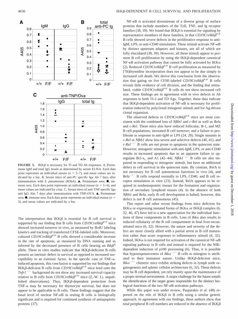
The interpretation that IKK� is essential for B cell survival issupported by our finding that B cells from CD19CreIkk�F/F miceshowed increased turnover in vivo, as measured by BrdU labelingkinetics and tracking of transferred CFSE-labeled cells. Moreover,cultured CD19CreIkk�F/F B cells showed a considerable increasein the rate of apoptosis, as measured by DNA staining and asinferred by the decreased presence of B cells bearing an Ikk��
allele. These in vitro studies indicate that IKK�-deficient B cellspossess an intrinsic defect in survival as opposed to increased sus-ceptibility to an extrinsic factor. In the specific case of TNF-�-induced apoptosis, this conclusion is supported by our findings thatIKK�-deficient B cells from CD19CreIkk�F/F mice bred onto theTnfr1�/� background do not show any increased survival capacityrelative to B cells from CD19CreIkk�F/F mice (Z.-W. Li, unpub-lished observations). Thus, IKK�-dependent protection fromTNF-� may be necessary for thymocyte survival, but does notappear to be applicable to B cells. These findings suggest that thebasal level of nuclear NF-�B in resting B cells is biologicallysignificant and is required for continued synthesis of antiapoptoticproteins (37).
NF-�B is activated downstream of a diverse group of surfaceproteins that include members of the Toll, TNF, and Ig receptorfamilies (38, 39). We found that IKK� is essential for signaling byrepresentative members of these families, in that CD19CreIkk�F/F
B cells showed severe defects in the proliferative response to anti-IgM, LPS, or anti-CD40 stimulation. These stimuli activate NF-�Bby distinct upstream adaptors and kinases, not all of which arefully elucidated (38, 39). However, all these stimuli appear to pro-mote B cell proliferation by using the IKK�-dependent canonicalNF-�B activation pathway that cannot be fully activated by IKK�(2). Reduced CD19CreIkk�F/F B cell proliferation as measured by[3H]thymidine incorporation does not appear to be due simply toincreased cell death. We derive this conclusion from the observa-tion that gating on live CFSE-labeled CD19CreIkk�F/F B cellsreveals little evidence of cell division, and the finding that stimu-lated, viable CD19CreIkk�F/F B cells do not show increased cellsize. These findings are in agreement with in vivo defects in Abresponses to both TI-2 and TD Ags. Together, these data indicatethat IKK�-dependent activation of NF-�B is necessary for prolif-eration induced by polyclonal mitogenic stimuli and for Ag-drivenclonal expansion.
The observed defects in CD19CreIkk�F/F mice are most con-sistent with the combined loss of Nfkb1 and c-Rel as well as Relaand c-Rel. These mice also have reduced follicular, B-1, and MZB cell populations; increased B cell turnover; and a failure to pro-liferate in response to anti-IgM or LPS (24, 28). Single mutants inc-Rel or Nfkb1 show less severe and selective defects (40, 41), andc-Rel�/� B cells are not prone to apoptosis in the quiescent state.However, mitogenic stimulation with anti-IgM, LPS, or anti-CD40results in increased apoptosis due to an apparent failure to up-regulate Bcl-xL and A1 (42–44). Nfkb1�/� B cells are also im-paired in responding to mitogenic stimuli, but have an additionaldefect in cell survival in the quiescent state. By contrast, RelA isnot necessary for B cell autonomous functions in vivo (4), andRela�/� B cells respond normally to LPS, CD40, and B cell re-ceptor stimulation in vitro (23). Instead, RelA appears to be re-quired in nonhemopoietic tissues for the formation and organiza-tion of secondary lymphoid tissues (4). In the absence of bothNfkb1 and Rela, early B cell development is halted; however, thisdefect is not B cell autonomous (45).
This report and other recent findings from mice deficient forIKK� or expressing mutated forms of IKK� or IKK� complex (6,32, 46, 47) have led to a new appreciation for the individual func-tions of these components in B cells. Loss of Ikk� also results inreduced cellularity of the B cell compartment in fetal liver-recon-stituted mice (6, 32). However, the nature and severity of the de-fect are more closely allied with a partial arrest in B cell matura-tion rather than acute responses to inflammatory stimuli (6, 32).Indeed, IKK� is not required for activation of the canonical NF-�Bsignaling pathway in B cells and instead is required for the NIK-dependent induction of p100 processing (6). Thus, it is possiblethat hyporesponsiveness of Ikk��/� B cells to mitogens is attrib-uted to their immature nature. Unlike IKK�-deficient mice,Ikk��/� chimeric mice exhibit striking defects in lymph node or-ganogenesis and splenic cellular architecture (6, 32). These defectsmay be B cell dependent, yet rely mainly upon the maintenance ofa proper stromal environment. A major challenge for the future entailsthe identification of the target genes responsible for the distinct bio-logical functions of the two NF-�B activation pathways.
While this paper was under review, Pasparakis et al. (48) re-ported on the role of IKK� in B cells using a similar geneticapproach. In agreement with our findings, these authors show thattotal peripheral B cell numbers are reduced in the absence of IKK�
FIGURE 5. IKK� is necessary for TI and TD Ab responses. A, Preim-mune IgM and total IgG levels as determined by serum ELISA. Each datapoint represents an individual mouse (n � 5–7), and mean values are in-dicated by a bar. B, Serum titers of anti-PC specific Ig� Ab 7 days afterimmunization with S. pneumoniae (R36A). Œ, Preimmune sera; F, im-mune sera. Each data point represents an individual mouse (n � 5–6), andmean values are indicated by a bar. C, Serum titers of anti-TNP specific Ig�and IgG Abs 7 days after immunization with TNP-OVA. Œ, Preimmunesera; F, immune sera. Each data point represents an individual mouse (n �3), and mean values are indicated by a bar.
4636 IKK�-DEPENDENT B CELL SURVIVAL AND PROLIFERATION
by guest on Decem
ber 1, 2018http://w
ww
.jimm
unol.org/D
ownloaded from

due to specific losses in the follicular and MZ B cell compart-ments. Using in vivo anti-IL-7R mAb treatment, they show that theremaining peripheral B cells have either only recently deleted thesecond Ikk�F allele and still presumably retain some IKK� proteinor have escaped deletion and still retain one intact Ikk�F allele.Importantly, the data presented by Pasparakis et al. (48) and ourpresent study clearly demonstrate that in addition to its establishedrole in suppression of receptor-induced apoptosis (37), the IKK�-dependent canonical NF-�B signaling pathway is also of impor-tance for sustaining nonstimulated resting B cells. Additionally,we have shown that IKK� may also play a role in B cell prolif-eration and activation in response to various mitogenic stimuli andboth TI and TD Ags. Dissecting IKK�-dependent survival vs ac-tivation mechanisms remains a challenging topic for future study.
AcknowledgmentsWe thank Dr. Dennis Otero and Kirsten Vroom for their assistance withsome of the experiments, and Dr. Gregg Silverman for providing the S.pneumoniae (R36A) extract and PC.
References1. Karin, M., and Y. Ben-Neriah. 2000. Phosphorylation meets ubiquitination: the
control of NF-�B activity. Annu. Rev. Immunol. 18:621.2. Ghosh, S., and M. Karin. 2002. Missing pieces in the NF-�B puzzle. Cell
109(Suppl.):S81.3. Caamano, J. H., C. A. Rizzo, S. K. Durham, D. S. Barton, C. Raventos-Suarez,
C. M. Snapper, and R. Bravo. 1998. Nuclear factor (NF)-�B2 (p100/p52) isrequired for normal splenic microarchitecture and B cell-mediated immune re-sponses. J. Exp. Med. 187:185.
4. Alcamo, E., N. Hacohen, L. C. Schulte, P. D. Rennert, R. O. Hynes, andD. Baltimore. 2002. Requirement for the NF-�B family member RelA in thedevelopment of secondary lymphoid organs. J. Exp. Med. 195:233.
5. Franzoso, G., L. Carlson, L. Poljak, E. W. Shores, S. Epstein, A. Leonardi,A. Grinberg, T. Tran, T. Scharton-Kersten, M. Anver, et al. 1998. Mice deficientin nuclear factor (NF)-�B/p52 present with defects in humoral responses, ger-minal center reactions, and splenic microarchitecture. J. Exp. Med. 187:147.
6. Senftleben, U., Y. Cao, G. Xiao, F. R. Greten, G. Krahn, G. Bonizzi, Y. Chen,Y. Hu, A. Fong, S. C. Sun, et al. 2001. Activation by IKK� of a second, evo-lutionary conserved, NF-�B signaling pathway. Science 293:1495.
7. Xiao, G., E. W. Harhaj, and S. C. Sun. 2001. NF-�B-inducing kinase regulatesthe processing of NF-�B2 p100. Mol. Cell 7:401.
8. Rothwarf, D. M., and M. Karin. 1999. The NF-�B activation pathway: a para-digm in information transfer from membrane to nucleus. Sci. STKE 1999:RE1.
9. Chu, W. M., D. Ostertag, Z. W. Li, L. Chang, Y. Chen, Y. Hu, B. Williams,J. Perrault, and M. Karin. 1999. JNK2 and IKK� are required for activating theinnate response to viral infection. Immunity 11:721.
10. Chu, W., X. Gong, Z. Li, K. Takabayashi, H. Ouyang, Y. Chen, A. Lois,D. J. Chen, G. C. Li, M. Karin, and E. Raz. 2000. DNA-PKcs is required foractivation of innate immunity by immunostimulatory DNA. Cell 103:909.
11. Hu, Y., V. Baud, M. Delhase, P. Zhang, T. Deerinck, M. Ellisman, R. Johnson,and M. Karin. 1999. Abnormal morphogenesis but intact IKK activation in micelacking the IKK� subunit of I�B kinase. Science 284:316.
12. Li, Z. W., W. Chu, Y. Hu, M. Delhase, T. Deerinck, M. Ellisman, R. Johnson, andM. Karin. 1999. The IKK� subunit of I�B kinase (IKK) is essential for nuclearfactor �B activation and prevention of apoptosis. J. Exp. Med. 189:1839.
13. Li, Q., Q. Lu, J. Y. Hwang, D. Buscher, K. F. Lee, J. C. Izpisua-Belmonte, andI. M. Verma. 1999. IKK1-deficient mice exhibit abnormal development of skinand skeleton. Genes Dev. 13:1322.
14. Li, Q., D. Van Antwerp, F. Mercurio, K. F. Lee, and I. M. Verma. 1999. Severeliver degeneration in mice lacking the I�B kinase 2 gene. Science 284:321.
15. Makris, C., V. L. Godfrey, G. Krahn-Senftleben, T. Takahashi, J. L. Roberts,T. Schwarz, L. Feng, R. S. Johnson, and M. Karin. 2000. Female mice heterozy-gous for IKK�/NEMO deficiencies develop a dermatopathy similar to the humanX-linked disorder incontinentia pigmenti. Mol Cell 5:969.
16. Rudolph, D., W. C. Yeh, A. Wakeham, B. Rudolph, D. Nallainathan, J. Potter,A. J. Elia, and T. W. Mak. 2000. Severe liver degeneration and lack of NF-�Bactivation in NEMO/IKK�-deficient mice. Genes Dev. 14:854.
17. Schmidt-Supprian, M., W. Bloch, G. Courtois, K. Addicks, A. Israel,K. Rajewsky, and M. Pasparakis. 2000. NEMO/IKK�-deficient mice model in-continentia pigmenti. Mol. Cell 5:981.
18. Takeda, K., O. Takeuchi, T. Tsujimura, S. Itami, O. Adachi, T. Kawai, H. Sanjo,K. Yoshikawa, N. Terada, and S. Akira. 1999. Limb and skin abnormalities inmice lacking IKK�. Science 284:313.
19. Tanaka, M., M. E. Fuentes, K. Yamaguchi, M. H. Durnin, S. A. Dalrymple,K. L. Hardy, and D. V. Goeddel. 1999. Embryonic lethality, liver degeneration,and impaired NF-�B activation in IKK-�-deficient mice. Immunity 10:421.
20. Alcamo, E., J. P. Mizgerd, B. H. Horwitz, R. Bronson, A. A. Beg, M. Scott,C. M. Doerschuk, R. O. Hynes, and D. Baltimore. 2001. Targeted mutation ofTNF receptor I rescues the RelA-deficient mouse and reveals a critical role forNF-�B in leukocyte recruitment. J. Immunol. 167:1592.
21. Doi, T. S., M. W. Marino, T. Takahashi, T. Yoshida, T. Sakakura, L. J. Old, andY. Obata. 1999. Absence of tumor necrosis factor rescues RelA-deficient micefrom embryonic lethality. Proc. Natl. Acad. Sci. USA 96:2994.
22. Senftleben, U., Z. W. Li, V. Baud, and M. Karin. 2001. IKK� is essential forprotecting T cells from TNF�-induced apoptosis. Immunity 14:217.
23. Horwitz, B. H., P. Zelazowski, Y. Shen, K. M. Wolcott, M. L. Scott,D. Baltimore, and C. M. Snapper. 1999. The p65 subunit of NF-�B is redundantwith p50 during B cell proliferative responses, and is required for germline CHtranscription and class switching to IgG3. J. Immunol. 162:1941.
24. Grossmann, M., L. A. O’Reilly, R. Gugasyan, A. Strasser, J. M. Adams, andS. Gerondakis. 2000. The anti-apoptotic activities of Rel and RelA required dur-ing B-cell maturation involve the regulation of Bcl-2 expression. EMBO J. 19:6351.
25. Cariappa, A., H. C. Liou, B. H. Horwitz, and S. Pillai. 2000. Nuclear factor �Bis required for the development of marginal zone B lymphocytes. J. Exp. Med.192:1175.
26. Weih, D. S., Z. B. Yilmaz, and F. Weih. 2001. Essential role of RelB in germinalcenter and marginal zone formation and proper expression of homing chemo-kines. J. Immunol. 167:1909.
27. Franzoso, G., L. Carlson, L. Xing, L. Poljak, E. W. Shores, K. D. Brown,A. Leonardi, T. Tran, B. F. Boyce, and U. Siebenlist. 1997. Requirement forNF-�B in osteoclast and B-cell development. Genes Dev. 11:3482.
28. Pohl, T., R. Gugasyan, R. J. Grumont, A. Strasser, D. Metcalf, D. Tarlinton,W. Sha, D. Baltimore, and S. Gerondakis. 2002. The combined absence of NF-�B1 and c-Rel reveals that overlapping roles for these transcription factors in theB cell lineage are restricted to the activation and function of mature cells. Proc.Natl. Acad. Sci. USA 99:4514.
29. Sun, Z., C. W. Arendt, W. Ellmeier, E. M. Schaeffer, M. J. Sunshine, L. Gandhi,J. Annes, D. Petrzilka, A. Kupfer, P. L. Schwartzberg, et al. 2000. PKC-� isrequired for TCR-induced NF-�B activation in mature but not immature T lym-phocytes. Nature 404:402.
30. Gerondakis, S., M. Grossmann, Y. Nakamura, T. Pohl, and R. Grumont. 1999.Genetic approaches in mice to understand Rel/NF-�B and I�B function: trans-genics and knockouts. Oncogene 18:6888.
31. Gugasyan, R., R. Grumont, M. Grossmann, Y. Nakamura, T. Pohl, D. Nesic, andS. Gerondakis. 2000. Rel/NF-�B transcription factors: key mediators of B-cellactivation. Immunol. Rev. 176:134.
32. Kaisho, T., K. Takeda, T. Tsujimura, T. Kawai, F. Nomura, N. Terada, andS. Akira. 2001. I�B kinase � is essential for mature B cell development andfunction. J. Exp. Med. 193:417.
33. Gu, H., J. D. Marth, P. C. Orban, H. Mossmann, and K. Rajewsky. 1994. Deletionof a DNA polymerase � gene segment in T cells using cell type-specific genetargeting. Science 265:103.
34. Rickert, R. C., K. Rajewsky, and J. Roes. 1995. Impairment of T-cell-dependentB-cell responses and B-1 cell development in CD19-deficient mice. Nature 376:352.
35. Zamorano, J., A. L. Mora, M. Boothby, and A. D. Keegan. 2001. NF-�B acti-vation plays an important role in the IL-4-induced protection from apoptosis. Int.Immunol. 13:1479.
36. Beg, A. A., W. C. Sha, R. T. Bronson, S. Ghosh, and D. Baltimore. 1995. Em-bryonic lethality and liver degeneration in mice lacking the RelA component ofNF-�B. Nature 376:167.
37. Karin, M., and A. Lin. 2002. NF-�B at the crossroads of life and death. Nat.Immunol. 3:221.
38. Silverman, N., and T. Maniatis. 2001. NF-�B signaling pathways in mammalianand insect innate immunity. Genes Dev. 15:2321.
39. Akira, S., K. Takeda, and T. Kaisho. 2001. Toll-like receptors: critical proteinslinking innate and acquired immunity. Nat. Immunol. 2:675.
40. Sha, W. C., H. C. Liou, E. I. Tuomanen, and D. Baltimore. 1995. Targeteddisruption of the p50 subunit of NF-�B leads to multifocal defects in immuneresponses. Cell 80:321.
41. Kontgen, F., R. J. Grumont, A. Strasser, D. Metcalf, R. Li, D. Tarlinton, andS. Gerondakis. 1995. Mice lacking the c-rel proto-oncogene exhibit defects inlymphocyte proliferation, humoral immunity, and interleukin-2 expression.Genes Dev. 9:1965.
42. Grumont, R. J., I. J. Rourke, and S. Gerondakis. 1999. Rel-dependent inductionof A1 transcription is required to protect B cells from antigen receptor ligation-induced apoptosis. Genes Dev. 13:400.
43. Owyang, A. M., J. R. Tumang, B. R. Schram, C. Y. Hsia, T. W. Behrens,T. L. Rothstein, and H. C. Liou. 2001. c-Rel is required for the protection of Bcells from antigen receptor-mediated, but not Fas-mediated, apoptosis. J. Immu-nol. 167:4948.
44. Tumang, J. R., A. Owyang, S. Andjelic, Z. Jin, R. R. Hardy, M. L. Liou, andH. C. Liou. 1998. c-Rel is essential for B lymphocyte survival and cell cycleprogression. Eur. J. Immunol. 28:4299.
45. Horwitz, B. H., M. L. Scott, S. R. Cherry, R. T. Bronson, and D. Baltimore. 1997.Failure of lymphopoiesis after adoptive transfer of NF-�B-deficient fetal livercells. Immunity 6:765.
46. Bendall, H. H., M. L. Sikes, D. W. Ballard, and E. M. Oltz. 1999. An intactNF-�B signaling pathway is required for maintenance of mature B cell subsets.Molecular Immunology 36:187.
47. Ren, H., A. Schmalstieg, D. Yuan, and R. B. Gaynor. 2002. I-�B kinase � iscritical for B cell proliferation and antibody response. J. Immunol. 168:577.
48. Pasparakis, M., M. Schmidt-Supprian, and K. Rajewsky. 2002. I�B kinase sig-naling is essential for maintenance of mature B cells. J. Exp. Med. 196:743.
4637The Journal of Immunology
by guest on Decem
ber 1, 2018http://w
ww
.jimm
unol.org/D
ownloaded from