review are the ikks and ikk-related kinases tbk1 and ikk-e ... review.pdf · bacterial infection...
TRANSCRIPT

Are the IKKs and IKK-related kinasesTBK1 and IKK-e similarly activated?Tieu-Lan Chau1*, Romain Gioia1*, Jean-Stephane Gatot1, Felicia Patrascu1,Isabelle Carpentier2, Jean-Paul Chapelle1, Luke O’Neill3, Rudi Beyaert2,Jacques Piette1 and Alain Chariot1
1 Interdisciplinary Cluster for Applied Genoproteomics, Medical Chemistry, and Virology/Immunology units,
University of Liege, Sart-Tilman, 4000 Liege, Belgium2 Unit of Molecular Signal Transduction in Inflammation, Department for Molecular Biomedical Research, VIB, Ghent, Belgium3 School of Biochemistry and Immunology, Trinity College, Dublin, Ireland
Review
the expression of a variety of cell surface markers and their responses to
pathogen molecules. pDCs are defined as a subset of cells, the appearance
under the microscope of which is similar to that of plasmablasts. These cells
are the main producers of type I IFNs in response to viral infections.
CpG DNAs: CpG DNAs are DNA oligodeoxynucleotide sequences that include a
cytosine–guanosine sequence and some flanking nucleotides. The CpG DNAs
induce innate immunity through binding to the TLR9 receptor.
Cytosolic NF-kB and IRF activating pathways: these pathways include the RIG-I
family (comprising MDA5 and RIG-I) and are triggered following infection with
RNA viruses and also the DAI-dependent pathway, which is activated when this
cytosolic receptor senses DNA from viruses or damaged cells.
E3 ligase: E3 ligases are defined as enzymes that facilitate the transfer of the
ubiquitin from the ubiquitin-conjugating enzyme (E2) to the e-amino group of a
lysine residue in a target protein.
Innate immunity: innate immunity is defined as the initial, rapidly induced
immune response of most multicellular organisms. This immunity relies on
receptors required for pathogen recognition and the recruitment and activation
of phagocytic cells.
Interferons (IFNs): IFNs are defined as cytokines that block viral replication and
infection of surrounding cells.
IFN-regulatory factors (IRFs): IRFs are a family of nine proteins (IRF1 to IRF9)
that share a well-conserved DNA-binding domain of �120 amino acids at their
N-terminus. This domain is required to bind to the consensus DNA sequence
that is known as the ISRE. IRFs are involved in the development and function of
immune cells. IRF3 and IRF7 are activated through TBK1 and IKK-e-mediated
phosphorylation on their C-terminal domain.
IFN-stimulated response element (ISRE): ISRE is a DNA motif that is bound by
The IkB kinases (IKKs) IKK-a and IKK-b, and theIKK-related kinases TBK1 and IKK-e, have essential rolesin innate immunity through signal-induced activation ofNF-kB, IRF3 and IRF7, respectively. Although the sig-naling events within these pathways have been exten-sively studied, the mechanisms of IKK and IKK-relatedcomplex assembly and activation remain poorly defined.Recent data provide insight into the requirement forscaffold proteins in complex assembly; NF-kB essentialmodulator coordinates some IKK complexes, whereasTANK, NF-kB-activating kinase-associated protein 1(NAP1) or similar to NAP1 TBK1 adaptor (SINTBAD)assemble TBK1 and IKK-e complexes. The different scaf-fold proteins undergo similar post-translational modifi-cations, including phosphorylation and non-degradativepolyubiquitylation. Moreover, increasing evidenceindicates that distinct scaffold proteins assemble IKK,and potentially TBK1 and IKK-e subcomplexes, in astimulus-specific manner, which might be a mechanismto achieve specificity.
Glossary
Caspase-recruitment domain (CARD): the CARD is found in some initiator
caspases, but also in some adaptor proteins, and mediates protein–protein
interactions.
Classical and alternative NF-kB-activating pathways: the classical pathway is
triggered by various stimuli, including proinflammatory cytokines and TLR
ligands, and leads to the activation of the IKK complex that includes IKK-a and
IKK-b and also the scaffold protein NEMO. This complex targets the inhibitory
IkBa protein for phosphorylation, which is followed by its degradation through
the proteasome pathway. NF-kB heterodimers (typically composed of p50 and
p65) subsequently move into the nucleus to drive the expression of
proinflammatory molecules and chemokines. The alternative pathway is
triggered by stimuli such as lymphotoxin-b and requires the kinase NIK in
addition to an IKK-a homodimer. NEMO is dispensable for this pathway to be
activated. The targeted inhibitory molecule is p100 instead of IkBa, and the NF-
kB heterodimers are typically composed of p52 and RelB. The target genes of
this pathway are required for adaptive immunity.
Conventional myeloid and plasmacytoid dendritic cells: dendritic cells (DCs)
take up antigens, are activated and migrate to lymphoid tissues in order to
present the antigenic peptides on the MHC molecules. They can be broadly
divided into plasmacytoid DCs (pDCs) and conventional myeloid DCs, based on
IRFs. The consensus sequence is GAAANNGAAAG/CT/C, where N denotes any
nucleotide.
Lipopolysaccharide (LPS): LPS is a component of the outer membrane of
Gram-negative bacteria and triggers NF-kB and IRF activation through binding
to the TLR4 receptor.
NF-kB essential modulator (NEMO): NEMO is required to assemble IKK-a and
IKK-b into an IkBa-phosphorylating complex following stimulation by various
stimuli, including proinflammatory cytokines and molecular components of
pathogens.
Nuclear factor (NF)-kB: NF-kB is a structurally and evolutionarily conserved
family of transcription factors initially identified as proteins harboring a DNA-
binding activity for the enhancer of the immunoglobulin k light-chain in
activated B cells. These proteins are crucial for the production of proinflam-
matory cytokines, growth factors and enzymes required for the initiation and
resolution of the immune response. These proteins include RelA (also known
as p65), RelB, c-Rel and also p50 and p52 (which are generated from processed
precursors – namely, p105 and p100, respectively).
Pathogen-associated molecular patterns: a molecular pattern which is found in
microorganisms but not in host cells.
Scaffold protein: a scaffold protein is referred to as a molecule that functions as
a platform in order to promote the recruitment and assembly of complexes.
These proteins do not harbor any enzymatic activity but are nevertheless
essential for the activation of the enzymes to which they bind.
Toll–IL-1 receptor (TIR) domain: the TIR domain is defined as an amino acid
sequence of the cytoplasmic region that is highly conserved among members
of the TLR and IL-1 receptor superfamily.
Type I IFNs: a family of proteins that includes IFN-a and IFN-b.
Ubiquitylation: the post-translational modification of proteins by the attach-
ment of one or more 7kDa ubiquitin molecules to lysine residues of the
substrates. This modification triggers protein degradation (K48-linked poly-
ubiquitylation) or is required for cell signaling (K63-linked polyubiquitylation).Corresponding author: Chariot, A. ([email protected]).* Authors contributed equally to this work.
0968-0004/$ – see front matter � 2008 Elsevier Ltd. All rights reserved. doi:10.1016/j.tibs.2008.01.002 Available online 18 March 2008 171
Review Trends in Biochemical Sciences Vol.33 No.4
The NF-kB- and IRF-activating signaling pathways ininnate immunityThe innate immune system (see Glossary) senses avariety of pathogen-associated molecular patterns(PAMPs), such as bacterial lipopeptides, viral and/orbacterial nucleic acids, by means of specific receptors.As a consequence, genes encoding the type I interferons(IFNs) IFN-a and -b, proinflammatory cytokines [e.g.tumor necrosis factor-a (TNF-a) and interleukin-1b
(IL-1b)] and chemokines [e.g. IL-8, monocyte chemoat-tractant protein-1 (MCP-1)] are induced [1,2]. Signalingpathways triggered by these viral or bacterial productsoccur through the Toll-like receptor (TLR) [3–5] or thecytosolic receptor pathway termed the retinoid-acid-indu-cible gene I (RIG-I)-dependent pathway [1,6]. Both path-ways rely on the coordinated activation of transcriptionfactors such as nuclear factor-kB (NF-kB) and IFN regu-latory factors (IRFs) [7,8].
Both the NF-kB and IRF families of transcription fac-tors are simultaneously activated in response to viral orbacterial infection but the target genes that are ultimatelyinduced through these signaling pathways are distinct.Indeed, whereas the induction of proinflammatory cyto-kines requires NF-kB, type I IFN gene induction mainlyrelies on IRF activation [2,7,8].
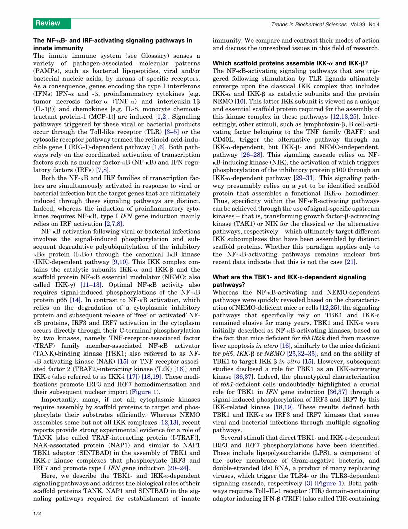
NF-kB activation following viral or bacterial infectionsinvolves the signal-induced phosphorylation and sub-sequent degradative polyubiquitylation of the inhibitorykBa protein (IkBa) through the canonical IkB kinase(IKK)-dependent pathway [9,10]. This IKK complex con-tains the catalytic subunits IKK-a and IKK-b and thescaffold protein NF-kB essential modulator (NEMO; alsocalled IKK-g) [11–13]. Optimal NF-kB activity alsorequires signal-induced phosphorylations of the NF-kBprotein p65 [14]. In contrast to NF-kB activation, whichrelies on the degradation of a cytoplasmic inhibitoryprotein and subsequent release of ‘free’ or ‘activated’ NF-kB proteins, IRF3 and IRF7 activation in the cytoplasmoccurs directly through their C-terminal phosphorylationby two kinases, namely TNF-receptor-associated factor(TRAF) family member-associated NF-kB activator(TANK)-binding kinase [TBK1; also referred to as NF-kB-activating kinase (NAK) [15] or TNF-receptor-associ-ated factor 2 (TRAF2)-interacting kinase (T2K) [16]] andIKK-e (also referred to as IKK-i [17]) [18,19]. These modi-fications promote IRF3 and IRF7 homodimerization andtheir subsequent nuclear import (Figure 1).
Importantly, many, if not all, cytoplasmic kinasesrequire assembly by scaffold proteins to target and phos-phorylate their substrates efficiently. Whereas NEMOassembles some but not all IKK complexes [12,13], recentreports provide strong experimental evidence for a role ofTANK [also called TRAF-interacting protein (I-TRAF)],NAK-associated protein (NAP1) and similar to NAP1TBK1 adaptor (SINTBAD) in the assembly of TBK1 andIKK-e kinase complexes that phosphorylate IRF3 andIRF7 and promote type I IFN gene induction [20–24].
Here, we describe the TBK1- and IKK-e-dependentsignaling pathways and address the biological roles of theirscaffold proteins TANK, NAP1 and SINTBAD in the sig-naling pathways required for establishment of innate
172
immunity. We compare and contrast their modes of actionand discuss the unresolved issues in this field of research.
Which scaffold proteins assemble IKK-a and IKK-b?The NF-kB-activating signaling pathways that are trig-gered following stimulation by TLR ligands ultimatelyconverge upon the classical IKK complex that includesIKK-a and IKK-b as catalytic subunits and the proteinNEMO [10]. This latter IKK subunit is viewed as a uniqueand essential scaffold protein required for the assembly ofthis kinase complex in these pathways [12,13,25]. Inter-estingly, other stimuli, such as lymphotoxin-b, B cell-acti-vating factor belonging to the TNF family (BAFF) andCD40L, trigger the alternative pathway through anIKK-a-dependent, but IKK-b- and NEMO-independent,pathway [26–28]. This signaling cascade relies on NF-kB-inducing kinase (NIK), the activation of which triggersphosphorylation of the inhibitory protein p100 through anIKK-a-dependent pathway [29–31]. This signaling path-way presumably relies on a yet to be identified scaffoldprotein that assembles a functional IKK-a homodimer.Thus, specificity within the NF-kB-activating pathwayscan be achieved through the use of signal-specific upstreamkinases – that is, transforming growth factor-b-activatingkinase (TAK1) or NIK for the classical or the alternativepathways, respectively – which ultimately target differentIKK subcomplexes that have been assembled by distinctscaffold proteins. Whether this paradigm applies only tothe NF-kB-activating pathways remains unclear butrecent data indicate that this is not the case [21].
What are the TBK1- and IKK-e-dependent signalingpathways?Whereas the NF-kB-activating and NEMO-dependentpathways were quickly revealed based on the characteriz-ation of NEMO-deficient mice or cells [12,25], the signalingpathways that specifically rely on TBK1 and IKK-eremained elusive for many years. TBK1 and IKK-e wereinitially described as NF-kB-activating kinases, based onthe fact that mice deficient for tbk1/t2k died from massiveliver apoptosis in utero [16], similarly to the mice deficientfor p65, IKK-b or NEMO [25,32–35], and on the ability ofTBK1 to target IKK-b in vitro [15]. However, subsequentstudies disclosed a role for TBK1 as an IKK-activatingkinase [36,37]. Indeed, the phenotypical characterizationof tbk1-deficient cells undoubtedly highlighted a crucialrole for TBK1 in IFN gene induction [36,37] through asignal-induced phosphorylation of IRF3 and IRF7 by thisIKK-related kinase [18,19]. These results defined bothTBK1 and IKK-e as IRF3 and IRF7 kinases that senseviral and bacterial infections through multiple signalingpathways.
Several stimuli that direct TBK1- and IKK-e-dependentIRF3 and IRF7 phosphorylations have been identified.These include lipopolysaccharide (LPS), a component ofthe outer membrane of Gram-negative bacteria, anddouble-stranded (ds) RNA, a product of many replicatingviruses, which trigger the TLR4- or the TLR3-dependentsignaling cascade, respectively [3] (Figure 1). Both path-ways requires Toll–IL-1 receptor (TIR) domain-containingadaptor inducing IFN-b (TRIF) [also called TIR-containing
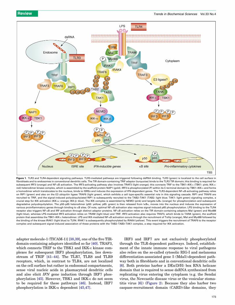
Figure 1. TLR3 and TLR4-dependent signaling pathways. TLR3-mediated pathways are triggered following dsRNA binding. TLR3 (green) is localized to the cell surface in
fibroblasts and to endosomes in conventional dendritic cells. The TIR domain-containing TRIF adaptor (turquoise) binds to the TLR3 TIR domain; this binding is required for
subsequent IRF3 (orange) and NF-kB activation. The IRF3-activating pathway also involves TRAF3 (light orange); this connects TRIF to the TBK1–IKK-e (TBK1: pink; IKK-e:red) heterodimer kinase complex, which is assembled by the scaffold protein NAP1 (gold). IRF3 is phosphorylated (P) within its C-terminal domain by TBK1–IKK-e and forms
a homodimer which translocates to the nucleus, binds to ISREs and induces the expression of IFN-dependent genes. The TLR3-dependent NF-kB-activating pathway relies
on RIP1 (green) and also on the E3 ubiquitin ligase TRAF6 (light green), which exhibits a cell type-specific essential role in this signaling cascade. RIP1 and TRAF6 are
recruited to TRIF, and the signal-induced polyubiquitylated RIP1 is subsequently recruited to the TAB2–TAK1 (TAB2: light blue; TAK1: light green) signaling complex, a
crucial step for IKK activation (IKK-a: orange; IKK-b: blue). The IKK complex is assembled by NEMO (pink) and targets IkBa (orange) for phosphorylation and subsequent
degradative polyubiquitylation. The p50–p65 heterodimer (p50: yellow; p65: green) is then released from IkBa, moves into the nucleus and induces the expression of
various proinflammatory genes through binding to kB sites. Of note, optimal NF-kB activation also requires signal-induced p65 phosphorylation. LPS binding to the TLR4
receptor also triggers NF-kB and IRF activation through distinct adaptor proteins. NF-kB activation relies on the TIR domain-containing adaptors Mal (green) and Myd88
(light blue), whereas LPS-mediated IRF3 activation relies on TRAM (light blue) and TRIF. IRF3 activation also requires TRAF3, which binds to TANK (green), the scaffold
protein that assembles the TBK1–IKK-e heterodimer. LPS and IKK-mediated NF-kB activation occurs through the recruitment of Tollip (orange), Mal and Myd88 followed by
the binding of the kinase IRAK1 (light blue) to TLR4. IRAK1 is subsequently phosphorylated by IRAK4 (yellow). This event triggers the recruitment of TRAF6 to the receptor
complex and subsequent signal-induced association of these proteins with the TAB2–TAB3–TAK1 complex, a step required for IKK activation.
Review Trends in Biochemical Sciences Vol.33 No.4
adapter molecule-1 (TICAM-1)] [38,39], one of the five TIR-domain-containing adaptors identified so far [40]. TRAF3,which connects TRIF to the TBK1 and IKK-e kinase com-plexes for subsequent IRF3 phosphorylation, lies down-stream of TRIF [41–44]. The TLR7, TLR8 and TLR9receptors, which, in contrast to TLR4, are not localizedon the cell surface but rather in endosomal compartments,sense viral nucleic acids in plasmacytoid dendritic cellsand also elicit IFN gene induction through IRF7 phos-phorylation [45]. However, TBK1 and IKK-e do not seemto be required for these pathways [46]. Instead, IRF7phosphorylation is IKK-a dependent [45,47].
IRF3 and IRF7 are not exclusively phosphorylatedthrough the TLR-dependent pathways. Indeed, establish-ment of the innate immune response to viral pathogensalso relies on the so-called cytosolic RIG-I and melanomadifferentiation-associated gene 5 (Mda5)-dependent path-way both in fibroblasts and in conventional dendritic cells[6]. Both proteins harbor a DEx(D/H) box RNA helicasedomain that is required to sense dsRNA synthesized fromreplicating virus entering the cytoplasm (e.g. the Sendaivirus, the Newcastle disease virus or the vesicular stoma-titis virus [6]) (Figure 2). Because they also harbor twocaspase-recruitment domain (CARD)-like domains, they
173

Figure 2. The cytosolic, TBK1- and IKK-e-dependent signaling pathways. IRF3 activation is triggered when cytosolic receptors sense intracellular nucleic acids (RNA from
viruses or DNA from viruses or damaged cells). RNA from viruses triggers the activation of the cytosolic receptors RIG-I and MDA-5 through binding to their DExD/H box
RNA helicase (red boxes). The CARD domains of these receptors (light blue boxes) subsequently undergo K63-linked polyubiquitylation by the E3 ubiquitin ligase TRIM25
(yellow), which might facilitate the interaction of RIG-I with the mitochondrial adaptor MAVS. Importantly, MAVS harbors a mitochondrial transmembrane domain (TM)
(orange box) that localizes this adaptor to the mitochondria. MAVS signals to TRAF3 (light orange), which triggers IRF3 phosphorylation through TBK1 and IKK-e-mediated
activations. Of note, MAVS can also signal to TRAF6 for the IKK-a–IKK-b-complex-mediated NF-kB activation but this pathway is not represented. TANK (green), NAP1 (gold)
and SINTBAD (light blue) are required for TBK1 and IKK-e assembly but it is currently unclear whether only one IKK-related kinase complex (illustrated as a heterodimer)
assembled by multiple scaffold proteins is involved or whether distinct subcomplexes assembled by only one scaffold protein (i.e. TANK, NAP1 or SINTBAB) are
simultaneously activated through this cytosolic pathway, as illustrated here. These adaptors might also be targeted for K63-linked polyubiquitylation but this hypothesis
awaits experimental validation. Intracellular DNA from damaged cells or from DNA viruses is sensed by DAI (depicted in light blue), a cytosolic DNA recognition receptor.
Activated DAI binds to TBK1 (represented as a homodimer) and triggers IRF3 phosphorylation. It is currently unknown whether TRAF3 is part of this signaling complex. The
adaptors for TBK1 homodimers remain to be elucidated.
Review Trends in Biochemical Sciences Vol.33 No.4
can transmit the signal through direct binding to mito-chondrial antiviral signaling (MAVS) [48] [also called IFN-b promoter stimulator 1 (IPS-1), virus-induced signalingadaptor (VISA) and Cardif [49–51]), a CARD domain-con-taining mitochondrial adaptor. This protein subsequentlytriggers NF-kB activation through TRAF6 and the IKK-a–b complex [50] or IRF3 and IRF7 activation throughTRAF3 and the TBK1 and IKK-e kinase complexes [48].Intracellular dsDNA from microbes or damaged cells alsomodulates the innate immune response by triggeringTBK1 and IKK-e activation [52] but the cytosolic receptorrequired in this pathway is not RIG-I. Indeed, the intra-cellular DNA sensor is DNA-dependent activator of IFN-regulatory factors (DAI), which binds to, and is activatedby, DNA from various sources. DAI subsequently recruits
174
TBK1, which triggers IRF3 phosphorylation and type IIFN gene expression [53].
Which scaffold proteins assemble the IKK-relatedkinases TBK1 and IKK-e?Similar to the IKKs, TBK1 and IKK-e require assembly bya scaffold protein to target their substrates efficiently.These scaffold proteins must constitutively interact withthe catalytic subunits of the complex, and three candi-dates, TANK, NAP1 and SINTBAD, seem to fulfill thisfunction.
TANK
TANK was originally identified as a TRAF-interacting protein that synergizes with TRAF2 to induce

Review Trends in Biochemical Sciences Vol.33 No.4
NF-kB-dependent reporter gene expression [54–56].Later, its role in the NF-kB-activating pathways wassupported by its binding to NEMO [57]. However, the roleforTANK in the IKK-dependentNF-kBsignaling cascadesremains controversial because TANK-depleted, knock-down cells do not show any defect in TNF-a-or LPS-mediated IKK activation [21,58]. Nevertheless, theseobservations do not rule out the possibility that TANKmight connect upstream kinases such as TBK1 and IKK-e,thereby promoting IKK-independent phosphorylation ofthe NF-kB proteins p65 [59,60], c-Rel [61] or p52 [62] inresponse to as yet poorly characterized signals. TANKconstitutively binds to IKK-e [63] and TBK1 [64] throughits N-terminal domain; this interaction is probably essen-tial for the positive regulation of signal transductionby TANK because recent studies demonstrated thatTANK is involved in some TLR-dependent IRF-activatingpathways by promoting TBK1 and IKK-e-mediated phos-phorylation of IRF3 and IRF7 [20,21].
NAP1
NAP1, a candidate scaffold protein that was initiallyidentified as a TBK1-interacting protein, shares severalstructural features with TANK [65]. Early studies indi-cated that NAP1 assembles TBK1 complexes for sub-sequent p65 phosphorylation and therefore has a role inthe NF-kB-activating pathways [65]. Enhanced sensitivityto apoptosis of the TNF-a-stimulated, NAP1-depletedknockdown cells, which is typical for cells having defectsin the prosurvival NF-kB signaling cascade, further sup-ported this hypothesis [65]. Similarly to TANK, NAP1constitutively binds to TBK1 and is also required forIRF3 phosphorylation through both the TLR3- and theRIG-I-dependent pathways [22,23].
SINTBAD
SINTBAD is the most recently identified scaffold proteinthat constitutively binds to TBK1 and IKK-e [24]. Inter-estingly, this protein shares a conserved TBK1- and IKK-e-binding domain (TBD) with TANK and NAP1, and thisregion is predicted to form an a-helix with the conservedresidues clustering on one side of it [24]. Owing to thestructural similarities between TANK, NAP1 and SINT-BAD, it is expected that they perform similar functions inTBK1 and IKK-e activation.
TANK-, NAP1- and SINTBAD-dependent signalingpathwaysIn addition to TANK and NAP1 possibly regulating NF-kBactivation through p65-mediated phosphorylation, bothscaffold proteins are also required for IRF3 and IRF7phosphorylation because they constitutively bind toTBK1 and IKK-e. Only recently have studies started toexplore TBK1- and IKKe-dependent pathways in whichTANK, NAP1 and SINTBAD are specifically required.Interestingly, some specificity at the level of TBK1 andIKK-e assembly might occur because NAP1 is essential forthe TLR-mediated IRF3 activation pathway [22], whereasTANK seems to have a similar role for IRF3 phosphoryl-ation through the TLR4 pathway in LPS-responsive cellssuch as macrophages [21] (Figure 1). In agreement with
this hypothesis, the early phase of dsRNA-mediated IRF3phosphorylation through the TLR3 receptor does notrequire TANK [21]. Thus, these results indicate thatTANK functions as a scaffold protein that assembles some,but not all, IRF3- and IRF7-phosphorylating TBK1 andIKK-e complexes. In other words, this observation opensthe possibility that distinct scaffold proteins might berequired for TBK1 and IKK-e assembly in a pathway-specific manner. The dispensable role of TBK1 and IKK-e in the TLR7-, TLR8- and TLR9-dependent IRF7-activat-ing pathwaysmakes an involvement of TANKandNAP1 inthese cascades unlikely. Of note, however, IRF7 was alsoidentified as a TANK-interacting protein through yeasttwo-hybrid analyses, and IRF7 can be phosphorylated byTANK-containing immune complexes in macrophagestreated with unmethylated CpG DNA motifs, which areTLR9 ligands [21]. These data indicate that TANK mightalso have a role in this IRF7-dependent signaling cascadeby connecting a yet to be identified kinase to this IRFmember. Alternatively, TANKmight connect TBK1 and/orIKK-e to IRF7 in a second wave of activation through theTLR4, a hypothesis supported by the LPS-inducibleexpression of TANK, IKK-e and IRF7 [17,66].
The signaling pathways that involve TANK, NAP1and SINTBAD do not exclusively include the TLR-de-pendent signaling pathways. Indeed, the cytosolic RIG-I-dependent pathway, which triggers TBK1 and IKK-eactivations, also requires these scaffold proteins(Figure 2). TANK is known to be one candidate becauseTANK-depleted knockdown cells infected with Sendaivirus exhibit defective IFN-b production [20]. Such amodel is further supported by the virus-inducibleTANK–MAVS association in fibroblasts [20]. Moreover,NAP1 and SINTBAD depletion through RNA interfer-ence also causes defects in Sendai virus-induced acti-vation of an IFN-stimulated responsive elements (ISRE)-containing reporter construct [24]. Thus, these resultsindicate that TANK, NAP1 and SINTBAB are allrequired for type I IFN induction through the cytosolicreceptor and IRF3-activating pathways.
Crosstalk between the NF-kB and IRF pathwaysCrosstalk is a common feature in signal transduction andalso seems to be relevant for the NF-kB- and IRF-activat-ing pathways (Figure 3). This hypothesis is supported bythe physical association between TANK and NEMO[57,67]. The consequence of this crosstalk remains unclearbut might underlie the molecular basis for the generationof specific IRF3–p65 complexes which are required for theproper expression of a subset of genes in LPS-stimulatedcells [68–70]. Additional evidence for crosstalk betweenthe two signaling pathways is provided by IKK-e-mediatedphosphorylation of p65 in TNF-a-stimulated cells [71].However, it still remains unclear whether the transcrip-tional complexes involving both the IRF and NF-kBproteins require a physical association between the IKKand IKK-related scaffold proteins (e.g. between NEMOand TANK) or whether such heterodimers are generatedfollowing simultaneous, but independent, activation of thepathways through the TLR- or RIG-I-dependent path-ways. Of note, some signaling molecules are involved in
175
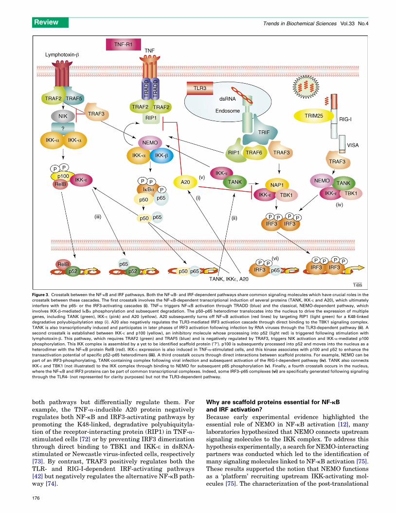
Figure 3. Crosstalk between the NF-kB and IRF pathways. Both the NF-kB- and IRF-dependent pathways share common signaling molecules which have crucial roles in the
crosstalk between these cascades. The first crosstalk involves the NF-kB-dependent transcriptional induction of several proteins (TANK, IKK-e and A20), which ultimately
interfere with the p65- or the IRF3-activating cascades (i). TNF-a triggers NF-kB activation through TRADD (blue) and the classical, NEMO-dependent pathway, which
involves IKK-b-mediated IkBa phosphorylation and subsequent degradation. The p50–p65 heterodimer translocates into the nucleus to drive the expression of multiple
genes, including TANK (green), IKK-e (pink) and A20 (yellow). A20 subsequently turns off NF-kB activation (red lines) by targeting RIP1 (light green) for a K48-linked
degradative polyubiquitylation step (i). A20 also negatively regulates the TLR3-mediated IRF3 activation cascade through direct binding to the TBK1 signaling complex.
TANK is also transcriptionally induced and participates in later phases of IRF3 activation following infection by RNA viruses through the TLR3-dependent pathway (ii). A
second crosstalk is established between IKK-e and p100 (yellow), an inhibitory molecule whose processing into p52 (light red) is triggered following stimulation with
lymphotoxin-b. This pathway, which requires TRAF2 (green) and TRAF5 (blue) and is negatively regulated by TRAF3, triggers NIK activation and IKK-a-mediated p100
phosphorylation. This IKK complex is assembled by a yet to be identified scaffold protein (’?’). p100 is subsequently processed into p52 and moves into the nucleus as a
heterodimer with the NF-kB protein RelB (red). IKK-e expression is also induced in TNF-a-stimulated cells, and this kinase associates with p100 and p52 to enhance the
transactivation potential of specific p52–p65 heterodimers (iii). A third crosstalk occurs through direct interactions between scaffold proteins. For example, NEMO can be
part of an IRF3-phosphorylating, TANK-containing complex following viral infection and subsequent activation of the RIG-I-dependent pathway (iv). TANK also connects
IKK-e and TBK1 (not illustrated) to the IKK complex through binding to NEMO for subsequent p65 phosphorylation (v). Finally, a fourth crosstalk occurs in the nucleus,
where the NF-kB and IRF3 proteins can be part of common transcriptional complexes. Indeed, some IRF3–p65 complexes (vi) are specifically generated following signaling
through the TLR4- (not represented for clarity purposes) but not the TLR3-dependent pathway.
Review Trends in Biochemical Sciences Vol.33 No.4
both pathways but differentially regulate them. Forexample, the TNF-a-inducible A20 protein negativelyregulates both NF-kB and IRF3-activating pathways bypromoting the K48-linked, degradative polyubiquityla-tion of the receptor-interacting protein (RIP1) in TNF-a-stimulated cells [72] or by preventing IRF3 dimerizationthrough direct binding to TBK1 and IKK-e in dsRNA-stimulated or Newcastle virus-infected cells, respectively[73]. By contrast, TRAF3 positively regulates both theTLR- and RIG-I-dependent IRF-activating pathways[42] but negatively regulates the alternative NF-kB path-way [74].
176
Why are scaffold proteins essential for NF-kBand IRF activation?Because early experimental evidence highlighted theessential role of NEMO in NF-kB activation [12], manylaboratories hypothesized that NEMO connects upstreamsignaling molecules to the IKK complex. To address thishypothesis experimentally, a search for NEMO-interactingpartners was conducted which led to the identification ofmany signaling molecules linked to NF-kB activation [75].These results supported the notion that NEMO functionsas a ‘platform’ recruiting upstream IKK-activating mol-ecules [75]. The characterization of the post-translational

Review Trends in Biochemical Sciences Vol.33 No.4
modifications targeting NEMO provided further insightinto its function in NF-kB signaling [76]. NEMO is phos-phorylated and also is subject to non-degradative polyubi-quitylation. This post-translational modification involvesthe internal lysine K63 of ubiquitin (i.e. K63-linked poly-ubiquitylation) and, in contrast to the degradative K48-linked polyubiquitylation, does not promote recognition bythe proteasome but rather triggers protein–protein inter-actions that mediate signal transduction. Recent findingsdemonstrate that the identity of both the E3 ubiquitinligase and the polyubiquitylated lysine residues arestimulus specific, as recently summarized [75,76].Although there is a general agreement on the signal-induced NEMO polyubiquitylation, and even if thismodification might not only regulate IKK, but also mito-gen-activated protein kinase activations in TLR- andIL-1b-stimulated B cells [77], it is currently unclear ifubiquitylation triggers NEMO oligomerization and/or therecruitment of upstream signaling molecules harboringubiquitin-binding domains [75]. Interestingly, in additionto being subject to non-degradative polyubiquitylation,NEMO also binds to K63-linked polyubiquitin chainsthrough its NEMO ubiquitin-binding domain (NUB)[78,79], indicating the existence of an interacting networkbetween polyubiquitin chains and a variety of ubiquitin-binding domains found inmultiple signalingmolecules suchas TAB2 and TAB3 which are involved in IKK activation[80,81]. Thus, the ability of NEMO to bind to polyubiquiti-nated proteins and to be polyubiquitinated itself is the
Figure 4. Structural similarities between the IKK and IKK-related scaffold proteins. The
represented. The structural motifs are illustrated as boxes (CC, coil–coil; NUB, NEMO
protein interactions are depicted as black bars and the interacting partners are listed b
scaffold proteins TANK, NAP1 and SINTBAD are represented in red and aligned based
region of NEMO, SINTBAD and TANK are depicted as hatched blue rectangles. The TAN
171 and 247, whereas those residues targeted by the TBK1- and IKK-e-dependent K63-lin
mechanismwhichunderlies theessential roleof this scaffoldprotein for NF-kB activation [75].
Because of structural similarities between TANK,NAP1, SINTBAD and NEMO (Figure 4), the findingsrelated to NEMO post-translational modifications mightbe relevant for the TBK1 and IKK-e scaffold proteins.Although TANK has not been shown to bind to K63-poly-ubiquitinated chains, it also is subject to phosphorylationand non-degradative polyubiquitination in a stimulus-de-pendent manner [21]. TBK1 and IKK-e phosphorylateTANK in macrophages and also are required for LPS-mediated TANKpolyubiquitylation, independently of theirkinase activity. This observation indicates that TBK1 andIKK-e function not only as IRF3 and IRF7 kinases, but alsoas signaling molecules required to connect a yet to beidentified E3 ligase for the non-degradative polyubiquity-lation of their own scaffold protein [21]. Whether thismodification of TANK is essential for LPS-mediatedIRF3 activation remains unclear [21]. TBK1 and IKK-ealso harbor a ubiquitin-like domain (ULD), adjacent totheir N-terminal kinase domain and located upstream oftheir C-terminal coil–coil domains [82]. The ULD does notseem to bind to known ubiquitin-binding domains but isnevertheless crucial for TBK1 and IKK-e substrate recog-nition and kinase activity [82]. Therefore, these recentstudies strongly indicate that optimal TBK1- and IKK-e-mediated phosphorylation requires distinct functionaldomains and highlight the importance of TANK non-degradative polyubiquitination for subsequent IFN gene
murine NEMO, SINTBAD, NAP1 and the human TANK proteins are schematically
ubiquitin binding domain; LZ, leucine zipper). The domains involved in protein–
elow them. NEMO is represented in blue, whereas the IKK-related TBK1 and IKK-eon their TBDs (depicted in green). The zinc finger motifs found in the C-terminal
K residues phosphorylated by TBK1 and IKK-e (P) are located between amino acids
ked polyubiquitination pathway (Ub) are located between amino acids 71 and 110.
177

Review Trends in Biochemical Sciences Vol.33 No.4
induction. It will be interesting to determine the extent towhich the ULD found in TBK1 and IKK-e is involved in therecognition and/or the binding to TANK polyubiquitinchains.
The RIG-I CARD domain also undergoes K63-linkedpolyubiquitylation in response to viral infection [83].TRIM25 is the RIG-I E3 ubiquitin ligase, and is crucialfor launching the proper cellular antiviral response.Whether TRIM25 also targets other substrates, such asTANK, for polyubiquitylation, remains unknown but it istempting to speculate that IRF3 and IRF7 activationinvolves sequential K63-linked polyubiquitylation ofmultiple scaffold proteins by potentially distinct E3 ligases.
Unresolved issuesDo TBK1 and IKK-e function as homo- and/or
heterodimers?
Whereas it is now well established that the activation ofseveral NF-kB-activating IKK subcomplexes is triggeredby distinct stimuli exist in the cell, little information isavailable on the existence of distinct IKK-related com-plexes. Although this issue remains to be addressed exper-imentally, key findings might help us to speculate on thenotion that TBK1 and IKK-emight, at least partially, exerttheir biological roles as homodimers. First, the expressionlevels of TBK1 and IKK-e are regulated differentially.Indeed, TBK1 is widely expressed and its level of expres-sion is not strongly regulated at the transcription level, incontrast to IKK-e, which is found in low levels unlessinduced by various proinflammatory cytokines [17]. Thus,these facts favor the hypothesis that TBK1 homodimersexist, especially in some cell types or in circumstanceswhere IKK-e expression is not transcriptionally induced.Moreover, and in contrast to TBK1-deficient cells, whichshow strong defects in IFN induction, IKK-e-deficient cellsinduce IFN normally; this is another piece of evidenceindicating that TBK1 functions alone [36]. The existenceof functional IKK-e homodimers is supported by the factthat this IKK-related kinase, but not TBK1, is recruited tothe mitochondria following viral infection [84] and also bythemore severe defects in IFN induction seen in TBK1 andIKK-e double-knockout versus TBK1 knockout cells [36].
Do TANK, NAP1 or SINTBAD preferentially assemble
TBK1–TBK1, TBK1–IKK-e or IKK-e–IKK-e dimers?
Similarly to the NF-kB-activating signaling pathways, wemight expect TANK, NAP1 and SINTBAD to assembledistinct TBK1 and IKK-e complexes in a signal-specificmanner. Still, it is currently unclear whether these scaffoldproteins function exclusively in distinct pathways orwhether they are also part of common cascades. Impor-tantly, these proteins mainly form homo-oligomers besidessome NAP1–SINTBAD hetero-oligomers. No direct associ-ation between TANK, NAP1 or SINTBAD was revealed[24]. These observations open the possibility that distinctscaffold proteins assemble some TBK1 and/or IKK-e sub-complexes. Studies addressing the subcellular localizationof each scaffold protein will most likely help to resolve thisissue. It is currently unknown if NAP1 and SINTBAD aretargeted for non-degradative polyubiquitylation through aTBK1- and IKK-e-dependent pathway but it is tempting to
178
speculate that this mechanism is not restricted toLPS-mediated and TANK-dependent IRF3 activation. Ifthis is indeed the case, it would be of interest to definewhether a single E3 ligase targets each of them or whethersome specificity occurs at that level.
Are TANK, NAP1 and SINTBAD required for TBK1
and IKK-e oncogenic potential?
Recent studies extended the roles of TBK1 and IKK-e tosignaling in cancer and defined both proteins as oncogenickinases [85,86]. For IKK-e, this oncogenic potential is theresult of an increased expression found in breast cancersamples having the amplified 1q32 locus that encompassesthe ikk-e gene [85]. Interestingly, enhanced IKK-e expres-sion in those samples is correlated with c-Rel nuclearlocalization [85]. It is currently unclear whether or notoverexpressed IKK-e targets other substrates aside fromIRF3 and IRF7, and the NF-kB proteins, and whetherTANK, NAP1 or SINTBAD are required in these path-ways. Because it is likely that K63-linked polyubiquityla-tion is required for proper NF-kB and IRF signaling,deregulation of this modification might contribute to can-cer development and/or progression.
Concluding remarksSignificant progress has recently been made regarding themolecular mechanisms underlying NF-kB and IRF acti-vation. Whereas the central roles of the IKK, TBK1 andIKK-e kinase complexes are now well established, themechanism of their assembly is only now becoming clear.Surprisingly, similarities between the two pathways haveemerged, such as the importance of signal-induced non-degradative polyubiquitylation of the constituent scaffoldproteins. A thorough understanding of the physiologicalrelevance of these post-translational modifications awaitsthe phenotypic characterization of mouse models in whichthe residues targeted for polyubiquitylation are specificallymutated (‘knock-in’ mice). Whereas such residues havebeen identified for NEMO, the targeted residues in TANKremain unknown. Future studies should tell usmore on theroles of these post-translational modifications for propersignaling in innate immunity. It will also be interesting toknow whether such modifications are impaired in cancer,especially in cases where TBK1 and IKK-e functions arederegulated. Importantly, numerous solid and hematologi-cal tumors have constitutive IKK activities [10]; some ofthese tumors also have enhanced IKK-e activity. Thus,targeting the IKKs by specific inhibitors might not be asefficient as initially thought to prevent cancer developmentand progression because these molecules will not targetthe IKK-related kinases. Thus, dissecting the TBK1 andIKK-e-dependent pathways through the characterizationof their interacting partners, and also through the estab-lishment and phenotypical characterization of mousemodels deficient for their scaffold proteins, will undoubt-edly lead to the identification of key targets for therapeuticpurposes.
AcknowledgementsWe apologize to all colleagues whose papers could not be cited owing tospace limitations. A.C. and J.P. are Research Associate and Research

Review Trends in Biochemical Sciences Vol.33 No.4
Director at the Belgian National Funds for Scientific Research (F.N.R.S.),respectively, whereas R.G. is a TELEVIE Research Assistant. Ourlaboratories are supported by grants from the F.N.R.S., TELEVIE,FWO-Vlaanderen, the Belgian Federation Against Cancer, the ConcertedResearch Action Program (04/09–323, University of Liege; 01G06B6,Ghent University), the Inter-University Attraction Pole 6/18 (FederalMinistry of Science), the Centre Anti-Cancereux and the Leon FredericqFundation (ULg).
References1 Akira, S. et al. (2006) Pathogen recognition and innate immunity. Cell
124, 783–8012 Stetson, D.B. and Medzhitov, R. (2006) Type I interferons in host
defense. Immunity 25, 373–3813 West, A.P. et al. (2006) Recognition and signaling by toll-like receptors.
Annu. Rev. Cell Dev. Biol. 22, 409–4374 Doyle, S.L. and O’Neill, L. (2006) Toll-like receptors: from the discovery
of NFkB to new insights into transcriptional regulations in innateimmunity. Biochem. Pharmacol. 72, 1102–1113
5 O’Neill, L.A. (2006) How Toll-like receptors signal: what we know andwhat we don’t know. Curr. Opin. Immunol. 18, 3–9
6 Kato, H. et al. (2005) Cell type-specific involvement of RIG-I in antiviralresponse. Immunity 23, 19–28
7 Wietek, C. and O’Neill, L.A. (2007) Diversity and regulation in the NF-kB system. Trends Biochem. Sci. 32, 311–319
8 Honda, K. and Taniguchi, T. (2006) IRFs: master regulators ofsignaling by Toll-like receptors and cytosolic pattern-recognitionreceptors. Nat. Rev. Immunol. 6, 644–658
9 Karin, M. and Ben-neriah, Y. (2000) Phosphorylation meetsubiquitination: the control of NF-kB activity. Annu. Rev. Immunol.18, 621–663
10 Perkins, N.D. (2007) Integrating cell-signaling pathways with NF-kBand IKK function. Nat. Rev. Mol. Cell Biol. 8, 49–62
11 Zandi, E. et al. (1997) The IkB kinase complex (IKK) contains twokinase subunits, IKKa and IKKb, necessary for IkB phosphorylationand NF-kB activation. Cell 91, 243–252
12 Yamaoka, S. et al. (1998) Complementation cloning of NEMO, acomponent of the IkappaB kinase complex essential for NF-kBactivation. Cell 93, 1231–1240
13 Rothwarf, D.M. et al. (1998) IKK-g is an essential regulatory subunit ofthe IkB kinase complex. Nature 395, 297–300
14 Viatour, P. et al. (2005) Phosphorylation of NF-kB and IkB proteins:implications in cancer and inflammation.TrendsBiochem.Sci.30,43–52
15 Tojima, Y. et al. (2000) NAK is an IkB kinase-activating kinase.Nature404, 778–782
16 Bonnard, M. et al. (2000) Deficiency of T2K leads to apoptotic liverdegeneration and impaired NF-kB-dependent gene transcription.EMBO J. 19, 4976–4985
17 Shimada, T. et al. (1999) IKK-i, a novel lipopolysaccharide-induciblekinase that is related to IkB kinases. Int. Immunol. 11, 1357–1362
18 Fitzgerald, K.A. et al. (2003) IKKe and TBK1 are essential componentsof the IRF3 signaling pathway. Nat. Immunol. 4, 491–496
19 Sharma, S. et al. (2003) Triggering the interferon antiviral responsethrough an IKK-related pathway. Science 300, 1148–1151
20 Guo, B. and Cheng, G. (2007) Modulation of the interferon antiviralresponse by the TBK1/IKKi adaptor protein TANK. J. Biol. Chem. 282,11817–11826
21 Gatot, J.S. et al. (2007) Lipopolysaccharide-mediated interferonregulatory factor activation involves TBK1/IKKe-dependent Lys63-linked polyubiquitination and phosphorylation of TANK/I-TRAF. J.Biol. Chem. 282, 31131–31146
22 Sasai, M. et al. (2005) Cutting edge: NF-kB-activating kinase-associated protein 1 participates in TLR3/Toll-IL-1 homologydomain-containing adapter molecule-1-mediated IFN regulatoryfactor 3 activation. J. Immunol. 174, 27–30
23 Sasai,M. et al. (2006) NAK-associated protein 1 participates in both theTLR3 and the cytoplasmic pathways in type I IFN induction. J.Immunol. 177, 8676–8683
24 Ryzhakov, G. and Randow, F. (2007) SINTBAD, a novel component ofinnate antiviral immunity, shares a TBK1-binding domain with NAP1and TANK. EMBO J. 26, 3180–3190
25 Rudolph, D. et al. (2000) Severe liver degeneration and lack of NF-kBactivation in NEMO/IKKg-deficient mice. Genes Dev. 14, 854–862
26 Dejardin, E. et al. (2002) The lymphotoxin-b receptor induces differentpatterns of gene expression via two NF-kB pathways. Immunity 17,525–535
27 Claudio, E. et al. (2002) BAFF-induced NEMO-independent processingof NF-k B2 in maturing B cells. Nat. Immunol. 3, 958–965
28 Coope, H.J. et al. (2002) CD40 regulates the processing of NF-kB2 p100to p52. EMBO J. 21, 5375–5385
29 Xiao, G. et al. (2001) NF-kB-inducing kinase regulates the processing ofNF-kB2 p100. Mol. Cell 7, 401–409
30 Senftleben, U. et al. (2001) Activation by IKKa of a second, evolutionaryconserved, NF-k B signaling pathway. Science 293, 1495–1499
31 Xiao, G. et al. (2004) Induction of p100 processing by NF-kB-inducingkinase involves docking IkB kinase a(IKKa) to p100 and IKKa-mediated phosphorylation. J. Biol. Chem. 279, 30099–30105
32 Beg, A.A. et al. (1995) Embryonic lethality and liver degeneration inmice lacking the RelA component of NF-kB. Nature 376, 167–170
33 Li, Q. et al. (1999) Severe liver degeneration in mice lacking the IkBkinase 2 gene. Science 284, 321–325
34 Li, Z.W. et al. (1999) The IKKb subunit of IkB kinase (IKK) is essentialfor nuclear factor kB activation and prevention of apoptosis. J. Exp.Med. 189, 1839–1845
35 Tanaka, M. et al. (1999) Embryonic lethality, liver degeneration, andimpaired NF-k B activation in IKK-b-deficient mice. Immunity 10,421–429
36 Hemmi, H. et al. (2004) The roles of two IkB kinase-related kinases inlipopolysaccharide and double stranded RNA signaling and viralinfection. J. Exp. Med. 199, 1641–1650
37 McWhirter, S.M. et al. (2004) IFN-regulatory factor 3-dependent geneexpression is defective in Tbk1-deficient mouse embryonic fibroblasts.Proc. Natl. Acad. Sci. U. S. A. 101, 233–238
38 Yamamoto, M. et al. (2002) A novel Toll/IL-1 receptor domain-containing adapter that preferentially activates the IFN-b promoterin the Toll-like receptor signaling. J. Immunol. 169, 6668–6672
39 Oshiumi, H. et al. (2003) TICAM-1, an adaptor molecule thatparticipates in Toll-like receptor 3-mediated interferon-b induction.Nat. Immunol. 4, 161–167
40 O’Neill, L.A. and Bowie, A.G. (2007) The family of five: TIR-domain-containing adaptors in Toll-like receptor signaling.Nat. Rev. Immunol.7, 353–364
41 Hoebe, K. and Beutler, B. (2006) TRAF3: a new component of the TLR-signaling apparatus. Trends Mol. Med. 12, 187–189
42 Saha, S.K. and Cheng, G. (2006) TRAF3: a new regulator of type Iinterferons. Cell Cycle 5, 804–807
43 Hacker, H. et al. (2006) Specificity in Toll-like receptor signalingthrough distinct effector functions of TRAF3 and TRAF6. Nature439, 204–207
44 Oganesyan, G. et al. (2006) Critical role of TRAF3 in the Toll-likereceptor-dependent and -independent antiviral response. Nature 439,208–211
45 Kawai, T. and Akira, S. (2006) Innate immune recognition of viralinfection. Nat. Immunol. 7, 131–137
46 Kawai, T. et al. (2004) Interferon-a induction through Toll-likereceptors involves a direct interaction of IRF7 with Myd88 andTRAF6. Nat. Immunol. 5, 1061–1068
47 Hoshino, K. et al. (2006) IkB kinase-a is critical for interferon-aproduction induced by Toll-like receptors 7 and 9.Nature 440, 949–953
48 Seth, R.B. et al. (2005) Identification of MAVS, a mitochondrialantiviral signaling protein that activates NF-kB and IRF 3. Cell122, 669–682
49 Kawai, T. et al. (2005) IPS-1, an adaptor trigerring RIG-I- and Mda5-mediated type I interferon induction. Nat. Immunol. 6, 981–988
50 Xu, L.G. et al. (2005) VISA is an adapter protein required for virus-triggered IFN-b signaling. Mol. Cell 19, 727–740
51 Meylan, E. et al. (2005) Cardif is an adaptor protein in the RIG-Iantiviral pathway and is targeted by hepatitis C virus. Nature 437,1167–1172
52 Ishii, K.J. et al. (2006) A Toll-like receptor-independent antiviralresponse induced by double-stranded B-form DNA. Nat. Immunol. 7,40–48
53 Takaoka, A. et al. (2007) DAI (DLM-1/ZBP1) is a cytosolic DNA sensorand an activator of innate immune response. Nature 448, 501–506
54 Cheng, G. and Baltimore, D. (1996) TANK, a co-inducer with TRAF2 ofTNF- and CD 40L-mediated NF-kB activation. Genes Dev. 10, 963–973
179

Review Trends in Biochemical Sciences Vol.33 No.4
55 Kaye, K.M. et al. (1996) Tumor necrosis factor receptor associatedfactor 2 is a mediator of NF-kB activation by latent infectionmembrane protein 1, the Epstein-Barr virus transforming protein.Proc. Natl. Acad. Sci. U. S. A. 93, 11085–11090
56 Rothe, M. et al. (1996) I-TRAF is a novel TRAF-interacting protein thatregulates TRAF-mediated signal transduction. Proc. Natl. Acad. Sci.U. S. A. 93, 8241–8246
57 Chariot, A. et al. (2002) Association of the adaptor TANK with the I kBkinase (IKK) regulator NEMO connects IKK complexes with IKKe andTBK1 kinases. J. Biol. Chem. 277, 37029–37036
58 Bonif, M. et al. (2006) TNFa- and IKKb-mediated TANK/I-TRAFphosphorylation: implications for interaction with NEMO/IKKg andNF-kB activation. Biochem. J. 394, 593–603
59 Adli, M. and Baldwin, A.S. (2006) IKK-i/IKKe controls constitutive,cancer cell-associated NF-kB activity via regulation of Ser-536 p65/RelA phosphorylation. J. Biol. Chem. 281, 26976–26984
60 Mattioli, I. et al. (2006) Inducible phosphorylation of NF-k B p65 atserine 468 by T cell costimulation is mediated by IKKe. J. Biol. Chem.281, 6175–6183
61 Harris, J. et al. (2006) Nuclear accumulation of cRel following C-terminal phosphorylation by TBK1/IKKe. J. Immunol. 177, 2527–2535
62 Wietek, C. et al. (2006) IkB kinase e interacts with p52 and promotestransactivation via p65. J. Biol. Chem. 281, 34973–34981
63 Nomura, F. et al. (2000) NF-kB activation through IKK-i-dependent I-TRAF/TANK phosphorylation. Genes Cells 5, 191–202
64 Pomerantz, J.L. and Baltimore, D. (1999) NF-kB activation by asignaling complex containing TRAF2, TANK and TBK1, a novelIKK-related kinase. EMBO J. 18, 6694–6704
65 Fujita, F. et al. (2003) Identification of NAP1, a regulatory subunit ofIkBkinase-related kinases that potentiatesNF-kB signaling.Mol. Cell.Biol. 23, 7780–7793
66 Schreiber, J. et al. (2006) Coordinated binding of NF-kB familymembers in the response of human cells to lipopolysaccharide. Proc.Natl. Acad. Sci. U. S. A. 103, 5899–5904
67 Zhao, T. et al. (2007) The NEMO adaptor bridges the nuclear factor-kBand interferon regulatory factor signaling pathways. Nat. Immunol. 8,592–600
68 Wietek, C. et al. (2003) Interferon regulatory factor-3-mediatedactivation of the interferon-sensitive response element by Toll-likereceptor (TLR) 4 but not TLR3 requires the p65 subunit of NF-k. J.Biol. Chem. 278, 50923–50931
69 Ogawa, S. et al. (2005) Molecular determinants of crosstalk betweennuclear receptors and toll-like receptors. Cell 122, 707–721
Reproduction of materia
Interested in reproducing part or all of an article pub
If so, please contact our Global Rights Departmen
material will be used. To submit a perm
www.elsevier.com/lo
180
70 Leung, T.H. et al. (2004) One nucleotide in a kB site can determinecofactor specificity for NF-kB dimers. Cell 118, 453–464
71 Wietek, C. et al. (2006) IkB kinase e interacts with p52 and promotestransactivation by p65. J. Biol. Chem. 281, 34973–34981
72 Wertz, I.E. et al. (2004) De-ubiquitination and ubiquitin ligase domainsof A20 downregulate NF-kB signaling. Nature 430, 694–699
73 Saitoh, T. et al. (2005) A20 is a negative regulator of IFN regulatoryfactor 3 signaling. J. Immunol. 174, 1507–1512
74 Hauer, J. et al. (2005) TNF receptor (TNFR)-associated factor (TRAF) 3serves as an inhibitor of TRAF2/5-mediated activation of thenoncanonical NF-kB pathway by TRAF-binding TNFRs. Proc. Natl.Acad. Sci. U. S. A. 102, 2874–2879
75 Sebban, H. et al. (2006) Posttranslational modifications of NEMO andits partners in NF-kB signaling. Trends Cell Biol. 16, 569–577
76 Israel, A. (2006) NF-kB activation: nondegradative ubiquitinationimplicates NEMO. Trends Immunol. 27, 395–397
77 Yamamoto, M. et al. (2006) Key function for the Ubc13 E2 ubiquitin-conjugating enzyme in immune receptor signaling. Nat. Immunol. 7,962–970
78 Wu, C.J. et al. (2006) Sensing of Lys 63-linked polyubiquitinationby NEMO is a key event in NF-kB activation. Nat. Cell Biol. 8, 398–406
79 Ea, C.K. et al. (2006) Activation of IKK by TNFa requires site-specificubiquitination of RIP1 and polyubiquitin binding by NEMO. Mol. Cell22, 245–257
80 Ishitani, T. et al. (2003) Role of the TAB2-related protein TAB3 in IL-1and TNF signaling. EMBO J. 22, 6277–6288
81 Kanayama, A. et al. (2004) TAB2 and TAB3 activate the NF-kBpathway through binding to polyubiquitin chains. Mol. Cell 15, 535–548
82 Ikeda, F. et al. (2007) Involvement of the ubiquitin-like domain ofTBK1/IKK-i kinases in regulation of IFN-inducible genes.EMBOJ. 26,3451–3462
83 Gack, M.U. et al. (2007) TRIM25 RING-finger E3 ubiquitin ligase isessential for RIG-I-mediated antiviral activity. Nature 446, 916–920
84 Lin, R. et al. (2006) Dissociation of a MAVS/IPS-1/VISA/Cardif-IKKemolecular complex from the mitochondria outer membrane byhepatitis C virus NS3-4A proteolytic cleavage. J. Virol. 80, 6072–6083
85 Boehm, J.S. et al. (2007) Integrative genomic approaches identifyIKBKE as a breast cancer oncogene. Cell 129, 1065–1079
86 Chien, Y. et al. (2006) RalB GTPase-mediated activation of the IkBfamily kinase TBK1 couples innate immune signaling to tumor cellsurvival. Cell 127, 157–170
l from Elsevier articles
lished by Elsevier, or one of our article figures?
t with details of how and where the requested
ission request online, please visit:
cate/permissions