observations with cytochemistry and ultracryotomy … · 2005-08-21 · for routine electron...
TRANSCRIPT

J. Cell Sci. 19, 239-259 (i975) 239
Printed in Great Britain
OBSERVATIONS WITH CYTOCHEMISTRY AND
ULTRACRYOTOMY ON THE FINE STRUCTURE
OF THE EXPANDING WALLS IN ACTIVELY
ELONGATING PLANT CELLS
J. C. ROLAND, BRIGITTE VIAN AND DANlfiLE REIS
Laboratoire de Biologie Vdgitale — Cytologie Expe'rimentale,University de Paris, E.N.S., 24 rite Lhomond, 75231 Paris Cedex 05, France
SUMMARY
Ultracryotomy with negative staining and cytochemistry (periodic acid - thiocarbohydrazide- silver proteinate test for polysaccharides, in conjunction with mild extractions) were used tostudy the architecture of the cell wall and its modifications during expansion. Those techniqueswere applied to the study in situ of the walls of actively elongating parenchyma of mung bean(Phaseolus aureus), and pea (Pisum sativum) root and of collenchyma of celery (Apium graveolens)petioles.
These complementary techniques provide information on the 3-dimensional disposition andfine structure of the subunits of the wall. In all the examples examined, the bulk of growingprimary wall appears well-ordered and no progressive evolution from a transverse texture nearthe plasmalemma to a scattered texture near the middle lamella was observed.
It seems unlikely that the development of the wall structure in relation to growth could beexplained mechanically by a passive shift of the fibrillar elements in response to cellular stress.There is no evidence for an inert change in fibrillar orientation in the major part of the wall.If such occurs, the process is limited to the outermost and senescent part of the wall. Thus, thetexture observed does not agree with the classical multinet growth hypothesis but rather withthe idea of an ordered structure of the primary wall. With the latter, the components should beable to respond in different ways to specific growth regulators and other environmental signalsand thus exert a more positive control over the processes of oriented cell growth.
INTRODUCTION
Growing plant cells are in a characteristic and paradoxical situation because theirwalls must allow expansion, yet be mechanically strong. During active growth, thecell length usually increases more than 2-fold within a few hours. This impliesdramatic and highly coordinated changes in the wall. In stems or roots, usually cellgrowth is not an isodiametric process but an elongation, i.e. only lateral surfaces of thecell increase. This is a highly anisotropic process, the structural basis of which mustbe determined.
The fine structure of the wall and the changes which occur during cell growth havealready received much attention (see Roelofsen, 1959; Clowes & Juniper, 1968;Preston, 1974). At the electron-microscope level, the components of the wall, speciallythe polysaccharides, react weakly with salts of heavy metals (osmium, lead, uranium,etc.) used as fixatives or stains. Thus the visualization of wall architecture has been

240 J. C. Roland, B. Vian and D. Reis
achieved chiefly with shadowing techniques, which are known to introduce positionalshifts in the components under study.
The results have led to various theories to explain growth in relation to the structureof the wall. The most generally assumed, the 'multinet growth hypothesis' (MGH),was proposed by Roelofsen & Houwink (1953). According to this theory the newfibrils should be laid down, at the inner face of the wall, more or less perpendicularto the cell axis (tubular texture). When the cell elongates the fibrils are stretched andbecome approximately parallel to the axis of growth. According to the multinettheory, growth should be characterized by a significant but passive change in orienta-tion of the fibrils, so that the texture of the primary wall during expansion would bedispersed and random. Although this theory is generally well accepted it cannotexplain all the textures observed in different tissues. For instance, collenchyma is awell known source of controversy (Chafe, 1970; Cox & Juniper, 1973; Preston, 1974;Roland, 1966; Reis & Roland, 1974).
Two recent techniques allow a new approach to elucidate the fine structure of thecell wall: ultrastructural cytochemistry associated with mild extraction of polysac-charides of the matrix; and ultracryotomy with subsequent negative staining of thecell wall fibrils.
MATERIAL AND METHODS
The biological materials were as follows, (a) Cortical parenchyma of pea root (Pisum sativuw)from the subapical zone of maximum elongation (from the 2nd to the 4th mm of the apex).(6) Cortical parenchyma of mung bean hypocotyl (P/iaseolus auretis). The latter organ ischaracterized by an intercalary growth concentrated near the tip, in the 5 mm just below thehook beneath the cotyledons. For (a) and (A) the specimens were picked from 5 -day-oldseedlings grown in darkness at 25 °C. (c) Collenchyma was from the upper part of 1-10-cmpetioles of celery (Apium graveolens) grown in a greenhouse. The regions and rate of growthwere followed by means of India ink marks, the separation of which was followed with time.
Light-microscope controls were performed after 2 h fixation in 6-25 % glutaraldehyde incacodylate buffer 0 2 M, pH 7-2. Two procedures were used: embedding in paraffin and stainingwith periodic acid - SchirT (PAS) + haematoxylin; and embedding in methacrylate and stainingof the thick sections with toluidine blue.
For routine electron microscopy, prefixation was for 2 h in 625 % glutaraldehyde in caco-dylate buffer, 0-2 M, pH 72 . Material was then washed for 2 h and postfixed for 1 h in 2 %osmium tetroxide in phosphate buffer 0 2 M, pH 72 . Embedding was in an Epon-Aralditemixture. The thin sections were stained with uranyl acetate and lead citrate.
For cytochemistry the specimens were prefixed in 6-25 % glutaraldehyde (2 h at roomtemperature), in cacodylate buffer 0 2 M, pH 72 , washed and incubated in ethylenediaminetetra-acetic acid (EDTA) 003 M or dimethylsulphoxide (DMSO) (Reis & Roland, 1974) followed bypostfixation in osmium tetroxide and embedding in an Epon-Araldite mixture. The cyto-chemical test for polysaccharides was periodic acid - thiocarbohydrazide - silver proteinate(PATAG) (Thiery, 1967) as previously described (Roland, 1974) (for discussion of the chemicalspecificity of the method see also Reis & Roland, 1974).
For ultracryotomy, specimens were prefixed in 625 % glutaraldehyde in cacodylate buffer0-2 M, pH 72 , and frozen in liquid nitrogen. Other specimens were directly frozen in liquidnitrogen. To prevent crystallization damage, some prefixed specimens were infused with1-5 M sucrose for 20-30 min before freezing. All steps were at 0—4 °C. Thin sections were cuton a Sorvall MT2 ultramicrotome supplied with a cryokit FTS. The sections were collectedeither on 50 % DMSO ( — 50 °C) or without floating medium ( — 70 °C) according to Tokuyasu(1973). The sections were stained on grids with 1 % sodium silicotungstate at 37 °C for 30-45 s(Vian & Rougier, 1974).

Fine structure of expanding cell wall 241
Ultrathin sections were examined and photographed with a Philips EM 300. Evaluations ofthe 3-dimensional texture of the wall were made with a goniometer stage tilted from —45° to+ 45°.
RESULTS
Growth studies confirmed that the cells were in a phase of active growth. The rateof relative growth was about 10%/hour. At the beginning of growth, the cells wereisodiametric (Fig. 3). Then they underwent typical elongation, with expansion ofonly the walls in the plane of the long axis of the organ (Figs. 3, 4). Whereas the trans-verse walls did not show significant changes in surface, the surface of the longitudinalwall increased 1- to 10-fold.
Along the expanding walls the plasmalemma becomes convoluted and underlaidby microtubules (Figs. 5, 6). With routine procedure the walls remain poorly con-trasted and no internal subunits are visible.
With ultracryotomy, contrast of the wall was provided by 45-s staining with 1 %sodium silicotungstate. Both fibrillar and granular components are observed withinthe wall. The granular components are 4-0-8-0 nm wide (Figs. 7, 12 and inset).The fibrillar elements have a diameter of 2-0-2-5 nm (Figs. 7, 12, 13). These fibrillarelements can be followed for several microns. No free ends have been observed. Innegative contrast the fibrils show a beaded appearance. One characteristic of thosefibrils is that they are frequently in pairs. The pairs of fibrils are visible throughoutthe wall, including the part proximal to the plasmalemma (Fig. 15). The spacing ofthe paired filaments (2-5-3-0 nm) approaches the thickness of a single fibril. Theinterpair spacing is less constant and always greater: 4-0-20-0 nm. In the presentationof the results we use the term 'fibril' to designate any linear structural component ofthe wall. The term is not meant to imply a specific chemical composition.
Cytochemically, all growing walls reacted strongly with the PATAG test forpolysaccharides. Silver grains were uniformly distributed, whereas the walls did notshow any contrast if hydrogen peroxide was substituted for periodic acid as the oxidant.A mild extraction, removing a part of the matrix, exposed the linear polysaccharidesand allowed the texture of the wall to be revealed.
Results obtained with the techniques of cytochemistry and ultracryotomy arecompared in Figs. 7 and 8 for cortical parenchyma of pea root at the beginning ofelongation. The linear elements of the wall are seen to be disposed in a transversedirection (perpendicular to the organ axis). The texture is entirely tubular. Thearrangement is uniform and ordered from the plasmalemma to the middle lamella.In contrast, the structure of the middle lamella is mostly granular. In young primarywall this region is the only one where granular components are seen. With both tech-niques, no fibrillar structures are seen, either in the secretory vesicles within thecytoplasm, or in the newly secreted contents of secretory vesicles (exocytes) at thesurface of the cell. With PATAG, the contents react positively but appear amorphousand non-linear (see also Vian & Roland, 1974). With ultracryotomy the contents ofthe secretory vesicles and of the exocytes remain granular and poorly contrasted.

242 J. C. Roland, B. Vian and D. Rets
The first fibrillar elements are visible only in the vicinity of the inner part of the wall(Fig. 7, arrow).
When the cell elongates, expanding longitudinal walls appear laminated (Figs. 4,9-11). The lamellae are not distributed uniformly around the cell but are mostconspicuous at the corners. The average number of lamellae is 5, the exact numberdepending on the age of the cell. Stratification is due to the alternation of fibrillarand granular layers as well as alternation in fibril orientation. The orientation of thelinear polysaccharides is clearly evident when they are observed in the plane of thesection (for example, transverse components in Figs. 9-11). When the fibrils areoriented perpendicular to the plane of the section, they can be followed only for ashort distance. To some extent, longitudinal units appear granular in cross-sections.To this extent, the texture of the entire wall and the 3-dimensional organization ofdifferent layers is incompletely resolved. To resolve this problem serial sections cutin various planes through the wall (especially tangential) were observed; and sectionswere viewed using the goniometer stage. The observations show transverse elementsthroughout the primary wall, independent of the age and kind of tissue. These ele-ments are not preferentially located on the inner surface of the wall, since they areencountered throughout. For example, Figs. 9 and 12 show transverse elementsadjacent to the middle lamella. Fig. 9 shows longitudinal elements in a more internalsituation than the transverse elements, contrary to that expected from a multinetmode of expansion.
Generally there is no evidence that longitudinal elements are derived from elementspreviously transverse. When a random texture is observed, it is only at the outermostedge of the wall and near the end of cell expansion (Fig. 14).
The collenchyma wall appears as an amplification of an organization of orderedalternation of layers. This thick growing wall is formed by a greater number of layers(20—40) than in parenchyma. The formation of lamellae coincides with an abruptchange in fibrillar orientation (Figs. 16-20). When the fibrils were oriented tooobliquely to determine their exact position or to know whether they were linear orgranular, the section was tilted with the goniometer stage (Fig. 18). The tilting analysisshows that the fibrils occur in the wall in 2 main directions, vertical and transverse.There is no progressive change through the wall, from inner and transverse to outerand random arrangement. In the preparations, components proximal to the plasmamembrane are either longitudinal or transverse (Figs. 19—21). There is no evidence ofsynchrony in orientation of layers among cells, since the thickness of the differentlayers is highly variable within one portion of tissue and even within a single cell(Figs. 17, 19, 20). Usually transverse elements predominate at the corners, whereasthe longitudinal ones predominate at the interangular faces of the cell (Fig. 22).
DISCUSSION
Ultracryotomy and cytochemistry serve as 2 complementary methods for elucidatingthe organization of walls of growing plant cells.
Previously, cytochemistry of polysaccharides has been used mainly to study the

Fine structure of expanding cell wall 243
sites of formation of wall precursors and their migration in the cytoplasm (see Roland,1973, 1974). Associated with a progressive extraction of the matrix, cytochemistry canprovide information on the 3-dimensional disposition of wall components. The extrac-tion must be sufficient to unmask the subunits but not severe enough to alter theirarrangement. For this purpose, EDTA and DMSO, which extract, respectively,pectin and xylan-rich portions of the matrix, with a rather good preservation of cyto-plasmic organelles (Thornber & Northcote, 1962; Reis & Roland, 1974), are veryconvenient.
Plant tissues appear suitable for studies with ultracryotomy. The occurrence of astiff wall around each cell constitutes a natural rigid support which helps in thepreparation of ultrathin frozen sections. For study of the fine structure of the wall,contrary to what could be expected because of its heterogeneity and its stiffness, theresults are fairly good and constant, even in differentiated cells. This method has theadvantage of employing a minimum of chemical treatments (no contact with anychemical solution or even water until staining). The method avoids the risk of creatingartifacts by removal of the embedding medium as is necessary for negative stainingof thin sections. In this way, subunits are directly displayed within the wall with aminimum of disturbance. Granular subunits appear in the middle lamella betweenfibrillar lamellae and in the Golgi vesicles. Some are associated with the fibrillar sub-units in the mature wall. In the growing cells, the fibrillar subunits are visible only inthe primary walls. The 2-0-2-5 nm wide fibrils are similar to the sub-elementaryfibrils of cellulose recently described by Hanna & Cote' (1974). In the cells examinedhere, a chemical analysis indicates a proportion of cellulose of 40-60 % of the dryweight of primary wall and electron diffraction of those elementary fibrils shows adiagramme of cellulose IV (H. Chanzy, unpublished data). The features shown in thesections obtained with ultracryotomy which must be emphasized are (a) regulardisposition of the fibrils inside the wall, (b) the matched appearance of the fibrils,(c) coiling of the matched subunits (but no intertwining of the non-matched fibrils)and (d) the interfibrillar volume exceeding the fibrillar volume.
The most characteristic feature of the expanding wall observed was the ordereddisposition of the subunits within and the presence of transverse elements throughoutthe wall. This contrasts with the majority of the previous results with shadow-casting(see Roelofsen, 1959; Wilson, 1964; Veen, 1971), which usually show a random,scattered texture in the primary walls. According to the basic concept of the multinetgrowth hypothesis, a scattered arrangement of microfibrils results from passivestretching of the primary walls. During expansion, the fibrils laid transversely shouldbe displaced inertly and reoriented according to principles of flow of viscous fluids(the wall matrix would act as a lubricant). The fibrils would behave passively and haveno morphogenetic impetus. This hypothesis was proposed initially for the particularexample of tip growth, for which it seems convincing. Generalized to cell elongationin higher plants, it seems less convincing, since the principal experimental data areresults from shadowing methods used under rather drastic conditions which may intro-duce risks of displacement, shrinkage and fasciation of the subunits. Now, it isevident that if these primary walls are made of superposed slender lamellae of criss-

244 J- c- Poland, B. Vian and D. Rets
crossed fibrils, the slightest dispersion of the subunits will produce a random appear-ance of the whole. Recently, Chafe (1970) and Chafe & Wardrop (1972), using shadow-ing methods with caution (in particular, Epon was substituted for methacrylate asthe embedding medium) have shown a crossed polylamellar structure consisting oflongitudinal and transverse fibrils, in epidermis and collenchyma walls. According toChafe & Chauret (1974) this type of wall structure may be thought of as typical forthe plant cell, rather than exceptional. However, these authors suggest that duringexpansion, the orientation of the transverse fibrils changes in the lamellae. Such apassive reorientation could be regarded as a modified form of MGH (Chafe & Ward-rop, 1972).
In spite of the general acceptance of multinet growth by many authors, the basicconcept of a passive shift and reorientation of primary wall subunits does not explainthe accuracy and harmonization implied by cell elongation, the polarity and anisotropyof the process, and the specific action of growth regulators on rate and direction ofgrowth. These unsolved problems emphasize that the mechanical explanation ofgrowth is at best an approximation.
An important point for structural interpretation is to determine whether the cellsunder study were actively growing at the time of specimen preparation. It is wellknown that in a plant organ only a narrow zone of cells may actually be expanding andthat expansion is completed for most walls. Yet, this point is seldom evaluated. Inthe effectively expanding cell wall studied here no change in the ratio of transverse tolongitudinal wall components was noted and there was no evidence of reorientationof the transverse components during growth. It seems more likely that crossed lamellaecontain ordered components which maintain their orientation during growth. As thecell elongates, the transverse subunits provide resistance to the major internal stress,which is transverse. At the same time the subunits running along the cell axis supportthe longitudinal stress to allow expansion by sliding against each other (Figs. 1, 2).The sliding could occur between the fibrils and/or between the crossed lamellae. Anordered sliding growth mechanism implies that the bonds between the fibrils and/orlamellae are labile (Morre & Eisinger, 1968; Davies, 1973; Albersheim, 1975). Theweaker the bonds, the higher the rate of growth if driven, by turgor pressure. Thus,the rupture or the reinforcement and stabilization of intrafibrillar bonds could controlthe rate and the direction of expansion as well as mediate the action of growth regu-lators.
The thickness of each wall lamella may vary with: (1) the rate of their synthesis orsecretion, (2) the rate of extension which may result in wall thinning, and (3) eventuallyan intussuception of new polysaccharides, for which the previously deposited com-ponents provide a template. Depending on the tissue, the same processes seem to occurbut in different proportions. The collenchyma wall, so far a source of controversy,seems in fact to have the same pattern of growth as other types of cells, although thenumber of lamellae is greater than usual. In this tissue, Cox & Juniper (1973) haveshown with the high-voltage electron microscope that the wall fibrils pass across fromthe plasmalemma into a longitudinal or transverse orientation. The implication maybe that fibrils are oriented by some mechanism in the plasma membrane. A morpho-

Fine structure of expanding cell wall 245
LS
TS
LS
Fig. 1. Diagram of 3 successive lamellae with crossed fibrils. The fibrils running alongthe transversal stress (TS) form annular bundles which act as rings opposed to cellenlargement. The others fibrils run along the longitudinal stress (LS). To allowelongation, fibrils and/or fibrillar lamellae have to slide against each other. This impliesthat they are linked by labile bonds (b) which could be sensitive sites for growthcontrol.
Fig. 2. Comparison between the multinet growth hypothesis (A) and the ordered fibrilhypothesis (B) for a cell wall increasing in length. The direction of cell elongation isindicated by a double-headed arrow. In (A) new fibrils laid transversely against thepla8malemma (p[) are progressively reoriented and passively shifted in an outwarddirection. In (B) the wall subunits are laid down alternately in transverse (resistant)or longitudinal (sliding) lamellae.

246 J. C. Roland, B. Vian and D. Reis
genetic role for the plasma membrane and cytoplasm has already been postulated byseveral authors (see reviews of Miihlethaler, 1967; Northcote, 1968; Roland, 1973;Hepler & Palevitz, 1974), especially in algae (Kiermayer & Dobberstein, 1973;Robinson & Preston, 1972), yeast (Miihlethaler, 1969), during secondary wall forma-tion and regeneration of protoplasts (Willison, 1972). Thus, it seems not unlikely thata precise morphogenetic process occurs when the primary wall is laid.
The question of how the orientation of the fibrils in each lamella is determined isstill unanswered. However, it seems that the primary wall structure may be consideredmore as a direct cause of oriented expansion rather than as a consequence of suchexpansion. By means of its specialized and 3-dimensional ordered subunits, the wallmay serve as a mechanical transducer for factors and information coming from thecytoplasm. This hypothesis may be of assistance in analysing the problem of physio-logical control of growth.
We thank D. J. Morr£, Purdue University, Indiana, U.S.A., for helpful discussion.
REFERENCES
ALBERSHEIM, P. (1975). The walls of growing plant cells. Scient. Am. 232, 4, 80-95.CHAFE, S. C. (1970). The fine structure of the collenchyma cell wall. Planta 90, 12-21.CHAFE, S. C. & CHAURET, G. (1974). Cell wall structure in the xylem parenchyma of trembling
aspen. Protoplasma 80, 129—147.CHAFE, S. C. & WARDROP, A. B. (1972). Fine structural observations on the epidermis. I. The
epidermal cell wall. Planta 107, 269-278.CLOWES, F. A. L. & JUNIPER, B. E. (1968). Plant Cells. Oxford: Blackwell.Cox, G. & JUNIPER, B. E. (1973). Electron microscopy of cellulose in entire tissue. J. Microscopy
97. 343-355-DAVIES, P. J. (1973). Current theories on the mode of action of auxin. Bot. Rev. 39, 139-171.HANNA, R. B. & COTE, W. A. (1974). The subelementary fibril of plant cell wall cellulose.
Cytobiologie 10, 102-116.HEPLER, P. K. & PALEVITZ, B. A. (1974). Microtubules and microfilaments. A. Rev. PI. Physiol.
25, 309-362.KIERMAYER, O. & DOBBERSTEIN, B. (1973). Membran-komplexe dictyosomaler Herkunft als
'Matrizen' fur die extraplasmatische Synthese und Orientierung von Mikrofibrillen.Protoplasma 77, 437-451.
MORRE, D. J. & ElSlNGER, W. R. (1968). Cell wall extensibility: its control by auxin andrelationship to cell elongation. In Biochemistry and Physiology of Plant Growth Substances(ed. F. Wightman & G. Setterfield), pp. 625-645. Ottawa: Runge Press.
MUHLETHALER, K. (1967). Ultrastructure and formation of plant cell walls. A. Rev. PI. Physiol.18, 1-24.
MUHLETHALER, K. (1969). Fine structure of natural polysaccharide systems. J. Polymer Sci. 28,305-316.
NORTHCOTE, D. H. (1968). Structure and function of plant cell membranes. Br. med. Bull.24, 107-112.
PRESTON, R. D. (1974). The Physical Biology of Plant Cell Wall. London: Chapman and Hall.REIS, D. & ROLAND, J. C. (1974). Mise en Evidence de l'organisation des parois des cellules
veg^tales en croissance par extractions menag^es des polysaccharides associees a la cytochimieultrastructurale. J. Microscopie 20, 271-284.
ROBINSON, D. G. & PRESTON, R. D. (1972). Plasmalemma structure in relation to microfibrilbiosynthesis in Oocystis. Planta 104, 234-246.
ROELOFSEN, P. A. (1959). The Plant Cell Wall. Berlin: Gebriider Borntraeger.ROELOFSEN, P. A. & HOUWINK, A. L. (1953). Architecture and growth of the primary cell wall
in some plant hairs and in the Phycomyces sporangiophore. Ada bot. neer. 2, 218-225.
Fine structure of expanding cell wall 247
ROLAND, J. C. (1966). Organisation de la membrane paraplasmique du collenchyme. J. Micro-scopie 5, 323-348.
ROLAND, J. C. (1973). The relationship between the plasmalemma and the plant cell wall.Int. Rev. Cytol. 36, 45-92.
ROLAND, J. C. (1974). Cytochimie des polysaccharides v6g6taux: detection et extractions£lectives. J. Microscopie 21, 233-244.
THIERY, J. P. (1967). Mise en evidence des polysaccharides sur coupes fines en microscopieelectronique. J. Microscopie 6, 987-1017.
THORNBER, J. P. & HORTHCOTE, D. H. (1962). Changes in the chemical composition of acambial cell during its differentiation into xylem and phloem tissue in trees. 3-xylan,glucomannan and a-cellulose fractions. Biochem. J. 82, 340—346.
TOKUYASU, K. T. (1973). A technique for ultracryotomy of cell suspensions and tissues.J. Cell Biol. 57, 551-565-
VEEN, B. W. (1971). Cell Wall Structure and Morphogenesis in Growing Stems of Pisunt sativum.Thesis, Groningen.
VIAN, B. & ROLAND, J. C. (1974). Cytochemical and ultrastructural observations on polysac-charides during secretion and exocytosis. Ada bot. port, (in press).
.VIAN, B. & ROUCIER, M. (1974). Ultrastructure des plasmodesmes apres cryo-ultramicrotomie.J. Microscopie 20, 307-312.
WILLISON, J. H. M. (1972). Fine structural changes occurring during the culture of isolatedtomato fruit protoplasts. Collogues int. cent. natn. Rech. scient. 215-241.
WILSON, K. (1964). The growth of plant cell walls. Int. Rev. Cytol. 17, 1-49.
(Received 24 May 1975)
37 C E L 19

248 J. C. Roland, B. Vian and D. Reis
ABBREVIATIONS ON PLATES
deeriLmml
dictyosomeexocyteendoplasmic reticulumintercellular gas spacelongitudinal subunitsmitochondriamiddle lamella
Vlt
nPipwTV
micro tubulenucleusplasmalemmaprimary walltransverse subunitsvacuole
The scale line on figures, unless specified otherwise, represents 05 /«n. Figs. 3, 4.Thick section; stain, toluidine blue. Figs. 5, 6. Fixation, glutaraldehyde-osmiumtetroxide; stain, uranyl acetate - lead citrate. Figs. 7, 12-15. Ultracryotomy. Prefixa-tion (with glutaraldehyde); no embedding; infusion of tissue in sucrose 15 M (20 min)for Fig. 7. Sectioning at —70 °C without floating medium for Figs. 7 and 12; section-ing at — 50 °C with flotation on DMSO solution for Figs. 13-15. Negative staining(sodium silicotungstate 1 %, 45 s). Figs. 8-11 and 16—22. Cytochemistry. Prefixationwith glutaraldehyde. Incubation, 6 h, in EDTA (Figs. 8, 9 and 11), or DMSO(Figs. 10, 16—18). Postfixation with osmium tetroxide. Embedding in Araldite-Epon. Test PATAG for polysaccharides on sections.
Fig. 3. Light micrograph. Mung bean hypocotyl. Thick section longitudinal to theorgan axis (top of the hypocotyl, inner face of the curved region under the cotyledons).The isodiametric cells are at the beginning of growth, x 2000.Fig. 4. Light micrograph. Same material and orientation as Fig. 3. 500 mm lower.The cell has just doubled in length. The section, slightly tangential to the cell, showslamellation in the longitudinal expanding wall. No change in the transverse non-expanding wall, x 2000.Figs. 5, 6. Electron micrographs. Same material and orientation. Beginning ofelongation. The organelles in the cytoplasm are visualized but the walls are poorlycontrasted. Note that the plasmalemma (p[) becomes convoluted against the longi-tudinal and expanding wall: microtubules (mt) underlie the plasmalemma. Fig. 5,X 40000; Fig. 6, X 70000.

Fine structure of expanding cell wall
17-2
250 J. C. Roland, B. Vian and D. Reis
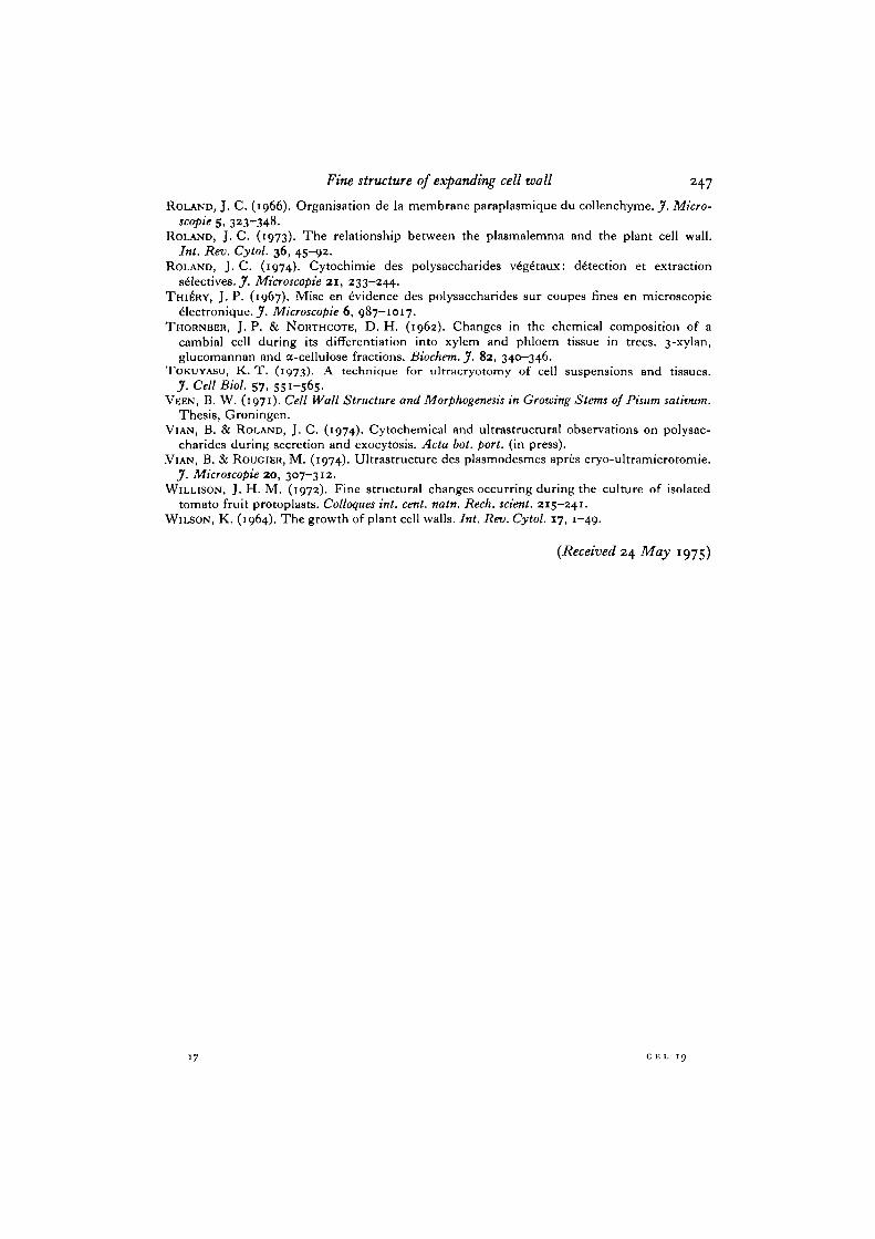
Fig. 7. Cortical parenchyma of pea. Beginning of growth. Ultracryotomy. Trans-verse section. Primary walls (pw) are entirely made of fibrils perpendicular to thedirection of elongation. Note an exocyte (e) without any visible structure. The arrowindicates a region where the fibrils are visible before their complete incorporation tothe wall. The middle lamella (m/) shows a granular structure, x 72 000.Fig. 8. Same tissue as Fig. 7. Cytochemistry. Transverse section. A parallel dis-position of the polysacchandes is shown in the primary wall (pw). The middle lamella(ml) shows a granular structure, x 52000.

Fine structure of expanding cell wall
pw
ml
•pw
pi
pw
I:•~.'2t

252 J. C. Roland, B. Vian and D. Rets
Fig. 9. Cortical parenchyma of pea. Maximum elongation zone. Cytochemistry.Transverse section. The primary wall is made of linear polysaccharides diverselyoriented. The arrow indicates the oldest and outmost layer in which the poly-saccharides have remained transverse despite the elongation, x 35000.Fig. 10. Cortical parenchyma of mung bean hypocotyle. Maximum elongation zone.Cytochemistry. Transverse section. Detail showing lamellation in the primary wall,x 60000.
Fig. 11. Same material as Fig. 5. Detail of the primary wall, x 60000.

Fine structure of expanding cell wall 253
10 pi-
•4

254 J- C- Roland, B. Vian and D. Reis
Fig. 12. Cortical parenchyma of pea. Maximum elongation zone. Ultracryotomy.Transverse section. Middle lamella (ml) and miter part of primary wall (pw). Themiddle lamella is made chiefly of granular components. Note that in the primary wallthe outermost elements remain transverse, x 70000. Inset: high magnification of thegranular components observed in a secretory vesicle, x 270000.
Fig. 13. Same material. Same technique. Detail of a row of parallel fibrils of theprimary wall observed longitudinally. Pairs of fibrils are seen. One couple istwisted (arrows), x 600000.
Fig. 14. Root cap of pea. Aged cell. Ultracryotomy. Tangential section. 'Scatteredtexture' in the outer part of the wall, x 160000.
Fig. 15. Cortical parenchyma of pea. Maximum elongation zone. Ultracryotomy.Oblique section. 'Parallel texture' in the inner part of the wall. Note that the pairsof fibrils are already seen near the plasmalemma (pi), x 160000.

Fine structure of expanding cell wall
256 J. C. Roland, B. Vian and D. Rets
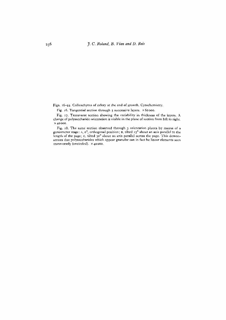
Figs. 16-22. Collenchyma of celery at the end of growth. Cytochemistry.Fig. 16. Tangential section through 3 successive layers, x 60000.Fig. 17. Transverse section showing the variability in thickness of the layers. A
change of polysaccharide orientation is visible in the plane of section from left to right,x 40000.
Fig. 18. The same section observed through 3 orientation planes by means of agoniometer stage: A, O°, orthogonal position; B, tilted 150 about an axis parallel to thelength of the page; c, tilted 300 about an axis parallel across the page. This demon-strates that polysaccharides which appear granular can in fact be linear elements seentransversely (encircled), x 40000.

Fine structure of expanding cell wall
17
O•41
18A 18B

258 J. C. Roland, B. Vian and D. Rets
Figs. 19-21. Inner part of the wall showing in cells of the same tissue great differencesin the thickness of the lamellae of transverse (T) and longitudinal (L) polysaccharides.Note the difference in the orientation of components in contact with the cytoplasm(transverse in Fig. 19, longitudinal in Fig. 21). x 60000.
Fig. 22. Architectural change according to the position of the wall. The lamellationdue to alternately transverse and longitudinal components in the corner of the cell (left)is no longer visible in the intercorner face (light) where the wall exhibits only trans-verse components, x 60000.

Fine structure of expanding cell wall
