· of transcription (jak/stat), etc. (otani, 2008), and regulate a large variety of cellular...
TRANSCRIPT

Conditioning Medicinewww.conditionmed.org
REVIEW ARTICLE | OPEN ACCESS
Regulat ion of gene expression in ischemic preconditioning in the brain
Tuo Yang1, Qianqian Li1, Feng Zhang1,2
[Received: 30 November 2017; accepted 11 December 2017; published online 15 December 2017]
Stroke is the third leading cause of death and the leading cause of long-term disability, with very few effective treatments and limited progress in the effort to search for novel therapeutic approaches. The phenomenon that a sublethal ischemic insult induces protection against a subsequent severe ischemia, termed ischemic preconditioning (IPC), represents an endogenous protective approach against ischemic brain injury, and may direct a breakthrough to future therapeutic strategies. It is broadly accepted that new protein synthesis is required for IPC-mediated long-term neuroprotection; however, their relative regulatory mechanisms are poorly understood. In the present review, we summarize genomic-based studies on alterations in gene expression and protein synthesis, particularly categorizing potential pathways regulated by IPC. We also review the role of epigenetics, an inheritable genetic regulatory mechanism without changes in DNA sequence, in IPC-mediated neuroprotection.
1Department of Neurology, Pittsburgh Institute of Brain Disorders and Recovery, University of Pittsburgh School of Medicine, Pittsburgh, PA 15213, USA. 2Geriatric Research, Educational and Clinical Center, Veterans Affairs Pittsburgh Health Care System, Pittsburgh, PA.
Correspondence should be addressed to Dr. Feng Zhang ([email protected]).
Conditioning Medicine 2017 | Volume 1 | Issue 1 | December 2017 47
REVIEW ARTICLE
Conditioning Medicine 2017 | www.conditionmed.org
Conditioning Medicine | 2017, 1(1):47-56
48
Stroke is the third leading cause of death and the leadingcause of long-term disability, with very few effective
treatments (Benjamin et al., 2017). The discovery of ischemic tolerance may lay the groundwork for future therapeutic development (Li et al., 2017). In 1986, Murry et al. reported that several episodes of non-injurious ischemia to the dog heart led to a 25% reduction in infarct size produced by a subsequent sustained occlusion of the coronary artery. They named this phenomenon “preconditioning” (Murry et al., 1986). Up to now, preconditioning has been observed in multiple animal species and across various organs, including the brain (Kitagawa et al., 1990), liver (Salomao et al., 2012), kidney (Cao et al., 2010), spinal cord (Liang et al., 2012), and pancreas (Hogan et al., 2012), etc. It should be noted that non-lethal ischemia is not the only means that confers brain ischemic tolerance. Other methods include hypothermia, hypoxia, cortical spreading depression, inflammation, oxidative stress, and epilepsy, etc. (Stetler et al., 2009). This phenomenon is termed “cross-conditioning” or “cross-tolerance”, denoting that stimulus of one type could provide protection against subsequent injury of an entirely different type (Stetler et al., 2014).
Various experimental models of brain IPC have been applied in the preclinical studies. In rodents, cerebral ischemia can be primarily induced by either global (forebrain) ischemia or focal ischemia models. Two global ischemia models have been reported: permanent occlusion of the vertebral arteries combined with brief occlusion of bilateral common carotid arteries (4VO), and bilateral carotid artery occlusion(2VO) with systemic hypotension. In the first study that described IPC in the brain in 1990, Kitagawa et al. reported that IPC rendered hippocampal neurons tolerant to subsequent lethal ischemia in gerbils. Interestingly, although they merely occluded bilateral carotid arteries, they actually induced a 4VO global ischemia model due to lack in the posterior communicating artery in gerbils (Kitagawa et al., 1990). Focal ischemia is typically induced by a temporary or permanent occlusion of the middle cerebral artery (MCA), because it represents a clinical course of prodromal transient ischemic attacks with a subsequent stroke. The intraluminal suture MCA occlusion (MCAO) model, also known as the so-called “filament model”, is induced by inserting a suture filament through the internal carotid artery up to the initial segment of the MCA, and removing the suture after a period of time, yielding local ischemia/reperfusion (I/R) injury (Glazier et al., 1994). The distal MCAO (Morancho et al., 2012) usually requires a craniectomy to expose the MCA, which can be occluded by electrocoagulation and additional transection, resulting in permanent occlusion, or alternatively by a microclip, a snare ligature, or a tungsten hook temporarily interrupting the blood flow of MCA (Shigeno et al., 1985; Buchan et al., 1992; Popa-Wagner et al., 1999). Notably, a novel laser-induced photochemical reaction technique enables us to make a pinpoint permanent occlusion of a vessel, leaving the dura and the skull intact (Dietrich et al., 1987).
Disruption of continuous oxygen and glucose supply can lead to neuronal death within a few minutes. The oxygen-glucose deprivation (OGD) in cell cultures or tissue slices is the most widely used model system mimicking ischemic injury in vitro. The OGD method was first established by Goldberg and Choi in mixed neocortical cultures (Goldberg and Choi, 1993), and Bruer et al., who further modeled in vitro IPC in a neuronal-enriched culture, observed that neuronal death was significantly reduced with sublethal OGD 48-72 h before lethal OGD (Bruer et al., 1997). The in vitro models provide a useful tool to study in the mechanisms of IPC, which may be generalized into the whole-animal model systems.
The two time-windows of protection in IPC are well-established and thoroughly reviewed (Stetler et al., 2014). The rapid window, occurring within minutes after IPC, provides
only a short-lived (1–2 hours) protection against lethal ischemia (Schurr et al., 1986; Perez-Pinzon et al., 1997). Moreover, the rapid window seems to be less universal, as no protection was observed in the rapid window in a global ischemia model in gerbils (Kato et al., 1991). Compared to the rapid window of IPC, the delayed window confers long-lasting and more robust neuroprotection. The delayed window starts 24 hours after IPC, peaking at 48 hours to 72 hours, and lasting up to 1 week (Chen and Simon, 1997). It is broadly accepted that de novo protein synthesis is required for IPC-mediated long-term neuroprotection; however, the identification of proteins that are necessary, and their relative regulatory mechanisms are poorly understood (Barone et al., 1998; Dirnagl et al., 2003; Koch et al., 2014).
Phases of IPC-mediated gene expressionTo understand the mechanisms underlying the delayed window of IPC, we need to map how brief ischemia leads to subsequent protein synthesis after IPC. Three distinct phases have been described in the process of protein synthesis: the triggering phase, the signal transduction phase, and the genomic phase (Della-Morte et al., 2012). The triggering phase involves release of receptor agonists that bind membrane receptors, mainly G protein-coupled receptors. Next, in the signal transduction phase, intracellular secondary messengers transduce the signals and activate transcription factors. And finally, the last phase, the genomic phase, refers to genetic regulation by transcription factors.
The triggering phaseThe most important receptor agonist in the triggering phase is adenosine, which is a purine nucleoside (Heurteaux et al., 1995; Perez-Pinzon et al., 1996). It is released by cultured neurons and can be detected as soon as 60 min after OGD (Parkinson and Xiong, 2004). The rapid release of adenosine potentiates it to mediate neuroprotection in both rapid window and delayed window, via different mechanisms, though. Adenosine-mediated rapid protection is via a decrease in glutamate release and inhibition of calcium fluxes, mainly though adenosine A1 receptor (Heurteaux et al., 1995; Zhou et al., 2004; Shen et al., 2011). Blockade of A1 receptor, leading to abrogation of IPC-mediated rapid protection (Perez-Pinzon et al., 1996). On the other hand, adenosine-mediated delayed protection is through passing on the signals to secondary messengers and pushing the story forward to the signal transduction phase and the genomic phase.
In addition to adenosine, release of other receptor activators and redox signaling are also reported in the triggering phase. For example, opioid receptors and nicotinic acetylcholine receptors are activated in IPC (Peart et al., 2005; Rehni et al., 2008; van Rensburg et al., 2009), and bradykinin and erythropoietin are released and confer neuroprotection through activation of multiple signaling pathways (Baker, 2005; Ping et al., 2005; Noguchi et al., 2007).
The signal transduction phaseProtein kinase C (PKC) pathway is a central player in the signal transduction phase. First, PKC targets the adenosine triphosphate (ATP)-sensitive potassium channel, which prevents calcium overload in the mitochondrion and slows the tricarboxylic acid (TCA) cycle (Critz and Byrne, 1992; Domanska-Janik and Zablocka, 1993; Nishi et al., 1999; Ivannikov et al., 2010). Consequently, scarce energy resources can be conserved, and excessive reactive oxygen species (ROS) product can be eliminated. In addition, opening ATP-sensitive potassium channels could promote uptake of glutamate by astrocytes (Sun et al., 2008), which reduces excitotoxicity in ischemia. Second, PKC also upregulates the
REVIEW ARTICLE
Conditioning Medicine 2017 | www.conditionmed.org
Conditioning Medicine | 2017, 1(1):47-56
49
expression of Sirtuin 1 (SIRT1) through activating nicotinamide phosphoribosyltransferase (Morris-Blanco et al., 2014), which boosts robust neuroprotection via both epigenetics-dependent and -independent mechanisms (Koronowski and Perez-Pinzon, 2015).
Other signaling pathways also play a role in the signal transduction phase, such as glycogen synthase kinase (GSK) 3β, mitogen-activated protein kinase (MAPK), c-Jun N-terminal kinase (JNK), and Janus kinase/signal transducers and activators of transcription (JAK/STAT), etc. (Otani, 2008), and regulate a large variety of cellular responses, including growth and proliferation, development and differentiation, inflammation, and apoptosis.
The genomic phaseThe genomic phase is initiated by nuclear translocation of transcription factors in response to intracellular signal transduction. Of note is that, transcription factors of the activator protein-1 family are activated that induce the translation of proto-oncogenes such as c-fos, jun B, c-jun and jun D (Truettner et al., 2002), and thereby play a critical role in determining the fate of cells after ischemic injury. Hundreds of genes are changed after IPC, by either upregulation or downregulation. This is termed “genomic reprogramming”, which expands the previous concept “new protein synthesis” in IPC by stating that downregulation of some genes should also be considered in this phase. The reprogramming profile following IPC is described by either genomic-based or proteomic-based studies and will be reviewed in Section 3.
In addition, the important role of epigenetics has recently been introduced to the stroke field, based on the finding that stroke is associated with increased DNA methylation (Endres et al., 2000) and histone acetylation (Meisel et al., 2006). However, it remains largely unexplored how epigenetics
regulates IPC responses; thus, it might be a promising future direction for a better understanding of the IPC biology. The remodeling of epigenetic marks is discussed in Section 4.
Genomic reprogramming after IPCGeneral features of genomic reprogramming after IPCAs previously mentioned, there exists a “cross-conditioning” phenomenon in the brain. Although multiple stimuli lead to similar ischemic tolerant status, the genomic reprogramming patterns they trigger are not the same. For example, IPC is associated with downregulation in genes related to metabolism and channels, while lipopolysaccharide preconditioning altered inflammatory patterns along with upregulation in defense genes are associated with type I interferons (Stenzel-Poore et al., 2007). Thus, it is concluded that the nature of preconditioning stimulus determines the neuroprotective phenotype and genomic reprogramming pattern (Stenzel-Poore et al., 2007; Vartanian et al., 2015).
Both IPC and I/R are of ischemic basis, though with different degrees in severity. As a result, one may speculate that the genomic reprogramming processes triggered by IPC should cover similar categories of genes and have similar pattern with I/R. Surprisingly, using microarray analysis to investigate gene expression after IPC and I/R in mice, Stenzel-Poore et al. found no common overlap among the IPC, I/R and IPC followed by I/R (IPC+I/R) groups (Stenzel-Poore et al., 2003). Moreover, when comparing IPC+I/R with I/R alone, up to 83% genomic responses were unique to IPC+I/R (Stenzel-Poore et al., 2007). This number is much lower, about 39%, in primary neuronal cultures with 45 min OGD mimicking in vivo IPC (Prasad et al., 2012). The difference between in vivo and in vitro setting is fairly reasonable, since other than neurons, glial cells and microvessel cells are also actively engaged in the adaptation to ischemia and/or IPC, and take part in forming
REVIEW ARTICLE
Conditioning Medicine 2017 | www.conditionmed.org
Conditioning Medicine | 2017, 1(1):47-56
50
an integrated network, termed neurovascular unit (Zhang et al., 2012). Collectively, IPC is associated with a unique programmed phenotypic alteration and confers a so-called “pro-survival” phenotype (Kawahara et al., 2004), which results in “programmed cell survival” (Gidday, 2006).
Both upregulation and downregulation of genes participate in IPC-mediated genomic reprogramming. Multiple studies have revealed that I/R prefers upregulation than downregulation, and IPC + I/R induces more pronounced downregulation. For example, up to 86% genes regulated showed increased expression in I/R, while 77% of regulated genes showed decreased expression in IPC + I/R (Stenzel-Poore et al., 2003). IPC alone is associated with a slight trend preferring upregulation than downregulation (Stenzel-Poore et al., 2003; Stenzel-Poore et al., 2004; Feng et al., 2007).
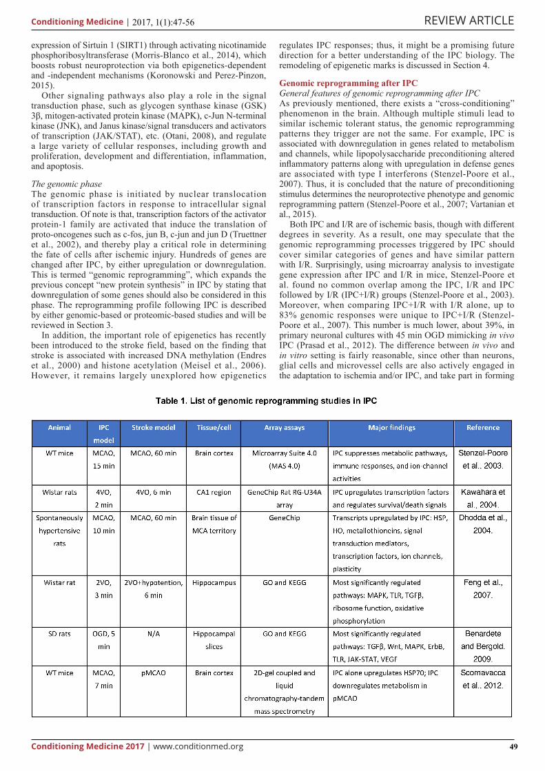
In t h i s s ec t ion , we r ev i ew the ove ra l l genomic reprogramming pattern mediated by IPC, particularly focusing on genomic-based studies and proteomic-based studies but not those studying an individual protein or pathway, and try to disclose the commonly shared mechanisms that are necessary for IPC (Table 1).
Categories of genes regulated by IPC1) Transcription factorsTranscription factors are effectors of IPC that directly regulategene expression and protein synthesis, and several groups oftranscription factors have been involved in IPC. For example,silencer factor B and C-jun are regulated in IPC (Dhodda et al.,2004), and c-Fos, Jun B, and zinc finger Egr family membersare also upregulated (Kawahara et al., 2004). In a studyconducted using a MetaCore’s network algorithm mappingof pathway analysis, it is revealed that protein expressionchanges in IPC are dependent mainly on three proteins:androgen receptor, HIF-1, and NF-κB (Scornavacca et al.,2012). Additionally, preconditioning in rat hippocampal slicessignificantly increases in transcription activities of Fosl1, Jun D,Med13 and Nr4a1, peaking around 3 h post IPC (Benardete andBergold, 2009).
2) Ion channels and transportersIon channels and other transporters are reported to bedownregulated by IPC (Stenzel-Poore et al., 2003; Stenzel-Poore et al., 2004). IPC significantly decreased potassiumchannel Kv1.5 expression in I/R brain in vivo. This finding isfurther validated by electrophysiological studies in in vitro ratcortical neuronal cultures which showed decreased outwardpotassium currents and whole-cell conductance after IPC(30 min OGD) (Stenzel-Poore et al., 2003). Consistently,downregulation of voltage-dependent anion channel 1 withproteomic analysis in the CA1 region after global IPC wasreported (Nakajima et al., 2015). In another study (Feng et al.,2007) applying global ischemia following global IPC, calciumsignaling pathway is among the most significantly modulatedpathways. Since a four-way pairwise comparison wasperformed, their findings are more likely to reveal pathwaysnecessary for IPC-mediated protection. Regrettably, they didnot identify whether and which genes in this pathway wereupregulated or downregulated. In addition, it is worth notingthat they were comparing it with tissues from IPC group vsIPC + MK801 group, in which MK801 is a non-competitiveNMDA receptor, which could abolish the protection providedby IPC (Zhang et al., 2009). Similarly, Kawahare et al. reporteddownregulation in calcium signaling genes, IP3 kinase and IP3receptor, mediated by a prior IPC (2 min global ischemia) inischemic rat brain (6 min global ischemia) (Kawahara et al.,2004).
However, Dhodda et al.’s study disagrees with the aforementioned results (Dhodda et al., 2004). They performed
a 10-min MCAO to induce IPC in spontaneously hypertensive rats, and total RNA from the MCA territory was subjected to GeneChip analysis. They reported upregulation in ion channels, including sodium channel scn6a, potassium channel KCNJ13, and calcium channel CNGA3 and P2X-associated ATP-gated channel. Consistently, Benardete and Bergold reported upregulated calcium signaling pathway genes after IPC in ex vivo hippocampal slices (Benardete and Bergold, 2009).
Acid-sensing ion channels, ASICs, are reported to be downregulated after IPC (Pignataro et al., 2011), although no whole-genomic studies have revealed a similar phenomenon.
3) Signaling transductionUpregulation in genes related to MAPK signaling pathway iswell-documented both in vivo (Dhodda et al., 2004; Kawaharaet al., 2004; Feng et al., 2007) and ex vivo (Benardete andBergold, 2009). An article also reported an upregulation of othergenes related to signaling transduction, including SMAD-1 and-7, guanylyl cyclase, and retinoid acid receptor, etc. (Dhodda etal., 2004). JAK/STAT signaling is reported to increase after IPCex vivo in hippocampal slice culture (Benardete and Bergold,2009), while cyclic adenosine triphosphate signaling andprotein kinase A signaling are decreased in neuronal culturesafter IPC (Prasad et al., 2012).
4) InflammationToll-like receptors (TLR) play a major role in innate immuneresponse against various insults. A hierarchical analysis revealedsignificant downregulation in genes in TLR signaling pathway(Feng et al., 2007). This is further confirmed by Western blot,showing decreased expression of cyclooxygenase 2, a TLR4downstream inflammatory factor. However, in an ex vivo studyusing rat hippocampal slice culture subjected to IPC (inducedby 5 min OGD), increased gene expression of TLR signalingwas reported, associated with increased inflammatory factorsinterleukin (IL)-1a, IL-1b, IL-6 and TNF as confirmed bypolymerase chain reaction (RT-PCR) (Benardete and Bergold,2009). Consistently, in primary neuronal cultures, increasedinflammatory responses after IPC were observed (Prasad et al.,2012).
5) NeuroplasticityIn spontaneously hypertensive rats, IPC was associated withsignificant upregulation in neurotrophic factors transforminggrowth factor (TGF)-α, TGF-β1, and TGF-β receptor, amongwhich the increase in TGF-β1 was confirmed by RT-PCR(Dhodda et al., 2004). Feng et al. (Feng et al., 2007) dugdeeper by focusing on the expression of bone morphogeneticprotein (BMP)-7, a member of TGF-β superfamily with uniqueneuroprotective and regenerative effects (Harvey et al., 2005;Heinonen et al., 2014). They confirmed upregulation in BMP-7in IPC with RT-PCR, Western blot and immunohistochemistryin the dentate gyrus. Increase in TGF-β pathway genes wasalso observed ex vivo in hippocampal slice culture after IPC,together with genes related to other neurotrophic pathways, likeWnt, ErbB, and vascular endothelial growth factor signaling(Benardete and Bergold, 2009). In another study with invitro neuronal culture, IPC increased cell cycle genes andneurotrophic genes (Prasad et al., 2012). Naylor et al. confirmedthat IPC alone could increase in a panel of growth factors,including insulin-like growth factor, fibroblast growth factor,TGF-β, epithelial growth factor and platelet-derived growthfactor-A (Naylor et al., 2005).
6) Metabolism and ribosome pathwayIn Stenzel-Poore’s study (Stenzel-Poore et al., 2003), theauthors reported significant decrease in metabolic geneexpression; interestingly, they further interpreted this feature asa stimulation to hibernation, in which a suppression of energyuse contributes to neuroprotection (Drew et al., 2001). Their
REVIEW ARTICLE
Conditioning Medicine 2017 | www.conditionmed.org
Conditioning Medicine | 2017, 1(1):47-56
51
findings agree with a later study using proteomic analysis (Scornavacca et al., 2012). While Feng et al.’s study, using a four-way pairwise comparison, elicited opposite results. They reported a significant increase in genes related to ribosome pathway, oxidative phosphorylation and protein synthesis (Feng et al., 2007). They also suggested that activated metabolism and ribosome pathway could facilitate the recovery protein processing machinery after injurious ischemia. Consistently, an ex vivo study inducing IPC in hippocampal slice culture reported increased genes related to glycolysis and gluconeogenesis (Benardete and Bergold, 2009). The above studies seem controversial, but in fact, a possible explanation for this discrepancy is the difference in treatment groups. In the first two studies, the authors compared IPC + I/R group versus I/R alone group, while in the third and fourth studies, the comparison was between IPC alone versus control or IPC alone versus IPC + MK801 group, without injurious ischemia.
However, one article reported opposite effect of IPC using proteomic analysis with two-dimensional electrophoresis, showing decreased mitochondrial aconitase, an enzyme related to TCA cycle, in CA1 region after IPC induced by 3 min global ischemia (Nakajima et al., 2015). A shortcoming of this study is that there was no entire pathway-based analysis, and change in one single protein can hardly reflect the general trend of one pathway.
7) Heat shock proteins (HSPs)HSPs are important stress sensors in the cellular system.Increased HSP expression by IPC is reported in multiplesstudies (Stenzel-Poore et al., 2003; Dhodda et al., 2004;Kawahara et al., 2004). To specify, after 15 min MCAO as IPCin mice, Hsp70 increased at 24 h, and Hspb2 at 72 h (Stenzel-Poore et al., 2003). IPC also contributed to an additionalincrease in Hsp70, 105 and 10 by I/R after 24 h (Stenzel-Poore et al., 2003). In a global IPC model, the only upregulatedHsp mRNA after IPC is Hsp70 (Kawahara et al., 2004). Inspontaneously hypertensive rats, IPC (10 min MCAO) inducedincrease in Hsp70, 27, 90, 10 and 60 as confirmed by RT-PCR(Dhodda et al., 2004), and the increase in HSP70 was thenvalidated by Western blot and immunohistochemistry in vivo.The upregulation of HSP70 after IPC was also reported byusing proteomic analysis by two-dimensional electrophoresiscoupled to liquid chromatography-tandem mass spectrometry(Scornavacca et al., 2012).
8) Redox signalingAlthough it was mentioned that genes related to oxidative stresswere regulated by IPC, Western blot reveals no significantchanges in the expression of superoxide dismutase, an ROSscavenger (Scornavacca et al., 2012). More glutathioneS-transferase expression was observed in CA1 than CA3/dentate gyrus region, but no significant changes between thesham and IPC groups (Nakajima et al., 2015). Although somesporadic studies did elicit increased antioxidants induced byIPC both in vivo and in vitro (Dozza et al., 2004; Holtzclawet al., 2004; Danielisova et al., 2005; Shokeir et al., 2014), noproteomic or genomic-based studies reported the indispensablerole of redox signaling.
Given that buffering oxidative stress abolished IPC-mediated protection (Puisieux et al., 2004; Narayanan et al., 2017), a couple groups have reported that the mild oxidative stress in preconditioning initiates redox signaling and activates nuclear factor erythroid 2-related factor 2 (Nrf2), a master transcription factor the upregulates anti-oxidative enzymes, in astrocytes (Bell et al., 2011; Narayanan et al., 2017). In support, our group also found an important role of Nrf2 after IPC. We found the nuclear translocation of Nrf2 after IPC and validated the indispensable role of Nrf2 for IPC (Yang et al., 2016a).
Limitations and future directionsDespite the progress in genomic-based and proteomic-based techniques in the past decade, all the above studies failed to make a breakthrough to reveal a common “tolerasome” or to provide clues in finding biomarkers for IPC (Meller and Simon, 2013; Koch et al., 2014). Besides different models and different comparison in different studies, most studies only applied correlation analysis, without further determination on the causal relationship. In addition, the sample sizes may be too small which lowered the power in these studies. A third limitation is lack of cellular specificity, since samples from most in vivo studies were extracted from whole tissue. Last but not least, gene regulation at an epigenetic level is largely ignored. Epigenetics regulates the transcriptional potential through modifying the accessibility of DNA to the transcriptional machinery. In the next section, we are going to review epigenetic reprogramming in IPC.
Remodeling of epigenetic marks after IPCDuring the past decade, there has been an emerging role of epigenetics in regulating pathologic processes and outcome of stroke. Nevertheless, fewer studies focus on the role of epigenetics in IPC. How IPC alters epigenetic reprogramming is still at a start-up stage and is attracting more and more attention. Mechanisms underlying epigenetic modulation on DNA expression include noncoding RNAs -- typically microRNAs (miRNAs) (Saugstad, 2010), global SUMOylation (SUMO: small ubiquitin-like modifier) (Lee et al., 2009), direct DNA modification by methylation, and histone protein modifications (Schweizer et al., 2015).
miRNAsIn an elegant study conducted by Lusardi et al. (Lusardi et al., 2010), miRNA array was performed in mouse cortex tissues following IPC (15 min MCAO) or I/R (60 min MCAO). IPC + I/R upregulated ~200 miRNAs, and downregulated ~100miRNAs. However, I/R alone only triggered ~100 miRNAdownregulation, suggesting a unique and robust miRNAresponse to IPC rather than I/R. Further target RNA analysison those downregulated miRNAs revealed a common target,methyl CpG binding protein 2 (MeCP2), which binds tomethylated DNA and helps with gene silencing. They alsoconfirmed that IPC increased MeCP2 expression, and MeCP2knockout abolished IPC-mediated neuroprotection.
Targeting pathways were also explored, though roughly. Dharap and Vemuganti profiled miRNAs in the cerebral cortices from spontaneously hypertensive rats after IPC and reported a quickly reactive miRNAome (Dharap and Vemuganti, 2010). Among 51 miRNAs that were altered, 26 were upregulated and 25 were downregulated. They further performed KEGG pathway analysis on their target proteins and found that MAPK pathway and mTOR pathway were top the 2 upregulated proteins, and Wnt pathway and GnRH pathway were the top 2 downregulated proteins. Lee et al. performed miRNA profiling covering a total of 360 miRNAs, and found selective upregulation of two miRNA families, miR-200 and miR-182, 3 hours after IPC effects (Lee et al., 2010b). They then transfected some of them and revealed that prolyl hydroxylase 2 and HIF-1 pathways had the best neuroprotective effects.
Global SUMOylationSUMOylation is correlated with transcriptional repression via modulating diverse chromatin enzymes, chromatin associated proteins, and promoter specific transcription factors (Ouyang and Gill, 2009). Elevated SUMOylation was protective against focal ischemia mouse brains (Lee et al., 2009). In the setting of IPC, elevated SUMO-1 conjugation levels was observed. Primary cortical neurons were more resistant to OGD when

REVIEW ARTICLE
Conditioning Medicine 2017 | www.conditionmed.org
Conditioning Medicine | 2017, 1(1):47-56
52
transfected with SUMO-1 or SUMO-2, and SUMO-1-siRNA transfection attenuated IPC-mediated protection (Lee et al., 2009).
DNA methylationIn general, DNA methylation at the promoter region results in repression in gene expression, and DNA hypomethylation is associated with gene transcription. DNA methylation is mediated by DNA methyltransferases (DNMTs). Both I/R in vivo and OGD in vitro led to increased DNA methylation (Endres et al., 2000; Hu et al., 2006), and a lower level, but not absence, of DNMT1 is protective against ischemic injury, associated with decreased DNA methylation (Endres et al., 2001). By contrast, in a study describing dynamics of overall DNA methylation after I/R, Meller et al. reported significantly decreased methylated DNA by up to 80% compared to sham, and IPC decreased methylated DNA by~50% (Meller et al., 2015). A possible reason for this discrepancy may lie in their different methodologies. In Endres et al.’s study, DNA methylation was measured by incorporating [3H]-methyl groups into the genomic DNA, which reflects dynamic DNA methylation after I/R-induced DNA injury, while in Meller et al.’s study, methylated DNA was measured by a pull-down of the methyl group and agarose gel analysis, which provides a snapshot of present methylated DNA. Taken together, it can be speculated that I/R-induced DNA damage leads to decrease in methylated DNA which can be attenuated by IPC; DNA remethylation process occurs after I/R.
A number of genes can be regulated by DNA methylation in IPC. The increase of NKCC1, an Na+/K+ co-transporter after I/R correlated to decreased methylation of the promoter (Lee et al., 2010a). Decrease in estrogen receptor methylation led to increased estrogen receptor expression following ischemia (Westberry et al., 2008; Wilson and Westberry, 2009). Moreover, IPC induced methylation to the promoter of thrombospondin 1, an anti-angiogenic factor (Hu et al., 2006; Lawler and Lawler, 2012). Nevertheless, detailed information and the causal relationship between DNA methylation and IPC remain largely unexplored.
Histone modificationHistone proteins include histone 2A, 2B, 3 and 4. They participate in forming nucleosomes, which blocks the access of transcription factors to DNA. Histones can be modulated by acetylation, phosphorylation, methylation, and ubiquitination.
1) Histone acetylationAcetylation of histones is the most extensively studied type ofepigenetics, not in IPC settings, though. In general, acetylationof histones opens the chromatin configuration and allowstranscription factors to bind DNA, and deacetylation of histonesis associated with repression in gene expression. Histoneacetylation is mediated by histone deacetylases (HDACs).Class I and II HDACs are Zn2+-dependent, and class IIIHDACs, otherwise known as Sirtuins, are nicotinamide adeninedinucleotide (NAD)+-dependent.
HDAC inhibitors are powerful tools to assess the role of HDACs. Class I and II HDAC inhibitors selectively inhibit the zinc hydrolase domain (Bradner et al., 2010). Multiples studies demonstrated neuroprotection against stroke by class I and II HDAC inhibitors via modulating oxidative stress, DNA damage, and inflammation (Qi et al., 2004; Ren et al., 2004; Faraco et al., 2006; Kim et al., 2007). The critical role of HDAC3, a member of HDAC class I, was also confirmed in IPC both in vivo and in vitro, possibly via induction of Hspa1a, Bcl2l1, and Prdx2 expression (Yang et al., 2016b).
NAD+ is a vital oxidizing agent of the glycolytic and TCA cycle. Being NAD+ sensors in the cellular system, Sirtuins, especially SIRT1, are required for IPC. In both in vivo and ex
vivo studies, SIRT1 activities were increased following IPC, and blockade SIRT1 abrogated IPC-mediated neuroprotection (Raval et al., 2006; Della-Morte et al., 2009; Koronowski et al., 2015). However, one should be aware of SIRT1’s diverse targets, including both histone and non-histone proteins. For example, SIRT1 can either deacetylate MeCP2 to elicit neuroprotection (Zocchi and Sassone-Corsi, 2012), or function indirectly via transcription factors, transcriptional co-factors, or nuclear receptors (Zhang et al., 2011).
2) Histone phosphorylationThe best-known function of histone phosphorylation occurswhen cells respond to DNA damage. Histone phosphorylationis associated with DNA repair, cell cycle and mitosis (Rossettoet al., 2012). Activation of the 5’-adenosine monophosphate-activated protein kinase signaling pathway, a master pathwayregulating cellular energy homeostasis, is the major mechanismthat phosphorylates histone. Bungard et al. reported that stressinduced histone 2B phosphorylation at S36 residue; S36Adecreased cell viability under stress conditions (Bungard et al.,2010). There is no report on histone phosphorylation in IPC sofar.
3) Histone mono-ubiquitinationMono-ubiquitination of histone 2A and 2B contributes torepression of genes that consume ATP. for example, voltage-gated potassium channels, a key factor during IPC (Stapels etal., 2010). Such mono-ubiquitination is mediated by polycombgroup proteins (PcG). PcG decreased potassium currents incultured neuronal cells, and PcG knockdown precluded theinduction of IPC. Some researchers believe PcG is one ofthe central modulators of IPC given its diverse target genesincluding those involved in electron and glucose transporters(Brand and Ratan, 2013).
ConclusionOver the past several decades since the discovery of IPC, piles of evidence have demonstrated the generality of this phenomenon across multiple species and organs that can be induced by diverse in vivo and in vitro models. This has largely expanded the broadness of our understanding in IPC. Disappointingly, our understanding in the depth of IPC mechanisms, such as which pathways, proteins, molecules, and genetic modulation account for IPC-endowed neuroprotection, remains superficial. Besides the two time-windows and the requirement of genomic reprogramming for long-term neuroprotection, no breakthrough has been made during the past two decades, despite extensive work on genomic-based and proteomic-based studies. Epigenetics in the stroke field has gotten increasingly hot, while its role in IPC remains poorly understood. Since drugs involving epigenetic regulation have already been put into clinical trials (Hwang et al., 2013), we expect that exploration on epigenetic regulation in IPC may lead to a new era for stroke and IPC research. The effects of IPC might be an integrated consequence from a complicated network, whose components actively interact with one another. Apparently, we are currently at the very beginning in understanding the integrated mechanisms of the genetic regulation in IPC. We still have a long way to go before we fully understand the biology of IPC, and, what is more important, bring it to clinic to fight against stroke.
AcknowledgementsThis work was supported by grants from the National Institutes of Health (NS092810 to FZ). We thank Pat Strickler for her administrative support.
References
REVIEW ARTICLE
Conditioning Medicine 2017 | www.conditionmed.org
Conditioning Medicine | 2017, 1(1):47-56
53
Bake r JE (2005 ) E ry th ropo i e t i n mimics i s chemic preconditioning. Vascular pharmacology 42:233-241.
Barone FC, White RF, Spera PA, Ellison J, Currie RW, Wang X, Feuerstein GZ (1998) Ischemic preconditioning and brain tolerance: temporal histological and functional outcomes, protein synthesis requirement, and interleukin-1 receptor antagonist and early gene expression. Stroke 29:1937-1950; discussion 1950-1931.
Bell KF, Al-Mubarak B, Fowler JH, Baxter PS, Gupta K, Tsujita T, Chowdhry S, Patani R, Chandran S, Horsburgh K, Hayes JD, Hardingham GE (2011) Mild oxidative stress activates Nrf2 in astrocytes, which contributes to neuroprotective ischemic preconditioning. Proceedings of the National Academy of Sciences of the United States of America 108:E1-2; author reply E3-4.
Benardete EA, Bergold PJ (2009) Genomic analysis of ischemic preconditioning in adult rat hippocampal slice cultures. Brain research 1292:107-122.
Benjamin EJ et al . (2017) Heart Disease and Stroke Statistics-2017 Update: A Report From the American Heart Association. Circulation 135:e146-e603.
Bradner JE, West N, Grachan ML, Greenberg EF, Haggarty SJ, Warnow T, Mazitschek R (2010) Chemical phylogenetics of histone deacetylases. Nature chemical biology 6:238-243.
Brand D, Ratan RR (2013) Epigenetics and the environment: in search of the "toleroasome" vital to execution of ischemic preconditioning. Transl Stroke Res 4:56-62.
Bruer U, Weih MK, Isaev NK, Meisel A, Ruscher K, Bergk A, Trendelenburg G, Wiegand F, Victorov IV, Dirnagl U (1997) Induction of tolerance in rat cortical neurons: hypoxic preconditioning. FEBS letters 414:117-121.
Buchan AM, Xue D, Slivka A (1992) A new model of temporary focal neocortical ischemia in the rat. Stroke 23:273-279.
Bungard D, Fuerth BJ, Zeng PY, Faubert B, Maas NL, Viollet B, Carling D, Thompson CB, Jones RG, Berger SL (2010) Signaling kinase AMPK activates stress-promoted transcription via histone H2B phosphorylation. Science 329:1201-1205.
Cao C, Wang S, Fan L, Wan X, Liu X, Chen X (2010) Renal protection by ischemic preconditioning is associated with p50/p50 homodimers. American journal of nephrology 31:1-8.
Chen J, Simon R (1997) Ischemic tolerance in the brain. Neurology 48:306-311.
Critz SD, Byrne JH (1992) Modulation of IK,Ca by phorbol ester-mediated activation of PKC in pleural sensory neurons of Aplysia. J Neurophysiol 68:1079-1086.
Danielisova V, Nemethova M, Gottlieb M, Burda J (2005) Changes of endogenous antioxidant enzymes during ischemic tolerance acquisition. Neurochem Res 30:559-565.
Della-Morte D, Dave KR, DeFazio RA, Bao YC, Raval AP, Perez-Pinzon MA (2009) Resveratrol pretreatment protects rat brain from cerebral ischemic damage via a sirtuin 1-uncoupling protein 2 pathway. Neuroscience 159:993-1002.
Della-Morte D, Guadagni F, Palmirotta R, Ferroni P, Testa G, Cacciatore F, Abete P, Rengo F, Perez-Pinzon MA, Sacco RL, Rundek T (2012) Genetics and genomics of ischemic tolerance: focus on cardiac and cerebral ischemic preconditioning. Pharmacogenomics 13:1741-1757.
Dharap A, Vemuganti R (2010) Ischemic pre-conditioning alters cerebral microRNAs that are upstream to neuroprotective signaling pathways. Journal of neurochemistry 113:1685-1691.
Dhodda VK, Sailor KA, Bowen KK, Vemuganti R (2004) Putative endogenous mediators of preconditioning-
induced ischemic tolerance in rat brain identified by genomic and proteomic analysis . Journal of neurochemistry 89:73-89.
Dietrich WD, Busto R, Watson BD, Scheinberg P, Ginsberg MD (1987) Photochemically induced cerebral infarction. II. Edema and blood-brain barrier disruption. Actaneuropathologica 72:326-334.
Dirnagl U, Simon RP, Hallenbeck JM (2003) Ischemic tolerance and endogenous neuroprotection. Trends in neurosciences 26:248-254.
Domanska-Janik K, Zablocka B (1993) Protein kinase C as an early and sensitive marker of ischemia-induced progressive neuronal damage in gerbil hippocampus. Mol Chem Neuropathol 20:111-123.
Dozza B, Smith MA, Perry G, Tabaton M, Strocchi P (2004) Regulation of glycogen synthase kinase-3beta by products of lipid peroxidation in human neuroblastoma cells. Journal of neurochemistry 89:1224-1232.
D r e w K L , R i c e M E , K u h n T B , S m i t h M A ( 2 0 0 1 ) Neuroprotective adaptations in hibernation: therapeutic implications for ischemia-reperfusion, traumatic brain injury and neurodegenerative diseases. Free radical biology & medicine 31:563-573.
Endres M, Fan G, Meisel A, Dirnagl U, Jaenisch R (2001) Effects of cerebral ischemia in mice lacking DNA methyltransferase 1 in post-mitotic neurons. Neuroreport 12:3763-3766.
Endres M, Meisel A, Biniszkiewicz D, Namura S, Prass K, Ruscher K, Lipski A, Jaenisch R, Moskowitz MA, Dirnagl U (2000) DNA methyltransferase contributes to delayed ischemic brain injury. The Journal of neuroscience : the official journal of the Society for Neuroscience 20:3175-3181.
Faraco G, Pancani T, Formentini L, Mascagni P, Fossati G, Leoni F, Moroni F, Chiarugi A (2006) Pharmacological inhibition of histone deacetylases by suberoylanilide hydroxamic acid specifically alters gene expression and reduces ischemic injury in the mouse brain. Molecular pharmacology 70:1876-1884.
Feng Z, Davis DP, Sasik R, Patel HH, Drummond JC, Patel PM (2007) Pathway and gene ontology based analysis of gene expression in a rat model of cerebral ischemic tolerance. Brain research 1177:103-123.
Gidday JM (2006) Cerebral preconditioning and ischaemic tolerance. Nat Rev Neurosci 7:437-448.
Glazier SS, O'Rourke DM, Graham DI, Welsh FA (1994) Induction of ischemic tolerance following brief focal ischemia in rat brain. J Cereb Blood Flow Metab 14:545-553.
Goldberg MP, Choi DW (1993) Combined oxygen and glucose deprivation in cortical cell culture: calcium-dependent and calcium-independent mechanisms of neuronal injury. The Journal of neuroscience : the official journal of the Society for Neuroscience 13:3510-3524.
Harvey BK, Hoffer BJ, Wang Y (2005) Stroke and TGF-beta proteins: glial cell line-derived neurotrophic factor and bone morphogenetic protein. Pharmacol Ther 105:113-125.
Heinonen AM, Rahman M, Dogbevia G, Jakobi H, Wolfl S, Sprengel R, Schwaninger M (2014) Neuroprotection by rAAV-mediated gene transfer of bone morphogenic protein 7. BMC Neurosci 15:38.
Heurteaux C, Lauritzen I, Widmann C, Lazdunski M (1995) Essential role of adenosine, adenosine A1 receptors, and ATP-sensitive K+ channels in cerebral ischemic preconditioning. Proceedings of the National Academy of Sciences of the United States of America 92:4666-4670.
Hogan AR, Doni M, Molano RD, Ribeiro MM, Szeto A,
REVIEW ARTICLE
Conditioning Medicine 2017 | www.conditionmed.org
Conditioning Medicine | 2017, 1(1):47-56
54
Cobianchi L, Zahr-Akrawi E, Molina J, Fornoni A, Mendez AJ, Ricordi C, Pastori RL, Pileggi A (2012) Beneficial effects of ischemic preconditioning on pancreas cold preservation. Cell transplantation 21:1349-1360.
Holtzclaw WD, Dinkova-Kostova AT, Talalay P (2004) Protection against electrophile and oxidative stress by induction of phase 2 genes: the quest for the elusive sensor that responds to inducers. Adv Enzyme Regul 44:335-367.
Hu CJ, Chen SD, Yang DI, Lin TN, Chen CM, Huang TH, Hsu CY (2006) Promoter region methylation and reduced expression of thrombospondin-1 after oxygen-glucose deprivation in murine cerebral endothelial cells. J Cereb Blood Flow Metab 26:1519-1526.
H w a n g J Y, A r o m o l a r a n K A , Z u k i n R S ( 2 0 1 3 ) Epigenet ic mechanisms in s t roke and epi lepsy. Neuropsychopharmacology : official publication of the American College of Neuropsychopharmacology 38:167-182.
Ivannikov MV, Sugimori M, Llinas RR (2010) Calcium clearance and its energy requirements in cerebellar neurons. Cell Calcium 47:507-513.
Kato H, Liu Y, Araki T, Kogure K (1991) Temporal profile of the effects of pretreatment with brief cerebral ischemia on the neuronal damage following secondary ischemic insult in the gerbil: cumulative damage and protective effects. Brain research 553:238-242.
Kawahara N, Wang Y, Mukasa A, Furuya K, Shimizu T, Hamakubo T, Aburatani H, Kodama T, Kirino T (2004) Genome-wide gene expression analysis for induced ischemic tolerance and delayed neuronal death following transient global ischemia in rats. J Cereb Blood Flow Metab 24:212-223.
Kim HJ, Rowe M, Ren M, Hong JS, Chen PS, Chuang DM (2007) Histone deacetylase inhibitors exhibit anti-inflammatory and neuroprotective effects in a rat permanent ischemic model of stroke: multiple mechanisms of action. The Journal of pharmacology and experimental therapeutics 321:892-901.
Kitagawa K, Matsumoto M, Tagaya M, Hata R, Ueda H, Niinobe M, Handa N, Fukunaga R, Kimura K, Mikoshiba K, et al. (1990) 'Ischemic tolerance' phenomenon found in the brain. Brain research 528:21-24.
Koch S, Della-Morte D, Dave KR, Sacco RL, Perez-Pinzon MA (2014) Biomarkers for ischemic preconditioning: finding the responders. J Cereb Blood Flow Metab 34:933-941.
Koronowski KB, Perez-Pinzon MA (2015) Sirt1 in cerebral ischemia. Brain circulation 1:69-78.
Koronowski KB, Dave KR, Saul I, Camarena V, Thompson JW, Neumann JT, Young JI, Perez-Pinzon MA (2015) Resveratrol Preconditioning Induces a Novel Extended Window of Ischemic Tolerance in the Mouse Brain. Stroke 46:2293-2298.
Lawler PR, Lawler J (2012) Molecular basis for the regulation of angiogenesis by thrombospondin-1 and -2. Cold Spring Harbor perspectives in medicine 2:a006627.
Lee HA, Hong SH, Kim JW, Jang IS (2010a) Possible involvement of DNA methylation in NKCC1 gene expression during postnatal development and in response to ischemia. Journal of neurochemistry 114:520-529.
Lee ST, Chu K, Jung KH, Yoon HJ, Jeon D, Kang KM, Park KH, Bae EK, Kim M, Lee SK, Roh JK (2010b) MicroRNAs induced during ischemic preconditioning. Stroke 41:1646-1651.
Lee YJ, Castri P, Bembry J, Maric D, Auh S, Hallenbeck JM (2009) SUMOylation participates in induction of ischemic tolerance. Journal of neurochemistry 109:257-267.
Li S, Hafeez A, Noorulla F, Geng X, Shao G, Ren C, Lu G, Zhao H, Ding Y, Ji X (2017) Preconditioning in
neuroprotection: From hypoxia to ischemia. Prog Neurobiol 157:79-91.
Liang CL, Lu K, Liliang PC, Chen TB, Chan SH, Chen HJ (2012) Ischemic preconditioning ameliorates spinal cord ischemia-reperfusion injury by triggering autoregulation. Journal of vascular surgery 55:1116-1123.
Lusardi TA, Farr CD, Faulkner CL, Pignataro G, Yang T, Lan J, Simon RP, Saugstad JA (2010) Ischemic preconditioning regulates expression of microRNAs and a predicted target, MeCP2, in mouse cortex. J Cereb Blood Flow Metab 30:744-756.
Meisel A, Harms C, Yildirim F, Bosel J, Kronenberg G, Harms U, Fink KB, Endres M (2006) Inhibition of histone deacetylation protects wild-type but not gelsolin-deficient neurons from oxygen/glucose deprivation. Journal of neurochemistry 98:1019-1031.
Meller R, Simon RP (2013) Tolerance to ischemia - an increasingly complex biology. Transl Stroke Res 4:40-50.
Meller R, Pearson A, Simon RP (2015) Dynamic changes in DNA methylation in ischemic tolerance. Frontiers in neurology 6:102.
Morancho A, Garcia-Bonilla L, Barcelo V, Giralt D, Campos-Martorell M, Garcia S, Montaner J, Rosell A (2012) A new method for focal transient cerebral ischaemia by distal compression of the middle cerebral artery. Neuropathology and applied neurobiology 38:617-627.
Morris-Blanco KC, Cohan CH, Neumann JT, Sick TJ, Perez-Pinzon MA (2014) Protein kinase C epsilon regulates mitochondrial pools of Nampt and NAD following resveratrol and ischemic preconditioning in the rat cortex. J Cereb Blood Flow Metab 34:1024-1032.
Murry CE, Jennings RB, Reimer KA (1986) Preconditioning with ischemia: a delay of lethal cell injury in ischemic myocardium. Circulation 74:1124-1136.
Nakajima T, Hata R, Kondo T, Takenaka S (2015) Proteomic analysis of the hippocampus in naive and ischemic-preconditioned rat. J Neurol Sci 358:158-171.
Narayanan SV, Dave KR, Perez-Pinzon MA (2017) Ischemic Preconditioning Protects Astrocytes against Oxygen Glucose Deprivation Via the Nuclear Erythroid 2-Related Factor 2 Pathway. Transl Stroke Res.
Naylor M, Bowen KK, Sailor KA, Dempsey RJ, Vemuganti R (2005) Preconditioning-induced ischemic tolerance stimulates growth factor expression and neurogenesis in adult rat hippocampus. Neurochemistry international 47:565-572.
Nishi A, Fisone G, Snyder GL, Dulubova I, Aperia A, Nairn AC, Greengard P (1999) Regulation of Na+, K+-ATPase isoforms in rat neostriatum by dopamine and protein kinase C. Journal of neurochemistry 73:1492-1501.
Noguchi CT, Asavaritikrai P, Teng R, Jia Y (2007) Role of erythropoietin in the brain. Critical reviews in oncology/hematology 64:159-171.
Otani H (2008) Ischemic preconditioning: from molecular mechanisms to therapeutic opportunities. Antioxidants & redox signaling 10:207-247.
Ouyang J, Gill G (2009) SUMO engages multiple corepressors to regulate chromatin structure and transcription. Epigenetics 4:440-444.
Parkinson FE, Xiong W (2004) Stimulus- and cell-type-specific release of purines in cultured rat forebrain astrocytes and neurons. Journal of neurochemistry 88:1305-1312.
Peart JN, Gross ER, Gross GJ (2005) Opioid-induced preconditioning: recent advances and future perspectives. Vascular pharmacology 42:211-218.
Perez-Pinzon MA, Mumford PL, Rosenthal M, Sick TJ (1996) Anoxic preconditioning in hippocampal slices: role of adenosine. Neuroscience 75:687-694.

REVIEW ARTICLE
Conditioning Medicine 2017 | www.conditionmed.org
Conditioning Medicine | 2017, 1(1):47-56
55
Perez-Pinzon MA, Xu GP, Dietrich WD, Rosenthal M, Sick TJ (1997) Rapid preconditioning protects rats against ischemic neuronal damage after 3 but not 7 days of reperfusion following global cerebral ischemia. J Cereb Blood Flow Metab 17:175-182.
Pignataro G, Cuomo O, Esposito E, Sirabella R, Di Renzo G, Annunziato L (2011) ASIC1a contr ibutes to neuroprotection elicited by ischemic preconditioning and postconditioning. Int J Physiol Pathophysiol Pharmacol 3:1-8.
Ping A, Chun ZX, Xue XY (2005) Bradykinin preconditioning induces protective effects against focal cerebral ischemia in rats. Brain research 1059:105-112.
Popa-Wagner A, Schroder E, Schmoll H, Walker LC, Kessler C (1999) Upregulation of MAP1B and MAP2 in the rat brain after middle cerebral artery occlusion: effect of age. J Cereb Blood Flow Metab 19:425-434.
Prasad SS, Russell M, Nowakowska M, Williams A, Yauk C (2012) Gene expression analysis to identify molecular correlates of pre- and post-conditioning derived neuroprotection. Journal of molecular neuroscience : MN 47:322-339.
Puisieux F, Deplanque D, Bulckaen H, Maboudou P, Gele P, Lhermitte M, Lebuffe G, Bordet R (2004) Brain ischemic preconditioning is abolished by antioxidant drugs but does not up-regulate superoxide dismutase and glutathion peroxidase. Brain research 1027:30-37.
Qi X, Hosoi T, Okuma Y, Kaneko M, Nomura Y (2004) Sodium 4-phenylbutyrate protects against cerebral ischemic injury.Molecular pharmacology 66:899-908.
Raval AP, Dave KR, Perez-Pinzon MA (2006) Resveratrol mimics ischemic preconditioning in the brain. J Cereb Blood Flow Metab 26:1141-1147.
R e h n i A K , S i n g h T G , J a g g i A S , S i n g h N ( 2 0 0 8 ) Pharmacological preconditioning of the brain: a possible interplay between opioid and calcitonin gene related peptide transduction systems. Pharmacological reports : PR 60:904-913.
Ren M, Leng Y, Jeong M, Leeds PR, Chuang DM (2004) Valproic acid reduces brain damage induced by transient focal cerebral ischemia in rats: potential roles of histone deacetylase inhibition and heat shock protein induction. Journal of neurochemistry 89:1358-1367.
Rosse t to D, Avvakumov N, Cote J (2012) His tone phosphorylation: a chromatin modification involved in diverse nuclear events. Epigenetics 7:1098-1108.
Salomao LS, Young SB, Galhardo MA, Pereira LA, Pires AR, Boaventura GT, Ferreira AM, Martinho JM (2012) Evaluation of liver regeneration by modulation with ischemic preconditioning after ischemia and reperfusion and partial hepatectomy. Revista do Colegio Brasileiro de Cirurgioes 39:211-215.
Saugstad JA (2010) MicroRNAs as effectors of brain function with roles in ischemia and injury, neuroprotection, and neurodegeneration. J Cereb Blood Flow Metab 30:1564-1576.
Schurr A, Reid KH, Tseng MT, West C, Rigor BM (1986) Adaptation of adult brain tissue to anoxia and hypoxia in vitro. Brain research 374:244-248.
Schweizer S, Harms C, Lerch H, Flynn J, Hecht J, Yildirim F, Meisel A, Marschenz S (2015) Inhibition of histone methyltransferases SUV39H1 and G9a leads to neuroprotection in an in vitro model of cerebral ischemia. J Cereb Blood Flow Metab 35:1640-1647.
Scornavacca G, Gesuete R, Orsini F, Pastorelli R, Fanelli R, de Simoni MG, Airoldi L (2012) Proteomic analysis of mouse brain cortex identifies metabolic down-regulation as a general feature of ischemic pre-conditioning. Journal
of neurochemistry 122:1219-1229.Shen HY, Lusardi TA, Williams-Karnesky RL, Lan JQ, Poulsen
DJ, Boison D (2011) Adenosine kinase determines the degree of brain injury after ischemic stroke in mice. J Cereb Blood Flow Metab 31:1648-1659.
Shigeno T, Teasdale GM, McCulloch J, Graham DI (1985) Recirculation model following MCA occlusion in rats. Cerebral blood flow, cerebrovascular permeability, and brain edema. Journal of neurosurgery 63:272-277.
Shokeir AA, Hussein AM, Barakat N, Abdelaziz A, Elgarba M, Awadalla A (2014) Activation of nuclear factor erythroid 2-related factor 2 (Nrf2) and Nrf-2-dependent genes byischaemic pre-conditioning and post-conditioning: newadaptive endogenous protective responses against renalischaemia/reperfusion injury. Acta Physiol (Oxf) 210:342-353.
Stapels M, Piper C, Yang T, Li M, Stowell C, Xiong ZG, Saugstad J, Simon RP, Geromanos S, Langridge J, Lan JQ, Zhou A (2010) Polycomb group proteins as epigenetic mediators of neuroprotection in ischemic tolerance. Science signaling 3:ra15.
Stenzel-Poore MP, Stevens SL, Simon RP (2004) Genomics of preconditioning. Stroke 35:2683-2686.
Stenzel-Poore MP, Stevens SL, King JS, Simon RP (2007) Preconditioning reprograms the response to ischemic injury and primes the emergence of unique endogenous neuroprotective phenotypes: a speculative synthesis. Stroke 38:680-685.
Stenzel-Poore MP, Stevens SL, Xiong Z, Lessov NS, Harrington CA, Mori M, Meller R, Rosenzweig HL, Tobar E, Shaw TE, Chu X, Simon RP (2003) Effect of ischaemic preconditioning on genomic response to cerebral ischaemia: similarity to neuroprotective strategies in hibernation and hypoxia-tolerant states. Lancet 362:1028-1037.
Stetler RA, Zhang F, Liu C, Chen J (2009) Ischemic tolerance as an active and intrinsic neuroprotective mechanism. Handb Clin Neurol 92:171-195.
Stetler RA, Leak RK, Gan Y, Li P, Zhang F, Hu X, Jing Z, Chen J, Zigmond MJ, Gao Y (2014) Preconditioning provides neuroprotection in models of CNS disease: paradigms and clinical significance. Prog Neurobiol 114:58-83.
Sun XL, Zeng XN, Zhou F, Dai CP, Ding JH, Hu G (2008) KATP channel openers facilitate glutamate uptake by GluTs in rat primary cultured astrocytes. Neuropsychopharmacology : official publication of the American College of Neuropsychopharmacology 33:1336-1342.
Truettner J, Busto R, Zhao W, Ginsberg MD, Perez-Pinzon MA (2002) Effect of ischemic preconditioning on the expression of putative neuroprotective genes in the rat brain. Brain research Molecular brain research 103:106-115.
van Rensburg R, Errington DR, Ennaceur A, Lees G, Obrenovitch TP, Chazot PL (2009) A new model for the study of high-K(+)-induced preconditioning in cultured neurones: role of N-methyl-d-aspartate and alpha7-nicotinic acetylcholine receptors. Journal of neuroscience methods 177:311-316.
Vartanian KB, Mitchell HD, Stevens SL, Conrad VK, McDermot t JE, Stenzel -Poore MP (2015) CpG preconditioning regulates miRNA expression that modulates genomic reprogramming associated with neuroprotection against ischemic injury. J Cereb Blood Flow Metab 35:257-266.
Westberry JM, Prewitt AK, Wilson ME (2008) Epigenetic regulation of the estrogen receptor alpha promoter in the cerebral cortex following ischemia in male and female
REVIEW ARTICLE
Conditioning Medicine 2017 | www.conditionmed.org
Conditioning Medicine | 2017, 1(1):47-56
56
rats. Neuroscience 152:982-989.Wilson ME, Westberry JM (2009) Regulation of oestrogen
receptor gene expression: new insights and novel mechanisms. Journal of neuroendocrinology 21:238-242.
Yang T, Sun Y, Mao L, Gao Y, Graham SH, Chen J, Zhang F (2016a) Endogenous lipid electrophiles mediate ischemic tolerance in brain via Nrf2. Abstract. Session 517.12 / EE6 - Ischemia: Molecular Mechasnisms [San Diego, CA]: Society for Neuroscience
Yang X, Wu Q, Zhang L, Feng L (2016b) Inhibition of Histone Deacetylase 3 (HDAC3) Mediates Ischemic Preconditioning and Protects Cortical Neurons against Ischemia in Rats. Frontiers in molecular neuroscience 9:131.
Zhang F, Wang S, Gan L, Vosler PS, Gao Y, Zigmond MJ, Chen J (2011) Protective effects and mechanisms of sirtuins in the nervous system. Prog Neurobiol 95:373-395.
Zhang JH, Badaut J, Tang J, Obenaus A, Hartman R, Pearce WJ (2012) The vascular neural network--a new paradigm in stroke pathophysiology. Nature reviews Neurology 8:711-716.
Zhang QG, Wang RM, Han D, Yang LC, Li J, Brann DW (2009) Preconditioning neuroprotection in global cerebral ischemia involves NMDA receptor-mediated ERK-JNK3 crosstalk. Neuroscience research 63:205-212.
Zhou AM, Li WB, Li QJ, Liu HQ, Feng RF, Zhao HG (2004) A short cerebral ischemic preconditioning up-regulates adenosine receptors in the hippocampal CA1 region of rats. Neuroscience research 48:397-404.
Zocchi L, Sassone-Cors i P (2012) SIRT1-media ted deacetylation of MeCP2 contributes to BDNF expression. Epigenetics 7:695-700.