ontogeny of normal and abnormal stomata in seedlings of ... · at an atmospheric temperature...
TRANSCRIPT

Phyton (Austria) Vol. 17 Fasc. 3 — 4 265-276 18. 8. 1976
Ontogeny of Normal and Abnormal Stomata in Seedlingsof Some Solanaceaex)
J. A. INAMDAR 2) and R. C. PATEL 3)
With 34 Figures and 2 Plates
Received June lth 1975
Abstract
Seven types of aberrent and five types of normal stomata of theseedlings of 21 species of Solanaceae are discussed. The normal stomataare anomocytic, anisocytic, paracytic, diacytic and have a single subsidiarycell, with a haplocheilic or perigenous development in the first type and asyndetocheilic or mesogenous development in the other types. Anomalousdevelopments noticed are: persistent stomatal initial which may be sphericalor ovoid in surface view; single guard cell with or without pore; singleguard cell looking like a stoma formed as a result of curvature and fusionof two ends; division of guard cell(s) through mitosis or due to ingrowth ofguard cell wall; secondarily developed cytoplasmic connection betweennearby stomata suggesting their physiological relation; aborted guardcells; and arrested development. Contiguous stomata develop either fromadjacently placed meristemoids or as a result of readjustment during matu-ration. The variable behaviour of the meristemoid is also discussed.
Zusammenfassung
An Keimpflanzen von 21 Solanaceen-Spezies werden sieben Typenabnormaler und fünf Typen normaler Spaltöffnungsapparate beschrieben.Die normalen Stomata sind anomocytisch, anisocytisch, paracytisch unddiacytisch oder mit nur einer Nebenzelle. Die anomocytischen Stomata
x) This paper is a part of the Ph. D. dissertation accepted by the SardarPatel University, Vallabh Vidyanagar, India.
2) Department of Botany, Sardar Patel University, Va l l abh Vidy-a n a g a r — 388120, Gujarat, India.
3) Present address: Department of Biology, Navjivan Science College,Dahod — 389151 (Panchmahals), Gujarat, India.
©Verlag Ferdinand Berger & Söhne Ges.m.b.H., Horn, Austria, download unter www.biologiezentrum.at

266
entstehen haplocheil (perigen), die übrigen syndetocheil (mesogen). Alsabnormale Bildungen werden festgehalten: in ihrer Entwicklung stecken-gebliebene, in Flächenansicht kugelig oder eiförmig erscheinende Stoma-Initialen, einzelne Schließzellen mit oder ohne Spalt oder infolge starkerKrümmung und Verwachsung der Enden wie eine Spaltöffnung aussehend,Teilung der Schließzelle(n) durch Mitose oder durch Einfaltung der Zell-wand; sekundär entstandene Plasmabrücken zwischen benachbartenSpaltöffnungen lassen auch physiologische Beziehungen zwischen diesenvermuten, schließlich verkümmerte Schließzellen und gehemmte Ent-wicklung. Einander berührende Spaltöffnungsapparate entstehen entwederaus eng beieinander liegenden Meristemoiden oder als Ergebnis einer Wieder-herstellung während der Ausbildung der Epidermis. Das veränderlicheVerhalten der Meristemoide wird besprochen. (Editor)
Introduction
INAMDAR & PATEL (1969) studied the development of foliar stomata in12 species of the Solanaceae. They reported some stomatal abnormalities asdid AHMAD (1964 a, b). Recently PATEL & INAMDAR (1971) studied thestructure and ontogeny of stomata in 51 species of the Polemoniales, butthe detailed development of abnormal types was not studied in thesestudies. DEHNEL (1961) studied the abnormal stomatal development in thefoliage of Begonia aridicaulis ZIES., INAMDAR, GOPAL & CHOHAN (1969)and INAMDAR and PATEL (1970) described the development of normal andabnormal stomata in the foliage leaves of three species of the Araliaceaeand Plumbago zeylanica respectively. We observed some anomalous develop-
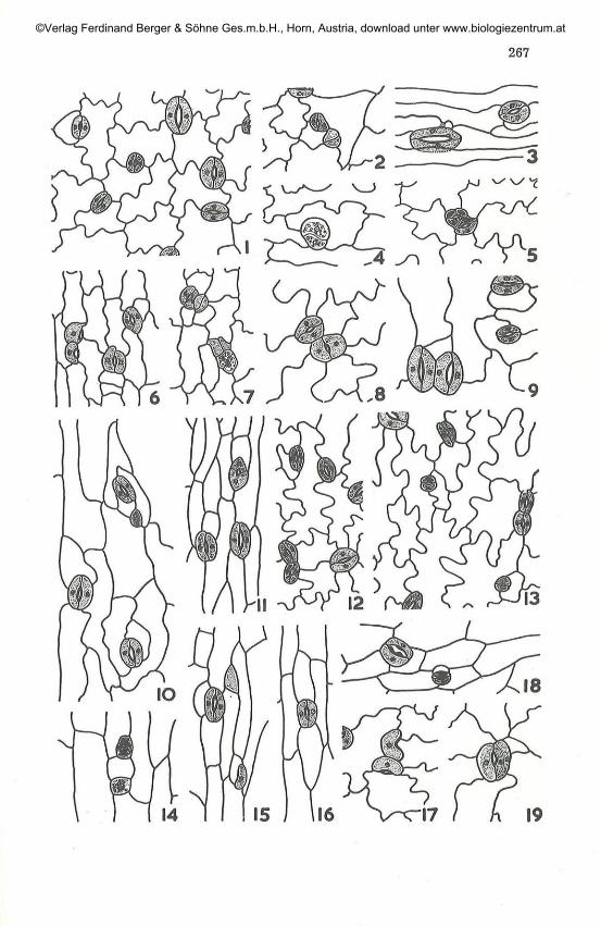
F;gs. 1 — 19. Epidermal peels showing normal and abnormal stomata (cotyledonsall abaxial surface). 225x. — 1 — 2: Browallia americana, cotyledon: arresteddevelopment contiguous with normal stoma; — 3 — 4: B. viscosa, cotyledon:cytoplasmic connection (stippled) between a stoma and an epidermal cell (3)and division of persistent stomatal intial (4); — 5 — 1': Datura gigantea, coty-ledon: division of guard cells (5), superimposed twin single guard cells (6),divided persistent stomatal initial and arrested development (7); — 8: D.stramonium, cotyledon: contiguous stomata; — 9 —10: Hyoscyamus niger,cotyledon: contiguous stomata (9) and meristemoid (10); — 11: Lyciumruthenicum, hypocotyl: unequal guard cells; — 12: Lycopersicon pimpinelli-folium, cotyledon: contiguous stomata; — 13: Nicandraphysaloides, cotyledon:contiguous stomata; — 14—15: N. physaloides f. alba, hypocotyl: meristemoids(14), arrested development (strippled without nucleus) and single guard cellformation (15); — 17: Nicotiana alata, cotyledon: twin single guard cells; —18: N. sanderae, hypocotyl: unequal thickening of cuticular ledges around thepore; — 19: N. sylvestris, cotyledon: longitudinal division in one of the guard
cells.
©Verlag Ferdinand Berger & Söhne Ges.m.b.H., Horn, Austria, download unter www.biologiezentrum.at
267
©Verlag Ferdinand Berger & Söhne Ges.m.b.H., Horn, Austria, download unter www.biologiezentrum.at

268
ments in the seedling stomata of 21 species of the Solanaceae and since it isa puzzle, decided to present an ontogenetic description of these types as itwould be of interest to plant morphologists.
Materials and Methods
The seeds of the species investigated were obtained from the botanicalgardens at Stockholm, Sweden and Berlin, West Germany through thecourtesy of the directors. The seeds of the following 21 species were grownat an atmospheric temperature (April—42.2° C, May—44° C) in the Botani-cal Garden of Sardar Patel University, Vallabh Vidyanagar: Browalliaamericana L., B. viscosa H. B. K., Datura gigantea L., D. stramonium L.,Hyoscyamus nig er L., Lycopersicon pimpinellifolium (JUSL.) MILL., Lyciumruthenicum MURR., Nicandra physaloides (L.) GAERTN., N. physaloides f.alba, Nicotiana alata LINK et OTTO, N. sanderae Hort., IV. sylvestris SPEG.et COMES, Petunia axillaris (LAMK.) B. S. P., P. hybrida Hort, ex DC,Salpiglossis sinuata Hui et PAV., Schizanthus wisetenensis Hort, ex GARD.,
Solanum capsicastrum LINK., and 8. dulcamara L.
Epidermal peels were taken from fresh as well as fixed material (1 :3acetic-ethanol) of cotyledon and hypocotyl, stained with Delafield'shaematoxylin and mounted in glycerin jelly by usual methods. Cameralucida drawings were prepared and series of photomicrographs were madeof stained as well as unstained peels.
Figs. 20 — 34. Epidermal peels showing normal and abnormal stomata (coty-ledons all abaxial surface except fig. 21). 225 x . — 20: Petunia axillaris, coty-ledon: single guard cell with pore and stoma with irregular outline; — 21:Petunia hybrida, cotyledon (upper surface): cytoplasmic connection betweensingle guard cell with pore and aborted stoma; — 22: idem, hypocotyl: divisionof persistent stomatal initial; — 23: P. nyctaginifolia, cotyledon: single guardcell in relationship with normal stoma and contiguous stomata; — 24: Physalisalhekengi, cotyledon: persistent stomatal initial; — 25 — 28: idem, hypocotyl:single guard cell without pore (25) and with pore (26), stoma with abnormallylarge pore (27), contiguous stomata (28); 29 — 30: P. ixocarpa, hypocotyl:epidermal cell like arrested development (stippled without nuclei) and stomawith displaced and unequal guard cells; — 31: Salpiglossis sinuata, hypocotyl:note one of the guard cells without nucleus and cytoplasm with an ingrowthfrom the pore side; — 32: Schizanthus wisetonensis, hypocotyl: note stoma withdisplaced guard cells; — 33: Solanum capsicastrum, cotyledon: note an arresteddevelopment (stippled without nuclei), contiguous stoma, single guard cell withpore, and cytoplasmic connection between two guard cells; — 34: S. dulcamara,hypocotyl: note transverse division in the guard cells (oblique cell plate in
lower stoma)
©Verlag Ferdinand Berger & Söhne Ges.m.b.H., Horn, Austria, download unter www.biologiezentrum.at

269
4
©Verlag Ferdinand Berger & Söhne Ges.m.b.H., Horn, Austria, download unter www.biologiezentrum.at

270
Observations
Mature Stomata: The cotyledons examined of all the species areamphistomatic. The epidermal walls are thick, being either sinuous, straightor arched (Pigs. 1—34). The stomata are irregularly arranged on the coty-ledon and parallel to the long axes or transversely or obliquely on thehypocotyl. Both normal and abnormal stomata may occur on the samesurface of an organ. The normal stomata on the cotyledons and hypocotylof investigated species are either anomocytic, anisocytic, paracytic, diacytic,or with a single subsidiary cell (Figs. 1 — 34). The mature stomatal apparatusconsists of lenticular pore surrounded by two kidney-shaped guard cells.The guard cells may be equal or unequal and displaced (Figs. 30—32). Thismay be due to unequal division of the guard mother cell (PL I I : L). Some-times one of the guard cells is abnormally large so that it appears to encroachupon the other (PL I : F). Rarely the pore is also quite large and not lenti-cular (Fig. 27). The cuticular thickening around the pore is in most casesuniform and even, or less often uneven and not uniform (Fig. 18).
Several types of anomalous developments have been noticed both onthe hypocotyl and cotyledons. In the species investigated by far the morecommon anomalies were the persistent stomatal initial (Figs. 4, 7, 22, 24;PL I I : R - S ) ; single guard cells (Figs. 6, 17, 20, 23, 26, 33; PL I I : N - P , V);and division of guard cells (Figs. 5, 19, 34; PL I : G - I ; PL I I : K). Theother abnormalities are single guard cells in relationship with normalstomata (Figs. 23; PL I I : 0—P); aborted guard cells (Fig. 21); arresteddevelopments (Figs. 2, 7, 15, 33) and cytoplasmic connection betweennearby stomata (Figs. 21, 33) or between stomata and epidermal cells(Figs. 3; PL I : J). On the foliage leaf surface of these species the anomaliesare not so common. Mean values of 15 observations showing the size ofepidermal cells and guard cells are given in the following Table 1.
Development: I. Normal (PL I: A—B)
a) Anomocytic stomata arise directly from the meristemoid by astraight division and without cutting off any subsidiary cells.
b) Paracytic stomata are formed by cutting off of two parallel subsidiarycells, one on either side of the meristemoid, and straight division of theguard mother cell parallel to the subsidiary cells.
c) Stoma with a single subsidiary cell arises after cutting off onesubsidiary cell from the meristemoid, parallel to the guard mother cell.
d) Diacytic stomata arise if the guard mother cell divides by a wall atright angles to the subsidiaries.
e) Anisocytic stomata: Here the meristemoid behaves like an apicalcell with three cutting faces, producing three subsidiary cells in a spiralfashion and then forming two equal guard cells by a straight divisionparallel to the last subsidiary cell.
©Verlag Ferdinand Berger & Söhne Ges.m.b.H., Horn, Austria, download unter www.biologiezentrum.at

271
Table 1
Average size of epidermal and guard cells
Species
Browallia americanaBrowallia viscosaDatura giganteaDatura stramoniumHyoscyamus nigerLycium ruthenicumLycopersicon
pimpinellifoliumNicandra physaloidesN. physoloides f. albaNicotiana alataNicotiana sanderaeNicotiana sylvestrisPetunia axillarisPettmia hybridaPetunia hybridaPetunia nyctaginifoliaPhysalis alkekengiPhysalis alkekengiPhysalis ixocarpaSchizanthus wisetonensisSalpiglossis sinuataSolanum capsicastrumSolanum dulcamara
(C)(C)(C)(C)(C)
(H)
(C)(C)
(H)(C)
(H)(C)(C)(C)
(H)(C)(C)
(H)(H)(H)(H)(C)(H)
Epidermal cellsLength
85999390
129108
679786
113849086
107117106
73106135106100
93165
Breadth
404228314517
3230184832424445214530332126263534
GuardLength
323731424539
3129463634363638373221343942464047
cellsBreadth
111010
81314
99
1010111313111110
9161412131321
(C) = Cotyledon, (H) = Hypocotyl
II. Abnormal Deve lopment
a) Persistant stomatal initial: The stomatal initial normally is devoidof chloroplasts, develops a uniform thickening, but lacks differential wallthickening. Chloroplasts appear later and become persistent (Figs. 4, 7,22, 24; PL I I : R—S). The persistent stomatal initial normally occupies theposition of a stoma. It is either spherical (PL I I : S) or ovoid (Figs. 22, 24)in outline, notched and sometimes shows protruberances (PL I I : R). Afterthe persistent stomatal initial is differentiated, it divides in any plane butan intervening pore doesnot develop (Figs. 4, 7, 22). It may occur solitaryor in close association with arrested development (Fig. 7).
b) Arrested development: During ontogeny the nucleus and thecytoplasm of the stomatal meristemoids degenerate at any stage of develop-
©Verlag Ferdinand Berger & Söhne Ges.m.b.H., Horn, Austria, download unter www.biologiezentrum.at

272
ment and become arrested (Figs. 2, 1, 15, 29, 33). Sometimes the arrestedstomatal cells come to resemble the epidermal cells (Fig. 29). Arresteddevelopment can be easily distinguished from the persistent stomatalinitial by lack of chloroplasts, a prominent nucleus and dense stainingproperties.
c) Single guard cell: The single guard cell (which for convenience maybe termed half stoma) also arises directly from the meristemoid lackingchloroplasts. The meristemoid, instead of giving rise to a pair of guard cells,is notched on one side, chloroplasts appear and the cell becomes reniform orsemilunar (Figs. 15, 20, 25-26; PL I: C; PL II : V). Here the differentialwall thickening develops on the side of the notch and the pore is usuallyabsent. The curvature sometimes is more pronounced in a half stoma, sothat it forms a complete circle around the pore and fuses with the other end,looking like a stoma (PL II : M). The half stoma shows division (PL II : N),occuring either solitary or in close association and variable orientation withnormal stomata (Fig. 23, PL II: 0—P). In plate II : P, the half stomaappears like a basket over the top of a normal stoma. Rarely the meristemoiddeveloping into a half stoma cuts off a small initial (PL I: D), which maydevelop into variously oriented twin half stomata (Figs. 6—7). The halfstoma also arises as a result of degeneration of one of the guard cells of anormal stoma (Fig. 33, PL II: T).
d) Division of guard cell(s): The guard cell(s) of stomata both onhypocotyl and cotyledons mostly divide by transverse divisions (Figs. 5, 34,PL I: G—H; PL II : K) and rarely by longitudinal divisions (Fig. 19). Thedivision of guard cell(s) is either due to mitosis and laying down of cellplates (Fig. 34) or may be due to ingrowth from the outer wall (PL I I :L, U). Due to this division it appears as if the stoma is made up of three tofour cells with a narrow (PL I: H; PL II : K, Q) or an abnormally large pore(PL 1:1). An abnormally large pore is formed due to stretching of the guardcells.
We observed transverse, oblique, and longitudinal divisions of theguard cell/s in both the hypocotyl and cotyledons of most of the speciesinvestigated without subjecting the plants to any type of special conditions,in other words, the morphological modifications noticed here are notartificially induced. We have failed to observe proliferation of guard cellsand formation of new stomata.
e) Cytoplasmic connection between nearby stomata: After the stomataare fully differentiated, sometimes a protruberence arises from one of theguard cells of a nearby stoma. The protruberences grow more and more,ultimately meeting and fusing in the center, their fusion wall disintigrated,and a communication channel is formed between two near by stomata.This tubular connection appears like a conjugation tube between two algalfilaments. These guard cells have a cytoplasmic connection which is secon-darily developed (PL I: J). We have also observed cytoplasmic connection
©Verlag Ferdinand Berger & Söhne Ges.m.b.H., Horn, Austria, download unter www.biologiezentrum.at

273
between nearby placed stomata (Figs. 21, 33) and sometimes between oneof the guard cells and an epidermal cell (Fig. 3). This indicates that there issome physiological connection between the nearby stomata and guard celland an epidermal cell.
f) Degeneration of guard cells: Sometimes both guard cells degenerateand only a central thickening remains (Fig. 21). Degeneration of guard cellsis commonly seen in mature leaves which are about to fall, but here it hasbeen observed in seedlings which are yet to mature.
g) Contiguous stomata: Contiguous stomata are very common (Figs. 5,8-9, 12, 13, 23, 28; PL I: E). They develop from the adjacently placedmeristemoids or as a result of readjustment during maturation of the epi-dermis. The contiguous stomata have variable orientations, they may bejuxtapposed, superimposed, obliquely oriented or at right angles to eachother. In the literature contiguous stomata are regarded as an abnormalityby several workers, but whether they are actually normal or abnormal canonly be decided by studying their ontogeny, when they develop from twoadjacently placed meristemoids or as a result of readjustment, we feel thatthey should not be regarded as abnormal, as there is a possibility here thatthe meristemoids are adjacently placed and the stomata have becomecontiguous during maturation of the epidermis.
The ontogeny of the anomocytic stomata resembles the haplocheilictype of FLORIN (1931, 1933) or perigenous type of PANT (1965), while thatof the other types are syndelocheilic (FLOEIN 1931, 1933) or mesogenous(PANT 1965).
!Discussion
The occurence of aberrent stomatal developments in foliage leaves isby no means new and has been reported by several workers, but theirrelatively wide range observed in the seedling stomata of 21 species of theSolanaceae is perhaps unique.
According to our observations the persistent stomatal initial noticedhere is not always a perfect circle and doesnot show attraction of nuclei ofthe adjacent cells. AHMAD (1964 a) also didnot observe attraction of nucleiof the adjacent cells by the persistent stomatal initial. The persistentstomatal initial may be spherical or ovoid in outline, notched, showsprotruberances and also divides in any plane.
The guard cell division under induced pathological conditions has beendiscussed at length by GERTZ (1919), KÜSTER (1925, 1930) and GUTTENBERG(1905). GERTZ (1919) induced several stomatal anomalies by growing seed-lings of plants in various combinations of warm-light and dark-humidconditions. He managed to effect morphological modifications in a hypocotylof Cucurbita pepo and in a cotyledon of Luffa cylindrica. DEHNEL'S (1957)
©Verlag Ferdinand Berger & Söhne Ges.m.b.H., Horn, Austria, download unter www.biologiezentrum.at
274
attempts to induce guard cell division in Begonia aridicaulis and resumptionof growth by artificial application of various chemical growth regulatorswere unsuccesful. According to KÜSTER (1930) division of guard cellsoccurs commonly in insect-induced galls. He also pointed out that there isno consistency of spindle-axis orientation during division as the wall maybe oblique, transverse or parallel to the long axis of the guard cells. RAO &RAMAYYA (1967) reported proliferation of guard cells producing stomata onthe connective of the anther in Momordica charantia L. and on the leaf ofScaevola frutescens KRAUSE.
The causes of such aberrent expression of morphological charactershave been variously interpreted. According to KLEBS (1903, 1904) it may bedue to "specific nature"; MORGON (1934) considered a "cytoplasmaticheterogeneity", MCCLINTOCK (1951, 1956) "gene action", BÜNNING (1952)"extrinsic factors", RAO & RAMAYYA (1967) "momentary developmentaldisturbances". INAMDAR, GOPAL & CHOHAN (1969) have pointed out thatit is difficult to draw any definite conclusion. DEHNEL (1961) has negatedthe gene-controlled reaction and also pointed out that it is clearly not yetpossible to determine whether extrinsic factors or intrinsic instability areresponsible for stomatal ontogenetic aberrations in the lamina of Begoniaaridicaulis. RAO & RAMAYYA (1967) have not given any causes for suchdevelopmental disturbances. There is little evidence to support the othertwo views.
By changing the external conditions GERTZ (1919), as pointed outearlier could induce stomatal abnormalities in the hypocotyl of Gucurbitapepo, and in the cotyledon of Luff a cylindrica. This is strong evidence infavour of BÜNNING'S (1952) hypothesis. But when such stomatal anomaliesoccur in nature explanation becomes difficult. More difficult is the variablebehaviour of the meristemoid on the same surface of an epidermis whichgives rise to either a pair of guard cells, a persistent stomatal initial, asingle guard cell, arrested development or trichome (see BÜNNING 1952).From our studies we feel that such stomatal aberrations may not be diie toone factor only. We obtained the seeds from Sweden and West Germanyand they were grown in our Botanical Garden; so naturally the environ-mental conditions (extrinsic factors) were different from these countries.What induces the meristemoids on the same surface of an epidermis tobehave in a variable way ? The possible answer which we can give from ourstudies is that the physiological conditions surrounding each meristemoidmay be different or may be due to inhibitory substances, which may beresponsible for abnormal stomatal development. Moreover, the cotyledonsare the main source of food supply for the embryo during germination. Thefood is continuously absorbed from the cotyledons by the germinatingembryo which might cause internal changes and might ultimately lead tostomatal aberrations. We believe that both the extrinsic and intrinsic
©Verlag Ferdinand Berger & Söhne Ges.m.b.H., Horn, Austria, download unter www.biologiezentrum.at

factors might be involved in the abnormal stomatal development. However,we feel that experimental studies under aseptic and controlled conditionsmight serve to throw some light on this aspect of the problem.
Acknowledgements
We are gratefully indebted to the Directors of Hortus Botanicus, Stock-holm, Sweden and of the Botanischer Garten und Museum, Berlin, WestGermany, for kindly supplying us with seeds and to Principal J. G. CHOHAN forencouragement and keen interest in our work. The Junior author thanks theUniversity Grants Commission for the financial support during his researchwork.
References
AHMAD K. J. 1964a. Cuticular studies with special reference to abnormalstomatal cells in Cestrum. — J. Indian bot. soc. 43: 165—177.
— 1964b. On the stomatal abnormalities in Solanaceae. — Sei. & Cult.30: 349-351.
BÜNNING E. 1952. Morphogenesis in plants. — In: AVERY G. S. jr. (ed.), Surveyof biological progress 2: 105—140, New York.
DEHNEL G. S. 1957. Ontogenetic and experimental studies of stomata. —Ph. D. Dissertation, Univ. of California, Berkley. (Original work notseen).
— 1961. Abnormal stomatal development in the foliage of Begonia aridi-caulis. — Amer. J. Bot. 48: 129—133.
FLORIN R. 1931. Untersuchungen zur Stammesgeschichte der Conifer ales undCordaitales. I. Morphologie und Epidermis-Struktur der Assimilations-organe bei den renzenten Koniferen. — K. Svenska Vetenskapsakad.Handl. ser. 3, 10: 1-588.
— 1933. Studien über die Cycadales der Mesozoikums nebst Erörterungenüber die Spaltöffnungsapparate der Bennettitales. — Ibid. 12: 1 — 134.
GERTZ O. 1919. Studier ofver klyfoppningarnas morphologi med sarskild hansyntill deras patologiska utbildnings-former. — Acta Univ. Lund 15: 3 — 84.
GUTTENBERG H. von 1905. Beiträge zur physiologischen Anatomie der Pilz-gallen. — Leipzig.
INAMDAR J. A., GOPAX B. V. & CHOHAN A. J. 1969. Development of normal andabnormal stomata in some Araliaceae. — Ann. Bot. 33: 67 — 73.
— & PATEL R. C. 1969. Development of stomata in in some Solanaceae. —Flora Abt. B, 158: 462-472.
— — 1970. Epidermal structure and normal and abnormal stomataldevelopment in vegetative and üoral organs of Plumbago zeylanica L. —Ibid. 159: 503 — 511.
KLEBS G. 1903. Willkürliche Entwickelungsänderungen bei Pflanzen. — Jena.— 1904. Über Probleme der Entwickelung. - Biol. Zbl. 24: 257-267,
289-305, 449-501, 545-559 and 601-614.KÜSTER E. 1925. Pathologische Pflanzenanatomie. 3. Aufl. — Jena.
— 1930. Anatomie der Gallen. — In: LINSBAUER K., Handb. d. Pflanzen-anatomie. Vol. 5 No. 1. — Berlin.
P h y t o n , Vol. 17, Fase. 3—4,1976. 18,
©Verlag Ferdinand Berger & Söhne Ges.m.b.H., Horn, Austria, download unter www.biologiezentrum.at

276
MCCLINTOCK B. 1951. Chromosome organisation and gene expression. — ColdSpring Harb. Symp. Quant. Biol. 16: 13 — 47.
— 1956. Intranuclear systems controlling gene action and mutation. —Brookhaven Symp. Biol. 8:58 — 74.
MORGON T. H. 1934. Embryology and genetics. — New York.PANT D. D. 1965. On the ontogeny of stomata and other homologous structures.
PI. Sei. Ser. (Allahabad) 1: 1-24.PATEL B. C. & INAMDAB J. A. 1971. Structure and ontogeny of stomata in some
Polemoniales. — Ann. Bot. 35: 389 — 409.RAO PV. B. & RAMAYYA N. 1967. Stomatal abnormalites in two dicotyledons. —
Curr. Sei. 36: 357-358.
©Verlag Ferdinand Berger & Söhne Ges.m.b.H., Horn, Austria, download unter www.biologiezentrum.at

Phyton, vol. 17 tab. 1(INAMDAII & PATEL)
=-.H ~™*rvW ^Datura stramonium, cotyledon: A: Meristemoids in stages of development(X720); — B : Formation of paracytic stoma (X550); — C: Formation ofsingle guard cell directly from the meristemoid ( X 550); — D: Formation ofsingle guard cells, note initial cut off at the end (xllOO); — E : Contiguous
stomata ( X 440);Physalis ixocarpa (F —H: hypocotyl, I —J: cotyledon): F : Stoma with unequalguard cells and one encroaching guard cell ( X 370); — G — H: Division in one ofthe guard cells, note unequal and compressed guard cells in H (X 370); —I: Stoma with four guard cells and a big pore formed as a result of transversedivision in both the guard cells ( X 400); — J : Cytoplasniic connection between
nearby stomata ( x 390).
©Verlag Ferdinand Berger & Söhne Ges.m.b.H., Horn, Austria, download unter www.biologiezentrum.at