open access neuromuscular deterioration in the … deterioration in the early stage of sepsis in...
TRANSCRIPT
Available online http://ccforum.com/content/11/1/R1
Open AccessVol 11 No 1ResearchNeuromuscular deterioration in the early stage of sepsis in ratsIlkin Cankayali1, Yusuf Hakan Dogan2, Ilhami Solak3, Kubilay Demirag1, Oguz Eris1, Serdar Demirgoren2 and Ali Resat Moral1
1Department of Anaesthesiology and Intensive Care Unit, Ege University, School of Medicine 35100, Izmir, Turkey2Department of Physiology, Ege University, School of Medicine 35100, Izmir, Turkey3Department of General Surgery, Ege University, School of Medicine 35100, Izmir, Turkey
Corresponding author: Ilkin Cankayali, [email protected]
Received: 20 Jan 2006 Revisions requested: 21 Feb 2006 Revisions received: 1 Sep 2006 Accepted: 4 Jan 2007 Published: 4 Jan 2007
Critical Care 2007, 11:R1 (doi:10.1186/cc5139)This article is online at: http://ccforum.com/content/11/1/R1© 2006 Cankayali et al.; licensee BioMed Central Ltd. This is an open access article distributed under the terms of the Creative Commons Attribution License (http://creativecommons.org/licenses/by/2.0), which permits unrestricted use, distribution, and reproduction in any medium, provided the original work is properly cited.
Abstract
Introduction Critical illness polyneuropathy (CIP) is a clinicalcondition frequently seen in patients being treated in criticalcare units in the final stage of sepsis. The etiopathology of CIPis still unclear, and the onset time of appearance of theelectrophysiological findings has not been elucidated. The verylittle research that has been carried out on this topic is limited toclinical electrophysiological and histopathological studies. Inthis study, electrophysiological alterations in the early stage ofexperimentally induced sepsis were investigated in septic rats.
Methods We conducted a prospective, randomized, controlledexperimental study in an animal basic science laboratory with 30male Sprague-Dawley rats, weighing 200 to 250 g. All of therats were randomly assigned to one of two groups. In the sepsisgroup (n = 20), cecal ligation and puncture (CLP) wasperformed to induce experimental sepsis. In the sham-operatedgroup (n = 10), laparotomy without CLP was performed. Beforeand 24 hours after CLP and laparotomy, the right sciatic nervewas stimulated from the sciatic notch and compound muscleaction potentials (CMAPs) were recorded from thegastrocnemius muscle. Recordings of latency, amplitude, andduration of the CMAP were evaluated.
Results CMAP durations before and 24 hours after surgerywere 0.45 ± 0.05 ms and 0.48 ± 0.05 ms, respectively, in thesham-operated group and 0.46 ± 0.05 ms and 0.55 ± 0.01 ms,respectively, in the sepsis group. Latency measurements in thesham-operated group were 0.078 ± 0.010 ms and 0.080 ±0.015 ms, respectively, whereas measurements were found tobe prolonged in the sepsis group: 0.094 ± 0.015 ms and 0.149± 0.054 ms before and 24 hours after surgery, respectively (p <0.05). CMAP amplitudes in the sham-operated group beforeand 24 hours after surgery were 8.41 ± 0.79 mV and 8.28 ±1.92 mV, respectively, whereas in the sepsis group theamplitude measurements decreased to 7.60 ± 1.75 mV and4.87 ± 3.44 mV, respectively (p < 0.05).
Conclusion The results of the study indicate thatelectrophysiological alterations appear in the first 24 hours afterexperimental sepsis and are characterized by an increase inlatency and a decrease in CMAP amplitude. The results alsosuggest that electrophysiological findings seen in patients withCIP might appear before clinical signs of CIP.
IntroductionCritical illness polyneuropathy (CIP) was described as a clini-cal disorder by Bolton and colleagues [1] in 1984. It is a pri-mary axonal degeneration of motor and sensory fibers whichoccurs mostly in patients who have systemic inflammatoryresponse syndrome (SIRS), sepsis, or multiple organ dysfunc-tion syndrome (MODS) [2-7]. Berek and colleagues [8] sug-gested that, in the course of sepsis, CIP has to be consideredpart of MODS.
CIP manifests with general weakness and sensory defects andespecially with weakness of the respiratory muscles, leadingto problems in weaning from mechanical ventilation in theintensive care unit (ICU). Physiopathology, onset of the symp-toms, and the treatment of CIP have not been clearly defined.
The studies on CIP are based mostly on clinical manifestationsand neurophysiological research. However, an experimentalstudy investigating possible neuromuscular changes in the
Page 1 of 7(page number not for citation purposes)
CIP = critical illness polyneuropathy; CLP = cecal ligation and puncture; CMAP = compound muscle action potential; EMG = electromyelographic; ICU = intensive care unit; MODS = multiple organ dysfunction syndrome; SIRS = systemic inflammatory response syndrome.

Critical Care Vol 11 No 1 Cankayali et al.
early stage of sepsis has not been performed yet. We aimedto observe electrophysiological alterations in the early stage ofsepsis. In this study, an experimental sepsis model was per-formed to investigate electrophysiological alterations in thefirst 24 hours of sepsis.
Materials and methodsAnimal Ethics Committee approval was obtained, and thestudy was conducted in the Research Laboratory of theDepartment of Anesthesiology and ICU of Ege University Med-ical School (Izmir, Turkey).
Experimental proceduresThirty adult male Sprague-Dawley rats two to three monthsold, each weighing approximately 250 g, were used. All ratswere housed in cages one week before the experiments in anacclimatized room at standard room temperature and withtwelve hour light/dark cycles. Rats were allowed free accessto water and standard chow. For surgical intervention, ratswere anesthetized with ketamine (80 mg/kg) and xylazine (10mg/kg) given intraperitoneally.
All rats were randomly divided into one of two groups: a cecalligation and puncture (CLP)-operated group (sepsis group) (n= 20) and a sham-operated group (sham group) (n = 10). Dueto the sepsis model's high mortality rate, more rats weregrouped in the sepsis group (n = 20) than in the sham group(n = 10). For reliable statistical results, at least six rats is suffi-cient. We decided to perform the study with at least 10 rats.
Sepsis was induced by CLP performed as described previ-ously [9,10]. In this sepsis model, five hours after CLP, ratswere accepted as septic. Under aseptic conditions, a 3-cmmidline laparotomy was performed to allow exposure of thececum with adjoining intestine. The cecum was ligated tightlywith a 3.0 silk suture at its base below the iliocecal valve andperforated once with a 22-gauge needle. The cecum was thengently squeezed to extrude a small amount of feces from theperforation site. The cecum was returned to the peritoneal cav-ity, and the laparotomy incision was closed with 4.0 silksutures. In the sham group, under aseptic conditions only,laparotomy was performed on rats, but their cecum was nei-ther ligated nor punctured.
Measurements and calculationsElectrophysiological recordings were obtained from the rightsciatic nerve stimulated supra-maximally (intensity 10 V, dura-tion 0.1 ms, frequency 1 Hz) by a Biopac HSTM01 surfacestimulation electrode (BIOPAC Systems, Inc., Santa Barbara,CA, USA) from the sciatic notch, and compound muscleaction potentials (CMAPs) were recorded by means of super-ficial disc electrodes located over the gastrocnemius musclebefore and 24 hours after surgery. Data were evaluated usingBiopac Student Lab Pro version 3.6.7 software (BIOPAC Sys-tems, Inc.), with latency, amplitude, and duration of CMAP as
the parameters (Figures 1, 2, 3, 4). During the electromyelo-graphic (EMG) recordings, rectal temperatures of the ratswere monitored by a rectal probe (HP Viridia 24-C; Hewlett-Packard Company, Palo Alto, CA, USA) and the temperatureof each rat was kept at approximately 36°C to 37°C by heatingpad. The animals were euthanized 24 hours after the CLP forthe next recording.
Because we aimed to assess EMG recordings in the earlystage of sepsis, we obtained EMG recordings of the rats in thefirst 24 hours after CLP. We did not aim to observe clinicalsigns of sepsis; therefore, the animals were euthanized 24hours after surgery.
Statistical analysisThe results were analyzed with the SPSS ver 14.0 statisticalprogram (SPSS Inc., 233 South wacker Drive, 11th floor, Chi-cago, IL 60606-6307) by using repeated measures (analysisof variance). Factors were session (before and 24 hours aftersurgery) and treatment (sepsis and sham groups). Dependentvariables were latency, amplitude, and duration. The groupswere compared by paired-sample t test, and results weregiven as mean ± standard deviation. A value of p > 0.05 wasaccepted as statistically significant.
ResultsIn the sepsis group, five rats died during the first 24 hours andwere excluded from the study. At 24 hours, the mortality ratewas 25% in the sepsis group, and there was no mortality in thesham group. The mortality rate was high in the sepsis groupbecause we did not use treatment materials (antibiotics andfluid resuscitation) for this study.
CMAP durations before and 24 hours after surgery wererecorded as 0.45 ± 0.05 ms and 0.48 ± 0.05 ms, respectively,in the sham group. Statistically significant difference was notfound (p > 0.05). CMAP durations before and 24 hours aftersurgery were recorded as 0.46 ± 0.05 ms and 0.55 ± 0.01ms, respectively, in the sepsis group. Statistically significantdifference was found in CMAP duration only for session (F1,23= 7.49, p = 0.012) but not for treatment (F1,23 = 4.02, p =0.057) (p > 0.05) (Table 1). CMAP amplitudes in the shamgroup before and 24 hours after surgery were 8.41 ± 0.79 mVand 8.28 ± 1.92 mV, respectively. Statistically significant dif-ference was not found (p > 0.05) (Table 1). However, in thesepsis group, the amplitudes decreased from 7.60 ± 1.75 mVto 4.87 ± 3.44 mV. This alteration was statistically significant(p < 0.05) (Table 1). CMAP amplitudes in the sham groupwere not different statistically but session (F1,23 = 5.56, p =0.027) and treatment (F1,23 = 8.40, p = 0.008) were signifi-cantly interacted (F1,23 = 4.38, p = 0.047) and the effect wasobserved only in the sepsis group (Table 1). Whereas CMAPamplitudes decreased profoundly in the sepsis group (ratio ofthe prolonged time = -33.3%), CMAP amplitudes were much
Page 2 of 7(page number not for citation purposes)
Available online http://ccforum.com/content/11/1/R1
less decreased in the sham group (ratio of the prolonged time= -0.7%).
Latency measurements were not significantly altered in thesham group (from 0.078 ± 0.010 ms to 0.080 ± 0.015 ms),whereas measurements were found to be prolonged in thesepsis group (from 0.094 ± 0.015 ms to 0.149 ± 0.054 ms)before and 24 hours after surgery, respectively (p < 0.05).Latency data showed significant difference only in the sepsisgroup, and interacted for session (F1,23 = 13.47, p = 0.001)and treatment (F1,23 = 15.86, p = 0.001) (F1,23 = 11.98, p =0.002). Whereas latency time was prolonged in the sepsisgroup in a significant manner (ratio of the prolonged amount =56.45%), latency time was prolonged much less in the shamgroup (ratio of the prolonged amount = 2.4%) (Table 1).
CMAP duration and latency increase and CMAP amplitudedecrease in the sham group was not statistically significant (p> 0.05).
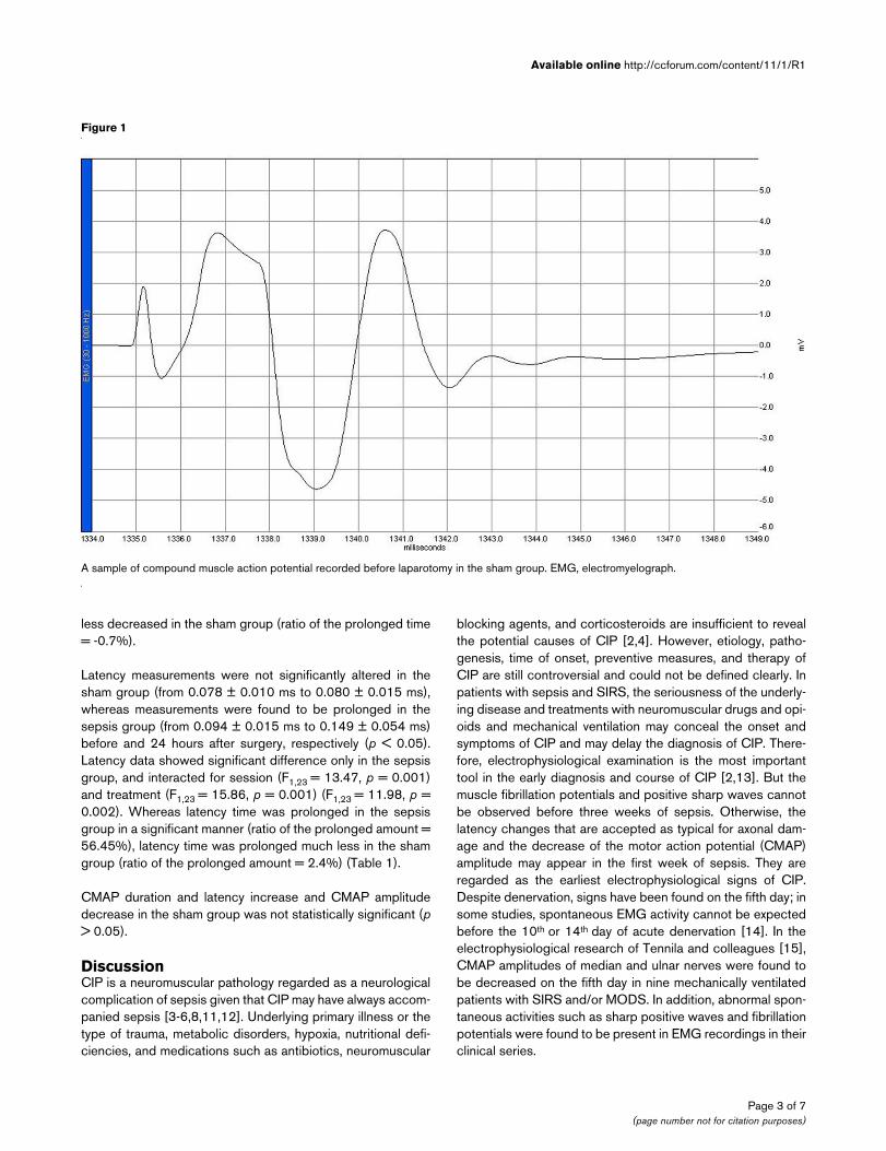
DiscussionCIP is a neuromuscular pathology regarded as a neurologicalcomplication of sepsis given that CIP may have always accom-panied sepsis [3-6,8,11,12]. Underlying primary illness or thetype of trauma, metabolic disorders, hypoxia, nutritional defi-ciencies, and medications such as antibiotics, neuromuscular
blocking agents, and corticosteroids are insufficient to revealthe potential causes of CIP [2,4]. However, etiology, patho-genesis, time of onset, preventive measures, and therapy ofCIP are still controversial and could not be defined clearly. Inpatients with sepsis and SIRS, the seriousness of the underly-ing disease and treatments with neuromuscular drugs and opi-oids and mechanical ventilation may conceal the onset andsymptoms of CIP and may delay the diagnosis of CIP. There-fore, electrophysiological examination is the most importanttool in the early diagnosis and course of CIP [2,13]. But themuscle fibrillation potentials and positive sharp waves cannotbe observed before three weeks of sepsis. Otherwise, thelatency changes that are accepted as typical for axonal dam-age and the decrease of the motor action potential (CMAP)amplitude may appear in the first week of sepsis. They areregarded as the earliest electrophysiological signs of CIP.Despite denervation, signs have been found on the fifth day; insome studies, spontaneous EMG activity cannot be expectedbefore the 10th or 14th day of acute denervation [14]. In theelectrophysiological research of Tennila and colleagues [15],CMAP amplitudes of median and ulnar nerves were found tobe decreased on the fifth day in nine mechanically ventilatedpatients with SIRS and/or MODS. In addition, abnormal spon-taneous activities such as sharp positive waves and fibrillationpotentials were found to be present in EMG recordings in theirclinical series.
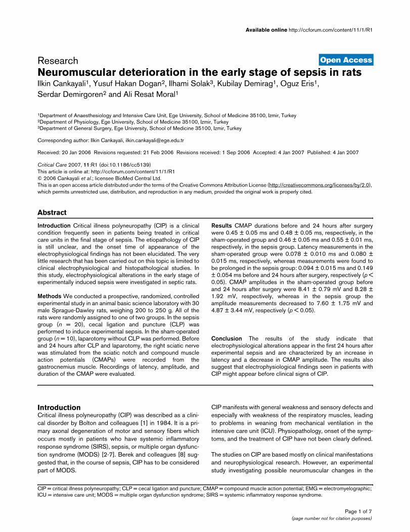
Figure 1
A sample of compound muscle action potential recorded before laparotomy in the sham groupA sample of compound muscle action potential recorded before laparotomy in the sham group. EMG, electromyelograph.
Page 3 of 7(page number not for citation purposes)

Critical Care Vol 11 No 1 Cankayali et al.
Figure 2
A sample of compound muscle action potential recorded 24 hours after laparotomy in the sham groupA sample of compound muscle action potential recorded 24 hours after laparotomy in the sham group. EMG, electromyelograph.
Figure 3
A sample of compound muscle action potential recorded before cecal ligation and puncture in the sepsis groupA sample of compound muscle action potential recorded before cecal ligation and puncture in the sepsis group. EMG, electromyelograph.
Page 4 of 7(page number not for citation purposes)

Available online http://ccforum.com/content/11/1/R1
In this study, CMAP amplitude was decreased; regarding thefirst electrophysiological finding in the early phase of CIP[2,6,16] and marked prolongation of latency values appearedin the first 24 hours of sepsis. Our findings are supported bythe previous studies. In some earlier studies, it was postulatedthat decrease in the CMAP amplitude was due primarily toneuromuscular blocking agents, steroids, and some medica-tions used widely in ICUs [4,17-19]. However, the latest pro-spective studies have shown that there is no correlation withCIP, critical illness myopathy, and medication used [5,6,20]. Inthe present study, the neurophysiological conduction altera-tions were seen in the experimental CIP model without usingneuromuscular blocking agents and/or steroids. Therefore,these findings support the concept that sepsis is mainlyresponsible for the neuromuscular changes. Likewise, antibiot-ics, especially aminoglycosides and their metabolites, are said
to be the other factors responsible for development of CIP thatresults from the increase of capillary membrane permeabilityand the invasion of antibiotics to peripheral nerves in sepsis[21]. However, there is no statistical evidence confirmingthese opinions, and thus further studies are needed. Giventhat antibiotics were not used in our experimental sepsismodel, our results support the idea that the observed electro-physiological alterations were attributable entirely to sepsis.
Disturbances in microcirculation and autoregulation of periph-eral nerves, as well as the other organs influenced in sepsis,are thought to be the principal causes of the development ofCIP. In addition, cytokines released in sepsis also cause anincrease in capillary permeability due to a histamine-like effectand the resulting endoneural edema leads to hypoxia andenergy deficit by increasing the intercapillary space [2].
Figure 4
A sample of compound muscle action potential sample recorded 24 hours after cecal ligation and puncture in the sepsis groupA sample of compound muscle action potential sample recorded 24 hours after cecal ligation and puncture in the sepsis group. EMG, electromyelograph.
Table 1
Measurements of compound gastrocnemius muscle action potentials
CMAP duration (ms) CMAP amplitude (mV) Latency (ms)
Groups Before surgery 24 hours after surgery Before surgery 24 hours after surgery Before surgery 24 hours after surgery
Sepsis group 0.46 ± 0.05 0.55 ± 0.01 7.60 ± 1.75 4.87 ± 3.44a 0.094 ± 0.015 0.149 ± 0.054a
Sham group 0.45 ± 0.005 0.48 ± 0.05 8.41 ± 0.79 8.28 ± 1.92 0.078 ± 0.010 0.080 ± 0.015
ap < 0.05. All values are presented as mean ± standard deviation for each group. CMAP, compound muscle action potential.
Page 5 of 7(page number not for citation purposes)

Critical Care Vol 11 No 1 Cankayali et al.
Because the axonal transportation of structural proteins ishighly energy-dependent, this energy deficit induces primaryaxonal degeneration of distal nerves [2]. Bolton and col-leagues [3] suggested that tumor necrosis factor, arachidonicacid, and metabolites of histamine, complement activation,cellular adhesion systems, and free radicals were principal fac-tors responsible for systemic effects of sepsis and SIRS andthese factors might lead to primary axonal degeneration.
Electrophysiological measurements in the early studiesobtained during early periods of clinical sepsis indicated thatthe decrease in amplitude of CMAP was accompanied by anincrease in duration without any change in latency. This findingdirected attention to the muscle fiber membrane as a physio-pathological explanation [22]. Decrease in CMAP amplitudeand increase in duration were suggested to be secondary tothe dysfunction of energy-dependent sodium-potassiumpumps in muscles [23]. In the present study, although therewas an increase in the duration 24 hours after sepsis wasinduced, the difference was not statistically significant whencompared with the sham group. Our results do not show theprolongation of duration which has been shown in the previousstudies, due to electrophysiological data that were obtained inthe early stages (24 hours) of sepsis in this study. Theprolongation of the duration due to sodium-potassium pumpinsufficiency in the muscles has been accepted as an indicatorobserved in the later stages of sepsis. Our results indicate nochange in CMAP duration but a decrease in amplitude andobservable prolongation of the latency which is regarded as anindicator of axonal degeneration. Further detailed studiesshould be designed to elucidate the pathogenesis properly.The common point in the majority of the related articles is thepresence of a decrease of CMAP amplitude. CMAP is pro-duced by synchronized activation of the muscle fibers afteraxonal innervations, which is the sum of the responses of thestriated muscles to stimuli. In addition, CMAP is a valuable toolboth for evaluating to descending motor axon and theresponse of the muscle fibers to the stimulus placed distallyand the conduction at the neuromuscular junction [24]. In thisstudy, CMAP changes indicate possible axonal conductionand/or neuromuscular junction pathologies or a reducednumber of fibers responding to stimulus. But we were not ableto distinguish and define the origin of the observed changessuch as axonal conduction defects, neuromuscular junctionpathologies, or reduction of the number of the muscle fibersthat led to the decrease of the amplitude and prolongation ofCMAP in the sepsis group.
ConclusionOur results indicate that electrophysiological findingsappeared in the first 24 hours after experimental sepsis andwere characterized by an increase in latency and a decreasein CMAP amplitude. Therefore, we conclude that electrophys-iological changes seen in sepsis might appear before clinicalsigns of CIP.
Competing interestsThe authors declare that they have no competing interests.
Authors' contributionsIC, IS, and ARM designed the study. IC, YHD, IS, KD, OE, andARM coordinated the study and drafted the manuscript. IC,YHD, and IS collected data. IC, IS, YHD, KD, OE, SD, andARM helped to draft the manuscript. IC, YHD, IS, and ARMconceived and designed the study and performed the statisti-cal analysis. All authors read and approved the finalmanuscript.
References1. Bolton CF, Gilbert JJ, Hahn AF, Sibbald WJ: Polyneuropathy in
critically ill patients. J Neurol Neurosurg Psychiatry 1984,47:1223-1231.
2. Zochodne DW, Bolton CF, Wells GA: Critical illness polyneu-ropathy. A complication of sepsis and multiple organ failure.Brain 1987, 110:819-841.
3. Bolton CF: Sepsis and the systemic inflammatory responsesyndrome: neuromuscular manifestations. Crit Care Med1996, 24:1408-1416.
4. Witt NJ, Zochodne DW, Bolton CF: Peripheral nerve function insepsis and multiple organ failure. Chest 1991, 99:176-184.
5. Leijten FS, de Weerd AW: Critical illness polyneuropathy. Areview of the literature, definition and pathophysiology. ClinNeurol Neurosurg 1994, 96:10-19.
6. Zifko UA, Zipko HT, Bolton CF: Clinical and electrophysiologicalfindings in critical illness polyneuropathy. J Neurol Sci 1998,159:186-193.
7. Motomura M: [Critical illness polyneuropathy and myopathy.](Abstract). Rinsho Shinkeigaku 2003, 43:802-804.
8. Berek K, Margreiter J, Willeit J: [Polyneuropathy in the criticallyill patient – critical illness polyneuropathy.] (Abstract). WienKlin Wochenschr 1998, 110:243-252.
9. Otero-Anton E, Gonzalez-Quintela A, Lopez-Soto A, Lopez-Ben S,Llovo J, Perez LF: Cecal ligation and puncture as a model ofsepsis in the rat: influence of the puncture size on mortality,bacteremia, endotoxemia and tumor necrosis factor alphalevels. Eur Surg Res 2001, 33:77-79.
10. Ritter C, Andrades M, Frota Junior ML, Bonatto F, Pinho RA, Poly-doro M, Klamt F, Pinheiro CT, Menna-Barreto SS, Moreira JC, Dal-Pizzol F: Oxidative parameters and mortality in sepsis inducedby cecal ligation and perforation. Intensive Care Med 2003,29:1782-1789.
11. Lorin S, Nierman DM: Critical illness neuromuscularabnormalities. Crit Care Clin 2002, 18:553-568.
12. van Mook WN, Hulsewe-Evers RP: Critical illnesspolyneuropathy. Curr Opin Crit Care 2002, 8:302-310.
13. Druschky A, Herkert M, Radespiel-Troger M, Druschky K, Hund E,Becker CM, Hilz MJ, Erbguth F, Neundorfer B: Critical illnesspolyneuropathy: clinical findings and cell culture assay of neu-rotoxicity assessed by a prospective study. Intensive Care Med2001, 27:686-693.
Key messages
• CMAP durations were increased in a sepsis model in rats in the first 24 hours.
• CMAP amplitudes were significantly decreased in a sepsis model in rats in the first 24 hours.
• Latency times were significantly prolonged in a sepsis model in rats in the first 24 hours.
• Electrophysiological changes seen in sepsis might appear before clinical signs of CIP.
Page 6 of 7(page number not for citation purposes)

Available online http://ccforum.com/content/11/1/R1
14. Bolton CF, Young GB, Zochodne DW: The neurological compli-cations of sepsis. Ann Neurol 1993, 33:94-100.
15. Tennila A, Salmi T, Pettila V, Roine RO, Varpula T, Takkunen O:Early signs of critical illness polyneuropathy in ICU patientswith systemic inflammatory response syndrome or sepsis.Intensive Care Med 2000, 26(9):1360-1363.
16. Schwarz J, Planck J, Briegel J, Straube A: Single-fiber electromy-ography, nerve conduction studies, and conventional electro-myography in patients with critical-illness polyneuropathy:evidence for a lesion of terminal motor axons. Muscle Nerve1997, 20:696-701.
17. Lacomis D, Giuliani MJ, Van Cott A, Kramer DJ: Acute myopathyof intensive care: clinical, electromyographic, and pathologicalaspects. Ann Neurol 1996, 40:645-654.
18. Wernig A, Pecot-Dechavassine M, Stover H: Sprouting andregression of the nerve at the frog neuromuscular junction innormal conditions and after prolonged paralysis with curare. JNeurocytol 1980, 9:278-303.
19. Coakley JH, Nagendran K, Ormerod IE, Ferguson CN, Hinds CJ:Prolonged neurogenic weakness in patients requiringmechanical ventilation for acute airflow limitation. Chest 1992,101:1413-1416.
20. Berek K, Margreiter J, Willeit J, Berek A, Schmutzhard E, Mutz NJ:Polyneuropathies in critically ill patients: a prospectiveevaluation. Intensive Care Med 1996, 22:849-855.
21. Spitzer AR, Giancarlo T, Maher L, Awerbuch G, Bowles A: Neu-romuscular causes of prolonged ventilator dependency.(Abstract). Muscle Nerve 1992, 15(6):682-686.
22. Bolton CF: Evidence of neuromuscular dysfunction in the earlystages of the systemic inflammatory response syndrome.Intensive Care Med 2000, 26:1179-1180.
23. Milner-Brown HS, Miller RG: Muscle membrane excitation andimpulse propagation velocity are reduced during musclefatigue. Muscle Nerve 1986, 9:367-374.
24. Daube JR: Compound muscle action potentials. In ClinicalNeurophysiology Edited by: Daube JR. Philadelphia: F A DavisCompany; 1996:199-234.
Page 7 of 7(page number not for citation purposes)