optical phenomenon of peri-implant soft tissue. part i
TRANSCRIPT

Optical phenomenon of peri-implantsoft tissue. Part I. Spectrophotometricassessment of natural tooth gingiva andperi-implant mucosa
Sang E. ParkJohn D. Da SilvaHans-Peter WeberShigemi Ishikawa-Nagai
Authors’ affiliations:Sang E. Park, Shigemi Ishikawa-Nagai,Department of Restorative Dentistry andBiomaterials Sciences, Harvard School of DentalMedicine, Boston, MA, USAJohn D. Da Silva, Department of Oral Health Policyand Epidemiology, Harvard School of DentalMedicine, Boston, MA, USAHans-Peter Weber, Professor and Chairman,Department of Restorative Dentistry andBiomaterials Sciences, Harvard School of DentalMedicine, Boston, MA, USA
Correspondence to:Sang E. ParkDepartment of Restorative Dentistry andBiomaterials SciencesHarvard School of Dental Medicine188 Longwood Avenue BostonMA 02115USATel.: þ1 617 432 2374Fax: þ1 617 432 3881e-mail: [email protected]
Key words: dental implants, esthetics, gingival color, gingival translucency, ITI, marginal
gingiva, peri-implant mucosa, soft tissues
Abstract
Objectives: The purpose of this study was to investigate the difference in optical
appearance of the soft tissues labial to dental implants and to analyze the effects of
titanium implant neck colors transmitted through the marginal mucosa.
Materials and methods: Fourteen patients with 15 Straumanns
single implant
replacements in the maxillary anterior region were recruited. Color measurements of the
peri-implant mucosa of test sites and the gingivae of contralateral or adjacent natural teeth
as controls were made at the facial aspect of the teeth using a spectrophotometer. The color
data (CIELAB color coordinates; Ln, an, bn and Cn) in five incremental areas of 1 � 2 mm from
the gingival margin toward the apical direction were obtained.
Results: A significant difference existed (Po0.01) between the test site and the control site
on the mean Ln and bn values in all five incremental areas (area 1–5). In contrast, there was
no significant difference in the mean an values. Discrepancies between color distributions of
soft tissues were stronger in areas close to the gingival margin and decreased toward the
apical direction. The mean color difference DE between the test site and the control site was
7.7 in area 1 and decreased toward area 5 with a value of 6.5. However, there was no
statistical difference in each of the mean values of differences in optical data, DLn, Dan and
Dbn, when five incremental areas of the control and the test sites were compared.
Conclusion: It was observed that the color of soft tissue around the titanium implant was
significantly different compared with the gingiva of natural teeth. Significantly lower
values of CIELAB color coordinates, Ln and bn were found in the peri-implant soft tissue.
Basic and clinical research in implant den-
tistry in the early years focused mainly on
the integration of endosseous dental im-
plants in hard tissue and the re-establish-
ment of masticatory function for patients
with complete and partial edentulism (Bra-
nemark et al. 1977; Adell et al. 1990; Buser
et al. 1991; Chaytor et al. 1991; Lekholm
et al. 1994; Avivi-Arber & Zarb 1996;
Scheller et al. 1998; Haas et al. 2002).
With the success of osseointegration based
dental rehabilitation in these indications,
the focus consequently shifted to the repla-
cement of missing teeth in the esthetic
region. In the maxillary anterior area, the
esthetic outcome is a critical determinant
in the overall success of implant therapy
and yet remains a challenge.
An important role in achieving an esthe-
tically optimal outcome is the morphology
and appearance of peri-implant soft tissue
associated with the implant and the result-
ing esthetics of the implant-supported
restoration (Reikie 1993, 1995; Garber
Date:Accepted 6 September 2006
To cite this article:Park SE, Da Silva JD, Weber HP, Ishikawa-Nagai N.Optical phenomenon of peri-implant soft tissue. Part I.Spectrophotometric assessment of natural tooth gingivaand peri-implant mucosa.Clin. Oral Impl. Res. 18, 2007; 569– 574doi: 10.1111/j.1600-0501.2007.01391.x
c� 2007 The Authors. Journal compilation c� 2007 Blackwell Munksgaard 569

1996). In natural teeth, a constant vertical
dimension of healthy periodontal soft tis-
sue, the biological width, is an essential
factor in establishing gingival esthetics
(Gargiulo et al. 1961; Vacek et al. 1994).
Studies on the biology of peri-implant soft
tissue indicated that supracrestal soft tis-
sues of approximately 3–4 mm are always
established following placement of an im-
plant and suggested that the position of
the peri-implant soft tissue margin may
be related to the level of bone support
around the implant (Berglundh et al.
1991; Abrahamsson et al. 1996; Berglundh
& Lindhe 1996). The biological width is an
important factor in achieving esthetic re-
sults of peri-implant soft tissue, especially
for the non-submerged, one-piece titanium
implants (Cochran et al. 1997; Hermann
et al. 2000, 2001).
Understanding the existence of the bio-
logical width, the recession of the peri-
implant soft tissue margin may result
from an attempt to establish appropriate
biological dimensions (Bengazi et al. 1996).
It is clear that gingival recession can cause
unacceptable soft tissue esthetics. Various
prosthetic and surgical techniques have
been proposed in an effort to overcome
these occurrences following implant ther-
apy, but the long-term success of these
procedures has been inconclusive. Many
surgical approaches have been used to en-
hance the esthetic appearance and gingival
contour of peri-implant tissues (Israelson &
Plemons 1993; Neale & Chee 1994; Pa-
lacci et al. 1995; Becker & Becker 1996;
Conte et al. 2002). However, questions
still remains regarding the viability and
predictability of surgical approaches
because the thickness of gingiva will
often define how restorations will appear
esthetically at the end of treatment (Kao
& Pasquinelli 2002). Unlike thick gingiva,
thin tissue is highly sensitive to trauma
and inflammation and, thus, more
susceptible to recession. In addition, thin
gingival tissue tends to be delicate and
almost translucent in appearance, contri-
buting to an undesirable shine-through
effect of the underlying titanium implants
with a grayish appearance of the gingival
cuff.
The predictability of an esthetic implant
outcome can be achieved by overcoming
this optical problem at the marginal peri-
implant mucosa of titanium implants. Few
studies exist in terms of color properties of
soft tissue around titanium dental im-
plants. Previous research on colorimetric
solutions to improve the esthetic appear-
ance of peri-implant soft tissue has not
been thoroughly performed and has re-
mained of subjective nature. The specific
aims of this study were to: (1) investigate
the difference in optical properties of the
soft tissues labial to dental implants and
the marginal gingivae of natural teeth, and
(2) analyze the effects of titanium implant
neck colors transmitted through the mar-
ginal mucosa in terms of areas. In this
study, the hypothesis that the peri-implant
soft tissue would exhibit a difference in
color compared with that of natural tooth
was verified, as measured by a color spec-
trophotometer.
Materials and methods
Color measurements of the peri-implant
mucosa of test sites and the gingivae of
contralateral or adjacent natural teeth as
controls were made at the facial aspect
of the teeth on five incremental areas in
1-mm increments (Fig. 1).
Human subjects and implants
Subjects who had already received Strau-
manns
implants (Institut Straumann AG,
Waldenburg/BL, Switzerland) during the
past 5 years for single-implant replace-
ments in the maxillary anterior region
were recruited from the patient pool at
the Harvard School of Dental Medicine.
At the first visit, an initial examination
including the location of the soft tissue
margin, plaque index, bleeding on probing,
probing depth and implant mobility was
performed. The patients who met the study
eligibility criteria were invited to a second
visit for color measurement. Fifteen sites
from 14 patients were included in this
study. This study was approved by the
Institutional Review Board (IRB) at Har-
vard Medical School.
Test siteControl site
54321
54321
Fig. 1. Five incremental areas for the color measure-
ment.Control Unit
USB 2.0
CCD sensor
(RGB)
Camera HeadNote PC
Fig. 2. A spectrophotometer used in this study.
Park et al . Optical phenomenon of peri-implant soft tissue
570 | Clin. Oral Impl. Res. 18, 2007 / 569–574 c� 2007 The Authors. Journal compilation c� 2007 Blackwell Munksgaard

Color measurements
Spectrophotometric measurements were
made using a multi-spectral camera system
(Handy-MSC; Olympus Co., Tokyo, Ja-
pan) as shown in Fig. 2. This spectrophot-
ometer (Handy-MSC) uses a new
technique of multi-band image acquisition
with a built-in light-emitting diode (LED)
lamp in the measuring head as a light
source. The acquired multiband image
data perform estimation of a spectrum
and colorimetric values with original algo-
rithm by exclusive software. Eight LED
lamps (Olympus Co.) are used as a source
of illumination. The area of illuminations
is 20 mm in diameter with a central area of
15 mm in diameter for measurement. Spec-
tra data acquisition requires about 0.2 s.
The spectrophotometer used in this study
has a 451/01 geometry and is accurate to
o0.1 DE for repeated measurements. Be-
fore specimen color measurement, a cali-
bration was performed with a standardized
calibration tile (Olympus Co.). The mea-
suring head was placed on the surface of the
object, and the display confirmed the ob-
ject and the area to be measured.
This machine generates a multi-spectral
image, that is, a digital image of an entire
object that features spectral data for each
pixel, and saves it to a computer (CF-W2;
Panasonic, Tokyo, Japan) to be used for
analysis. The image is expanded on the
computer, where areas of interest for spec-
tral analysis can be selected. Actual mea-
surement is performed for each area chosen
from the display image. Reflectance values
ranged from 380 to 780 nm at 1 nm inter-
vals. CIELAB color coordinates Ln (light-
ness), an (redness), bn (yellowness) and Cn
(chroma) were provided. The total DE
between the colors being compared was
determined by the following equation:
DE¼ (DLn2þDan2þDbn2)1/2 (Ishikawa-
Nagai et al. 2005).
The multi-spectral images of soft tissues
for the implant crown (test site) and the
natural tooth (control) were taken and saved
to the computer. The color data (CIELAB
color coordinates; Ln, an, bn and Cn) in five
incremental areas of 1 � 2 mm from the
gingival margin toward the apical direction
were calculated based on the reflectance
values from the wavelength of 400–
700 nm. Triplet color measurement was
performed, and the average of these mea-
surements was considered to be as the mea-
sured data. Color difference DE values and
its components (DLn, Dan, Dbn and DCn)
were analyzed in each of five incremental
areas. According to the CIELAB units, close
color mismatch was in the range of 2–4 DE
units. A DE of o1 was considered to be
excellent and that of over 3.6 was considered
a clinically distinguishable color difference
(Ruyter et al. 1987; Johnston & Kao 1989;
Ishikawa-Nagai et al. 2005).
Statistical analysis
Statistical analyses were performed using
Statcel computer software (OMS Ltd., Sai-
tama, Japan) and data were expressed as the
mean� 1 SD. Outcome values (DE, DLn,
Dan, Dbn) were plotted and skewness and
kurtosis values were computed to evaluate
the normality of the distributions. Those
distributions with confirmed normality
were analyzed using parametric methods
(one-way ANOVA). Scheffe’s test was
chosen a priori as a test for multiple
comparisons.
Results
The color of the test site (peri-implant
mucosa) demonstrated lower mean values
of Ln, an, bn and Cn than the control site
(gingiva of adjacent or contralateral natural
tooth) in all five areas (Table 1 and Fig. 3).
There was a significant difference
(Po0.01) between the test site and control
site in the mean Ln and bn values in all five
incremental areas, and in the mean Cn
values in all areas except for area 5. In
contrast, there was no significant differ-
ence in the mean an values. Both the Ln–
Cn map and the an–bn map showed discre-
pancies between color distributions of soft
tissues measured in the test and control
sites (Fig. 4). Lower Ln–Cn and an–bn va-
lues for the test site were observed com-
pared with those of the control site. The
color coordinates showed lower values
of Ln, an, bn and Cn for the peri-implant
mucosa compared with those of the control
site, indicating darkness, less redness, less
yellowness and less chroma, respectively.
The discrepancies were stronger in areas
close to the gingival margin and decreased
toward the apical direction.
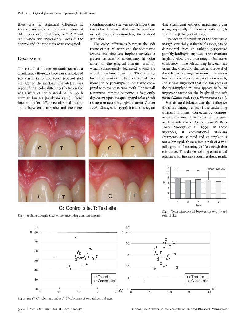
The mean color difference DE between
the test and control site was 7.7 in area 1
and decreased toward area 5, which was 6.5
(Fig. 5). In addition, the mean DE value in
area 5 was45, which is a much greater
value than the clinical perceptual threshold
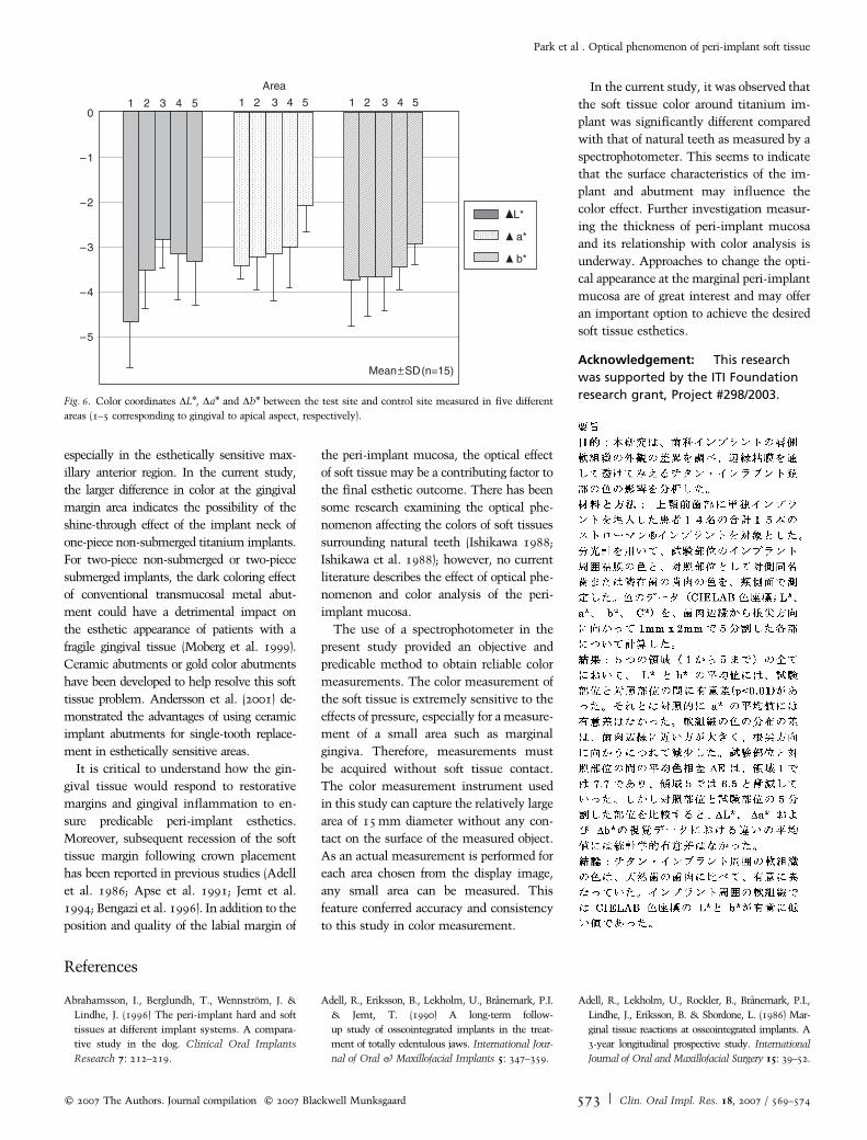
of 3.6. A trend toward decrease in color
coordinates DLn, Dan and Dbn between the
test site and control site was observed
toward the apical aspect (Fig. 6). Area 1
(gingival) showed the greatest mean values
of DLn, Dan and Dbn and these values
decreased toward area 5 (apical). However,
Table 1. Optical data of five incremental areas for the control (C) and test (T) sites
Area 1 2 3 4 5
Site C T C T C T C T C T
Ln
Mean 56.2 49.9 55.7 50.8 54.4 49.8 53.9 49.1 53.8 48.9SD 6.7 4.9 6.7 4.2 6.3 3.2 6.7 3.6 6.8 3.8
an
Mean 17 15.7 18.6 16.3 20.1 17.8 20.5 18.8 20.5 19.4SD 3.2 4.3 3.8 3.3 3.9 3.2 3.8 3.4 3.5 3.4
bn
Mean 17.2 12.7 14.7 11.2 15 11.7 15.3 12.3 15.3 12.6SD 3 4.2 3.1 2.8 3.1 2.4 3 2.6 2.9 2.8
Cn
Mean 24.1 20.4 23.6 19.8 25 21.4 25.5 22.5 25.4 23.2SD 3.6 5.2 4.2 3.9 4.4 3.6 4.2 3.9 3.9 4
C, control site; T, test site.
Park et al . Optical phenomenon of peri-implant soft tissue
c� 2007 The Authors. Journal compilation c� 2007 Blackwell Munksgaard 571 | Clin. Oral Impl. Res. 18, 2007 / 569–574
there was no statistical difference at
Po0.05 on each of the mean values of
differences in optical data, DLn, Dan and
Dbn, when five incremental areas of the
control and the test sites were compared.
Discussion
The results of the present study revealed a
significant difference between the color of
soft tissue in natural tooth (control site)
and around the implant (test site). It was
reported that color differences between the
soft tissues of contralateral natural teeth
were within 2.7 (Ishikawa 1988). There-
fore, the color difference obtained in this
study between a test site and the corre-
sponding control site was much larger than
the color difference that can be observed
in soft tissues surrounding the natural
dentition.
The color differences between the soft
tissue of natural teeth and the soft tissue
around the titanium implant revealed a
greater amount of discrepancy in color
closer to the gingival margin (area 1),
which subsequently decreased toward the
apical direction (area 5). This finding
further supports the effect of optical phe-
nomenon of peri-implant soft tissue com-
pared with that of natural teeth. The overall
restorative esthetic outcome is frequently
dependent upon the quality and color of soft
tissue at or near the gingival margin (Garber
1996; Chang et al. 1999). It is in this region
that significant esthetic impairment can
occur, especially in patients with a high
smile line (Chang et al. 1999).
Changes in the position of the soft tissue
margin, especially at the facial aspect, can be
detrimental from an esthetic perspective
possibly leading to exposure of the titanium
implant below the crown margin (Furhauser
et al. 2005). The relationship between soft
tissue thickness and changes in the level of
the soft tissue margin in terms of recession
has been investigated in previous research,
and it was suggested that the thickness of
the peri-implant mucosa appears to be an
important factor for the height of the soft
tissue (Warrer et al. 1995; Wennstrom 1996).
Soft tissue thickness can also influence
the shine-through effect of the underlying
titanium implant, consequently compro-
mising the overall esthetics of the peri-
implant soft tissue (Ochsenbein & Ross
1969; Moberg et al. 1999). In these
instances, if conventional titanium
abutments are selected and an implant is
not submerged, there exists a risk of a me-
tallic gray tint becoming visible through thin
soft tissue. This darker coloring effect could
produce an unfavorable overall esthetic result,
C: Control site, T: Test site
C CT
T
T
TC C
Fig. 3. A shine-through effect of the underlying titanium implant.
C*
80
70
60
50
40
30
0 10 20 30 40
L*
0 0
5
10
15
20
25
0 10 20 30 40a*
b*
: Test site: Control site
: Test site: Control site
a b
Fig. 4. An Ln–Cn color map and a an–bn color map of test and control sites.
Area
∆E
0
2
4
6
8
10
12
1 2 3 4 5
Mean±D(n=15)
Fig. 5. Color difference DE between the test site and
control site.
Park et al . Optical phenomenon of peri-implant soft tissue
572 | Clin. Oral Impl. Res. 18, 2007 / 569–574 c� 2007 The Authors. Journal compilation c� 2007 Blackwell Munksgaard
especially in the esthetically sensitive max-
illary anterior region. In the current study,
the larger difference in color at the gingival
margin area indicates the possibility of the
shine-through effect of the implant neck of
one-piece non-submerged titanium implants.
For two-piece non-submerged or two-piece
submerged implants, the dark coloring effect
of conventional transmucosal metal abut-
ment could have a detrimental impact on
the esthetic appearance of patients with a
fragile gingival tissue (Moberg et al. 1999).
Ceramic abutments or gold color abutments
have been developed to help resolve this soft
tissue problem. Andersson et al. (2001) de-
monstrated the advantages of using ceramic
implant abutments for single-tooth replace-
ment in esthetically sensitive areas.
It is critical to understand how the gin-
gival tissue would respond to restorative
margins and gingival inflammation to en-
sure predicable peri-implant esthetics.
Moreover, subsequent recession of the soft
tissue margin following crown placement
has been reported in previous studies (Adell
et al. 1986; Apse et al. 1991; Jemt et al.
1994; Bengazi et al. 1996). In addition to the
position and quality of the labial margin of
the peri-implant mucosa, the optical effect
of soft tissue may be a contributing factor to
the final esthetic outcome. There has been
some research examining the optical phe-
nomenon affecting the colors of soft tissues
surrounding natural teeth (Ishikawa 1988;
Ishikawa et al. 1988); however, no current
literature describes the effect of optical phe-
nomenon and color analysis of the peri-
implant mucosa.
The use of a spectrophotometer in the
present study provided an objective and
predicable method to obtain reliable color
measurements. The color measurement of
the soft tissue is extremely sensitive to the
effects of pressure, especially for a measure-
ment of a small area such as marginal
gingiva. Therefore, measurements must
be acquired without soft tissue contact.
The color measurement instrument used
in this study can capture the relatively large
area of 15 mm diameter without any con-
tact on the surface of the measured object.
As an actual measurement is performed for
each area chosen from the display image,
any small area can be measured. This
feature conferred accuracy and consistency
to this study in color measurement.
In the current study, it was observed that
the soft tissue color around titanium im-
plant was significantly different compared
with that of natural teeth as measured by a
spectrophotometer. This seems to indicate
that the surface characteristics of the im-
plant and abutment may influence the
color effect. Further investigation measur-
ing the thickness of peri-implant mucosa
and its relationship with color analysis is
underway. Approaches to change the opti-
cal appearance at the marginal peri-implant
mucosa are of great interest and may offer
an important option to achieve the desired
soft tissue esthetics.
Acknowledgement: This research
was supported by the ITI Foundation
research grant, Project #298/2003.
References
Abrahamsson, I., Berglundh, T., Wennstrom, J. &
Lindhe, J. (1996) The peri-implant hard and soft
tissues at different implant systems. A compara-
tive study in the dog. Clinical Oral Implants
Research 7: 212–219.
Adell, R., Eriksson, B., Lekholm, U., Branemark, P.I.
& Jemt, T. (1990) A long-term follow-
up study of osseointegrated implants in the treat-
ment of totally edentulous jaws. International Jour-
nal of Oral & Maxillofacial Implants 5: 347–359.
Adell, R., Lekholm, U., Rockler, B., Branemark, P.I.,
Lindhe, J., Eriksson, B. & Sbordone, L. (1986) Mar-
ginal tissue reactions at osseointegrated implants. A
3-year longitudinal prospective study. International
Journal of Oral and Maxillofacial Surgery 15: 39–52.
L*
a*
b*
Area
–1
–2
–3
–4
–5
01 2 3 4 5
Mean±SD(n=15)
1 2 3 4 5 1 2 3 4 5
Fig. 6. Color coordinates DLn, Dan and Dbn between the test site and control site measured in five different
areas (1–5 corresponding to gingival to apical aspect, respectively).
Park et al . Optical phenomenon of peri-implant soft tissue
c� 2007 The Authors. Journal compilation c� 2007 Blackwell Munksgaard 573 | Clin. Oral Impl. Res. 18, 2007 / 569–574

Andersson, B., Taylor, A., Lang, B.R., Scheller, H.,
Scharer, P., Sorensen, J.A. & Tarnow, D. (2001)
Alumina ceramic implant abutments used for
single-tooth replacement: a prospective 1- to 3-
year multi-center study. International Journal of
Prosthodontics 14: 432–438.
Apse, P., Zarb, G.A., Schmitt, A. & Lewis, D.W.
(1991) The longitudinal effectiveness of osseoin-
tegrated dental implants. The Toronto study: peri-
implant mucosal response. The International
Journal of Periodontics and Restorative Dentistry
11: 95–111.
Avivi-Arber, L. & Zarb, G.A. (1996) Clinical effec-
tiveness of implant-supported single-tooth repla-
cement: Toronto study. International Journal of
Oral & Maxillofacial Implants 11: 311–321.
Becker, W. & Becker, E.B. (1996) Flap designs for
minimization of recession adjacent to maxillary
anterior implant sites: a clinical study. Interna-
tional Journal of Oral & Maxillofacial Implants
11: 46–54.
Bengazi, F., Wennstrom, J.L. & Lekholm, U. (1996)
Recession of the soft tissue margin at oral im-
plants. A 2-year longitudinal prospective study.
Clinical Oral Implants Research 7: 303–310.
Berglundh, T. & Lindhe, J. (1996) Dimension of the
peri-implant mucosa. Biologic width revisited.
Journal of Clinical Periodontology 23: 971–973.
Berglundh, T., Lindhe, J., Ericsson, I., Marinello,
C.P., Liljenberg, B. & Thomsen, P. (1991) The
soft tissue barrier at implants and teeth. Clinical
Oral Implants Research 2: 81–90.
Branemark, P.I., Hansson, B.O., Adell, R., Breine,
U., Lindstrom, J., Hallen, O. & Ohman, A. (1977)
Osseointegrated implants in the treatment of the
edentulous jaw. Experience from a 10-year period.
Scandinavian Journal of Plastic and Reconstruc-
tive Surgery 11 (Suppl.): 1–132.
Buser, D., Weber, H.P., Bragger, U. & Balsiger, C.
(1991) Tissue integration of one-stage ITI im-
plants: 3-year results of a longitudinal study
with hollow-cylinder and hollow-screw implants.
International Journal of Oral & Maxillofacial
Implants 6: 405–412.
Chang, M., Wennstrom, J.L., Odman, P. & Anders-
son, B. (1999) Implant supported single-tooth
replacements compared to contralateral natural
tooth. Crown and soft tissue dimensions. Clinical
Oral Implants Research 10: 185–194.
Chaytor, D.V., Zarb, G.A., Schmitt, A. & Lewis,
D.W. (1991) The longitudinal effectiveness of os-
seointegrated dental implants. The Toronto study:
bone level changes. International Journal of
Periodontics and Restorative Dentistry 11:
113–125.
Cochran, D.L., Hermann, J.S., Schenk, R.K., Hig-
ginbottom, F.L. & Buser, D. (1997) Biologic width
around titanium implants. A histometric analysis
of the implanto-gingival junction around unloaded
and loaded nonsubmerged implants in the
canine mandible. Journal of Periodontology 68:
186–198.
Conte, G.J., Rhodes, P., Richards, D. & Kao, R.T.
(2002) Considerations for anterior implant es-
thetics. Journal of California Dental Association
30: 528–534.
Furhauser, R., Florescu, D., Benesch, T., Hass, R.,
Mailath, G. & Watzek, G. (2005) Evaluation of
soft tissue around single-tooth implant crowns:
the pink esthetic score. Clinical Oral Implants
Research 16: 639–644.
Garber, D.A. (1996) The esthetic dental implant:
letting restoration be the guide. Journal of Oral
Implantology 22: 45–50.
Gargiulo, A.W., Wentz, F.M. & Orban, B. (1961)
Dimensions and relations of the dentogingival
junction in humans. Journal of Periodontology
32: 261–267.
Haas, R., Pollak, Ch., Furhauser, R., Mailath-Pok-
omy, G., Dortbudak, O. & Watzek, G. (2002) A
long-term follow-up of 76 Branemark single tooth
implants. Clinical Oral Implants Research 13:
38–43.
Hermann, J.S., Buser, D., Schenk, R.K., Higginbot-
tom, F.L. & Cochran, D.L. (2000) Biologic width
around titanium implants. A physiologically
formed and stable dimension over time. Clinical
Oral Implant Research 12: 559–571.
Hermann, J.S., Buser, D., Schenk, R.K., Schoolfield,
JD. & Cochran, D.L. (2001) Biologic width around
one- and two-piece titanium implants. A histo-
metric evaluation of unloaded nonsubmerged and
submerged implants in the canine mandible. Clin-
ical Oral Implant Research 12: 559–571.
Ishikawa, S. (1988) Colorimetric study of marginal
gingiva inflammatory effects on color difference
analyses from the standpoint of gingival transmis-
sion. Journal of the Japan Prosthodontic Society
32: 829–838.
Ishikawa, S., Furukawa, K. & Ishibashi, K. (1988)
Colorimetric studies of gingival color variations in
the upper anterior region. Journal of the Japan
Prosthodontic Society 32: 821–828.
Ishikawa-Nagai, S., Ishibashi, K., Tsuruta, O. &
Weber, H.P. (2005) Reproducibility of tooth color
gradation using a computer color-matching tech-
nique applied to ceramic restorations. Journal of
Prosthetic Dentistry 93: 129–137.
Israelson, H. & Plemons, J.M. (1993) Dental im-
plants, regenerative techniques, and periodontal
plastic surgery to restore maxillary anterior es-
thetics. International Journal of Oral & Maxillo-
facial Implants 8: 555–561.
Jemt, T., Book, K., Lie, A. & Borjesson, T. (1994)
Mucosal topography around implants in eden-
tulous upper jaws. Photo-grammetric three-
dimensional measurements of the effect of repla-
cement of a removable prosthesis with a fixed
prosthesis. Clinical Oral Implants Research 5:
220–228.
Johnston, W.M. & Kao, E.C. (1989) Assessment of
appearance match by visual observation and clin-
ical colorimetry. Journal of Dental Research 68:
819–822.
Kao, R.T. & Pasquinelli, K. (2002) Thick vs. thin
gingival tissue: a key determinant in tissue re-
sponse to disease and restorative treatment. Jour-
nal of California Dental Association 30: 521–
526.
Lekholm, U., van Steenberghe, D., Herrmann, I.,
Bolender, C., Folmer, T., Gunne, J., Henry, P.,
Higuchi, K., Laney, W. & Linden, U. (1994)
Osseointegrated implants in the treatment of
partially edentulous jaws. A prospective 5-year
multicenter study. International Journal of Oral
& Maxillofacial Implants 9: 627–635.
Moberg, L.E., Kondell, P.A., Kullman, L., Heim-
dahl, A. & Gynther, G.W. (1999) Evaluation
of single-tooth restorations on ITI dental im-
plants. Clinical Oral Implants Research 10:
45–53.
Neale, D. & Chee, W.W.L. (1994) Development of
implant soft tissue emergence profile: a techni-
que. Journal of Prosthetic Dentistry 71:
364–368.
Ochsenbein, C. & Ross, A. (1969) A re-evaluation
of osseous surgery. Dental Clinics of North Amer-
ica 13: 87–103.
Palacci, P., Ericsson, I. & Engstrand, P. (1995)
Implant placement. In: Palacci, P., Ericsson, I.,
Engstrand, P. & Rangert, B., eds. Optimal Im-
plant Positioning and Soft Tissue Management
for the Branemark System, 35–39. Chicago:
Quintessence.
Reikie, D.F. (1993) Esthetic and functional consid-
erations for implant restoration of the partially
edentulous patient. Journal of Prosthetic Dentis-
try 70: 443–437.
Reikie, D.F. (1995) Restoring gingival harmony
around single tooth implants. Journal of Prosthe-
tic Dentistry 74: 47–50.
Ruyter, I.E., Niler, K. & Mollar, B. (1987) Color
stability of dental composite resin materials for
crown and bridge veneers. Dental Materials 3:
246–251.
Scheller, H., Urgell, J.P., Kultje, C., Klineberg, I.,
Goldberg, P.V., Stevenson-Moore, P., Alonso,
J.M.N., Schaller, M., Corria, R.M., Engquist, B.,
Toreskog, S., Kastenbaum, F. & Smith, C.R.
(1998) A 5-year multi-center study on implant-
supported single crown restorations. International
Journal of Oral & Maxillofacial Implants 13:
212–218.
Vacek, J.S., Gher, M.E., Assad, D.A., Richardson,
A.C. & Giambarresi, L.I. (1994) The dimensions
of the human dentogingival junction. Interna-
tional Journal of Periodontics and Restorative
Dentistry 14: 155–165.
Warrer, K., Buser, D., Lang, N.P. & Karring, T.
(1995) Plaque-induced peri-implantitis in the pre-
sence or absence of keratinized mucosa. Clinical
Oral Implants Research 6: 131–138.
Wennstrom, J.L. (1996) Mucogingival considera-
tions in orthodontic treatment. Seminars in
Orthodontics and Dentofacial Orthopedics 2:
46–54.
Park et al . Optical phenomenon of peri-implant soft tissue
574 | Clin. Oral Impl. Res. 18, 2007 / 569–574 c� 2007 The Authors. Journal compilation c� 2007 Blackwell Munksgaard