osteogenic and angiogenic potentials of monocultured and
TRANSCRIPT

Acta Biomaterialia 9 (2013) 4906–4915
Contents lists available at SciVerse ScienceDirect
Acta Biomaterialia
journal homepage: www.elsevier .com/locate /ac tabiomat
Osteogenic and angiogenic potentials of monocultured and co-culturedhuman-bone-marrow-derived mesenchymal stem cellsand human-umbilical-vein endothelial cells on three-dimensional porousbeta-tricalcium phosphate scaffold
Yunqing Kang a, Sungwoo Kim a, Monica Fahrenholtz b, Ali Khademhosseini c,d,e, Yunzhi Yang a,⇑a Department of Orthopedic Surgery, Stanford University, 300 Pasteur Drive, Stanford, CA 94305, USAb Department of Bioengineering, Rice University, 6100 Main St., Houston, TX 77030, USAc Center for Biomedical Engineering, Department of Medicine, Brigham and Women’s Hospital, Harvard Medical School, Cambridge, MA 02139, USAd Harvard-MIT Division of Health Sciences and Technology, Massachusetts Institute of Technology, Cambridge, MA 02139, USAe Wyss Institute for Biologically Inspired Engineering, Harvard University, Boston, MA 02115, USA
a r t i c l e i n f o
Article history:Received 14 April 2012Received in revised form 23 July 2012Accepted 8 August 2012Available online 16 August 2012
Keywords:b-TCPhBMSCsHUVECsOsteogenesisAngiogenesis
1742-7061/$ - see front matter � 2012 Acta Materialhttp://dx.doi.org/10.1016/j.actbio.2012.08.008
⇑ Corresponding author. Tel.: +1 650 723 0772; faxE-mail address: [email protected] (Y. Yang).
a b s t r a c t
The use of biodegradable beta-tricalcium phosphate (b-TCP) scaffolds holds great promise for bone tissueengineering. However, the effects of b-TCP on bone and endothelial cells are not fully understood. Thisstudy aimed to investigate cell proliferation and differentiation of mono- or co-cultured human-bone-marrow-derived mesenchymal stem cells (hBMSCs) and human-umbilical-vein endothelial cells(HUVECs) on a three-dimensional porous, biodegradable b-TCP scaffold. In co-culture studies, the ratiosof hBMSCs:HUVECs were 5:1, 1:1 and 1:5. Cellular morphologies of HUVECs, hBMSCs and co-culturedHUVECs/hBMSCs on the b-TCP scaffolds were monitored using confocal and scanning electron micros-copy. Cell proliferation was monitored by measuring the amount of double-stranded DNA (dsDNA)whereas hBMSC and HUVEC differentiation was assessed using the osteogenic and angiogenic markers,alkaline phosphatase (ALP) and PECAM-1 (CD31), respectively. Results show that HUVECs, hBMSCs andhBMSCs/HUVECs adhered to and proliferated well on the b-TCP scaffolds. In monoculture, hBMSCs grewfaster than HUVECs on the b-TCP scaffolds after 7 days, but HUVECs reached similar levels of proliferationafter 14 days. In monoculture, b-TCP scaffolds promoted ALP activities of both hBMSCs and HUVECs whencompared to those grown on tissue culture well plates. ALP activity of cells in co-culture was higher thanthat of hBMSCs in monoculture. Real-time polymerase chain reaction results indicate that runx2 and alpgene expression in monocultured hBMSCs remained unchanged at days 7 and 14, but alp gene expressionwas significantly increased in hBMSC co-cultures when the contribution of individual cell types was notdistinguished.
� 2012 Acta Materialia Inc. Published by Elsevier Ltd. All rights reserved.
1. Introduction
Tissue engineering holds great promise for regenerating func-tional tissues and organs. One important component of tissue engi-neered materials is the scaffold, which is an artificial extracellularmatrix (ECM) that serves as a temporary support structure and im-parts the necessary biophysical, biomechanical and biochemicalcues required for cell attachment, proliferation and differentiationto form tissue [1]. In particular, it is highly desirable for scaffolds topossess an interconnected and open macroporous structure tofacilitate cellular in-growth and neovascularization of regeneratedtissue in vivo.
ia Inc. Published by Elsevier Ltd. A
: +1 650 724 5401.
Calcium phosphate (CaP) bioceramics have been widely usedin clinical settings for bone repair and reconstruction [2,3].Hydroxyapatite (HA) and beta-tricalcium phosphate (b-TCP) arethe most popular bioceramics due to their close resemblance tonatural bone, excellent biocompatibility and biodegradability.Bone-marrow-derived mesenchymal stem cells (BMSCs) havebeen used in combination with CaP scaffolds in bone tissue engi-neering [4–7]. Many studies involving the use of BMSC-seededbioceramics have shown promising results in the context of boneregeneration [8–11]; however, most of these investigations havehighlighted the osteogenic potential of BMSCs on these CaPbioceramics.
Bone tissue contains multiple cell types, including osteogeniccells and endothelial cells. A number of studies in bone repairand regeneration have highlighted the intimate interactions
ll rights reserved.

Y. Kang et al. / Acta Biomaterialia 9 (2013) 4906–4915 4907
between endothelial cells and osteoprogenitor cells [12,13]. In-deed, it has been well established that angiogenesis is a prerequi-site for osteogenesis in vivo [14]. For example, insufficientneovascularization of bone constructs/tissues after scaffoldimplantation resulted in hypoxia and cellular necrosis [15–19].Thus, a key factor in repairing large bone defects is vascularizationof the scaffold. Within this context, it is important to characterizethe effects of bioceramic scaffolds with regards to osteogenic andendothelial differentiation. It has been reported that co-culturedhuman endothelial cells with human osteoblast cells on porousHA, b-TCP or Ca-deficient HA containing polycaprolactone sup-ported the formation of capillary-like structures [20,21]. Zhouet al. pre-vascularized b-TCP scaffold by co-seeding MSCs andMSC-derived endothelial cells (ECs) and used it to promote the re-pair of segmental bone defects in rabbits [22].
In this study, we investigated the effect of a porous, biodegrad-able b-TCP scaffold on the behavior of mono- or co-cultured hu-man-bone-marrow-derived mesenchymal stem cells (hBMSCs)and human-umbilical-vein endothelial cells (HUVECs). We hypoth-esize that different cell ratios of hBMSCs and HUVECs may behavedifferently on a porous, biodegradable b-TCP scaffold. In this exper-iment, interconnected porous b-TCP scaffolds were prepared by atemplate-casting method [23–25]. Cell proliferation was moni-tored by measuring the amount of double-stranded DNA (dsDNA),whereas hBMSC and HUVEC differentiation was assessed using theosteogenic and angiogenic markers alkaline phosphatase (ALP) andPECAM-1 (CD31), respectively.
2. Materials and methods
2.1. Materials
b-TCP powder with a specific surface area of 17 m2 g–1 waspurchased from Nanocerox, Inc. (Ann Arbor, Michigan).Carboxymethyl cellulose powder, paraffin beads and ethyl alcoholwere purchased from Fisher Scientific (Pittsburgh, USA).
Dulbecco’s modified Eagle’s medium (DMEM), fetal bovine ser-um (FBS) and L-glutamine, 100 � antibiotic–antimycotic solutionwere purchased from Invitrogen Co. (Grand Island, NY, CA). EBM™endothelial basal medium containing EGM™ endothelial growthmedium and a Single Quots™ kit were purchased from Lonza, Inc.Alexa Fluor� 594 goat anti-mouse secondary antibody (2 mg ml–1)was purchased from Invitrogen Inc. An RNeasy mini kit for extract-ing RNA was purchased from QIAGEN (Valencia, CA, USA).
2.2. Preparation of b-TCP scaffolds
Porous b-TCP scaffolds were fabricated using a template-castingmethod as previously described [23–25]. Briefly, b-TCP powder,carboxymethyl cellulose powder, surfactant (Surfonal�) and dis-persant (Darvan� C) were mixed in distilled water to form a cera-mic slurry. Paraffin beads were packed into a customized mold andheated to induce partially melting and formation of a template. Theb-TCP ceramic slurry was then cast into the mold under vacuum,solidified and subsequently dehydrated in a series of ethyl alcoholsolutions (70%, 90% and 95%). After removing the dehydrated greenbody from the mold, the green body was dried in an oven for 2 h,and then placed into an electric high temperature furnace and sin-tered at 1250 �C for 3 h. The morphology of b-TCP scaffolds wascharacterized by scanning electron microscopy. The average poresize of scaffold was obtained from scanning electron microscopy(SEM) images and at least six pores were measured. The intercon-nected pore structure of scaffold was scanned using micro-computed tomography (micro-CT; Imtek Micro CAT II, Knoxville,TN) at a resolution of 80 lm. Raw images were further
reconstructed and analyzed by GE microView software (GeneralElectric Co.). The scaffolds used in this study were 7–8 mm indiameter and 5–6 mm in height.
2.3. Cell culture of hBMSCs and HUVECs
hBMSCs were purchased from Lonza Inc. (Allendale, NJ) [26].According to the manufacturer’s certificate of analysis, the cellsare more than 90% positive for CD105, CD166, CD29 and CD44,and less than 10% positive for CD14, CD34 and CD45. The cells werecultured in basal medium consisting of DMEM, (Invitrogen, USA)with 10% FBS, 1% L-glutamine (200 mM) and 1% antibiotic–antimy-cotic solution under standard conditions (5% CO2, 95% humidity,and 37 �C). Passages 6–8 were used for all the experiments.
Immortalized HUVECs constitutively expressing green fluores-cent protein (GFP) were a generous gift from the late Dr. J. Folkman,Children’s Hospital, Boston. HUVECs were cultured in endothelialbasal medium (EBM-2, Lonza) with endothelial growth supplementSingle Quots (EGM-2, Lonza) in a 5% CO2 atmosphere at 37 �C.
A preliminary study was performed to test the culture mediumfor co-culture experiments (Supplementary Fig. S1). In this study,the medium used in all co-culture experiments was a 1:1 mixtureof EBM-2 and DMEM.
When the cells reached �85–90% confluence in flasks, theywere subcultured using 0.25% trypsin–EDTA (Invitrogen, USA)and resuspended in culture medium. Next, 100,000 cells in100 ll medium were gently seeded into the sterilized b-TCP scaf-folds and incubated at 37 �C for 1 h to allow cells to attach ontothe inner construct surface of the scaffold. New medium was thenadded for further incubation. The medium was changed every3 days. To investigate the reciprocal effect of the two cell typeson osteogenesis and angiogenesis, three mixture ratios (1:5, 1:1,5:1) of hBMSCs and HUVECs were seeded and co-cultured on thescaffolds. In mono- and co-culture experiments, the seeding den-sity (100,000 cells per scaffold) was held constant regardless of cellmixture ratios. To indicate the cell distribution in the scaffolds,scaffolds with seeded HUVEC-GFP cells were recaptured using afluorescent microscope (Nikon 2000) and shown in SupplementaryFig. S2.
2.4. Morphologies of HUVECs, hBMSCs and co-cultured cellson the b-TCP scaffolds
The morphology of HUVECs on b-TCP scaffolds was monitoredusing a confocal laser scanning microscope (CLSM; OlympusIX81) after 1, 3 and 7 days of incubation in EBM-2 medium. Mor-phologies of HUVECs, hBMSCs and hBMSC/HUVEC mixtures seededon the b-TCP scaffolds were also observed under a scanning elec-tron microscope (SEM, FEI Quanta 400). HUVECs and hBMSCs inmonoculture were incubated in EBM-2 and DMEM, respectively,and hBMSC/HUVEC mixture cells (50%:50%) were co-cultured inEBM-2:DMEM mixture medium (1:1) for 3 days. Following this,scaffolds were fixed with 2.5% glutaraldehyde for 2 h, followedby graded dehydration using a series of ethanol solutions (gener-ally 70%, 80%, 90% and 100%). Samples were then dried in a hoodand sputtered with gold before observation under an SEM (FEIQuanta 400) at 20 kV.
2.5. Visualization of F-actin
F-actin was stained by rhodamine phalloidin to assess cytoskel-etal organization on the b-TCP scaffold. After 7 days of incubationin the same medium as used in the experiments for SEM, cells/scaf-folds were rinsed twice using phosphate buffered saline (PBS) andthen fixed by 4% paraformaldehyde solution (PFA) for 15 min atroom temperature. The fixed cells were further permeabilized in

Table 1Sequences of primers used for real-time PCR analysis.
Genes Sequences
GAPDH For: 50-AACAGCGACACCCACTCCTCRev: 50-CATACCAGGAAATGAGCTTGACAA
runx-2 For: 50-AGATGATGACACTGCCACCTCTGRev: 50-GGGATGAAATGCTTGGGAACT
alp For: 50-ACATTCCCACGTCTTCACATTTRev: 50-AGACATTCTCTCGTTCACCGCC
opn For: 50-ATGAGATTGGCAGTGATTRev: 50-TTCAATCAGAAACTGGAA
oc For: 50-TGTGAGCTCAATCCGGACTGTRev: 50-CCGATAGGCCTCCTGAAGC
bsp For: 50-ATGGCCTGTGCTTTCTCAATGRev:50-GGATAAAAGTAGGCATGCTTG
bmp-2 For:50-GCCCTTTTCCTCTGGCTATRev:50-TTGACCAACGTCTGAACAATGG
cd31 For: 50-GAGTCCTGCTGACCCTTCTGRev: 50-CACTCCTTCCACCAACACCT
4908 Y. Kang et al. / Acta Biomaterialia 9 (2013) 4906–4915
0.5% Triton X-100 and incubated in 100 nM rhodamine phalloidinworking solution (Cytoskeleton Inc., USA) at room temperaturefor 2 h. After a brief rinse with PBS, DAPI solution (5 lg ml–1)was added to counterstain cell nuclei. After a thorough washingwith PBS, cells on scaffolds were visualized with a CLSM.
2.6. Immunofluorescent staining of PECAM-1
Platelet-endothelial cell adhesion molecule (PECAM-1, or CD31)is an endothelial-specific adhesion protein and a specific marker ofHUVECs. It was assessed by immunefluorescent staining. The effectof b-TCP scaffolds on PECAM-1 expression of HUVECs in monocul-ture or co-culture with hBMSCs in various ratios (1:5, 1:1, 5:1)were investigated at 7 and 14 days. They were cultured in a 1:1mixture of EBM-2 and DMEM, and then rinsed twice with PBSand fixed in a solution of 4% PFA for 15 min at room temperature.After washing three times in PBS, the fixed cells/scaffolds wereplaced in 3% bovine serum albumin (BSA)/PBS blocking buffer for1 h, and then were incubated with mouse anti-human CD31 pri-mary antibody (1:3000, Cell Signaling Technology) in 1% BSA/PBSovernight at 4 �C. The cells/scaffolds were then washed three timesusing PBS, and incubated in an anti-mouse secondary antibodyAlexa Fluor�594 (1:1000; 2 lg ml–1, Invitrogen) for 1 h at roomtemperature. After a brief rinse using PBS, the cell nuclei werecounterstained with DAPI solution (5 lg ml–1) for 1 min. The scaf-fold samples were then extensively washed with PBS, and visual-ized with a CLSM.
2.7. Osteogenic differentiation assay of hBMSCs
The effect of b-TCP scaffolds on the early osteogenic differenti-ation of hBMSCs in monoculture and co-culture was assessedthrough ALP specific activity. hBMSCs were co-cultured with vari-ous ratios of HUVECs (5:1, 1:1, 1:5) on the scaffolds. MonoculturedhBMSCs and HUVECs were used as controls. The medium used inthese co-culture experiments was a 1:1 mixture of EBM-2 andDMEM, as determined in our preliminary study (S.1).
At the end of each time point, cells/scaffolds were washed twicewith PBS and preserved at �80 �C. Cells/scaffolds experiencingthree freeze/thaw cycles in �80 �C/37 �C, were then lysed in500 ll of 0.2% Triton X-100 in PBS, and finally homogenized bysonication for 30 s on ice. The ALP activity was assayed using a col-orimetric p-NPP method [23,24]. The absorbance was measured ona microplate reader (TECAN) at 405 nm after 30 min incubation at37 �C. ALP specific activity levels were quantified with a standardcurve and normalized to the amount of total cellular dsDNA fromthe same sample. dsDNA content was determined using a PicoGreen assay (Molecular Probe, Invitrogen). A 50 ll volume of work-ing reagent was added to the 50 ll cell lysate of the sample. Thesample was read at 485/528 nm (excitation/emission) on a fluores-cence spectrophotometer (Biotek, Flx800, USA). The amount ofdsDNA was calculated by comparing the standard curves of theknown dsDNA sample according to the manufacturer’s instruction.
2.8. Quantitative real-time polymerase chain reaction (PCR)
Total RNA of cells was extracted from the monocultured and co-cultured cells after incubation of 7 and 14 days using an RN easymini Kit (QIAGEN) following the manufacturer’s protocol. RNA con-centration was determined on an Eppendorf Biophotometer. To re-verse-transcribe RNA of the samples into cDNA, an iScript cDNAsynthesis kit (BIO-RAD) was used according to the manufacturer’sprotocols. Using cDNA product template, specific primers and iQ-SYBR Green supermix (BIO-RAD), real-time PCR was performedon an ABI 7900HT Sequence Detection system (ABI, Foster city,USA). The total reaction volume was 10 ll. Primer sequences are
shown in Table 1, including runt-related transcription factor 2(runx2), alkaline phosphatase (alp), osteopontin (opn), osteocalcin(oc), bone sialoprotein (bsp), bone morphogenetic protein-2(bmp-2), cd31 and glyceraldehyde 3-phosphate dehydrogenase(GAPDH). These primers were purchased from Invitrogen Co. andused to evaluate gene expression [27–29]. The relative expressionlevels of genes were analyzed using the 2�DDCt method [30] bynormalizing with the housekeeping gene GAPDH as an endogenouscontrol and calibrating with efficiency, where DDCt is calculatedfrom (Ct, target � Ct, control)target gene � (Ct, target � Ct, control)GAPDH.
2.9. Statistical analysis
All the groups in the experiments were performed in triplicate,and the statistical significance was analyzed by Student’s t-test. Ifthe p-values obtained from the t-test were less than 0.05, the dif-ference was considered to be significant.
3. Results
3.1. Porous morphologies of the b-TCP scaffold
Representative morphology of a b-TCP scaffold is shown inFig. 1. The porous structure and interconnected pores of a b-TCPscaffold is shown in Fig. 1A at a lower magnification. The pores sizeof the scaffold is in the range of 350–500 lm and the average poresize measured from SEM images is �396 ± 49 lm. Fig. 1B indicatesa local strut of the scaffold at a higher magnification. The strut sur-face appears dense and consists of microscale grains. Fig. 1C–Eshows the representative three-dimensional (3-D) and two-dimensional (2-D) reconstructed micro-CT images. The intercon-nected pores are observed across the scaffold in Fig. 1C–E.
3.2. Cell morphologies on the b-TCP scaffold
Morphologies of GFP-tagged HUVECs on the b-TCP scaffoldwere observed with a CLSM. Fig. 2 shows cell morphology changeson a b-TCP scaffold over time. Fluorescent images in Fig. 2A showthat HUVECs adhered and spread well on the surface of strutsand inner pores of the scaffold at day 1, and cells exhibited a cob-blestone-like morphology. HUVECs proliferated well on the scaf-fold with increasing culture time. At day 3, HUVECs covered themajority of the strut surface (Fig. 2B) and a dense endothelial layerwas formed by day 7 (Fig. 2C). In this study, SEM was used to

Fig. 1. SEM morphologies of the b-TCP scaffold with different magnifications: (A) interconnected pores at low magnification (50�), and (B) the local strut surface at highmagnification of the square area in (A) (3000�). Micro-CT images indicate the interconnected pores of scaffold in 3-D (C), and 2-D reconstruction (D) and (E).
Fig. 2. HUVEC adhesion and spread morphology on the struts of a b-TCP scaffold observed by confocal laser scanning microscopy. HUVECs are observed to attach and growwell on the scaffold for (A) 1 day, (B) 3 days and (C) 7 days. Images were captured on the same site of one scaffold.
Y. Kang et al. / Acta Biomaterialia 9 (2013) 4906–4915 4909
further investigate cell morphologies of hBMSCs and HUVECs onthe b-TCP scaffolds. Results showed that both hBMSCs and HUVECscan adhere well on the scaffolds (Fig. 3). In monocultures, HUVECsexhibited cobblestone-like morphology and formed a flattenedendothelial cell layer (Fig. 3A), while hBMSCs exhibited a typicalspindle-like morphology (Fig. 3B). In co-culture of hBMSCs and HU-VECs, both spindle-like cells and cobble-like cells could be ob-served, suggesting the co-existence of both cell types on thescaffold (Fig. 3C).
3.3. Visualization of F-actin filament of cells
In HUVEC and hBMSC monocultures, an abundance of actin fi-bers was observed in the cytoplasm of the cells (Fig. 4A, G and C,H). The fluorescent image in Fig. 4B shows cobblestone-like mor-phology. In hBMSC monocultures, the rhodamine phalloidin/DAPIstaining of hBMSCs in Fig. 4C indicated a homogenous distributionof actin fibers and filament elongation of cells on the scaffold.Additionally, as shown in Fig. 4D, hBMSCs were GFP-negative,
distinguishing them from the GFP-positive HUVECs. In co-cultures,comparing the rhodamine phalloidin/DAPI staining image inFig. 4E with the GFP fluorescent image in Fig. 4F, the distributionof hBMSCs in co-culture can be distinguished from that of GFP-tagged HUVECs. Contacting zones or overlapping growths were ob-served. Fig. 4G and H shows the F-actin staining of HUVECs andhBMSCs at a higher magnification, respectively. Obvious actin fiberbundles can be observed in the cytoplasm.
3.4. Immunofluorescent staining of PECAM-1
In HUVECs monocultures, immunofluorescent images show theexpression of PECAM-1 at the cell–cell interface after 7 days of cul-ture (Fig. 5). With increasing culture time, PECAM-1 expression ofHUVECs became more pronounced and cells were observed form-ing network after 14 days. For both the 1:5 hBMSCs/HUVECs andHUVEC alone groups, PECAM-1 expression increased with culturetime. The higher magnification images in the inserts showed theformation of small sprouts (Fig. 5). In the 5:1 and 1:1 hBMSC/HU-

Fig. 3. Scanning electron micrographs showing cells growing on the b-TCP scaffolds after 3 days of culture: (A) HUVECs, (B) hBMSCs and (C) co-cultured hBMSCs/HUVECs (1:1ratio).
Fig. 4. Cytoskeletal organization of cells grown on the b-TCP scaffolds at day 7, as observed under a CLSM and demonstrated by rhodamine phalloidin/DAPI staining for F-actin/cell nuclei (A, C, E) and endogenous GFP (B, D, F). An abundance of F-actin fiber is observed in HUVECs (A, B), hBMSCs (C, D) and co-cultured hBMSCs/HUVECs (E, F),indicating a homogenous distribution of F-actin. (G) and (H) show F-actin staining of HUVECs and hBMSCs at a higher magnification, respectively.
4910 Y. Kang et al. / Acta Biomaterialia 9 (2013) 4906–4915
VEC groups, these network structures were not apparent due to thelower proportion of HUVECs present.
3.5. Cell proliferation and ALP activity of hBMSCs
The proliferation of hBMSCs and HUVECs on the b-TCP scaffoldsis shown in Fig. 6a. For hBMSC monocultures, cell proliferation in-creased from day 3 to day 7. After 7 days, cellular dsDNA contentstarted to decrease. For HUVEC monocultures, cellular dsDNAamount increased from day 3 to day 14. The growth rate of HUVECs
was slower compared to that of hBMSCs, but reached the same le-vel of proliferation within 14 days of incubation. Compared tomonocultures, the total cellular dsDNA amount in co-culture wasproportional to the cell ratios and growth rates (Fig. 6a).
To evaluate the effect of HUVECs on the ALP activity of hBMSCs,hBMSCs were co-cultured with HUVECs at various ratios in non-osteogenic medium. The ALP activity of cells in monoculture andco-culture is shown in Fig. 6b. Results show that, in both mono-and co-culture, the ALP activity level of hBMSCs continually in-creased over 14 days (Fig. 6b). For the 5:1 and 1:1 hBMSC/HUVEC

Fig. 5. CLSM images showing expression of endothelial marker, PECAM-1 (CD31), by HUVECs in monoculture and co-culture with hBMSCs at day 7 and day 14 on b-TCPscaffolds. Confocal images through a z-stack demonstrated the expression of CD31. Insert images represent CD31 expression of hBMSC/HUVEC (1:5) and HUVEC groups onday 14 at a higher magnification. Formation of small sprouts can be observed (white arrows). CD31 is in red (labeled with Alexa Fluor 594), and nuclei are in blue (labeledwith DAPI).
Fig. 6. dsDNA contents of cells in monocultured hBMSCs and HUVECs, and co-cultured hBMSCs/HUVECs at various ratios on the b-TCP scaffolds (a), and ALP activityexpression in hBMSCs on the b-TCP scaffolds in monocultures and co-culture after 3, 7 and 14 days of incubation (b). ALP activity in monoculture hBMSCs (c) and HUVECs (d)on well plate and b-TCP scaffolds. An asterisk is used to indicate significant difference (p < 0.05).
Y. Kang et al. / Acta Biomaterialia 9 (2013) 4906–4915 4911
groups, the ALP activity of co-cultured cells was significantly high-er compared to those in monoculture (p < 0.05), except for the 1:5hBMSC/HUVEC groups at day 14. ALP expression of HUVECs washigher than that in other groups at day 3, and it remained constantduring 14 days of culture. ALP expression of hBMSCs or HUVECsalone on well plates was also investigated, and the results showthat ALP activity could not be detected until day 14 (Fig. 6c and
d), but the cells on the b-TCP scaffolds produced detectable ALPactivity level at days 3 and 7 (Fig. 6b).
3.6. Gene expression
Quantitative real-time PCR was performed to evaluate theexpression of osteogenic and endothelial markers. Fig. 7 shows

4912 Y. Kang et al. / Acta Biomaterialia 9 (2013) 4906–4915
the osteogenic gene expression of hBMSCs and the endothelialgene expression of HUVECs in monoculture and co-culture. Allgene expression was normalized to the expression levels of mono-cultured hBMSCs at day 7 (reference value set to 1).
For monocultures, the expression of runx2 of hBMSCs did notchange at 7 and 14 days of incubation. In the co-culture, runx2down-regulation increased with a decrease in the hBMSC/HUVECratio. Alp expression did not significantly change for monoculturedhBMSCs, but it was significantly increased in the 5:1 hBMSC/HU-VEC ratio group at day 7 and in the 5:1 and 1:1 hBMSC/HUVEC ra-tio groups at day 14. In monocultures, hBMSCs increased theexpressions of bsp and opn, two early bone matrix genes, and re-duced the expression of oc, a late bone matrix gene between day7 and day 14 [31–33]. The addition of HUVECs significantly de-creased the expression of the three genes in all co-culture groups.Additionally, these early osteogenic differentiation genes and bonematrix genes including runx2, alp, bsp and opn were detected atvery low levels in HUVECs. In contrast, HUVECs expressed higherlevels of bmp-2 and cd31, when compared to hBMSCs. The expres-sion of these two genes was detected in hBMSC monocultures. Theexpression of these two genes in co-culture groups increased as thehBMSC/HUVEC ratio decreased.
4. Discussion
b-TCP bioceramics have been widely used in clinical settings forbone repair and reconstruction [2,3]. In bone tissue engineering, itis common to merge biodegradable, biomimetic, porous CaP scaf-folds with stem cells such as MSCs to regenerate bone tissue[4,8–11]. However, most of these works focused mainly on the oste-ogenic potential of MSCs on bioceramic scaffolds. It is well knownthat osteogenic and angiogenic processes are interdependent viathe intimate interaction between bone-forming cells and endothe-lial cells [34–36]. An increasing number of studies have focused onthe reciprocal effects between mesenchymal stem cells and endo-thelial cells or their corresponding precursors [37–40]. Althoughthese studies provide new insights into the relationship betweenbone-forming cells and endothelial cells, the influence of the porousb-TCP ceramic scaffolds on the osteogenic and angiogenic poten-tials of hBMSCs and HUVECs in monoculture or co-culture is stillnot well understood.
In this study we investigated the cell behavior of HUVECs,hBMSCs and co-cultured hBMSCs/HUVECs on b-TCP scaffolds,including cell attachment, proliferation, and cell-specific markerexpression. In co-culture, we mixed two types of cells in suspen-sion and then seeded them on the b-TCP scaffolds. In this model,the interactions between two types of cells occurred by directcell–cell contacts and diffusible paracrine signaling [38,41]. Inthese experiments, one major factor impacting cell behavior isthe co-culture medium. Different culture media may lead to stemcell differentiation towards osteogenic, chondrogenic, endothelial,adipogenic and vascular smooth muscle phenotypes [42]. To selectthe appropriate co-culture medium in this study, we first co-cultured the two types of cells on well plates using three differentmedia, including EBM-2, DMEM, and a 1:1 mixture of both. Wefound that the morphology of hBMSCs changed from spindle shapein DMEM to a slightly long, narrow spindle shape in EBM-2. In themixture medium, the morphology of hBMSCs did not show thiskind of change (S.1). For HUVECs, we found that they hardly sur-vived or proliferated in DMEM. In a 1:1 mixture, the co-culturedcells can both display normal cell morphology as in their own pre-ferred monoculture medium, regardless of their co-culture ratio(5:1, 1:1, 1:5; see S.1). To take into consideration the behavior ofboth cell types, the combined medium DMEM:EBM-2 (1:1) wasselected to culture the cells on scaffolds. In this study, we were
interested in the effect of b-TCP scaffolds on hBMSC and HUVECmonoculture and co-culture. Therefore, we used non-osteogenicmedium in all experiments to exclude the medium effect on oste-ogenic activity level of hBMSCs.
The morphological observations by the confocal and SEM in Figs.2 and 3 clearly indicate that the cells, in either monoculture or co-culture, can attach and proliferate well on the highly interconnectedporous scaffolds. Cytoskeletal organization of cells on the scaffoldsin Fig. 4 was also clearly observed. Cytoskeletal proteins such as ac-tin have been used to illustrate spreading and attachment of cells onsubstrates [43]. Our F-actin labeling results show that HUVECs andhBMSCs exhibit uniform and homogenous distribution of actin incytoplasm. These results indicate that the cells can attach and spreadon the scaffolds. Previous studies have reported that biomaterialcoated with extracellular matrix proteins significantly enhancesattachment of endothelial cells to biomaterials and in some casesprotein coating is a prerequisite for attachment of endothelial cells[20,44,45]. In this study, b-TCP scaffolds without pre-coating alsosupported attachment and spreading of endothelial cells.
The ALP activity level produced by hBMSCs on b-TCP scaffoldswas significantly higher than that on well plates (Fig. 6c). This re-sult implies that b-TCP scaffolds significantly promote early differ-entiation of hBMSCs. This may be attributed to the release ofcalcium and phosphate ions from b-TCP scaffolds. Our results indi-cated that Ca and P concentrations increased with the incubationtime (S.3a). It has been indicated from previous studies that extra-cellular Ca2+ and inorganic P released by CaP biomaterials favorosteoblast differentiation, proliferation and matrix mineralizationthrough activating Ca-sensing receptors in osteoblast cells [46–48]. Surprisingly, HUVECs seeded on b-TCP scaffolds also expressedALP activity, although it remained constant over 14 days of incuba-tion (Fig. 6b). Furthermore, HUVECs on well plates also exhibitedALP activity at day 14 (Fig. 6d). The HUVECs used in this study isa GFP transfected, immortalized cell line. The reason that HUVECshave a detectable ALP activity may be related to the mixture med-ium employed in this study or due to the release of Ca and P ions,which can alter the pH to a weak alkaline environment (S.3b).Alternatively, the change of pH from 7.4 to 7.9 due to the releaseof Ca and P ions may have induced ALP activity (S.3b). Wanget al. have also reported ALP expression in HUVECs in vitro [49].Although the reasons remain unknown, it is clear that b-TCP scaf-folds stimulated higher ALP activity in HUVECs on b-TCP scaffoldscompared to those on well plates. This suggests that culture on a 3-D porous b-TCP bioceramic may change the behavior of HUVECs.More mechanistic studies to investigate the effects of 3-D struc-tures on the morphology and calcium chemistry of cells shouldbe performed to address these questions.
Our results further indicated that early ALP activities of the 5:1and 1:1 co-culture groups were significantly higher than that ofhBMSCs in monoculture. This may be a result of the BMP-2 se-creted by HUVECs [49]. In real-time PCR, bmp-2 gene expressionwas significantly higher in HUVECs when compared to hBMSCs,which further implies the possibility of HUVECs releasing solubleBMP-2 into the co-culture medium [49].
Additionally, the total dsDNA content of co-cultured cells wasnot the same as that of cells in monoculture, as shown in thedsDNA results (Fig. 6a). The proliferation rate of co-cultured mix-ture cells did not exceed that in monocultured hBMSCs but signif-icantly exceeds that in monocultured HUVECs. This result could beexplained by the dynamic change of percentage of both cell typesin co-culture, as the growth rate of hBMSCs and HUVECs is differ-ent. hBMSCs grown in monoculture showed a clearly higher cellgrowth rate as compared with HUVECs in monoculture at an earlystage (Fig. 6a). These results collectively suggested the interactionsbetween hBMSCs and HUVECs in co-culture changed the individualgrowth rate of HUVECs and hBMSCs. This result further implied
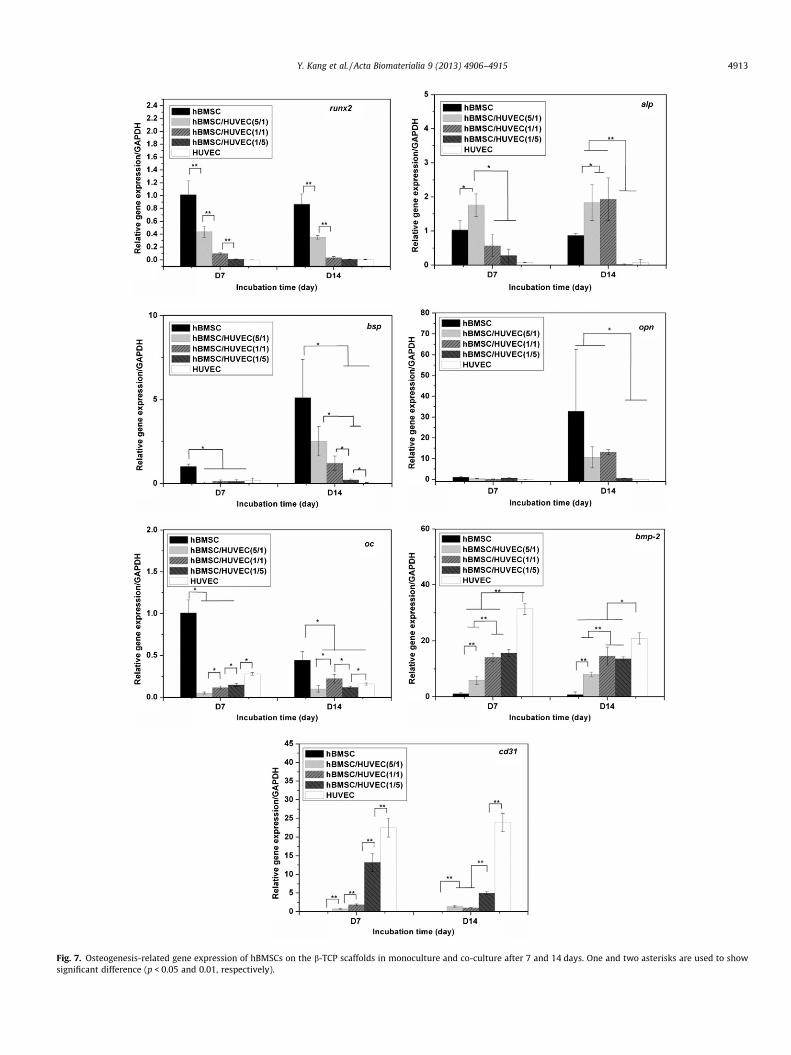
Fig. 7. Osteogenesis-related gene expression of hBMSCs on the b-TCP scaffolds in monoculture and co-culture after 7 and 14 days. One and two asterisks are used to showsignificant difference (p < 0.05 and 0.01, respectively).
Y. Kang et al. / Acta Biomaterialia 9 (2013) 4906–4915 4913

4914 Y. Kang et al. / Acta Biomaterialia 9 (2013) 4906–4915
that the HUVECs in co-culture may not stimulate hBMSCs prolifer-ation; meanwhile, hBMSCs could suppress HUVEC expansion dueto their high proliferative capacity in co-culture.
We further investigated the angiogenic potential of co-culturedcells in vitro on the b-TCP ceramic scaffolds. Cell adhesion mole-cule PECAM-1 expression at the cell–cell interface could be usedto indicate the microcapillary-like structure or lumina. Cell adhe-sion molecule PECAM-1 expressed by HUVECs is known to be cru-cial for vessel formation and maintenance [50]. Our results showthat in the HUVEC monoculture, a large amount of PECAM-1expression can be clearly observed on the scaffolds shown inFig. 5. Some elongated networks but no obvious lumina were ob-served during this experimental period. The ability of HVUECs toform lumina may depend on the properties of the extracellular ma-trix, which can affect the migration of HUVECs [51]. Co-culturingendothelial cells with bone-forming cells may facilitate the tubeformation in vitro on scaffolds since the bone-forming cells canproduce cytokines and angiogenic growth factors such as VEGF[52,53]. Also, the extracellular matrix produced by bone-formingcells may promote the formation of microcapillary-like structurewhen the cells co-cultured in direct contact [20,54]. Our data inco-culture did not show that hBMSCs promoted the tube formationof HUVECs on the scaffolds. Unlike the continuous PECAM-1expression by the monocultured HUVECs at day 14 of incubation,a lower, scattered PECAM-1 expression can be observed in thegroups of lower ratio of HUVECs (1:5 and 1:1). This is probably aresult of decreased HUVEC population and the separation of HU-VECs by hBMSCs. In the co-culture groups, hBMSCs and HUVECswere loaded simultaneously onto the porous scaffolds. A higher ra-tio of hBMSCs (5:1) in the mixed cell suspension could separateindividual HUVECs, leading to the inability of HUVECs to contactand form a microcapillary-like structure.
The real-time PCR studies indicated that ALP gene expressionlevels in the co-culture group were significantly increased, butmost of other genes were decreased. It is worth noting that weare unable to distinguish the contribution of individual cell typebecause gene expression in this study came from the total mRNAof two types of cells. As the expression level of hBMSCs is a partof the total expression level in the co-culture, therefore geneexpression of all these osteogenic markers of hBMSCs specificallyin co-culture could be higher [29]. Although further evaluationand new strategies will be needed to investigate the interactionsbetween mesenchymal stem cells and endothelial cells on the bio-degradable b-TCP, these present results implied that b-TCP scaffoldwith 3-D spatial features could be a useful platform to furtherinvestigate the interactions between the two cell types in 3-Dstructures for better understanding of bone regeneration, as thisb-TCP ceramic scaffold can provide biocompatible and suitable sur-face properties for hBMSC and HUVEC development.
5. Conclusions
In the present study we examined the effect of an intercon-nected, macroporous, biodegradable b-TCP scaffold on cell behav-iors of HUVECs and hBMSCs in both monoculture and co-culture.Our results demonstrate that b-TCP scaffolds supported the attach-ment and proliferation of HUVECs and hBMSCs in both monocul-tures and co-cultures. b-TCP scaffolds stimulated ALP activity ofboth hBMSCs and HUVECs. In co-cultures, HUVECs enhanced thevery early osteogenic differentiation of hBMSCs. Meanwhile, theformation of an elongated network-like structure was observedin vitro on the b-TCP scaffolds. This b-TCP scaffold could providea 3-D platform for studying interactions between multiple cells in-volved in bone regeneration. In particular, vascularization andosteogenesis of biodegradable b-TCP scaffolds can be promotedby manipulating structural and biological cues.
Acknowledgements
This work was supported by grants from the following agen-cies: NIH R01AR057837 (NIAMS), NIH R01DE021468 (NIDCR),DOD W81XWH-10-1-0966 (PRORP), W81XWH-10-200-10 (AirliftResearch Foundation), W81XWH-11-2-0168-P4 (Alliance of NanoHealth), and Wallace H. Coulter Foundation.
Appendix A. Figures with essential colour discrimination
Certain figures in this article, particularly Figs. 2, 4, and 5, aredifficult to interpret in black and white. The full colour imagescan be found in the on-line version, at http://dx.doi.org/10.1016/j.actbio.2012.08.008.
Appendix B. Supplementary data
Supplementary data associated with this article can be found, inthe online version, at http://dx.doi.org/10.1016/j.actbio.2012.08.008.
References
[1] Dietmar WH. Scaffolds in tissue engineering bone and cartilage. Biomaterials2000;21:2529–43.
[2] Kalita SJ, Bhardwaj A, Bhatt HA. Nanocrystalline calcium phosphate ceramicsin biomedical engineering. Mater Sci Eng C 2007;27:441–9.
[3] Descamps M, Duhoo T, Monchau F, Lu J, Hardouin P, Hornez JC, et al.Manufacture of macroporous [beta]-tricalcium phosphate bioceramics. J EurCeram Soc 2008;28:149–57.
[4] Pittenger MF, Mackay AM, Beck SC, Jaiswal RK, Douglas R, Mosca JD, et al.Multilineage potential of adult human mesenchymal stem cells. Science1999;284:143–7.
[5] Chai YC, Roberts SJ, Desmet E, Kerckhofs G, van Gastel N, Geris L, et al.Mechanisms of ectopic bone formation by human osteoprogenitor cells on CaPbiomaterial carriers. Biomaterials 2012;33:3127–42.
[6] Kasten P, Vogel J, Luginbühl R, Niemeyer P, Tonak M, Lorenz H, et al. Ectopicbone formation associated with mesenchymal stem cells in a resorbablecalcium deficient hydroxyapatite carrier. Biomaterials 2005;26:5879–89.
[7] Kruyt MC, de Bruijn JD, Wilson CE, Oner FC, van Blitterswijk CA, Verbout AJ,et al. Viable osteogenic cells are obligatory for tissue-engineered ectopic boneformation in goats. Tissue Eng 2003;9:327–36.
[8] Arinzeh TL, Tran T, McAlary J, Daculsi G. A comparative study of biphasiccalcium phosphate ceramics for human mesenchymal stem-cell-induced boneformation. Biomaterials 2005;26:3631–8.
[9] Dong J, Uemura T, Shirasaki Y, Tateishi T. Promotion of bone formation usinghighly pure porous b-TCP combined with bone marrow-derivedosteoprogenitor cells. Biomaterials 2002;23:4493–502.
[10] Grynpas MD, Pilliar RM, Kandel RA, Renlund R, Filiaggi M, Dumitriu M. Porouscalcium polyphosphate scaffolds for bone substitute applications in vivostudies. Biomaterials 2002;23:2063–70.
[11] Jafarian M, Eslaminejad MB, Khojasteh A, Mashhadi Abbas F, Dehghan MM,Hassanizadeh R, et al. Marrow-derived mesenchymal stem cells-directed boneregeneration in the dog mandible: a comparison between biphasic calciumphosphate and natural bone mineral. Oral Surg Oral Med O 2008;105:e14–24.
[12] Gerber HP, Vu TH, Ryan AM, Kowalski J, Werb Z, Ferrara N. VEGF coupleshypertrophic cartilage remodeling, ossification and angiogenesis duringendochondral bone formation. Nat Med 1999;5:623–8.
[13] Grellier M, Bordenave L, Amedee J. Cell-to-cell communication betweenosteogenic and endothelial lineages: implications for tissue engineering.Trends Biotechnol 2009;27:562–71.
[14] McCarthy I. The physiology of bone blood flow: a review. J Bone Joint Surg Am2006;88(Suppl. 3):4–9.
[15] Rose FR, Cyster LA, Grant DM, Scotchford CA, Howdle SM, Shakesheff KM. Invitro assessment of cell penetration into porous hydroxyapatite scaffolds witha central aligned channel. Biomaterials 2004;25:5507–14.
[16] Ishaug SL, Crane GM, Miller MJ, Yasko AW, Yaszemski MJ, Mikos AG. Boneformation by three-dimensional stromal osteoblast culture in biodegradablepolymer scaffolds. J Biomed Mater Res 1997;36:17–28.
[17] Rouwkema J, Rivron NC, van Blitterswijk CA. Vascularization in tissueengineering. Trends Biotechnol 2008;26:434–41.
[18] Smith MK, Peters MC, Richardson TP, Garbern JC, Mooney DJ. Locally enhancedangiogenesis promotes transplanted cell survival. Tissue Eng 2004;10:63–71.
[19] Klenke FM, Liu Y, Yuan H, Hunziker EB, Siebenrock KA, Hofstetter W. Impact ofpore size on the vascularization and osseointegration of ceramic bonesubstitutes in vivo. J Biomed Mater Res A 2008;85:777–86.
[20] Unger RE, Sartoris A, Peters K, Motta A, Migliaresi C, Kunkel M, et al. Tissue-likeself-assembly in cocultures of endothelial cells and osteoblasts and theformation of microcapillary-like structures on three-dimensional porousbiomaterials. Biomaterials 2007;28:3965–76.
Y. Kang et al. / Acta Biomaterialia 9 (2013) 4906–4915 4915
[21] Yu H, VandeVord PJ, Mao L, Matthew HW, Wooley PH, Yang SY. Improvedtissue-engineered bone regeneration by endothelial cell mediatedvascularization. Biomaterials 2009;30:508–17.
[22] Zhou J, Lin H, Fang T, Li X, Dai W, Uemura T, et al. The repair of large segmentalbone defects in the rabbit with vascularized tissue engineered bone.Biomaterials 2010;31:1171–9.
[23] Liu Y, Kim JH, Young D, Kim S, Nishimoto SK, Yang Y. Novel template-castingtechnique for fabricating beta-tricalcium phosphate scaffolds with highinterconnectivity and mechanical strength and in vitro cell responses. JBiomed Mater Res A 2010;92:997–1006.
[24] Kang Y, Kim S, Khademhosseini A, Yang Y. Creation of bony microenvironmentwith CaP and cell-derived ECM to enhance human bone-marrow MSC behaviorand delivery of BMP-2. Biomaterials 2011;32:6119–30.
[25] Kang Y, Scully A, Young DA, Kim S, Tsao H, Sen M, et al. Enhanced mechanicalperformance and biological evaluation of a PLGA coated beta-TCP compositescaffold for load-bearing applications. Eur Polym J 2011;47:1569–77.
[26] Zhao J, Zhang N, Prestwich GD, Wen X. Recruitment of endogenous stem cellsfor tissue repair. Macromol Biosci 2008;8:836–42.
[27] Yim EK, Wan AC, Le Visage C, Liao IC, Leong KW. Proliferation anddifferentiation of human mesenchymal stem cell encapsulated inpolyelectrolyte complexation fibrous scaffold. Biomaterials 2006;27:6111–22.
[28] Anderson JM, Vines JB, Patterson JL, Chen H, Javed A, Jun HW. Osteogenicdifferentiation of human mesenchymal stem cells synergistically enhanced bybiomimetic peptide amphiphiles combined with conditioned medium. ActaBiomater 2011;7:675–82.
[29] Hofmann A, Ritz U, Verrier S, Eglin D, Alini M, Fuchs S, et al. The effect ofhuman osteoblasts on proliferation and neo-vessel formation of humanumbilical vein endothelial cells in a long-term 3D co-culture onpolyurethane scaffolds. Biomaterials 2008;29:4217–26.
[30] Livak KJ, Schmittgen TD. Analysis of relative gene expression data using real-time quantitative PCR and the 2-[Delta][Delta]CT method. Methods2001;25:402–8.
[31] Pham QP, Kurtis Kasper F, Scott Baggett L, Raphae RM, Jansen JA, Mikos AG. Theinfluence of an in vitro generated bone-like extracellular matrix onosteoblastic gene expression of marrow stromal cells. Biomaterials2008;29:2729–39.
[32] Sommer B, Bickel M, Hofstetter W, Wetterwald A. Expression of matrixproteins during the development of mineralized tissues. Bone1996;19:371–80.
[33] Arafat MT, Lam CXF, Ekaputra AK, Wong SY, Li X, Gibson I. Biomimeticcomposite coating on rapid prototyped scaffolds for bone tissue engineering.Acta Biomater 2011;7:809–20.
[34] Kanczler JM, Oreffo RO. Osteogenesis and angiogenesis: the potential forengineering bone. Eur Cell Mater 2008;15:100–14.
[35] Towler DA. The osteogenic-angiogenic interface: novel insights into thebiology of bone formation and fracture repair. Curr Osteoporos Rep2008;6:67–71.
[36] Finkenzeller G, Arabatzis G, Geyer M, Wenger A, Bannasch H, Stark GB. Geneexpression profiling reveals platelet-derived growth factor receptor alpha as atarget of cell contact-dependent gene regulation in an endothelial cell-osteoblast co-culture model. Tissue Eng 2006;12:2889–903.
[37] Choong CS, Hutmacher DW, Triffitt JT. Co-culture of bone marrow fibroblastsand endothelial cells on modified polycaprolactone substrates for enhancedpotentials in bone tissue engineering. Tissue Eng 2006;12:2521–31.
[38] Guillotin B, Bareille R, Bourget C, Bordenave L, Amedee J. Interaction betweenhuman umbilical vein endothelial cells and human osteoprogenitors triggerspleiotropic effect that may support osteoblastic function. Bone2008;42:1080–91.
[39] Scherberich A, Galli R, Jaquiery C, Farhadi J, Martin I. Three-dimensionalperfusion culture of human adipose tissue-derived endothelial andosteoblastic progenitors generates osteogenic constructs with intrinsicvascularization capacity. Stem Cells 2007;25:1823–9.
[40] Rouwkema J, de Boer J, Van Blitterswijk CA. Endothelial cells assemble into a 3-dimensional prevascular network in a bone tissue engineering construct.Tissue Eng 2006;12:2685–93.
[41] Villars F, Guillotin B, Amedee T, Dutoya S, Bordenave L, Bareille R, et al. Effectof HUVEC on human osteoprogenitor cell differentiation needs heterotypic gapjunction communication. Am J Physiol Cell Physiol 2002;282:C775–85.
[42] Vater C, Kasten P, Stiehler M. Culture media for the differentiation ofmesenchymal stromal cells. Acta Biomater 2011;7:463–77.
[43] Moghe PV, Berthiaume F, Ezzell RM, Toner M, Tompkins RG, Yarmush ML.Culture matrix configuration and composition in the maintenance ofhepatocyte polarity and function. Biomaterials 1996;17:373–85.
[44] Unger RE, Huang Q, Peters K, Protzer D, Paul D, Kirkpatrick CJ. Growth ofhuman cells on polyethersulfone (PES) hollow fiber membranes. Biomaterials2005;26:1877–84.
[45] Unger RE, Peters K, Wolf M, Motta A, Migliaresi C, Kirkpatrick CJ.Endothelialization of a non-woven silk fibroin net for use in tissueengineering: growth and gene regulation of human endothelial cells.Biomaterials 2004;25:5137–46.
[46] Barradas AMC, Fernandes HAM, Groen N, Chai YC, Schrooten J, van de Peppel J,et al. A calcium-induced signaling cascade leading to osteogenicdifferentiation of human bone marrow-derived mesenchymal stromal cells.Biomaterials 2012;33:3205–15.
[47] Valerio P, Pereira MM, Goes AM, Leite MF. Effects of extracellular calciumconcentration on the glutamate release by bioactive glass (BG60S)preincubated osteoblasts. Biomed Mater 2009;4:045011.
[48] Marie PJ. The calcium-sensing receptor in bone cells: a potential therapeutictarget in osteoporosis. Bone 2010;46:571–6.
[49] Wang J, Ye Y, Tian H, Yang S, Jin X, Tong W, et al. In vitro osteogenesis ofhuman adipose-derived stem cells by coculture with human umbilical veinendothelial cells. Biochem Biophys Res Commun 2011;412:143–9.
[50] Simon AM, McWhorter AR. Vascular abnormalities in mice lacking theendothelial gap junction proteins connexin37 and connexin40. Dev Biol2002;251:206–20.
[51] Soucy PA, Romer LH. Endothelial cell adhesion, signaling, and morphogenesisin fibroblast-derived matrix. Matrix Biol 2009;28:273–83.
[52] Deckers MM, van Bezooijen RL, van der Horst G, Hoogendam J, van Der Bent C,Papapoulos SE, et al. Bone morphogenetic proteins stimulate angiogenesisthrough osteoblast-derived vascular endothelial growth factor A.Endocrinology 2002;143:1545–53.
[53] Furumatsu T, Shen ZN, Kawai A, Nishida K, Manabe H, Oohashi T, et al. Vascularendothelial growth factor principally acts as the main angiogenic factor in theearly stage of human osteoblastogenesis. J Biochem 2003;133:633–9.
[54] Stahl A, Wenger A, Weber H, Stark GB, Augustin HG, Finkenzeller G. Bi-directional cell contact-dependent regulation of gene expression betweenendothelial cells and osteoblasts in a three-dimensional spheroidal coculturemodel. Biochem Biophys Res Commun 2004;322:684–92.