pathology of parasitic diseases
TRANSCRIPT

Department of Pathology,
Faculty of Veterinary Medicine,
Zagazig University, Egypt
Pathology of Veterinary Parasitic Diseases
BY
Professor Dr. Mohamed Hamed [email protected]
+20124067373

Diseases caused by parasitesThe parasitic diseases are classified into 2 categories:
I-Diseases cased by Protozoa.
II-Diseases caused by Helminths and Arthropods.
N.B:
Infestation: Parasitic invasion to the body organs without
multiplication and may be decreased.
Infection: Invasion and multiplication of microorganisms in the
body tissue.
Inoculation: Introduction of microorganisms or other substance in
the body tissue.
I-Diseases caused by protozoaProtozoa: They are single-celled eukaryotic animals.
Classification of the Protozoa:
Kingdom: Protista.
Subkingdom: Protozoa.
The protozoa are divided into 2 main phyla that divided
into subphylum.

Phylum Sarcomastigophora Ciliophora
Subphylum 1-Mastigophora
2-Sarcodina
3-Apicomplixa (sporozoa)
4-Microspora
It is protozoa with cilia for
locomotion e.g. Blantidium
Definitions:
Mastigophora: Protozoa with one or more flagella (Giardia, Trichomonas).
Sarcodina: Protozoa with pseudopodia for locomotion e.g. Entameba.
Apicomplixa: Protozoa without locomotor organs and produce spores at
the end life span. They are either:
A-Coccidia: It has direct life span e.g. Eimeria, Isospora.
B-Hemospordia: It has indirect life span (has IMH) e.g.
Babesia, Thileria.
Microspora: Protozoa have unique spores with 1–6 polar filaments
e.g. Encephalitozoon.

Tissue response in protozoan infection1-Necrosis and degeneration due to production of toxic substances.
2-Tissue damage due to activation of complement system through
immediate hypersensitivity type II as in African trypanosomiasis
3-Granulomatous inflammation and caseous necrosis results from
cellular immunity due to intracellular invasion of some protozoa as
in case of leishmaniasis
4-Vasculitis results from formation of immune complex which
deposit under the endothelial lining in kidney and other tissue.
CoccidiosisDefinition: It is a disease caused by Eimeria or Isospora. There are 3 types:
1-Enteric Coccidiosis: characterized by dysentery, dehydration and death.
2-Hepatic Coccidiosis: It is usually in rabbits less than 3 months old and
characterized by enlarged abdomen and obstructive jaundice.
3-Renal Coccidiosis: in young geese and caused by Eimeria truncate or in
equine, mice and guinea pigs and caused by Klossiella species.

Pathognomonic Lesions:
Enteric Coccidiosis:In Schizogony Stage:
1-The intestine is hemorrhagic and eroded or ulcerated.
2- Excessive epithelial necrosis.
3-Petecial hemorrhages and pin point white foci are seen the
serosa, particularly with large schizonts.
4- Hemorrhagic cores in the cecum of chickens.
5- Coccidia in various stages (schizogony and gametogony) are
adjacent to hemorrhagic and eroded areas.
6- Villous atrophy as a result of loss of epithelial cells.
7-The schizonts are oval and with basophilic banana-shaped
merozoites.
In Gametogenesis:
1-Regeneration and hyperplasia of the lining epithelium.
2-lymphocytes and plasma cells infiltrations (predominant).
3-Developmental stages of Eimeria (macro and micro gametes).

Hepatic Coccidiosis: (in rabbits less 3 months old):
1-The liver is yellowish and severely enlarged.
2-Adenomatous hyperplasia in the lining epithelium of bile ducts.
2-developmental stages of E. stiedae are detected.
3-lymphocytes and plasma cells infiltration are seen in the portal
areas.
Renal Coccidiosis: (in young geese).
1-Developmental stages of Eimeria truncate in the epithelial cells of
convoluted tubules without inflammation.
2- Slight destruction of the renal epithelial cells.
3-The protozoan maybe hyalinized and shed in the lumen of the
tubules.
NB: Klossiella
1-Tiny gray foci on cortical surface of the kidneys.
2-These foci are areas of necrosis.
3- Perivascular infiltration of lymphocytes and histiocytes.
4-An increase of interstitial fibroblasts.
5-Numerous sporoblasts and sporocysts in the tubular epithelium.

Types Schizogony Gametogony1-Enteric Intestinal epithelium Intestinal epithelium
2-Hepatic Bile duct epithelium Bile duct epithelium
3-Renal
Glomerular epithelium (BC) and endothelium Tubular epithelium4-Klossiella
NB:Sporogony: It is the sporulation of the oocysts outside the host
(formation of sporocysts).
In Rabbit: The sporozoites reach the bile ducts via the portal veins or
lymphatics.
In sheep and goats: The Eimeria are found in the gallbladder and
mesenteric veins.

Organ : Chicken digestive tract Disease : Coccidiosis.Macro : Inflamed small intestine due to Eimeria nicatrix (left), and
inflamed ceca due to Eimeria tenella (right).

Organ : Ceca of chicken.Disease : Coccidiosis (Eimeria tenella-experimental infection).Macro : Inflamed, hemorrhagic and necrotic ceci.

Organ : Small intestine (cross-section).Stain : H&E.Disease : Coccidiosis (Eimeria nicatrix).Micro : Extensive hemorrhage and necrosis.

Organ : Small intestine of a chicken.Stain : H&E.Disease : Coccidiosis (Eimeria nicatrix).Micro : A higher magnification to show the numerous schizonts,
hemorrhage and necrosis in submucosa.

Organ : Small intestine of a chicken.Stain : H&E.Disease : Coccidiosis (Eimeria nicatrix).Micro : Mature schizonts containing merozoites.

Organ : Small intestine of a chickenStain : H&E.Disease : Coccidiosis (Eimeria nicatrix).Micro : Microgammonts containing microgametes.

Organ : Kidney of goose.Stain : H&E.Disease : Coccidiosis (EImeria truncata).Micro : Gamonts in tubular eoithelial cells.

Organ : Kidney of a goose.Stain : H&E.Disease : Coccidiosis (Eimeria truncata).Micro : A higher magnification of gamonts.

Organ : Liver of a rabbit.Stain : H&E.Disease : Hepatic coccidiosis.Micro : Dilated bile-duct due to Eimeria stiedae.

Organ : Liver of a rabbit.Stain : H&E.Disease : Hepatic coccidiosis.Micro : Numerous macrogamonts of
Eimeria stiedae in epithelium of bile-duct.

Organ : Liver of a rabbit.Stain : H&E.Disease : Hepatic coccidiosis.Micro : A high magnification to show numerous macrogamonts of
Eimeria stiedae in epithelium of bile-duct.

Organ : Kidney of a horse.Stain : H&E.Disease : Klosiellosis.Micro : Young budding sporont, sporont with radiating
sporoblasts and mature sporocystwithin tubular epithelium.

Organ : Kidney of a horse.Stain : H&E.Disease : Klosiellosis.Micro : A high magnification of sporont, sporont with
radiating sporoblasts and mature sporocystwithin tubular epithelium.

ToxoplasmosisDefinition: It is acute or chronic disease of cats (definitive host),
mammals and birds (IMH).
Causes: Tissue phase of Toxoplasma gondii.
NB: The infective stage is the oocysts or tissue containing bradyzoites or
tachyzoites. The tachyzoites can cross the placenta and pass with milk
and semen.
Endodyogony: The tachyzoites of toxoplasma multiply by internal
budding into 2 organisms.
Endopolygony: The tachyzoites of toxoplasma multiply by internal
budding into several organisms.
Parasitemia: It the circulation of parasite in the blood as toxoplasma.
Pathognomonic Lesions:
In Definitive Host (Cats): They are similar to coccidiosis (enteric type).
1-Necrosis of the submucosal lymphoid follicles and ulceration.
2-Sexual and asexual developmental stages are seen in the lining epithelium.
3-Large granulomatous nodules in muscularis. They are consisted of
i-Tachyzoites
ii-Excessive granulation tissues.
iii-Macrophages, lymphocytes and plasma cells infiltrations.

In IMH (mammals and Birds):Brain:
1-Diffuse non-suppurative meningoencephalitis. It is characterized by
mononuclear cell infiltration and leukoencephalomalacia.
2-Tachyzoites (crescentic or round bodies) inside the neurons and
astrocytes.
3-Tissue cysts (containing more than 50 bradyzoites without
inflammatory cells.
4-Small areas of necrosis and calcification.
Liver:
1-Focalcoagulative necrosis.
2-Tachyzoites inside the hepatic and kupffer cells.
3-Tissue cysts containing bradyzoites are seen.
Lungs:
1-Small gray (tumor-like) nodules on the lungs.
2-Tachyzoites inside the type I and II pneumocytes and bronchial
epithelium.
3-Focal necrosis with alveolar fetalization or pulmonary adenomatosis.
4-Lymphocytes and macrophages infiltration and inside the alveoli.

Lymph nodes:
1-The regional Lns are enlarged, firm and congested.
2-Focal necrosis. 3-Tachyzoites inside the endothelial cells of veins.
4-Hyperplasia of the lymphocytes and REC.
Myocardium:
1-Tachyzoites inside the cardiac muscles. 2-Round cells infiltration.
Placenta:
1-Focal necrosis and calcification.
2-Tachyzoites are seen free or inside the trophoblasts.
3-Abortion with or without invasion of fetus.
4-The affected fetus show brain lesions.
Eye:
1-Granulomatous chorioretinitis (choroid and retina) characterized by
infiltration with macrophages and lymphocytes.
NB:Hammondiasis: Hammondia hammondi
It is a non-pathogenic coccidian of cats similar to T. gondii. Its life cycle and
structure are essentially as those of T. gondii with the following exceptions:
1-It has no extraintestinal cycle in cat and dogs (definitive hosts).
2-intermediate hosts (rodents) infected only by ingestion of oocysts
3-The tachyzoites proliferate in the lymphoid cells and the bradyzoites
are limited in skeletal and cardiac muscles (forming tissue cysts, as in
toxoplasma).

Organ : Small intestine of a cat.Stain : H&E.Disease : Txoplasmosis.Micro : Numerous asexual and sexual stages in epithelial cells with
microgamont in cell and meront in cell

Organ : Brain of mouse.Stain : PASH.Disease : Toxoplasmosis.Micro : Four cysts with PAS-positive bradyzoites.

Organ : Skeletal muscle of mouse.Stain : PASH. Disease : Hammondosis.Micro : Hammondia hammondi cyst.

Neosporiasis:Neospora caninum affects the dogs of all ages (definitive host) and
similar to toxoplasma in the IMH (cattle, sheep, goats, horses, dogs, and
deer); the wall of the cyst is thicker and only found in CNS.
Pathognomonic Lesions:
1-Multifocal non suppurative necrotizing encephalomyelitis.
2-Focal gliosis, lymphoid cuffing, and hydrocephalus.
3-Numerous tachyzoites found in the brain of aborted feti.
4-Tissue cysts containing bradyzoites are detected in cerebrum.
5-Periportal hepatitis with focal hepatocellular necrosis.
6-Focal non-suppurative myocarditis.
7-The placenta show focal necrosis, tachyzoites and neospora cysts.
Caryospora:It affects the dogs and causes pyogranulomatous dermatitis and
lymphadenitis.

Organ : Brain of aborted bovine fetus.Stain : H&E.Disease : Neosporosis.Micro : A focus of central necrosis, surrounded by
inflammatory cells.

Organ : Spinal cord of a congenitally-infected bovine-calf.Stain : H&E.Disease : Neosporosis.Micro : A thick-walled tissue-cyst in a neuron.

SarcosporidiosisIt is a disease characterized by tubular cysts in the striated muscles.
Causes: Sarcocystis species or Balbiania gigantica in the tongue and esophagus of sheep.
Intestinal Stage: Isospora (in final host).
Tissue Phase: Sarcocystis (in IMH).
Pathognomonic Lesions:1-Numerous intact Sarcocystis in the cardiac and skeletal muscles without
inflammatory reactions.
2-These cysts are variable in sizes with clear or hyaline wall and contain
strongly basophilic crescentic bradyzoites.
3-Hyaline degeneration, eosinophilic polymyositis and Zenker’s necrosis
are seen in some heavily parasitized muscles.
4-The ruptured cysts stimulate granulomatous reactions.
5-Focal leptomeningitis, non suppurative encephalitis, pneumonia, keratitis
and abortion are recorded in cattle, sheep and goats due to multiplication
of the merozoites in the endothelial cells of blood vessels and induce
vasculitis, thrombosis and necrosis of all tissue including placenta
(abortion). The placentas were thickened and edematous, and the caruncles
were atrophied.
6-Enlargement of the superficial cervical lymph nodes, serous atrophy and
edema of the body fat, hydrothorax, hydropericardium and ascites are
found.

Organ : Trachea of sheep. Disease : Sarcosporidiosis.Macro : Large sarcocysts.

Tissue : Muscle of bird.Disease : Sarcosporidiosis.Macro : Large sarcocysts.

Tissue : Placental lamina propria of a cow.Stain : H&E..Disease : Sarcosporidiosis.Micro : Numerous free and intracellular merozoites,
and immature meronts in area of necrosis.

Organ : Heart of a calf.Stain : H&E.Disease : Sarcosporidiosis.Micro : Immature sarcocyst, containing globular
immature metrocytes.

Tissue : Bovine mesenteric artery.Stain : H&E.Disease : Sarcosporidiosis.Micro : Multinucleated first generation meront of
S. bovifelis) protruding into lumen

Organ : Tongue of a calf.Stain : H&E.Disease : Sarcosporidiosis.Micro : Numerous basophilic mature intramuscular and
microscopic sarcocysts.

Organ : Tongue of a calf.Stain : H&E.Disease : Sarcosporidiosis.Micro : Numerous basophilic mature intramuscular and
microscopic sarcocysts.

Tissue : Esophageal muscle.Stain : H&E.Disease : Sarcosporidiosis.Micro : Thick-walled sarcocyst of Sarcocyctis .

BesnoitosisCauses: Besnoitia besnoiti similar to Sarcocystis.
Pathognomonic Lesions:
1-The skin is thick, wrinkled and hairless on legs, thighs and
scrotum.
2-Large cysts containing spores in the wall of small blood
vessels of skin and upper GIT.
3-Granulomatous reactions around the rupture cysts.
4-These cysts may present in cardiovascular system e.g.
Antelopes: in jugular vein and veins of limbs.
Impala: in S/C lymphatics and endocardium.
Enigmatic bodies: It is membrane-bounded spindle bodies with
electron-dense core in the tachyzoites of Besnoitia.

Organ : Hind leg .Disease : Besnoitiosis.Macro : Numerous white raised subcutaneous cysts of Besnitia.

Organ : Skin of an ox.Stain : H&E.Disease : Besnoitiosis (globidiosis).Micro : Numerous cysts of Benoitia besnoiti in dermis.

Organ : Skin of an ox.Stain : H&E.Disease : Besnoitiosis (globidiosis).Micro : A high magnification of a cyst of Benoitia
besnoiti in dermis.

Tissue : Jugular vein.Stain : H&E.Disease : Besnoitiosis.Micro : Numerous Besnoitia sp. cysts protruding into lumen.

Organ : Spleen.Stain : H&E.Disease : Besnoitiosis.Micro : Numerous cysts of Besnoitia sp.

CryptosporidiosisIt is a protozoal disease of most domestic and wild animals, birds,
fish and reptiles which caused by Cryptosporidium parvum.
Pathognomonic Lesions:
1-Small basophilic organisms on the villi or embedded inside the
enterocytes of the small intestine, colon and abomasum.
2-Atrophy and fusion of intestinal villi besides dilation of crypt.
3-The lamina propria is infiltrated with lymphocytic.
4-The parasite is stained by Giemsa or cold Ziehl-Neelsen stains.
Encephalitozoonosis
(Nosematosis)It is a subclinical protozoal disease of human, rabbits, dogs and some
laboratory animals. It causes encephalitis, hepatitis and nephritis.
Causes: Encephalitozoon caniculi.
Pathognomonic Lesions:
1-Large areas of necrosis at cerebral cortex.
2-The parasites (Encephalitozoon) in parasitophorous vacuoles.
3-Granulomatous reaction (Epithelioid cells and macrophages)
around the necrosis.
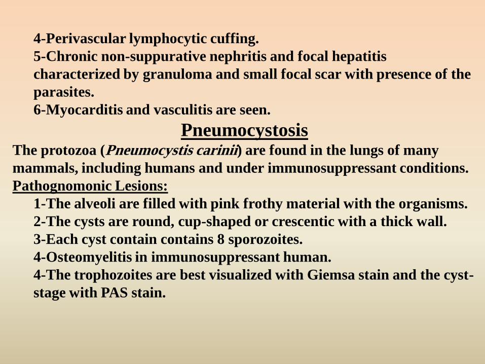
4-Perivascular lymphocytic cuffing.
5-Chronic non-suppurative nephritis and focal hepatitis
characterized by granuloma and small focal scar with presence of the
parasites.
6-Myocarditis and vasculitis are seen.
PneumocystosisThe protozoa (Pneumocystis carinii) are found in the lungs of many
mammals, including humans and under immunosuppressant conditions.
Pathognomonic Lesions:
1-The alveoli are filled with pink frothy material with the organisms.
2-The cysts are round, cup-shaped or crescentic with a thick wall.
3-Each cyst contain contains 8 sporozoites.
4-Osteomyelitis in immunosuppressant human.
4-The trophozoites are best visualized with Giemsa stain and the cyst-
stage with PAS stain.

Organ : Intestine.Stain : H&E.Disease : Cryptosporidiosis.Micro : Numerous developmental stages of Cryptosporidia.

Organ : Lung of horse. Stain : H&E.Disease : Pneumocystosis.Micro : Pneumocystis sp. in the lungs.

Organ : Lung of a horse.Stain : H&E.Disease : Pneumocystosis.Micro : Alveoli are filled with foamy eosinophilic material.

Organ : Lung of horse. Stain : H&E.Disease : Pneumocystosis.Micro : A high magnification of Pneumocystis sp. in the lungs.

Organ : Lung of a horse.Stain : Gomori methanamine silver (GMS).Disease : Pneumocystosis.Micro : Numerous darkly-stained ovoid organisms.

Organ: Brain.
Stain: H&E
Lesion: Encephalitozoonosis
Micro: Necrosis and Lymphocytic perivascular cuffing.
Organ : Brain of rabbitStain : Brown and Hopps tissue gram-stain.Disease : Encephalitozoonosis.Micro : Dark-stained immature spores.

Organ : Kidney of rabbitStain : H&E.Disease : Encephalitozoonosis.Micro : Lymphocytic interstitial nephritis.

Organ : Kidney of rabbitStain : Brown and Hopps tissue gram-stain.Disease : Encephalitozoonosis.Micro : Dark-stained immature spores.

Organ : Liver of rabbitStain : H&E.Disease : Encephalitozoonosis.Micro : Multifocal hepatic necrosis and leukocytes
infiltrations.

Organ : Liver.Stain : PAS.Disease : Encephalitozoonosis.Micro : Visible polar granule in each spore.

Organ : Muscle.Stain : Ziehl Nielsen acid-fast stain.Disease : Encephalitozoonosis.Micro : Dark-blue stained annular rings, and red-stained
(acid-fast) mature spores.

AmebiasisIt is protozoal disease of human and non-human primates and induces
severe dysentery.
Causes: Entameba histolytica and Entameba bovis.
Pathognomonic Lesions:
1-Flask-shape ulcers containing trophozoites in the intestinal mucosa.
2-Amebic abscesses (liquefactive necrosis without leukocytes) in the
liver and brain.
3-The lamina propria and submucosa show necrotic intestinal crypts
and lymphocytic infiltrations.
NB:
Amebic Meningoencephalitis (Naegleria; Acanthamoeba; Hartmannella)
1-Naegleria (N. fowleri): It induces purulent, hemorrhagic or necrotic
meningoencephalitis (gray matter).
2-Acanthamoeba: It induces granulomatous encephalitis around the
protozoa (microglia cells).

Preparation : Human fecal smear.Stain : Iron hematoxylin.Disease : Amoebiasis.Micro : Trophozoit of Entamoeba histolytica with small
endosome and chromatin plaques at periphery of nucleus.

Organ : Intestine of monkey.Stain : H&E.Disease : Amoebiasis.Micro : Trophozoite of Entamoeba histolytica

Organ : Heart of a dog.Stain : H&E.Disease : Amoebiasis.Micro : Trophozoite of Acanthamoeba sp.
With large dark endosome.

Organ : Brain of experimentally-infected mouse.Stain : H&E.Disease : Amoebiasis.Micro : Trophozoites of Acanthamoeba culbertsoni
with large endosome.

Organ : Brain of experimentally infected monkey.Stain : H&E.Disease : Amoebiasis.Micro : Trophozoit of Naegleria fowleri with large endosome.

GiardiasisIt is a protozoal disease affecting the small intestine and colon of
human and animals without clinical signs (may induce chronic
diarrhea).
Causes: G. bovis, G. cati, G. canis, G. muris.
Pathognomonic Lesions:
1-Minimal villous atrophy (stunting of the villi).
2-Leukocytic infiltration in the lamina propria.
3-Presence of the parasitic trophozoites or cysts on the
epithelial surface.
BalantidiasisBalantidium coli are naturally inhabitant of the digestive tract of
animals. The organisms may invade the intestinal mucosa, penetrating
into the submucosa, localized particularly in lymphoid nodules.
Extraintestinal spread to mesenteric lymph nodes, liver, pleura, lungs
and urogenital tract is reported. Balantidiasis as well as amebiasis is
characterized by diarrhea in calves. Enteritis, peritonitis, pneumonia
and lymphadenitis are seen associated with balantidium trophozoites.

Organ : Small intestine of canary.Stain : H&E.Disease : Giardiasis.Micro .: Numerous Giardia sp.

Organ : Small intestine of canary.Stain : H&E.Disease : Giardiasis.Micro : A higher power to show trophozoites of Giardia
sp. attached to intestinal villus.

Preparation : Human fecal smear.Stain : Iron –hematxylin stain.Disease : Giardiasis.Micro : Trophozoites of Giardia lamblia with two nuclei.

TrichomoniasisIt is a protozoal disease affecting cattle (bovine trichomoniasis) and
poultry (avian Trichomoniasis), and caused by the family
trichomonadidae.
I-Bovine Trichomoniasis: It is venereal disease (transmitted by coitus
and artificial insemination), caused by Trichomonas fetus. The
organisms live in preputial cavity of bull and vagina and uterus of cow.
Pathognomonic lesions:
1-Vaginitis, endometritis (closed pyometra) and placentitis
2-Early abortion (in the first half of pregnancy).
3-Pyogrnaulomatous bronchopneumonia with multinucleated
giant cells in the aborted fetus.
4-In bull: Balanoposthitis, seminal vesiculitis and epididymitis.
II-Avian Trichomoniasis: It is caused by Trichomonas gallinae and
affecting pigeons, turkey and chickens.
Pathognomonic lesions:
1-Masses of caseated material in mouth, throat and crop.
2-Necrosis and ulceration of crop.
3-Yellowish caseated nodules in the liver.

Preparation : Fecal smear of guinea pig.Stain : Protargol stain.Disease : Trichomoniasis.Micro : Trophozoites of Trichomonas with
undulating membrane and large single nucleus.

2-Histomoniasis (Blackhead disease or infectious
typhlohepatitis)It is a common protozoal disease of turkeys, chickens, peafowl, quail.
Etiology: Histomonas meleagridis
Cecal worm, Heterakis gallinarum and earthworms act as accessory hosts.
Lesions:
-The lesions are restricted to the ceca and liver.
-The ceca are enlarged; show ulceration and necrosis
(fibrinonecrotic) in the mucosa.
-The liver shows yellow necrotic areas surrounding a darker
hemorrhagic depressed center (saucer shaped depressions).
-The skin of affected birds is bluish-black in color, particularly on
the head (blackhead).
-Micro:
The lesions are represented by caseous necrosis with spherical
trophozoites of H. meleagridis (8 - 21 um in diameter) and
surrounded by granulomatous reaction in liver and ceca.

Liver and ceci infected with Histomonas meleagridis.Macro : Characteristic depressed liver-lesions (saucer
shaped depressions).

Organ : Liver of a turkey.Stain : H&E.Disease : Histomoniasis.Micro : Hepatic necrosis and faintly-stained
trophozoites (5-20 µ).

Organ : Liver.Stain : PAS.Disease : Histomoniasis.Micro : Red trophozoites (5-15 µ ) throughout
hepatic parenchyma.

Organ : Cecum.Stain : PAS.Disease : Histomoniasis.Micro : Red trophozoites ( 5-15 µ ) throughout wall.

TrypanosomiasisIt is exotic disease that occurs only in tropical Africa, India, and other
far-off places. This disease is most serious in humans and animals and
transmitted by arthropods, which act as biological vectors (IMH).
Specific infection Causes Affecting animals
1-Dourine T. equiperdum Equines (by coitus)
2-Nagana (Tsetse-fly
disease)
T. vivax, T. congolense and
T. brucei
Cattle
Horse, camels and dogs.
3-Surra T. evansi Horse (by horse fly).
4-Mal de Caderas T. equinum Equines
5-Chagas disease T. cruzi Human (by kissing bug)
Pathogenesis and Clinical Signs:
The pathogenesis of trypanosomiasis is based upon the presence of the
parasites in the blood circulation, where they block capillaries, causing
edema and hemolyze the erythrocytes, inducing anemia besides fever
during parasitemia, incoordination (obstruction to flow of CSF), coma
and death.

Pathognomonic lesions:A-Nagana: (Tsetse-fly disease):
I-Acute form: It characterized by sudden deaths with excessive
hemorrhages.
II-Chronic form:1-Emaciation and serous atrophy of fat.
2-The protozoa are detected in the blood.
3-Hemosiderosis in the spleen and liver, which become enlarged.
4-Hemorrhages on pericardium and endocardium.
5-Hydropericardium.
6-The lymph nodes are swollen and edematous.
B-Dourine: (venereal disease, transmitted by coitus).1-Ulceration (ulcerous plaques) in the genitalia and skin.
2-Edema in the genital tract and lower abdomen.
3-The protozoa are seen in the lesions (genitalia)
4-Mononuclear or granulomatous reaction.
C-Surra: (transmitted by horsefly).1-Emaciation with patches of alopecia and serous atrophy of fat.
2-Edema in the lower abdomen, limbs and thorax.
3-Hemosiderosis and icterus (hemolytic type).
4-Petichiae and ecchymoses on visible mucous membranes.

D-Chagas disease: (transmitted by kissing bugs).
The lesions are seen in different organs and amastigotes (trypanosomes)
are found in the blood and other body fluid as well as free in tissues.
1-The site of bite:-hard, red, painful and edematous mass.
-T. cruzi (leishmanial form) are present in the lesions.
2-Lymph nodes and Spleen:-Enlarged (due to hyperplasia of lymphoid tissues) and edematous.
-Intense histiocytes and giant cells proliferation contain T. cruzi.
-Microabscesses may be seen.
3-Heart:-Hydropericardium and severe myocarditis (due to penetration of the protozoa
the cardiac muscle fibers) which accompanied with chronic general passive
hyperemia.
4-Brain:-Non-suppurative meningoencephalitis, characterized by:
i-Edema and congestion of meninges.
ii-Perivascular lymphocytic cuffing and gliosis.
5-Testes:-Interstitial lymphocytic infiltration.
-The cytoplasm of the lining epithelium contained leishmanial form.

Trypanosomiasis (Dourine): anemic with ventral edema

Tissue : Peripheral blood-smear from infected mouseStain : Giemsa stain.Disease : Trypanosomiasis.Micro : Trypomastigotes of Trypanosoma brucei.

Organ : Heart of dog.Stain : H&E.Disease : Trypanosomiasis.Micro : Amastigotes of Trypanosoma cruzi.

Organ : Heart of a dog.Stain : H&E.Disease : Trypanosomiasis.Micro : Amastigotes of Trypanosoma cruzi, each
with a large basophilic kinetoplast.

Organ: Brain.
Stain: H&E.
Lesion: Trypanosomiasis.
Micro: Non suppurative meningoencephalitis.

LeishmaniasisVisceral and cutaneous Leishmaniasis are recorded in animals and caused by
Leishmania species. The disease is transmitted by sand flies.
Causes: Leishmania donovani, L. tropica and l. braziliensis.
Pathognomonic Lesions: leishmaniasis is divided into three major forms:
i-Visceral Leishmaniasis: (Kala-azar, Dum dum fever):
1-Severe emaciation and anemia (pallor mucous membranes).
2-The bone marrow become soft, red and heavily infiltrated with
macrophages contained the protozoa.
3-All viscera (lymph nodes, spleen and liver) are enlarged and massively
infiltrated with lymphocytes and macrophages whose cytoplasm is filled with
leishmaniae.
4-Ulceration of the intestine and immune complex glomerulonephritis.
ii-Cutaneous Leishmaniasis:1-Multiple nodules or ulcers of the skin that heal spontaneously by fibrous
tissue proliferation.
2-The dermis is infiltrated with macrophages accompanied by lymphocytes,
plasma cells and rarely eosinophils.
3-Numerous parasites are present within the macrophages.
iii-Mucocutaneous Leishmaniasis: It is similar to cutaneous form but the
lesions (chronic ulcers) occur at mucocutaneous junction or oral and nasal mucosa.

MalariaCauses: Plasmodium species (P. vivax, P. falciparum, …….).
Pathogenesis and Clinical Signs:
Two hosts are required:
1-Vertebrates (human or animals): in which schizogony takes place
in erythrocytes and other cells.
2-Invertebrate blood-sucking insects (mosquitoes): The life cycle is
started when a female mosquito penetrates the skin of vertebrate
host, introducing sporozoites into the peripheral circulation. The
macro- and microgametocytes ingested by female mosquitoes are
developed (in the stomach) into sporozoites which released into the
hemolymph and then migrated to salivary glands of mosquito. From
this site, the sporozoites are available to infect a new vertebrate host
when the female mosquito takes her blood meal.
Pathognomonic Lesions:
1-The destruction of parasitized erythrocytes (hemolysis and anemia).
2-Marked hepatosplenomegaly resulting from congestion, hemorrhage,
hyperplasia of REC, which contains the protozoa.
3--The erythrocytes show the parasites.

4-Hemozoin (brown pigments) is seen in tissue.
5-Brain hemorrhages
6-Thrombi and the blood vessels occluded with parasitized RBCs.
7-Multiple infarcts in the liver and affected organs.
8-Chronic immune complex glomerulonephritis.
HepatocystisIt is plasmodial protozoa, live in the erythrocytes of monkeys, particularly in
tropical countries in Africa and Asia.
Causes: Hepatocytis kochi, H. bouillezi, H. cercopitheci, H. simiae,
Pathognomonic Lesions:
1-The liver show large cysts contain merozoites (called
merocysts) with or without tissue reaction.
2-The most frequent reaction is granulomatous (giant cells and
macrophages besides few lymphocytes).
3-The parasites (trophozoites, macro- and microgametes) are
seen in the erythrocytes and hemozoin (malarial pigments) in the
tissue.

HepatozoonHepatozoon (H. canis) infect rodents, canines and others and
transmitted by arthropods. Schizonts found in the spleen and
liver and the gametocytes in leukocytes of such vertebrates.
Gametes released in the invertebrate gut and undergo
fertilization to form an ookinete, and then undergo sporogony to
form sporozoites. In the vertebrate host, release of sporozoites
and penetrate the intestinal wall to bloodstream and then to liver
and other organs.
Pathognomonic Lesions:
1-Emaciation and anemia (pallor mucous membranes).
2-Splenomegaly and hepatomegaly due to extensive
reticuloendothelial cell hyperplasia.
3-Focal necrosis with cellular infiltrations in the affected
organs.

Preparation : Blood-smear from a starling.Stain : Giemsa stain.Disease : Plasmodiosis.Micro : Gamont of Plasmodium reluctum.

Preparation : Blood-smear from Pigeon.Stain : Giemsa stain.Disease : Plasmodiosis.Micro : Gamont of Plasmodium reluctum.

Organ : Lung of a penguin’Stain : H&E.Disease : Plasmodiosis.Micro : Exoerythrocytic scizont of Plasmodium sp.

Tissue : Endothelial cell of turkey-brain.Stain : H&E.Disease : Plasmodiosis.Micro : Exaerythrocytic schizont of Plasmodium durae.

Preparation : Endothelium of turkey-brainStain : H&E.Disease : Plasmodiosis.Micro : Releasing of merozoites from schizont of
Plasmodium durae.

Organ : Endothelium of turkey-brainStain : H&E.Disease : Plasmodiosis.Micro : Infarct areas of coagulative necrosis.

Organ : Endothelium of turkey-brainStain : H&E.Disease : Plasmodiosis.Micro : Infarct areas of coagulative necrosiswith large schizont.

Organ : Liver of a monkey.Stain : H&E.Disease : Hepatocystosis.Micro : Merocyst of Hepatocystis sp.

Organ : Liver of a monkey.Stain : H&E.Disease : Hepatocystosis.Micro : A high magnification of merocyst to show
formation of merozoites.

Organ : Liver of a monkey.Stain : H&E.Disease : Hepatocystosis.Micro : A higher magnification of merozoit-formation in the
wall of a merocyst.

Organ : Liver of boa constrictor.Stain : H&E.Disease : HepatozoonMicro : Developing schizont of H. sp.

Organ : Spleen of a dog.Stain : H&E.Disease : Hepatozoon.Micro : Developing schizont of H. canis.

Preparation : Blood-smear from a dog.Stain : Giemsa stain.Disease : Hepatozoon.Micro : Macrogamete or microgametocyte of H.
canis in neutrophil.

Babesiosis(Piroplasmosis, Cattle tick fever, Red water fever)Babesiosis is an infectious disease caused by intra-erythrocytic protozoan parasite, usually transmitted
by ticks (IMH) and is capable of producing acute febrile and sometimes fatal infection.
Causes: B. bovis, B. bigemina, B. major and B. divergens.
Pathogenesis and Clinical Signs:The organisms live in erythrocytes and lead to intravascular hemolysis inducing
In early stage: the mucous membranes are pale (severe anemia).
In the terminal stages: the mm become yellow (severe jaundice), hemoglobinuria
(red water disease and Hb nephrosis) and ascites are seen. Incoordination, mania,
convulsions, paraplegia and coma are observed due to thrombosis (DIC).
Pathognomonic Lesions:
1-Emaciation of carcass with presence of ticks on the skin and serous
atrophy of fat.
2-The blood is watery (anemia) with red plasma.
3-Icterus is described as yellow mucous membranes.
4-The gallbladder is overdistended with dark green bile.
5-The liver and spleen are enlarged (presence of hemosiderosis).
6-Centrolobular hepatic necrosis (due to hypoxia from anemia).
7-The urinary bladder contains red urine (hemoglobin-urea).
8-Hb nephrosis 9-Hydro-thorax - peritoneum – pericardium.
10-Focal hemorrhages, thrombosis, and necrosis in the brain.

Preparation : Blood-smear.Stain : Neitz stain.Disease : Babesiosis.Micro : Intraerythrocytic Babesia sp.

Organ : Brain.
Disease: Babesiosis.
Macro: Hemorrhages in the brain

Organ : Brain.
Stain : H&E.
Disease: Babesiosis.
Micro : Fibrinous thrombosis in the b.v.

Organ : Intestine.
Stain : H&E.
Disease: Babesiosis.
Micro : Hemorrhagic enteritis.

Babesiosis: Splenomegaly

Theileriasis(Turning Disease, East Coast Fever)
Theileriasis is a hemoprotozoal infection, like Babesia, responsible for high mortality
among native and imported cattle and caused by protozoan parasites of the genus
Theileria and transmitted by ticks.
Causes: T. anulata (in Egypt), T. parva, T. mutans.
Pathogenesis and Clinical Signs:Theileria, after inoculated by ticks, invaded the lymphocytes and histiocytes, first in
regional lymph nodes, then throughout all lymph nodes, spleen, liver and circulating
cells. Macroschizonts (Koch’s blue bodies) and microschizonts are detected in these cells.
Released micromerozoites from the microschizonts could parasitize the erythrocytes.
Fever, rough hair coat, anemia, emaciation, delirium and coma (called turning disease)
are observed. Bloody diarrhea, hemoglobinuria, jaundice and corneal opacity are seen.
Pathognomonic Lesions: 1-severe emaciation of carcass.
2-All body lymph nodes are enlarged, edematous and hemorrhagic.
3-The spleen is enlarged.
4-Koch’s blue bodies (macroschizonts, contain 8 or more nuclei) are seen
in the lymphocytes in lymph nodes, spleen and blood.
5-Some blood vessels may be occluded with parasitized lymphocytes,
producing thrombosis and infarction in the brain (turning disease).
6-Hydrothorax and Hydropericardium.

Organ : Lymph-node and spleen of a cow.Disease : Theileriosis.Macro : Enlargement of spleen and lymph nodes.

Organ : Brain of a cow.Disease : Theileriosis.Macro : Hemorrhages and infarctions.

Preparation : Blood-smear.Stain : H&E.Disease : Theileriasis.Micro : Trophozoites of Theileria in erythrocytes.

Tissue : Splenic artery .Stain : H&E.Disease : Theileriosis.Micro : Schizonts of Theileria, occluding lumen.

Organ : Lung.Stain : H&E.Disease : Theileriasis.Micro : Macrophage containing schizont of Theileria
that seems to bud from endothelium of pulmonary vein.

Organ : Lung.Stain : H&E.Disease : Theileriasis.Micro : Pulmonary vessel with three developing schizonts.

Organ : Lung.Stain : H&E.Disease : Theileriosis.Micro ; Pulmonary vessel with developing and mature stages.

Organ : Kidney.Stain : H&E.Disease : Theileriasis.Micro : Capillary of a glomerular tuft with schizony.

Organ : Liver.Stain : H&E.Disease : Theileriasis.Micro : Several schizonts are seen in hepatic sinusoids.

Diseases Schizogony stage Gametogony stage
Toxoplasmosis
Intestinal epithelium of cats
Intestinal epithelium of catsTachyzoites in tissue cells (IMH)
Bradyzoites in tissue cyst (IMH)
Neosporiasis
Intestinal epithelium of dogs
Intestinal epithelium of dogsTachyzoites in tissue (IMH)
Bradyzoites in tissue cyst (IMH)
Hammondiasis
Intestinal epithelium of dogs
Intestinal epithelium of dogsTachyzoites in lymphoid (rodents)
Bradyzoites in tissue cyst in cardiac and skeletal
muscles (rodents)
Caryosporiasis Intestinal epithelium of rodents Intestinal epithelium of
rodents
Besnoitosis Cysts contain spores in b. vs., skin Tachyzoites in tissues
Sarcosporidiosis Isospora in definitive host (intestine) Isospora in definitive host
(intestine)Sporozoites in endothelium (IMH)
Bradyzoites in sarcocystis
Cryptosporidiosis Both are seen in intestinal epithelium of mammals and birds
Theileriasis In lymphocytes, erythrocytes, macs In ticks
Babesiosis In erythrocytes In tick

II-Diseases Caused by Helminths and ArthropodsThe parasitic helminths are classified into 3 phyla:
A-Phylum Platyhelminths (Flatworm):
e.g. Trematodes (Flukes).
Cestodes (Tapeworms).
B-Phylum Nemathelminths:
e.g. Nematodes (roundworms).
C-Phylum Acanthocephala:
e.g. Thorny-headed worms.
Pathological changes associated with helminths infection:
1-Mechanically interfere with functions:
i-Obstruct blood or lymph vessels.
ii-Obstruct ducts or tracts (intestine).
iii-Attach to or use functional tissue.
iv-Act as foreign bodies and displaces the normal structures.
2-Invade and destruct the tissue and cells.
3-Devour blood and cause anemia.
4-Devour tissues of hosts.
5-Secrete toxic products.
6-Introduce bacteria or other infections.
7-Induce neoplasms.
NB:
The eosinophils play a major role in the body defense against helminths besides the
basophils and mast cells.

AscariasisThe ascarids, among the most common intestinal parasites of young animals, can
cause digestive disturbance, enteritis and poor growth.
Causes:Species of Ascarid Definitive hosts
Toxocara lumbricoides (suum) Human and swine
Toxocara canis Dog
Toxocara cati Cat, lion, leopard
Toxocara leonine Dog, cat, lion, tiger, fox
Toxocara vitulorum Cattle
Parascaris equorum
Ascaridia galli
Equine
Chickens and turkeys
NB: Ascarid infection is rare or not present in sheep and goat.
The life cycle of ascarids has 3 types of migration:
1-Tracheal migration: When the embryonated eggs containing L2 are
ingested and they hatched (in the stomach and intestine) to second-stage
larvae. These larvae penetrate the intestinal wall and enter the portal vein
and invade the hepatic parenchyma, where they may molt to third-stage
larvae. Thereafter, these larvae enter the general circulation to reach the
lungs, bronchi, and trachea and then swallowed to intestine. In the intestine,
two additional molts occur before maturation to adult worms.

2-Intestinal migration: The second-stage larvae penetrate the intestinal
wall and do not migrate further. They undergo their molts here and reenter
the lumen as fourth-stage larvae, which mature to adults.
3-Somatic Migration: (Visceral Larva Migrans). It is the migration of the
ascarids larvae in paratenic hosts (abnormal hosts) without formation of
adults in the intestine. When the larvae reach the general circulation, they
invade all body tissues (brain, eyes, placenta, mammary gland and others)
without further development. These larvae either pass with milk or across
the placenta to infect the newborn.
Pathognomonic Lesions:
In early stages (visceral larva migrans):
1-The ascaris larvae are seen in the body tissues (liver, kidneys, lungs, heart,
brain, spleen and others).
2-Several necrotic migratory tracks are represented by parasitic larvae
embedded in cellular debris, extravasated erythrocytes and few leukocytes.
3-The degenerated larvae are seen inside granulomatous nodules consisting
of eosinophils, giant cells, macrophages and lymphocytes.
4-Focal coagulative necrosis and hemorrhages.
5-The larvae are seen inside the portal blood vessels, bronchi and glomeruli.
In late stages (Adult):
1-Numerous adult worms in the small intestine.
2-Catarrhal enteritis with eosinophils infiltrations.

3-Obstruction of the bile or pancreatic ducts with adult worms,
producing obstructive jaundice.
4-Peritonitis due to penetration the wall of intestine.
Ancylostomiasis
Hookworm diseaseHookworms are important parasites of mammals, and in some parts of the
world, they produce widespread disease in humans. They inhabit the small
intestine and feed on blood producing anemia.
Causes: Ancylostoma species.
Pathogenesis and Clinical Signs:
The adult worms attach to the intestinal mucosa with hook like structures
(hookworms) leading to hemorrhages and feed on blood with hypoproteinemia
and anemia. There are two types of anemia:
i-In acute phase is hemorrhagic (normocytic normochromic)
ii-In chronic phase is iron-deficiency anemia (microcytic hypochromic).
Pathognomonic Lesions:
1-Hemorrhagic enteritis.
2-The larvae or adult worms are seen in the intestinal lumen.
3-Villus atrophy, round (blunt) and fuse together.
4-Eosinophils infiltrations in the lamina propria.
5-Severe dermatitis (creeping eruption, ground or water itch
6-Pulmonary hemorrhages and pneumonia (due to larval migration).

Organ : Liver.Disease : Larva migrans.Macro : Grayish-white necrotic foci on the hepatic surface.

Organ : Kidney.Disease : Larva migrans.Macro : Grayish-white necrotic foci on the kidney surface.

Organ : Lung.Stain : H&E.Disease : Larva migrans.Micro : Several larvae in the bronchiole.

Organ : Kidney.Stain : H&E.Disease : Larva migrans.Micro : Granulomatous nodule around the larvae.

Organ : Liver (swine).Disease : Larva migrans.Macro : Milky spots on the hepatic surface.

Organ : Intestine.Disease : Adult ascarids.Macro : Intestinal perforation.

Organ : Liver.Disease : Adult ascarids.Macro : The worm reside the bile duct.

Organ : Intestine.Disease : Adult ascarids.Macro : Intestinal lumen stuffed with ascarids worms.

Organ : Intestine.Disease : Adult ascarids.Macro : Intestinal lumen stuffed with ascarids worms.

Calf-Infected with hookwormsshowing severe dysentery.

Organ : Intestine.Disease : Ancylostomiasis (Hookworm).Macro : Hemorrhages with adult worms.

Organ : Intestine.Disease : Ancylostomiasis (Hookworm).Macro : Hemorrhages with adult worms.

NB:
Creeping Eruption: It is severe dermatitis characterized by irregular
reddish patches or zigzag tunnels due to penetration the skin by Ancylostoma.
TrichostrongyloidosisI-Abomasal TrichostrongyloidosisAbomasal trichostrongyles are included Haemonchus, Ostertagia and
Trichostrongylus in the abomasum of sheep, goat and cattle.
Causes: Trichostrongylus axei, Ostertagia ostertagi, Haemonchus placei (in
cattle) and H. contortus (in sheep and goats).
Pathogenesis and Clinical Signs:The lesions of Ostertagia spp. are produced by the developing larvae in the
gastric glands, which lead to a rise in abomasal pH and a fall in the pepsin
concentration. The result is gastric dysfunction and diarrhea. The
Trichostrongylus spp. is detected between the abomasal epithelium and
basement membrane, which induced chronic irritation with excessive mucus
production. Meanwhile, the Haemonchus spp. (4th stage larvae and adults) are
attached to the abomasal mucosa with their lancets (buccal teeth) and sucked
blood, leading to severe blood loss (induce anemia and hypoproteinemia).
Pathognomonic Lesions:

A-Haemonchosis:1-Many blood clots or hemorrhages on the mucosa of the abomasum
(hemorrhagic abomasitis).
2-Free nematodes (Barber-pole or twisted stomach worms) on the mucosa.
3-The abomasal contents are reddish-brown.
4-Whitish nodules consist of worms, fibrous tissue and eosinophils.
5-Mucous membranes are pale (anemic).
6-Hydopericardium, hydrothorax, ascites and edema in submandibular
space (bottle jaw) are seen.
7-The body fat was edematous and gelatinous (serous atrophy of fat).
8-The liver was large, mottled and friable (due to fatty change).
NB: Hypobiosis of larvae: During cold winter and hot dry seasons the larval
development is arrested with the host.
B-Ostertagiosis: cattle, sheep and goats.
1-The abomasal mucosa is thick and granular (cobblestone appearance).
2-Multiple nodules containing larvae and adult worms, giving morocco
leather appearance.
3-These nodules are seen as a result of dilation of parasitized glands, which
are covered by marked epithelial hyperplasia and mucinous degeneration.
4-The surrounding tissues are displaced by fibrous connective tissue.
5-Eosinophils and mononuclear cells in the lamina propria and around the
nodules. 6-Focal areas of mucosal necrosis (ulceration).

C-Trichostrongylosis: Smallest type and invisible.
1-Catarrhal abomasitis characterized by
i-Greatly inflamed, congested mucosa with thick mucus.
ii-Mucinous degeneration.
iii-Hyperplasia and desquamation of lining epithelium.
iv-Eosinophils infiltrations.
2-Presence of the parasites which extended to the small intestine.
3-Catarrhal enteritis with edematous and congested mucosa.
4-Erosions and villous atrophy with increased goblet cells.
5-Loss of chief, zymogene and parietal cells leading to achlorhydria.
II-Intestinal Trichostrongyloidosis:They included Cooperiosis and Nematodiriosis that parasitize the upper
small intestine of cattle, sheep and other ruminants.
Causes: Cooperia punctata, Nematodirus spathiger and N. filicollis.
Pathogenesis and Clinical Signs:Cooperia and Nematodirus species are found on the intestinal villi, inducing
villous atrophy, reduction in disaccharase and alkaline phosphatase activities in
the mucosa. Thus, inappetence, weight loss and diarrhea are seen.
Pathognomonic Lesions:
1-Irregular patches of mucosal atrophy with atrophic villi.
2-The intestinal glands are dilated with mucus containing the parasite.
3-Erosions of the mucosa and leukocytic infiltration of the lamina propria.

ChabertiidasisThis family Chabertiidae included the genera Oesophagostomum and
Chabertia, which is similar to gastrointestinal nematode infestation.
Causes: Oesophagostomum radiatum, O. venulosum and Chabertia ovina.
Pathogenesis and Clinical Signs:The larvae of these parasites developed in the wall of the ileum and the large
intestine and the adults are found in the colon and cecum (sheep, goat and cattle). The
heavy infestation with these parasites induces a protein losing enteropathy and anemia.
The loss of albumin into the bowel causes a diarrhea with excessive mucus contents.
Pathognomonic Lesions:
A-Oesophagostomiasis (nodular-worm disease): in large intestine
1-The intestinal mucosa is ulcerated and covered by mucus.
2-Caseated and calcified nodules are seen in the intestinal wall (pimply gut).
3-These nodules are represented by central larvae surrounded by large
numbers of eosinophils, lymphocytes, macrophages and giant cells and then
encapsulated by a dense fibrous connective tissue capsule.
4-These nodules are also seen in mesentery, mesenteric lymph nodes and
liver. 5-Local or generalized peritonitis.
B-Chabertiasis:
1-The wall of the colon is thickened, edematous and with congested or
hemorrhagic areas.
2-The intestinal nodules are absent.

Goat-infected with Haemonchosis:
Showing Bottle jaw (Submandibular edema).

Goat-infected with Haemonchosis:
Showing worms in abomasum.

Organ : Abomasum.Disease : Ostertagiosis.Macro : The mucosa is hemorrhagic with blackish content.

Organ : Abomasum.Disease : Ostertagiosis.Macro : The mucosa is thick and granular
(cobblestone appearance).

Organ : Abomasum.Disease : Ostertagiosis.Macro : The adult worms are embedded in the mucosa.

FilariasisI-Canine Filariasis
They included 1-Dirofilaria immitis (heart worms).
2-D. repens (adult in S/C)
3-Dipetalonema reconditum (adult in S/C)
A-Dirofilariasis “Heart-Worm Disease”
Dirofilaria immitis are found in the right ventricle of the heart and
pulmonary artery. The females are viviparous, release highly motile microfilariae
which circulate in the blood. These microfilariae are taken up from the cutaneous
circulation by mosquitoes where they develop, and reinfect other host.
Pathognomonic Lesions:
Adult D. immitis (in the heart):
1-Presence the adult worms
i-In the right ventricle leading to general venous congestion and
edema (cardiac type).
ii-In pulmonary artery producing, pulmonary embolism,
thrombosis and infarctions. Hypertrophy of the right heart due to
back pressure (Core pulmonal).
2-The intema of the arterial tree is uneven, rough or shaggy (due to villous
proliferation of the intema and focal proliferation of smooth muscles).
3-Endarteritis with intemal infiltration of eosinophils.

4-The dead worms induce pulmonary granulomatous reactions with
hemosiderosis and fibrosis.
5-Phleboscleosis of the vena cava and hepatic veins (due to fibrous C.T.
Proliferation).
Microfilaria (in the blood):1-These larvae circulate in the blood with little tissue damage (congestion
and hemosiderosis).
2-Dead microfilariae produce small granulomas.
3-Immune complex glomerulonephritis may be present.
II-Bovine FilariasisThe adult of both sexes live in the lymphatics and connective tissues and
the microflariae in the blood. They included Stephanofilaria, Onchocerca,
Setaria and Parafilaria in the buffaloes and cattle.
A-Stephanofilariasis:Stephanofilaria is a nematode dermatitis of buffaloes and cattle
characterized by summer wound, hump or filarial sore. The microfilariae and
adults are only detected in the lesions (in S/C tissue). The disease is also
transmitted by various species of flies
Causes: Stephanofilaria stilesi.
Pathognomonic lesions:

1-The adult worms are found either in small cyst with epithelial lining in the
base of hair follicles or in the dermis near the epidermis.
2-These worms are surrounded by a zone of inflammatory cells (eosinophils,
neutrophils and macrophages) and a layer of fibrous connective tissue.
3-The microfilariae are found near the adults in spaces in the dermal
papillae.
4-Hyperkeratosis and parakeratosis are seen in the parasitized areas.
5-Severe dermatitis is detected around dead worms.
6-Chocolate blood is noticed due to formation of methemoglobin.
NB: The larvae in eggs whose shells don’t stained by HE stain; but can be
stained by Gram stain.
B-OnchocerciasisIt is a disease of cattle, sheep and goats. The adults are seen in tunnels, nodules
or cyst in the wall aorta, ligaments and connective tissues of the tendons and fascia.
They also recorded in the spleen particularly under thick capsule.
Causes: Onchocerca armillata. O. bovis and O. gibsoni
Pathognomonic Lesions:
1-Fibrotic nodules in the wall aorta and legamentum nuchae.
2-These nodules are represented by
i-Centrally located adult worms and microfilariae
ii-Surrounded by fibrous connective tissue
iii-Chronic inflammatory cell infiltration.

3-Granulomatous reactions and local calcification are seen around
degenerated larvae in the subcutaneous tissue.
4-Cracks, filled with blood, are detected in the skin of the teat.
5-These nodules are also seen in the spleen under the thick capsule.
NB: In the horse, O. cervicalis is isolated from fistulous withers, poll evil,
periodic ophthelmitis and remittent dermatitis.
C-Bovine Setariasis (Neurofilariasis):The adults of Setaria digitata are parasitized the peritoneal cavity of cattle.
The larvae are migrated randomly throughout the viscera and the microfilaria
was seen in the blood. It is characterized by
i-Eosinophilia, leukopenia and anemia. ii-Splenomegaly.
iii-The immature worms may seen in the brain leaving necrotic tracks. Gitter
cells and a slight to moderate infiltration of eosinophils and neutrophils
surrounded these tracks. Incoordination, weakness and paralysis (lumber
paralysis) particularly in sheep, goats and horses (kumri).
D-ParafilariasisParafilaria bovicola are seen in bovine subcutaneous nodules (firm and raised) or
skin-ulcer and also isolated from hemorrhagic, gelatinous and edematous areas in the
subcutis. This worm has distinct predilection sites (neck and back) as revealed by the
fixed pattern of distribution of the lesions among naturally and experimentally infected
calves. Intermittent bleeding is noticed from ulcerated skin. Microscopically, the lesions
reveal eosinophilic infiltration around microfilaria and adult worms. These worms are
degenerated and calcified.

III-Ovine FilariasisElaeophoriasis (Filarial Dermatosis of sheep):
Elaeophora species affect sheep (filarial dermatosis), elk and deer. It is
arterial worms that found in the carotid, mesenteric and iliac arteries. The
microfilariae are localized in the dermis and lead to circumscribed dermatitis
over the head, poll and face.
Pathognomonic Lesions:
1-The adult worms are found in the carotid, mesenteric and iliac arteries.
2-Granulomatous or necrotizing dermatitis. The lesions are accompanied
by pruritus, vesicles (bullae), ulcers and small pustules with crusts.
3-Hyperkeratosis, parakeratosis and acanthosis.
4- Thrombosis of the affected arteries and infarctions (in brain and eyes).
5-Eosinophils and hemorrhages around the microfilariae.
DracunculosisThis nematode is present in man and dogs. Two species of Dracunculus (guinea-worm)
Dracunculus medinensis (serpent-worm, dragon-worm or Medina-worm) is a parasite
of human and D. insignis parasitizes dogs and wild carnivores.
NB: The adult females reach up to 400 cm in length and reside in the subcutis,
particularly of the limbs.
Pathognomonic Lesions:
1-Painful inflammatory swellings.
2-Ulceration and protrusion of the worms from these swellings.

Organ : Heart.
Disease: Dirofilaria immitis.
Macro : Heart worms are seen in the right heart.

Organ : Heart.
Disease: Dirofilaria immitis.
Macro : Heart worms are seen in the right heart.

Organ : Subcutis.
Disease: Dirofilaria repens.
Macro : Adult worms are seen in the S/C.

Organ : Subcutis.
Disease: Dipetalonema reconditum .
Macro : Adult worms are seen in the S/C.

Organ : Skin.
Disease: Stephanofilariasis.
Macro : Skin lesions of severe dermatitis.

Organ : Skin.
Stain : H&E.
Disease: Stephanofilariasis.
Micro : Adult worms are seen in cysts and
the microfilaria in the blood capillaries.

Organ : Legamentum nuchae.
Disease: Onchocerciasis.
Macro : Fibrotic nodules in legamentum nuchae.

Organ : Legamentum nuchae.
Stain : H&E.
Disease: Onchocerciasis.
Micro : Fibrotic nodules containing adult worms.

Organ : Legamentum nuchae.
Stain : H&E.
Disease: Onchocerciasis.
Micro : Fibrotic nodules containing adult worms.

Organ : Diaphragm.
Disease: Setariasis.
Macro : Fibrotic nodules with the adult worm.

Organ : Skin.
Disease: Parafilariasis.
Macro : Skin injury and hemorrhages.

Organ : Skin.
Disease: Parafilariasis.
Macro : Subcutaneous hemorrhages.

Organ : Skin.
Disease: Parafilariasis.
Macro : Adult worms in the subcutaneous tissue.

Organ : Brain.
Stain : H&E.
Disease: Parafilariasis.
Micro : Adult worms in meninges (meningitis).

Organ : Liver.
Disease: Elaeophoriasis.
Macro : Adult worms in the liver.

Microfilaria
Preparation : Blood smear (thick).
Stain : Giemsa stain.
Disease : Microfilaria.
Micro : Microfilariae are seen among the erythrocytes.

Organ : Skin.
Stain : H&E.
Disease: Microfilaria.
Micro : Microfilariae are seen S/C.

Organ : Liver.
Stain : H&E.
Disease: Microfilaria.
Micro : Microfilariae are seen among the hepatocytes.

Organ : Kidney.
Stain : H&E.
Disease: Microfilaria.
Micro : Microfilariae are seen among the renal tubules and in capillaries.

Organ : Lung.
Stain : H&E.
Disease: Microfilaria.
Micro : Granulomatous pneumonia around the microfilaria.

Organ : Lung.
Stain : H&E.
Disease: Microfilaria.
Micro : A high magnification of the granulomatous
pneumonia around the dead microfilaria.

Equine StrongylidosisThe family Strongylidae contains 2 types of strongyles:
A-Large Strongyles B-Small Strongyles
Pathogenic (in colon and cecum) Non-pathogenic (less tissue damage)
Adult live in the intestine Live in the intestine
Sucking blood Eating debris
There is larval migration as S. vulgaris No larval migration
Pathognomonic Lesions:A-Strongylus vulgaris:
i-With the adult worms: They are double-toothed strongyle, found in the mucosa of the
cecum and colon and ingested blood.
1-Enteritis with focal hemorrhages.
2-The submucosa and lamina propria are infiltrated with eosinophils.
ii-With the larvae They are found in the lumen and wall of anterior mesenteric artery
branches
1-The wall of the affected artery is severely thickened by proliferation of
fibrous tissue and showed hemorrhages, necrosis, fibrin and cellular debris
accumulation (verminous arteritis).
2-Verminous aneurysm (false, thickening without dilation), thrombosis and
embolism.
3-The larvae (L5) are seen in the spinal cord and brain producing verminous
encephalitis.

B-Strongylus equinus:1-The adults (triple-toothed strongyle) are found in cecum and rarely
colon of equines causing catarrhal inflammation.
2-The larvae (L3) penetrate the wall of cecum or colon, causing small
hemorrhages and inflammatory nodules.
3-These larvae migrate into the peritoneal cavity and the liver leading to
tracks of hepatic necrosis, hemorrhages and eosinophilic inflammation.
4-Larvae leave the liver (after 6-7 weeks) by way of the hepatic ligament,
enter the pancreas and then the peritoneal cavity, where they molt (L5)
and migrate directly to the intestinal lumen.
C-Strongylus edentatus:The adults are toothless strongyle and found in the cecum and colon of
equines. The larvae penetrate the wall of the intestine and reach the liver
by way of portal veins, producing migratory tracks of necrosis,
hemorrhages and eosinophilic inflammation. Larvae leave the liver also
via hepatic ligament to mesentery and then to the lumen of the cecum
and colon. Small hemorrhages and inflammatory nodules are seen in the
large intestine. These nodules may show central caseous necrosis besides
edema, leukocytes and fibrous connective tissue.

Organ : Ileum.
Disease: Strongylus vulgaris .
Macro : Adult worms in the lumen.

Organ : Intestine.
Disease: Strongylus vulgaris .
Macro : Mucosal infarct (Red).

Organ : Aorta.
Disease: Strongylus vulgaris .
Macro : Adult worms in the lumen.

StrognyloidosisSeveral species of Strongyloides (not to be confused with Strongylus).
These worms are similar to other intestinal nematodes.
Causes: Strongyloid papillosus (cattle, sheep and goats), S. westeri (horses), S.
stercoralis (human, foxes, dogs and cat).
Pathogenesis and Clinical Signs:Strongyloides occur in the intestinal tract or as free living non-parasitic
worms. The female worm is trisomic {presence of an additional “third”
chromosome of one type “xxx” or “diploid cell (2n) + 1”} and parthenogenetic
(asexual reproduction without fertilization with sperms). In the intestine, the
female give three types of eggs:
i-Diploid eggs (in intestine) hatch to adult females.
ii-Haploid eggs (in intestine) hatch to adult males .
iii-Triploid eggs (in soil) hatch to 3rd-stage larvae. These larvae penetrate
the skin of the host to enter the venous circulation, carried to the lungs
and then coughed up swallowed and reach the intestine to give the adult
females and males. Transmammary and prenatal infection as well as
oral infection may occur. Diarrhea and coughing are intermittently seen.
Pathognomonic Lesions:1-Allergic dermatitis and pneumonia due to migration of the larvae.

2-Catarrhal enteritis, in which the mucosa of small intestine is
edematous and infiltrated with neutrophils, eosinophils, lymphocytes
and rarely epithelioid cells to form small granulomas (nodules).
3-Small erosions and hemorrhages may occur in the mucosa.
4-The villi become round or blunted.
5-Numerous filariform larvae are present in the wall of intestine, liver,
lungs and brain.
PinwormsSeveral members of the family Oxyuridae are parasitized the cecum and
colon of animals and human. These include Entrobius vermicularis (human),
Oxyuris equi (equines).
Pathognomonic Lesions:
1-Pruritus due to increase the activities of female worms.
2-Ulcerative colitis may detect with heavy infested hosts.
3-Rectal polyps are seen with Oxyuris equi in horses.
TrichuriasisTrichuris means “hair-tail” and so-called whipworm. This worm is found in the
cecum and colon of cattle, sheep, goats and others.
Causes: Trichuris ovis, T. discolor, T. globulosa.
Pathognomonic Lesions:
-Catarrhal to hemorrhagic or necrotic typhlitis and colitis according to
the number of parasitized worms.

Organ : Rectum of horse.
Disease: Pinworm.
Macro : Adult worms in the lumen.

Organ : Colon.
Disease: Trichuriasis.
Macro : Adult worms embedded in the mucosa.

Organ : Colon.
Disease: Trichuriasis.
Macro : Adult worms embedded in the mucosa.

Organ : Colon.
Disease: Trichuriasis.
Macro : Adult worms embedded in the mucosa.

Organ : Colon.
Stain : H&E.
Disease: Trichuriasis.
Macro : Adult worms embedded in the mucosa.

SpirocercosisSpirocerca lupi, esophageal worms, are usually bright red and have been found
coiled in nodules in the wall of the esophagus, aorta, stomach and other organs
of the dogs with dysphagia (difficult swallowing) and vomition or sudden death.
NB: The infection occurs by ingestion of paratenic host containing larvae
(beetles, frog, snake and lizard).
Pathognomonic Lesions:1-Tumor-like nodules, containing red worms are seen in the submucosa
of esophagus, adventitia of aorta and serosa of stomach.
2-Aneurysm of aorta, rupture and fatal hemorrhages.
3-Microscopically, these nodules are consisted of:
i-Central worms and boat-like eggs are embedded in fibrous connective
tissue infiltrated with macrophages and eosinophils.
ii-Mineralization and ossification are seen in these nodules.
iii-Fibroma and fibrosarcoma developed from the fibrous tissue.
4-Spondylosis (degenerative changes in spin) and spondylitis
(inflammation of spin) of thoracic vertebrae are seen with the
fibrosarcoma (decalcification).

Organ : Esophagus.
Disease: Spirocerca lupi.
Macro : Fibrotic nodules are seen in the esophageal mucosa.

Organ : Esophagus.
Disease: Spirocerca lupi.
Macro : Fibrotic nodules are seen with red worms.

Organ : Esophagus.
Disease: Spirocerca lupi.
Macro : Fibrotic nodules are seen (neoplasm).

Organ : Esophagus.
Stain : H&E.
Disease: Spirocerca lupi.
Micro : Fibrotic nodule containing adult worms.

Cerebrospinal nematodiasisCertain nematodes have selective affinity for the central nervous system,
and several may accidentally wander into the brain or spinal cord especially in
an aberrant host.
They include: i-Setaria digitata (Filaroid worms).
ii-Pneumostrongylus tenuis.
iii-Angiostrongylus cantonensis.
iv-Halicephalobus (Micronema) deletrix.
v-Migrating larvae of Strongylus vulgaris.
vi-Toxocara species.
vii-Horse stomach worm (Habronema megastoma).
Pathognomonic Lesions:
1-Hemorrhages in the brain and spinal cord.
2-Malacia (encephalomalacia or myelomalacia) and gliosis.
3-Perivascular lymphocytic cuffing.
4-Some eosinophils and neutrophils infiltration adjacent the parasitic larvae
5-Eosinophilic meningoencephalitis in rats infected with A. cantonensis.
6-Encephalitis and nasal granulomas in horse infected with Micronema
deletrix.
7-Extensive hemorrhage in the subdural space of white-tailed deer infected with
Pneumostrongylus tenuis.

Organ : Lung.
Disease: Angiostrongylus cantonensis.
Macro : Granulomatous nodules with large infarct area.

Organ : Lung.
Stain : H&E.
Disease: Angiostrongylus cantonensis.
Micro : Fibrinous thrombus and numerous larvae
in the b.vs. and adjacent alveoli.

Organ : Lung.
Stain : Mallory trichrom.
Disease: Angiostrongylus cantonensis.
Micro : Fibrinous thrombus and adult worms
in the b.vs. and adjacent alveoli.

Organ : Lung.
Stain : Mallory trichrom.
Disease: Angiostrongylus cantonensis.
Micro : Granulomatous reaction and fibrous tissue
around the larvae.

Organ : Brain.
Disease: Angiostrongylus cantonensis.
Macro : Adult worms and hemorrhages in the meninges.

Organ : Brain (cerebellum).
Stain : H&E.
Disease: Angiostrongylus cantonensis.
Micro : Numerous larvae in the meninges and brain tissue.

Organ : Brain.
Stain : H&E.
Disease: Angiostrongylus cantonensis.
Micro : Necrosis and numerous larvae in the brain tissue.

Organ : Brain.
Stain : H&E.
Disease: Angiostrongylus cantonensis.
Micro : A high magnification to show larva in the brain tissue.

Organ : Kidney.
Disease: Halicephalobus (Micronema) deletrix.
Macro : Necrosis and fibrosis of the renal tissue.

Organ : Kidney.
Stain : H&E.
Disease: Halicephalobus (Micronema) deletrix.
Micro : Necrosis in the renal tissue around the worm.

Organ : Brain.
Stain : H&E.
Disease: Halicephalobus (Micronema) deletrix.
Micro : Meningoencephalitis and worms are seen.

Organ : Brain.
Stain : H&E.
Disease: Halicephalobus (Micronema) deletrix.
Micro : A high magnification to show the worm in brain.

Trichinosis
TrichinelliasisTrichinosis is a serious disease of human and animals (swine).
Causes: Trichinella spiralis.
Pathogenesis and Clinical Signs:
The females are viviparous, inhabit the small intestine and the larvae are
encysted in striated muscles without specific clinical signs. Muscular pain,
nausea, vomiting, diarrhea, fever, edema of face, increased respiratory rat, and
urticaria.
Invasion the larvae of cardiac muscles result in dicrotic pulse (feeble),
muffled heart sound, systolic murmur or palpitation.
While, invasion the larvae of central nervous system, stupor, delirium,
paralysis and coma are seen.
Leukocytosis with eosinophilia is characteristic.
NB: The dicrotic pulse (feeble pulse) is measured by sphygmograph (instrument
for record movement and force of arterial pulse “sphygma = pulse”).
Pathognomonic Lesions:
A-The Adults:
-The adults induce transient catarrhal enteritis.
B-The Larvae:
1-The larvae are encysted in the skeletal muscle fibers (diaphragmatic,
intercostals, masseteric, laryngeal, lingual and ocular).

Organ : Diaphragm.
Disease : Trichinelliasis.
Trichinoscope: Multiple larvae are seen.

Organ : Skeletal muscles.
Stain : H&E.
Disease: Trichinelliasis.
Micro : Encysted larvae are seen replacing the muscle fibers.

Organ : Skeletal muscles.
Stain : H&E.
Disease: Trichinelliasis.
Micro : A high magnification of encysted larva in muscle fibers.

Organ : Skeletal muscles.
Stain : H&E.
Disease: Trichinelliasis.
Micro : Eosinophilic myositis.

Site of adult parasitePrincipal hostsParasite
Bronchi, bronchioles
Bronchi, bronchioles
Bronchi, bronchioles
Cattle
Sheep and goats
Horses and donkey
Dictyocaulus viviparous
D. filaria
D. arnfieldi
Bronchi, bronchiolesSwineMetastrongylus species
BronchiolesSheep and goatsProtostrongylus species
Alveoli
Alveoli
Sheep and goats
Sheep and goats
Muellerius capillaries
Cystocaulus ocreatus
Trachea, bronchiFoxes, dogs and catsCapillaria aerophila
Trachea, bronchi, bronchioleFoxes and dogsCrenosoma vulpis
Trachea, bronchiDogsFilaroides species
Bronchioles, alveolar ductsCatsAelurostrongylus abstrusus
Pulmonary arteries, capillariesRatsAngiostrongylus cantonensis

Pathogenesis and Clinical Signs:The migrating larvae induce little damage, until they reach the lungs, to the
intestinal mucosa, mesenteric lymph nodes and the alveolar tissue of the lungs.
Adults parasitized airways produce chronic irritation, which persist for long
period. The main clinical signs were labored respiration, cough, anorexia,
diarrhea and stunted growth.
Pathognomonic Lesions:
A-Dictyocaulus:
In Early Stage:
1-Pulmonary hemorrhages and necrosis due to migration of the larva.
2-Hyperplasia of type II pneumocytes, edema and interstitial thickening.
3-The dead larvae are surrounded by granulomatous reaction.
In Late Stage:
1-Presence of adult worms in the bronchi and bronchioles.
2-Catarrhal bronchitis and bronchioleitis.
3-Hypertrophy of peribronchial smooth muscles with persistent cough.
4-The adults may accidentally displace the alveolar tissue and become
surrounded by granulomatous reactions.
5-Chronic focal compensatory emphysema.
NB: Filaroides, Capillaria and Crenosoma in trachea and bronchi of carnivores,
produced the same lesions of Dictyocaulus.
B-Protostrongylus: Similar to Dictyocaulus, where it reside in small bronchioles.

C-Muellerius capillaries (Nodular-Lungworm):1-Granulomatous nodules in the subpleura (with central caseation
and calcification).
2-Pulmonary hemorrhages.
3-The adults, larvae and eggs are seen in the alveolar tissue.
4-Numerous eosinophils, lymphocytes, macrophages and giant cells
are detected around the parasites.
5-Chronic focal compensatory emphysema.
NB: Cystocaulus ocreatus is similar to Muellerius capillaries.
D-Angiostrongylus: it is parasitized the lungs and pulmonary arteries of rat.
1-In the lungs, the eggs and larvae damage capillaries and small
arterioles, leading to thrombosis and necrosis.
2-Granulomatous reaction around the parasites.
3-The larvae may be seen in the brain inducing eosinophilic
meningoencephalitis.
4-Some species of Angiostrongylus reside the pulmonary artery and
right ventricle of dogs and foxes as Dirofilaria immitis.
NB: Angiostrongylus costaricensis is not lungworm, but a parasite of intestinal
subserosal arteries and the anterior mesenteric artery of rat and characterized
by eosinophilic granulomas in the wall of the intestine.

2-The cardiac muscles rarely invaded by young larvae without
encystment (active).
3-The sarcoplasm of the affected muscles is displaced by numerous
large nuclei at the two poles of parasitic cysts (Nurse Cells).
4-Proliferation of sarcolemmal nuclei are seen.
5-Later on, these polar nuclei become pyknotic and the muscle fibers
become granular basophilic with loss of their striations.
6-At this stage, the larvae are died and calcified.
7-Some inflammatory cells are seen around the larvae.
NB: Nurse Cells: They are seen in the muscle fibers infested with the larvae
of Trichinella spiralis. The sarcoplasm of these muscles is displaced by numerous
large nuclei at the 2 poles. The larvae became died and calcified when they
undergo degeneration and necrosis.
Pulmonary nematodiasis
Lungworm DiseasesCauses:

Organ : Lung (bronchus).
Disease: Dictyocaulus sp.
Macro : Adult worms in bronchus.

Organ : Lung (bronchus).
Disease: Dictyocaulus sp.
Macro : Adult worms in bronchus.

Organ : Lung (bronchiole).
Stain : H&E.
Disease: Dictyocaulus sp.
Micro : Adult worms in bronchiole and leukocytes infiltrations.

Organ : Lung (bronchiole).
Stain : H&E.
Disease: Dictyocaulus sp.
Micro : Adult worms in bronchiole and leukocytes infiltrations.

Organ : Lung (subpleura).
Disease: Muellerius capillaries .
Macro : Subpleural grayish nodules.

Organ : Lung (subpleura).
Stain : H&E.
Disease: Muellerius capillaries .
Micro : Subpleural tissue showed larvae in the alveolar spaces.

Organ : Lung (subpleura).
Stain : H&E.
Disease: Muellerius capillaries .
Micro : Subpleural tissue showed larvae in the alveolar and
bronchiolar spaces.

Organ : Lung (subpleura).
Stain : H&E.
Disease: Muellerius capillaries .
Micro : Granulomatous reaction around the larvae.

E-Aelurostrongylus: They reside the terminal bronchioles and alveolar
ducts of cats.1-The alveoli are impacted with eggs and larvae, which are surrounded
by eosinophils, macrophages, giant cells and lymphocytes.
2-Medial hyperplasia and hypertrophy of the smooth muscle of
pulmonary arteries.
F-Metastrongylus: They reside the bronchi and bronchioles of swine.
1-Alveolitis and bronchitis.
2-Granulomas around dead parasites and their eggs.
DioctophymosisThe giant kidney worm Dioctophyma renale reside in the renal pelvis and is
rarely seen in the peritoneal cavity (eggs pass with urine) of dogs, cats, foxes,
cattle, horses, pigs and human. Presence of the parasites in the renal pelvis lead
to slight destruction of the renal parenchyma (right kidney of dogs is highly
affected). Fluid-filled sac is detected replacing the destructed renal tissue.
Chronic peritoneitis is present with adhesion due to presence of worms and eggs.
Stephanuriasis
Kidney-worm diseaseThe swine kidney worms, Stephanurus dentatus, reside in the peritoneal fat
and adjacent tissue. These worms form cystic cavities (tract) that communicate
with the lumen of the ureters and permit the discharge of ova with the urine.

The infection occurs either by ingestion of the larvae, which reach the liver
by the way of portal vein and then penetrate the liver to peritoneal cavity or
penetrate the skin to reach the general circulation, lungs, liver and then to
peritoneal cavity.
Pathognomonic Lesions:1-Subcutaneous nodules and edema due to larval penetration.
2-Liver show coagulative necrosis and extensive portal fibrosis (as
migratory tacks).
3-Intense eosinophils infiltration in the affected tissue.
4-The parasites are seen in the lungs, kidneys, heart and lumbar spinal
cord (paralysis).
HabronemiasisI-Gastric Habronemiasis (Stomach worms of horse):
These worms include three species:
i-Habronema megastoma (Draschia megastoma).
ii-H. muscae
iii-H. majus
Pathognomonic Lesions:
1-Chronic catarrhal gastritis. 2-Small erosions and ulcers are seen.
3-Multiple large nodules in the gastric mucosa.
4-These nodules are consisted of central parasites and necrotic debris,
surrounded by granulomatous and eosinophils.

II-Cutaneous Habronemiasis (Summer sores):It is a persistent disease of the skin of equines results from the activity of larvae
of H. megastoma. The larvae are deposited on ulcerated or injured skin by flies.
Pathognomonic Lesions:1-Skin ulcers (sores) on pectoral region and between the forelegs.
2-Granulomatous dermatitis (around the larvae).
3-Alopacia and encrustation of the affected areas.
4-Granulomatous conjunctivitis (larvae in eyes) and pneumonia.
Capillaria, Trichosomoides and AnatrichosomaA-Capillaria hepatica: These worms are inhabited the liver of rats and mice.
The adults, eggs and excreta accumulate in the liver causing necrosis and fibrosis.
B-Capillaria plica (in cats) and Trichosomoides crassicauda (in rats): They
inhabit the urinary bladder and occasionally ureters and renal pelvis.
1-Hemorrhages and granulomatous nodules in the urinary bladder.
2-Neoplasia (transitional cell papilloma) in the bladder.
3-The adult worms and eggs are seen in lumen.
C-Anatrichosoma cutaneum: This worm resides in the nasal mucosa and less
frequently the skin around the nares, lips and eyes of monkeys.
1-The coiled worms and eggs are found in the submucosa or dermis,
surrounded by inflammatory cells.
2-Hyperkeratosis and parakeratosis in the skin and nasal mucosa are
also evident.

Organ : Kidneys.
Disease: Stephanuriasis.
Macro : Nephritis with extensive fibrosis.

Organ : Kidneys and ureters.
Disease: Stephanuriasis.
Macro : Nephritis and ureteritis with extensive fibrosis.

Organ : Skin (horse).
Disease: Chronic cutaneous Habronemiasis.
Macro : Skin ulcerations.

Organ : Skin (horse).
Disease: Chronic cutaneous Habronemiasis.
Macro : Alopecia and encrustation.

Organ : Skin (horse).
Stain : H&E.
Disease: Chronic cutaneous Habronemiasis.
Micro : Granulomatous dermatitis around the larvae.

Organ : Skin (horse).
Stain : H&E.
Disease: Chronic cutaneous Habronemiasis.
Micro : A high magnification to show the larvae.

Organ : Liver.
Disease: Capillaria hepatica.
Macro : Grayish-white necrotic foci on hepatic surface.

Organ : Liver.
Stain : H&E.
Disease: Capillaria hepatica.
Micro : Numerous eggs surrounded by fibrous connective tissue.

Organ : Urinary bladder.
Stain : H&E.
Disease: Capillaria plica.
Micro : Numerous eggs and adult worms.

Organ : Urinary bladder.
Stain : H&E.
Disease: Capillaria plica.
Micro : Numerous eggs and adult worms.

Thelaziasis
Eye wormsThelazia species reside in the conjunctival sac and lacrymal duct of cattle,
sheep, horse and humans. The flies deposit the 2nd and 3rd stage larvae in the
eyes, producing corneal opacity (with photophobia and excessive lacrimation)
and blindness.
Cestodiasis (Taeniasis)
Tapeworm DiseaseCestodes or tapeworms (Phylum: Platyhelminths, Class Cestoda) are
common parasites of all vertebrate animals. These worms reside the intestine,
interfere with digestion and absorption, and have little apparent effect on the
health except in heavy infestation induced catarrhal enteritis and eosinophils
infiltration.
Causes: Taenia species, Monieza species, Stilesia hepatica (reside the bile
ducts), Avitellina species and Hymenolepis species.
Cysticercosis and HydatidosisCysticercosis (beef or pork bladderworm disease) and hydatidosis are
diseases of human and animals (IMH), caused by the presence of larva of
tapeworms in tissue.
Causes:

Tapeworm Definitive host Anatomic site Type of larvae IMH
Taenia solium Humans Skm, heart C. cellulosae Swine
T. saginata Humans Heart, skm C. bovis Cattle
T. hydatigena Dog Peritoneum C. tenuicollis Sheep, goat
T. ovis Dog, fox, wolf Muscles C. ovis Sheep, goat
T. pisiformis Dog, fox, cat Liver, peritoneum C. pisiformis Rabbits
T. multiceps
T. serialis
Dog
Dog,
Brain, sp.cord
S/C
Coenurus cerebralis
Coenurus serialis
Sheep, goat
Rabbits
T. taeniaeformis Cat, dog, fox Liver C. fasciolaris Rodents
Echinococcus
granulosus
Dog, fox, man Liver, lung,
viscera
Hydatid cysts Man, cattle,
sheep, goats
E. multilocularis Dog, fox, man As E.g. Hydatid cysts Man, cattle, swine
NB: Procercoid and plerocercoid are the larval stage of Diphyllobothrium
latum, which grow to form solid (not bladder-like) larvae, resembling the mature
worms in fish.
Pathognomonic Lesions:
I-Cysticercosis:
C. tenuicollis and C. pisiformis:
1-The larvae migrate in the liver, leaving recent hemorrhagic tracks of central
necrosis, fibrin exudate and erythrocytes besides numerous leukocytes.

2-Some onchospheres are surrounded by granulomatous reactions.
3-Later on, these larvae mature and drop into the peritoneal cavity
leaving old, yellow tracks of fibrous connective tissue infiltrated with
macrophages, lymphocytes, eosinophils and siderocytes.
C. fasciolaris:1-The larvae (mature and immature) reside in the liver and are
surrounded by fibrous connective tissue capsule.
2-Meanwhile, the degenerated cysts are represented by central
caseation and calcification surrounded by granulomatous reactions
and eosinophils.
C. bovis:1-The larvae are found in the masseter muscles, tongue, diaphragm,
heart, liver, lungs and lymph nodes.
2-These cysts are represented by small white or gray cysts (up to 1 cm.
in diameter) and slightly protruded on the organ-surface.
3-The degenerated cyst is represented by focal yellowish or whitish
foci of caseous necrosis and calcification.
4-The affected heart reveals Zenker’s necrosis adjacent viable and
degenerated cysts.
II-Echinococcosis (Hydatidosis)The larval or intermediate stages of Echinococcus species are known as
hydatid cysts, which localize in the liver, lungs and other vital organs.

There are two types of hydatid cysts:
1-The granulosa hydatid cyst: It is the larvae of E. granulosus (dog) and is unilocular cysts. This cyst
consists of:
a-Germinal membrane (thick concentric and lamellated membrane
without nuclei).
b-Broad capsule (contain scolices and hydatid sand).
c-Daughter cysts within the parent cyst (internal).
2-The multilocularis hydatid cyst:It is the larvae of E. multilocularis (fox), and is multilocular cysts with
external daughter cysts.
a-The viable cysts are surrounded by fibrous connective tissue capsule.
b-Ruptured cysts may lead to anaphylactic shock or infiltrated by
eosinophils, macrophages, lymphocytes and giant cells.
c-The degenerated cysts represented by caseation and calcification,
surrounding by granulomatous reactions.
d-The surrounding tissues show pressure atrophy (collapse in the lungs).

Organ : Liver.
Disease : Hydatidosis.
Macro : Several cysts in the liver.

Organ : Lungs (camel).
Disease : Hydatidosis.
Macro : Several cysts in the liver.
Organ : Liver, lungs, spleen and kidney .
Disease : Hydatidosis (multilocularis).
Macro : Several cysts in these organs.

Organ : Skeletal muscles.
Disease : Cysticercosis.
Macro : Several cysts in the muscles.

Organ : Heart.
Disease : Cysticercosis.
Macro : Several cysts in the heart.

Organ : Liver.
Disease : Hydatidosis.
Macro : Several cysts in the liver.

Organ : Liver.
Stain : H&E.
Disease : Cysticercosis.
Micro : A cyst focally replaced the liver.

Organ : Intestine.
Disease : Tapeworm.
Macro : Granulomatous nodules are seen in the serosa.

Organ : Intestine.
Disease : Tapeworm.
Macro : Granulomatous nodules in the mucosa.

Organ : Intestine.
Disease : Tapeworm.
Macro : Granulomatous nodules in the mucosa.

Trematodiasis (Distomiasis)Trematodes are flat worms of family Platyhelminths (leaflike and have one
or two suckers). The term distoma is synonym to trematodes. They reside liver
(bile ducts, gallbladder and parenchyma), intestine and rarely pancreatic ducts,
lungs and veins.
I-Hepatic Distomiasis (Liver Flukes)
FascioliasisCauses: Fasciola gigantica is only the cause of fascioliasis in Egypt due to the
presence of its IMH (Lymnae caillaudi snail); meanwhile the imported cattle
showed Fasciola hepatica.
Pathognomonic Lesions:
A-Acute Form:
1-Recent (hemorrhagic) migratory tracks are seen in the liver.
2-These tracks are represented by cavities filled with coagulated blood,
fibrin, cellular debris, and neutrophils and eosinophils infiltration.
3-Extensive hemorrhages and coagulative necrosis in liver tissue.
4-Focal peritoneitis due to initial migration of the larvae.
B-Chronic Form:
1-Old (yellow) migratory tracks are seen in the liver.
2-These tacks are represented by scar (fibrous connective tissue)
infiltrated with macrophages, epithelioid cells, giant cells.
3-Chronic cholangitis with hyperplasia of the bile duct epithelium.

4-The wall of bile ducts is thickened by extensive fibrosis, calcification and
numerous leukocytes infiltration (Pipe-stem liver).
5-The adult worms are detected in the bile ducts and gallbladder.
6-Adenoma and adenocarcinoma in the bile ducts and gallbladder.
NB: Pipe-stem liver: It is chronically inflamed, thickened and calcified bile ducts
particularly in fascioliasis.
Fascioloides magnaIt is a large liver fluke occurs in the liver parenchyma of cattle, sheep and horses.
These flukes wander through the liver parenchyma, destroying its tissue and eliciting a
reaction of the host, which encapsulates the parasites.
Dicrocoelium dendriticum (Lancet fluke)D. dendriticum is a small flukes (5-12 mm. long and 1 mm. wide), slender, flat
and lancet-shaped with pointed ends. It is parasitized the bile ducts of sheep,
goats, rabbits and cattle.The life cycle of this parasite is most unusual, where it require 2 intermediate hosts.
Eggs, fully embryonated when passed in feces, are ingested by land snails (first IMH), in
which the cercariae are developed. Such cercariae are liberated from the snails during
rainy periods and become massed in “slime balls”. These slime balls, each of which
contains large numbers of cercariae, are eaten by ants, Formica fusca (second IMH). In
the brain of the ants, the cercariae become encysted to form metacercaria, cause the ants
to remain on herbage after normal ants return to their nest. This presumably increases the
possibility that infected ants will be ingested by grazing animals to form adult flukes in the
small bile ducts.

Cattle-infected with Fascioliasis showed watery diarrhea.

Organ : Liver.
Disease : Fascioliasis.
Macro : Pipe-stem liver due to chronic cholangitis.

Organ : Liver.
Stain : H&E.
Disease : Fascioliasis.
Micro : Adult fluke in bile duct suffered chronic cholangitis.

Organ : Liver.
Disease : Dicrocoelium dendriticum.
Macro : White migratory tracks on the liver surface.

Organ : Liver.
Stain : H&E.
Disease : Dicrocoelium dendriticum.
Micro : Adult worm in the bile duct with adenomatous hyperplasia.

Organ : Liver.
Stain : H&E.
Disease : Dicrocoelium dendriticum.
Micro : A high magnification.

NB: Hepatic Distomiasis in dogs and cats (Clonorchis sinensis) reside the bile
ducts causing cholangiohepatitis characterized by biliary necrosis, hyperplasia,
and fibrosis besides leukocytes infiltration.
II-Intestinal Trematodiasis or Distomiasis
ParamphistomiasisParamphistomiasis is a disease of cattle, sheep and goats. It is responsible for a high
morbidity and mortality among young animals.
Causes: Paramphistomum species.
Pathogenesis and Clinical Signs:Clinical incidence is produced by the immature flukes in the duodenum, rarely ileum
and abomasum inducing acute enteritis. The mature flukes in the forestomachs cause
little damage. A persistent fetid diarrhea was characteristic in early stages (with
immature flukes). Submandibular edema and pallor mucous membranes are reported
with adult flukes.
Pathognomonic Lesions:
1-Emaciation, subcutaneous and edema.
2-The fat in fatty depots is gelatinous.
3-The mucosa of duodenum is thickened, covered with blood stained
mucus and the serosa show patches of hemorrhages.
4-Large numbers of immature flask-colored flukes in the duodenum.
5-The paramphistomes are seen in the biliary passages and the bile ducts
are cystically dilated with flukes and the wall of gallbladder is thickened
and fibrosed.

6-Ulceration and necrosis in the tips of the ruminal papillae (adults).
7-Lymphocytes, macrophages and eosinophils in the lamina propria.
8-Adult flukes were detected under the keratinized layer.
NB: Hetrophyes, Alaria and Anophallus: They reside the intestine of dogs and cats,
producing hemorrhagic duodenitis with immature flukes and catarrhal enteritis
with mature flukes.
III-Other Distomiasis
SchistosomiasisThe schistosomes live in the blood vessels of the human and animals.
Causes: Schistosoma mansoni, S. hematobium, S. bovis, S. jabonicum,
Pathogenesis and Clinical Signs:
The principal lesions are produced by cercarial penetration of the host-skin,
adult flukes in the veins, and ova (circulate in the blood as emboli) in different
tissue. Dermatitis, dysentery, and signs of cirrhosis are the common clinical
signs.
Pathognomonic Lesions:
1-Cercarial dermatitis with neutrophils, eosinophils and lymphocytes,
particularly around dead cercariae.
2-Adult flukes are seen in the mesenteric, urinary bladder or other veins.
3-Thrombosis, infarction and black bilharzial pigments are detected inside
the macrophages and REC.

Organ : Rumen.
Disease : Paramphistomiasis.
Macro : Red worms attached to the mucosa.

Organ : Rumen.
Disease : Paramphistomiasis.
Macro : Red worms attached to the mucosa.

Organ : Rumen.
Disease : Paramphistomiasis.
Macro : Red worms attached to the mucosa.

Organ : Rumen.
Stain : H&E.
Disease : Paramphistomiasis.
Micro : adult worms attached to the ruminal papillae.

4-The ova are circulated in the blood and lodged in the tissue with the formation
of egg-granulomas. These granulomas are seen in the liver, intestine, spleen,
lungs, urinary bladder, nasal cavity and uterus. The granulomas are consisted of
central ova, surrounded by fibrous connective tissue and granulomatous reactions
(macrophages, giant cells and lymphocytes).
5-Sandy patches are elevated nodules of egg-granulomas on the intestinal serosa.
6-Hemorrhagic enteritis with small ulcers in the intestinal mucosa.
7-Numerous egg-granulomas in the liver produces cirrhosis.
ParagonimiasisParagonimus species are hermaphroditic flukes, live in the lungs of humans, dogs,
pigs and cats. The flukes are found in pairs in lung-cysts, lined with bronchial epithelium
and surrounded by granulomatous reactions. Their eggs are embedded in the lungs and
cause eosinophilic granulomas.
EurytremiasisEurytrema pancreaticum reside in pancreatic duct of sheep, goats and cattle.
Pathognomonic Lesions:1-Mild hyperplasia and desquamation of the lining epithelium are noticed beside
few numbers of the parasites.
2-Erosion of the mucosa and the parasites and their eggs are located in the lumen
and on the luminal surface of the mucosa.
3-Fibrosis of the ducts with large numbers of eggs and inflammatory cells are
detected in their wall.
4-The pancreatic parenchyma is displaced by fibrous connective tissue infiltrated
with macrophages, lymphocytes and eosinophils.

Organ : Lungs.
Disease : Paragonimiasis (hermaphrodite worm).
Macro : Several cysts are seen in the lungs.

Organ : Pancreas (cow).
Disease : Eurytrema pancreaticum.
Macro : Several brown worms are seen.

Organ : Pancreas (cow).
Stain : H&E.
Disease : Eurytrema pancreaticum.
Micro : Adult worm inside fibrosed pancreatic duct.

Parasitic ArthropodsBiting and blood sucking insects (Pediculosis):
Many species in both of the class Insecta and Arachnid are parasite that bite
or suck blood of infested animals. Biting and blood sucking insects include:
1-Horse flies 2-Stable flies 3-Tsetse flies
4-Mosquitoes 5-Kissing bugs 6-Lice and fleasi-Animals injure themselves and damage their hides by biting and scratching.
ii-The hairs appear soiled by feces of the lice and the coat become rough and shaggy.
iii-The coat of the infested animals show broken hair with untidy appearance and
gross excoriation of the skin with the production of raw abraded areas are seen.
iv-Dermatitis, anemia and hypersensitivity skin diseases (delayed type) are noticed.
Myiasis: It is the invasion of the living tissue of animals by the
larval stage of flies. 1-Cutaneous myiasis: The larvae live in or under the skin (ox warble flies)
2-Intestinal myiasis: Live in the stomach and intestine (horse bots).
3-Atrial myiasis: The larvae in the oral, nasal, ocular (Oestrus ovis).
4-Wound-invading myiasis: (screw-worm larvae).
5-External myiasis: (blood sucking larvae).

Cutaneous Myiasis in ram.

Cutaneous Myiasis (Hypoderma).

Cutaneous Myiasis (Hypoderma).

Gastric Myiasis (Gastrophilus).

Organ : Skin.
Stain : H&E.
Disease : Cutaneous Myiasis (Hypoderma).
Micro : Presence of the larvae in the keratinized layer.

Atrial Myiasis (Oestrus ovis) induce severe sinusitis

AcariasisI-External or cutaneous Acariasis
Mange, Scab or Mite infestationThe mites parasitized the skin of animals and many of them produce serous
lesions. In general, each species of mites has its preferred host.
Causes: Demodex, Sarcoptes, Psoroptes and Chorioptes species.
Pathognomonic Lesions:A-Demodectic Mange (Follicular or Red Mange):
1-Demodex folliculorum mites (cigar-shape) in the hair follicles and
sebaceous glands.
2-Deep folliculitis and dermatitis are detected.
3-Alopecic, scaly and eroded epidermis.
4-Scab formation on eroded surface due to exudation of serum.
5-Suppuration (abscesses), granulomas and lymphocytes infiltration.
6-The mites may be seen in the regional lymph nodes.
B-Sarcoptic Mange (Burrowing mites):
Sarcoptes scabiei mites attack cattle, sheep, dogs, swine, horses and
humans. This mite burrows in the deeper parts of the stratum corneum or the
superficial layers of the prickle cell-layer of the skin, and rarely goes deeper.
1-Hyperkeratosis and acanthosis due to severe itching.
2-Erosions and alopecia are seen.

3-Adults, nymphs and eggs may be detected in epidermal tunnels.
4-Secondary dermatitis may be seen.
C-Psoroptic Mange (Sucking mites):Psoroptes ovis and P. cuniculi are infect sheep, cattle and rabbits, where it
lives in the external meatus and produce severe lesions that may lead to middle
or inner ear infection, and death. Psoroptes species do not burrow into the
epidermis, but remain on the surface.
D-Chorioptic mange (Scab eating mites):This type is caused by Chorioptes and localize on the legs of the horse, cattle
and sheep inducing “Foot-Mange or Leg Itch”.
E-Notoedric Mange: It occurs around ears, face and neck of cats and rats.
F-Otodectic Mange:It occurs in ear canal of dogs and cats, producing hematoma of the pinna
due to head shaking and scratching. Otitis media or interna and encephalitis
(non-suppurative) are present beside the mites.
G-Psorergatic Mange:This type localizes on the skin surface of sheep, causing pruritus and dermatitis.

Organ : Skin.
Stain : H&E.
Disease : Demodectic mange.
Micro : Numerous mites inside the hair follicle (Cigar-shape).

Organ : Skin.
Stain : H&E.
Disease : Demodectic mange.
Micro : A high magnification.

Organ : Skin (calf).
Disease : Sarcoptic mange.
Macro : Alopecia and encrustation.

Organ : Skin (goat).
Disease : Sarcoptic mange.
Macro : Alopecia and encrustation.

Organ : Skin.
Disease : Sarcoptic mange.
Macro : Mites are seen in keratinized tunnels.

Organ : Skin.
Disease : Sarcoptic mange.
Macro : Mites are seen in dermal tunnels.

II-Internal or Pulmonary AcariasisPentastomiasis
LinguatulasisVisceral linguatulasis, larval or nymphal stages of Linguatula serrata, are
seen in different organs especially the mesenteric lymph nodes of sheep, goats,
rabbits, horse and cattle. They are the intermediate hosts of L. serrata. The
adults are tongue-shaped, worm-like arthropods and recorded in the
respiratory passages of dogs, foxes and human.
Pathognomonic Lesions:
I-With the arthropods:1-Presence of tongue-shaped, worm-like arthropods with serrated
cuticle in the respiratory passages.
2-Extensive hemorrhages and granulomatous reactions around the
arthropods.
3-The bronchioles are inflamed or necrotic.
II-With the nymph or larvae:1-The larvae are seen in the mesenteric lymph nodes without
inflammatory reactions.
2-The degenerated larvae are represented by central caseation and
calcification and surrounded by granulomatous reactions.

Organ : Peritoneal cavity and mesenteric L.n.
Disease : Pentastomiasis.
Macro : Peritonitis and lymphadenitis.

Organ : Liver (snake).
Disease : Pentastomiasis.
Macro : Nymph and larvae in the liver.

Organ : Liver.
Stain : H&E.
Disease : Pentastomiasis.
Micro : Larvae are seen in the liver.

شكــــرا
محمد حامد./د.أ