pelvic congestion syndrome: diagnosis and treatment challenges · pelvic congestion syndrome:...
TRANSCRIPT

Pelvic Congestion Syndrome: Diagnosis and Treatment Challenges
Kathleen Gibson, MD
Lake Washington Vascular Surgeons, Bellevue, WA, USA

Disclosure
Speaker name: Kathleen Gibson, MD
.................................................................................
I have the following potential conflicts of interest to report:
Consulting: Consultant for Medtronic

Pelvic Venous Disorders:
Leg veins
Pelvic congestion syndrome
Vulvar veins
A common problem with a
wide variety of
presentations. Term “pelvic
congestion syndrome”
overly simplistic and does
not cover whole spectrum of
these disorders.

Pathophysiology/Etiology
• Incompetence of the ovarian or internal iliac veins (PCS with or without labial/vulvar varices)
• Nutcracker syndrome: compression of the left renal vein by the superior mesenteric artery v. “stretch” of renal vein across aorta leading to incompetence of the left ovarian/pelvic/perirenal veins
• In almost all cases (nutcracker being the exception), syndrome occurs during/after pregnancy

Ovarian Vein Anatomy
• Provides drainage of the parmetrium, cervix, mesosalpinx, pampiniformplexus
• Drainage Pattern: 2 - 3 trunks form single vein at L4. Right empties into IVC, left the left renal vein.
• Mean diameter 3.1 mm• 2 - 3 valves• Valvular incompetence in 47% of
women
Ovarian
Veins
IIV

Pubis, Vulva, Labia Majora
Round Ligament
Obturator Vein
Inner Thigh, Posterior Vulva
Internal Pudendal
Ovarian Veins
Gluteal, Posteromedial Thigh
Gluteal
Internal Pudendal
Villavicencio, et al
Internal Iliac Vein Anatomy

Questions/Controversies
• Who are these patients?
• How should they be worked up?
• What is the best treatment?

Recognition/Physical Examination:Leg Veins with Pelvic Source
Anterior
Medial
Posterior

Characteristics of Patients in our Practice: Perineal Veins
• 72 symptomatic patients seen over 18 mos (7/2012-12/2013), compared to 1164 women seen in same time period without pelvic source varicose veins
• Mean age 44.15 (compared to mean age of 51.8 in 1163 women seen in our clinic with vvs during same period, p<0.0001)
• Median births 3, mean birth weight 3538 g (7 lb 12 oz), mean largest baby 3770 g (8 lb 5 oz)
• Mean BMI 21.9 (compared to 25.8 for “vv all” population, p<0.0001)

Patient Reported Symptoms
Symptoms % (N)
Aching 68% (49)
Throbbing 47% (34)
Heavy 35% (25)
Pressure 33% (24)
Fullness 18% (13)
Painful 12% (9)
Swollen 11% (8)
Other (stabbing, burning,inflammed, bursting)
17% (12)

Activity/Temporal Relationships
Symptoms % (N)
Menses 65% all, 73% premenopausal (47)
Exercise 38% (27)
Standing 36% (26)
Dyspareunia 25% (18)
Sitting 17% (12)

Duplex and Medical History
N (%)
GSV incompetence 39 (54.1%)
Terminal valve incompetence 22 (30.6%)
Leg pain 60 (83.3%)
Pelvic pain 5 (6.9%)
Hx of hemorrhoids 38 (52.8%)

Conclusions from Study Duplex Scans
• The majority of patients had some GSV incompetence
• The minority of patients had terminal valve incompetence
• Pelvic source veins could be missed as a source-”if you don’t look for them, you can miss them”
• Be suspicious in patients that “fit the profile”

The Profile
• Primary symptoms are throbbing and aching, worse during menstrual cycle
• Patients with perineal/pelvic source varicose veins are younger, and thinner than the “general” population of patients with varicose veins
• Symptomatic perineal veins occur in women who have been pregnant, and data suggests they may have infants with higher birth weights than the general population
• Pain scales show inverse correlation with age, no correlation with BMI or GSV involvement

Typical pelvic symptoms
• Pelvic heaviness/pain (at least 6 months)
• Dysparuenia
• Urinary frequency
• Lumbar pain
• Often patients have had alternative diagnoses proposed-endometriosis, leiomyomata, adenomyosis, etc.

Improving Diagnosis
• Clinical suspicion: pattern recognition, symptoms worse with menses, symptoms during pregnancy
• Duplex ultrasound: follow to highest proximal point in the limb, plus transabdominal duplex
• Transvaginal ultrasound
• Cross-sectional imaging
• Venography (with intent to treat)-the gold standard

Transabdominal Ultrasound
Uterus
ovarian vein
L CIV
R EIA

Duplex: Pelvis/Abdomen

Transvaginal Ultrasound
Courtesy of Mark Meissner, MD

Cross-Sectional Imaging

Venography-Gold Standard

Our duplex technique
• Fasting, supine, head at 30 degrees (can be upright)
• Curvilinear probe, 2-5 mHz
• Image IVC, left renal vein, ovarian veins, periuterineveins, and internal iliac veins (deep and posterior to common iliac veins)
• Look for obstruction, direction of flow
• Ovarian veins are along psoas-may have multiple trunks
• Look for ovarian vein reflux and periuterine vein reflux with Valsalva
Treatment of Pelvic source veins: Current Controversies
• No consensus on best mode of treatment
• No long term outcome papers, quality of evidence is poor
• Recurrence rates are not well established or defined (clinical recurrence v. imaging recurrence)
• Treat the “reservoir” v. what is bothering the patient
• How to measure success?
• Options: coil embolization/sclerotherapy of pelvic veins v. sclerotherapy of vulvar veins alone

Treatment: Perineal Veins
• Treat the patient, not the diagnostic image
• Careful history - what is bothering your patient, why did they come to see you?
• Can treat the source (“top down”) or the branches/reservoir (“bottom up”)
• Considerations: expense, recovery, insurance coverage, radiation exposure

My Technique (Bottom Up Approach) for vulvar veins: Ultrasound Guided Foam Sclerotherapy
• In office procedure
• Small needle, ultrasound guidance
• 1% polidocanol mixed in a 1:4 ratio with O2/CO2
• Use warm gel, “stepoff”, I have an RVT assist as each of us flattens the skin with one hand
• Occasional microphlebectomy for unusually large branches
• Normal activities, “Spanx” for two weeks
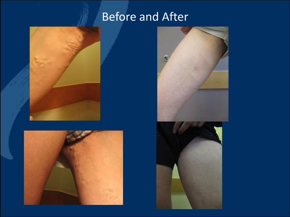
Before and After

Alternative Technique
• Ultrasound guided puncture of extrapelvic varices (Leg, vulvar, gluteal)
• Fluoroscopic calibration of varicose venous volume with contrast (angio suite)
• Foam sclerotherapy to level of broad ligament

When to Treat the Pelvic Source?
• When the primary symptom complex is pelvic: low back pain, heaviness in the pelvis, pain with intercourse, worse with menses
• Failure of treatment/frequent or early recurrence of vulvar and leg veins treated from the “bottom up” approach
• Patients with significant symptoms often eager for treatment

Considerations
• Investigate/rule out compression (May - Thurner or Nutcracker) can mimic reflux symptoms
• How old is the patient? Symptoms improve after menopause.
• How severe are the symptoms? What anatomic location?
• Treat the patient, not the diagnostic image
• Careful history - what is bothering your patient, why did they come to see you?
• Considerations: expense, recovery, insurance coverage, radiation exposure

Treatment Approaches for Coil Embolization
• Via jugular (my favorite) v. femoral vein approach
• Select both ovarian veins, both internal iliac veins
• Verify diagnosis: rule out obstruction (Nutcracker/May Thurner)
• Coil/sclerotherapy “sandwich” technique v. coils alone for the ovarian veins
• Balloon occlusion sclerotherapy for the internal iliac branches

Tools/Techniques I Like
• IJ approach
• Renal curve sheath/MPA
• Microcatheters
• Occlusion balloon
• STS foam with or without Lipiodol
• Toradol during procedure

Ovarian Vein: Sclerotherapy/Coil Embolization

Pelvic Venous Insufficiency (My Protocol):
Asymptomatic
Minimally Symptomatic
Symptomatic
Don’t Treat
Pelvic
SymptomsNo Pelvic
Symptoms
Coils &
SclerotherapySclerotherapy
From Below

Conclusions
• Pelvic source veins of the pelvis, vulva, perineum, and thigh share a common anatomic source with pelvic congestion syndrome, but patient presentation can differ
• As disease state has increased attention, improvements and standardization for diagnostic techniques needed
• Treatment methods vary, no data to tell us what is best
• Costs must be considered: we must be responsible with our health care dollars
• We need to learn more! Validated assessment tools are needed

THANK YOU!

Pelvic Congestion Syndrome: Diagnosis and Treatment Challenges
Kathleen Gibson, MD
Lake Washington Vascular Surgeons, Bellevue, WA, USA