phys ther.€2006; 86:672-682. - shockwave | ther.€2006; 86:672-682. massimo frascarelli, valter...
TRANSCRIPT

2006; 86:672-682.PHYS THER. Massimo Frascarelli, Valter Santilli and Giorgio SpaccaDon, Fosco de Paulis, Vittorio Calvisi, Alberto Ranavolo, Angelo Cacchio, Marco Paoloni, Antonio Barile, RomildoRandomized Clinical StudyCalcific Tendinitis of the Shoulder: Single-Blind, Effectiveness of Radial Shock-Wave Therapy for
http://ptjournal.apta.org/content/86/5/672found online at: The online version of this article, along with updated information and services, can be
Collections
Tendinitis Randomized Controlled Trials
Physical Agents/Modalities Injuries and Conditions: Shoulder
in the following collection(s): This article, along with others on similar topics, appears
e-Letters
"Responses" in the online version of this article. "Submit a response" in the right-hand menu under
or click onhere To submit an e-Letter on this article, click
E-mail alerts to receive free e-mail alerts hereSign up
by guest on January 11, 2013http://ptjournal.apta.org/Downloaded from
Effectiveness of Radial Shock-WaveTherapy for Calcific Tendinitis of theShoulder: Single-Blind, RandomizedClinical Study
Background and Purpose. Radial shock-wave therapy (RSWT) is apneumatically generated, low- to medium-energy type of shock-wavetherapy. This single-blind, randomized, “less active similar therapy”-controlled study was performed to evaluate the effectiveness of RSWTfor the management of calcific tendinitis of the shoulder. Subjects.Ninety patients with radiographically verified calcific tendinitis of theshoulder were tested. Methods. Subjects were randomly assigned toeither a treatment group (n�45) or a control group (n�45). Pain andfunctional level were evaluated before and after treatment and at a6-month follow-up. Radiographic modifications in calcifications wereevaluated before and after treatment. Results. The treatment groupdisplayed improvement in all of the parameters analyzed after treat-ment and at the 6-month follow-up. Calcifications disappeared com-pletely in 86.6% of the subjects in the treatment group and partially in13.4% of subjects; only 8.8% of the subjects in the control groupdisplayed partially reduced calcifications, and none displayed a totaldisappearance. Discussion and Conclusion. The results suggest that theuse of RSWT for the management of calcific tendinitis of the shoulderis safe and effective, leading to a significant reduction in pain andimprovement of shoulder function after 4 weeks, without adverseeffects. [Cacchio A, Paoloni M, Barile A, et al. Effectiveness of radialshock-wave therapy for calcific tendinitis of the shoulder: single-blind,randomized clinical study. Phys Ther. 2006;86:672–682.]
Key Words: Calcific tendinitis, Lithotripsy, Radial shock-wave therapy, Rotator cuff, Shoulder.
Angelo Cacchio, Marco Paoloni, Antonio Barile, Romildo Don, Fosco de Paulis, Vittorio Calvisi,Alberto Ranavolo, Massimo Frascarelli, Valter Santilli, Giorgio Spacca
672 Physical Therapy . Volume 86 . Number 5 . May 2006
Rese
arch
Repo
rt �
�������������������������������������������������������������������������������������������������������������������������������������������������������������������������������������������
������
������
������
������
������
������
������
������
������
�
by guest on January 11, 2013http://ptjournal.apta.org/Downloaded from
Calcific tendinitis of the shoulder is a commonlyobserved problem1 characterized by calciumphosphate crystal deposition in the rotator cufftendons, typically occurring between the fourth
and the fifth decades of life.2–4 It most frequently affectsthe supraspinatus tendon near its insertion, followed bythe infraspinatus, teres minor, and subscapularis ten-dons in descending order.5 The etiology and pathogen-esis of shoulder calcific tendinitis are still unclear. Hypo-vascularization and local degenerative and proliferativechanges in tendinous tissue of the rotator cuff have beensuggested as possible causes.2,6,8 The incidence of calcifictendinitis varies, depending on different reports, from2.7% to 63%.1,9,10 This wide variability may be due to theuse of different clinical and radiographic criteria,11
because the incidence of the calcification may be over-estimated when evaluated by radiographs rather than byclinical parameters.
The disorder leads to pain, particularly nocturnal dis-comfort, in about 50% of patients1,12 and frequently to aconsiderable restriction of range of motion. The clinicalpresentation varies considerably, and symptoms may lastfor several days and then either disappear or becomechronic,1,13 which means that it has not yet been possible
to clearly predict the natural history of the disease. Forexample, Bosworth1 described the disappearance ofcalcifications in 9.3% of patients within 3 years of theinitial diagnosis. According to Wagenhauser,14 calcifica-tions disappeared in 27.1% of patients after 10 years, andGartner15 reported that calcifications with sharp marginsand a homogeneous or heterogeneous structure disap-peared spontaneously in 33% of patients over a period of3 years. The time required for a spontaneous disappear-ance of the calcifications, however, often is too long andunacceptable for the patient’s quality of life.
Treatment of patients with calcific tendinitis is typicallyconservative and includes the use of nonsteroidal anti-inflammatory drugs, subacromial injection with steroids,percutaneous needle aspiration,16 transcutaneous elec-trical nerve stimulation,17 and therapeutic exercise,18 allof which have a limited effect19; the only interventionthat has been shown to result in a clinical improvementis therapeutic ultrasound.20,21 Open or arthroscopicsurgical procedures have been proposed to relieve symp-toms for patients with chronic pain, with goodresults.3,22,23
A Cacchio, MD, is Assistant of PM&R, Department of Neuroscience, Physical Medicine and Rehabilitation Unit, “San Salvatore” Hospital ofL’Aquila, Coppito-L’Aquila, Italy, and Assistant of PM&R, Department of Physical Medicine and Rehabilitation, School of Medicine, “La Sapienza”University of Roma, Rome, Italy. Address all correspondence to Dr Cacchio Angelo at Dipartimento di Neuroscienze, Unita Operativa di MedicinaFisica e Riabilitazione, Ospedale “San Salvatore” di L’Aquila, via L.Natali 1, 67100 Coppito-L’Aquila, Italy ([email protected]).
M Paoloni, MD, is Assistant of PM&R, Department of Physical Medicine and Rehabilitation, School of Medicine, “La Sapienza” University of Roma.
A Barile, MD, is Researcher of Radiology, Department of Radiology, School of Medicine, University of L’Aquila.
R Don, MD, is Assistant of PM&R, Department of Physical Medicine and Rehabilitation, School of Medicine, “La Sapienza” University of Roma.
F de Paulis, MD, is Chief of Radiology at CT Unit, Department of Radiology, “San Salvatore” Hospital of L’Aquila.
V Calvisi, MD, is Professor of Orthopaedic Surgery, Department of Surgery, Orthopaedic Surgery, School of Medicine, University of L’Aquila.
A Ranavolo, MEng, is Researcher of Biomechanics, Department of Physical Medicine and Rehabilitation, School of Medicine, “La Sapienza”University of Roma.
M Frascarelli, MD, is Professor of PM&R, Department of Physical Medicine and Rehabilitation, School of Medicine, “La Sapienza” University ofRoma.
V Santilli, MD, is Professor and Chief of PM&R Unit, Department of Physical Medicine and Rehabilitation, School of Medicine, “La Sapienza”University of Roma.
G Spacca, MD, is Chief of PM&R Unit, Department of Neuroscience, Physical Medicine and Rehabilitation Unit, “San Salvatore” Hospital ofL’Aquila.
All authors provided concept/idea/research design, data analysis, and subjects. Dr Cacchio and Dr Paoloni provided writing and consultation(including review of manuscript before submission). Dr Don and Mr Ranavolo provided data collection. Dr Cacchio and Dr Spacca providedproject management.
No author or related institution has received any financial benefit from research in this study.
This article was received April 7, 2005, and was accepted December 7, 2005.
Physical Therapy . Volume 86 . Number 5 . May 2006 Cacchio et al . 673 by guest on January 11, 2013http://ptjournal.apta.org/Downloaded from

When conservative therapy has not been effective in reliev-ing pain and other symptoms, extracorporeal shock-wavetherapy (ESWT) has been used,11,24–28 yielding results suchas relief of pain29–31 and improved function25,29,31,32 thatare sometimes as good as those achieved by means ofsurgical procedures.28 However, recent randomized con-trolled trials have shown negative results with the use ESWTfor the management of calcific tendinitis.33,34
A radial shock wave (RSW) is a low- to medium-energyshock wave that is pneumatically generated through theacceleration of a projectile inside the handpiece of thetreatment device and then transmitted radially from thetip of the applicator to the target zone. The pressure andthe energy density decrease by the third power of thepenetration depth in the tissue. Radial shock waves showa lower peak pressure and a considerably longer risetime than extracorporeal shock waves (ESWs) (Fig. 1).In radial shock-wave therapy (RSWT), the focal point isnot centered on the target zone, as occurs in ESWT, but
on the tip of the applicator (Fig. 2).The energy at the focal point of theshock wave per impulse is called the“energy flux density” (EFD) and isrecorded as joules per area. The ef-fective total energy of a treatmentis defined by the number and EFD ofthe single impulses and by the geomet-rical measurement of the focal point.Low-energy shock waves (EFD less than0.1 mJ/mm2) are generally differenti-ated from high-energy waves (EFD of0.2–0.4 mJ/mm).2,25
Recent studies by Loew et al25 andRompe et al27 compared high-energyshock-wave therapy with low-energyshock-wave therapy in the managementof calcific tendinitis of the shoulder,and the results showed energy-dependent success.25 It was assumedthat low-energy and unfocused shock-wave therapy (eg, RSWT), althougheffective for achieving pain relief, couldnot be effective in disintegrating thecalcific deposit of the rotator cuff.35
Recent findings, however, have demon-strated that ultrasound treatment inpatients with calcific tendinitis of theshoulder leads to calcific deposit dem-olition, and not only to relief of pain21;moreover, a considerable total levelenergy can be administered throughRSWT by using an adequate number ofimpulses per treatment.
Potential benefits could derive from RSWT, comparedwith ESWT, because it is less painful and thus can beadministered without anesthesia, thereby reducing therisks of treatment for patients. Furthermore, due to theradial emission of RSWT, the calcification, once locatedradiographically, is surely included inside the wave prop-agation area. Contrarily, when the shock wave is focused,as occurs in the ESWT, refocusing of the applicator isperiodically necessary to be certain that the waves hit thecalcification.36 Moreover, no ultrasound guide is neededto perform therapeutic applications of RSWT.
Although RSWT has been successfully used since the late1990s for the management of various orthopedic disor-ders such as epicondylitis of the elbow and chronic heelpain,37,38 which represent 2 of the 3 musculoskeletalindications for ESWT (plantar fasciitis, lateral epicondy-litis, and calcific tendinitis39), no randomized clinicalstudy has yet been performed in the treatment ofshoulder calcifications.
Figure 1.Physical characteristics of extracorporeal shock-wave therapy (ESWT) (left) and radial shock-wave therapy (RSWT) (right).
Figure 2.Wave propagation of extracorporeal shock-wave therapy (ESWT) and radial shock-wavetherapy (RSWT).
674 . Cacchio et al Physical Therapy . Volume 86 . Number 5 . May 2006
������
������
������
�����
by guest on January 11, 2013http://ptjournal.apta.org/Downloaded from

The aim of our study was to evaluate the effectiveness ofRSWT on pain relief, restoration of shoulder function,and resolution of calcific tendinitis of the shoulder,using a single-blind, randomized, “less active similartherapy”-controlled study. We considered functionalityand pain as primary end points, because we initially didnot expect a reduction in calcification, and we consid-ered the radiographic disappearance of calcifications asa secondary end point.
Subjects and MethodBetween November 2002 and December 2003, we con-ducted a single-center, single-blind, “less active similartherapy”-controlled study. Inclusion criteria were: cal-cific tendinitis of the shoulder, detected on standardizedradiographs, with type I (homogenous and with well-defined borders) or type II (heterogeneous in structurewith sharp outline or homogenous in structure with nodefined border) calcifications according to the Gartnerand Simons radiographic classification6; visual analogscale (VAS) score of �4 cm at the moment of the
evaluation; presence of symptoms for atleast 6 months; failure of previous con-servative treatments (anti-inflammatorydrugs, ultrasound and exercises, lasertherapy and exercises, electrical stimu-lation and exercises, acupuncture, andsteroid injection) (Tab. 1). Exclusioncriteria were: rotator cuff tear, glenohu-meral or acromioclavicular arthritis oracromioclavicular spur to rule out alter-native explanations for the pain; preg-nancy; implanted pacemaker; bloodcoagulation disorders or use of antico-agulant drugs; age of �18 years; inflam-matory or neoplastic disorders; pres-ence of type III (cloudy andtransparent) calcifications according tothe Gartner and Simons radiographicclassification6; and conservative treat-ments administered in the last 4 weeks.
Presence of shoulder pathologies wasclinically evaluated by an expert ortho-pedic physician, and, when suspectedon the basis of the clinical findings,ultrasound and magnetic resonanceimaging (MRI) examinations were per-formed. All subjects were verballyinformed of the potential risks of treat-ment as well as of the possibility ofunknowingly being included in thecontrol group and receiving a “lessactive similar therapy.” Writteninformed consent was obtained fromall subjects, and the procedures fol-
lowed were in accordance with the ethical standards ofthe Committee on Human Experimentation of “SanSalvatore” Hospital of L’Aquila. Subjects who met theeligibility criteria were randomly assigned, by the use ofa computer-based 1:1 randomization scheme and sealedenvelopes, to either a treatment group or a controlgroup (Tab. 2), so that each subject had an equalprobability of being assigned to either group.
Ninety-five patients were enrolled and assessed for eligi-bility between November 2002 and December 2003; 2patients did not meet the inclusion criteria, and 3patients met the exclusion criteria (Fig. 3), showing atear of the rotator cuff muscles with diagnostic ultra-sound. Six patients whom the orthopedic physicianbelieved had a rotator cuff tear received diagnosticultrasound. In 3 of the 6 patients (the 3 patients whowere excluded), positive findings for rotator cuff tearwere found. In the remaining 3 patients, due to uncer-tain findings for rotator cuff tear using diagnos-tic ultrasound, an MRI examination revealed no rotator
Table 1.Failed Previous Conservative Interventions
Failed Interventions
TreatmentGroup(n�45)
ControlGroup(n�45)
Total(% of 90Patients)
Anti-inflammatory drugs 42 40 82 (91.1%)Ultrasound and exercise 13 16 29 (32.2%)Laser therapy and exercise 10 8 18 (20.0%)Transcutaneous electrical nerve
stimulation and exercise16 12 28 (31.1%)
Acupuncture 3 3 6 (6.7%)Corticosteroid injection 39 33 72 (80.0%)
Table 2.Baseline Characteristics of the Treatment and Control Groupsa
CharacteristicsTreatmentGroup
ControlGroup
Subjects (n) 45 45Age (y)b 56.12�1.98 56.42�2.09Duration (mo)b 14�4.95 13�5.03Male/female (n) 27/18 28/17Shoulder 45 45Treatment side (right/left) 27/18 23/22UCLA Shoulder Rating Scale score
(range�0–35)b10.25�2.08 10.14�1.96
VAS (range�0–10)b 7.96�0.88 7.72�1.03Calcification size (mm)b 21.30�7.50 19.70�8.30Type of calcificationc
I 11 13II 34 32
a UCLA�University of California—Los Angeles, VAS�visual analog scale.b Values are mean � standard deviation.c Gartner and Simons radiographic classification.6
Physical Therapy . Volume 86 . Number 5 . May 2006 Cacchio et al . 675 by guest on January 11, 2013http://ptjournal.apta.org/Downloaded from
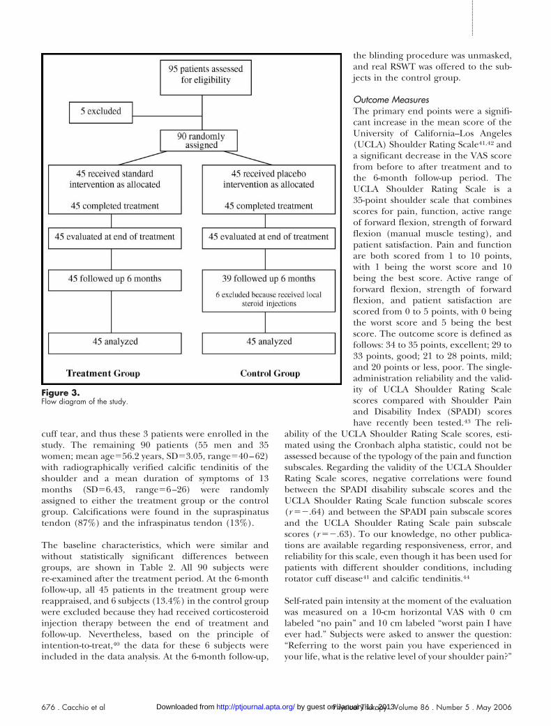
cuff tear, and thus these 3 patients were enrolled in thestudy. The remaining 90 patients (55 men and 35women; mean age�56.2 years, SD�3.05, range�40–62)with radiographically verified calcific tendinitis of theshoulder and a mean duration of symptoms of 13months (SD�6.43, range�6–26) were randomlyassigned to either the treatment group or the controlgroup. Calcifications were found in the supraspinatustendon (87%) and the infraspinatus tendon (13%).
The baseline characteristics, which were similar andwithout statistically significant differences betweengroups, are shown in Table 2. All 90 subjects werere-examined after the treatment period. At the 6-monthfollow-up, all 45 patients in the treatment group werereappraised, and 6 subjects (13.4%) in the control groupwere excluded because they had received corticosteroidinjection therapy between the end of treatment andfollow-up. Nevertheless, based on the principle ofintention-to-treat,40 the data for these 6 subjects wereincluded in the data analysis. At the 6-month follow-up,
the blinding procedure was unmasked,and real RSWT was offered to the sub-jects in the control group.
Outcome MeasuresThe primary end points were a signifi-cant increase in the mean score of theUniversity of California–Los Angeles(UCLA) Shoulder Rating Scale41,42 anda significant decrease in the VAS scorefrom before to after treatment and tothe 6-month follow-up period. TheUCLA Shoulder Rating Scale is a35-point shoulder scale that combinesscores for pain, function, active rangeof forward flexion, strength of forwardflexion (manual muscle testing), andpatient satisfaction. Pain and functionare both scored from 1 to 10 points,with 1 being the worst score and 10being the best score. Active range offorward flexion, strength of forwardflexion, and patient satisfaction arescored from 0 to 5 points, with 0 beingthe worst score and 5 being the bestscore. The outcome score is defined asfollows: 34 to 35 points, excellent; 29 to33 points, good; 21 to 28 points, mild;and 20 points or less, poor. The single-administration reliability and the valid-ity of UCLA Shoulder Rating Scalescores compared with Shoulder Painand Disability Index (SPADI) scoreshave recently been tested.43 The reli-
ability of the UCLA Shoulder Rating Scale scores, esti-mated using the Cronbach alpha statistic, could not beassessed because of the typology of the pain and functionsubscales. Regarding the validity of the UCLA ShoulderRating Scale scores, negative correlations were foundbetween the SPADI disability subscale scores and theUCLA Shoulder Rating Scale function subscale scores(r ��.64) and between the SPADI pain subscale scoresand the UCLA Shoulder Rating Scale pain subscalescores (r ��.63). To our knowledge, no other publica-tions are available regarding responsiveness, error, andreliability for this scale, even though it has been used forpatients with different shoulder conditions, includingrotator cuff disease41 and calcific tendinitis.44
Self-rated pain intensity at the moment of the evaluationwas measured on a 10-cm horizontal VAS with 0 cmlabeled “no pain” and 10 cm labeled “worst pain I haveever had.” Subjects were asked to answer the question:“Referring to the worst pain you have experienced inyour life, what is the relative level of your shoulder pain?”
Figure 3.Flow diagram of the study.
676 . Cacchio et al Physical Therapy . Volume 86 . Number 5 . May 2006
������
������
������
�����
by guest on January 11, 2013http://ptjournal.apta.org/Downloaded from

The secondary end point was the radio-graphic disappearance of calcificationsat the end of treatment. Success wasdefined as complete disappearance ofcalcification. An anteroposterior radio-graph of the shoulder obtained in 45degrees of external rotation and 45degrees of internal rotation was takenfor each subject under standardizedconditions in terms of distance fromradiographic film and exposure set-ting45 in order to evaluate the pres-ence, type, and size of calcifications, aswell as their location within a specifictendon. Type of calcification was evalu-ated according to the Gartner andSimons classification.6 A caliper thatevaluated calcification length (in milli-meters) was used for size measurement.The radiographic assessments wereobtained before treatment; post-treatment assessment was performed 1week after the end of treatment so as tobe able to correlate the disappearance of the calcifica-tion with the therapy performed.
Primary outcome measurements were performed by 2experienced physicians. The secondary outcome mea-surements were assessed by an experienced radiologist.The subjects, the outcome assessors, and the radiologistwere all blinded to the treatment performed.
Method of TreatmentA Physio Shock Wave Therapy device* consisting of acontrol unit, a handpiece with 3 different head applica-tors (8, 10, and 15 mm), and a medical air compressorwas used. The compressor generates a pneumatic energythat is used to accelerate a projectile inside the hand-piece. When the projectile strikes the applicator, a shockwave is generated and radially spreads from the tip of theapplicator to the target zone.
Subjects were seated with the shoulder abducted at 45degrees, the elbow flexed at 90 degrees, and the forearmresting on a flat surface, and the shock-wave applicatorwas placed in the direction of the calcifications. No localanesthetics or analgesic drugs were administered beforeor during the treatment and no therapeutic cointerven-tion was administered in either the treatment group orthe control group. Radial shock-wave therapy was admin-istered in both groups by the same experienced physi-cian (in accordance with Italian law, shock-wave therapymust be administered by a physician and not by aphysical therapist).
The RSWT was administered using a 15-mm-head appli-cator. Each subject in the treatment group received 4sessions at 1-week intervals, with 2,500 impulses persession (500 impulses with a pressure of 1.5 bar and afrequency of 4.5 Hz and 2,000 impulses with a pressureof 2.5 bar and a frequency of 10 Hz), an EFD of0.10 mJ/mm2, and a fixed impulse time of 2 milli-seconds. The treatment area was prepared with a cou-pling gel (Aquasonic 100†) to minimize the loss ofshock-wave energy at the interface between applicatortip and skin.
The same treatment procedure was followed for thesubjects in the control group, except that the totalnumber of impulses administered was only 25 (5impulses with a pressure of 1.5 bar and a frequency of 4.5Hz and 20 impulses with a pressure of 2.5 bar and afrequency of 10 Hz). Because we were not able toperform a simulated treatment, we had to give someshock-wave impulse to the control group to avoid possi-ble blinding failure. Other researchers46,47 also haveused a “less active similar therapy,” and the rationale forthis technique is that the efficacy of shock-wave therapyseems to be dose-dependent.25
Data AnalysisStatistical analysis was performed using the SSP 2.5statistical package (Smith’s Statistical Package, version2.75, 2004‡). All analyses of the primary and secondary
* Elettronica Pagani Srl, Via De Nicola 4/D, 20037 Paderno Dugnano (MI), Italy.
† Parker Laboratories Inc, 286 Eldridge Rd, Fairfield, NJ 07004.‡ Gary Smith, Pomona College, Claremont, Calif 91711 (http://www.economics.pomona.edu/StatSite/framepg.html).
Table 3.Mean (�SD) Values and Outcomes of the University of California—Los Angeles ShoulderRating Scale in the Treatment and Control Groups
Mean (�SD)Values
Treatment Group(n�45)
Control Group(n�45) P
Before treatment 10.25�2.08 10.14�1.96 .9144a
After treatment 33.12�2.94 11.28�2.82 .0056b
P .00000002c .1438c
Follow-up 32.12�3.02 10.57�3.96 .0023d
P .1151e .3302e
Outcomesf E G M P Total E G M P Total
Before treatment 45 45 45 45After treatment 41 4 45 8 37 45Follow-up 39 5 1 45 3 36 39
a Comparison between treatment and control groups before treatment.b Comparison between treatment and control groups after treatment.c Comparison between before and after treatment within each group.d Comparison between treatment and control groups at 6-month follow-up.e Comparison between after treatment and 6-month follow-up within each group.f E�excellent (34–35 points), G�good (29–33 points), M�mild (21–28 points), P�poor (�20 points).
Physical Therapy . Volume 86 . Number 5 . May 2006 Cacchio et al . 677 by guest on January 11, 2013http://ptjournal.apta.org/Downloaded from

outcomes were performed according to the principle ofintention-to-treat. The intention-to-treat analysis was car-ried out according to a “worst-case scenario” analysis:subjects who did not complete the treatment or had notundergone the post-treatment or final follow-up assess-ments were assigned a poor outcome, corresponding tothe final average change recorded in the per-protocolcompleter population in the control group.40 A 2-samplet test was applied to compare the differences of thebaseline data. A 2-way analysis of variance (ANOVA) withgroup (treatment versus control) as the between-subjectsfactor and time as the within-subjects factor was used toassess the presence of significant differences between
the treatment and control groups andwithin each group before and aftertreatment and at the 6-month follow-up. A Tukey post hoc comparison wasused to determine significant differ-ences between mean values when asignificant main effect and interactionwere found. Two-sample paired andunpaired t tests were applied to com-pare the differences of average size ofcalcium deposits on radiographicexamination before and after treat-ment and between the treatment andcontrol groups, respectively. For allanalyses, the level of significance wasset at P�.05.
To allow a clinical translation of thestatistical results, the number neededto treat (NNT)48 was evaluated. TheNNT is expressed in terms designed tohelp decide whether the interventionmight be valuable in clinical practice.48
For example, when comparing treat-ment X and treatment Y, an NNT scoreof 5 for treatment X indicates that, onaverage, after treating 5 patients, treat-ment X will have achieved one morepositive outcome than if treatment Yhad been used.48 For the primary out-come, the NNT was calculated consid-ering the “excellent” category (34–35points on the UCLA Shoulder RatingScale) as a positive outcome and the“good,” “mild,” and “poor” categories(below 34 points on the UCLA Shoul-der Rating Scale) as negative outcomes.For the secondary outcome, number ofdisappearance of calcifications wasused to calculate the NNT.
Results
Primary Outcome MeasuresThe ANOVA demonstrated a significant effect of treat-ment (P �.0001) and a significant treatment-time inter-action (P �.0001). One week after the end of treatmentand at the 6-month follow-up, statistically significantimprovements in mean total scores (Tab. 3) and single-item scores (Tab. 4) on the UCLA Shoulder Rating Scalewere observed in the treatment group. Statistically sig-nificant improvements in scores on the pain subscale ofUCLA Shoulder Rating Scale also were observed in thecontrol group. No statistically significant difference wasfound between the treatment and control groups for thefunction subscale of UCLA Shoulder Rating Scale at the
Table 4.Comparison of Single Items of the Unviersity of California–Los Angeles (UCLA) ShoulderRating Scale Before and After Treatment With Radial Shock-Wave Therapy and at 6-MonthFollow-up in the Treatment and Control Groups
UCLA ShoulderRating Scale Item
TreatmentGroup(n�45)
ControlGroup(n�45) P
Pain (range�1–10)Before treatment 1.39�0.97 1.04�1.03 .8966a
After treatment 7.90�1.09 2.85�2.03 .0044b
P .00000001c .0386c
Follow-up1 7.95�0.92 2.64�1.14 .0023d
P .8147e .5471e
Active range of forward flexionBefore treatment 66.75�15.41 68.14�18.77 .2033a
After treatment 134.35�24.93 85.00�32.45 .0084b
P .000002c .0693c
Follow-up 152.00�28.99 90.00�26.15 .0127d
P .0026e .4232e
Strength of forward flexion(range�0–5)
Before treatment 3.49�0.75 3.16�0.32 .6590a
After treatment 4.98�0.35 3.66�0.95 .0067b
P .0000009c 0.1611c
Follow-up 4.85�0.46 3.42�0.95 .0045d
P .1352e 0.2340e
Function (range�0–5)Before treatment 2.10�0.33 2.18�0.45 .4738a
After treatment 4.48�0.85 2.98�1.23 .0748b
P .000001c .2145c
Follow-up 4.50�0.82 2.45�1.61 .0163d
P .9098e .0830e
Patient satisfaction (range�0–5)Before treatment 0.80�0.50 0.84�0.45 .7494a
After treatment 4.80�1.02 1.70�1.90 .0017b
P .0000001c .0921c
Follow-up 4.60�1.03 1.05�0.95 .0011d
P .3572e .0442e
a Comparison between treatment and control groups before treatment.b Comparison between treatment and control groups after treatment.c Comparison between before and after treatment within each group.d Comparison between treatment and control groups at 6-month follow-up.e Comparison between after treatment and 6-month follow-up within each group.
678 . Cacchio et al Physical Therapy . Volume 86 . Number 5 . May 2006
������
������
������
�����
by guest on January 11, 2013http://ptjournal.apta.org/Downloaded from
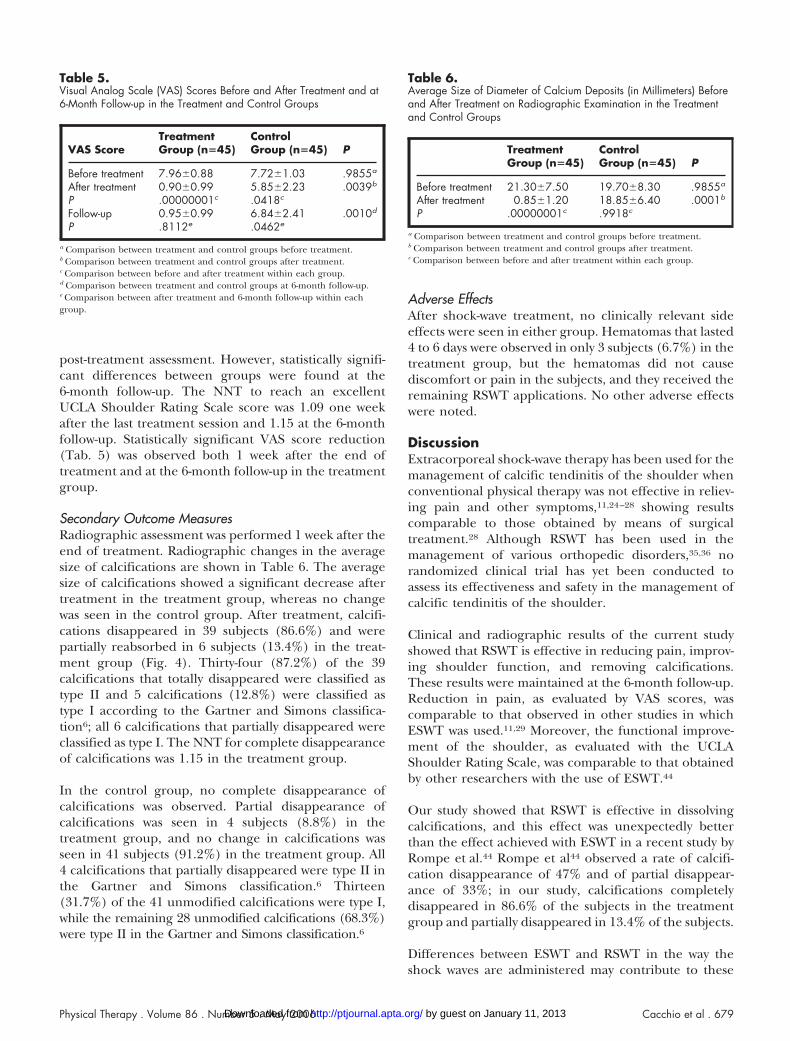
post-treatment assessment. However, statistically signifi-cant differences between groups were found at the6-month follow-up. The NNT to reach an excellentUCLA Shoulder Rating Scale score was 1.09 one weekafter the last treatment session and 1.15 at the 6-monthfollow-up. Statistically significant VAS score reduction(Tab. 5) was observed both 1 week after the end oftreatment and at the 6-month follow-up in the treatmentgroup.
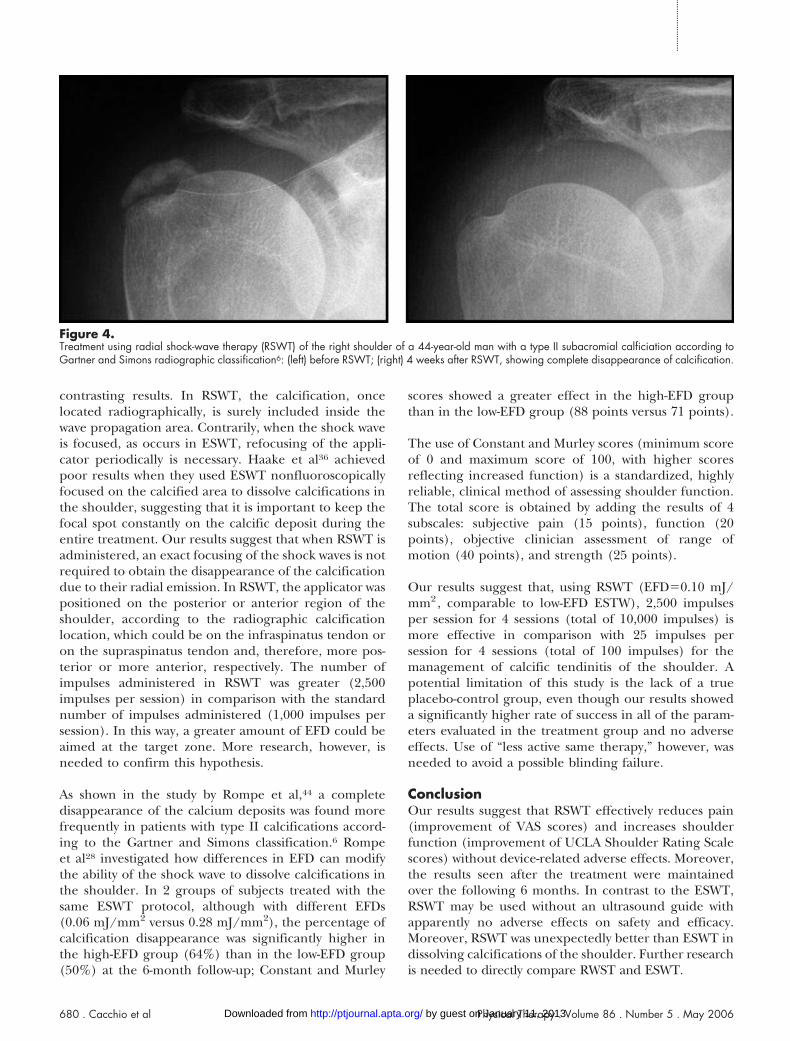
Secondary Outcome MeasuresRadiographic assessment was performed 1 week after theend of treatment. Radiographic changes in the averagesize of calcifications are shown in Table 6. The averagesize of calcifications showed a significant decrease aftertreatment in the treatment group, whereas no changewas seen in the control group. After treatment, calcifi-cations disappeared in 39 subjects (86.6%) and werepartially reabsorbed in 6 subjects (13.4%) in the treat-ment group (Fig. 4). Thirty-four (87.2%) of the 39calcifications that totally disappeared were classified astype II and 5 calcifications (12.8%) were classified astype I according to the Gartner and Simons classifica-tion6; all 6 calcifications that partially disappeared wereclassified as type I. The NNT for complete disappearanceof calcifications was 1.15 in the treatment group.
In the control group, no complete disappearance ofcalcifications was observed. Partial disappearance ofcalcifications was seen in 4 subjects (8.8%) in thetreatment group, and no change in calcifications wasseen in 41 subjects (91.2%) in the treatment group. All4 calcifications that partially disappeared were type II inthe Gartner and Simons classification.6 Thirteen(31.7%) of the 41 unmodified calcifications were type I,while the remaining 28 unmodified calcifications (68.3%)were type II in the Gartner and Simons classification.6
Adverse EffectsAfter shock-wave treatment, no clinically relevant sideeffects were seen in either group. Hematomas that lasted4 to 6 days were observed in only 3 subjects (6.7%) in thetreatment group, but the hematomas did not causediscomfort or pain in the subjects, and they received theremaining RSWT applications. No other adverse effectswere noted.
DiscussionExtracorporeal shock-wave therapy has been used for themanagement of calcific tendinitis of the shoulder whenconventional physical therapy was not effective in reliev-ing pain and other symptoms,11,24–28 showing resultscomparable to those obtained by means of surgicaltreatment.28 Although RSWT has been used in themanagement of various orthopedic disorders,35,36 norandomized clinical trial has yet been conducted toassess its effectiveness and safety in the management ofcalcific tendinitis of the shoulder.
Clinical and radiographic results of the current studyshowed that RSWT is effective in reducing pain, improv-ing shoulder function, and removing calcifications.These results were maintained at the 6-month follow-up.Reduction in pain, as evaluated by VAS scores, wascomparable to that observed in other studies in whichESWT was used.11,29 Moreover, the functional improve-ment of the shoulder, as evaluated with the UCLAShoulder Rating Scale, was comparable to that obtainedby other researchers with the use of ESWT.44
Our study showed that RSWT is effective in dissolvingcalcifications, and this effect was unexpectedly betterthan the effect achieved with ESWT in a recent study byRompe et al.44 Rompe et al44 observed a rate of calcifi-cation disappearance of 47% and of partial disappear-ance of 33%; in our study, calcifications completelydisappeared in 86.6% of the subjects in the treatmentgroup and partially disappeared in 13.4% of the subjects.
Differences between ESWT and RSWT in the way theshock waves are administered may contribute to these
Table 5.Visual Analog Scale (VAS) Scores Before and After Treatment and at6-Month Follow-up in the Treatment and Control Groups
VAS ScoreTreatmentGroup (n�45)
ControlGroup (n�45) P
Before treatment 7.96�0.88 7.72�1.03 .9855a
After treatment 0.90�0.99 5.85�2.23 .0039b
P .00000001c .0418c
Follow-up 0.95�0.99 6.84�2.41 .0010d
P .8112e .0462e
a Comparison between treatment and control groups before treatment.b Comparison between treatment and control groups after treatment.c Comparison between before and after treatment within each group.d Comparison between treatment and control groups at 6-month follow-up.e Comparison between after treatment and 6-month follow-up within eachgroup.
Table 6.Average Size of Diameter of Calcium Deposits (in Millimeters) Beforeand After Treatment on Radiographic Examination in the Treatmentand Control Groups
TreatmentGroup (n�45)
ControlGroup (n�45) P
Before treatment 21.30�7.50 19.70�8.30 .9855a
After treatment 0.85�1.20 18.85�6.40 .0001b
P .00000001c .9918c
a Comparison between treatment and control groups before treatment.b Comparison between treatment and control groups after treatment.c Comparison between before and after treatment within each group.
Physical Therapy . Volume 86 . Number 5 . May 2006 Cacchio et al . 679 by guest on January 11, 2013http://ptjournal.apta.org/Downloaded from
contrasting results. In RSWT, the calcification, oncelocated radiographically, is surely included inside thewave propagation area. Contrarily, when the shock waveis focused, as occurs in ESWT, refocusing of the appli-cator periodically is necessary. Haake et al36 achievedpoor results when they used ESWT nonfluoroscopicallyfocused on the calcified area to dissolve calcifications inthe shoulder, suggesting that it is important to keep thefocal spot constantly on the calcific deposit during theentire treatment. Our results suggest that when RSWT isadministered, an exact focusing of the shock waves is notrequired to obtain the disappearance of the calcificationdue to their radial emission. In RSWT, the applicator waspositioned on the posterior or anterior region of theshoulder, according to the radiographic calcificationlocation, which could be on the infraspinatus tendon oron the supraspinatus tendon and, therefore, more pos-terior or more anterior, respectively. The number ofimpulses administered in RSWT was greater (2,500impulses per session) in comparison with the standardnumber of impulses administered (1,000 impulses persession). In this way, a greater amount of EFD could beaimed at the target zone. More research, however, isneeded to confirm this hypothesis.
As shown in the study by Rompe et al,44 a completedisappearance of the calcium deposits was found morefrequently in patients with type II calcifications accord-ing to the Gartner and Simons classification.6 Rompeet al28 investigated how differences in EFD can modifythe ability of the shock wave to dissolve calcifications inthe shoulder. In 2 groups of subjects treated with thesame ESWT protocol, although with different EFDs(0.06 mJ/mm2 versus 0.28 mJ/mm2), the percentage ofcalcification disappearance was significantly higher inthe high-EFD group (64%) than in the low-EFD group(50%) at the 6-month follow-up; Constant and Murley
scores showed a greater effect in the high-EFD groupthan in the low-EFD group (88 points versus 71 points).
The use of Constant and Murley scores (minimum scoreof 0 and maximum score of 100, with higher scoresreflecting increased function) is a standardized, highlyreliable, clinical method of assessing shoulder function.The total score is obtained by adding the results of 4subscales: subjective pain (15 points), function (20points), objective clinician assessment of range ofmotion (40 points), and strength (25 points).
Our results suggest that, using RSWT (EFD�0.10 mJ/mm2, comparable to low-EFD ESTW), 2,500 impulsesper session for 4 sessions (total of 10,000 impulses) ismore effective in comparison with 25 impulses persession for 4 sessions (total of 100 impulses) for themanagement of calcific tendinitis of the shoulder. Apotential limitation of this study is the lack of a trueplacebo-control group, even though our results showeda significantly higher rate of success in all of the param-eters evaluated in the treatment group and no adverseeffects. Use of “less active same therapy,” however, wasneeded to avoid a possible blinding failure.
ConclusionOur results suggest that RSWT effectively reduces pain(improvement of VAS scores) and increases shoulderfunction (improvement of UCLA Shoulder Rating Scalescores) without device-related adverse effects. Moreover,the results seen after the treatment were maintainedover the following 6 months. In contrast to the ESWT,RSWT may be used without an ultrasound guide withapparently no adverse effects on safety and efficacy.Moreover, RSWT was unexpectedly better than ESWT indissolving calcifications of the shoulder. Further researchis needed to directly compare RWST and ESWT.
Figure 4.Treatment using radial shock-wave therapy (RSWT) of the right shoulder of a 44-year-old man with a type II subacromial calficiation according toGartner and Simons radiographic classification6: (left) before RSWT; (right) 4 weeks after RSWT, showing complete disappearance of calcification.
680 . Cacchio et al Physical Therapy . Volume 86 . Number 5 . May 2006
������
������
������
�����
by guest on January 11, 2013http://ptjournal.apta.org/Downloaded from

References1 Bosworth BM. Calcium deposits in the shoulder and subacromialbursitis: a survey of 12,122 shoulders. JAMA. 1941;116:2477–2482.
2 Uhthoff HK, Sarkar K. Calcifiyng tendinitis. In: Rockwood CA,Matsen FA III, eds. The Shoulder. Philadelphia, Pa: WB Saunders Co;1990:774–790.
3 Ark JW, Flock TJ, Flatow EL, Bigliani LU. Arthroscopic treatment ofcalcific tendinitis of the shoulder. Arthroscopy. 1992;8:183–188.
4 Wainner RS, Hasz M. Management of acute calcific tendinitis of theshoulder. J Orthop Sports Phys Ther. 1998;27:231–237.
5 Uhthoff HK. Calcifying tendinitis. Ann Chir Gynaecol. 1996;85:111–115.
6 Gartner J, Simons B. Analysis of calcific deposits in calcifyingtendinitis. Clin Orthop Relat Res. 1990;254:111–120.
7 Hayes CW, Conway WF. Calcium hydroxyapatite depositing disease.Radiographics. 1990;10:1031–1048.
8 Moseley HF. The natural history and clinical syndromes produced bycalcified deposits in the rotator cuff. Surg Clin North Am. 1963;43:1489–1493.
9 Refior HJ, Krodel A, Melzer C. Examinations of the pathology of therotator cuff. Arch Orthop Trauma Surg. 1987;106:301–308.
10 Milone FP, Copeland MM. Calcific tendinitis of the shoulder joint:presentation of 136 cases treated by irradiation. Am J Roentgenol Ther.1961;85:901–913.
11 Gerdesmyer L, Wagenpfeil S, Haake M, et al. Extracorporeal shockwave therapy for the treatment of chronic calcifying tendonitis of therotator cuff: a randomized controlled trial. JAMA. 2003;290:2573–2580.
12 McKendry RJR, Uhthoff HK, Sakar K, Hyslop PS. Calcifying tendi-nitis of the shoulder: prognostic value of clinical, histologic, andradiologic features in 57 surgically treated cases. J Rheumatol. 1982;9:75–80.
13 Uhthoff HK, Sakar K, Maynard JA. Calcifying tendinitis: a newconcept of its pathogenesis. Clin Orthop Relat Res. 1976;118:164–168.
14 Wagenhauser JF. Die periarthropatie-syndrome. Therapiewoche.1972;37:3187–3192.
15 Gartner J. Tendinosis calcarea: results of treatment with needling.Z Orthop Ihre Grenzgeb. 1993;131:461–469.
16 Parlier-Cuau C, Champsaur P, Nizard R, et al. Percutaneous treat-ments of painful shoulder. Radiol Clin North Am. 1998;36:589–596.
17 Kaada B. Treatment of peritendinitis of the shoulder by transcuta-neous nerve stimulation. Acupunct Electrother Res. 1984;9:115–125.
18 Gimblett PA, Saville J, Ebrall P. A conservative management proto-col for calcific tendinitis of the shoulder. J Manipulative Physiol Ther.1999;22:622–627.
19 Green S, Buchbinder R, Glazier R, Forbes A. Systematic review ofrandomised controlled trials of interventions to painful shoulder:selection criteria, outcome assessment, and efficacy. BMJ. 1998;316:354–360.
20 Philadelphia Panel Evidence-Based Clinical Practice Guidelines onSelected Rehabilitation Interventions for Shoulder Pain. Phys Ther.2001;81:1719–1730.
21 Ebenbichler GR, Erdogmus CB, Resch KL, et al. Ultrasound therapyfor calcific tendinitis of the shoulder. N Engl J Med. 1999;340:1533–1538.
22 Rochwerger A, Franceschi JP, Viton JM, et al. Surgical managementof calcific tendinitis of the shoulder: an analysis of 26 cases. ClinRheumatol. 1999;18:313–316.
23 Jerosch J, Strauss JM, Schmiel S. Arthroscopic treatment of calcifictendinitis of the shoulder. J Shoulder Elbow Surg. 1998;7:30–37.
24 Loew M, Jurgowski W, Mau HC, Thomsen M. Treatment of calcify-ing tendinitis of rotator cuff by extracorporeal shock waves: a prelim-inary report. J Shoulder Elbow Surg. 1995;4:101–106.
25 Loew M, Daecke D, Kusnierczak D, et al. Shock-wave therapy iseffective for chronic calcifying tendinitis of the shoulder. J Bone J SurgBr. 1999;81:863–867.
26 Wang C-J, Ko J-Y, Chen H-S. Treatment of calcifying tendinitis of theshoulder with shock wave therapy. Clin Orthop Relat Res. 2001;387:83–89.
27 Rompe JD, Rumler F, Hopf C, et al. Extracorporeal shock wavetherapy for calcifying tendinitis of the shoulder. Clin Orthop Relat Res.1995;321:196–201.
28 Rompe JD, Burger R, Hopf C, Eysel P. Shoulder function afterextracorporeal shock wave therapy for calcific tendinitis. J ShoulderElbow Surg. 1998;7:505–509.
29 Cosentino R, De Stefano R, Selvi E, et al. Extracorporeal shock wavetherapy for chronic calcific tendinitis of the shoulder: single blindstudy. Ann Rheum Dis. 2003;62:248–250.
30 Daeke W, Kusnierczak D, Loew M. Long-term effects of extracor-poreal shock wave therapy in chronic calcific tendinitis of the shoul-der. J Shoulder Elbow Surg. 2002;11:476–480.
31 Pan P-J, Chou C-L, Chiou H-J, et al. Extracorporeal shock wavetherapy for chronic calcific tendinitis of the shoulders: a functionaland sonographic study. Arch Phys Med Rehabil. 2003;84:988–993.
32 Wang CJ, Yang KD, Wang FS, et al. Shock wave therapy for calcifictendinitis of the shoulder. Am J Sports Med. 2003;31:425–430.
33 Schmitt J, Tosch A, Hunerkopf M, Haake M. Extracorporeal shock-wave therapy (ESWT) as therapeutic option in supraspinatus tendonsyndrome? One-year results of a placebo controlled study. Orthopade.2002;31:652–657.
34 Speed CA, Richards C, Nichols D, et al. Extracorporeal shock-wavetherapy for tendonitis of the rotator cuff: a double-blind, randomized,controlled trial. J Bone Joint Surg Br. 2002;84:509–512.
35 Gerdesmeyer L, Maier M, Haake M, Schmitz C. Physikalisch-technische Grundlagen der extrakorporalen Sto�wellentherapie(ESWT). Orthopade. 2002;31:610–617.
36 Haake M, Deike B, Thon A, Schmitt J. Exact focusing of theextracorporeal shock wave therapy for calcifying tendinopathy. ClinOrthop Relat Res. 2002;397:323–331.
37 Straub T, Penninger E, Froelich T, et al. Prospective, multicentricand placebo-controlled study on shockwave treatment of tennis elbowand plantar fasciitis. Int J Sports Med. 1999;20:S105–S106.
38 Lohrer H, Schoell J, Arentz S, et al. Effectiveness of radial shock-wave therapy (RSWT) on tennis elbow and plantar fasciitis. In:Proceedings of the Annual Meeting of the Canadian Academy of SportsMedicine; July 4–7, 2001; Calgary, Alberta, Canada.
39 Health Technology Assessment: Extracorporeal Shockwave Therapy for theTreatment of Musculoskeletal Disorders. Olympia, Wash: Department ofLabor and Industries; March 29, 2004.
40 Hollis S, Campbell F. What is meant by intention-to-treat analysis?Survey of a published randomised controlled trials. BMJ. 1999;319:670–674.
Physical Therapy . Volume 86 . Number 5 . May 2006 Cacchio et al . 681 by guest on January 11, 2013http://ptjournal.apta.org/Downloaded from
41 Ellman H. Arthroscopic subacromial decompression: analysis ofone- to three-years results. Arthroscopy. 1987;3:173–181.
42 Kay SP, Amstutz HC. Shoulder hemiarthroplasty at UCLA. ClinOrthop Relat Res. 1988;228:42–48.
43 Roddey TS, Olson SL, Cook KF, et al. Comparison of the Universityof California–Los Angeles Shoulder Scale and the Simple ShoulderTest with the Shoulder Pain and Disability Index: single-administrationreliability and validity. Phys Ther. 2000;80:759–768.
44 Rompe JD, Zoellner J, Nafe B. Shock wave therapy versus conven-tional surgery in the treatment of calcifying tendinitis of the shoulder.Clin Orthop Relat Res. 2001;387:72–82.
45 Kilcoyne RF, Reddy PK, Lyons F, Rockwood CA Jr. Optimal plainfilm imaging of the shoulder impingement syndrome. Am J Roentgenol.1989;153:795–797.
46 Pleiner J, Crevenna R, Langenberger H, et al. Extracorporealshockwave treatment is effective in calcific tendonitis of the shoulder:a randomized controlled trial. Wien Klin Wochenschr. 2004;116:536–541.
47 Rompe JD, Hopf C, Kullmer K, et al. Analgesic effect of extracor-poreal shock-wave therapy on chronic tennis elbow. J Bone Joint Surg Br.1996;78:233–237.
48 Dalton GW, Keating JL. Number needed to treat: a statistic relevantfor physical therapists. Phys Ther. 2000;80:1214–1219.
682 . Cacchio et al Physical Therapy . Volume 86 . Number 5 . May 2006
������
������
������
�����
by guest on January 11, 2013http://ptjournal.apta.org/Downloaded from

2006; 86:672-682.PHYS THER. Massimo Frascarelli, Valter Santilli and Giorgio SpaccaDon, Fosco de Paulis, Vittorio Calvisi, Alberto Ranavolo, Angelo Cacchio, Marco Paoloni, Antonio Barile, RomildoRandomized Clinical StudyCalcific Tendinitis of the Shoulder: Single-Blind, Effectiveness of Radial Shock-Wave Therapy for
References
http://ptjournal.apta.org/content/86/5/672#BIBLfor free at: This article cites 38 articles, 9 of which you can access
Cited by
http://ptjournal.apta.org/content/86/5/672#otherarticles
This article has been cited by 9 HighWire-hosted articles:
Information Subscription http://ptjournal.apta.org/subscriptions/
Permissions and Reprints http://ptjournal.apta.org/site/misc/terms.xhtml
Information for Authors http://ptjournal.apta.org/site/misc/ifora.xhtml
by guest on January 11, 2013http://ptjournal.apta.org/Downloaded from