pi urachal cancer underlined terms are listed in the glssary. - 2 - what is urachal cancer? 3 what...
TRANSCRIPT

Information for Patients English
Urachal cancer
* The underlined terms are listed in the glossary. - 2 -
What is urachal cancer? ................................................................................................ 3
What is the urachus? ........................................................................................................ 3
Risk factors for urachal cancer ............................................................................... 3
What are the symptoms? ............................................................................................ 3
Diagnosis ....................................................................................................................................... 3
Physical Exam and Imaging .......................................................................................... 3
Transurethral resection (TUR) ................................................................................... 4
Classification .............................................................................................................................. 4
Treatment ..................................................................................................................................... 4
Surgery ............................................................................................................................................ 5
Tumour removal ..................................................................................................................... 5
Bladder removal ..................................................................................................................... 5
Diagnostic laparoscopy .................................................................................................... 5
Chemotherapy ......................................................................................................................... 5
Palliative Care .......................................................................................................................... 6
Follow-up after treatment .......................................................................................... 6
FAQs .................................................................................................................................................. 6
Links to more information ......................................................................................... 7
Glossary of terms ................................................................................................................. 8
Table of contents
This information was produced by the European Association of Urology
This leaflet contains general information about urachal cancer. If you have any specific questions about your individual medical situation you should consult your doctor or other professional healthcare provider.
This information was produced by the European Association of Urology (EAU) Patient Information Working Group and is based on a narrative review of the literature.
- Dr. Mark Behrendt, Amsterdam (NL)- Dr. Henning Reis, Essen (DE)
You can find this and other information on urological diseases at our website: http://patients.uroweb.org

* The underlined terms are listed in the glossary. - 3 -
• Blood in the urine (haematuria)• Pain• Bladder irritation• Recurrent bladder infections• Mucus from the navel (umbilical discharge) or in the urine
(mucusuria)
Diagnosis
The diagnosis of urachal cancer is usually based on:• Blood in the urine• A tumour located outside, in the middle, or on top of the
urinary bladder• Mucus-producing cells in the tissue
Physical Exam and ImagingAn initial diagnosis will include a physical examination, ultrasound of the abdomen, and a urine test (urinary swab test). Urachal cancer is suggested by a tumour located outside, in the middle, or on top of the urinary bladder and blood in the urine.
Urachal cancer
What is urachal cancer?
A growth of cancerous cells that starts in the urachus is called urachal cancer.
What is the urachus?
The urachus is a tube-like structure that forms in a developing embryo. It connects the umbilical cord to the urinary bladder before birth. After birth, the urachus usually shrinks into a small ligament. However, traces of the urachus (called urachal residues) can be detected in up to one-third of adults. Urachal residues usually do not cause any symptoms.
Risk factors for urachal cancer
There is a risk of urachal cancer if part of the urachus or the whole urachus is present. Years of chronic inflammation and remodelling or possibly leftover embryologic cells can cause urachal cancer.
An intact urachus is indicated by:• Recurrent infections of the navel or bladder• Production of mucus by the navel or bladder• Navel (umbilical) hernia that has not been identified as a
persistent urachusNo other risk factors have been identified yet.
Urachal cancer is very rare. Approximately one in 1 million people per year develop urachal cancer, depending on the region of the world. The disease seems to be less common in Europe, for example, than in Japan. Statistically, more men are affected by urachal cancer than women. Most patients are diagnosed in their 50s.
Because of its location, urachal cancer can grow into the abdominal wall and the abdominal cavity. Often it infiltrates the roof of the urinary bladder. Urachal cancer can grow for a long time before causing symptoms. As a result, it is often detected at later stages.
What are the symptoms?
Symptoms of urachal cancer become noticeable when the cancer cells grow into surrounding tissue or organs (advanced stage). Symptoms may include:
Fig. 1. Anatomical illustration of an intact (patent) urachus. This situation is very rare in an adult. If urachal residues remain, they are
usually partial or microscopic.
©2018 patients.uroweb ALL RIGHTS RESERVED
Umbilicus
Urachus
Anterior abdominal wall
Bladder

* The underlined terms are listed in the glossary. - 4 -
Treatment
Urachal cancer is often diagnosed at later stages. Based on your disease stage and predicted outcomes, recommended treatment may include:
• Surgery• Chemotherapy• Palliative care
The planned treatment approach should be discussed by a multidisciplinary tumour board. This board is made up of practitioners from different medical specialties. They share their different professional opinions to plan appropriate care for individual cancer patients.
Diagnostic Imaging Techniques
Ultrasound Ultrasound is done outside the body (non-invasive) using high-frequency sounds to make an image of the inside of the body
CT scan CT stands for Computed Tomography. This imaging technique makes a series of x-ray images of the body.
MRI Magnetic Resonance Imaging uses strong magnetic fields and radio waves to make images of the body.
Transurethral resection (TUR)If urachal cancer is suspected, the next step is to examine tissue under a microscope (histologic examination). Surgery must be performed to get the tissue for that examination.
Transurethral resection (TUR) is the most accessible way to acquire tissue from the urinary bladder. TUR is performed by the insertion of a rigid tube-like instrument (endoscope) through the urethra (the canal through which the urine is passed) into the bladder. The tissue is removed with the endoscope. The patient is asleep (under general anaesthesia) during this procedure.
If examination of the tissue shows cancerous mucus-producing cells (mucinous histology), the diagnosis is then formed in accordance with the imaging results.
ClassificationYour doctor will classify the severity and aggressiveness of urachal cancer. A cancer stage will be determined based on:• The histologic examination of the tissue• The size of the tumour and whether it has grown into
surrounding organs or tissue• Whether the tumour cells have spread to other organs or
tissue (metastases)
Classification by cancer stage guides treatment and prediction of outcomes (prognosis).
Fig. 2: partially patent urachus, opening externally, blind internally.
Fig. 3: partially patent urachus, opening internally, blind externally.
Fig. 4: Cyst of urachus.
Umbilicus Umbilicus Umbilicus
Urachus Urachus cyst
©2018 patients.uroweb ALL RIGHTS RESERVED ©2018 patients.uroweb ALL RIGHTS RESERVED ©2018 patients.uroweb ALL RIGHTS RESERVED
* The underlined terms are listed in the glossary. - 5 -
anaesthesia). If cancerous cells are found, a larger operation will be performed to remove all mucus and tumours, and chemotherapy drugs will be given directly in the abdomen to kill remaining cancer cells.
What to ask your doctor about urachal cancer surgery• Why do I need surgery?• Will I have to stay in hospital?• How long will I be in hospital?• What are the possible side effects of the surgery?• Will I have any pain?• Are there any possible complications?• How long will I need to be off work? ChemotherapySystemic chemotherapy is not a substitute for surgical treatment, and it does not provide a cure. Chemotherapy drugs containing platinum and 5-fluorouracil (5-FU) have shown success in:
• Urachal cancer that has spread to other tissues or organs• Patients with a high risk of recurrence after the operation
General side effects of chemotherapyMany of the side effects are mild and can be managed at home. Side effects may include:• Lower levels of white blood cells• Anaemia• Hair loss• Fluid retention• Vomiting• Allergic reactions• Fatigue• Muscle pain• Diarrhoea• Loss of appetite• Hearing loss• Kidney damage
Surgery
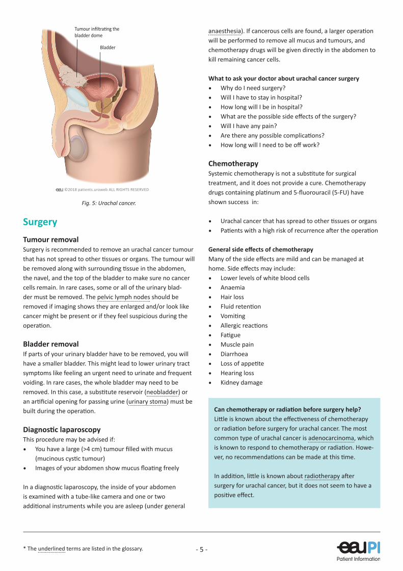
Tumour removalSurgery is recommended to remove an urachal cancer tumour that has not spread to other tissues or organs. The tumour will be removed along with surrounding tissue in the abdomen, the navel, and the top of the bladder to make sure no cancer cells remain. In rare cases, some or all of the urinary blad-der must be removed. The pelvic lymph nodes should be removed if imaging shows they are enlarged and/or look like cancer might be present or if they feel suspicious during the operation.
Bladder removalIf parts of your urinary bladder have to be removed, you will have a smaller bladder. This might lead to lower urinary tract symptoms like feeling an urgent need to urinate and frequent voiding. In rare cases, the whole bladder may need to be removed. In this case, a substitute reservoir (neobladder) or an artificial opening for passing urine (urinary stoma) must be built during the operation.
Diagnostic laparoscopyThis procedure may be advised if:• You have a large (>4 cm) tumour filled with mucus
(mucinous cystic tumour)• Images of your abdomen show mucus floating freely
In a diagnostic laparoscopy, the inside of your abdomen is examined with a tube-like camera and one or two additional instruments while you are asleep (under general
Fig. 5: Urachal cancer.
Tumour infiltrating the bladder dome
Bladder
©2018 patients.uroweb ALL RIGHTS RESERVED
Can chemotherapy or radiation before surgery help?Little is known about the effectiveness of chemotherapy or radiation before surgery for urachal cancer. The most common type of urachal cancer is adenocarcinoma, which is known to respond to chemotherapy or radiation. Howe-ver, no recommendations can be made at this time.
In addition, little is known about radiotherapy after surgery for urachal cancer, but it does not seem to have a positive effect.

* The underlined terms are listed in the glossary. - 6 -
Chemotherapy increases your risk of infection. This is because the chemotherapy lowers your white blood cell levels. If you have a temperature or other signs of infection, such as a cough, contact your treatment team immediately. If you have an infection, you may need antibiotics to treat it.
What to ask your doctor about urachal cancer chemotherapy• Why do I need chemotherapy?• Are there other treatment options besides
chemotherapy?• Which drugs will I have?• How do I take the drugs?• Will I have to stay in hospital?• What are the side effects?• Can I do anything to help prevent side effects?• Who can help me cope with side effects?• Who can I contact if I am worried about side effects?• What should I do if I get an infection?• How long will I have chemotherapy for?• How many courses of chemotherapy will I need?• How long will I need to be off work?• How can you tell if the chemotherapy is working?• How long will it take me to recover from the treatment?
Palliative Care
If your tumour has spread to other organs or tissues (metastases), surgery is not a treatment option. At this point, treatment should reduce symptoms and maintain your quality of life. This is the main focus of palliative care.
During palliative care, you and your loved ones are supported by a multidisciplinary team. Together you address physical, psychological, social, and spiritual issues. Palliative care includes controlling your symptoms and medical treatment for pain management.
The palliative care team can provide care in the hospital or at your home. Another option is hospice care. A hospice is an institution that provides care during the final phase of your life.
Follow-up after treatment
After the operation, you will be scheduled for doctor visits and imaging at regular intervals (follow-ups). The purpose of these visits is to look for treatment side effects and to scan for possible recurrences.
Early detection of recurrence is crucial to make sure you are treated. A consultation and imaging should be scheduled between one to four times a year for 5 to 10 years after the operation. You should personally look for symptoms like blood or mucus in your urine, abdominal pain, or swelling. If you experience any of these symptoms, please contact your doctor.
FAQs
What is urachal cancer?Urachal cancer is a very rare tumour that begins in the urachus. If part of the urachus or the whole urachus remains, symptoms like chronic infection and mucus production can sometimes be seen. Urachal cancer can occur due to years of chronic inflammation and remodelling processes or embryologic cells that remain in the urachus region.
What are the symptoms of urachal cancer?Symptoms of urachal cancer typically appear when the tumour grows into surrounding tissues or organs. Symptoms such as blood in the urine (haematuria), pain, irritative complaints, recurrent bladder infections, or mucus production from the naval or bladder (mucusuria) can be observed.
What tests are done to diagnose urachal cancer?Physical examination, ultrasound of the abdomen, and a urinary swab test are the first diagnostic tests to be performed.
Why are urachal tumours classified?The classification of a tumour by stage guides treatment and predicts outcomes. Treatment and outcome are influenced by: • The type of cells in the tumour (histology)• The size of the tumour and whether it has grown into
adjacent tissues and organs (local extent)• Whether the cancer cells have spread to other tissues or
organs (presence of metastases)
Does everyone have a urachus?Yes, before birth we all do. In most people, the urachus goes away before birth. However, about one-third still have remnants at birth. These remnants degenerate within the first year. If the urachus persists as a bigger structure, it often
OutcomeAbout 20% of urachal cancer patients cannot be cured by the time they develop symptoms. After treatment, about one-third will have relapse or their disease will spread. Average survival is 50% at 5 years.

* The underlined terms are listed in the glossary. - 7 -
causes symptoms and is mostly found and removed in early childhood. If it exists without symptoms or only microscopic residues are present, in rare cases, cancer can develop over decades.
Are there risk factors for developing urachal cancer?No genetic or lifestyle risk factors are known currently.
Is urachal cancer hereditary?To our current knowledge, no.
Are there any screening tests for urachal cancer?No. Urachal cancer is quite rare (about one in 1 million), so there is not a rationale to screen for it.
What is TUR and when is it recommended?TUR is the most direct way to take tissue for examination and diagnosis in most cases. This assumes that the tumour has grown into the bladder.
What is local resection and when is it recommended?Local resection is surgery to remove a urachal cancer tumour that has not spread to other tissues or organs. It is the recommended treatment for urachal cancer. The tumour will be removed along with surrounding tissue in the abdomen, the navel, and the top of the bladder to make sure no cancer cells remain. In rare cases, some or all of the urinary bladder must be removed. The pelvic lymph nodes should be removed if imaging shows they are enlarged and/or look like cancer might be present or if they feel suspicious during the operation.
What is chemotherapy and when is it recommended?Chemotherapy treats cancer with drugs that specifically kill cancer cells. Systemic chemotherapy is not a substitute for surgical treatment, and it does not provide a cure. Chemotherapy drugs containing platinum and 5-fluorouracil (5-FU) have shown success in:
• Urachal cancer that has spread to other tissues or organs• Patients with a high risk of recurrence after the operation
What if my urachal cancer cannot be cured?Sometimes recovery from urachal cancer is not possible. When treatment is no longer successful, you may be offered palliative care to make you more comfortable. Palliative care is focussed on reducing your symptoms and maintaining your quality of life.
Links to more information
http://urachalcancer.org/
https://en.wikipedia.org/wiki/Urachal_cancer
https://www.cancercompass.com/message-board/message/all,1991,0.htm
Literature[1] Schubert GE, Tubular urachal remnants in adult bladders. J Urol. 1982
Jan;127(1):40-2.
[2] Begg RC. The Urachus: its Anatomy, Histology and Development. J Anat
1930;64:170–83.
[3] Ashley RA, Inman BA, Routh JC, Rohlinger AL, Husmann DA, Kramer SA.
Urachal anomalies: a longitudinal study of urachal remnants in children
and adults. J Urol 2007;178:1615–8.
[4] Paner GP, McKenney JK, Barkan GA, Yao JL, Frankel WL, Sebo TJ, et al.
Immunohistochemical analysis in a morphologic spectrum of urachal
epithelial neoplasms: diagnostic implications and pitfalls. Am J Surg
Pathol 2011;35:787–98.
[5] Siefker-Radtke A. Urachal adenocarcinoma: a clinician’s guide for
treatment. Semin Oncol 2012;39:619–24.
[6] Yazawa S, Kikuchi E, Takeda T, Matsumoto K, Miyajima A, Nakagawa K,
et al. Surgical and chemotherapeutic options for urachal carcinoma:
report of ten cases and literature review. Urol Int 2012;88:209–14.
[7] Siefker-Radtke AO, Gee J, Shen Y, Wen S, Daliani D, Millikan RE, et al.
Multimodality management of urachal carcinoma: the M. D. Anderson
Cancer Center experience. J Urol 2003;169:1295–8.
[8] Szarvas T, Módos O, Niedworok C, Reis H, Szendröi A, Szász MA, et al.
Clinical, prognostic, and therapeutic aspects of urachal carcinoma—A
comprehensive review with meta-analysis of 1,010 cases. Urol Oncol
Semin Orig Investig 2016.
[9] Herr HW, Bochner BH, Sharp D, Dalbagni G, Reuter VE. Urachal
carcinoma: contemporary surgical outcomes. J Urol 2007;178:74–8;
discussion 78.
10] Behrendt M, De Jong J, Van Rhijn B. Urachal Cancer: contemporary
review of the pathological, surgical, and prognostic aspects of this rare
disease. Minerva Urol Nefrol 2015.
[11] Sheldon CA, Clayman R V, Gonzalez R, Williams RD, Fraley EE. Malignant
urachal lesions. J Urol 1984;131:1–8.
[12] Kumar N, et al. Urachal carcinoma: clinicopathological features,
treatment and outcome. J Cancer Res Ther. 2014 Jul-Sep;10(3):571-4.
[13] Molina JR, Quevedo JF, Furth AF, Richardson RL, Zincke H, Burch PA.
Predictors of survival from urachal cancer: a Mayo Clinic study of 49
cases. Cancer 2007;110:2434–40.
[14] Collazo-Lorduy A, Castillo-Martin M, Wang L, Patel V, Iyer G, Jordan E,
et al. Urachal Carcinoma Shares Ge-nomic Alterations with Colorectal
Carcinoma and May Respond to Epidermal Growth Factor Inhibition.
Eur Urol 2016.

* The underlined terms are listed in the glossary. - 8 -
Glossary of terms
Abdominal cavityThe space in the body that contains all the abdominal organs (including bladder, kidneys, urinary tract, genital structures)
Abdominal wallThe muscle and tissue that surrounds the abdominal cavity
AdenocarcinomaA type of cancer that starts in and has features of glandular cells
Anaesthesia (general, spinal, or local)Before a procedure, you will get medication to make sure that you do not feel pain. Under general anaesthesia, you are unconscious and unaware of what is happening to you. Under spinal or local anaesthesia, you will not feel pain in the part of your body where the procedure is done. Anaesthesia wears off gradually after the procedure.
BladderOrgan that collects urine from the kidneys
ChemotherapyTreatment of cancer with drugs that aim to kill cancer cells
Chronic infectionAn infection that lasts over a long period of time
CT scanCT stands for Computed Tomography. This imaging technique makes a series of x-ray images of the body.
Embryologic structureA tissue or structure formed during development of an embryo
Histologic examinationExamination of tissue cells under a microscope
ImagingTaking images of the body with ultrasound, x-ray, or other scanning techniques
Local resectionSurgery to remove a tumour that has not spread to other tissues or organs
MetastasesCancer cells that have spread to other organs, tissues, or lymph nodes
MRIMagnetic Resonance Imaging is an imaging technique that uses strong magnetic fields and radio waves to make images of the body
Mucinous histologyMucus-producing cells that can be found in histologic examination under the microscope or by using a specific colouring test.
MucusuriaMucus in the urine
Multidisciplinary tumour boardA team of practitioners from different medical specialties who share their professional opinions to plan care for individual cancer patients
NeobladderA substitute reservoir to hold urine after the bladder is removed
Palliative careA concept of care with the goal to optimise your quality of life if you cannot recover from your illness. It involves physical, psychological, social, and spiritual issues.
Pelvic lymph nodesThe sum of lymph nodes collecting the lymphatic drainage of the legs, pelvis and pelvic organs.
RadiotherapyA type of cancer treatment that uses radiation to control or destroy malignant cells.
RecurrenceThe return of cancer after treatment and after a period of time in which the cancer could not be detected. This can happen at the place where the cancer first was detected or somewhere else in the body. There is no standard period of time, but most doctors would consider it a recurrence if the cancer had not been detected for at least 1 year.
* The underlined terms are listed in the glossary. - 9 -
Glossary of terms
TURTUR stands for Transurethral Resection. A tube-like instrument is used to remove tissue through the urethra (the canal through which the urine is passed).
UltrasoundImaging technique that uses high-frequency sounds to make an image of the inside of the body
Umbilical dischargeSubstance produced by the navel
UrachusA tube-like embryologic structure that connects the forming urinary bladder and the navel before birth
Urachal residuesTraces of the tissue or cells that formed the urachus before birth
Urinary stomaAn artificial opening for passing urine
Urinary swab testAlso known as “urine dip test” or just a “urine test”. A test strip is dipped into collected urine to colour-indicate the pH level and the presence of electrolytes and cells.

European Association of UrologyPO Box 30016NL-6803 AA ARNHEMThe Netherlands
e-Mail: [email protected]: patients.uroweb.org