prenatal counseling series - eapsa.org · includes: omphalocele, gastroschisis, and numerous...
TRANSCRIPT

American Pediatric Surgical Association
Prenatal Counseling SeriesOmphalocele
TM
from the Fetal Diagnosis and Treatment Committee
of the American Pediatric Surgical Association
Editor-in-Chief: Ahmed I. Marwan, MD
Special thanks to Ryan Phillips, MD, Niti Shahi, MD, and Jill Stein, MD
©2019, American Pediatric Surgical Association

American Pediatric Surgical Association
Prenatal Counseling SeriesOmphalocele
2
Definition and Etiology• Omphalocele is one of the two most common abdominal wall defects
encountered by pediatric surgeons.
• Etiology: failure of the migration of lateral folds to form the umbilical ring
and failure of the herniated midgut to return to the abdominal cavity early in
gestation.
• Incidence: 2.5/10,000 to 4/10,000 (1)
Axial ultrasound and sagittal MR images of a fetus with a ventral abdominal wall defect that contains a small portion of the liver. The umbilical cord inserts onto the defect.
American Pediatric Surgical Association
Prenatal Counseling SeriesOmphalocele
3
Definition and Etiology
▪ Omphalocele is one of the two most common abdominal wall defects encountered by pediatric surgeons.
▪ Etiology: failure of the migration of lateral folds to form the umbilical ring and failure of the herniated midgut to return to the abdominal cavity early in gestation.
▪ Incidence: 2.5/10,000 to 4/10,000 (1)
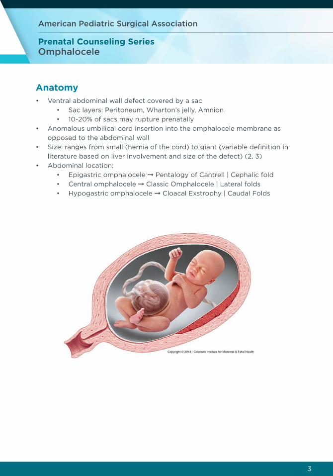
Anatomy
▪ Ventral abdominal wall defect covered by a sac
▪ Sac layers: Peritoneum, Wharton’s jelly, Amnion
▪ 10-20% of sacs may rupture prenatally
▪ Anomalous umbilical cord insertion into the omphalocele membrane as opposed to the abdominal wall
▪ Size: ranges from small (hernia of the cord) to giant (variable definition in literature based on liver involvement and size of the defect) (2, 3)
▪ Abdominal location:
▪ Epigastric omphalocele ! Pentalogy of Cantrell | Cephalic fold
▪ Central omphalocele ! Classic Omphalocele | Lateral folds
▪ Hypogastric omphalocele ! Cloacal Exstrophy | Caudal Folds
!
Anatomy • Ventral abdominal wall defect covered by a sac
• Sac layers: Peritoneum, Wharton’s jelly, Amnion
• 10-20% of sacs may rupture prenatally
• Anomalous umbilical cord insertion into the omphalocele membrane as
opposed to the abdominal wall
• Size: ranges from small (hernia of the cord) to giant (variable definition in
literature based on liver involvement and size of the defect) (2, 3)
• Abdominal location:
• Epigastric omphalocele ➞ Pentalogy of Cantrell | Cephalic fold
• Central omphalocele ➞ Classic Omphalocele | Lateral folds
• Hypogastric omphalocele ➞ Cloacal Exstrophy | Caudal Folds
American Pediatric Surgical Association
Prenatal Counseling SeriesOmphalocele
4
Differential DiagnosisDifferential diagnosis of a prenatally diagnosed congenital abdominal wall defect
includes:
• Ecopia cordis
• Bladder exstrophy
• Cloacal exstrophy
• Urachal anomalies
• Complex abnormalities
o Pentalogy of Cantrell (abdominal wall defect, anterior diaphragmatic
hernia, cardiac anomaly, pericardial defect and sternal cleft)
o Limb-body wall complex/body stalk anomaly
o OEIS: Omphalocele, Exstrophy, Imperforate anus and spinal dysraphism
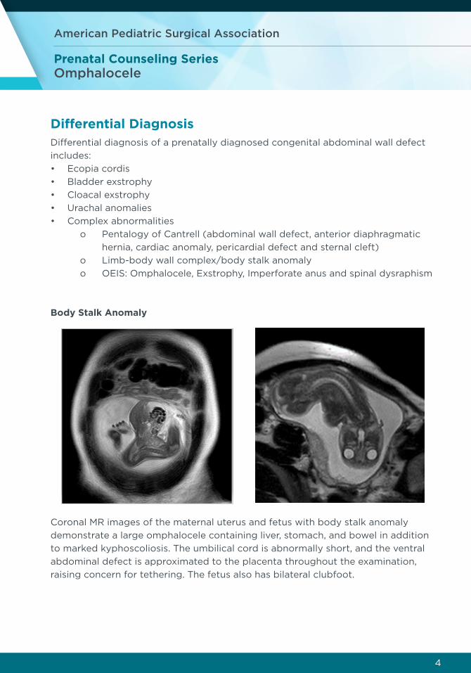
Body Stalk Anomaly
Coronal MR images of the maternal uterus and fetus with body stalk anomaly
demonstrate a large omphalocele containing liver, stomach, and bowel in addition
to marked kyphoscoliosis. The umbilical cord is abnormally short, and the ventral
abdominal defect is approximated to the placenta throughout the examination,
raising concern for tethering. The fetus also has bilateral clubfoot.
Differential Diagnosis
▪ Differential diagnosis of a prenatally diagnosed congenital abdominal wall defect includes: omphalocele, gastroschisis, and numerous complex malformations such as ectopia cordis, bladder exstrophy, cloacal exstrophy, urachal anomalies and limb-body wall complex/Body stalk anomaly.
Body Stalk Anomaly
Coronal MR images of the maternal uterus and fetus with body stalk anomaly demonstrate a large omphalocele containing liver, stomach, and bowel in addition to marked kyphoscoliosis. The umbilical cord is abnormally short, and the ventral abdominal defect is approximated to the placenta throughout the examination, raising concern for tethering. The fetus also has bilateral clubfoot.

American Pediatric Surgical Association
Prenatal Counseling SeriesOmphalocele
5
OEIS
Sagittal MR image of a fetus with OEIS (omphalocele, cloacal exstrophy,
imperforate anus, spinal defects) complex shows a large ventral wall defect
involving the abdomen and pelvis with dominant cystic component consistent with
omphalocele and cloacal exstrophy. Also, there is a lumbosacral spinal defect with
a dorsal cyst.
OEIS
Sagittal MR image of a fetus with OEIS (omphalocele, cloacal exstrophy, imperforate anus, spinal defects) complex shows a large ventral wall defect involving the abdomen and pelvis with dominant cystic component consistent with omphalocele and cloacal exstrophy. Also, there is a lumbosacral spinal defect with a dorsal cyst.
Associated Anomalies
▪ Omphaloceles are commonly associated with other anomalies (50-70%)
▪ Chromosomal abnormalities (20-30%), especially Trisomy 18,13, 21
▪ Beckwith-Wiedemann Syndrome
▪ Pentalogy of Cantrell (abdominal wall defect, anterior diaphragmatic hernia, cardiac anomaly, pericardial defect, and sternal cleft)
▪ Bladder exstrophy
▪ Congenital heart defects (3)
American Pediatric Surgical Association
Prenatal Counseling SeriesOmphalocele
6
Associated Anomalies• Omphaloceles are commonly associated with other anomalies (50-70%)
• Chromosomal abnormalities (20-30%), especially Trisomy 18, 13, and 21
• Beckwith-Wiedemann Syndrome
• Bladder exstrophy
• Congenital heart defects (3)
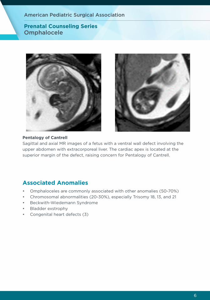
Pentalogy of Cantrell
Sagittal and axial MR image of a fetus with a ventral wall defect involving the upper abdomen with the extracorporeal liver. The cardiac apex is located at the superior margin of the defect, raising concern for Pentalogy of Cantrell.
Giant Omphalocele Giant omphalocele lacks a consensus definition (2,3):
▪ Historically defined by various criteria including: the diameter of the sac or the
abdominal wall defect, inability to primarily close the defect, liver evisceration, and
volume disproportion between the abdominal viscera and abdominal cavity.
▪ Based on recent larger series, omphaloceles are generally considered giant when:
▪ Defect size: >5 cm
▪ Liver herniation: >50% of the liver within the sac
▪ Giant omphaloceles can be associated with varying degrees of (4):
▪ Pulmonary hypoplasia
▪ Pulmonary hypertension
▪ Systemic hypertension
▪ Inguinal hernias
Pentalogy of Cantrell
Sagittal and axial MR images of a fetus with a ventral wall defect involving the
upper abdomen with extracorporeal liver. The cardiac apex is located at the
superior margin of the defect, raising concern for Pentalogy of Cantrell.
American Pediatric Surgical Association
Prenatal Counseling SeriesOmphalocele
7
Giant OmphaloceleGiant omphalocele lacks a consensus definition (2,3):
• Historically defined by various criteria including: the diameter of the sac or the
abdominal wall defect, inability to primarily close the defect, liver evisceration,
and volume disproportion between the abdominal viscera and abdominal
cavity.
• Based on recent larger series, omphaloceles are generally considered giant
when:
• Defect size: >5 cm
• Liver herniation: >50% of the liver within the sac
• Giant omphaloceles can be associated with varying degrees of (4):
• Pulmonary hypoplasia
• Pulmonary hypertension
• Systemic hypertension
• Inguinal hernias
• Undescended testes
• GERD
• Feeding difficulties
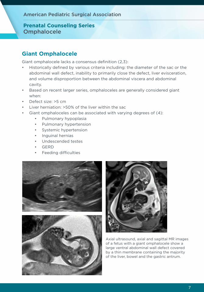
Axial ultrasound, axial and sagittal MR images of a fetus with a giant omphalocele show a large ventral abdominal wall defect covered by a thin membrane containing the majority of the liver, bowel and the gastric antrum.

American Pediatric Surgical Association
Prenatal Counseling SeriesOmphalocele
8
Initial EvaluationObstetrical ultrasound
Fetal echocardiography
Fetal magnetic resonance imaging (MRI)
Genetic testing
Obstetrical ultrasound (5,6,7)
• Assess for protrusion of viscera through the abdominal wall
• Presence of an overlying membrane (sac)
• Insertion of umbilical cord into the defect
• Presence of associated anomalies
• Measure the size of the defect
• Presence of extracorporeal liver
Echo
• Cardiac structure and function
• Assess for any cardiac anomalies
Fetal MRI
MRI is complementary to ultrasound and is particularly useful for distinguishing
liver from the bowel that at times may appear similar by ultrasound
• Measure the proportion of extracorporeal liver
• Quantify defect size
• Useful for associated abnormalities
• Total lung volume (degree of pulmonary hypoplasia)
• Observed/expected total lung volume (O/E TLV) calculated using
normative data by gestational age
• O/E TLV <50% predictive of increased postnatal morbidity
Genetic Testing
• Should be performed regardless of whether it is a small omphalocele or hernia
of the cord
• ➞ in maternal serum AFP and B-HCG
• Offer genetic testing for Beckwith-Wiedemann syndrome
• Aneuploidies

American Pediatric Surgical Association
Prenatal Counseling SeriesOmphalocele
9
Prenatal Counseling• Can be diagnosed as early as 10-12 weeks on prenatal screening ultrasound
• Serial ultrasound examinations to assess fetal growth and amniotic fluid
volumes
• Monitor pregnancy for preterm labor and intrauterine growth restriction
• Close antenatal surveillance is recommended due to the possibility of late
gestational fetal demise
• Isolated omphalocele defects have a good prognosis
Giant Omphalocele
• Referral to a fetal center for multidisciplinary counseling including pediatric
surgery, MFM, neonatology and genetics
• Obstetrical ultrasounds every four weeks
• Consider fetal MRI at 34 weeks in giant omphaloceles to assess for pulmonary
hypoplasia and to obtain an objective evaluation of total lung volume
• Twice weekly non-stress tests or biophysical profiles starting at 32 weeks
• Major consideration for the mode of delivery is related to the risk of rupture
o Pregnancy should be allowed to proceed as close to term as possible
o For giant omphaloceles, Cesarean section may be justified
• Rupture is an emergent situation!
Postnatal Considerations• The clinical management of these defects varies from straight forward to
complex. The morbidity and mortality are often linked to the associated
congenital anomalies and size of the defect.
• Evaluation for syndromic features (enlarged tongue, heart defects, and
hemihypertrophy)
• Full genetic evaluation as the presence of chromosomal anomalies, cardiac
defects, and syndromic conditions impacts outcomes and timing of surgical
repair
• Assess for pulmonary hypertension and pulmonary hypoplasia
• Surgical repair via either primary closure or delayed primary/secondary closure
American Pediatric Surgical Association
Prenatal Counseling SeriesOmphalocele
10
References1) Campbell KH, Copel JA. Omphalocele. In: Obstetric Imaging: Fetal Diagnosis
and Care (Second Edition). Elsevier; 2018. p. 84,91. e1.
2) Danzer E, Gerdes M, D’Agostino JA, Bernbaum J, Hoffman C, Rintoul NE, et
al. Patient characteristics are important determinants of neurodevelopmental
outcome during infancy in giant omphalocele. Early Hum Dev. 2015;91(3):187-
93.
3) Ein SH, Langer JC. Delayed management of giant omphalocele using silver
sulfadiazine cream: an 18-year experience. J Pediatr Surg. 2012;47(3):494-
500.
4) Partridge EA, Hanna BD, Panitch HB, Rintoul NE, Peranteau WH, Flake AW,
et al. Pulmonary hypertension in giant omphalocele infants. J Pediatr Surg.
2014;49(12):1767-70.
5) Kleinrouweler CE, Kuijper CF, van Zalen-Sprock MM, Mathijssen IB, Bilardo CM,
Pajkrt E. Characteristics and outcome and the omphalocele circumference/
abdominal circumference ratio in prenatally diagnosed fetal omphalocele.
Fetal Diagn Ther. 2011;30(1):60-9.
6) Kleinrouweler CE, Kuijper CF, van Zalen-Sprock MM, Mathijssen IB, Bilardo CM,
Pajkrt E. Characteristics and outcome and the omphalocele circumference/
abdominal circumference ratio in prenatally diagnosed fetal omphalocele.
Fetal Diagn Ther. 2011;30(1):60-9.
7) Kamata S, Usui N, Sawai T, Nose K, Fukuzawa M. Prenatal detection of
pulmonary hypoplasia in giant omphalocele. Pediatr Surg Int. 2008;24(1):107-
11.
8) Van Eijck FC, de Blaauw I, Bleichrodt RP, et al. Closure of giant omphaloceles
by the abdominal wall component separation technique in infants. J Pediatr
Surg. 2008;43(1):246-50
9) Levy S, Tsao K, Cox CS, et al. Component separation for complex congenital
abdominal wall defects: not just for adults anymore. J Pediatr Surg.
2013;48(12):2525-9
10) Tsakayannis DE, Zurakowski D, Lillehei CW. Respiratory insufficiency at
birth: a predictor of mortality for infants with omphalocele. J Pediatr Surg.
1996;31(8):1088-90; discussion 1090-1.
11) Edwards EA, Broome S, Green S, et al. Long-term respiratory support in
children with giant omphalocele. Anaesth Intensive Care. 2007;35(1):94-8.