powerpoint presentationamos3.aapm.org/abstracts/pdf/99-30883-365478-117368.pdf•noninvasive method...
TRANSCRIPT
7/15/2015
1
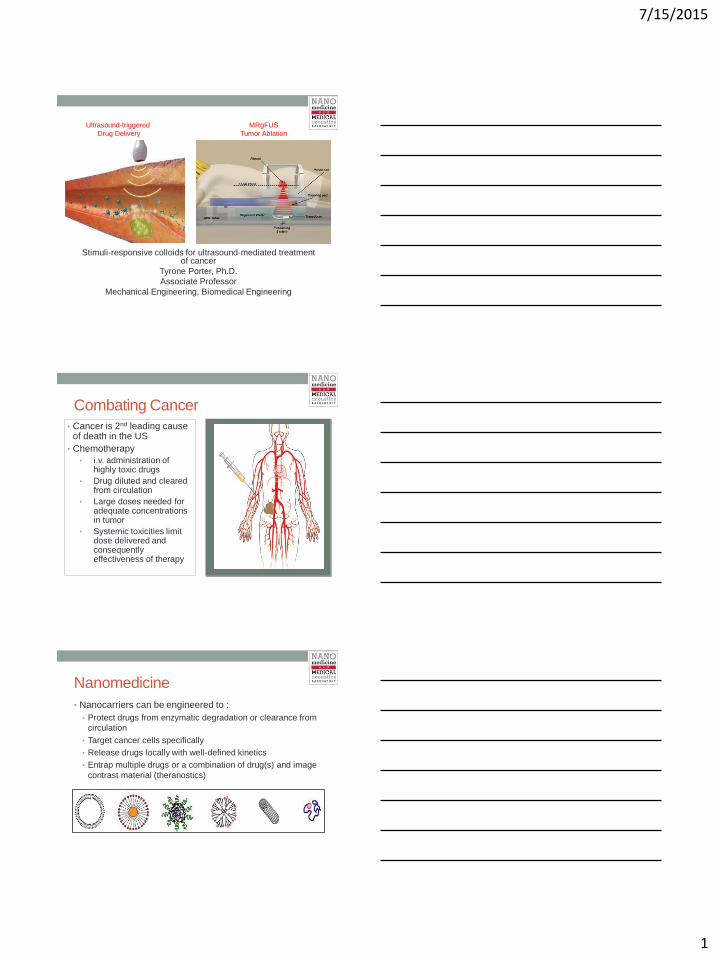
Stimuli-responsive colloids for ultrasound-mediated treatment of cancer
Tyrone Porter, Ph.D.
Associate Professor
Mechanical Engineering, Biomedical Engineering
Ultrasound-triggered
Drug Delivery
MRgFUS
Tumor Ablation
Combating Cancer
• Cancer is 2nd leading cause of death in the US
• Chemotherapy
• i.v. administration of highly toxic drugs
• Drug diluted and cleared from circulation
• Large doses needed for adequate concentrations in tumor
• Systemic toxicities limit dose delivered and consequently effectiveness of therapy
Nanomedicine
• Nanocarriers can be engineered to :
• Protect drugs from enzymatic degradation or clearance from
circulation
• Target cancer cells specifically
• Release drugs locally with well-defined kinetics
• Entrap multiple drugs or a combination of drug(s) and image
contrast material (theranostics)

7/15/2015
2
DOXIL ® • Clinically approved liposome encapsulated Doxorubicin
(DOX)
• Surface modified with Poly(ethylene glycol) to avoid
detection from MPS
Figure adapted from Ortho Biopharmaceuticals, subsidiary of
Johnson and Johnson ©2008
Slow drug release:
Passive diffusion
Degradation of lipid
shell
Reduces side effects
Marginal
improvement in
therapeutic efficacy:
drug release rate too
slow; resultant
intratumoral dose too
low
Thermosensitive Liposomes
From Kong et al Cancer Research 2000
Criteria for Success
• Thermosensitive liposome must circulate for
hours and be stable in blood
• This will optimize extravasation and drug delivery to
cancer cells
• Heating must be localized to tumor and sustained
long enough to release > 50% of payload
• An external noninvasive energy source is ideal
• Noninvasive method for measuring temperature
elevation and feedback control of heating source
• This is critical as the body will attempt to cool heated
tissue

7/15/2015
3
Documented Formulations
From Ta and Porter J Cont Release 2013
• DPPC is main lipid
• Ttrans = 42-44oC
• Stably retains payload in
blood until heated
• Long-circulating
• Slow release kinetics
• Heat > 15 min
• Lysolipid (MPPC) added,
drops Ttrans to 39-40oC
• Rapid release kinetics
• Heat 1-2 min
• Unstable in blood,
significant release within
60 minutes
Polymer-modified TSL
• DPPC main constituent…ensures stability in serum and sensitivity to heat
• PEGylated for long-circulation in blood
• Thermosensitive polymer terminated with fatty acid, which inserts into lipid bilayer
• Polymer goes from hydrophilic-to-hydrophobic when heated above LCST and
disrupts lipid bilayer…enhances payload release
From Ta and Porter J Cont Release 2013
DOX Release as function of T and pH
0
20
40
60
80
100
20 24 28 32 36 40 44
Temperature (°C)
% D
OX
re
lea
se
d
NTSL (n=4)
TSL (n=4)
pTSL (pH 7.5) (n=4)
pTSL (pH 5) (n=4)
5.44
4.55E-02 3.83E-030
1
2
3
4
5
6
7
TSL pTSL (pH 7.5) pTSL (pH 5)
The
rmal do
se (
min
)
5.44
4.55E-02 3.83E-030
1
2
3
4
5
6
7
TSL pTSL (pH 7.5) pTSL (pH 5)
The
rmal do
se (
min
)
5.44
4.55E-02 3.83E-030
1
2
3
4
5
6
7
TSL pTSL (pH 7.5) pTSL (pH 5)
The
rmal do
se (
min
)
Ta et al Biomacromolecules 2010 DOX:lipid mass ratio ~ 6%
N-isopropylacrylamide
thermosensitive
Propyl acrylic acid
pH-sensitive
Terminal
hydrophobic
unit
Copolymer Structure

7/15/2015
4
Thermal Dose (50% DOX release)
5.44
4.55E-02 3.83E-030
1
2
3
4
5
6
7
TSL pTSL (pH 7.5) pTSL (pH 5)
The
rmal do
se (
min
)
5.44
4.55E-02 3.83E-030
1
2
3
4
5
6
7
TSL pTSL (pH 7.5) pTSL (pH 5)
The
rmal do
se (
min
)
* * * *
*
5.44
4.55E-02 3.83E-030
1
2
3
4
5
6
7
TSL pTSL (pH 7.5) pTSL (pH 5)
The
rmal do
se (
min
)
* * * *
*
5.44
4.55E-02 3.83E-030
1
2
3
4
5
6
7
TSL pTSL (pH 7.5) pTSL (pH 5)
Th
erm
al d
ose
(m
in)
p < 0.0001 p < 0.0001
p < 0.001
Ta et al Biomacromolecules 2010
DOX Release @ 37oC (20% serum)
Ta et al Biomacromolecules 2010
In vitro system for FUS-mediated heating
• 50% glycerol:saline attenuation coefficient @ 5 MHz: 0.795 dB/cm
• Mixture used to characterize FUS-mediated heating
• Optically transparent…ideal for quantifying DOX released from
liposomes via spectrofluorophotometry

7/15/2015
5
Ultrasound-induced heating measured
with MR Thermometry
MR-guided ultrasound was used to heat implanted
tumor to 42-44oC for 5 min. MR thermometry used
to measure temperature change.
Untreated
TSL-DOX (5mg/kg)
+ FUS (43°C)
PTSL-DOX (5mg/kg)
+ FUS (43°C)
Average intensity /
area
2.205 ± 0.435
4.137 ± 0.348
0.891 ±
0.028
Evidence of Tissue Remodeling
Extracted tumors
sectioned and
stained for extra
cellular matrix
(ECM). More ECM
was detected in
tumors treated with
pTSL and HIFU than
other treatments,
suggesting that this
treatment
combination was
more effective.
Ta et al J Cont Release 2014

7/15/2015
6
Ta et al J Cont Release 2014
Summary
• Engineered nanocarrier that is responsive to both
mild heating and mild acidity
• Incorporation of copolymer significantly reduced
thermal dose required for triggered DOX release
• Sustained heating and pre-defined thermal dose
delivered to solid tumor possible with MRgFUS
• pTSL released more DOX in solid tumors when
heated to 43oC for 5 min
• Combination of ultrasound-mediated heating and
pTSL effective at slowing tumor growth significantly
Ultrasound-triggered
Drug Delivery
MRgFUS
Tumor Ablation

7/15/2015
7
Time (s)
Tem
per
atu
re C
han
ge
(°C
)
PC
D O
utp
ut (A
rb.)
0 1 2 3 4 5 0
10
20
30
0
0.5
1 1.65 MPa
0
10
20
30
0
0.5
1 1.8 MPa
0
0.5
1
0
10
20
30 1.9 MPa
Good correlation
between onset of
enhanced heating &
onset of broadband
acoustic emissions
Inertial Cavitation
Matters Most
Cavitation-enhanced HIFU Heating (courtesy of Holt & Roy)
Bubble-enhanced Heating at
1.1-MHz in Rabbit Thigh
21
CW exposure for 20 s
(from Sokka et al Phys Med Biol 2003)
0.5 s tone burst; 19.5 s CW
Cavitation was initiated with 0.5 s 300 W tone burst, followed by 19.5 W
continuous wave exposure for ablation.
7/15/2015
8
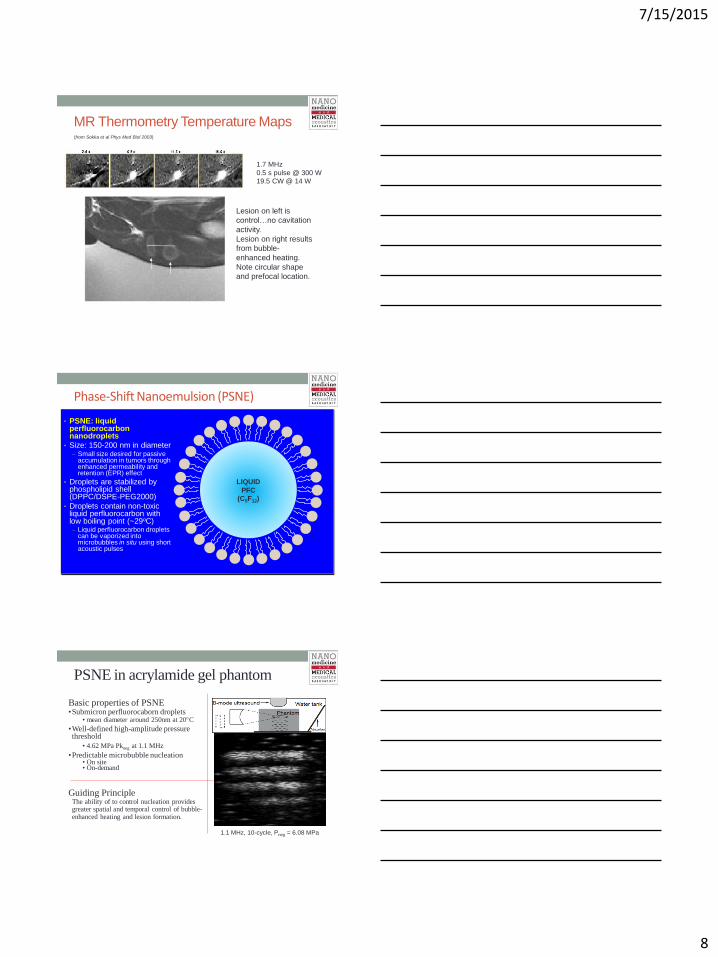
MR Thermometry Temperature Maps
1.7 MHz
0.5 s pulse @ 300 W
19.5 CW @ 14 W
(from Sokka et al Phys Med Biol 2003)
Lesion on left is
control…no cavitation
activity.
Lesion on right results
from bubble-
enhanced heating.
Note circular shape
and prefocal location.
Phase-Shift Nanoemulsion (PSNE)
LIQUID
PFC
(C5F12)
• PSNE: liquid perfluorocarbon nanodroplets
• Size: 150-200 nm in diameter – Small size desired for passive
accumulation in tumors through enhanced permeability and retention (EPR) effect
• Droplets are stabilized by phospholipid shell (DPPC/DSPE-PEG2000)
• Droplets contain non-toxic liquid perfluorocarbon with low boiling point (~29oC) – Liquid perfluorocarbon droplets
can be vaporized into microbubbles in situ using short acoustic pulses
PSNE in acrylamide gel phantom
Basic properties of PSNE •Submicron perfluorocaborn droplets
• mean diameter around 250nm at 20C
•Well-defined high-amplitude pressure threshold
• 4.62 MPa Pkneg at 1.1 MHz
•Predictable microbubble nucleation • On site • On-demand
1.1 MHz, 10-cycle, Pneg = 6.08 MPa
Guiding Principle The ability of to control nucleation provides greater spatial and temporal control of bubble-enhanced heating and lesion formation.

7/15/2015
9
Experimental Setup
Rabbit
Magnet
Bore
Direction of
Ultrasound
Beam Imaging
Plane
Lesion
Degassed
Water
MR coil
MRI Table
HIFU Transducer
RF
Degassed
Water Plastic Bag
GE Signa 3T MRI System
MR-compatible, 1.5 MHz HIFU Transducer
PSNE Accumulation
26
• PSNE tagged with gadolinium, injected intravenously through ear vein • T1-weighted MR images acquired before injection and 0.5, 2, and 6 hours after
PSNE injection Kopechek et al J Heathcare Eng 2013
HIFU Parameters – Center Frequency: 1.5 MHz
– Pulse Repetition Period: 670 ms
– Total Sonication Time: 30 s
– Pulsing Scheme:
1. Short, high-amplitude pulse (100 cycles) to vaporize PSNE
2. Long (1 million cycles), low-amplitude pulse to drive inertial
cavitation and accelerate heating
27
Vaporization pulse
Long pulse to drive cavitation
7/15/2015
10
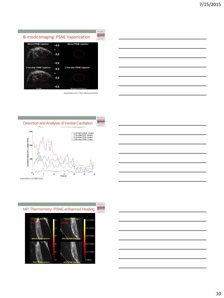
B-mode Imaging: PSNE Vaporization
Kopechek et al J Ther Ultrasound 2014
Detection and Analysis of Inertial Cavitation
Kopechek et al PMB 2014
MR Thermometry: PSNE-enhanced Heating
30

7/15/2015
11
PSNE-enhanced Heating
0
2
4
6
8
10
12
14
-10 10 30 50 70 90 110
Me
an ∆
T (
°C)
Time (s)
Before PSNE Injection
After PSNE Injection
N=3 Animals 1.E-01
1.E+00
1.E+01
1.E+02
1.E+03
1.E+04
1.E+05
1.E+06
1.E+07
0 20 40 60 80 100 120
Ther
mal
Do
se (
CEM
43)
Time (s)
Before PSNE Injection
After PSNE Injection
N=3 Animals
Green box indicates time when HIFU was on (from t=0 to t=30 seconds)
Kopechek et al PMB 2014
Cavitation-enhanced Lesion Formation
• Typically, tumors are treated with > 10 W of HIFU for ablation
• It was possible to ablate tumor in the presence of vaporized PSNE with acoustic power as low as 3 W
• Size of ablated volume depended upon acoustic power
• Polyacrylamide gel phantom • Mean size: 208 nm (+/- 23 nm)
• 8.3x10⁶ – 8.3x10⁸ PSNE/mL
1.6 MHz focused ultrasound 5 MPa PNP 100 cycle vaporization pulse
2 MPa PNP 1,000,000 cycle heating pulse
50 second treatment time (~95% duty cycle)
600 kHz ring passive
cavitation detector
(PCD)
MR Imaging and Thermometry Proton resonant frequency (PRF) shift
128 element linear array
4.8 MHz
(3.1 – 6.5 MHz)
Passive Cavitation Mapping
Integrated MR Thermometry and Cavitation Monitoring

7/15/2015
12
Cavitation Activity • Data collected throughout entire treatment (50 seconds, 78 sonications)
• Each sonication time trace was enveloped and summated
Cavitation Value (dB) = 20 log10
Vsum
Vnoise
æ
è
ç ç
ö
ø
÷ ÷
0
2
4
6
8
10
12
0 10 20 30 40 50 60 70
Cavitation
Valu
e
(dB
ref
nois
e)
Sonication # (0 - 50 seconds)
8.4e7 PSNE/mL No PSNE 4.2e8 PSNE/mL
Control Gel
0
2
4
6
8
10
12
14
8.E+06 8.E+07 8.E+08
Cavitation
Valu
e
(dB
ref
nois
e)
Concentration (PSNE/mL)
*
MR Thermometry
Control gel (no PSNE)
Max ΔT=8.60C (+/- 0.4, n=3)
1.66x10⁸ PSNE/mL
Max ΔT=19.40C (+/- 0.8, n=3)
4.15x10⁸ PSNE/mL
Max ΔT=15.70C (+/- 0.6, n=3)
200C
100C
00C
0
1
2
3
4
5
6
7
8
9
8.E+06 8.E+07 8.E+08
Mean
ΔT
(0C
)
Concentration (PSNE/mL)
Control Gel
*
Passive Cavitation Mapping
4.15x10⁸ PSNE/mL 1.66x10⁸ PSNE/mL Control gel (no PSNE)
Linear Array
7/15/2015
13
Summary • PSNE can accumulate in established tumors and seed
inertial cavitation
• PSNE-nucleated cavitation enhanced heating, applied
thermal dose, and reduced acoustic intensity required for
lesion formation in vivo
• Combined MR thermometry and ultrasound monitoring in
PSNE-loaded hydrogels:
• Capture cavitation and heating migration
• Multimodality feedback control of cavitation-
enhanced tumor ablation
• Ongoing work for treatment of established tumors in
rabbit kidney
Acknowledgements
• Nanomedicine and medical acoustics laboratory
• Focused ultrasound laboratory
• National Institute of Health (NIH) • Grant #: R01EB016102
• Focused Ultrasound
Foundation