practical hematology leukocytosis wendy blount, dvm august 28-19, 2010
Post on 21-Dec-2015
230 views
TRANSCRIPT
Practical HematologyLeukocytosis
Wendy Blount, DVM
August 28-19, 2010
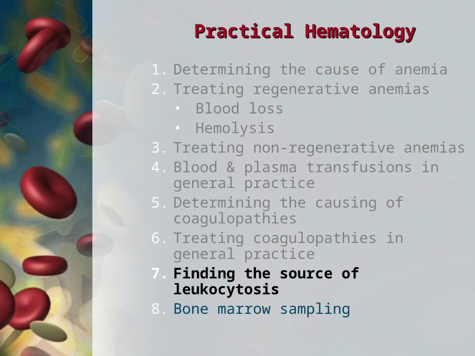
Practical HematologyPractical Hematology
1. Determining the cause of anemia2. Treating regenerative anemias
• Blood loss• Hemolysis
3. Treating non-regenerative anemias4. Blood & plasma transfusions in general
practice5. Determining the causing of
coagulopathies6. Treating coagulopathies in general
practice7. Finding the source of leukocytosis8. Bone marrow sampling
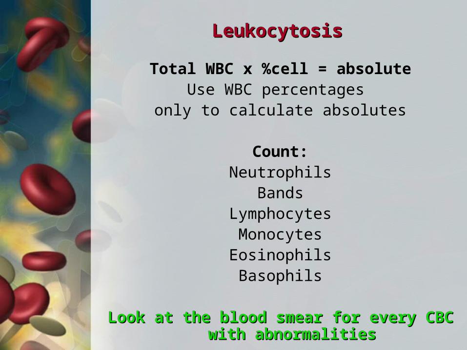
LeukocytosisLeukocytosis
Total WBC x %cell = absoluteUse WBC percentages
only to calculate absolutes
Count:Neutrophils
BandsLymphocytesMonocytesEosinophilsBasophils
Look at the blood smear for every CBC with Look at the blood smear for every CBC with abnormalitiesabnormalities
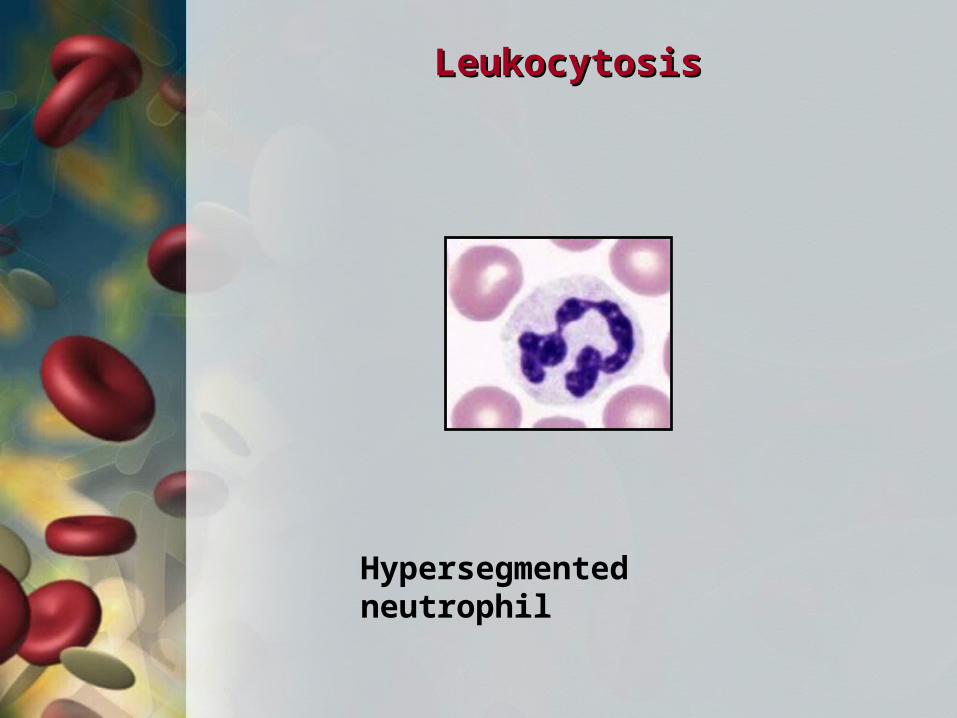
LeukocytosisLeukocytosis
Hypersegmented neutrophil
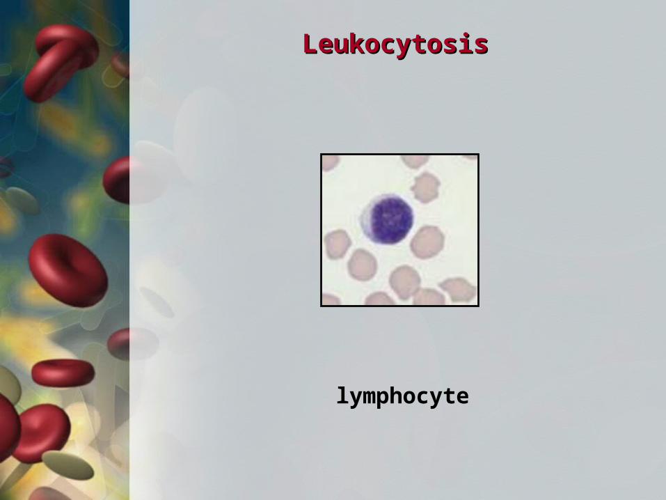
LeukocytosisLeukocytosis
lymphocyte
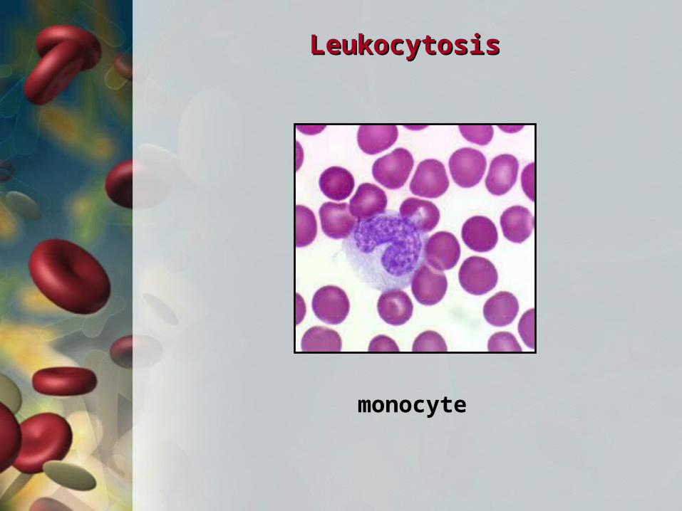
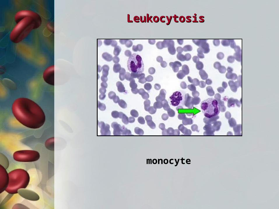
LeukocytosisLeukocytosis
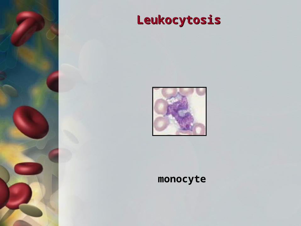
monocyte
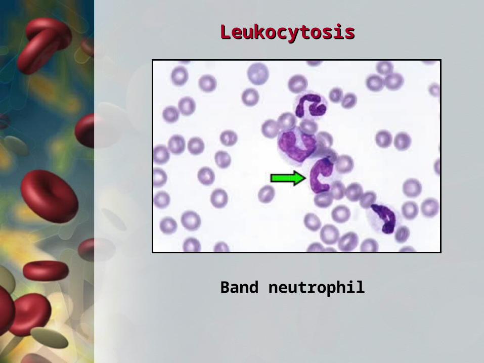
LeukocytosisLeukocytosis
Band neutrophil
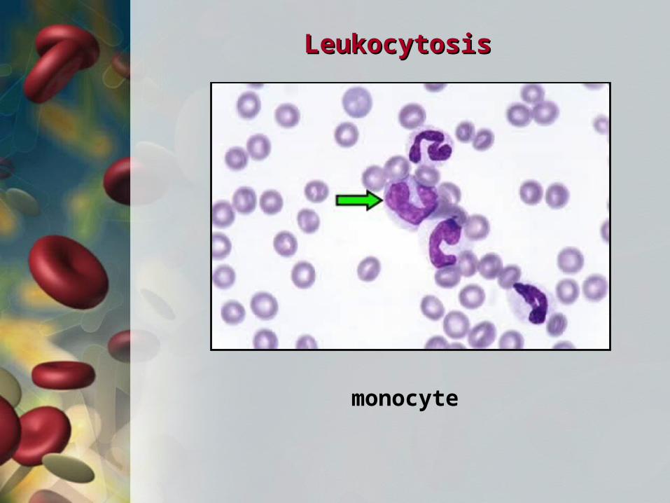
LeukocytosisLeukocytosis
monocyte
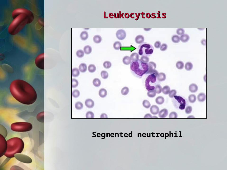
LeukocytosisLeukocytosis
Segmented neutrophil
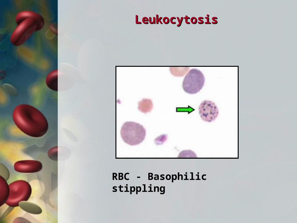
LeukocytosisLeukocytosis
RBC - Basophilic stippling
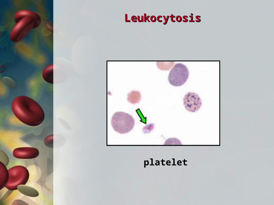
LeukocytosisLeukocytosis
platelet
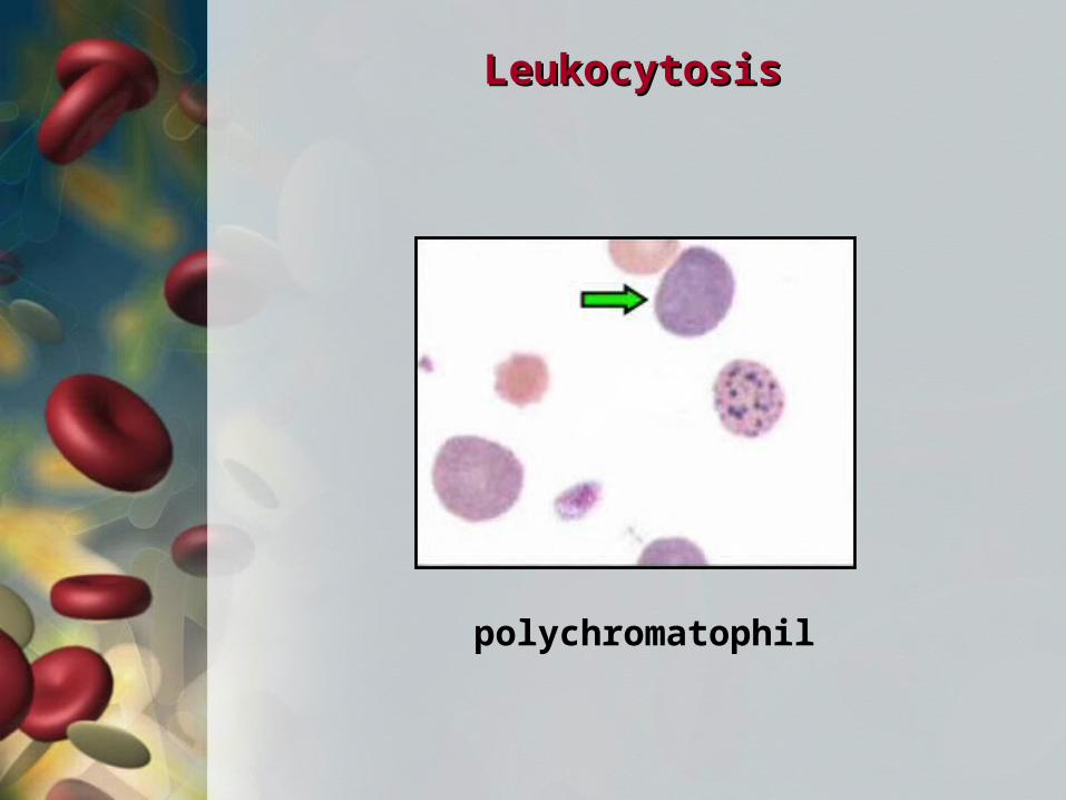
LeukocytosisLeukocytosis
polychromatophil
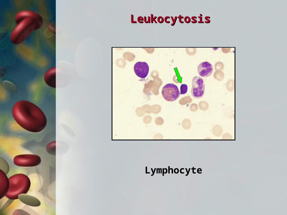
LeukocytosisLeukocytosis
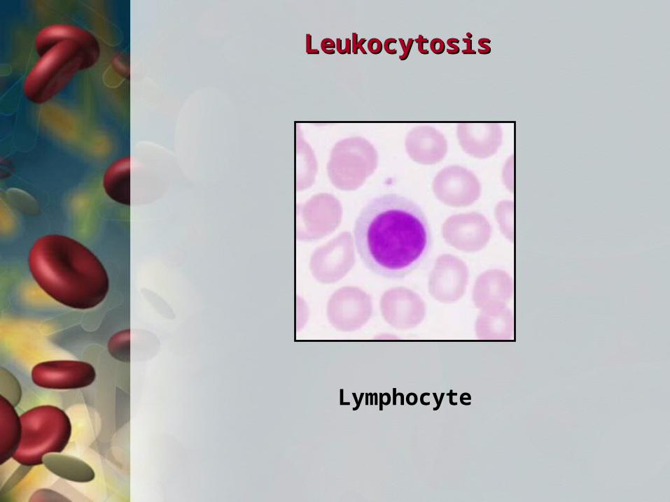
Lymphocyte
LeukocytosisLeukocytosis
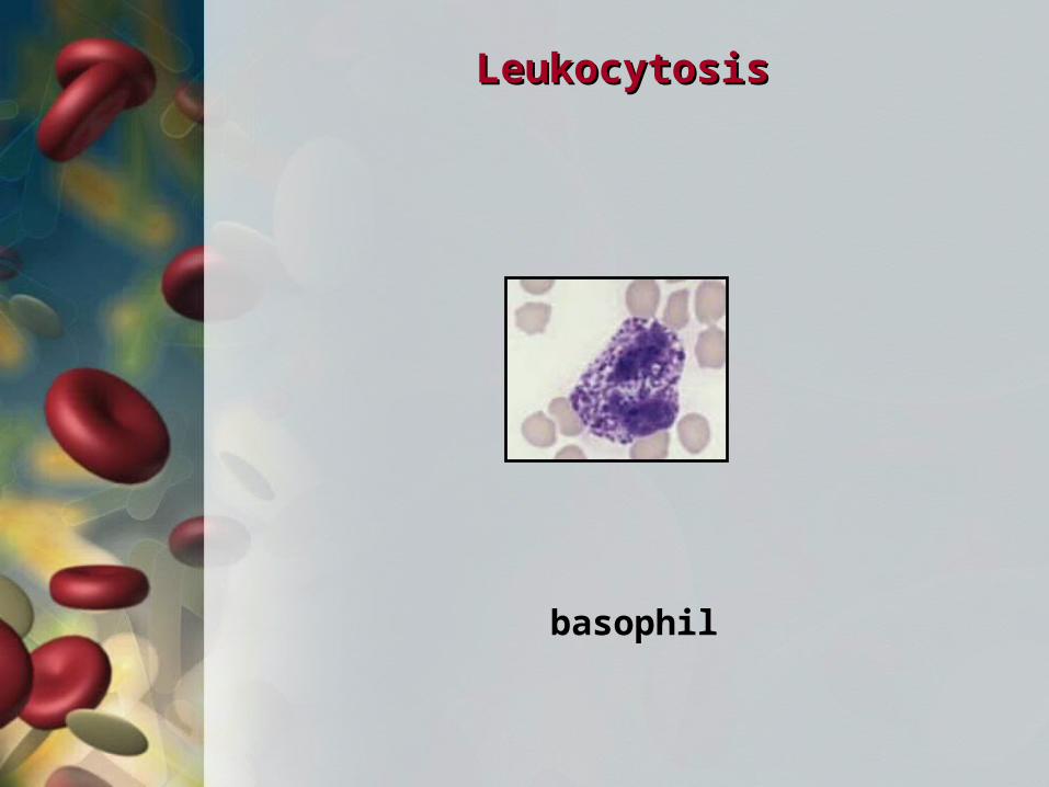
basophil
LeukocytosisLeukocytosis
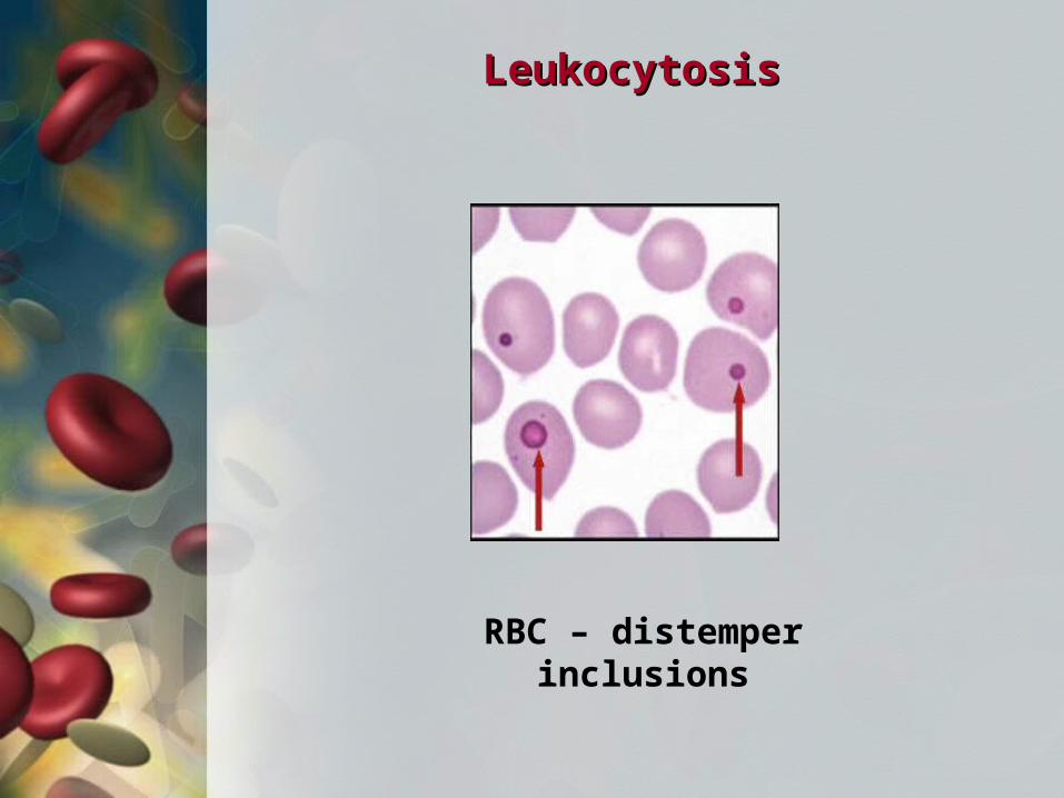
RBC – distemper inclusions
LeukocytosisLeukocytosis
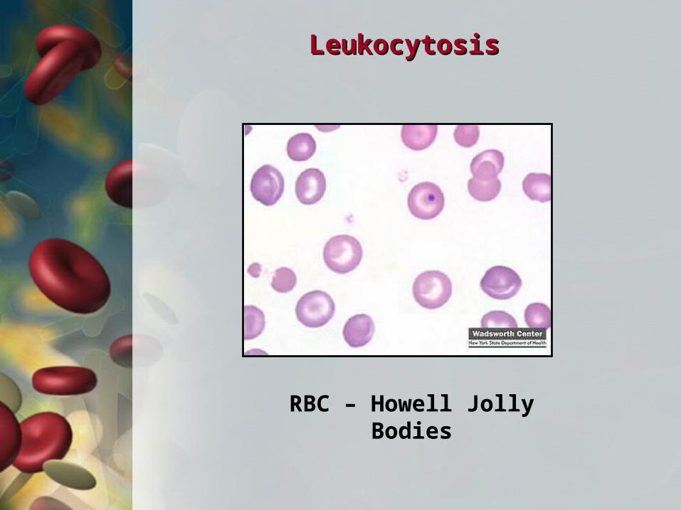
RBC – Howell Jolly Bodies
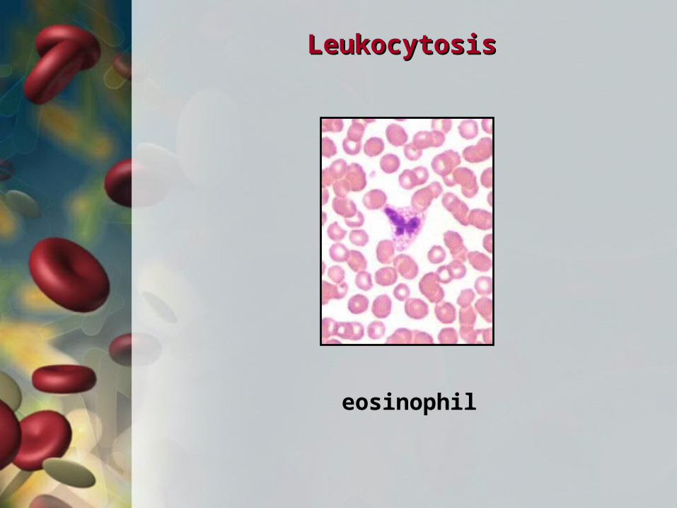
LeukocytosisLeukocytosis
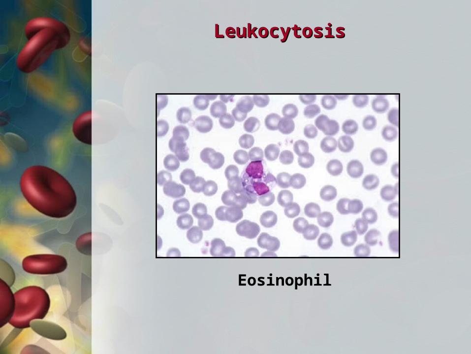
Eosinophil
LeukocytosisLeukocytosis
monocyte
LeukocytosisLeukocytosis
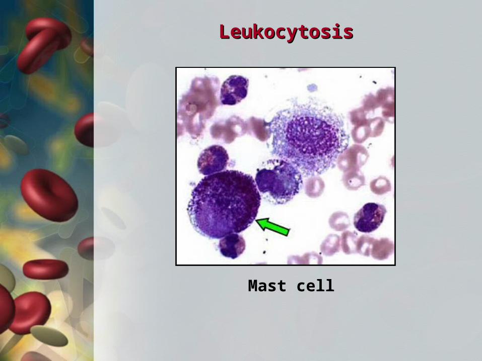
Mast cell
LeukocytosisLeukocytosis
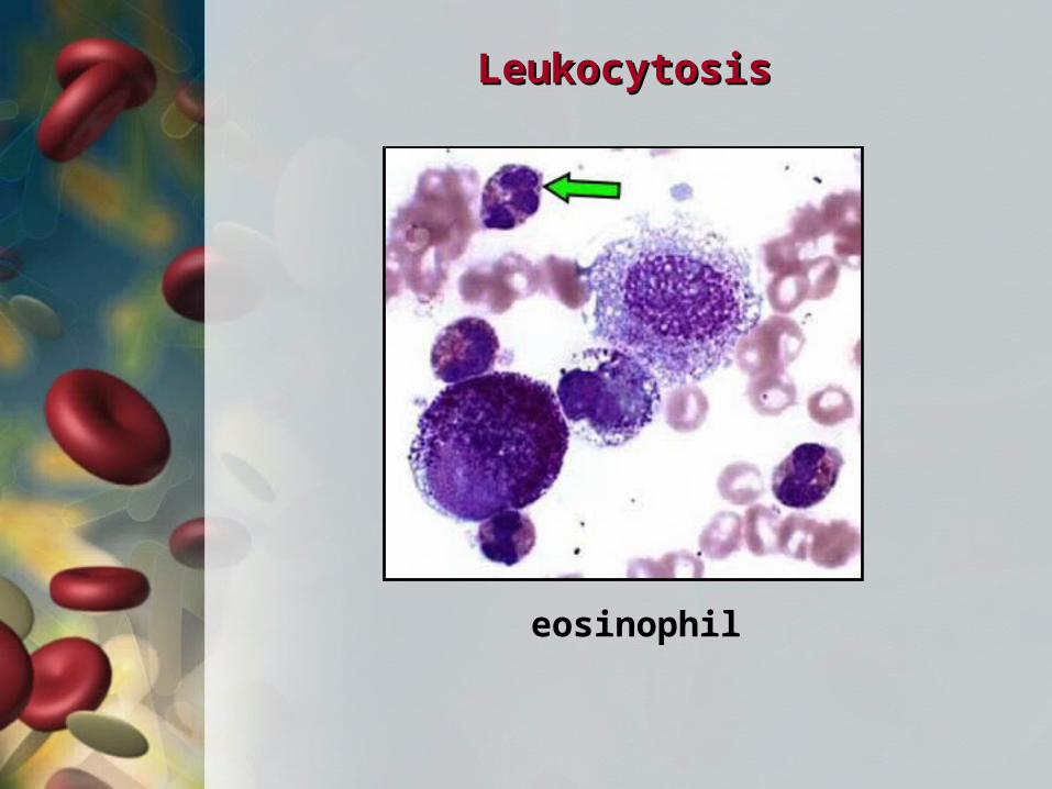
eosinophil

LeukocytosisLeukocytosis
basophil
LeukocytosisLeukocytosis
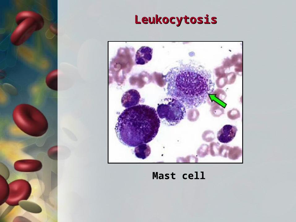
Mast cell
LeukocytosisLeukocytosis
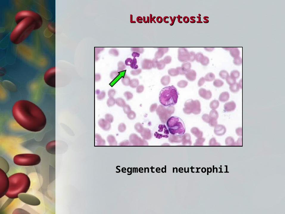
Segmented neutrophil
LeukocytosisLeukocytosis
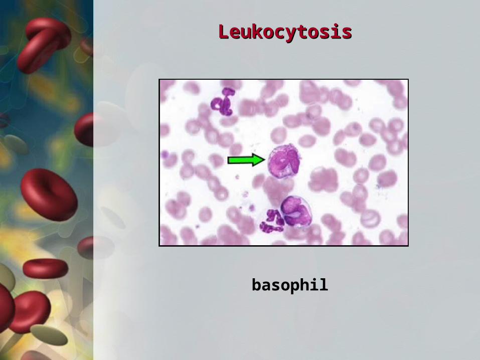
basophil

LeukocytosisLeukocytosis
monocyte
LeukocytosisLeukocytosis
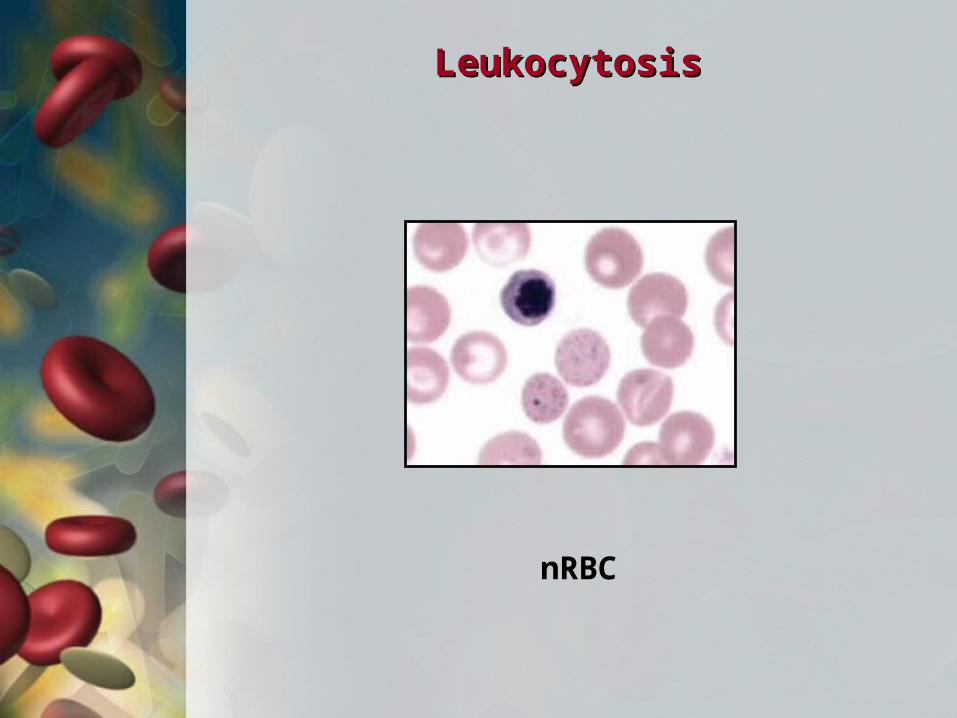
nRBC
LeukocytosisLeukocytosis
eosinophil
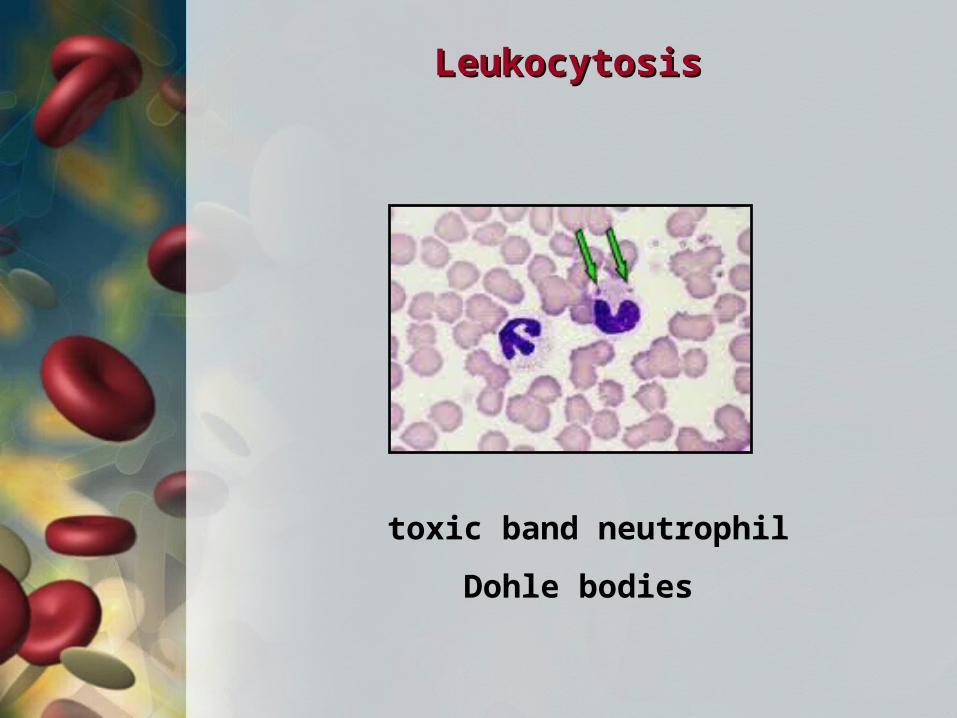
LeukocytosisLeukocytosis
toxic band neutrophil
Dohle bodies
LeukocytosisLeukocytosis
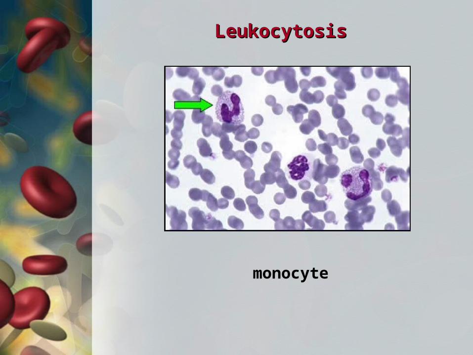
monocyte
LeukocytosisLeukocytosis
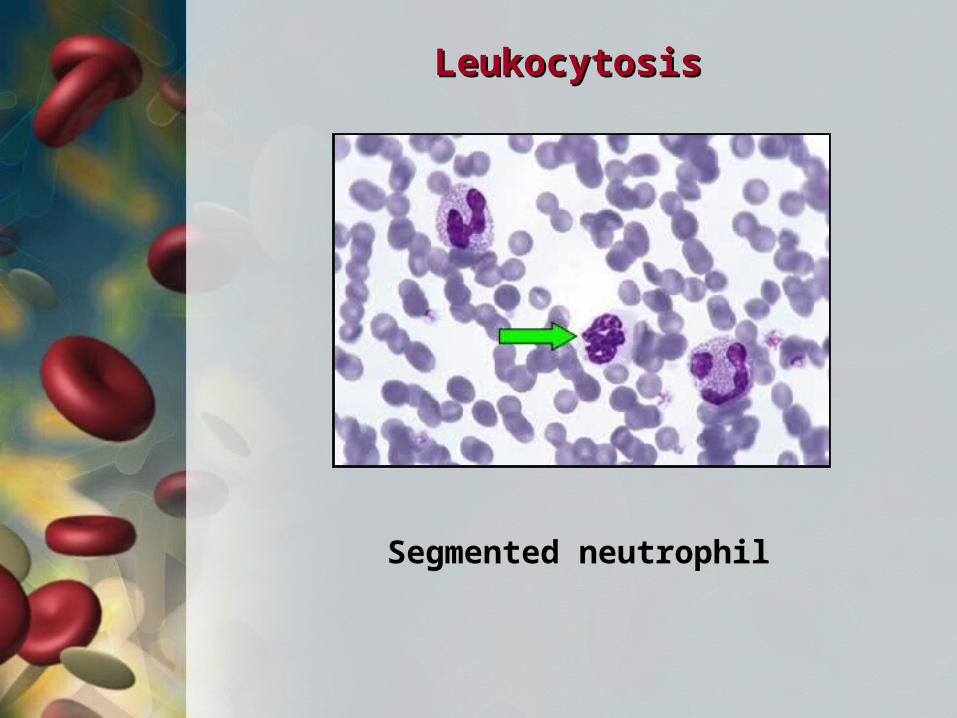
Segmented neutrophil
LeukocytosisLeukocytosis
monocyte
LeukocytosisLeukocytosis
Lymphocyte

LeukocytosisLeukocytosis
band eosinophil

LeukocytosisLeukocytosis
Segmented neutrophil

LeukocytosisLeukocytosis
Activated lymphocyte
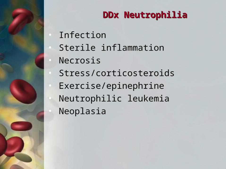
DDx NeutrophiliaDDx Neutrophilia
• Infection • Sterile inflammation• Necrosis• Stress/corticosteroids• Exercise/epinephrine• Neutrophilic leukemia• Neoplasia
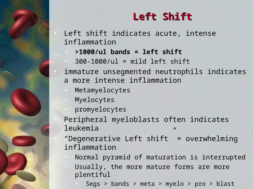
Left ShiftLeft Shift
• Left shift indicates acute, intense inflammation• >1000/ul bands = left shift
• 300-1000/ul = mild left shift
• immature unsegmented neutrophils indicates a more intense inflammation• Metamyelocytes
• Myelocytes
• promyelocytes
• Peripheral myeloblasts often indicates leukemia• “Degenerative Left shift” = overwhelming
inflammation• Normal pyramid of maturation is interrupted
• Usually, the more mature forms are more plentiful• Segs > bands > meta > myelo > pro > blast
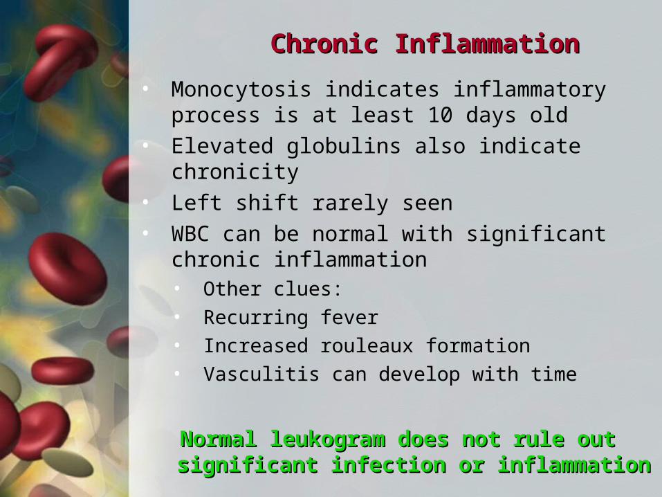
Chronic InflammationChronic Inflammation
• Monocytosis indicates inflammatory process is at least 10 days old
• Elevated globulins also indicate chronicity• Left shift rarely seen• WBC can be normal with significant chronic
inflammation• Other clues:
• Recurring fever
• Increased rouleaux formation
• Vasculitis can develop with time
Normal leukogram does not rule out significant Normal leukogram does not rule out significant infection or inflammationinfection or inflammation
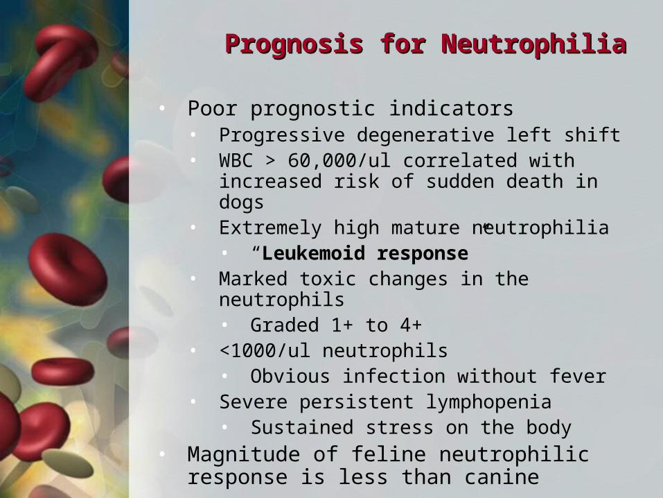
Prognosis for NeutrophiliaPrognosis for Neutrophilia
• Poor prognostic indicators• Progressive degenerative left shift• WBC > 60,000/ul correlated with increased
risk of sudden death in dogs• Extremely high mature neutrophilia
• “Leukemoid response”• Marked toxic changes in the neutrophils
• Graded 1+ to 4+• <1000/ul neutrophils
• Obvious infection without fever• Severe persistent lymphopenia
• Sustained stress on the body• Magnitude of feline neutrophilic response is
less than canine
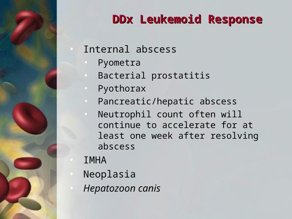
DDx Leukemoid ResponseDDx Leukemoid Response
• Internal abscess• Pyometra
• Bacterial prostatitis
• Pyothorax
• Pancreatic/hepatic abscess
• Neutrophil count often will continue to accelerate for at least one week after resolving abscess
• IMHA• Neoplasia• Hepatozoon canis
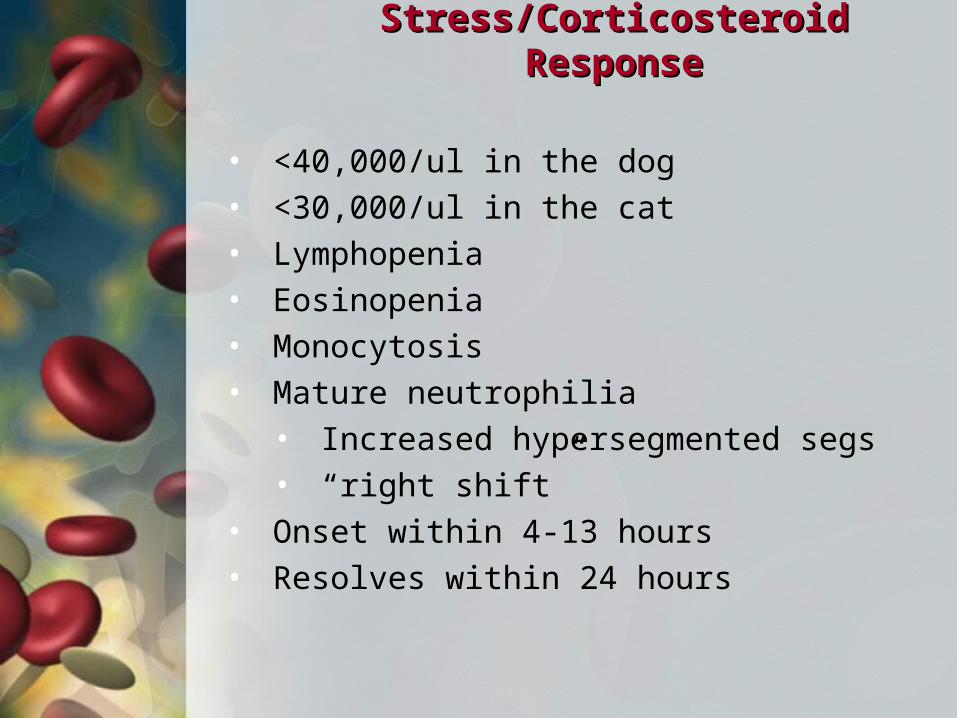
Stress/Corticosteroid ResponseStress/Corticosteroid Response
• <40,000/ul in the dog• <30,000/ul in the cat• Lymphopenia• Eosinopenia• Monocytosis• Mature neutrophilia
• Increased hypersegmented segs • “right shift”
• Onset within 4-13 hours• Resolves within 24 hours
Epinephrine/Exercise ResponseEpinephrine/Exercise Response
• <40,000/ul in the dog• <30,000/ul in the cat• More of a problem in cats• Lymphocytosis• Increased HCT
NeutropeniaNeutropenia
• DDx:• Excessive peripheral consumption
• Infection• Necrosis• IM neutropenia
• Bone marrow disease• See non-regenerative anemia
• Test for parvovirus• Diarrhea• < 2 years of age• Immunosuppressed• Swab tonsils then rectum
NeutropeniaNeutropenia
• Treatment• Treat obvious causes of infection,
necrosis or inflammation
• If no obvious causes, work up for occult infection
• Discontinue myelosuppressive drugs
• Prophylactic antibiotics• 1500/2000/ul - amoxicillin• <1500/ul – amoxicillin and quinolone
• Clindamycin and quinolone• Metronidazole and quinolone
• If septic, IV antibiotics
NeutropeniaNeutropenia
• Treatment• Recheck CBC weekly
• Bone marrow sampling of no response
• Sooner if bicytopenia or pancytopenia
• FeLV IFA in cats
• Neupogen if maturation arrest
• GCSF - Granulocyte colony stimulation factor
• Doxycycline then Immunosuppressive therapy for IM neutropenia
Work-Up for Occult InfectionWork-Up for Occult Infection
• FeLV/FIV test in cats• Heartworm test in dogs• CBC• General health profile• Electrolytes and venous blood gases• Thoracic and abdominal x-rays• Abdominal ultrasound• Urinalysis and urine culture• Look especially hard for infection if:
• Toxic neutrophils• Degenerative left shift• Pronounced rouleaux

Work-Up for Occult InfectionWork-Up for Occult Infection
• Echocardiogram if murmur• “to and fro” murmur at left heart base• bounding pulses
• Blood culture when febrile• use ARD (antimicrobial removal device) if
on antibiotics• 2 samples several hours apart• Collect aseptically
• CSF tap if neck pain or CNS deficits• Joint taps if joint swelling• CPK if muscle pain• Muscle biopsies if Hepatozoon suspected or
increased CPK
MonocytosisMonocytosis
• Chronic infection• >10 days
• Necrosis• Infection
• viral (especially FIP)• Fungal• Mycobacterial• L-form, mycoplasma, Ureaplasma• Parasitic
• Foreign body• Neoplasia• Immune mediated inflammation• Corticosteroids (lymphopenia, eosinopenia)
LymphocytosisLymphocytosis
• Stress/corticosteroid response• Chronic infection
• viremia• Immune mediated disease• Recent vaccination• Lymphoid neoplasia• Ehrlichia spp.• Addison’s Disease
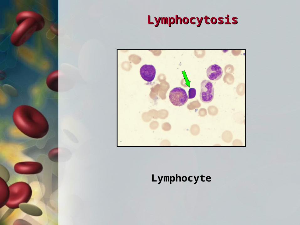
Lymphocyte
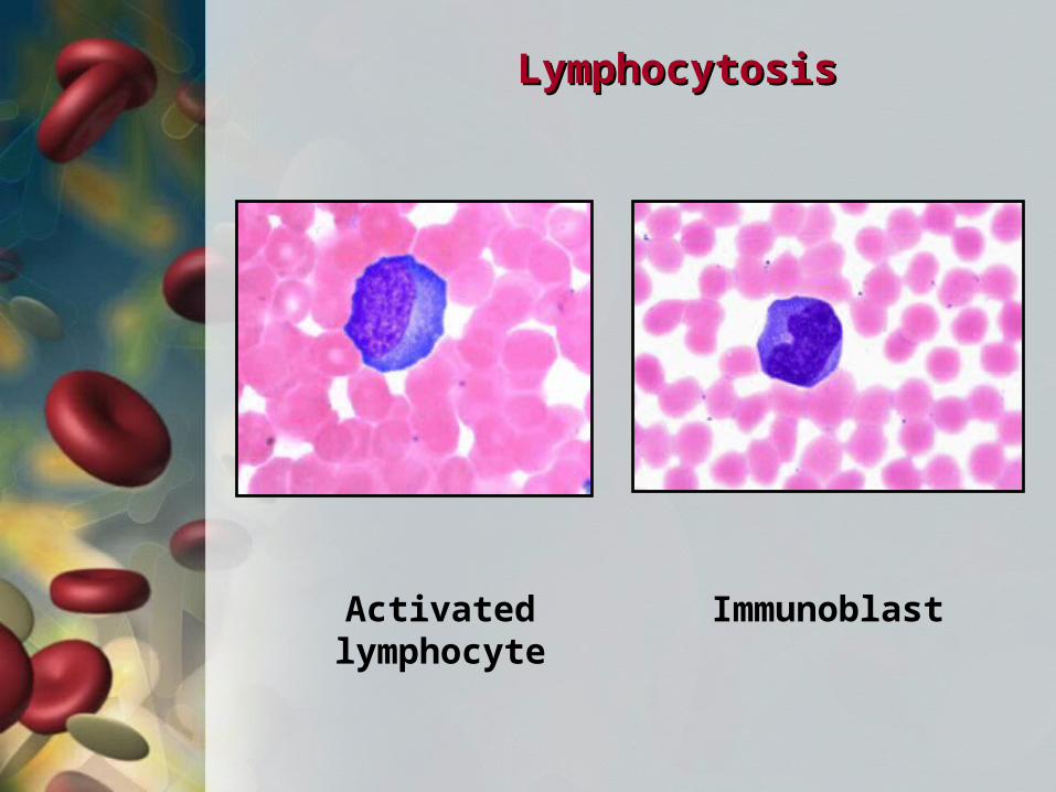
LymphocytosisLymphocytosis
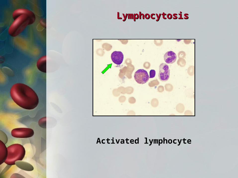
Activated lymphocyte
LymphocytosisLymphocytosis
Activated lymphocyte
LymphocytosisLymphocytosis
Immunoblast
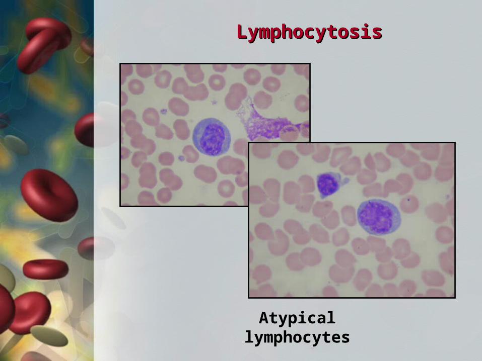
Atypical lymphocytes
LymphocytosisLymphocytosis
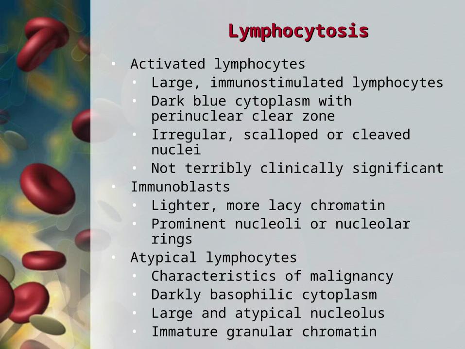
LymphocytosisLymphocytosis
• Activated lymphocytes• Large, immunostimulated lymphocytes• Dark blue cytoplasm with perinuclear
clear zone• Irregular, scalloped or cleaved nuclei• Not terribly clinically significant
• Immunoblasts• Lighter, more lacy chromatin• Prominent nucleoli or nucleolar rings
• Atypical lymphocytes• Characteristics of malignancy• Darkly basophilic cytoplasm• Large and atypical nucleolus• Immature granular chromatin
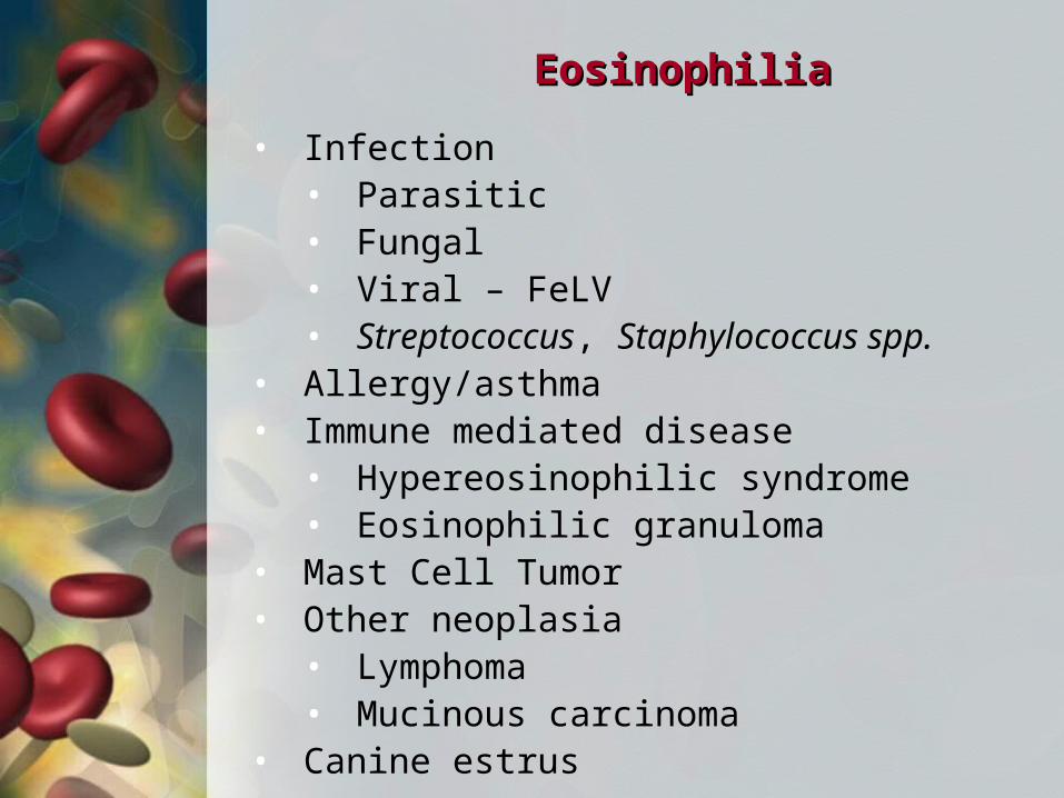
EosinophiliaEosinophilia
• Infection • Parasitic• Fungal• Viral – FeLV• Streptococcus, Staphylococcus spp.
• Allergy/asthma• Immune mediated disease
• Hypereosinophilic syndrome• Eosinophilic granuloma
• Mast Cell Tumor• Other neoplasia
• Lymphoma• Mucinous carcinoma
• Canine estrus
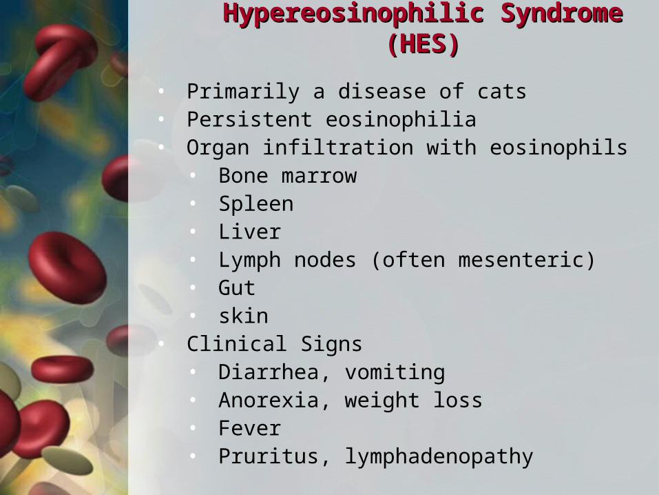
Hypereosinophilic Syndrome (HES)Hypereosinophilic Syndrome (HES)
• Primarily a disease of cats• Persistent eosinophilia• Organ infiltration with eosinophils
• Bone marrow• Spleen• Liver• Lymph nodes (often mesenteric)• Gut• skin
• Clinical Signs• Diarrhea, vomiting• Anorexia, weight loss• Fever• Pruritus, lymphadenopathy
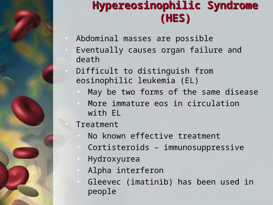
Hypereosinophilic Syndrome (HES)Hypereosinophilic Syndrome (HES)
• Abdominal masses are possible• Eventually causes organ failure and death• Difficult to distinguish from eosinophilic
leukemia (EL)• May be two forms of the same disease• More immature eos in circulation with EL
• Treatment• No known effective treatment• Cortisteroids – immunosuppressive• Hydroxyurea• Alpha interferon• Gleevec (imatinib) has been used in
people
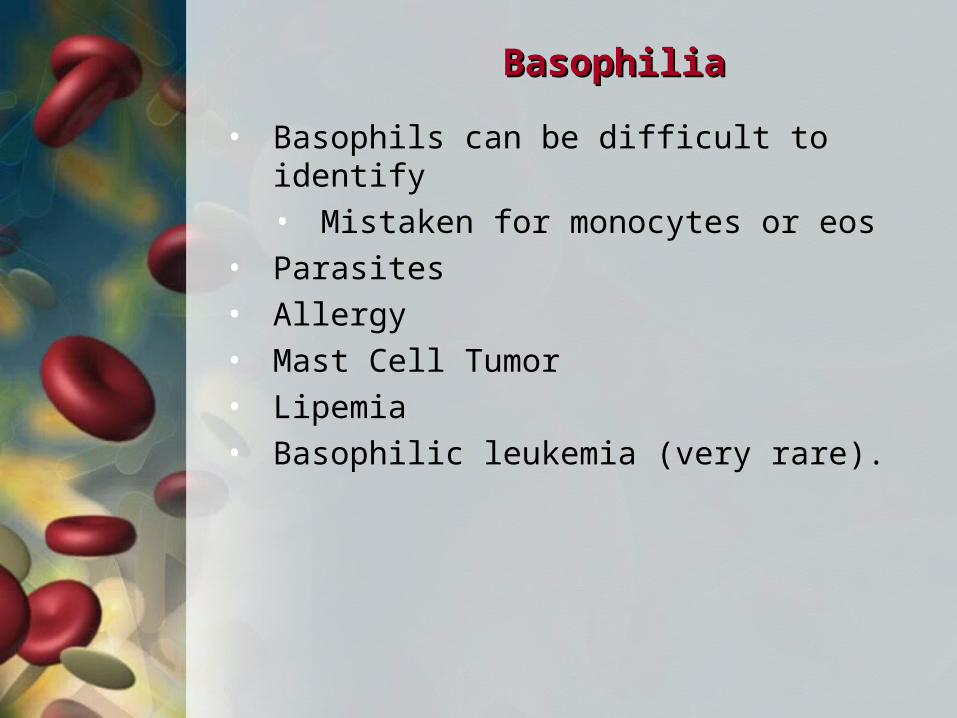
BasophiliaBasophilia
• Basophils can be difficult to identify• Mistaken for monocytes or eos
• Parasites• Allergy• Mast Cell Tumor• Lipemia• Basophilic leukemia (very rare).
LeukemiaLeukemia
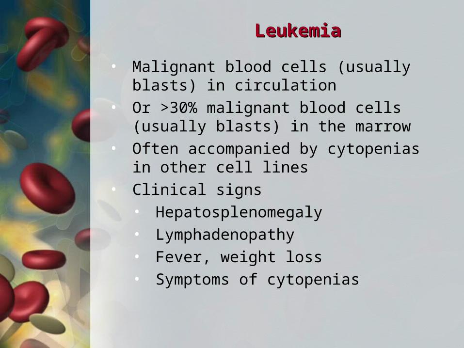
• Malignant blood cells (usually blasts) in circulation
• Or >30% malignant blood cells (usually blasts) in the marrow
• Often accompanied by cytopenias in other cell lines
• Clinical signs• Hepatosplenomegaly• Lymphadenopathy• Fever, weight loss• Symptoms of cytopenias
LeukemiaLeukemia
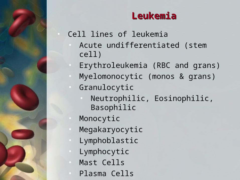
• Cell lines of leukemia• Acute undifferentiated (stem cell)• Erythroleukemia (RBC and grans)• Myelomonocytic (monos & grans)• Granulocytic
• Neutrophilic, Eosinophilic, Basophilic• Monocytic• Megakaryocytic• Lymphoblastic• Lymphocytic• Mast Cells• Plasma Cells
LeukemiaLeukemia
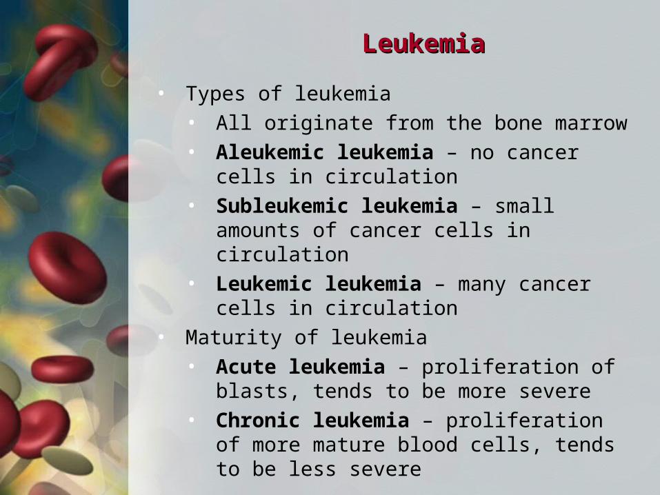
• Types of leukemia• All originate from the bone marrow• Aleukemic leukemia – no cancer cells in
circulation• Subleukemic leukemia – small amounts
of cancer cells in circulation• Leukemic leukemia – many cancer cells
in circulation• Maturity of leukemia
• Acute leukemia – proliferation of blasts, tends to be more severe
• Chronic leukemia – proliferation of more mature blood cells, tends to be less severe
LeukemiaLeukemia
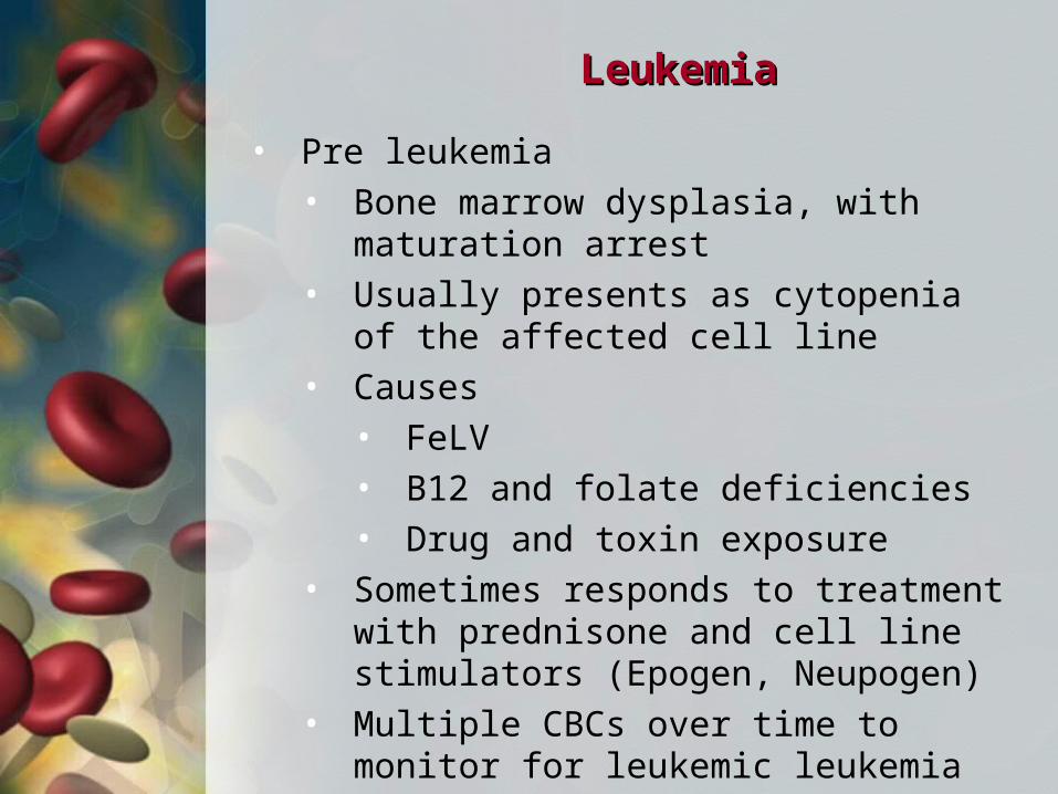
• Pre leukemia• Bone marrow dysplasia, with maturation
arrest• Usually presents as cytopenia of the
affected cell line• Causes
• FeLV• B12 and folate deficiencies• Drug and toxin exposure
• Sometimes responds to treatment with prednisone and cell line stimulators (Epogen, Neupogen)
• Multiple CBCs over time to monitor for leukemic leukemia