preleukemic mutations in human acute myeloid leukemia ... · preleukemic mutations in human acute...
TRANSCRIPT
Preleukemic mutations in human acute myeloidleukemia affect epigenetic regulators andpersist in remissionM. Ryan Corces-Zimmermana, Wan-Jen Honga,b, Irving L. Weissmana,1, Bruno C. Medeirosb, and Ravindra Majetia,b,1
aProgram in Cancer Biology, Ludwig Center, Cancer Institute, Institute for Stem Cell Biology and Regenerative Medicine, and bDepartment of Medicine,Division of Hematology, Stanford University School of Medicine, Stanford, CA 94305
Contributed by Irving L. Weissman, January 9, 2014 (sent for review December 23, 2013)
Cancer is widely characterized by the sequential acquisition ofgenetic lesions in a single lineage of cells. Our previous studies haveshown that, in acute myeloid leukemia (AML), mutation acquisitionoccurs in functionally normal hematopoietic stem cells (HSCs). Thesepreleukemic HSCs harbor some, but not all, of the mutations foundin the leukemic cells. We report here the identification of patterns ofmutation acquisition in human AML. Our findings support a model inwhich mutations in “landscaping” genes, involved in global chroma-tin changes such as DNA methylation, histone modification, andchromatin looping, occur early in the evolution of AML, whereasmutations in “proliferative” genes occur late. Additionally, we ana-lyze the persistence of preleukemic mutations in patients in remis-sion and find CD34+ progenitor cells and various mature cells thatharbor preleukemic mutations. These findings indicate that preleu-kemic HSCs can survive induction chemotherapy, identifying thesecells as a reservoir for the reevolution of relapsed disease. Finally,through the study of several cases of relapsed AML, we demonstratevarious evolutionary patterns for the generation of relapsed diseaseand show that some of these patterns are consistent with involve-ment of preleukemic HSCs. These findings provide key insights intothe monitoring of minimal residual disease and the identification oftherapeutic targets in human AML.
preleukemia | clonal evolution
Acute myeloid leukemia (AML) is an aggressive malignancyof the bone marrow characterized by uncontrolled pro-
liferation of immature myeloid lineage cells (1, 2). Recent advancesin high-throughput sequencing have led to the identification ofmany recurrently mutated genes implicated in the pathogenesis ofAML (3–10). The coding genomes of over 200 AML patients havebeen sequenced by The Cancer Genome Atlas (TCGA) consor-tium, identifying ∼30 genes that are mutated in greater than 2% ofpatients (11). In addition, several studies have investigated the ge-netic changes occurring between diagnosis and relapse in the samepatient (12, 13). Despite this extensive characterization of the ge-netic variation in AML, the evolutionary processes that precede theonset of frank leukemia and shape the architecture of the dis-ease have not been well elucidated.These sequencing efforts have demonstrated that an individual
AML case is associated with an average of five different recurrentmutations (11), raising the question of how these multiple muta-tions accumulate in a single lineage of cells given the generally lowspontaneous mutation rate in hematopoietic cells (14) and thelack of hypermutator phenotypes in this disease (15). To explainthese observations, we proposed a model in which the majority ofAML mutations are sequentially acquired in successive clones ofself-renewing hematopoietic stem cells (HSCs), unless the muta-tion confers self-renewal potential on a more differentiated cell(16, 17). Early evidence consistent with this model comes fromstudies of AML patients harboring the AML1-ETO translocation(18). In these patients, HSCs isolated during durable remissionproduced normal myeloid colonies in vitro with detectableAML1-ETO transcripts. Additionally, in chronic myeloid leukemia,the breakpoint cluster region–Abelson tyrosine-protein kinase 1
(BCR-ABL) translocation occurs in HSCs, causing chronic disease,but subsequent progression to blast crisis involves mutation evolu-tion at the level of granulocyte–macrophage progenitors (19, 20).More recently, a study of elderly women with clonal hematopoiesisindicated by X-inactivation skewing showed that these individualsharbored somatic ten eleven translocation methylcytosine dioxyge-nase 2 (TET2) mutations, occurring at the level of the HSC, thatmay predispose them to hematologic malignancies (21).Recent work from our laboratory has provided direct evidence
supporting this model by demonstrating that, in a small subset ofAML patients harboring mutations in the FMS-related tyrosinekinase 3 (FLT3) receptor tyrosine kinase, multiple genetic lesionsserially accumulate in functionally normal HSCs (22). We definedthese HSCs as “preleukemic” because they are genetically distinctfrom germ-line HSCs in that they harbor only a subset of the leu-kemia-specific mutations which identifies them as the evolutionaryancestors of the frankly leukemic cells. These preleukemic muta-tions included mutations in recurrently mutated genes that areknown drivers of leukemogenesis, such as TET2, SMC1A, nucleo-phosmin 1 (NPM1), and CTCF, implicating preleukemic mutationsas functional components of AML evolution. Interestingly, muta-tion of FLT3 never occurred in preleukemic cells, leading to thehypothesis that genetic lesions in AML may occur in a nonrandompattern. These data are supported by studies of mutation stabilitybetween diagnosis and relapse in AML, where mutations in FLT3were found to often be unstable, indicating that these mutationsare late events (13). Additionally, in certain patients, only onerecurrent coding mutation distinguished the functionally normalHSCs from the frankly leukemic cells, suggesting that these pre-leukemic HSCs may be a putative reservoir for relapsed disease by
Significance
A growing body of evidence has determined that somatic muta-tions in acute myeloid leukemia (AML) accumulate in self-renew-ing hematopoietic stem cells (HSCs). Thus, at the time of diagnosis,AML patients harbor preleukemic HSCs containing some, but notall, of the mutations in the downstream leukemia. Despite thesefindings, common patterns of preleukemic clonal evolution havenot been determined, nor has the response of preleukemic HSCsto standard therapy been identified. This report addresses both ofthese questions determining that there are common patterns ofpreleukemic clonal evolution in AML, and that these preleukemicHSCs often survive standard induction chemotherapy. This studyis of interest to the AML field, and broadly in cancer genomics asthe principle that stem cells acquire initial cancer-initiating muta-tions is likely to extend beyond AML.
Author contributions: M.R.C.-Z., I.L.W., and R.M. designed research; M.R.C.-Z. performedresearch; W.-J.H. and B.C.M. analyzed data; and M.R.C.-Z., W.-J.H., I.L.W., B.C.M., and R.M.wrote the paper.
The authors declare no conflict of interest.1To whom correspondence may be addressed. E-mail: [email protected] or [email protected].
This article contains supporting information online at www.pnas.org/lookup/suppl/doi:10.1073/pnas.1324297111/-/DCSupplemental.
2548–2553 | PNAS | February 18, 2014 | vol. 111 | no. 7 www.pnas.org/cgi/doi/10.1073/pnas.1324297111
surviving chemotherapy and acquiring additional mutations thattransform them into AML.From these observations, we proposed a model of AML evo-
lution where preleukemic HSCs can persist during clinical re-mission and have the potential to give rise to relapsed disease(16). More specifically, we proposed that relapsed disease canoriginate from multiple sources including (i) treatment refrac-tory primary disease, (ii) further evolution of a dominant clonepresent at diagnosis, (iii) outgrowth of a subclone present atdiagnosis, or (iv) further evolution of disease from a preleukemicHSC. Here, through the genomic and functional analysis of denovo AML and patient-matched HSCs, we provide evidencesupporting these mechanisms by determining patterns of pre-leukemic mutation acquisition at diagnosis, and by trackingpreleukemic cells and mutations in clinical remission and relapse.
ResultsIdentification of Preleukemic Mutations and HSCs in a Diverse Cohortof Human AML Patients.Our previous studies identified preleukemicmutations and HSCs in a small cohort of AML patients. We aimedto expand upon these studies to investigate preleukemic clonalevolution in a larger cohort and broader spectrum of AML patients(Tables S1 and S2). Exome sequencing (23, 24) of FACS-purifiedleukemia cells (CD99+ TIM3+) (22, 25) and patient-matchedCD3+ T cells was used to identify leukemia-specific mutationsin 10 patients. Sequencing was performed to a median coveragedepth of 56-fold (Fig. S1A) and all mutations were validated usingSanger sequencing of genomic DNA (gDNA) (Table S3).All leukemia-specific mutations were then sequenced at high
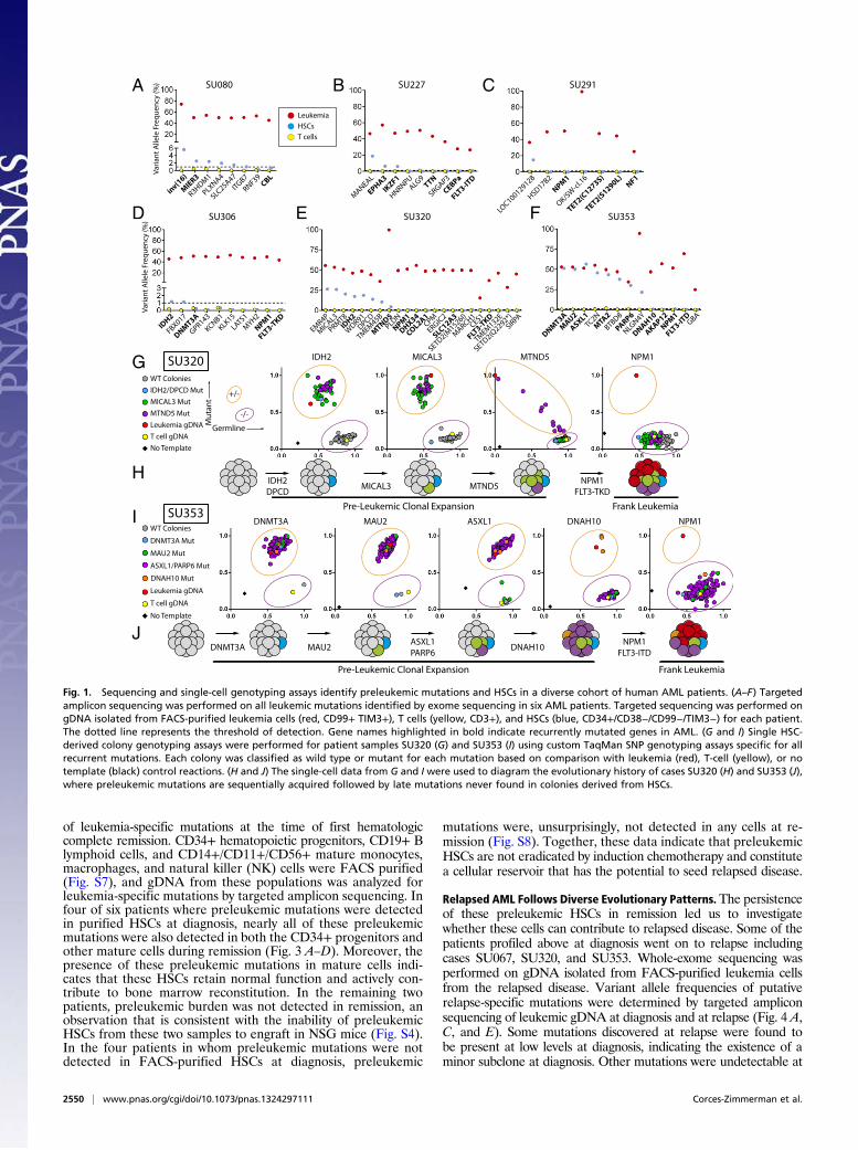
depth (Fig. S1E) using targeted amplicon sequencing of FACS-purified leukemia cells, CD3+ T cells, and Lin−CD34+CD38−TIM3−CD99− HSCs (Fig. S2). Importantly, apart from surfacemarker expression, these HSCs were also functionally defined bytheir ability to generate long-term myeloid and lymphoid engraft-ment in immunodeficient NOD/SCID/IL2R-γ (NSG) mice, aspreviously described (Fig. S3A) (22, 25). Preleukemic mutationswere identified as those mutations occurring at >1% variant allelefrequency in the purified HSC subpopulation, a thresholdidentified by control sequencing reactions of known admixtures ofleukemic and normal DNA (Fig. S4). As expected, deep se-quencing of the HSC subpopulation for leukemia-specific muta-tions never detected all leukemia-specific mutations, which wouldbe an indicator of leukemia cell contamination. Leukemia-spe-cific mutations found to be absent from purified HSCs wereclassified as late events. Six of 10 patients had detectable pre-leukemic mutations in purified HSCs (Fig. 1 A–F). These pre-leukemic mutations included recurrent mutations in isocitratedehydrogenase 2 (IDH2), DNA (cytosine-5-) methyltransferase3 alpha (DNMT3A), additional sex combs like 1 (ASXL1), andIKAROS family zinc finger 1 (IKZF1), and inversion of chromosome16 resulting in the core-binding factor beta subunit–myosin heavychain 11 (CBFB-MYH11) fusion gene. In a select number of cases,leukemic mutations were detectable not only in purified HSCs, butalso in a fraction of sorted B and T lymphocytes at diagnosis, in-dicating a clear contribution of preleukemic HSCs to hematopoiesisin humans (Fig. S5). Interestingly, multiple patients that lacked de-tectable preleukemic mutations in purified HSCs showed pre-leukemic mutation burden in CD19+ lymphoid cells that developedin NSG mice transplanted with those same purified HSCs (SU067,SU072; Fig. S4). This implies that, in these cases, rare preleukemicHSCs exist that have a competitive advantage in the xenotrans-plantation setting (Table S1). Of the 10 samples analyzed, 2 (SU290,SU336) showed no detectable preleukemic mutations.To validate these findings and determine the order of mutation
acquisition in a subset of samples, colonies derived from singleHSCs were genotyped using custom TaqMan SNP genotypingassays for all recurrent mutations, and a select subset of the otherleukemia-specific mutations (Fig. 1 G and I). In case SU320, 104colonies were tested for the presence of mutations in IDH2, deletedin primary ciliary dyskinesia homolog (DPCD), microtubule asso-ciated monooxygenase, calponin and lim domain containing 3(MICAL3), mitochondrially encoded NADH dehydrogenase 5(MTND5), NPM1, and FLT3 (Fig. 1G). Of these colonies, 61 werefound to be wild type for all mutations profiled, 1 was found to
harbor mutations in IDH2 and DPCD only, 30 were found withmutations in IDH2, DPCD, and MICAL3, and 12 were found toharbor mutations in IDH2, DPCD, MICAL3, and MTND5. Im-portantly, none of the colonies contained mutations in NPM1or FLT3, suspected late mutations based on amplicon sequenc-ing (Fig. 1E). These single-cell–based assays determined thestepwise pattern of mutation acquisition in case SU320 (Fig. 1H).Intriguingly, mutation ofMTND5, a mitochondrially encoded gene,showed a progressive increase in mutation burden in preleukemiccells, indicated by a somewhat stepwise increase in mutantallele frequency at the single-cell level (Fig. 1G).Similarly, in case SU353, 192 single-cell–derived myeloid col-
onies were genotyped for mutations in DNMT3A, MAU2, ASXL1,poly (ADP-Ribose) polymerase family member 6 (PARP6), dyneinaxonemal heavy chain 10 (DNAH10), NPM1, and FLT3 (Fig. 1I).Of these 192 colonies, only 1 was found to be wild type for allmutations profiled, indicating that the preleukemic cells in thispatient have strongly outcompeted their genetically normalcounterparts. Of the remaining 191 colonies, 1 was found toharbor only the DNMT3Amutation, 5 were found with both theDNMT3A and maternally affected uncoordination (MAU2)mutations, 182 were found to harbor mutations in DNTM3A,MAU2, ASXL1, and PARP6, and 3 were found to contain muta-tions in DNMT3A, MAU2, ASXL1, PARP6, and DNAH10. Im-portantly, none of the colonies harbored mutations in NPM1 orFLT3. These single-cell–based assays determined the order ofacquisition of mutations in case SU353 (Fig. 1J).
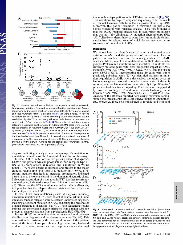
Mutation Acquisition in AML Occurs in Patterns. The identificationof preleukemic mutations and late mutations in a diverse cohortof AML patients led us to investigate the possibility that thereare patterns of mutation acquisition in human AML. Pre-leukemic mutation analysis from 16 patients [10 described hereand 6 previously reported (22)] was used to identify early andlate mutations in recurrently mutated genes (Fig. 2A). Theserecurrently mutated genes were subdivided into categories basedon the classification system described by the TCGA AML con-sortium (Table S4). Of 74 total mutations in recurrently mutatedgenes, 36 (49%) were found to be preleukemic and 38 (51%)were found to be late. Mutations in genes involved in DNAmethylation, chromatin modification, and chromatin topology(cohesin complex) were significantly overrepresented in thesubset of preleukemic mutations and underrepresented in thesubset of late mutations. In contrast, mutations in genes involvedin activated signaling were found to be significantly overrepre-sented in the subset of late mutations and underrepresented inthe subset of preleukemic mutations.Additional patient samples were selected based on prior
knowledge of somatic mutations in IDH1/IDH2, DNMT3A, NPM1,FLT3, or Kirsten rat sarcoma viral oncogene homolog (KRAS)/neuroblastoma rat sarcoma viral oncogene homolog (NRAS).Targeted amplicon sequencing of gDNA from purified HSCs wasused to determine whether these mutations were preleukemic(Fig. 2B and Fig. S6). Mutations in IDH1 and IDH2 were found tobe preleukemic in 80% of patients. Similarly, mutations inDNMT3A were found to be preleukemic in 75% of patients. Incontrast, only 12% of patients with mutation of NPM1 and 0% ofpatients with mutation in FLT3 or KRAS/NRAS exhibited de-tectable preleukemic burden of these mutations in purified HSCs.From this analysis, we developed a model of mutation acquisi-tion (Fig. 2C) where the earliest founding mutations occur in“landscaping” genes, involved in global regulation of gene ex-pression through epigenetic mechanisms, whereas late progressormutations occur in genes that generally lead to an increase in ac-tivated signaling and cellular proliferation such as RAS and FLT3.
Preleukemic HSCs Survive Induction Chemotherapy and Persist inRemission. We hypothesized that preleukemic HSCs may persistin remission and contribute to relapse through the acquisition ofnew mutations. In most AML patients, some normal HSCs sur-vive induction chemotherapy and eventually repopulate the bonemarrow, leading to hematologic remissions. To investigate thechemosensitivity of preleukemic HSCs, we analyzed bone mar-row samples from the patients investigated above for the presence
Corces-Zimmerman et al. PNAS | February 18, 2014 | vol. 111 | no. 7 | 2549
CELL
BIOLO
GY
of leukemia-specific mutations at the time of first hematologiccomplete remission. CD34+ hematopoietic progenitors, CD19+ Blymphoid cells, and CD14+/CD11+/CD56+ mature monocytes,macrophages, and natural killer (NK) cells were FACS purified(Fig. S7), and gDNA from these populations was analyzed forleukemia-specific mutations by targeted amplicon sequencing. Infour of six patients where preleukemic mutations were detectedin purified HSCs at diagnosis, nearly all of these preleukemicmutations were also detected in both the CD34+ progenitors andother mature cells during remission (Fig. 3 A–D). Moreover, thepresence of these preleukemic mutations in mature cells indi-cates that these HSCs retain normal function and actively con-tribute to bone marrow reconstitution. In the remaining twopatients, preleukemic burden was not detected in remission, anobservation that is consistent with the inability of preleukemicHSCs from these two samples to engraft in NSG mice (Fig. S4).In the four patients in whom preleukemic mutations were notdetected in FACS-purified HSCs at diagnosis, preleukemic
mutations were, unsurprisingly, not detected in any cells at re-mission (Fig. S8). Together, these data indicate that preleukemicHSCs are not eradicated by induction chemotherapy and constitutea cellular reservoir that has the potential to seed relapsed disease.
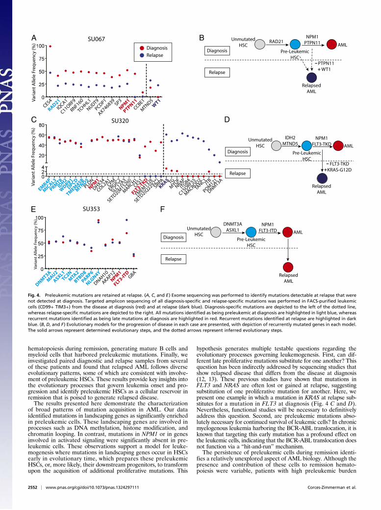
Relapsed AML Follows Diverse Evolutionary Patterns. The persistenceof these preleukemic HSCs in remission led us to investigatewhether these cells can contribute to relapsed disease. Some of thepatients profiled above at diagnosis went on to relapse includingcases SU067, SU320, and SU353. Whole-exome sequencing wasperformed on gDNA isolated from FACS-purified leukemia cellsfrom the relapsed disease. Variant allele frequencies of putativerelapse-specific mutations were determined by targeted ampliconsequencing of leukemic gDNA at diagnosis and at relapse (Fig. 4 A,C, and E). Some mutations discovered at relapse were found tobe present at low levels at diagnosis, indicating the existence of aminor subclone at diagnosis. Other mutations were undetectable at
SU227
ALG9
CEBPa
EPHA3
FLT3-ITD
HNRNPU
IKZF1
MANEAL
SRGAP3TTN
B
DNMT3A MAU2 ASXL1 DNAH10 NPM1I
SU080
Va
ria
nt
Alle
le F
req
ue
ncy
(%
)
CBL
inv(16
)IT
GB7
MIER3
PLXNA4
R3HDM1
RNF39
SLC25A47
HSD17B2
NPM1 NF
1
SU291C
WT Colonies
DNMT3A Mut
MAU2 Mut
ASXL1/PARP6 Mut
Leukemia gDNA
T cell gDNA
No Template
DNAH10 Mut
SU353
Leukemia
HSCs
T cells
SU353
AKAP12
ASXL1
BTBD8
DNAH10
DNMT3A
FLT3-ITD
GBA
MAU2
MTA2
NLGN4Y
NPM1
PARP6
TC2N
SU320
MARCH1
CES1
COL2A1
CPM
DHX34
DPCD
EMR4P
ERGIC2
FLT3-TK
DIDH2
MIC
AL3
MTND5NPM1
PLDN
PRMT8
SETD2(Q2292*)
SETD2(M1526I)
SIRPA
SLC12A3
TMEM
132E
TMEM
45B
WDR91
SU306
Va
ria
nt
Alle
le F
req
ue
ncy
(%
)
DNMT3A
FBX017
FLT3-TK
D
GPR143
IDH2
KCNB1
KLK15
LATS1
MYH2
NPM1
D E F TET2(C1273S)
TET2(S1290L)
LOC100129128
G IDH2 MICAL3 MTND5 NPM1
Mu
tan
t
Germline
+/-
-/-
WT Colonies
IDH2/DPCD Mut
MICAL3 Mut
MTND5 Mut
Leukemia gDNA
T cell gDNA
No Template
SU320
HIDH2
DPCDMICAL3 MTND5
NPM1
FLT3-TKD
Pre-Leukemic Clonal Expansion Frank Leukemia
DNMT3A MAU2ASXL1
PARP6
NPM1
FLT3-ITDDNAH10
Pre-Leukemic Clonal Expansion Frank Leukemia
J
OK/S
W-c
l.16
A
Fig. 1. Sequencing and single-cell genotyping assays identify preleukemic mutations and HSCs in a diverse cohort of human AML patients. (A–F) Targetedamplicon sequencing was performed on all leukemic mutations identified by exome sequencing in six AML patients. Targeted sequencing was performed ongDNA isolated from FACS-purified leukemia cells (red, CD99+ TIM3+), T cells (yellow, CD3+), and HSCs (blue, CD34+/CD38−/CD99−/TIM3−) for each patient.The dotted line represents the threshold of detection. Gene names highlighted in bold indicate recurrently mutated genes in AML. (G and I) Single HSC-derived colony genotyping assays were performed for patient samples SU320 (G) and SU353 (I) using custom TaqMan SNP genotyping assays specific for allrecurrent mutations. Each colony was classified as wild type or mutant for each mutation based on comparison with leukemia (red), T-cell (yellow), or notemplate (black) control reactions. (H and J) The single-cell data from G and I were used to diagram the evolutionary history of cases SU320 (H) and SU353 (J),where preleukemic mutations are sequentially acquired followed by late mutations never found in colonies derived from HSCs.
2550 | www.pnas.org/cgi/doi/10.1073/pnas.1324297111 Corces-Zimmerman et al.
diagnosis indicating a newly acquired relapse-specific mutation, ora mutation present below the threshold of detection of this assay.In case SU067, mutations in two genes present at diagnosis,
CCBE1 and protein tyrosine phosphatase, non-receptor type 11(PTPN11), were absent at relapse, and a mutation in Wilmstumor 1 (WT1) was absent at diagnosis and present in the majorclone at relapse (Fig. 4A). Loss of a mutation in PTPN11, a re-current mutation that leads to increased proliferation, indicateda step back to a clone ancestral to the dominant diagnostic clone.Subsequent acquisition of a mutation in WT1, another recurrentlymutated gene, indicated a step forward in clonal evolution (Fig.4B). Given that the WT1 mutation was undetectable at diagnosis,it is possible that the relapsed disease originated from a rare an-tecedent preleukemic HSC.In case SU320, four mutations present at diagnosis, including
a recurrent mutation in FLT3, were absent at relapse. Of the 11 newmutations found at relapse, 4were detected at low levels at diagnosis,including a recurrent mutation in KRAS, indicating the presence ofa minor subclone at diagnosis (Fig. 4C). These sequencing resultsindicate that the relapsed disease originated from a minor subclonepresent at diagnosis and not from a preleukemic HSC (Fig. 4D).In case SU353, no mutation differences were found between
the disease at diagnosis and the disease at relapse (Fig. 4E). Thisobservation is consistent with the clinical course of this patientwho, at the time of complete hematologic remission, harboredevidence of residual disease based on the presence of an abnormal
immunophenotypic pattern in the CD34+ compartment (Fig. S7).This was shown by targeted amplicon sequencing to be the resultof residual leukemic cells from the diagnostic clone (Fig. 3D).Moreover, this patient remained in remission for just 2 mobefore presenting with relapsed disease. These results indicatethat the SU353 relapsed disease was, in fact, refractory diseasethat was not fully eliminated by induction chemotherapy (Fig.4F). Collectively, these three patients illustrate multiple distinctmechanisms for relapse, some of which do not preclude the in-volvement of preleukemic HSCs.
DiscussionWe report here the identification of patterns of mutation ac-quisition in AML and the persistence of preleukemic HSCs inpatients in complete remission. Sequencing studies of 10 AMLcases identified preleukemic mutations in multiple diverse sub-groups. Preleukemic mutations were identified in multiple re-currently mutated genes with clear prognostic impact in AML,including DNMT3A, IDH1, IDH2, ASXL1, IKZF1, and the fusiongene CBFB-MYH11. Incorporating these 10 cases with our 6previously published cases (22), we identified patterns in muta-tion acquisition in AML where the earliest mutations occur inlandscaping genes, involved primarily in regulation of the epi-genome, whereas late mutations occur primarily in “proliferative”genes, involved in activated signaling. These data were supportedby directed profiling of 16 additional patients harboring muta-tions in NPM1, IDH1/IDH2, DNMT3A, FLT3, or KRAS/NRAS.Analysis of the 10 cases reported here during remission demon-strated that preleukemic HSCs can survive induction chemother-apy. Moreover, these cells contributed to myeloid and lymphoid
A
B
Founder
“Landscaping”
Mutation
Expansion of
Pre-Leukemic
HSC Clone
Progressor
“Proliferative”
Mutation
Expansion of
Leukemic
Clone
C
IDH1/2
12/15
(80%)
Va
ria
nt
Alle
le F
req
ue
ncy
in H
SC
s (%
)
NPM1
2/17
(12%)
FLT3
0/13
(0%)
DNMT3A
3/4
(75%)
KRAS/NRAS
0/4
(0%)
0 5 10 1551015
Pre-Leukemic Late
Unknown
DNA Methylation
Activated Signaling
Cohesin Complex
NPM1
Myeloid TFs
TF Fusions
**
*
*
*
***** *NS
Number of Mutations Observed
Fig. 2. Mutation acquisition in AML occurs in patterns with preleukemiclandscaping mutations followed by late proliferative mutations. (A) Exomesequencing and targeted amplicon sequencing data identifying preleukemicand late mutations from 16 patients (Table S1) were pooled. Recurrentmutations (74 total) were stratified according to the classification systemestablished by the TCGA, and assigned to be preleukemic or late based ondetection in HSCs as described in Table S4. The number of mutations in eachcategory is indicated on the x axis. (B) Additional AML cases were selectedfor the presence of recurrent mutations in IDH1/IDH2 (n = 15), DNMT3A (n =4), NPM1 (n = 17), FLT3 (n = 13), or KRAS/NRAS (n = 4). Each dot representsone case (see Table S2 for patient information). The dotted line representsthe threshold of detection. The ratio of cases with preleukemic mutation ofa given gene to the total number of cases with that mutation analyzed isshown below the plot. (C) A model for the acquisition of mutations in AML.(**P < 0.001, *P < 0.05; NS, not significant, χ2 test).
SU306
DNMT3A
FBX017
FLT3-T
KD
GPR143
IDH2
KCNB1
KLK15
LATS1
MYH2
NPM1
SU320
Va
ria
nt
Alle
le F
req
ue
ncy
(%
)
SU291
Va
ria
nt
Alle
le F
req
ue
ncy
(%
)
A
C
D
Mature Mono/Mac/NK cellsCD34+ cellsB cells
HSD17B2
NPM1
NF1
TET2(C1273S)
TET2(S1290L)
LOC100129128
OK/S
W-c
l.16
AKAP12
ASXL1
BTBD8
DNAH10
DNMT3A
FLT3-IT
DGBA
MAU2
MTA2
NLGN4Y
NPM1
PARP6
TC2N
MARCH1
CES1
COL2A1
CPM
DHX34
DPCD
EMR4P
ERGIC2
FLT3-T
KDIDH2
MICAL3
MTND5
NPM1
PLDN
PRMT8
SETD2(Q2292*)
SETD2(M1526I)
SIRPA
SLC12A3
TMEM
132E
TMEM45B
WDR91
SU353
B
Fig. 3. Preleukemic mutations and HSCs persist in remission. (A–D) Bonemarrow mononuclear cells from patient remission samples were sorted forCD19+ B cells, CD14+/CD11b+/CD56+ mature monocytes, macrophages, andNK cells, and CD34+ hematopoietic progenitors. Targeted amplicon sequenc-ing was performed for all leukemic mutations in all sorted populations. Thedotted line represents the threshold of detection. All mutations identified asbeing preleukemic at diagnosis are highlighted in blue.
Corces-Zimmerman et al. PNAS | February 18, 2014 | vol. 111 | no. 7 | 2551
CELL
BIOLO
GY
hematopoiesis during remission, generating mature B cells andmyeloid cells that harbored preleukemic mutations. Finally, weinvestigated paired diagnostic and relapse samples from severalof these patients and found that relapsed AML follows diverseevolutionary patterns, some of which are consistent with involve-ment of preleukemic HSCs. These results provide key insights intothe evolutionary processes that govern leukemia onset and pro-gression and identify preleukemic HSCs as a cellular reservoir inremission that is poised to generate relapsed disease.The results presented here demonstrate the characterization
of broad patterns of mutation acquisition in AML. Our dataidentified mutations in landscaping genes as significantly enrichedin preleukemic cells. These landscaping genes are involved inprocesses such as DNA methylation, histone modification, andchromatin looping. In contrast, mutations in NPM1 or in genesinvolved in activated signaling were significantly absent in pre-leukemic cells. These observations support a model for leuke-mogenesis where mutations in landscaping genes occur in HSCsearly in evolutionary time, which prepares these preleukemicHSCs, or, more likely, their downstream progenitors, to transformupon the acquisition of additional proliferative mutations. This
hypothesis generates multiple testable questions regarding theevolutionary processes governing leukemogenesis. First, can dif-ferent late proliferative mutations substitute for one another? Thisquestion has been indirectly addressed by sequencing studies thatshow relapsed disease that differs from the disease at diagnosis(12, 13). These previous studies have shown that mutations inFLT3 and NRAS are often lost or gained at relapse, suggestingsubstitution of one proliferative mutation for another. Here, wepresent one example in which a mutation in KRAS at relapse sub-stitutes for a mutation in FLT3 at diagnosis (Fig. 4 C and D).Nevertheless, functional studies will be necessary to definitivelyaddress this question. Second, are preleukemic mutations abso-lutely necessary for continued survival of leukemic cells? In chronicmyelogenous leukemia harboring the BCR-ABL translocation, it isknown that targeting this early mutation has a profound effect onthe leukemic cells, indicating that the BCR-ABL translocation doesnot function via a “hit-and-run” mechanism.The persistence of preleukemic cells during remission identi-
fies a relatively unexplored aspect of AML biology. Although thepresence and contribution of these cells to remission hemato-poiesis were variable, patients with high preleukemic burden
A B
C
Diagnosis
Relapse
Unmutated
HSC
Pre-Leukemic
HSC
AMLRAD21
NPM1
PTPN11
Diagnosis
Relapse
Relapsed
AML
D
Unmutated
HSC
Pre-Leukemic
HSC
AML
IDH2
MTND5NPM1
FLT3-TKD
Diagnosis
Relapse
Relapsed
AML
E F
Unmutated
HSCPre-Leukemic
HSC
AML
DNMT3A
ASXL1NPM1
FLT3-ITD
Diagnosis
Relapse
Relapsed
AML
PTPN11
WT1-+
FLT3-TKD
KRAS-G12D-+
AX746839
C11ORF9
CCBE1CES4
IQCA1
NPM1
NUDT9
PCDP1
PTPN11
RAD21
RNF160SP3
TCHHL1
MTND5
WT1
C15ORF55
DNMT3A
ITGB4
JTB
KRAS
MACRO
D2
MYO
F
POM
121
TANC1TG
NRAP
Va
ria
nt
Alle
le F
req
ue
ncy
(%
)V
ari
an
t A
llele
Fre
qu
en
cy (
%)
Va
ria
nt
Alle
le F
req
ue
ncy
(%
)
MARCH1
CES1
COL2A1
CPM
DHX34
DPCD
EMR4P
ERGIC2
FLT3-TK
DIDH2
MICAL3
MTND5NPM1
PLDN
PRMT8
SETD2(Q2292*)
SETD2(M1526I)
SIRPA
SLC12A3
TMEM
132E
TMEM45B
WDR91
AKAP12
ASXL1
BTBD8
DNAH10
DNMT3A
FLT3-ITD
GBA
MAU2
MTA2
NLGN4Y
NPM1
PARP6
TC2N
SU067
SU320
SU353
Fig. 4. Preleukemic mutations are retained at relapse. (A, C, and E) Exome sequencing was performed to identify mutations detectable at relapse that werenot detected at diagnosis. Targeted amplicon sequencing of all diagnosis-specific and relapse-specific mutations was performed in FACS-purified leukemiccells (CD99+ TIM3+) from the disease at diagnosis (red) and at relapse (dark blue). Diagnosis-specific mutations are depicted to the left of the dotted line,whereas relapse-specific mutations are depicted to the right. All mutations identified as being preleukemic at diagnosis are highlighted in light blue, whereasrecurrent mutations identified as being late mutations at diagnosis are highlighted in red. Recurrent mutations identified at relapse are highlighted in darkblue. (B, D, and F) Evolutionary models for the progression of disease in each case are presented, with depiction of recurrently mutated genes in each model.The solid arrows represent determined evolutionary steps, and the dotted arrows represent inferred evolutionary steps.
2552 | www.pnas.org/cgi/doi/10.1073/pnas.1324297111 Corces-Zimmerman et al.
seemed to maintain similar or elevated levels of preleukemicburden during remission, indicating that these cells are not tar-geted by induction chemotherapy. Although formal proof of theinvolvement of preleukemic HSCs in generation of relapseddisease has not yet been obtained, we believe that these cells arepoised to acquire additional mutations and transform, onceagain, into leukemic cells. As more studies are performed onpreleukemia in AML, it will be important to rigorously de-termine whether patients with preleukemic cells at remissiondevelop relapsed disease at an increased frequency. Moreover,the identification of preleukemic cells in remission has importantimplications for the monitoring of minimal residual disease(MRD) by molecular analyses that might instead detect residualpreleukemic cells. As we have identified patterns of mutationacquisition, it will be important to probe for the presence of theearliest founder mutations so as to capture both preleukemic andleukemic contributions during remission. MRD studies usingrecurrent mutations in NPM1 and FLT3, shown here to be fre-quently late events in AML evolution, undoubtedly underes-timate the contribution of preleukemic cells and leukemicsubclones with different progressor mutations.We have previously hypothesized that relapsed disease in
AML can originate from multiple sources including reevolutionof disease from a poised preleukemic HSC (16). If a role forthese preleukemic cells in relapse disease evolution can beestablished, it will be necessary to target these cells. Such a resultwould shift the focus of drug development from late leukemo-genic events, such as mutation of FLT3, to the earliest events,such as mutation of IDH1/IDH2, DNMT3A, or members of thecohesin complex. Even so, drugs that are designed to target thesemutations in leukemic cells may not be similarly capable oftargeting preleukemic HSCs. Therefore, it will be important toidentify functional differences between preleukemic HSCs andwild-type HSCs that may provide actionable drug targets to se-lectively eradicate preleukemic HSCs and allow for more dura-ble remissions and the eventual cure of AML.
Materials and MethodsHuman Samples. Human AML samples were obtained from patients at theStanford Medical Center with informed consent, according to InstitutionalReview Board (IRB)-approved protocols (Stanford IRB no. 18329 and 6453).Mononuclear cells from each sample were isolated by Ficoll separation andcryopreserved in liquid nitrogen. All analyses conducted here used freshlythawed cells. Individual case information is presented in Table S2.
Animal Care. Experiments were conducted under a Stanford University Insti-tutional Animal Care and Use Committee-approved protocol and in adherence
to the National Institutes of Health’s Guide for the Care and Use of LaboratoryAnimals (26).
Flow Cytometry Analysis and Cell Sorting. A panel of antibodies was used foranalysis and sorting of HSCs from diagnosis and relapse AML samples aspreviously described (25). In addition to the antibodies used previously, thefollowing antibodies were used: CD11b antibody clone ICRF44, CD14 anti-body clone MφP9, CD56 antibody clone B159, CD235a antibody clone GA-R2(all BD Pharmingen), and CD45 antibody clone J.33 (Beckman Coulter).
Exome Sequencing. Exome sequencing was performed using the SeqCap EZExome SR kit, version 3.0, per themanufacturer’s instructions (Roche/Nimblegen)as described previously on an Illumina HiSEq. 2000 (22).
Targeted Amplicon Sequencing of Leukemia-Associated Mutations. TargetedAmplicon Sequencing was performed as described previously (22). The var-iant allele frequency was defined as follows: (mutant read no.)/(germ-lineread no. +mutant read no.). Read counts and primer pairs from all assays areavailable upon request. Each locus was sequenced to high depth (>500-foldcoverage for over 99% of assays; 19,551 median fold coverage).
To determine the approximate threshold of detection for each PCR assay,control experiments were conducted on admixtures of gDNA from the givenleukemia patient and from normal control DNA from unrelated individuals.Admixtures of 2%, 1%, and 0.5%were used. Assays that did not closely followa linear increase in variant allele frequency in these assays were either rede-signed or ignored (in the case of a small number of likely passengermutations).
NSG Xenotransplantation Assay. FACS-purified cells from each sample weretransplanted into three newborn NSG mice conditioned with 100 rad of ir-radiation as described previously (27). After 12 wk, mice were euthanizedand the bone marrow was analyzed for bilineage human engraftment(hCD45+, CD33+/CD19+).
Single HSC-Derived Colony Genotyping Assay. Colony genotyping assays wereperformed as described previously (22).
ACKNOWLEDGMENTS. We acknowledge Feifei Zhao, Serena Tseng, VivianZhang, and Julie Koenig for laboratory management, and Max Jan and DanWebster for review of the manuscript. We acknowledge the HematologyDivision Tissue Bank and the patients for donating their samples. M.R.C.-Z. issupported by the Smith Fellowship, the National Science Foundation Grad-uate Research Fellowship Program, and the National Institutes of Health(NIH) F31 Predoctoral Fellowship. W.-J.H. is supported by the California In-stitute of Regenerative Medicine Stanford Training Program (TG2-01159)and the Stanford Training Program in Investigative Oncology (T32CA009287-35). R.M. holds a Career Award for Medical Scientists from the BurroughsWellcome Fund and is a New York Stem Cell Foundation Robertson Investiga-tor. This research was supported by grants from the Stinehardt–Reed Founda-tion and Ludwig Foundation, and NIH Grant U01HL099999.
1. Estey E, Döhner H (2006) Acute myeloid leukaemia. Lancet 368(9550):1894–1907.2. Löwenberg B, Downing JR, Burnett A (1999) Acute myeloid leukemia. N Engl J Med
341(14):1051–1062.3. Ley TJ, et al. (2010) DNMT3A mutations in acute myeloid leukemia. N Engl J Med
363(25):2424–2433.4. Welch JS, et al. (2012) The origin and evolution of mutations in acute myeloid leu-
kemia. Cell 150(2):264–278.5. Ley TJ, et al. (2008) DNA sequencing of a cytogenetically normal acute myeloid leu-
kaemia genome. Nature 456(7218):66–72.6. Mardis ER, et al. (2009) Recurring mutations found by sequencing an acute myeloid
leukemia genome. N Engl J Med 361(11):1058–1066.7. Yan X-J, et al. (2011) Exome sequencing identifies somatic mutations of DNA meth-
yltransferase gene DNMT3A in acute monocytic leukemia. Nat Genet 43(4):309–315.8. Nikoloski G, et al. (2010) Somatic mutations of the histone methyltransferase gene
EZH2 in myelodysplastic syndromes. Nat Genet 42(8):665–667.9. Ernst T, et al. (2010) Inactivating mutations of the histone methyltransferase gene
EZH2 in myeloid disorders. Nat Genet 42(8):722–726.10. Abdel-Wahab O, et al. (2009) Genetic characterization of TET1, TET2, and TET3 al-
terations in myeloid malignancies. Blood 114(1):144–147.11. Cancer Genome Atlas Research Network (2013) Genomic and epigenomic landscapes
of adult de novo acute myeloid leukemia. N Engl J Med 368(22):2059–2074.12. Ding L, et al. (2012) Clonal evolution in relapsed acute myeloid leukaemia revealed by
whole-genome sequencing. Nature 481(7382):506–510.13. Krönke J, et al. (2013) Clonal evolution in relapsed NPM1-mutated acute myeloid
leukemia. Blood 122(1):100–108.14. Araten DJ, et al. (2005) A quantitative measurement of the human somatic mutation
rate. Cancer Res 65(18):8111–8117.
15. Alexandrov LB, et al. (2013) Signatures of mutational processes in human cancer.Nature 500(7463):415–421.
16. Jan M, Majeti R (2013) Clonal evolution of acute leukemia genomes. Oncogene 32(2):135–140.
17. Weissman IL (2005) Stem cell research: Paths to cancer therapies and regenerativemedicine. JAMA 294(11):1359–1366.
18. Miyamoto T, Weissman IL, Akashi K (2000) AML1/ETO-expressing nonleukemic stemcells in acute myelogenous leukemia with 8;21 chromosomal translocation. Proc NatlAcad Sci USA 97(13):7521–7526.
19. Jamieson CHM, et al. (2004) Granulocyte-macrophage progenitors as candidate leu-kemic stem cells in blast-crisis CML. N Engl J Med 351(7):657–667.
20. Abrahamsson AE, et al. (2009) Glycogen synthase kinase 3β missplicing contributes toleukemia stem cell generation. Proc Natl Acad Sci USA 106(10):3925–3929.
21. Busque L, et al. (2012) Recurrent somatic TET2 mutations in normal elderly individualswith clonal hematopoiesis. Nat Genet 44(11):1179–1181.
22. Jan M, et al. (2012) Clonal evolution of preleukemic hematopoietic stem cells pre-cedes human acute myeloid leukemia. Sci Transl Med 4(149):149ra118.
23. Ng SB, et al. (2009) Targeted capture and massively parallel sequencing of 12 humanexomes. Nature 461(7261):272–276.
24. Choi M, et al. (2009) Genetic diagnosis by whole exome capture and massively parallelDNA sequencing. Proc Natl Acad Sci USA 106(45):19096–19101.
25. Jan M, et al. (2011) Prospective separation of normal and leukemic stem cells based ondifferential expression of TIM3, a human acute myeloid leukemia stem cell marker.Proc Natl Acad Sci USA 108(12):5009–5014.
26. Committee on Care and Use of Laboratory Animals (1996) Guide for the Care and Useof Laboratory Animals (Natl Inst Health, Bethesda), DHHS Publ No (NIH) 85–23.
27. Majeti R, Park CY, Weissman IL (2007) Identification of a hierarchy of multipotenthematopoietic progenitors in human cord blood. Cell Stem Cell 1(6):635–645.
Corces-Zimmerman et al. PNAS | February 18, 2014 | vol. 111 | no. 7 | 2553
CELL
BIOLO
GY