prenatal determinants of early behavioral and … · prenatal determinants of early behavioral and...
TRANSCRIPT

Prenatal Determinants of Early Behavioral and Cognitive Development
The Generation R Study
Jens Henrichs

Acknowledgements
The Generation R Study is conducted by the Erasmus Medical Center Rotterdam in close collaboration with the Faculty of Social Sciences of the Erasmus University Rotterdam, the Municipal Health Service Rotterdam area, and the Stichting Trom-bosedienst & Artsenlaboratorium Rijnmond, Rotterdam. We gratefully acknowledge the contribution of the participating children and their parents, general practitioners, hospitals, midwives and pharmacies in Rotterdam. The first phase of Generation R was made possible by the Erasmus Medical Center Rotterdam, the Erasmus University Rotterdam; and the Netherlands Organization for Health Research and Development (ZonMw, Grant No. 10.000.1003).
The work presented in this thesis was conducted at the Institute of Psychology, Erasmus University Rotterdam, in collaboration with the Department of Child & Adolescent Psychiatry, Erasmus Medical Center - Sophia Children’s Hospital, Rotterdam and partly financially supported by RISBO, Erasmus University Rotterdam.
ISBN: 978-90-8559-958-6
Layout and printing: Optima Grafische Communicatie, Rotterdam, The Netherlands

Prenatal Determinants of Early Behavioral and Cognitive Development
The Generation R Study
Prenatale determinanten van de vroegkinderlijke gedrags- en cognitieve ontwikkeling
Het Generation R Onderzoek
ProefschriftTer verkrijging van de graad van doctor aan de
Erasmus Universiteit Rotterdamop gezag van de
rector magnificus
Prof.dr. H. G. Schmidt
en volgens besluit van het College voor Promoties.
De openbare verdediging zal plaatsvinden opwoensdag 19 mei 2010 om 11.30 uur
door
Jens Henrichs
geboren te Wesel, Duitsland

Promotiecommissie
Promotoren: Prof.dr. H. G. Schmidt Prof.dr. F. C. Verhulst
Overige leden: Prof.dr. H. J. de Koning Prof.dr. B. R. H. M. Van den Bergh Prof.dr. R. A. Zwaan
Copromotoren: Dr. J. J. Schenk Dr. H.Tiemeier
Paranimfen: Tamara van Batenburg Paul Span

Contents
Chapter 1. Introduction 9
Chapter 2. Prenatal determinants of behavior and cognition
2.1 Maternal psychological distress and fetal growth trajectories 21
2.2 Maternal pre- and postnatal anxiety and infant temperament 43
2.3 Parental family stress during pregnancy and cognitive development
65
2.4 Maternal thyroid function in early pregnancy and cognitive development
87
2.5 Maternal thyroid function during pregnancy and behavioral problems
103
Chapter 3. Fetal growth as determinant of behavior and cognition
3.1 Fetal size and infant alertness 123
3.2 Fetal growth and infant development 145
Chapter 4. Early language functioning
4.1 Predictors of continuity and discontinuity of early language functioning
165
Chapter 5. General discussion 193
Chapter 6. Summary / Samenvatting 227
Dankwoord 236
About the Author 240


Manuscripts based on this thesis
Chapter 2.1
Henrichs J., Schenk J. J., Roza S. J., van den Berg M. P., Schmidt H. G., Steegers E. A. P., Hofman A., Jaddoe V. W. V., Verhulst F. C., & Tiemeier H. (2009). Maternal psychological distress affects fetal growth trajectories. The Generation R Study. Psy-chological Medicine. Published online: 6 Aug 2009.
Chapter 2.2
Henrichs, J., Schenk J. J., Schmidt H. G., Velders, F. P., Hofman A., Jaddoe V. W. V., Verhulst F. C., & Tiemeier H. (2009). Maternal pre- and postnatal anxiety and infant temperament. The Generation R Study.Infant and Child Development, 18, 556-572.
Chapter 2.3
Henrichs, J., Schenk J. J., Ftitache B., Schmidt H. G., Hofman A., Jaddoe V. W. V., Verhulst F. C., & Tiemeier H. (2009). Parental family stress during pregnancy and cognitive development in toddlers. The Generation R Study.submitted
Chapter 2.4
Henrichs, J., Bongers-Schokking J. J., Schenk J. J., Ghassabian A., Schmidt H. G., Visser, T. J., Hooijkaas H., de Muinck Keizer-Schrama S. M. P. F., Visser W., Hofman A., Jaddoe V. W. V., Steegers E. A. P., Verhulst F. C., de Rijke Y. B., & Tiemeier H. (2009). Maternal thyroid function during early pregnancy and cognitive development in early childhood. The Generation R Study.submitted
Chapter 2.5
Ghassabian A., Bongers-Schokking J. J., Henrichs J., Jaddoe V. W. V. , Visser T. J.,Visser W., de Muinck Keizer-Schrama S. M. P. F., Hooijkaas H., Steegers E. A. P., Hofman A., Verhulst F. C., van den Ende J., de Rijke Y. B., & Tiemeier H. (2009). Maternal thyroid function during pregnancy and emotional and behavioral problems of the offspring. The Generation R Study.submitted

8 Manuscripts based on this thesis
Chapter 3.1
Henrichs, J., Schenk J. J., Schmidt H. G., Arends L. R., Steegers E. A. P., Hofman A., Jaddoe V. W. V., Verhulst F. C., & Tiemeier H. (2009). Fetal size in mid- and late pregnancy is related to infant alertness. The Generation R Study.Developmental Psychobiology, 51 (2), 119-130.
Chapter 3.2
Henrichs, J., Schenk J. J., Barendregt C. S., Schmidt H. G., Steegers E. A. P., Hofman A., Jaddoe V. W. V., Moll H. A., Verhulst F. C., & Tiemeier H. (2009). Fetal growth from mid- to late pregnancy is associated with infant development. The Generation R Study.Developmental Medicine & Child Neurology. Published online: 13 Oct 2009.
Chapter 4.1
Henrichs, J., Rescorla L., Schenk J. J., Schmidt H. G., Raat H., Hofman A., Jaddoe V. W. V., Verhulst F. C., & Tiemeier H. (2009). Predictors of continuity and discontinu-ity of early language functioning. The Generation R Study.submitted

C hapter 1Introduction


Introduction 11
Child development is fascinating in its complexity and for more than 120 years psychologists have applied scientific methods to its examination, but the concept of child development did not receive much attention from philosophers during classical antiquity and the Middle Ages (Oerter & Montada, 2002). Based on his analysis of art work the historian Philippe Ariès (1962) assumed that the concept of childhood did not exist in the medieval period and concluded that children were considered as little adults. In the medieval period, most young people were apprentices, became workers in the fields and normally entered the adult world very early in life (Ariès, 1962).
Very important for the emergence of the concept of child development were two opposing philosophical views of human nature from the 17th and 18th century (De-Hart, Sroufe, & Cooper, 2004). On the one hand, the English empiricist John Locke (1632-1704) argued that at birth the mind of a child is tabula rasa, “a totally blank slate to be written on by life’s experience” (DeHart et al., 2004). This blank slate view suggests that differences among children can be explained in terms of differences in their environments (Boyd & Bee, 2009). On the other hand, Jean Jacques Rous-seau (1712-1778) claimed that all human beings possess innate goodness and seek out experiences that help them grow (Boyd & Bee, 2009). According to Rousseau, child development unfolds naturally in positive ways as long as society allows it to do so (Boyd & Bee, 2009). To this day, these two opposing views of human nature are still reflected in the so-called nature-nurture debate addressing of how heredity and environment influence development.
At the end of the 18th century the concept of development was widespread and the need of an empirical psychology was formulated (Oerter & Montada, 2002). Johann Herder (1744-1803) was the first to postulate that development could be characterized by passage through an orderly series of stages (Lamb & Keller, 1991). In 1787, Tiede-mann was a pioneer in publishing his Tagebuch einer Kindlichen Entwicklung (Diary of a Child’s Development) (Lamb & Keller, 1991). The diary was widely recognized, but as it failed to demonstrate an integrated view of either development or psychology, it established no discipline or school of thought (Lamb & Keller, 1991). Nonetheless, Tiedemann’s work inspired a number of 19th-century intellectuals to accumulate and publish diaries reporting observations of the development of their own children. No other than Charles Darwin was among these intellectuals. In 1877, he published the observations that he had made of his young son, Doddy, in 1840 and 1841 (Lamb & Keller, 1991). Darwin’s observations of his son were detailed, informative and interesting. However, they could never match the influence of his masterpiece The Origin of Species (1859), in which Darwin proposed that the development of species through structural changes over time, i.e. evolution, is based on the interplay between genes and environment (DeHart et al., 2004). His groundbreaking ideas stimulated

12 Chapter 1
contemporaries like Haeckel (1866) and Spencer (1855) to make it commonplace to discuss developmental processes, phylogenesis, and parallels between the psyche and developmental phases of animals and humans (Lamb & Keller, 1991).
In contrast to these anecdotal observations G. Stanley Hall searched for more objec-tive ways to study child development (Boyd & Bee, 2009). He used questionnaires and interviews to assess large numbers of children (Boyd & Bee, 2009). This led to the first scientific study of child development that was published by Hall as an article entitled “The Contents of Children’s Minds on Entering School” in 1891 (White, 1992). He claimed that developmentalists should identify norms, or average ages at which developmental milestones are attained (Boyd & Bee, 2009). In line with this, around the turn of the century a growing concern for disturbed, impaired, and disabled children provided an additional impulse to the emergence of developmental psychology particularly emphasizing the assessment and formal testing of children’s cognitive abilities (Binet & Simon, 1905a, 1905b, 1905c; Lamb & Keller, 1991).
The 20th century thus dawned as a “century of the child“ (Lamb & Keller, 1991). In both Europe and the United States, research, theory building, and speculations started to flourish, both in newly founded research institutes and in the salons and consulting rooms, in which the revolutionary ideas of psychoanalysis were being for-mulated (Freud, 1899; Lamb & Keller, 1991). Between 1890 and 1915, 26 institutes and 21 journals that focused on child development were founded (Bühler, 1928). The rich heritage of this early phase of developmental psychology was both conceptual and empirical (Lamb & Keller, 1991). Sigmund Freud (1856-1939) increasingly concen-trated on developmental processes and formulated the “crucial formative importance of early experiences” (Lamb & Keller, 1991). However, Freud’s data were primarily based on increasingly circuitous interpretations of the free associations and recalled memories of neurotic adults (Lamb & Keller, 1991). On both sides of the Atlantic this methodology was criticized by the majority of academic developmentalists. As a consequence, researchers in both Europe and North America started to develop descriptive developmental chronologies using observations, interviews and question-naires as sources of information (Lamb & Keller, 1991). Furthermore, the founding father of behaviorism, John B. Watson (1913), conducted some of the earliest and most noteworthy laboratory experiments on child behavior. In his infamous study known as the “Little Albert” experiment he showed the development of conditioned fear (Watson & Rayner, 1920).
Three different lines of research methodology evolved: studies of a single child, studies of small groups of children and large-scale parametric studies. One of the most famous parametric studies began in 1921 when Lewis M. Terman started his longitudinal study designed to investigate the maintenance of early intellectual su-periority among 1,528 children who had intelligence quotients above 135 and were

Introduction 13
followed up until the end of their lives (Terman, 1925). One year later Walter F. Dearborn began the Harvard Growth Study, which examined the physical and mental development of 1,553 children over a period of 12 years (Dearborn, Rothney, & Shuttleworth, 1938). Growth studies became more and more popular and remained an important and valuable research method until this day.
In the following decades, developmental psychology evolved into a well established scientific discipline accumulating a rich body of theories and research attempting to identify factors that influence and explain developmental processes in several domains of psychological development, including behavioral and cognitive development. De-velopmental psychology has expanded to include adolescence and adult development, and aging and thus now addresses psychological changes and functioning across the entire life span. In the second half of the 20th century, inspired by medical science, also an increasing scientific interest in prenatal development and its consequences for subsequent psychological development, in particular behavioral and cognitive development, evolved within the field.
The prenatal period is a time of enormous growth and change, in which tissues develop in a specific sequence from conception to maturity (Moore & Persaud, 1993). Fetal organs, metabolic systems and body parts are particularly vulnerable to disrupting influences during their critical period of development (Moore, 1974). Brain development is an ongoing process throughout the entire prenatal period and is still not complete at birth (Cowan, 1979; Gazzaniga, Ivry, & Mangun, 1998). As suboptimal brain maturation is associated with behavioral problems and lower cognitive functioning in childhood and adolescence (Castellanos et al., 2002; Lenroot & Giedd, 2006; Shaw et al., 2006), it seems plausible that adverse environmental influences on behavioral and cognitive development possibly originate in utero.
Indications for the effect of human fetal experience on adverse neuropsychological functioning later in life stem from the Dutch Famine Study, which was a “natural experiment” based on an extraordinary historical event known as the Dutch Hunger Winter. The first examinations of the effects of prenatal exposure to famine on neuro-development addressed cognitive development and mental retardation (Stein, Susser, Saenger, & Marolla, 1972), long before David Barker postulated his famous hypoth-esis that intrauterine growth restriction due to maternal undernutrition permanently changes the body’s structure, physiology and metabolism resulting in a higher risk of chronic diseases in adulthood (Barker, Winter, Osmond, Margetts, & Simmonds, 1989). However, these initial examinations found no evidence for an association of prenatal exposure to undernutrition with cognitive development, including intel-ligence quotient, and mental retardation at age 18 years (Stein, Susser, & Saenger, 1975; Stein et al., 1972). On the contrary, with regard to other neurodevelopmental outcomes, there was a single but salient finding, i.e. a higher prevalence of congenital

14 Chapter 1
anomalies of the nervous system, including spina bifida, hydrocephalus and cerebral palsy, due to maternal undernutrition during pregnancy (Stein et al., 1975). In addi-tion, decades later investigations based on data from the Dutch Famine Study revealed that maternal undernutrition during pregnancy increases the risk of schizophrenia (Susser et al., 1996), antisocial personality disorder (Neugebauer, Hoek, & Susser, 1999), and affective disorders (Brown, van Os, Driessens, Hoek, & Susser, 2000).
The general aim of this thesis is to extend existing knowledge on prenatal deter-minants of behavioral and cognitive development in infancy and toddlerhood. The studies were carried out in the Generation R Study, which is a prospective population-based cohort study from fetal life onwards in Rotterdam, the Netherlands. The Gen-eration R Study thus offers a unique opportunity to examine the effects of prenatal and postnatal environmental factors on growth and development.
The main aims of this thesis were: 1) to examine whether adverse prenatal factors are associated with poor fetal growth or less optimal early behavioral and cognitive functioning, 2) to investigate whether reduced fetal growth negatively affects early behavioral, cognitive and motor development, and 3) to explore which perinatal, socio-demographic and maternal psychological factors predict the continuity and discontinuity of early verbal cognitive functioning.
The Generation R Study is a prospective population-based cohort study from fetal life onwards. For the current thesis, data from three different study populations within this cohort were used. All mothers who were resident in the study area at their delivery date between April 2002 and January 2006 were eligible for enrolment in the Genera-tion R Study from early pregnancy until birth. In total, 9,778 mothers were enrolled in the cohort (Figure 1). Of these mothers, 8,880 (91%) were enrolled in pregnancy (Sample 1). For postnatal consent, 8,544 mothers and their live born children were approached (Sample 2). Of these 8,544 mothers, 7,620 (96.5%) were prenatally re-cruited (Sample 3). Differences in the prenatal and postnatal definition of the samples are due to twin pregnancies, withdrawal or loss to follow-up during pregnancy, time of enrollment, perinatal death of the child, and exclusion of participants in the pilot phase who lived outside the definite study area (Figure 1).
Outline
In Chapter 2, the effects of prenatal environmental factors on fetal growth and behavioral and cognitive development are studied. These environmental factors include maternal prenatal psychological distress, i.e. anxiety, depression and stress, and maternal thyroid function during early pregnancy. Chapter 3 shows whether reduced fetal growth affects infant behavior and developmental milestone attainment.

Introduction 15
In Chapter 4, we examine to what extent multiple perinatal, socio-demographic and maternal postnatal psychological factors can explain the continuity and discontinuity of language functioning in toddlerhood.
Finally, Chapter 5 provides a more general discussion of the main findings, and addresses methodological aspects of the study. The present thesis concludes with implications for clinical practice and future research.
CohortEnrollment Prenatal Birth
Pregnancies 8880 898
Pregnancy outcomesSingleton pregnancy 8638 872
Twin pregnancy 93 26Abortion 29
IUVD 75Loss to follow up during pregnancy 45
Live birth 8821 924pilot participants 1167
neonatal deaths 34Children eligible for postnatal participation 7620 924
Total 8544
Figure 1. Flow chart of the Generation R cohort

16 Chapter 1
References
Ariès, P. (1962). Centuries of Childhood: A Social History of Family Life. New York: Afred A. Knopf.Barker, D. J., Winter, P. D., Osmond, C., Margetts, B., & Simmonds, S. J. (1989). Weight in
infancy and death from ischaemic heart disease. Lancet, 2(8663), 577-580.Binet, A., & Simon, T. (1905a). Application des methodes nouvelles ou diagnostic du niveau
intellectueel chez des enfants normaux et anormaux d’hospice et d’ecole primaire. L’Annee Psychologique, 11, 245-336.
Binet, A., & Simon, T. (1905b). Methodes nouvelles pour la diagnostic du niveau intellectuel des anormaux. L’Annee Psychologique, 11, 191-244.
Binet, A., & Simon, T. (1905c). Sur la necessite d’etablir un diagnostic scientifique des etats inferieur de líntelligence. L’Annee Psychologique, 11, 163-190.
Boyd, D., & Bee, H. (2009). Lifespan development (5th ed.). Boston: Allyn and bacon.Brown, A. S., van Os, J., Driessens, C., Hoek, H. W., & Susser, E. S. (2000). Further evidence of
relation between prenatal famine and major affective disorder. American Journal of Psychiatry, 157(2), 190-195.
Bühler, C. (1928). Kindheit und Jugend. Leipzig: Hirzel.Castellanos, F. X., Lee, P. P., Sharp, W., Jeffries, N. O., Greenstein, D. K., Clasen, L. S., et al.
(2002). Developmental trajectories of brain volume abnormalities in children and adolescents with attention-deficit/hyperactivity disorder. Journal of American Medical Association, 288(14), 1740-1748.
Cowan, W. M. (1979). The development of the brain. Scientific American, 241(3), 113-133.Dearborn, W. F., Rothney, J. W. M., & Shuttleworth, F. K. (1938). Data on the growth of public
school children Monograhps of the Society for Research in Child Development, 3, 1-90.DeHart, G. B., Sroufe, L. A., & Cooper, R. G. (2004). Child develeopmet: its nature and course (5th
ed.). New York: McGraw-Hill.Freud, S. (1899). Die Traumdeutung. Leipzig and Vienna: Franz Deuticke.Gazzaniga, M. S., Ivry, R. B., & Mangun, G. R. (1998). Cognitive neuroscience: the biology of the
mind. New York Norton & Company.Lamb, M. E., & Keller, H. (1991). Infant development; Perspectives from German-speaking countries.
Hillsdale, NJ: Lawrence Erlbaum Associates.Lenroot, R. K., & Giedd, J. N. (2006). Brain development in children and adolescents: insights
from anatomical magnetic resonance imaging. Neuroscience and Biobehavioral Reviews, 30(6), 718-729.
Moore, K. L. (1974). Before we are born. Philadelphia: Saunders.Moore, K. L., & Persaud, T. V. N. (1993). The developing human: clinically oriented embryology (5th
ed.). Philadelphia: Saunders.Neugebauer, R., Hoek, H. W., & Susser, E. (1999). Prenatal exposure to wartime famine and devel-
opment of antisocial personality disorder in early adulthood. Jama, 282(5), 455-462.Oerter, R., & Montada, L. (2002). Entwicklungspsychologie (5th ed.). Weinheim: Beltz Verlage.Shaw, P., Greenstein, D., Lerch, J., Clasen, L., Lenroot, R., Gogtay, N., et al. (2006). Intellectual
ability and cortical development in children and adolescents. Nature, 440(7084), 676-679.Stein, Z., Susser, M., & Saenger, G. (1975). Famine and human development: the Dutch Hunger
Winter of 1944-45. New York, NY: Oxford University Press.Stein, Z., Susser, M., Saenger, G., & Marolla, F. (1972). Nutrition and mental performance. Science,
178(62), 708-713.

Introduction 17
Susser, E., Neugebauer, R., Hoek, H. W., Brown, A. S., Lin, S., Labovitz, D., et al. (1996). Schizo-phrenia after prenatal famine. Further evidence. Archives of General Psychiatry, 53(1), 25-31.
Terman, L. M. (1925). Genetic studies of genius: Volume I. Mental and Physical Traits of a Thousand Gifted Children. Stanford: Stanford University Press.
Watson, J. B., & Rayner, R. (1920). Conditioned emotional reactions. Journal of Experimental Psychology, 3, 1-14.
White, W. H. (1992). G. Stanley Hall: From philosophy to developmental psychology. Developmen-tal Psychology, 28, 25-34.


Chapter 2Prenatal determinants of
behavior and cognition


2 .1Maternal psychological distress
and fetal growth trajectories

22 Chapter 2.1
Abstract
Background: Previous research suggests, though not consistently, that maternal psy-chological distress during pregnancy leads to adverse birth outcomes. We investigated whether maternal psychological distress affects fetal growth during the period of mid-pregnancy until birth.Method: Pregnant women (n = 6,313) reported levels of psychological distress using the Brief Symptom Inventory (anxious and depressive symptoms) and the Family Assessment Device (family stress) at 20.6 weeks pregnancy and had fetal ultrasound measurements in mid- and late pregnancy. Estimated fetal weight was calculated using head circumference, abdominal circumference and femur length.Results: In mid-pregnancy, maternal distress was not linked to fetal size. In late pregnancy, however, anxious symptoms were related to fetal size after controlling for potential confounders. Anxious symptoms were also associated to a 37.73 grams (95% Confidence Interval (CI) -69.22; -6.25, p = 0.019) lower birth weight. When we related maternal distress to fetal growth curves using multilevel models more con-sistent results emerged. Maternal symptoms of anxiety or depression were associated with impaired fetal weight gain and impaired fetal head and abdominal growth. For example, depressive symptoms reduced fetal weight gain by 2.86 grams (95% CI -4.48; -1.23, p < 0.001) per week.Conclusions: The study suggests that, starting in mid-pregnancy, fetal growth can be affected by different aspects of maternal distress. In particular, children of prenatally anxious mothers seem to display impaired fetal growth patterns during pregnancy. Future work should address the biological mechanisms underlying the association of maternal distress with fetal development and focus on the effects of reducing psycho-logical distress in pregnancy.

Maternal psychological distress and fetal growth trajectories 23
Introduction
The belief that the emotional state of the pregnant woman affects the development of the fetus is ancient and found in all cultures (Ferreira, 1965). Animal research shows that exposure to prenatal stress is related to lower fetal and birth weight of the offspring (Lesage et al., 2004; Pinto & Shetty, 1995). In humans, maternal prenatal depression, anxiety and stress are associated with higher rates of spontaneous abortion and pre-eclampsia (Kurki, Hiilesmaa, Raitasalo, Mattila, & Ylikorkala, 2000; Nakano et al., 2004). Moreover, maternal psychological distress in pregnancy is related to an increased risk of preterm delivery (Hedegaard, Henriksen, Sabroe, & Secher, 1993; Mancuso, Schetter, Rini, Roesch, & Hobel, 2004; Rondo et al., 2003). Earlier research investigating the relation between maternal psychological distress and lower birth weight was inconsistent. Although some studies reported that maternal psychological distress is negatively related to birth weight (Lou et al., 1994; Rahman, Bunn, Lovel, & Creed, 2007; Rondo et al., 2003), other studies observed no (independent) relation between maternal psychological distress and low birth weight (Anderson, Doyle, & Victorian Infant Collaborative Study, 2003; Andersson, Sundstrom-Poromaa, Wulff, Astrom, & Bixo, 2004; Evans, Heron, Patel, & Wiles, 2007; Nordentoft et al., 1996).
Previous studies have investigated the influences of maternal prenatal distress on birth outcomes, such as birth weight. Birth outcomes are only crude summary measures of intrauterine growth and cannot provide information on the growth of the fetal head, abdomen, femur, and body across different time periods in preg-nancy. Furthermore, individuals can reach the same birth weight by different fetal growth trajectories (Bloomfield, Oliver, & Harding, 2006). Therefore, in the current population-based cohort study, we examined the effect of maternal distress during pregnancy not only on birth weight but also on repeatedly measured fetal growth parameters such as head and abdominal circumference and femur length in mid- and late pregnancy. Furthermore, we also studied the ratio of abdominal and head circumference, which assesses levels of symmetry of fetal growth and is an indicator of brain sparing. We hypothesized that maternal distress in pregnancy negatively affects fetal size and growth from mid-pregnancy onwards.
Method
Design
This study was embedded in the Generation R Study, a population-based cohort study from fetal life onwards in Rotterdam, The Netherlands. The Generation R Study has previously been described in detail (Jaddoe et al., 2008). The cohort includes 9,778

24 Chapter 2.1
mothers and their children that were born between April 2002 and January 2006. Assessments in pregnant women consisted of physical examinations, fetal ultrasounds, biological samples and questionnaires.
The study has been approved by the Medical Ethics Committee of the Erasmus Medical Center, Rotterdam (number: MEC 198.782/2001/31). Written informed consent was obtained from all pregnant women.
Population for analysis
Of the total cohort of 9,778 mothers, 8,880 (91%) were enrolled in pregnancy (Jad-doe et al., 2008). In this study, 104 fetal deaths and 93 mothers with twin pregnancies were excluded because growth potentials of fetuses in multiple pregnancies are not comparable to those of fetuses in singleton pregnancies. For mothers with multiple pregnancies, data on their second (n = 500) or third (n = 8) pregnancy enrolled in the study, were excluded to avoid effects of paired data. The remaining 8,130 mothers were eligible. There were 45 losses to follow-up during pregnancy. In 22.2% (n = 1806) of the eligible mothers no information on any of the three types of maternal distress was available. For 11 mothers there were no data on fetal ultrasounds. Of the remaining 6,313 (77.7% of 8,130) mothers, 5,976 mothers (94.7%) had two ultrasound assessments in mid- and late pregnancy and 337 (5.3%) mothers attended only one ultrasound assessment.
Maternal psychological distress in pregnancy
Information on maternal distress was obtained by postal questionnaires that were returned at, on average, 20.6 (SD = 1.2) weeks of gestation. Anxious and depressive symptoms were assessed with the Brief Symptom Inventory (BSI), a validated self-report questionnaire with 53 items (De Beurs, 2004). These items define a spectrum of psychiatric symptoms in the preceding seven days. For this study, the 6-item anxiety scale and the 6-item depression scale were used (Table 1). Each item was rated on 5-point uni-dimensional scales ranging from ‘0’ (not at all) to ‘4’ (extremely). Total scores for each scale were calculated by summing the item scores (range: 0-4) and dividing by the number of endorsed items. Following the BSI manual instruc-tions (De Beurs, 2004) we allowed one missing item per scale to minimize selective non-response. For depressive symptoms, 1.6% (n = 100) of the participating mothers only filled in 5 of the 6 items. For anxious symptoms, one item was missing in 1.9% (n = 120) of the mothers. The internal consistencies were α = 0.80 for the depression scale and α = 0.75 for the anxiety scale. Mothers scoring in the top 15% (or as close as possible) of the anxiety or depression scale scores of the BSI were considered to have anxious or depressive symptoms. The applied top 15% cut-offs were 0.50 for depressive symptoms and 0.66 for anxious symptoms, and lie within the range used

Maternal psychological distress and fetal growth trajectories 25
to describe ‘above average’ scores, i.e. scores > 0.33 and < 0.67, on both the depression and the anxiety scale of the BSI in the Dutch norm population (De Beurs, 2004). An earlier study used a very similar percentile cut-off to define increased antenatal anxi-ety using the Crown-Crisp Index (Birtchnell, Evans, & Kennard, 1988; O’Connor, Heron, Glover, & Alspac Study, 2002).
Within a Generation R subgroup of 917 women, we tested the BSI’s ability to identify clinical depression and anxiety using the applied cut-off scores. Data on clinical depression and anxiety during the last year were obtained with the Composite International Diagnostic Interview (CIDI). The CIDI is a structured interview based on DSM-IV criteria. Good reliability and validity have been reported (Andrews & Pe-ters, 1998). A home interview was conducted during pregnancy by research assistants. The cut-offs for each scale had low positive predictive values for depressive (6.8%) and anxious (10.4%) disorders, but they were very good at assessing that a person is not depressed or anxious (negative predictive value = 99.2% or negative predictive value = 99.3%, respectively). However, if the prevalence is as low as in this subgroup, i.e. < 2% for clinical depression and anxiety, the positive predictive value will not be close to 1 even if sensitivity and specificity are high. Inevitably most people with positive test results will be false positives (Altman & Bland, 1994). Therefore, we also calculated the positive likelihood ratio (LR+) of the top 15% cut-offs for depressive (LR+ = 5.62) and anxious symptoms (LR+ = 9.68), which accounts for the prevalence. This demonstrated moderate quality of the cut-offs as indicators of certainty of diagnosis.
Family stress was assessed by the 7th subscale General Functioning (GF) of the Family Assessment Device (Byles, Byrne, Boyle, & Offord, 1988). GF is a validated
Table 1. Listing of items included in the depression and anxiety scale of the Brief Symptom Inventory
Depression scale
During the past 7 days, how much were you distressed by:
1. Thoughts of ending your life 0 Not at all 1 A little bit 2 Moderately 3 Quite a bit 4 Extremely
2. Feeling lonely 0 Not at all 1 A little bit 2 Moderately 3 Quite a bit 4 Extremely
3. Feeling blue 0 Not at all 1 A little bit 2 Moderately 3 Quite a bit 4 Extremely
4. Feeling no interest in things 0 Not at all 1 A little bit 2 Moderately 3 Quite a bit 4 Extremely
5. Feeling hopeless about the future 0 Not at all 1 A little bit 2 Moderately 3 Quite a bit 4 Extremely
6. Feelings of worthlessness 0 Not at all 1 A little bit 2 Moderately 3 Quite a bit 4 Extremely
Anxiety scale
During the past 7 days, how much were you distressed by:
1. Nervousness or shaking inside 0 Not at all 1 A little bit 2 Moderately 3 Quite a bit 4 Extremely
2. Suddenly scared for no reason 0 Not at all 1 A little bit 2 Moderately 3 Quite a bit 4 Extremely
3. Feeling fearful 0 Not at all 1 A little bit 2 Moderately 3 Quite a bit 4 Extremely
4. Feeling tense or keyed up 0 Not at all 1 A little bit 2 Moderately 3 Quite a bit 4 Extremely
5. Spells of terror or panic 0 Not at all 1 A little bit 2 Moderately 3 Quite a bit 4 Extremely
6. Feeling so restless you couldn’t sit still 0 Not at all 1 A little bit 2 Moderately 3 Quite a bit 4 Extremely

26 Chapter 2.1
12-item measure of family health. The item scores were summed and divided by 12 yielding a total score from 1 to 4. We allowed 25% of the 12 GF items to be missing, which was the case in 4.2% (n = 263) of the participating mothers. When data were missing weighted sum-scores were calculated. A GF score > 2.17 (cut-off) denotes un-healthy family functioning. In this study, just as in the Ontario Child Health Study, 10 percent of the families scored above this cut-off (Byles et al., 1988). The internal consistency of GF was α = 0.90.
Fetal ultrasound measurements and birth weight
Trained sonographers conducted fetal ultrasound examinations at the visits to the research centres in early (gestational age < 18 weeks), mid-pregnancy (gestational age 18-25 weeks) and late pregnancy (gestational age ≥ 25 weeks). These examinations were used for establishing gestational age and assessing fetal growth characteristics. Gestational age was established by the fetal ultrasound assessments since women do not remember the exact date of their last menstrual period or have irregular menstrual cycles (Altman & Chitty, 1997).
Online measurements included head and abdominal circumference, and femur length in mid- and late pregnancy that were all measured to the nearest millimetre us-ing standardized techniques. The intra- and inter-observer reliability of fetal biometry in early pregnancy within the Generation R Study was high. Intra-observer intraclass correlation coefficients based on relative agreement varied from 0.982 for femur length to 0.995 for head circumference, inter-observer intraclass correlation coefficients varied from 0.982 for abdominal circumference to 0.988 for head circumference and femur length, with coefficients of variation between 2.2% and 5.9% (Verburg, Mulder et al., 2008). The ratio of abdominal and head circumference, which was calculated by dividing abdominal circumference through head circumference, measures symmetry of fetal growth and indicates brain sparing. Estimated fetal weight was calculated using the formula by Hadlock et al. (1984) including head and abdominal circumference, and femur length. This formula by Hadlock et al. (1984) is frequently used in research and applied within Dutch medical practice. Before 18 weeks of gestation an accurate estimation of fetal weight cannot be achieved (Hadlock, Harrist, Carpenter, Deter, & Park, 1984). Gestational-age-adjusted standard deviation (SD) scores of estimated fetal weight were constructed using reference growth curves from the total Generation R Study population (Verburg, Steegers et al., 2008). Birth weight was obtained from medical records completed by midwives and gynaecologists.
Covariates
Information on maternal age, pre-pregnancy body mass index, educational level, ethnicity and parity (0, or ≥1) was obtained by questionnaire at enrolment. Following

Maternal psychological distress and fetal growth trajectories 27
the definition of Statistics Netherlands we divided education into 5 categories: pri-mary education (no education, primary school), secondary education 1st phase (lower vocational training or ≤ 3 years secondary school), secondary education 2nd phase (> 3 years secondary school, intermediate vocational training), higher education 1st phase (higher vocational training) and higher education 2nd phase (university degree). Ethnicity of the mother was based on the country of birth of herself and her parents. Maternal height was measured during the first visit to the research centre. Informa-tion about maternal prenatal smoking and alcohol use was obtained by questionnaires in early, mid- and late pregnancy. Based on these questionnaires maternal smoking or alcohol use were categorized into ‘no’, ‘until pregnancy was known’ and ‘continued during pregnancy’ as described previously (Roza et al., 2007). Fetal gender and in-formation on gestational diabetes, pre-eclampsia, and maternal hypertension during pregnancy were obtained from medical records.
Statistical analysis
To examine whether non-response was selective, we compared core data of pregnant women with information on psychological distress and fetal ultrasound assessments to eligible women not included because of missing data on one or the other assessment.
Multiple linear regression was used to examine the associations of maternal distress with absolute measures of fetal size in mid- and late pregnancy and birth weight. To investigate whether the wide range of gestational ages, in which fetal size was assessed, influenced our results we reran analysis using gestational-age-adjusted SD scores of estimated fetal weight as outcome measures. All models were controlled for maternal education and known determinants of fetal development, i.e. maternal height, age, body mass index, ethnicity, prenatal smoking, parity, gestational diabetes, pre-eclampsia, hypertension, and fetal gender (Kramer, 1987). Models including absolute measures of fetal size were additionally controlled for gestational age. Fur-thermore, all analyses were also adjusted for maternal anxious symptoms or for family stress to determine whether a type of maternal distress was independently related to fetal size. To avoid collinearity and over-adjustment maternal anxious and depressive symptoms were not included in the same model. Anxiety and depression as measured by the BSI were highly comorbid (correlation: r = 0.7, p < 0.001). Maternal prenatal alcohol use did not significantly improve the models and was therefore not included in the analysis. On average data was incomplete in 3.5% (range: 0.0% - 15.7%) of the confounders. To avoid the bias of a complete case analysis we accounted for miss-ing information on confounders by using a missing dummy category for categorical variables or imputing the mean or median. The number of missing data per covariate is shown in Table 2. Using a categorical distinction of a top 15% cut-off for anxious and depressive symptoms and the established cut-off of a GF score > 2.17 for family

28 Chapter 2.1
Table 2. Maternal and child characteristics by level of depressive symptoms
No depressive symptoms(n = 5,372)
Depressive symptoms(n= 941)
Maternal characteristicsAge, years 30.1 (5.0) 27.9 (5.7)***
Height, cm 167.8 (7.3) 165.2 (7.2)***
Pre-pregnancy BMI, kg/m2, median (95% range) 22.5 (18.0; 34.6) 22.8 (17.6; 35.6)
Parity (% nulli) 37.8 41.1
Education (%)
Primary education 8.0 18.6
Secondary education 1st phase 14.2 23.9
Secondary education 2nd phase 29.9 37.7
Higher education 1st phase 21.6 12.1
Higher education 2nd phase 26.3 7.8***
Ethnicity (%)
Dutch 57.4 25.2
Cape Verdian 3.1 8.2
Moroccan 4.7 10.2
Dutch Antilles 2.9 6.1
Surinamese 7.8 14.5
Turkish 6.7 17.9
Other Western 12.3 9.8
Other non-western 5.1 8.2***
Smoking during pregnancy (%)
No 76.9 62.5
Until pregnancy was known 7.6 6.9
Continued during pregnancy 15.5 30.6***
Alcohol use in pregnancy (%)
No 43.2 53.9
Until pregnancy was known 13.4 10.8
Continued during pregnancy 43.3 35.3***
Gestational diabetes (%, Yes) 1.1 0.7
Pre-eclamplsia (%, Yes) 1.8 2.2
Hypertension (%, Yes) 4.3 2.9
Child characteristicsGender (% girls) 50.7 47.5
Gestational age in mid-pregnancy, weeks 20.6 (1.1) 20.7 (1.3)
Head circumference in mid-pregnancy (mm) 179.3 (14.2) 179.7 (15.6)
Abdominal circumference in mid-pregnancy (mm) 156.6 (14.6) 156.9 (15.4)
Femur length in mid-pregnancy (mm) 33.4 (3.5) 33.6 (3.8)
Estimated fetal weight in mid-pregnancy (grams) 380.9 (91.9) 383.2 (97.5)
Gestational age in late pregnancy, weeks 30.4 (1.1) 30.4 (1.1)

Maternal psychological distress and fetal growth trajectories 29
stress (Byles et al., 1988), we established dichotomized main determinants that were used in our primary analyses.
The associations of maternal distress with repeatedly measured parameters of fetal growth were analyzed using longitudinal multilevel analysis to account for the depen-dency between measurements in the same subject. As fetal growth trajectories follow a non-linear pattern we used fractional polynomials of gestational age to model fetal growth. Fractional polynomial account for non-linearity and offer greater flexibility in curve shape than conventional polynomials (Royston & Altman, 1994; Royston, Ambler, & Sauerbrei, 1999). We fitted an additive regression model where the result-ing variable was a sum of transformations of gestational age and which was used to estimate fetal growth. The transformation functions were chosen from first- or second-degree powers among from P = {-2,-1, -0.5, 0, 0.5, 1, 2, 3}. By a fractional polynomial of second-degree in a variable X (in this case gestational age), we mean a linear combination of power transformations of the form: β0 + β1x
p1 + β2xp2. The
best fitting model was chosen by comparing the deviance difference of the respective fractional polynomial regression model with the straight line model using approxi-mate χ2 tests with significance level set at 0.1 (Royston et al., 1999). To build the best fitting model, random effects for both intercept and gestational age were included. Then, type of maternal distress was brought into the model as main determinant. The interaction term of maternal distress with gestational age was included in the model to compare the slope of the curves between the different categories of affective symptoms and family stress. We tested whether this interaction term resulted in a significant improvement by comparing the -2 log likelihood of the model with the interaction
Table 2. Maternal and child characteristics by level of depressive symptoms (continued)
No depressive symptoms(n = 5,372)
Depressive symptoms(n= 941)
Head circumference in late pregnancy (mm) 285.1 (12.3) 283.5 (12.5)***
Abdominal circumference in late pregnancy (mm) 264.1 (16.2) 261.8 (17.3)***
Femur length in late pregnancy (mm) 57.4 (2.9) 57.3 (3.0)
Estimated fetal weight in late pregnancy (grams) 1618 (251) 1592 (265)**
Birth weight, grams 3431 (554) 3347 (552)***
Gestational age at birth, weeks 39.9 (1.7) 39.8 (1.8)
Values are means (SD) unless otherwise indicated. Independent t-tests were used for continuous normal distributed variables, Chi-square tests were used for categorical variables and Mann-Whitney-U tests for continuous non-normal distributed variables. * p < 0.05, ** p < 0.01, *** p < 0.001. Data were missing on height (n = 16), pre-pregnancy body mass index (n = 989), parity (n = 37), education (n = 319), ethnicity (n = 232), alcohol use during pregnancy (n = 380), gestational diabetes (n = 225), pre-eclampsia (n = 225), hypertension during pregnancy (n = 224).

30 Chapter 2.1
term to the -2 log likelihood of the model without the interaction term. The following models were used:
Head circumference = β0 + β1 × maternal distress + β2 × gestational age + β3 × gestational age2 + β4 × gestational age2 × ln(gestational age) + β5 × maternal distress × gestational age.
Ratio of abdominal and head circumference = β0 + β1 × maternal distress + β2 × gestational age + β3 × ln(gestational age) + β4 × gestational age-0.5 + β5 × maternal distress × gestational age.
Femur length = β0 + β1 × maternal distress + β2 × gestational age + β3 × gesta-tional age3 + β4 × maternal distress × gestational age.
Fetal weight gain = β0 + β1 × maternal distress + β2 × gestational age + β3 × gestational age × ln(gestational age) + β4 × maternal distress × gestational age.
The model of abdominal circumference was the same as that of head circumference. The model of fetal weight gain represents the increase in weight of the fetus from mid-pregnancy onwards and is based on estimated fetal weight in mid- and late pregnancy and birth weight. In these models, ‘β0 + [β1 × maternal distress]’ reflects the intercept and the terms including ‘βx × gestational age (or βx × polynomials of gestational age)’ reflect the slope of fetal growth per week. Terms including ‘βx × maternal distress × gestational age’ represent the differences in growth per week of the respective fetal body part (or in fetal weight gain) between the categories of maternal distress. Models were based on 11,856 observations for head circumference, 11,915 observations for abdominal circumference, 11,570 observations for the ratio of abdominal and head circumference, 11,925 observations for femur length and 18,010 observations for fetal and birth weight. All models were controlled for potential confounders. Then, all models were additionally adjusted for maternal anxious symptoms or for family stress as well as for the respective interaction with gestational age to determine whether a type of maternal distress was independently related to fetal growth trajectories. SPSS for Windows (version 15.0) and SAS (version 9.1) including the Proc Mixed module for longitudinal multilevel analysis were used.
Non-response analysis
The non-response analysis showed that mothers included in the study were more likely to be Dutch (52.6% vs. 30.6%, χ2 = 327.9, df = 7, p < 0.001) and to be higher educated (% higher education with a university degree 23.7% vs. 13.3%, χ2 = 266.3, df = 4, p <

Maternal psychological distress and fetal growth trajectories 31
0.001) than non-responders. Children of mothers included in the study had a higher birth weight (M =3,416 grams (SD = 556) vs. 3,343 grams (SD = 581), t = 4.83, p < 0.001) and gestational age at birth (M = 39.9 weeks (SD = 1.8) vs. 39.6 weeks (SD = 2.3), t = 5.99, p < 0.001).
Results
Table 2 presents maternal and child characteristics of mothers with and without depressive symptoms during pregnancy. Mothers reporting depressive symptoms were younger, less tall, had higher rates of education, were less often Dutch, and contin-ued smoking during pregnancy more often than mothers not reporting depressive symptoms. Children of mothers with depressive symptoms during pregnancy had lower fetal weight in late pregnancy and lower birth weight (Table 2). Distributions for mothers who reported anxious symptoms (n = 937) or family stress (n = 625) and who did not report anxious symptoms (n = 5,376) or family stress (n = 5,688) were similar (data not shown).
Family stress in pregnancy was moderately correlated with both anxiety (r = 0.3, p < 0.001) and depression (r = 0.4, p < 0.001). In late pregnancy, head and abdominal circumference and femur length were all highly correlated (r = 0.6, p < 0.001). Corre-lations between these ultrasound measurements in mid-pregnancy were similar (data not shown).
Table 3 shows that maternal distress was not related to estimated fetal weight in mid-pregnancy. In contrast, a crude analysis demonstrated that all types of mater-
Table 3. Associations of maternal distress during pregnancy with fetal size in mid- and late pregnancy and size at birtha
Estimated fetal weight in mid-pregnancy (grams)
Estimated fetal weight in late pregnancy (grams)
Birth weight (grams)
Type of maternal distress Bb (95% CI) Bb (95% CI) Bb (95% CI)
Depressive symptoms -0.53 (-3.85; 2.79) -2.09 (-15.62; 11.44) -22.42 (-53.05; 8.21)
Anxious symptoms -1.94 (-5.35; 1.47) -15.72 (-29.57; -1.87)* -37.73 (-69.22; -6.25)*
Family stress -0.92 (-4.82; 2.98) -1.24 (-17.09; 14.60) -2.35 (-38.29; 33.58)
B, beta; CI, confidence intervalaAdjusted for gestational age in mid- or late pregnancy or at birth, fetal gender, maternal age, height, body mass index, education, ethnicity, smoking during pregnancy, parity, gestational diabetes, hypertension in pregnancy, pre-eclampsia and for maternal anxious symptoms in pregnancy in case of family stress or for family stress in case of maternal anxious/depressive symptomsbBetas represent the differences in fetal or birth weight expressed in grams between high levels of maternal distress and low levels of maternal distress*p<0.05

32 Chapter 2.1
nal distress were negatively associated with fetal weight in late pregnancy (data not shown). However, only anxious symptoms were negatively linked to estimated fetal weight in late pregnancy after controlling for potential confounders (Table 3). Almost identical results were found when we used SD scores of estimated fetal weight as outcome. Anxious symptoms were negatively related to SD scores of estimated fetal weight in late pregnancy (B= -0.09 (95% CI -0.17; -0.02, p = 0.013)) but not in mid-pregnancy (B = -0.06 (95% CI -0.14; 0.02, p = 0.123)). The other forms of maternal distress were not related to SD scores of estimated fetal weight in mid- and late pregnancy after adjustment for potential confounders (data not shown). Similarly, all forms of maternal distress were related to SD scores of estimated fetal weight in mid- and late pregnancy after adjustment for potential confounders (data not shown). Similarly, all forms of maternal distress were negatively related to birth weight before adjustments were made (data not shown). After controlling for potential confounders, only anxious symptoms were associated with lower birth weight (Table 3).
Table 4 presents the adjusted associations between maternal distress and repeatedly measured fetal growth characteristics. The effect estimates of the different forms of maternal distress, the respective slope based on fractional polynomials of gestational age and the interaction terms of maternal distress with gestational age are shown. The main effects of maternal distress on the fetal growth characteristics cannot be inter-preted because the interaction effects were included in the models. Anxious symptoms were negatively associated with growth trajectories of the fetal head and abdomen and with fetal weight gain but not with growth patterns of the femur or asymmetric growth. Depressive symptoms had a negative association with fetal head growth and fetal weight gain but not with growth of the femur, abdomen or asymmetric growth. Family stress was not related to any parameter of fetal growth. However, when the association between family stress and the different fetal growth characteristics was not additionally adjusted for anxious symptoms but only for the other confounders we did find significant associations. Family stress was negatively linked to fetal head growth (B = -0.09 mm/week (95% CI -0.16; 0.01, p = 0.024)) and fetal weight gain (B = -2.53 grams/week (95% CI -4.41; -0.13, p = 0.009)). To illustrate the non-linear pattern of the modelled fetal growth trajectories Figure 1 presents patterns of weight gain of fetuses of mothers with and without anxious symptoms during pregnancy.
To place the magnitude of observed effects on the rate of fetal weight gain we also investigated the association of maternal prenatal smoking with fetal weight gain. Ma-ternal smoking during pregnancy was linked to a 7.33 grams (95% CI -8.84; -5.82, p < 0.001) lower fetal weight gain per week after control for potential confounders. In comparison to, for example, the negative effect of maternal anxious symptoms on fetal weight gain (i.e. B = -3.23 grams/week (95% CI -4.91; -1.55, p = 0.002)), the negative effect of maternal smoking on fetal weight gain was 2.3 times higher.

Maternal psychological distress and fetal growth trajectories 33
Tabl
e 4.
Ass
ocia
tions
of m
ater
nal d
epre
ssiv
e an
d an
xiou
s sy
mpt
oms
and
fam
ily s
tres
s in
pre
gnan
cy w
ith fe
tal g
row
tha
Type
of m
ater
nal d
istr
ess
Hea
d ci
rcum
fere
nce
Bb (9
5% C
I)
Abd
omin
al c
ircum
fere
nce
Bb (9
5% C
I)
Ratio
of a
bdom
inal
and
he
ad c
ircum
fere
nce
Bb (9
5% C
I)
Fem
ur le
ngth
Bb (9
5% C
I)
Feta
l wei
ght g
ain
Bb (9
5% C
I)
No
depr
essi
ve s
ympt
oms
Refe
renc
eRe
fere
nce
Refe
renc
eRe
fere
nce
Refe
renc
e
Dep
ress
ive
sym
ptom
s1.
52 (0
.01;
3.0
4)*
0.90
(-1.
16; 2
.96)
-0.0
0 (-0
.01;
0.0
1)0.
34 (-
0.04
; 0.7
3)61
.13
(24.
99; 9
7.27
)***
GA
-59.
01 (-
67.3
6; -5
0.66
)***
-33.
61 (-
45.0
2; -2
2.19
)***
0.08
(0.0
4; 0
.11)
***
3.35
(3.2
8; 3
.42)
***
-752
.2 (-
763.
.5; -
740.
9)**
*
G
A2
7.14
(6.3
5; 7
.93)
***
4.33
(3.2
4; 5
.41)
***
--
-
G
A3
--
--0
.00
(-0.0
0; -0
.00)
***
-
G
A-0
.5-
--3
0.92
(-47
.45;
-14.
39)*
**-
-
ln
(GA
)-
--4
.87
(-7.3
7; -2
.37)
***
--
G
A x
ln(G
A)
--
--
207.
2 (2
04.6
; 209
.7)*
**
G
A2 x
ln(G
A)
-1.5
4 (-1
.71;
-1.3
7)**
*-0
.92
(-1.1
5; -0
.69)
***
--
-
GA
x n
o de
pres
sive
sy
mpt
oms
Refe
renc
eRe
fere
nce
Refe
renc
eRe
fere
nce
Refe
renc
e
GA
x d
epre
ssiv
e sy
mpt
oms
-0.0
7 (-0
.13;
-0.0
1)*
-0.0
6 (-0
.15;
0.0
3)0.
00 (-
0.00
; 0.0
0)-0
.01
(-0.0
2; 0
.00)
-2.8
6 (-4
.48;
-1.
23)*
**
No
anxi
ous
sym
ptom
sRe
fere
nce
Refe
renc
eRe
fere
nce
Refe
renc
eRe
fere
nce
Anx
ious
sym
ptom
s2.
23 (0
.66;
3.8
0)**
1.85
(-0.
28; 3
.98)
-0.0
0 (-0
.01;
0.0
1)0.
23 (-
0.16
; 0.6
3)66
.75
(23.
37; 9
8.63
)**
GA
-58.
93 (-
67.2
8; -5
0.58
)***
-33.
31 (-
44.7
3; -2
1.90
)***
0.08
(0.0
4; 0
.11)
***
3.35
(3.2
8; 3
.42)
***
-752
.2 (-
763.
5; -7
40.3
)***
G
A2
7.13
(6.3
4; 7
.92)
***
4.30
(3.2
1; 5
.38)
***
--
-
G
A3
--
--0
.00
(-0.0
0; -0
.00)
***
-
G
A-0
.5-
--3
1.30
(-47
.83;
-14.
77)*
**-
-
ln
(GA
)-
--4
.93
(-7.4
3; -2
.42)
***
--
G
A x
ln(G
A)
--
--
207.
2 (2
04.6
; 209
.7)*
**
G
A2 x
ln(G
A)
-1.5
4 (-1
.71;
-1.3
7)**
*-0
.92
(-1.1
5; -0
.69)
***
--
-
GA
x n
o an
xiou
s sy
mpt
oms
Refe
renc
eRe
fere
nce
Refe
renc
eRe
fere
nce
Refe
renc
e
GA
x a
nxio
us s
ympt
oms
-0.1
0 (-0
.17;
-0.0
4)**
-0.1
1 (-0
.20;
-0.0
2)*
0.00
(-0.
00; 0
.00)
-0.0
1(-0
.03;
0.0
0)-3
.23
(-4.9
1; -
1.55
)**

34 Chapter 2.1
Tabl
e 4.
Ass
ocia
tions
of m
ater
nal d
epre
ssiv
e an
d an
xiou
s sy
mpt
oms
and
fam
ily s
tres
s in
pre
gnan
cy w
ith fe
tal g
row
tha (c
ontin
ued)
Type
of m
ater
nal d
istr
ess
Hea
d ci
rcum
fere
nce
Bb (9
5% C
I)
Abd
omin
al c
ircum
fere
nce
Bb (9
5% C
I)
Ratio
of a
bdom
inal
and
he
ad c
ircum
fere
nce
Bb (9
5% C
I)
Fem
ur le
ngth
Bb (9
5% C
I)
Feta
l wei
ght g
ain
Bb (9
5% C
I)
No
fam
ily s
tres
sRe
fere
nce
Refe
renc
eRe
fere
nce
Refe
renc
eRe
fere
nce
Fam
ily s
tres
s0.
93 (-
0.87
; 2.7
4)0.
67 (-
1.77
; 3.1
2)0.
00 (-
0.01
; 0.0
1)0.
33 (-
0.13
; 0.7
9)39
.14
(-3.5
4; 8
1.83
)
GA
-58.
93 (-
67.2
8; -5
0.58
)***
-33.
31 (-
44.7
3; -2
1.90
)***
0.08
(0.0
4; 0
.11)
***
3.35
(3.2
8; 3
.42)
***
-752
.2 (-
763.
5; -7
40.3
)***
G
A2
7.13
(6.3
4; 7
.92)
***
4.30
(3.2
1; 5
.38)
***
--
-
G
A3
--
--0
.00
(-0.0
0; -0
.00)
***
-
G
A-0
.5-
--3
1.30
(-47
.83;
-14.
77)*
**-
-
ln
(GA
)-
--4
.93
(-7.4
3; -2
.42)
***
--
G
A x
ln(G
A)
--
--
207.
2 (2
04.6
; 209
.7)*
**
G
A2 x
ln(G
A)
-1.5
4 (-1
.71;
-1.3
7)**
*-0
.92
(-1.1
5; -0
.69)
***
--
-
GA
x n
o fa
mily
str
ess
Refe
renc
eRe
fere
nce
Refe
renc
eRe
fere
nce
Refe
renc
e
GA
x fa
mily
str
ess
-0.0
6 (-0
.14;
0.0
1)-0
.03
(-0.1
4; 0
.07)
0.00
(-0.
00; 0
.00)
-0.0
2 (-0
.03;
0.0
0)-1
.78
(-3.7
0; 0
.13)
B, b
eta;
CI,
confi
denc
e in
terv
al; G
A, g
esta
tiona
l age
a Mod
els
wer
e co
nstr
ucte
d us
ing
frac
tiona
l pol
ynom
ials
for g
esta
tiona
l age
and
adj
uste
d fo
r fet
al g
ende
r, m
ater
nal a
ge, h
eigh
t, bo
dy m
ass
inde
x, e
duca
tion,
eth
nici
ty,
smok
ing
durin
g pr
egna
ncy,
par
ity, g
esta
tiona
l dia
bete
s, hy
pert
ensi
on in
pre
gnan
cy a
nd p
re-e
clam
psia
.b Be
tas
are
rela
tive
to th
e re
spec
tive
grou
p of
no
mat
erna
l dis
tres
s du
ring
preg
nanc
y.*
p <
0.05
, **
p <
0.01
, ***
p <
0.0
01

Maternal psychological distress and fetal growth trajectories 35
Discussion
In this study we showed that affective symptoms during pregnancy were negatively associated with growth trajectories of, in particular, fetal head and abdominal circum-ference. Furthermore, children of mothers with anxious or depressive symptoms had reduced fetal weight gain during pregnancy. Only maternal anxious symptoms during pregnancy were related to lower birth weight.
So far, studies relating maternal psychological distress to lower birth weight showed inconsistent findings. While some studies found no (independent) association be-tween maternal psychological distress and low birth weight (Andersson et al., 2004; Evans et al., 2007; Nordentoft et al., 1996), our results as regards maternal anxious symptoms in pregnancy are in line with the positive findings from earlier studies of birth weight (Lou et al., 1994; Rahman et al., 2007; Rondo et al., 2003). However, birth weight is only a summative measure of a long, rapid, and non-linear period of intrauterine growth. While undergoing fetal growth restriction due to environmental influences an individual fetus may still reach a normal birth weight because of his/her
0
500
1000
1500
2000
2500
3000
3500
4000
20 25 30 35 40
Gestational age (weeks)
Feta
l wei
ght g
ain
(gra
ms)
No anxious symptoms Anxious symptoms
Figure 1. Maternal anxious symptoms during pregnancy and fetal weight gain. Values are weight gain patterns of fetuses of mothers with and without anxious symptoms during pregnancy based on linear mixed models that were adjusted for gestational age, fetal gender, maternal age, height, body mass index, education, ethnicity, smoking during pregnancy, parity, gestational diabetes, hypertension in pregnancy and pre-eclampsia.

36 Chapter 2.1
high genetic growth potential. Nevertheless, fetal growth restriction may affect fetal physiology and lifetime health (Hanson, 2002).
Only a single cross-sectional study reported an association between maternal psy-chological distress and fetal size in mid-pregnancy, indexed by fetal weight (Diego et al., 2006). This study was based on a small sample (n = 98) with measurements in mid-pregnancy only and an incomplete control for confounders.
In our study, maternal distress was related to fetal size in late pregnancy and at birth but not to fetal size in mid-pregnancy, which suggests that influences of maternal distress on fetal growth are strongest in the last trimester of pregnancy. This is not surprising because fetal growth prior to 20 weeks is predominantly determined by genetic predisposition, whereas growth in the third trimester is more likely to be re-lated to intrauterine environment. The finding may also reflect that effects of maternal distress on fetal growth are cumulative and easier to detect in the last trimester of pregnancy because of the increasing discriminative power of the measurements.
Maternal distress was associated with reduced fetal weight gain, and growth of the fetal head and abdomen but not with growth of the femur. Probably, maternal distress affects development of central organs more than that of distal body parts and bone structure.
Family stress was not related to fetal growth independently of maternal anxious symptoms. Arguably, we over-corrected our analyses by also adjusting for anxious symptoms. It is possible that aspects of family stress, such as lack of trust in family members, is one cause of anxious symptoms in pregnant women.
Our findings support the notion that maternal distress affects fetal head growth. As head circumference correlates with brain volume (Cooke, Lucas, Yudkin, & Pryse-Davies, 1977), fetal head growth can be interpreted as indicators of fetal brain development. Earlier studies reported a relation of maternal distress in pregnancy with childhood behavioural problems and poorer growth in infancy (O’Connor et al., 2002; Rahman, Iqbal, Bunn, Lovel, & Harrington, 2004). Moreover, previous research showed that intrauterine growth restriction indexed by birth length is as-sociated with childhood behavioural problems and that head circumference at birth predicts cognitive functioning in childhood (Gale, O’Callaghan, Bredow, Martyn, & Avon Longitudinal Study of Parents and Children Study, 2006; Wiles et al., 2006). Possibly, fetal head growth is an intermediate in the relation of maternal psychological distress during pregnancy and subsequent child development.
Whereas our results showed that maternal distress is negatively related to several indicators of growth we observed no association with asymmetric fetal growth. This suggests that maternal distress during pregnancy leads to generally reduced fetal growth patterns but not to asymmetric growth restriction. Furthermore, these find-

Maternal psychological distress and fetal growth trajectories 37
ings imply that the fetal brain is not spared when the fetus is exposed to maternal psychological distress.
Several mechanisms have been put forward to explain the association between maternal distress in pregnancy and fetal growth. Human and animal research suggests that maternal stress and distress during pregnancy leads to an elevated maternal HPA-axis activity, which causes an increased release of glucocorticoids (Diego et al., 2006; Huizink, Mulder, & Buitelaar, 2004; Mancuso et al., 2004), that in turn negatively affect fetal development (Diego et al., 2006; Mancuso et al., 2004). Maternal stress hormones may be transduced to the fetus by transplacental transport and by stress-induced release of placental hormones that enter the fetal circulation (Huizink et al., 2004). It was shown that maternal cortisol levels are strongly correlated with fetal levels, although fetal concentrations are lower compared to maternal concentrations (Gitau, Cameron, Fisk, & Glover, 1998). Glucocorticoids are involved in fetal tis-sue proliferation and differentiation and are growth inhibiting (Fowden & Forhead, 2004; Huizink et al., 2004). It is also possible that the association between maternal distress and fetal growth might also be partly accounted for by a general reduced food intake of the mother or by a low intake of essential fatty acids or vitamins, such as folic acid or Vitamin B 12.
Our results might also be explained by an underlying common genetic factor affect-ing both maternal distress and fetal growth. Although we controlled for genetic effects on fetal growth by adjusting for maternal height and pre-pregnancy body-mass index, residual genetic influences are likely.
The main strength of this large prospective population-based cohort study was that the repeated fetal ultrasound assessments were combined with information on birth weight, so that we were able to assess fetal growth from mid-pregnancy until birth. In addition, we controlled for many confounders known to affect fetal development.
Several potential limitations must be considered. As maternal psychological distress was only assessed at 20 weeks pregnancy, we do not know whether maternal affective symptoms and family stress varied in intensity or were persistent throughout preg-nancy. Second, the anxiety and depression scale of the BSI were strongly correlated. It seems plausible that these scales measure very similar concepts. This reflects the comorbidity between anxiety and depression, which has frequently been reported (Beekman et al., 2000). We could not disentangle whether maternal anxiety and de-pression have independent effects on fetal growth, because of collinearity and possible over-adjustment in our analysis. Moreover, we were not able to control for antidepres-sant drug use during pregnancy. A recent population-based study (n = 29,005) of Dutch pregnant women showed, however, that only 1.8-2% took antidepressants at some point during pregnancy (Ververs et al., 2006). Our data also do not allow us to determine, which physiological mechanisms may account for the findings of this
38 Chapter 2.1
study. As data on maternal distress were more complete in Dutch and higher-educated mothers whose children had a higher birth weight, we cannot rule out selection effects on fetal growth trajectories. Finally, while the size of the association between maternal psychological distress and fetal growth was small, such effects may be important in public health terms. The relations between maternal psychological distress and outcomes were evident within the normal range of maternal distress and fetal growth. Possibly the observed effects would have been larger if more individuals with higher rates of maternal distress and lower rates of fetal growth had been studied.
In conclusion, maternal psychological distress during pregnancy affects fetal devel-opment. Future research should address mechanisms underlying the relation between maternal psychological distress and fetal growth, e.g. disregulation of the HPA-axis, and long-term effects on child development. Furthermore, our findings highlight the importance of distress in pregnant women because this may affect the fetus. Infor-mation about distress can easily be obtained by questionnaires. Pregnant women at elevated risk could then be invited to participate, for example, in stress reduction programs.

Maternal psychological distress and fetal growth trajectories 39
References
Altman, D. G., & Bland, J. M. (1994). Diagnostic tests 2: Predictive values. British Medical Journal, 309(6947), 102.
Altman, D. G., & Chitty, L. S. (1997). New charts for ultrasound dating of pregnancy. Ultrasound in Obstetrics & Gynecology, 10(3), 174-191.
Anderson, P., Doyle, L. W., & Victorian Infant Collaborative Study, G. (2003). Neurobehavioral outcomes of school-age children born extremely low birth weight or very preterm in the 1990s. Jama, 289(24), 3264-3272.
Andersson, L., Sundstrom-Poromaa, I., Wulff, M., Astrom, M., & Bixo, M. (2004). Neonatal out-come following maternal antenatal depression and anxiety: a population-based study. American Journal of Epidemiology, 159(9), 872-881.
Andrews, G., & Peters, L. (1998). The psychometric properties of the Composite International Diagnostic Interview. Social Psychiatry and Psychiatric Epidemiology, 33(2), 80-88.
Beekman, A. T., de Beurs, E., van Balkom, A. J., Deeg, D. J., van Dyck, R., & van Tilburg, W. (2000). Anxiety and depression in later life: Co-occurrence and communality of risk factors. American Journal of Psychiatry, 157(1), 89-95.
Birtchnell, J., Evans, C., & Kennard, J. (1988). The total score of the Crown-Crisp Experiential Index: a useful and valid measure of psychoneurotic pathology. The British Journal of Medical Psychology, 61 ( Pt 3), 255-266.
Bloomfield, F. H., Oliver, M. H., & Harding, J. E. (2006). The late effects of fetal growth patterns. Archives of Disease in Childhood. Fetal and Neonatal Edition, 91(4), F299-304.
Byles, J., Byrne, C., Boyle, M. H., & Offord, D. R. (1988). Ontario-Child-Health-Study - Reli-ability and Validity of the General Functioning Subscale of the Mcmaster Family Assessment Device. Family Process, 27(1), 97-104.
Cooke, R. W., Lucas, A., Yudkin, P. L., & Pryse-Davies, J. (1977). Head circumference as an index of brain weight in the fetus and newborn. Early Human Development, 1(2), 145-149.
De Beurs, E. (2004). Brief Symptom Inventory, handleiding [Dutch manual]. Leiden, The Netherlands.Diego, M. A., Jones, N. A., Field, T., Hernandez-Reif, M., Schanberg, S., Kuhn, C., et al. (2006).
Maternal psychological distress, prenatal cortisol, and fetal weight. Psychosomatic Medicine, 68(5), 747-753.
Evans, J., Heron, J., Patel, R. R., & Wiles, N. (2007). Depressive symptoms during pregnancy and low birth weight at term: longitudinal study. British Journal of Psychiatry, 191, 84-85.
Ferreira, A. J. (1965). Emotional factors in prenatal environment. A review. Journal of Nervous and Mental Disease, 141(1), 108-118.
Fowden, A. L., & Forhead, A. J. (2004). Endocrine mechanisms of intrauterine programming. Reproduction, 127(5), 515-526.
Gale, C. R., O’Callaghan, F. J., Bredow, M., Martyn, C. N., & Avon Longitudinal Study of Parents and Children Study, T. (2006). The influence of head growth in fetal life, infancy, and childhood on intelligence at the ages of 4 and 8 years. Pediatrics, 118(4), 1486-1492.
Gitau, R., Cameron, A., Fisk, N. M., & Glover, V. (1998). Fetal exposure to maternal cortisol. Lancet, 352(9129), 707-708.
Hadlock, F. P., Harrist, R. B., Carpenter, R. J., Deter, R. L., & Park, S. K. (1984). Sonographic estimation of fetal weight. The value of femur length in addition to head and abdomen measure-ments. Radiology, 150(2), 535-540.

40 Chapter 2.1
Hanson, M. (2002). Birth weight and the fetal origins of adult disease. Pediatric Research, 52(4), 473-474.
Hedegaard, M., Henriksen, T. B., Sabroe, S., & Secher, N. J. (1993). Psychological distress in pregnancy and preterm delivery. British Medical Journal, 307(6898), 234-239.
Huizink, A. C., Mulder, E. J., & Buitelaar, J. K. (2004). Prenatal stress and risk for psychopathology: specific effects or induction of general susceptibility? Psychological Bulletin, 130(1), 115-142.
Jaddoe, V. W., Mackenbach, J. P., Moll, H. A., Steegers, E. A., Tiemeier, H., Verhulst, F. C., et al. (2006). The Generation R Study: Design and cohort profile. European Journal of Epidemiology, 21(6), 475-484.
Jaddoe, V. W., van Duijn, C. M., van der Heijden, A. J., Mackenbach, J. P., Moll, H. A., Steegers, E. A., et al. (2008). The Generation R Study: design and cohort update until the age of 4 years. European Journal of Epidemiology, 23(12), 801-811.
Kramer, M. S. (1987). Determinants of low birth weight: methodological assessment and meta-analysis. Bulletin of the World Health Organization, 65(5), 663-737.
Kurki, T., Hiilesmaa, V., Raitasalo, R., Mattila, H., & Ylikorkala, O. (2000). Depression and anxiety in early pregnancy and risk for preeclampsia. Obstetrics and Gynecology, 95(4), 487-490.
Lesage, J., Del-Favero, F., Leonhardt, M., Louvart, H., Maccari, S., Vieau, D., et al. (2004). Prenatal stress induces intrauterine growth restriction and programmes glucose intolerance and feeding behaviour disturbances in the aged rat. Journal of Endocrinology, 181(2), 291-296.
Lou, H. C., Hansen, D., Nordentoft, M., Pryds, O., Jensen, F., Nim, J., et al. (1994). Prenatal stressors of human life affect fetal brain development. Developmental Medicine and Child Neurol-ogy, 36(9), 826-832.
Mancuso, R. A., Schetter, C. D., Rini, C. M., Roesch, S. C., & Hobel, C. J. (2004). Maternal prenatal anxiety and corticotropin-releasing hormone associated with timing of delivery. Psycho-somatic Medicine, 66(5), 762-769.
Nakano, Y., Oshima, M., Sugiura-Ogasawara, M., Aoki, K., Kitamura, T., & Furukawa, T. A. (2004). Psychosocial predictors of successful delivery after unexplained recurrent spontaneous abortions: a cohort study. Acta Psychiatrica Scandinavica, 109(6), 440-446.
Nordentoft, M., Lou, H. C., Hansen, D., Nim, J., Pryds, O., Rubin, P., et al. (1996). Intrauterine growth retardation and premature delivery: the influence of maternal smoking and psychosocial factors. American Journal of Public Health, 86(3), 347-354.
O’Connor, T. G., Heron, J., Glover, V., & Alspac Study, T. (2002). Antenatal anxiety predicts child behavioral/emotional problems independently of postnatal depression. Journal of the American Academy of Child and Adolescent Psychiatry, 41(12), 1470-1477.
Pinto, M. L., & Shetty, P. S. (1995). Influence of exercise-induced maternal stress on fetal outcome in Wistar rats: inter-generational effects. British Journal of Nutrition, 73(5), 645-653.
Rahman, A., Bunn, J., Lovel, H., & Creed, F. (2007). Association between antenatal depression and low birthweight in a developing country. Acta Psychiatrica Scandinavica, 115(6), 481-486.
Rahman, A., Iqbal, Z., Bunn, J., Lovel, H., & Harrington, R. (2004). Impact of maternal depres-sion on infant nutritional status and illness: a cohort study. Archives of General Psychiatry, 61(9), 946-952.
Rondo, P. H., Ferreira, R. F., Nogueira, F., Ribeiro, M. C., Lobert, H., & Artes, R. (2003). Maternal psychological stress and distress as predictors of low birth weight, prematurity and intrauterine growth retardation. European Journal of Clinical Nutrition, 57(2), 266-272.
Royston, P., & Altman, D. G. (1994). Regression using fractional polynomials of continuous covari-ates: parsimonious parametric modelling (with discussion). Applied Statistics, 43, 429-467.

Maternal psychological distress and fetal growth trajectories 41
Royston, P., Ambler, G., & Sauerbrei, W. (1999). The use of fractional polynomials to model con-tinuous risk variables in epidemiology. International Journal of Epidemiology, 28(5), 964-974.
Roza, S. J., Verburg, B. O., Jaddoe, V. W., Hofman, A., Mackenbach, J. P., Steegers, E. A., et al. (2007). Effects of maternal smoking in pregnancy on prenatal brain development. The Genera-tion R Study. European Journal of Neuroscience, 25(3), 611-617.
Verburg, B. O., Mulder, P. G., Hofman, A., Jaddoe, V. W., Witteman, J. C., & Steegers, E. A. (2008). Intra- and interobserver reproducibility study of early fetal growth parameters. Prenatal Diagnosis, 28(4), 323-331.
Verburg, B. O., Steegers, E. A., De Ridder, M., Snijders, R. J., Smith, E., Hofman, A., et al. (2008). New charts for ultrasound dating of pregnancy and assessment of fetal growth: longitudinal data from a population-based cohort study. Ultrasound in Obstetrics & Gynecology, 31(4), 388-396.
Ververs, T., Kaasenbrood, H., Visser, G., Schobben, F., de Jong-van den Berg, L., & Egberts, T. (2006). Prevalence and patterns of antidepressant drug use during pregnancy. European Journal of Clinical Pharmacology, 62(10), 863-870.
Wiles, N. J., Peters, T. J., Heron, J., Gunnell, D., Emond, A., & Lewis, G. (2006). Fetal growth and childhood behavioral problems: results from the ALSPAC cohort. American Journal of Epidemiology, 163(9), 829-837.


2 .2Maternal pre- and postnatal
anxiety and infant temperament

44 Chapter 2.2
Abstract
The aim of this study was to investigate whether maternal anxiety that is temporary or chronic during the pre- and postnatal period predicts infant temperament. Mothers of 2,997 infants in a population-based birth cohort reported levels of pregnancy-specific anxiety (Pregnancy Outcome Questionnaire) and general anxiety symptoms (Brief Symptom Inventory) prenatal and at 6 months postnatal. Temperament characteris-tics were assessed by maternal report using the Infant Behavior Questionnaire-Revised when the infants were 6 months of age. Maternal pregnancy-specific and general anxi-ety during the pre- and postnatal period were all independently associated with per-ceived infant temperamental difficulties. Chronically high maternal anxiety predicted the highest perceived infant activity level and negative affectivity. These findings show that different forms of maternal anxiety during both the pre- and postnatal period are independently related to perceived temperamental problems in infancy. They also emphasize the significance of chronic maternal anxiety for infant mental health.

Maternal anxiety and infant temperament 45
Introduction
Animal studies suggest that exposure to maternal prenatal stress may negatively influence offspring development. It can affect both somatic health outcomes, e.g. birth weight and brain development (Lesage et al., 2004; Weinstock, 2001), and psychological outcomes, e.g. cognitive and behavioral functioning (Weinstock, 1997, 2001). In non-human primates experimentally-induced prenatal stress predicted neuromotor delays, shorter attention spans, and more infant irritability (Schneider & Coe, 1993; Schneider, Coe, & Lubach, 1992). Animal research suggests that the effect of maternal prenatal stress on fetal development and later behavioral problems is mediated by an increased maternal hypothalamic-pituitary-adrenal (HPA) axis activity (Huizink, Mulder, & Buitelaar, 2004). Increased maternal HPA axis activity is characterized by elevated stress hormone levels (Barbazanges, Piazza, Le Moal, & Maccari, 1996). These maternal stress hormones may enter the fetal circulation by transplacental transport or by stress-induced release of placental hormones (Huizink, Mulder, & Buitelaar, 2004). Prenatal glucocorticoid exposure can affect fetal brain development (Antonow-Schlorke, Schwab, Li, & Nathanielsz, 2003) as cortisol eas-ily crosses the blood-brain barrier (Zarrow, Philpott, & Denenberg, 1970). Cortisol influences regions of the limbic system, e.g. the amygdala, which are involved in the regulation of behavior, such as fearfulness and behavioral inhibition (Zarrow et al., 1970).
In humans, the psychological state of the pregnant woman (i.e. maternal anxiety, depression and perceived stress) may predispose the offspring to behavioral problems. Recent prospective studies indicate that maternal prenatal anxiety in late pregnancy is associated with childhood emotional and behavioral problems (O’Connor, Heron, Glover, & Alspac Study, 2002) and difficult infant temperament (Austin, Hadzi-Pavlovic, Leader, Saint, & Parker, 2005; Mohler, Parzer, Brunner, Wiebel, & Resch, 2006). Furthermore, maternal anxiety and depression in late pregnancy predicted infant distress to novelty (Davis et al., 2004). Endogenous maternal stress hormones during pregnancy (Davis et al., 2005; de Weerth, van Hees, & Buitelaar, 2003) and prenatal exposure to synthetic glucocorticoids (Trautman, Meyer-Bahlburg, Postel-nek, & New, 1995) are also linked to emotional disturbances, such as behavioral inhibition and infant negative behavior. Davis et al. (2007) reported associations of maternal prenatal depression, based on assessments at three time points in mid- and late pregnancy, and maternal cortisol levels around 30 weeks of gestation with infant negative reactivity at age 2 months. Inconsistent results regarding associations of the maternal prenatal psychological state with behavioral and psychological functioning of the offspring have also been reported. In a longitudinal study, maternal prenatal cortisol levels based on three assessments between 15-38 weeks of gestation, were not

46 Chapter 2.2
associated with behavioral problems and temperamental difficulties at age 27 months (Gutteling, de Weerth, Willemsen-Swinkels et al., 2005), but were positively linked to children’s cortisol levels in response to a vaccination during the preschool period (Gutteling, de Weerth, & Buitelaar, 2004). Mean scores of perceived stress over preg-nancy predicted more observed problems with attention regulation of 3-month-old infants (Huizink, de Medina, Mulder, Visser, & Buitelaar, 2002), but did not predict children’s cortisol levels at the first day of school (Gutteling, de Weerth, & Buite-laar, 2005). However, pregnancy-specific anxiety was positively related to children’s cortisol levels at the first day of school (Gutteling, de Weerth, & Buitelaar, 2005). Furthermore, pregnancy-specific anxiety may give rise to deviant infant behavior and development (Huizink et al., 2002; Huizink, Robles de Medina, Mulder, Visser, & Buitelaar, 2003).
In the present study, we examined whether different forms of maternal prenatal anxiety predict maternal perceptions of infant temperament. The concept of infant temperament can be defined as constitutionally based differences in self-regulation and emotional reactivity (Rothbart & Bates, 1998). Temperamental continuity be-comes apparent after the first few months of infancy (Komsi et al., 2006). Infant temperament characteristics are crucial for the development of mother-infant interac-tions (Crockenberg & Smith, 1982). Moreover, temperament plays a significant role in the etiology and maintenance of behavioral problems in childhood and adolescence (Muris & Ollendick, 2005; Nigg, 2006). For example, infant negative emotionality and activity predicted externalizing disorders at age 5.5 years (Shaw, Owens, Giovan-nelli, & Winslow, 2001).
Not only the prenatal but also the postnatal maternal psychological state affects child development. Recent studies observed that postnatal maternal mood is associ-ated with childhood emotional and behavioral problems (O’Connor et al., 2002), and infant temperamental difficulties (Galler, Harrison, Ramsey, Butler, & Forde, 2004; McGrath, Records, & Rice, 2008). In particular, mothers experiencing chronic postnatal depression reported that children had more behavioral difficulties in in-fancy and childhood (Cornish et al., 2006; Trapolini, McMahon, & Ungerer, 2007). Moreover, negative emotional reactions from anxious and depressed mothers elicit reactions of fear and withdrawal in infants (Pauli-Pott, Mertesacker, & Beckmann, 2004). Postnatal maternal depression also affects mother-infant interactions (Beck, 1995; Field, Healy, Goldstein, & Guthertz, 1990; Murray, 1992; Murray & Cooper, 1997). Impaired maternal care-giving and disturbed mother-infant interactions medi-ate the association between postnatal mood and infant temperament (Mantymaa, Puura, Luoma, Salmelin, & Tamminen, 2006; Pauli-Pott, Mertesacker, Bade, Bauer, & Beckmann, 2000).

Maternal anxiety and infant temperament 47
Pregnancy and early motherhood are major transitions in a woman’s life. These periods are physically and mentally demanding and likely to predispose mothers to stress and anxiety. As both the pre- and postnatal psychological state of the mother af-fect infant temperament it seems likely that the effects of high levels of maternal stress or anxiety during the pre- and postnatal period may add up. Therefore, infants of continuously, i.e. pre- and postnatally, stressed and anxious mothers may particularly be at risk to develop temperamental difficulties. Most of the previously described studies examined whether the prenatal maternal psychological state affects children’s behavioral development independently of the postnatal maternal psychological state (Davis et al., 2007; Huizink et al., 2002; O’Connor et al., 2002). To our knowledge, only one study by Pesonen et al. (2005) specifically studied mothers who felt stressed during pre- and postnatal periods. However, the study by Pesonen et al. (2005) did not provide a decisive answer to this matter as the analyses were not adjusted for important potential demographic and obstetric confounders and as prenatal stress was measured retrospectively only. The current study seeks to address this matter by using self-report measures of maternal general and pregnancy-specific anxiety prospectively within a large population-based cohort. General anxiety represents a cluster of affec-tive symptoms concerning subjective feelings of fear and internal reactions to anxiety. Pregnancy-specific anxiety reflects fears concerning pregnancy, e.g. fear of bearing a handicapped child, and seems to be a unique form of stress in pregnancy (Huizink et al., 2002). General anxiety explains only a small part of the variance of these pregnancy-specific fears (Huizink, Mulder, Robles de Medina, Visser, & Buitelaar, 2004).
In sum, the current study examines whether maternal pre- and postnatal anxiety predict reported infant temperament within a large population-based cohort. It will investigate whether the effects of maternal pregnancy-specific, pre- and postnatal gen-eral anxiety, and continuity of maternal general anxiety during the pre- and postnatal period are independently related to infant temperament, assessed at 6 months. We hypothesized that infants of highly anxious mothers, in particular of those who re-ported high general anxiety levels in the pre- and postnatal period, would be perceived to display more temperamental difficulties compared to infants of mothers reporting continuously low levels of anxiety.
Method
Design
This study was embedded in the Generation R Study, a population-based cohort study from fetal life onwards in Rotterdam, The Netherlands. The Generation R Study

48 Chapter 2.2
has previously been described in detail (Jaddoe et al., 2008). All children were born between April 2002 and January 2006. Of all eligible children in the study area, 61% participated in the study.
The study has been approved by the Medical Ethics Committee of the Erasmus Medical Center, Rotterdam (numbers: prenatal, MEC 198.782/2001/31 and postna-tal, MEC 217.595/2002/202). Written informed consent was obtained from all adult participants.
Population for analysis
In the follow-up of the Generation R cohort 4,927 mothers provided information on pregnancy-specific and prenatal general anxiety. Data on postnatal general anxiety was available for 3,541 mothers. In total, information on pregnancy-specific and general pre- and postnatal anxiety was complete for 3,152 mothers. Of these, 155 mothers did not report infant temperament. In our analyses 2,997 infants (61.0% of the 4,927 eligible subjects) were included. Of the mothers of these 2,997 infants, 8.4% had two (n = 250) or three (n = 2) participating children in the study. Since results were substantively identical after exclusion of these children, they were included in our analyses. Moreover, 149 preterm infants (5.0% of 2,997) were included in the sample. Again results were very similar when replicating our analyses without preterm infants. We therefore present only the analyses including preterm infants.
Maternal general prenatal and postnatal anxiety and pregnancy-specific anxiety
Information about maternal prenatal and postnatal general anxiety and pregnancy-specific anxiety was obtained by postal questionnaires. Symptoms of general anxiety at 20 weeks pregnancy and 6 months postpartum were assessed with the Brief Symptom Inventory (BSI), a validated self-report questionnaire with 53 items (De Beurs, 2004). These items define a spectrum of psychiatric symptoms in the preceding seven days. For this study, the 6-item anxiety scale, e.g. “nervousness or shaking inside”, was used. Items were rated on 5-point scales ranging from ‘0’ (not at all) to ‘4’ (extremely). Total scores were calculated by summing the item scores (range: 0-4) and dividing by the number of endorsed items. In this study, internal consistencies of the prenatal and postnatal anxiety scales were high (α = 0.81 and α = 0.84, respectively).
Pregnancy-specific anxiety was assessed by an adapted version of the Pregnancy Outcome Questionnaire (Theut, Pederson, Zaslow, & Rabinovich, 1988) at 12 weeks pregnancy. This version consisted of 13 items that represent fears concerning the out-come of pregnancy. Items were rated on 4-point scales ranging from ‘0’ (almost never) to ‘3’ (almost always). Total scores for pregnancy-specific anxiety were calculated by summing the item scores (range: 0-3) and dividing by the number of endorsed items. In this study, the internal consistency was α = 0.70.

Maternal anxiety and infant temperament 49
Infant temperament
Approximately 6 months postpartum, infant temperament was assessed using an adapted version of the Infant Behavior Questionnaire-Revised (IBQ-R) (Gartstein & Rothbart, 2003). This adapted parent-report measure, which has previously been described by Roza et al. (2008), consists of 74 items and six scales of the IBQ-R and asks mothers to report the frequency of specific infant behaviors observed over the past week. Using the entire original instrument of 191 items was not feasible, as time-consuming questionnaires in a multidisciplinary study with various assessments could increase the risk of attrition. Therefore, we chose six out of the original 14 scales of the IBQ-R, that were considered as crucial for the prediction of the most prevalent childhood behavioral problems. These six scales included ‘Activity Level’, ‘Distress to Limitations’, ‘Fearfulness’, ‘Duration of Orienting’, ‘Recovery from Distress’, and ‘Sadness’. ‘Activity level’ represents gross motor and locomotor activity, squirming and temperamental aspects of extraversion and impulsivity (Rothbart, Ahadi, & Evans, 2000). ‘Distress to limitations’ relates to fussing, crying, or showing distress. ‘Fearful-ness’ refers to distress to novelty or sudden changes. ‘Duration of orienting’ includes attention to a single object for extended periods of time and refers to attention-related and regulatory temperament characteristics (Rothbart et al., 2000). ‘Recovery from distress’ represents the rate of recovery from excitement and distress. Finally, ‘Sadness’ refers to lowered mood due to personal suffering. Distress to limitations, fearfulness, recovery from distress and sadness represent temperamental characteristics of negative affectivity (Gartstein & Rothbart, 2003). The original 7-point scale was adapted to a 3-point scale (0 = never present, 1 = sometimes present, and 2 = often present), since participants of a pilot study seldom used the extreme positions of scales (Roza et al., 2008). Higher scores on the scales, except for ‘Recovery from distress’, represent difficult temperament characteristics. Total scores for each scale were calculated by dividing the sum of the items by the number of endorsed items. Internal consistencies for the adapted IBQ-R ranged from 0.71 for duration of orienting to 0.85 for fearful-ness, which is similar to the internal consistencies of the original IBQ-R (Gartstein & Rothbart, 2003).
Covariates
Information about maternal age, education, ethnicity, family income, long lasting difficulties and family functioning was obtained by questionnaires during pregnancy. The highest completed education (primary school, secondary school, and higher education) represents the maternal educational level. Child ethnicity was based on the country of birth of the parents and grandparents. Household income was dichoto-mized into ‘low income’ (less than 1200 euros income per month), and ‘high income’ (more than 1200 euros income per month). Using the Long Lasting Difficulties List

50 Chapter 2.2
(Hendriks, Ormel, & van de Willige, 1990) long lasting situational and relational difficulties that occurred in the preceding year were measured. Family functioning was assessed using the 7th subscale (General Functioning) of the Family Assessment Device (Byles, Byrne, Boyle, & Offord, 1988). Maternal prenatal smoking (dichotomized in: ‘no smoking’ and ‘smoking during pregnancy’) and alcohol use (classified into: ‘no alcohol use’, ‘< 1 unit of alcohol per week’, ‘1-6 units of alcohol per week’, and ‘> 1 unit of alcohol per day’) were asked for at 12, 20 and 30 weeks pregnancy. Maternal prenatal depression was measured using the BSI at 20 weeks pregnancy. We assessed postnatal depression using the validated Dutch version of the Edinburgh Postnatal Depression Scale (Pop, Komproe, & van Son, 1992) 2 months after birth. Infant gender and birth weight were obtained from medical records. Gestational age was established by fetal ultrasound examinations within the Generation R Study.
Statistical analysis
Multiple linear regression models were conducted to examine associations of maternal general pre- and postnatal anxiety and pregnancy-specific anxiety with perceived infant temperament. Continuous measures of maternal anxiety were used in the analysis and expressed in standard deviation scores. All models were controlled for the earlier described potential demographic and obstetric confounders, long lasting difficulties and prenatal family functioning and maternal postnatal depression. The different forms of maternal anxiety were entered jointly in the final regression models to address which form of maternal anxiety was independently related to infant tem-perament. The consideration of potential confounders was determined a priori and based on earlier research of the association between maternal antenatal anxiety and childhood behavioral problems (O’Connor et al., 2002). To check for confounding we added the considered potential confounders to the unadjusted models. Covariates were selected and included in the analyses if the effect estimates of forms of maternal anxiety changed meaningfully (defined as > 5%). As maternal prenatal general anxiety and maternal prenatal depression as measured with the BSI were highly comorbid (r = 0.71, p < 0.001), we did not include prenatal depression in our analysis to avoid over-adjustment and collinearity. To address whether there were any gender differ-ences in the associations we included multiplicative interaction terms between gender and maternal anxiety in our analyses. Using a categorical distinction of a cut-off for high general anxiety of the top 15%, which is in line with earlier studies using a very similar cut-off when defining increased antenatal anxiety (O’Connor et al., 2002), we differentiated between mothers reporting high or low levels of pre- and postnatal general anxiety. Based on this dichotomization four categories of maternal pre- and postnatal general anxiety were created: (1) ‘Continuously low anxiety’, (2) ‘Prenatal anxiety only’, (3) ‘Postnatal anxiety only’ and (4) ‘Chronically high anxiety’.

Maternal anxiety and infant temperament 51
Linear regression models were used to determine whether high maternal prenatal and postnatal general anxiety were specifically associated with infant temperament and whether chronically high anxiety has an even stronger effect on infant temperament. ANCOVAs were conducted to study the mean levels of the respective temperament characteristics according to the categorization of maternal pre- and postnatal general anxiety. We adjusted for the confounders described above and pregnancy-specific anxiety. Scores on each temperamental scale were z-standardized. For each form of anxiety we calculated an effect size (Cohen’s d): the standardized difference between the means of the respective form of anxiety and the non-anxious group. According to Cohen’s criteria, ds of < 0.20 are considered small effects; ds about 0.50, moderate effects; and ds > 0.80, large effects (Cohen, 1988). SPSS for Windows (Version 15.0) was used for data analysis.
Non-response analysis
Analyses of missing data showed that responders had a higher birth weight (M = 3.45 kg (SD = 0.55) vs. M = 3.39 kg (SD = 0.58), t = 3.67, p < 0.001), and gestational age (M = 39.9 weeks (SD = 1.7) vs. M = 39.8 weeks (SD = 1.8), t = 2.50 p < 0.012) and were more likely to be Dutch (66.2% vs. 56.8%, χ2 = 75.65, df = 2, p < 0.001) than non-responders. Participating mothers were more likely to be higher educated (% higher education: 58.0 % vs. 40.0%, χ2 = 190.30, df = 2, p < 0.001) than non-responders.
Results
Table 1 presents baseline characteristics of mothers and their children. Maternal prenatal anxiety was moderately correlated with pregnancy-specific anxiety (r = 0.37, p < 0.001) and maternal postnatal anxiety (r = 0.50, p < 0.001). Based on the categorical distinction for high general anxiety specific groups with high levels of general anxiety at particular times were identified; 270 mothers (9.0%) reported high anxiety in pregnancy only, 315 mothers (10.5%) experienced elevated anxiety 6 months postpartum only and 273 (9.1%) mothers reported chronically high anxiety, whereas 2139 mothers (71.4%) experienced continuously low anxiety.
Aspects of maternal anxiety and perceived infant temperament
Table 2 shows associations of continuous measures of pregnancy-specific anxiety and maternal general pre- and postnatal anxiety with infant temperament. We report both the crude and the fully adjusted associations between maternal anxiety and infant temperament. The latter models were adjusted for all confounders and also included

52 Chapter 2.2
all forms of maternal anxiety. In the unadjusted analyses, continuous measures of pregnancy-specific anxiety and maternal general pre- and postnatal anxiety were significantly associated with all dimensions of difficult infant temperament. After full adjustments were made, pregnancy-specific anxiety predicted higher levels of infant activity, fearfulness and sadness but not the remaining temperament characteristics. In the final model, maternal prenatal anxiety was associated to a higher activity level, more distress to limitations, and more sadness but not to the other temperamental dimensions. After full adjustments were made, maternal postnatal anxiety was only positively related to activity level, distress to limitations, and sadness. None of the in-
Table 1. Subject characteristics (n = 2,997)
Maternal characteristics M (SD)a
Age, years 31.1 (5.1)
Education (%)
Primary education 15.5
Secondary education 26.4
Higher education 58.0
Low income (%) 9.6
Smoking during pregnancy (Yes, %) 20.6
Alcohol use in pregnancy (%)
No alcohol use 39.6
< 1 unit of alcohol per week 31.7
1-6 units of alcohol per week 24.6
> 1 unit of alcohol per day 4.0
Prenatal anxiety, score 0.24 (0.40)
Pregnancy specific anxiety, score 0.81 (0.40)
Prenatal depression, score 0.18 (0.41)
Family stress during pregnancy, score 1.50 (0.45)
Long lasting difficulties in pregnancy, score 2.35 (3.1)
Postnatal depression at age 2 months, score 4.82 (4.5)
Postnatal anxiety at age 6 months, score 0.26 (.44)
Infant characteristics M (SD)a
Gender (boys, %) 48.3
Birth weight, kg 3.45 (.55)
Gestational age, weeks 39.9 (1.7)
Child age, months 6.6 (1.2)
Ethnicity (%)
Dutch 66.2
Other Western 10.2
Other Non-western 23.6
Note. a Unless otherwise indicated

Maternal anxiety and infant temperament 53
Tabl
e 2.
Ass
ocia
tions
bet
wee
n m
ater
nal p
regn
ancy
-spe
cific
anx
iety
and
pre
- and
pos
tnat
al a
nxie
ty p
er s
tand
ard
devi
atio
n an
d in
fant
tem
pera
men
t at a
ge 6
mon
ths
Activ
ity le
vel
Dis
tres
s to
lim
itatio
nsFe
arfu
lnes
sbD
urat
ion
of o
rient
ing
Reco
very
from
dis
tres
scSa
dnes
s
Type
of m
ater
nal a
nxie
tyB
(95%
CI)
B (9
5% C
I)B
(95%
CI)
B (9
5% C
I)B
(95%
CI)
B (9
5% C
I)
Preg
nanc
y-sp
ecifi
c an
xiet
y, p
er S
D
Una
djus
ted
mod
el
Fully
adj
uste
d m
odel
a
0.25
(0.
21; 0
.29)
***
0.10
(0.
06; 0
.14)
***
0.17
(0.
13; 0
.21)
***
0.04
(-0
.01;
0.0
8)0.
20 (0
.16;
0.2
4)**
*0.
05 (
0.01
; 0.0
9)*
0.07
(0.0
3; 0
.11)
***
0.01
(-0.
03; 0
.06)
-0.1
5 (-0
.19;
-0.1
1)**
*-0
.03
(-0.0
8; 0
.10)
0.13
(0.0
9; 0
.16)
***
0.05
(0.
01; 0
.09)
*
Pren
atal
anx
iety
, per
SD
U
nadj
uste
d m
odel
Fu
lly a
djus
ted
mod
ela
0.23
(0.
19; 0
.27)
***
0.07
(0.
02; 0
.12)
**0.
21 (
0.17
; 0.2
5)**
*0.
07 (
0.02
; 0.1
2)**
0.20
(0.1
6; 0
.24)
***
0.04
(-0.
00; 0
.09)
0.07
(0.0
3; 0
.11)
***
-0.1
1 (-
0.24
; 0.0
2)d
-0.1
6 (-0
.20;
-0.1
2)**
*0.
13 (
-0.0
0; 0
.26)
e
0.20
(0.1
6; 0
.24)
***
0.08
(0.0
3; 0
.13)
***
Post
nata
l anx
iety
, per
SD
U
nadj
uste
d m
odel
Fu
lly a
djus
ted
mod
ela
0.18
(0.1
4; 0
.22)
***
0.07
(0.
03; .
11)*
*0.
18 (0
.14;
0.2
1)**
*0.
07 (
0.03
; 0.1
1)**
*0.
15 (0
.12;
0.1
9)**
*0.
04 (-
0.00
; 0.0
8)0.
04 (0
.00;
0.0
8)*
0.01
(-0.
04; 0
.05)
-0.1
3 (-0
.17;
-0.0
9)**
*-0
.04
(-0.
08; 0
.01)
0.19
(0.1
6; 0
.23)
***
0.09
(0.
04; 0
.14)
***
Not
e. B
= B
eta,
whi
ch re
pres
ents
the
incr
ease
or d
ecre
ase
of th
e z-
stan
dard
ized
sco
res
of th
e di
ffere
nt te
mpe
ram
ent s
cale
s pe
r sta
ndar
d de
viat
ion
of th
e di
ffere
nt fo
rms
of m
ater
nal a
nxie
ty. C
I = c
onfid
ence
inte
rval
.a T
he fu
lly a
djus
ted
mod
el w
as c
ontr
olle
d fo
r mat
erna
l age
, edu
catio
nal l
evel
, sm
okin
g an
d al
coho
l use
dur
ing
preg
nanc
y, fa
mily
inco
me,
infa
nt a
ge a
nd e
thni
city
, birt
h w
eigh
t, ge
stat
iona
l age
and
gen
der,
long
last
ing
diffi
culti
es a
nd fa
mily
func
tioni
ng d
urin
g pr
egna
ncy,
and
mat
erna
l pos
tnat
al d
epre
ssio
n at
age
2 m
onth
s an
d al
so
incl
uded
all
form
s of
mat
erna
l anx
iety
.b F
earf
ulne
ss w
as lo
g-tr
ansf
orm
ed.
c In c
ontr
ast t
o al
l oth
er s
cale
s, hi
gher
sco
res
on R
ecov
ery
from
dis
tres
s in
dica
te le
ss te
mpe
ram
enta
l pro
blem
s.d T
here
was
a s
igni
fican
t int
erac
tion
effec
t bet
wee
n m
ater
nal p
rena
tal a
nxie
ty a
nd c
hild
gen
der (
B =
0.10
, 95
% C
I = 0
.01;
0.1
8, p
= 0
.021
) with
rega
rd to
dur
atio
n of
or
ient
ing.
e The
re w
as a
sig
nific
ant i
nter
actio
n eff
ect b
etw
een
mat
erna
l pre
nata
l anx
iety
and
chi
ld g
ende
r (B
= -0
.11,
95%
CI =
-0.1
9; -0
.02,
p =
0.0
12) w
ith re
gard
to re
cove
ry fr
om
dist
ress
* p
≤ 0.
05, *
* p
≤ 0.
01, *
** p
≤ 0
.001
.

54 Chapter 2.2
teractions between gender and the different forms of maternal anxiety were significant with two exceptions. Gender moderated the association between maternal prenatal anxiety and duration of orienting and the relation between maternal prenatal anxiety and recovery from distress (Table 2). After stratifying for gender, maternal prenatal anxiety predicted more duration of orienting (B = 0.10, 95% CI = 0.03; 0.18, p = 0.010) and less recovery from distress (B = -0.09, 95% CI = -0.16; -0.01, p = 0.024) in boys but not in girls (data not shown).
Table 3 presents associations of the different categories of high levels of maternal prenatal, postnatal and chronic general anxiety with infant temperament. In the unadjusted analyses, all types of high levels of maternal prenatal and postnatal general anxiety predicted all infant temperamental dimensions with one exception. Maternal postnatal anxiety only was not related to duration of orienting. After adjustments were made, maternal prenatal anxiety only was associated with more distress to limita-tions, fearfulness, duration of orienting and sadness but no longer to activity level and recovery from distress. Maternal postnatal anxiety only predicted a higher activity level, more distress to limitations and sadness and lower rates of recovery from distress
Chapter 2.2 (Figure 1):
-0.15
-0.1
-0.05
0
0.05
0.1
0.15
0.2
0.25
0.3
0.35
0.4
Mea
n z-
scor
es o
f in
fant
tem
pera
men
t ch
arac
teri
stic
Continuously low anxiety High prenatal anxiety only High postnatal anxiety only Chronically high anxiety
Activity level
Sadness
p for trend < 0.001 p for trend < 0.001
* if p-value < 0.05; ** if p- value < 0.01; *** if p-value < 0.001 for the comparison between the group of mothers reporting continuously low anxiety and the groups of mothers reporting high anxiety levels at different time points (p-values based on LSD-method)
**
***
* **
***
Figure 1. Mean z-scores of activity level and sadness according to categories of maternal anxiety levels in the pre- and postnatal period. Mean z-scores of the two infant temperament characteristics were derived from ANCOVAs, that were adjusted for maternal age, education, smoking and alcohol drinking during pregnancy, levels of family income, birth weight, gestational age, infant age, gender and ethnicity, long lasting difficulties and family functioning during pregnancy, maternal pregnancy-specific anxiety and postnatal depression at age 2 months.

Maternal anxiety and infant temperament 55
Tabl
e 3.
Ass
ocia
tions
bet
wee
n m
ater
nal h
igh
pren
atal
, pos
tnat
al a
nd c
hron
ical
ly h
igh
anxi
ety
and
infa
nt te
mpe
ram
ent a
t age
6 m
onth
s
Activ
ity le
vel
Dis
tres
s to
lim
itatio
nsFe
arfu
lnes
sbD
urat
ion
of o
rient
ing
Reco
very
from
dis
tres
scSa
dnes
s
Type
of m
ater
nal p
rena
tal
and
post
nata
l anx
iety
B (9
5% C
I)B
(95%
CI)
B (9
5% C
I)B
(95%
CI)
B (9
5% C
I)B
(95%
CI)
Cont
inuo
usly
low
anx
iety
Refe
renc
eRe
fere
nce
Refe
renc
eRe
fere
nce
Refe
renc
eRe
fere
nce
Hig
h pr
enat
al a
nxie
ty o
nly
U
nadj
uste
d m
odel
Fu
lly a
djus
ted
mod
ela
0.34
(0.
22; 0
.47)
***
0.11
(-0
.01;
0.2
4)0.
33 (
0.20
; 0.4
5)**
*0.
14 (
0.01
; 0.2
6)*
0.37
(0.
25; 0
.50)
***
0.13
(0.
01; 0
.26)
*0.
20 (0
.08;
0.3
3)**
0.14
(0.
01; 0
.27)
*-0
.30
(-0.
43; -
0.17
)***
-0.1
2 (-
0.26
; 0.0
1)0.
27 (
0.14
; 0.3
9)**
*0.
14 (
0.01
; 0.2
7)*
Hig
h po
stna
tal a
nxie
ty o
nly
U
nadj
uste
d m
odel
Fu
lly a
djus
ted
mod
ela
0.26
(0.
14; 0
.38)
***
0.18
(0.
06; 0
.29)
**0.
32 (
0.20
; 0.4
3)**
*0.
20 (
0.08
; 0.3
2)**
*0.
16 (
0.04
; 0.2
8)**
0.05
(-0
.07;
0.1
7)-0
.00
(-0.
12; 0
.12)
-0.0
0 (-
0.13
; 0.1
2)-0
.23
(-0.
35; -
0.11
)***
-0.1
4 (-
0.26
; -0.
02)*
0.27
(0.
16; 0
.39)
***
0.16
(0.
04; 0
.28)
**
Chro
nica
lly h
igh
anxi
ety
U
nadj
uste
d m
odel
Fu
lly a
djus
ted
mod
ela
0.61
(0.
48; 0
.74)
***
0.29
(0.
15; 0
.43)
***
0.56
(0.
43; 0
.69)
***
0.27
(0.
13; .
40)*
**0.
46 (0
.33;
0.5
8)**
*0.
11 (
-0.0
2; 0
.25)
0.15
(0.
02; 0
.27)
*0.
07 (
-0.0
8; 0
.21)
-0.4
5 (-
0.58
; -0.
32)*
**-0
.18
(-0.
32; -
0.04
)*0.
54 (
0.42
; 0.6
7)**
*0.
30 (
0.15
; 0.4
4)**
*
Not
e. B
= B
eta,
whi
ch re
pres
ents
diff
eren
ces
in th
e z-
stan
dard
ized
tem
pera
men
t sca
les
of a
cer
tain
type
of m
ater
nal a
nxie
ty c
ompa
red
to th
e re
fere
nce
grou
p, i.
e.
cont
inuo
usly
low
mat
erna
l anx
iety
.CI
= c
onfid
ence
inte
rval
.a T
he fu
lly a
djus
ted
mod
el w
as c
ontr
olle
d fo
r mat
erna
l age
, edu
catio
nal l
evel
, sm
okin
g an
d al
coho
l use
dur
ing
preg
nanc
y, fa
mily
inco
me,
infa
nt a
ge a
nd e
thni
city
, birt
h w
eigh
t, ge
stat
iona
l age
and
gen
der,
long
last
ing
diffi
culti
es a
nd fa
mily
func
tioni
ng d
urin
g pr
egna
ncy,
pre
gnan
cy-s
peci
fic a
nxie
ty a
nd m
ater
nal p
ostn
atal
dep
ress
ion
at a
ge 2
mon
ths.
b Fea
rful
ness
was
log-
tran
sfor
med
.c In
con
tras
t to
all o
ther
sca
les,
high
er s
core
s on
Rec
over
y fr
om d
istr
ess
indi
cate
less
tem
pera
men
tal p
robl
ems.
* p
≤ 0.
05, *
* p
≤ 0.
01, *
** p
≤ 0
.001
.

56 Chapter 2.2
but not the other two temperamental dimensions after controlling for confounders. Chronically high maternal anxiety was associated with an increased activity level, more distress to limitations and sadness, and lower recovery from distress after adjust-ments were made. Effect sizes of chronically high maternal anxiety on the different temperamental dimensions were in tendency larger than those of the other forms of maternal anxiety except for fearfulness and duration of orienting. Nevertheless, effect sizes were generally small according to the criteria of Cohen’s d. Figure 1 illustrates mean z-scores of infant activity level and sadness within the groupings of mothers with different levels of maternal anxiety in the pre- and postnatal periods.
Discussion
The present study showed that different forms of maternal pre- and postnatal anxi-ety were independently associated with perceived temperamental difficulties at age 6 months. Maternal general pre- and postnatal anxiety predicted several perceived temperamental problems, i.e. higher activity level, more distress to limitations, and more sadness. Mothers with more pregnancy-specific anxiety reported higher levels of activity, fearfulness and sadness in their infants. Furthermore, mothers who had chronically high levels of general anxiety in the pre- and postnatal period perceived their infants to display the highest activity level and negative affectivity.
Goodman and Gotlib (1999) proposed four explanatory mechanisms to account for the negative influence of maternal depression on children’s behavioral development, which can be applied to the interpretation of our results. First, it has been postulated that the impact of maternal depression, anxiety and stress is mediated by abnormal maternal HPA axis functioning during pregnancy (Goodman & Gotlib, 1999; Van den Bergh, Mulder, Mennes, & Glover, 2005). In humans, maternal depression, anxi-ety and stress during pregnancy are correlated with elevated levels of maternal prenatal stress hormones, such as cortisol (Diego et al., 2006; Field et al., 2004; Mancuso, Schetter, Rini, Roesch, & Hobel, 2004). It was shown that maternal cortisol levels are strongly correlated with fetal levels (Gitau, Cameron, Fisk, & Glover, 1998). Maternal stress hormones also affect fetal and behavioral development in the offspring (Davis et al., 2007; Diego et al., 2006). Future studies should address whether physiological correlates of anxiety mediate the association between maternal prenatal anxiety and infant temperament.
Second, Goodman and Gotlib (1999) proposed that the negative effect of maternal depression on behavioral development is mediated by negative maternal cognitions, maladaptive interaction patterns with the child and inefficient care-giving. Given the high comorbidity of anxiety and depression (Beekman et al., 2000) these mediators

Maternal anxiety and infant temperament 57
could also explain the observed association between maternal anxiety and infant tem-perament. Depressed adults display negatively-tuned self-perceptions and cognitions including more attention to negative stimuli and events (Gotlib & Neubauer, 2000). Earlier work also emphasized the possible distortion of reported infant temperament by maternal depression (Edhborg, Seimyr, Lundh, & Widstrom, 2000). Postnatal maternal mood also negatively influences mother-infant interactions (Beck, 1995; Field et al., 1990; Murray, 1992; Murray & Cooper, 1997). For example, depressed mothers provide a lower quality of care-giving for their infants (Field et al., 1990; Livingood, Daen, & Smith, 1983) and demonstrate more sad and irritable affect in interaction with their children (Cohn, Campbell, Matias, & Hopkins, 1990). Nega-tive emotional reactions from anxious and depressed mothers result in reactions of fear and withdrawal in children (Pauli-Pott et al., 2004). In this study, we had no data on mother-infant interactions and maternal care-giving. Therefore, future research should address whether the association between maternal anxiety and infant tempera-ment is mediated by mother-infant interactions and care-giving.
Third, Goodman and Gotlib (1999) proposed that children of depressed mothers grow up in family contexts that expose them to more environmental stressors, such as marital conflict, affecting their behavioral development. Marital conflict exacerbates the negative impact of maternal mood on child functioning (Fendrich, Warner, & Weissman, 1990). We included a measure of family functioning and associated stressors (Byles et al., 1988), e.g. lack of support within the family context, in our adjustments. Family functioning was indeed an important explanatory factor in this study, but only partly explained the observed effects.
Fourth, Goodman and Gotlib (1999) postulated that children of depressed mothers inherit vulnerabilities to personality traits, including inhibited temperamental style and negative affectivity. Therefore, one could argue that a common genetic predisposi-tion underlies both maternal anxiety and infant temperamental problems. Anxiety has a genetic basis (Clement, Calatayud, & Belzung, 2002). Temperament is also strongly heritable and stable across developmental periods (Cyphers, Phillips, Fulker, & Mrazek, 1990; Plomin, Owen, & McGuffin, 1994). However, twin studies showed that temperament characteristics can be partly explained by shared environmental in-fluences (Cyphers et al., 1990). Animal models of the relation between prenatal stress and behavioral development with random assignment (Weinstock, 2001) as well as human studies evaluating consequences for development of randomly occurring stress-ful events, e.g. natural disasters (Laplante et al., 2004), suggest that effects of prenatal stress cannot be explained by genetic predispositions. In this study, we were not able to address genetic effects. Future studies should investigate whether specific genetic fac-tors, such as polymorphisms of the glucocorticoid receptor gene, explain or moderate the association between maternal anxiety and adverse behavioral development.

58 Chapter 2.2
Finally, Goodman and Gotlib (1999) proposed a number of factors, such as child gender and course of maternal depression, which could moderate the association between maternal mood and children’s risk of behavioral difficulties. In this study, maternal prenatal general anxiety predicted more duration of orienting and less re-covery from distress in boys but not in girls. Because of the higher fetal and neonatal mortality in boys it has been suggested that boys may also be more vulnerable to prenatal environmental risk factors than girls. Before adolescence boys are also at a higher risk for psychopathology than girls (Petersen, 1988; Rutter & Quinton, 1984).
In line with the proposed moderating effect of the course of the maternal mood dis-order on behavioral development (Goodman & Gotlib, 1999) our findings highlight the significance of a longitudinal perspective to the maternal psychological state as mothers with chronically high anxiety levels in the pre- and postnatal periods had in-fants with more temperamental difficulties. This finding indicates a cumulative effect of maternal pre- and postnatal anxiety and suggests that maternal chronic anxiety has a particular negative effect in these periods. Similar results were reported by Pesonen et al. (2005) regarding the association between maternal stress and infant temperament. Previous studies also emphasized that in particular children of mothers with chronic postnatal depression demonstrated more behavioral difficulties in infancy and child-hood (Cornish et al., 2006; Trapolini et al., 2007). Sugawara et al. (1999) stressed the significance of the maternal psychological state over time. They showed a bidirectional relationship between postnatal maternal mood and infant temperamental difficulties suggesting a vicious circle of maternal mood and temperamental problems during infancy (Sugawara, Kitamura, Toda, & Shima, 1999).
The main strengths of this population-based cohort study were the large sample size, that we addressed continuity and discontinuity of maternal anxiety in the pre- and postnatal period using a prospective study design and had information on a large number of potential confounders.
Several potential limitations must be discussed. First, data were based on maternal reports. Anxious mothers might be more likely to overreport difficult infant tempera-ment characteristics. However, earlier research demonstrated that parental perceptions of infant temperament temporally preceded temperament characteristics that were observable for external raters (Pauli-Pott, Mertesacker, Bade, Haverkock, & Beck-mann, 2003), showing that parental reports have a good predictive validity. Parental assessments of infant temperament may be more accurate than observers’ assessments because parents know their children best and see a wide range of behaviors in different contexts. Moreover, mother-infant interactions shape the infant’s characteristics from birth onwards and are important for cognitive development (Feldman, Greenbaum, Yirmiya, & Mayes, 1996). Furthermore, mothers react to, support and mirror specific emotional expressions of their infant that fit their own behavioral style (Cohn &

Maternal anxiety and infant temperament 59
Tronick, 1989). Second, our data do not allow us to determine, which physiologi-cal mechanisms, e.g. HPA-axis activity level, may explain the observed association between maternal prenatal anxiety and infant temperament. However, general and pregnancy-specific anxiety are correlated with endocrine parameters (Diego et al., 2006; Mancuso et al., 2004). Third, although we controlled for maternal postnatal depression, we did not adjust for prenatal depression in our analysis. Prenatal depres-sion also predicts infant temperament (Davis et al., 2007). However, the anxiety and depression scales of the BSI were strongly correlated suggesting a substantial overlap between anxiety and depression and reflecting the well-known comorbidity of these disorders (Beekman et al., 2000). We did not control for maternal prenatal depression to avoid collinearity and possible over-adjustment in our analysis. The substantial overlap between anxiety and depression makes it very difficult to disentangle whether maternal general prenatal anxiety is related to infant temperament independent of maternal prenatal depression. Maternal postnatal depression was assessed at 2 months postpartum and not concurrently with the assessments of infant temperament at 6 months. Finally, we cannot rule out that selective non-response influenced our find-ings since data on infant temperament were more complete in healthier, Dutch chil-dren of higher-educated mothers. This suggests that we observed associations among the less severely disturbed individuals. It is possible, but less likely that the selective non-response led to spurious associations, because the relations between forms of maternal anxiety and outcomes were evident within the normal range of maternal anxiety and infant temperament.
Maternal pregnancy-specific anxiety and general anxiety in the pre- and postnatal period were all independently associated with perceived infant temperamental dif-ficulties. Our results also stress the significance of chronic maternal anxiety in the pre- and postnatal period for infant temperament. Temperamental characteristics in infancy are important for early mother-infant interaction, continue into childhood and are associated with childhood externalizing disorders (Crockenberg & Smith, 1982; Komsi et al., 2006; Shaw et al., 2001). Future research should address the mechanisms underlying the relations between maternal pre- and postnatal anxiety and infant temperament. As infants of mothers reporting chronically high levels of anxiety were perceived as most temperamentally difficult, the critical time for prevention and intervention may be the prenatal period. Information about anxiety can easily be obtained by means of maternal questionnaires. Pregnant women at elevated risk could then be invited to participate, for example, in anxiety reduction programs.

60 Chapter 2.2
References
Antonow-Schlorke, I., Schwab, M., Li, C., & Nathanielsz, P. W. (2003). Glucocorticoid exposure at the dose used clinically alters cytoskeletal proteins and presynaptic terminals in the fetal baboon brain. Journal of Physiology, 547(Pt 1), 117-123.
Austin, M. P., Hadzi-Pavlovic, D., Leader, L., Saint, K., & Parker, G. (2005). Maternal trait anxiety, depression and life event stress in pregnancy: relationships with infant temperament. Early Hu-man Development, 81(2), 183-190.
Barbazanges, A., Piazza, P. V., Le Moal, M., & Maccari, S. (1996). Maternal glucocorticoid secretion mediates long-term effects of prenatal stress. Journal of Neuroscience, 16(12), 3943-3949.
Beck, C. T. (1995). The effects of postpartum depression on maternal-infant interaction: a meta-analysis. Nursing Research, 44(5), 298-304.
Beekman, A. T., de Beurs, E., van Balkom, A. J., Deeg, D. J., van Dyck, R., & van Tilburg, W. (2000). Anxiety and depression in later life: Co-occurrence and communality of risk factors. American Journal of Psychiatry, 157(1), 89-95.
Byles, J., Byrne, C., Boyle, M. H., & Offord, D. R. (1988). Ontario-Child-Health-Study - Reli-ability and Validity of the General Functioning Subscale of the Mcmaster Family Assessment Device. Family Process, 27(1), 97-104.
Clement, Y., Calatayud, F., & Belzung, C. (2002). Genetic basis of anxiety-like behaviour: a critical review. Brain Research Bulletin, 57(1), 57-71.
Cohen, J. (1988). Statistical power analysis for the behavioral sciences (2nd ed.). Hillsdale, NJ: Law-rence Earlbaum Associates.
Cohn, J. F., Campbell, S. B., Matias, R., & Hopkins, J. (1990). Face-to-Face Interactions of Postpartum Depressed and Nondepressed Mother Infant Pairs at 2 Months. Developmental Psychology, 26(1), 15-23.
Cohn, J. F., & Tronick, E. (1989). Specificity of Infants Response to Mothers Affective Behavior. Journal of the American Academy of Child and Adolescent Psychiatry, 28(2), 242-248.
Cornish, A. M., McMahon, C. A., Ungerer, J. A., Barnett, B., Kowalenko, N., & Tennant, C. (2006). Maternal depression and the experience of parenting in the second postnatal year. Journal of Reproductive and Infant Psychology, 24(2), 121-132.
Crockenberg, S. B., & Smith, P. (1982). Antecedents of mother-infant interaction and infant ir-ritability in the first 3 months of life. Infant Behavior & Development, 5(3), 105-119.
Cyphers, L. H., Phillips, K., Fulker, D. W., & Mrazek, D. A. (1990). Twin temperament during the transition from infancy to early childhood. Journal of the American Academy of Child and Adolescent Psychiatry, 29(3), 392-397.
Davis, E. P., Glynn, L. M., Dunkel Schetter, C., Hobel, C., Chicz-Demet, A., & Sandman, C. A. (2005). Corticotropin-releasing hormone during pregnancy is associated with infant tempera-ment. Developmental Neuroscience, 27(5), 299-305.
Davis, E. P., Glynn, L. M., Schetter, C. D., Hobel, C., Chicz-Demet, A., & Sandman, C. A. (2007). Prenatal exposure to maternal depression and cortisol influences infant temperament. Journal of the American Academy of Child and Adolescent Psychiatry, 46(6), 737-746.
Davis, E. P., Snidman, N., Wadhwa, P. D., Glynn, L. M., Schetter, C. D., & Sandman, C. (2004). Prenatal maternal anxiety and depression predict negative behavioral reactivity in infancy. Infancy, 6(3), 319-331.
De Beurs, E. (2004). Brief Symptom Inventory, handleiding (Dutch manual). Leiden, The Netherlands.

Maternal anxiety and infant temperament 61
de Weerth, C., van Hees, Y., & Buitelaar, J. K. (2003). Prenatal maternal cortisol levels and infant behavior during the first 5 months. Early Human Development, 74(2), 139-151.
Diego, M. A., Jones, N. A., Field, T., Hernandez-Reif, M., Schanberg, S., Kuhn, C., et al. (2006). Maternal psychological distress, prenatal cortisol, and fetal weight. Psychosomatic Medicine, 68(5), 747-753.
Edhborg, M., Seimyr, L., Lundh, W., & Widstrom, A. M. (2000). Fussy child-difficult parenthood? Comparisons between families with a ‘depressed’ mother and non-depressed mother 2 months postpartum. Journal of Reproductive and Infant Psychology, 18(3), 225-238.
Feldman, R., Greenbaum, C. W., Yirmiya, N., & Mayes, L. C. (1996). Relations between cyclicity and regulation in mother-infant interaction at 3 and 9 months and cognition at 2 years. Journal of Applied Developmental Psychology, 17(3), 347-365.
Fendrich, M., Warner, V., & Weissman, M. M. (1990). Family Risk-Factors, Parental Depression, and Psychopathology in Offspring. Developmental Psychology, 26(1), 40-50.
Field, T., Diego, M., Dieter, J., Hernandez-Reif, M., Schanberg, S., Kuhn, C., et al. (2004). Prenatal depression effects on the fetus and the newborn. Infant Behavior & Development, 27(2), 216-229.
Field, T., Healy, B., Goldstein, S., & Guthertz, M. (1990). Behavior-State Matching and Synchrony in Mother Infant Interactions of Nondepressed Versus Depressed Dyads. Developmental Psychol-ogy, 26(1), 7-14.
Galler, J. R., Harrison, R. H., Ramsey, F., Butler, S., & Forde, V. (2004). Postpartum maternal mood, feeding practices, and infant temperament in Barbados. Infant Behavior & Development, 27(3), 267-287.
Gartstein, M. A., & Rothbart, M. K. (2003). Studying infant temperament via the Revised Infant Behavior Questionnaire. Infant Behavior & Development, 26(1), 64-86.
Gitau, R., Cameron, A., Fisk, N. M., & Glover, V. (1998). Fetal exposure to maternal cortisol. Lancet, 352(9129), 707-708.
Goodman, S. H., & Gotlib, I. H. (1999). Risk for psychopathology in the children of depressed mothers: A developmental model for understanding mechanisms of transmission. Psychological Review, 106(3), 458-490.
Gotlib, I. H., & Neubauer, D. L. (2000). Information processing approaches to the study of cogni-tive biases in depression. In S. Johnson, A. Hayes, T. Field, P. McCabe & N. Schneiderman (Eds.), Stress, coping and depression (pp. 117-143). Mahwah: Erlbaum.
Gutteling, B. M., de Weerth, C., & Buitelaar, J. K. (2004). Maternal prenatal stress and 4-6 year old children’s salivary cortisol concentrations pre- and post-vaccination. Stress, 7(4), 257-260.
Gutteling, B. M., de Weerth, C., & Buitelaar, J. K. (2005). Prenatal stress and children’s cortisol reaction to the first day of school. Psychoneuroendocrinology, 30(6), 541-549.
Gutteling, B. M., de Weerth, C., Willemsen-Swinkels, S. H., Huizink, A. C., Mulder, E. J., Visser, G. H., et al. (2005). The effects of prenatal stress on temperament and problem behavior of 27-month-old toddlers. European Child and Adolescent Psychiatry, 14(1), 41-51.
Hendriks, A. A. J., Ormel, J., & van de Willige, G. (1990). Long lasting difficulties measured with a self-assessment questionnaire and semistructured interview: a theoretical and empirical comparison [in Dutch]. Gedrag en Gezondheid, 18, 273-283.
Huizink, A. C., de Medina, P. G., Mulder, E. J., Visser, G. H., & Buitelaar, J. K. (2002). Psycho-logical measures of prenatal stress as predictors of infant temperament. Journal of the American Academy of Child and Adolescent Psychiatry, 41(9), 1078-1085.
Huizink, A. C., Mulder, E. J., & Buitelaar, J. K. (2004). Prenatal stress and risk for psychopathology: specific effects or induction of general susceptibility? Psychological Bulletin, 130(1), 115-142.
62 Chapter 2.2
Huizink, A. C., Mulder, E. J., Robles de Medina, P. G., Visser, G. H., & Buitelaar, J. K. (2004). Is pregnancy anxiety a distinctive syndrome? Early Human Development, 79(2), 81-91.
Huizink, A. C., Robles de Medina, P. G., Mulder, E. J., Visser, G. H., & Buitelaar, J. K. (2003). Stress during pregnancy is associated with developmental outcome in infancy. Journal of Child Psychology and Psychiatry, 44(6), 810-818.
Jaddoe, V. W., van Duijn, C. M., van der Heijden, A. J., Mackenbach, J. P., Moll, H. A., Steegers, E. A., et al. (2008). The Generation R Study: design and cohort update until the age of 4 years. European Journal of Epidemiology, 23(12), 801-811.
Komsi, N., Raikkonen, K., Pesonen, A. K., Heinonen, K., Keskivaara, P., Jarvenpaa, A. L., et al. (2006). Continuity of temperament from infancy to middle childhood. Infant Behavior & Development, 29(4), 494-508.
Laplante, D. P., Barr, R. G., Brunet, A., Galbaud du Fort, G., Meaney, M. L., Saucier, J. F., et al. (2004). Stress during pregnancy affects general intellectual and language functioning in human toddlers. Pediatric Research, 56(3), 400-410.
Lesage, J., Del-Favero, F., Leonhardt, M., Louvart, H., Maccari, S., Vieau, D., et al. (2004). Prenatal stress induces intrauterine growth restriction and programmes glucose intolerance and feeding behaviour disturbances in the aged rat. Journal of Endocrinology, 181(2), 291-296.
Livingood, A. B., Daen, P., & Smith, B. D. (1983). The depressed mother as a source of stimulation for her infant. Journal of Clinical Psychology, 39(3), 369-375.
Mancuso, R. A., Schetter, C. D., Rini, C. M., Roesch, S. C., & Hobel, C. J. (2004). Maternal prenatal anxiety and corticotropin-releasing hormone associated with timing of delivery. Psycho-somatic Medicine, 66(5), 762-769.
Mantymaa, M., Puura, K., Luoma, I., Salmelin, R. K., & Tamminen, T. (2006). Mother’s early perception of her infant’s difficult temperament, parenting stress and early mother-infant inter-action. Nordic Journal of Psychiatry, 60(5), 379-386.
McGrath, J. M., Records, K., & Rice, M. (2008). Maternal depression and infant temperament characteristics. Infant Behavior & Development, 31(1), 71-80.
Mohler, E., Parzer, P., Brunner, R., Wiebel, A., & Resch, F. (2006). Emotional stress in pregnancy predicts human infant reactivity. Early Human Development, 82(11), 731-737.
Muris, P., & Ollendick, T. H. (2005). The role of temperament in the etiology of child psychopa-thology. Clinical Child and Family Psychology Review, 8(4), 271-289.
Murray, L. (1992). The impact of postnatal depression on infant development. Journal of Child Psychology and Psychiatry, 33(3), 543-561.
Murray, L., & Cooper, P. (1997). Effects of postnatal depression on infant development. Archives of Disease in Childhood, 77(2), 99-101.
Nigg, J. T. (2006). Temperament and developmental psychopathology. Journal of Child Psychology and Psychiatry, 47(3-4), 395-422.
O’Connor, T. G., Heron, J., Glover, V., & Alspac Study, T. (2002). Antenatal anxiety predicts child behavioral/emotional problems independently of postnatal depression. Journal of the American Academy of Child and Adolescent Psychiatry, 41(12), 1470-1477.
Pauli-Pott, U., Mertesacker, B., Bade, U., Bauer, C., & Beckmann, D. (2000). Contexts of relations of infant negative emotionality to caregiver’s reactivity/sensitivity. Infant Behavior & Develop-ment, 23(1), 23-39.
Pauli-Pott, U., Mertesacker, B., Bade, U., Haverkock, A., & Beckmann, D. (2003). Parental percep-tions and infant temperament development. Infant Behavior & Development, 26(1), 27-48.

Maternal anxiety and infant temperament 63
Pauli-Pott, U., Mertesacker, B., & Beckmann, D. (2004). Predicting the development of infant emotionality from maternal characteristics. Development and Psychopathology, 16(1), 19-42.
Pesonen, A. K., Raikkonen, K., Strandberg, T. E., & Jarvenpaa, A. L. (2005). Continuity of mater-nal stress from the pre- to the postnatal period: associations with infant’s positive, negative and overall temperamental reactivity. Infant Behavior & Development, 28(1), 36-47.
Petersen, A. C. (1988). Adolescent development. Annual Review of Psychology, 39, 583-607.Plomin, R., Owen, M. J., & McGuffin, P. (1994). The genetic basis of complex human behaviors.
Science, 264(5166), 1733-1739.Pop, V. J., Komproe, I. H., & van Son, M. J. (1992). Characteristics of the Edinburgh Post Natal
Depression Scale in The Netherlands. Journal of Affective Disorders, 26(2), 105-110.Rothbart, M. K., Ahadi, S. A., & Evans, D. E. (2000). Temperament and personality: Origins and
outcomes. Journal of Personality and Social Psychology, 78(1), 122-135.Rothbart, M. K., & Bates, J. K. (1998). Temperament. In W. Damon & E. N. (Eds.), Handbook of
child psychology. New York: Wiley.Roza, S. J., van Lier, P. A., Jaddoe, V. W., Steegers, E. A., Moll, H. A., Mackenbach, J. P., et al.
(2008). Intrauterine growth and infant temperamental difficulties: the Generation R Study. Journal of the American Academy of Child and Adolescent Psychiatry, 47(3), 264-272.
Rutter, M., & Quinton, D. (1984). Parental psychiatric disorder: effects on children. Psychological Medicine, 14(4), 853-880.
Schneider, M. L., & Coe, C. L. (1993). Repeated social stress during pregnancy impairs neuromotor development of the primate infant. Journal of Developmental and Behavioral Pediatrics, 14(2), 81-87.
Schneider, M. L., Coe, C. L., & Lubach, G. R. (1992). Endocrine activation mimics the adverse effects of prenatal stress on the neuromotor development of the infant primate. Developmental Psychobiology, 25(6), 427-439.
Shaw, D. S., Owens, E. B., Giovannelli, J., & Winslow, E. B. (2001). Infant and toddler pathways leading to early externalizing disorders. Journal of the American Academy of Child and Adolescent Psychiatry, 40(1), 36-43.
Sugawara, M., Kitamura, T., Toda, M. A., & Shima, S. (1999). Longitudinal relationship between maternal depression and infant temperament in a Japanese population. Journal of Clinical Psychology, 55(7), 869-880.
Theut, S., Pederson, F., Zaslow, M., & Rabinovich, B. (1988). Pregnancy subsequent to perinatal loss: Prenatal anxiety and depression. Journal of the American Academy of Child and Adolescent Psychiatry, 27, 289-292.
Trapolini, T., McMahon, C. A., & Ungerer, J. A. (2007). The effect of maternal depression and marital adjustment on young children’s internalizing and externalizing behaviour problems. Child: Care, Health and Development, 33(6), 794-803.
Trautman, P. D., Meyer-Bahlburg, H. F., Postelnek, J., & New, M. I. (1995). Effects of early prena-tal dexamethasone on the cognitive and behavioral development of young children: results of a pilot study. Psychoneuroendocrinology, 20(4), 439-449.
Van den Bergh, B. R., Mulder, E. J., Mennes, M., & Glover, V. (2005). Antenatal maternal anxiety and stress and the neurobehavioural development of the fetus and child: links and possible mechanisms. A review. Neuroscience and Biobehavioral Reviews, 29(2), 237-258.
Weinstock, M. (1997). Does prenatal stress impair coping and regulation of hypothalamic-pituitary-adrenal axis? Neuroscience and Biobehavioral Reviews, 21(1), 1-10.

64 Chapter 2.2
Weinstock, M. (2001). Alterations induced by gestational stress in brain morphology and behaviour of the offspring. Progress in Neurobiology, 65(5), 427-451.
Zarrow, M. X., Philpott, J. E., & Denenberg, V. H. (1970). Passage of 14C-4-corticosterone from the rat mother to the foetus and neonate. Nature, 226(5250), 1058-1059.

2 .3Parental family stress during pregnancy
and cognitive development in toddlers

66 Chapter 2.3
Abstract
We investigated whether parental experiences of family stress during pregnancy predict verbal and nonverbal cognitive functioning of toddlers (n = 3,139) in a population-based cohort. Experiences of family stress were assessed at 20 weeks pregnancy with the Family Assessment Device in mothers and in fathers. Mothers completed the MacArthur Communicative Development Inventory when the child was 1.5 years and the Parent Report of Children’s Abilities when the child was 2 years old measuring verbal and nonverbal cognitive functioning, respectively. Maternal prenatal family stress was associated with low word comprehension and poorer nonverbal cognitive functioning independent of paternal reports. Paternal experience of prenatal family stress predicted only poorer nonverbal cognitive development independent of the mother. Children of parents who both experienced high levels of family stress during pregnancy had a particularly increased risk of low verbal and nonverbal cognitive functioning in toddlerhood. These findings show that parental family stress during pregnancy affects toddlers’ cognitive outcomes.

Parental family stress and cognitive development 67
Introduction
In all societies, young parents are confronted with numerous pressures, which contrib-ute to their experiences of stress or distress. Parents may experience stress or distress for several reasons, such as financial and relationship strain and unhealthy family functioning (Keitner et al., 1995; Scaramella, Sohr-Preston, Callahan, & Mirabile, 2008). A line of research has evolved addressing the effect of parental stress or distress, e.g. anxiety or depression, on child-developmental outcomes showing that parental stress and distress have a negative impact on children’s behavioral and cognitive functioning (Creasey & Reese, 1996; Kliewer & Kung, 1998; Kurstjens & Wolke, 2001; Ramchandani, Stein, Evans, O’Connor, & team, 2005; Scaramella et al., 2008; Stein et al., 2008; Wanless, Rosenkoetter, & McClelland, 2008). Previous research also suggests that the association of parental distress with adverse child development is mediated by impaired parental care-giving and disturbed parent-child interactions (Goodman & Gotlib, 1999; Pauli-Pott, Mertesacker, Bade, Bauer, & Beckmann, 2000; Stein et al., 2001). Most of the evidence for a negative association between parental stress or distress and child development, however, stems from research in-vestigating the effect of postnatal parental distress. In these studies, the direction of the association of parental postnatal stress or distress with child development remains unclear due to the presence of the child, i.e. it cannot be ruled out that a child with developmental problems affected the parent’s experience of stress or distress. Indeed, previous work showed bidirectional relations between maternal postnatal mood and infant temperamental difficulties over time (Sugawara, Kitamura, Toda, & Shima, 1999).
Recent studies overcame this potential limitation by studying the association of ma-ternal stress or distress during pregnancy with child neurodevelopmental outcomes, i.e. behavioral and cognitive functioning in infancy and childhood. Niederhofer and Reiter (2004) showed that maternal prenatal stress is associated with perinatal temperament of the child and his/her school marks at the age of 6 years. It was also shown that maternal stress during pregnancy predicts a delay in motor and mental development of infants (Huizink, Robles de Medina, Mulder, Visser, & Buitelaar, 2003). In addition, studies suggest that the relationship with the partner may partly explain the association between maternal prenatal stress and subsequent child devel-opmental outcomes. Maternal experiences of relationship strain and personal tensions during pregnancy were linked to developmental delays in both infancy and childhood (Bergman, Sarkar, O’Connor, Modi, & Glover, 2007; Stott, 1973). The association between maternal prenatal stress and child neurodevelopmental outcomes has often been accounted for by an increased activity of the stress-responsive maternal prenatal hypothalamic-pituitary-adrenal (HPA) axis and its hormonal end-product cortisol,
68 Chapter 2.3
which might exert programming effects on the fetus with consequences for subse-quent neurodevelopment in the offspring (Talge et al., 2007; Van den Bergh, Mulder, Mennes, & Glover, 2005).
Most research, so far, investigated the effects of stress or distress experienced by the mother from pregnancy onwards on child development. The effects of paternal stress or distress on children’s development are under-studied, in particular during the early child developmental periods, i.e. infancy and toddlerhood (Kane & Garber, 2004). Furthermore, only a few studies addressed the effects of paternal stress or distress during pregnancy on child functioning (Howes & Markman, 1989; Perren, von Wyl, Burgin, Simoni, & von Klitzing, 2005; Ramchandani et al., 2008; van den Berg et al., 2009). Paternal experiences of low premarital relationship quality were found to be related to higher dependency of the child (Howes & Markman, 1989). Ramchandani et al. (2008) investigated the effects of pre- and postnatal depression in fathers on their children’s subsequent psychological functioning. The study by Ramchandani et al. (2008) showed that children of fathers who were depressed during pregnancy had a higher risk of behavioral problems at age 3.5 years. In particular, children of fathers who were chronically depressed during both the pre- and postnatal period had a higher risk of psychiatric diagnoses at age 7 years. Van den Berg et al. (2009) reported that paternal depressive symptoms during pregnancy were associated with a higher risk of excessive infant crying at age 2 months. Although underlying mechanisms of the association between paternal prenatal distress and subsequent child development have not clearly been understood, prenatal paternal distress may also affect the fetus indirectly through an associated increase in maternal prenatal stress (Bergman et al., 2007). In addition, while there is some initial evidence for a negative effect of paternal distress during pregnancy on child behavior (Howes & Markman, 1989; Perren et al., 2005; Ramchandani et al., 2008; van den Berg et al., 2009), studies addressing the association between paternal prenatal stress or distress on cognitive development are lacking.
Furthermore, Goodman and Gotlib (1999) proposed that father’s psychological functioning moderates the negative impact of maternal distress or stress on children’s development. On the one hand, fathers may exacerbate the risk for adverse child development if they are also stressed or distressed. On the other hand, fathers may represent a protective factor against the negative impact of maternal distress on child development if they are psychologically healthy and supportive. For example, a recent cross-sectional study showed that parental perceptions of their infant may be inter-dependent: one parent’s stress enhanced the other stressed parent’s negatively-tuned perception of infant temperament whereas low levels of stress of the other parent buffered the more stressed parent’s negatively-tuned perceptions (Raikkonen et al., 2006). In addition, it was shown that infants of depressed mothers with a partner

Parental family stress and cognitive development 69
who drank heavily were more likely to be insecurely attached than infants of depressed mothers whose partner did not drink heavily (Eiden & Leonard, 1996). The presence of a psychologically healthy father in the home was related to lower rates of disorder among school-aged children of mothers who were depressed (Conrad & Hammen, 1989). Previous studies also suggest that paternal depression is related to childhood and adolescent emotional or behavioral problems independent of maternal depression (Ramchandani et al., 2005; Thomas & Forehand, 1991). Indeed, paternal and ma-ternal depression have an additive effect on, e.g., adolescents’ externalizing disorders (Brennan, Hammen, Katz, & Le Brocque, 2002). It is less clear, however, whether father’s stress or distress during pregnancy influences or moderates the possible rela-tion between maternal prenatal stress or distress and child developmental outcome.
In the current population-based study, we examined whether prenatal maternal and paternal experiences of family stress predict verbal and nonverbal cognitive devel-opment in toddlerhood. Using both maternal and paternal reports of family stress during pregnancy made it possible to investigate the potential association between family stress experienced by both parents and cognitive development independent of child-to-parent effects. The availability of data on maternal and paternal family stress also allowed us to address whether not only maternal prenatal family stress but also whether the father’s experience of family stress during pregnancy specifi-cally affects cognitive development. Furthermore, we examined whether maternal and paternal experiences of family stress during pregnancy interact in predicting cognitive development. We hypothesized that in particular children of parents who both experi-ence high levels of family stress during pregnancy have a high risk of low verbal and nonverbal cognitive functioning.
Method
Design
The current study was embedded in the Generation R Study, a population-based multi-ethnic cohort study from fetal life onwards in Rotterdam, The Netherlands. The Generation R Study has previously been described in detail (Jaddoe et al., 2008). Assessments in pregnant women consisted of physical examinations, fetal ultrasounds, biological samples and questionnaires. All children were born between April 2002 and January 2006.
The study has been approved by the Medical Ethics Committee of the Erasmus Medical Center, Rotterdam (numbers: prenatal, MEC 198.782/2001/31 and postna-tal, MEC 217.595/2002/202). Written informed consent was obtained from all adult participants.

70 Chapter 2.3
Population of analysis
As part of the Generation R Study, 4,677 married or cohabitating pregnant women with a partner provided information on family stress during pregnancy. Of these 4,677 couples, 731 fathers did not participate in the Generation R Study and 357 fathers did not provide information on family stress. Furthermore, 450 mothers of the 3589 remaining couples did not provide any information on verbal or nonverbal cognitive development. This left 3,139 couples with data on both family stress and verbal or nonverbal cognitive development (67.1% of the 4,677 eligible subjects, 87.5% of the 3,589 subjects with complete information at baseline). Due to missing data on the different cognitive outcomes the number of subjects included in our analyses differed. Analyses of verbal development were based on 2,922 observations and those of nonverbal cognitive development on 2,729 observations. In total, 3,139 participants were included in one or more of the analyses.
Parental experiences of family stress during pregnancy
Information about parental experiences of family stress during pregnancy was obtained by postal questionnaires. Family functioning and associated stressors were assessed by the 7th subscale General Functioning (GF) of the Family Assessment Device (Byles, Byrne, Boyle, & Offord, 1988) at 20 weeks pregnancy. GF is a validated self-report measure of family health and pathology as well as individual perceptions of the family unit functioning with regard to essential tasks. Byles et al. (1988) found evidence sup-porting the construct validity of GF as a measure of family functioning and showed that GF is associated with a number of family variables, including marital disharmony and violence. The GF scale consists of 12 items. Half of the items describe healthy family functioning, e.g. “In times of crisis we turn to each other for support”, and the other half defines unhealthy family functioning, e.g. “There are lots of bad feelings in our family”. The specific items of the GF subscale of the Family Assessment Device are shown in Table 1. Parents were asked to rate how well each statement describes their family by selecting from among four alternative responses: strongly agree, agree, disagree and strongly disagree. The item scores were summed and then divided by 12 yielding a total score from 1 to 4. In the present study, the internal consistency of GF was α = 0.89 for mothers and α = 0.86 for fathers.
Verbal cognitive development
At the age of 18 months, children already speak their first meaningful words and start to rapidly accelerate their use of words. We used the Dutch version of the MacArthur Short Form Vocabulary Checklists (N-CDI 2A), which is appropriate for measuring the word production and comprehension of children aged 16 to 30 months (Zink & Lejaegere, 2003). This instrument contains a list of 112 words and is based on the

Parental family stress and cognitive development 71
original MacArthur Communicative Development Inventory (MCDI) consisting of 680 words (Fenson et al., 1994; Fenson et al., 2000). In the short form of the MCDI, parents report on their children’s production and comprehension of monomorphemic root words. For the vocabulary measure reported here, participating mothers checked the words they think their child understands and the words they have heard their child say. The number of positive responses was summed and converted into age- and gender-specific percentile scores. The percentile scores were based on gender-specific age norms, which were calculated using gender-specific one month age brackets as described in the manual of the Dutch short form of the MCDI (Zink & Lejaegere, 2003). As the percentile scores of word production and comprehension were not normally distributed, they were dichotomized for analyses. Low word production or comprehension was defined as scores below the 10th percentile for each scale in line with a previous definition of low language functioning based on the MCDI (Dale, 1996). In our sample, 15.2% of the children had a low word production and 12.0% had a low word comprehension based on the 10th percentile cut-off derived from the age- and gender specific norms of the Dutch short form of the MCDI (Zink & Lejaegere, 2003).
Table 1. Mean differences between maternal and paternal report on the items included in the General Functioning subscale of the Family Assessment Device
General Functioning subscale Maternal reportM (SD)
Paternal reportM (SD)
1. In times of crisis we turn to each other for supporta 1.27 (0.53) 1.27 (0.50)
2. Planning family activities is difficult because we misunderstand each othera 1.48 (0.67) 1.53 (0.68)**
3. Individuals are accepted for what they area 1.58 (0.62) 1.67 (0.61)***
4. We cannot talk to each other about the sadness that we feela 1.52 (0.70) 1.54 (0.68)
5. We express feelings towards each othera 1.52 (0.62) 1.57 (0.61)***
6. We avoid discussing our fears and concernsa 1.62 (0.69) 1.68 (0.67)***
7. We feel accepted for what we area 1.48 (0.61) 1.54 (0.58)***
8. There are lots of bad feelings in our familya 1.38 (0.65) 1.44 (0.65)***
9. We are able to make decisions about how to solve problemsa 1.50 (0.61) 1.47 (0.56)
10. Making decisions is a problem for our familya 1.57 (0.73) 1.60 (0.73)
11. We confide in each othera 1.27 (0.50) 1.25 (0.47)
12. We don’t get along well togethera 1.24 (0.54) 1.23 (0.51)
Total General Functioning score 1.45 (0.42) 1.48 (0.38)***
Note. a Half of the items describe healthy family functioning, e.g. “In times of crisis we turn to each other for support”, and the other half defines unhealthy family functioning, e.g. “There are lots of bad feelings in our family”. Parents were asked to rate how well each statement describes their family by selecting from among four alternative responses: strongly agree, agree, disagree and strongly disagree. Item scores as well as the mean sum of the total General Functioning score range from 1 to 4.*p < 0.05, **p < 0.01, ***p < 0.001

72 Chapter 2.3
The Dutch short form of the MCDI has excellent internal consistency and test-retest reliability, as well as concurrent validity (Zink & Lejaegere, 2003). Internal consistencies of word production and comprehension were very high, i.e. α > 0.97 and α > 0.98, respectively (Zink & Lejaegere, 2003). Furthermore, validity results revealed that both language production and comprehension scores on the short form predicted the respective scores on the original form of the MCDI with very high accuracy, i.e. r = 0.97 and r = 0.99, respectively (Zink & Lejaegere, 2003). Fenson et al. (1994) reported a correlation of 0.73 between the original MCDI and a standard tester-administered measure of expressive vocabulary. In the current sample, internal consistencies of word production and comprehension were 0.97 and 0.98, very similar to the values reported by Zink & Lejaegere (2003).
Nonverbal cognitive development
Nonverbal cognitive development at 24 months of age was assessed using the parent report part of the Dutch version of the Parent Report of Children’s Abilities (PARCA; Saudino et al., 1998). The parent-report part of the PARCA comprises 26 questions assessing quantitative skills, spatial abilities, symbolic play, planning and organizing, adaptive behaviors and memory. The questions are formulated in terms of specific ‘activities’, and mothers reported whether or not they had seen their child perform the activity (e.g. “Can your child put a simple piece, such as a square or an animal, into the correct piece on a puzzle board?”). The normally distributed sum of the ‘Yes’ responses was z-standardized across the sample of the present study. In addition, we defined low nonverbal cognitive functioning as scores below the 10th percentile.
In a pilot study of the original PARCA based on a sample of 107 two-year-old children internal consistency of the parent-report component was estimated 0.74 (Saudino et al., 1998). Scores on the parent report part of the PARCA predicted performance on the Mental Development Index (MDI) of the Bayley Scales of Infant Development-II (r = 0.49).
Covariates
The choice of potential confounding variables in our analyses was primarily deter-mined a priori and based on earlier research (Huizink et al., 2003; O’Connor, Heron, Glover, & Alspac Study, 2002; Ramchandani et al., 2005). Measures of maternal and paternal prenatal depression and maternal postnatal depression were considered as very important confounders because earlier research showed an association of parental depression with cognitive development and of depression with family stress (Fornari et al., 1999; Keitner et al., 1995; Nulman et al., 2002; Wanless et al., 2008). We assessed maternal and paternal depressive symptoms at 20 weeks pregnancy with the depression scale of the Brief Symptom Inventory (BSI) (De Beurs, 2004; Derogatis

Parental family stress and cognitive development 73
& Melisaratos, 1983). Maternal postnatal depressive symptoms 2 months after deliv-ery were again measured with the BSI. The depression scale of the BSI consists of 6 items, e.g. “feeling lonely”. Each item was rated on 5-point uni-dimensional rating scales ranging from ‘0’ (not at all) to ‘4’ (extremely). Information about parental age, parental educational level, country of birth of the parents and grandparents, maternal smoking and drinking and family income was obtained by questionnaires during pregnancy. Following the definitions of Statistics Netherlands (2004), we di-vided parental education into three categories: low education (no education, primary school or ≤ 3 years secondary school), medium education (> 3 years secondary school, intermediate vocational training), and high education (higher vocational training or university degree). Ethnicity of the child was based on the country of birth of the parents and grandparents classified into ‘Dutch’, ‘Other Western’ and ‘non-Western’. Household income was dichotomized into ‘low income’ (less than 1200 euros net per month), and ‘high income’ (more than 1200 euros net per month). Information about maternal smoking and alcohol use during pregnancy was categorized into ‘no’, or ‘during pregnancy’. Child gender and birth weight were obtained from medical records that were completed by midwives and gynecologists. Gestational age was established by fetal ultrasound examinations within the Generation R Study (Verburg et al., 2008). Mothers reported child age when assessing their children’s verbal and nonverbal cognitive development.
Statistical analysis
To examine whether non-response was selective, we compared core data of toddlers included in the analyses to eligible children without data on cognitive development.
Continuous measures of maternal or paternal family stress were used in the analysis and expressed in standard deviation scores. Multiple logistic regression analyses were conducted to examine the association of maternal or paternal family stress during pregnancy with low word comprehension or low word production (defined as scores below the 10th percentile of the age- and gender-specific word production or compre-hension percentile scores). Multiple linear regression models were used to investigate the relation between maternal or paternal family stress and the z-scores on nonverbal cognitive development. All models were controlled for parental age, maternal educa-tion, maternal drinking and smoking during pregnancy, family income, birth weight, gestational age at birth, child age, gender and ethnicity and parental prenatal depressive symptoms and maternal postnatal depressive symptoms at 2 months. The percentile scores of word comprehension and production were age- and gender-specific, hence the analyses of low language functioning did not include child age and gender as covariates. Covariates were selected on an a priori basis or after exploratory analysis if they changed the main effect estimates by more than 5%. Paternal education was

74 Chapter 2.3
omitted in our analyses because the model fit did not improve after entering paternal education when maternal education was already included in the model. In a next step, maternal and paternal family stress were entered jointly into the regression models to examine whether maternal and paternal experiences of family stress during pregnancy were independently related to cognitive development. To test whether the effect of family stress as experienced by one parent is moderated by the experience of family stress by the other parent we entered a multiplicative interaction term of maternal and paternal family stress in our analyses.
By using a categorical distinction of a cut-off for family stress of the top 15%, we differentiated between couples that experienced low or high levels of family stress during pregnancy. Based on this dichotomization of parental experiences of family stress during pregnancy, four categories of couples were created: (1) ‘Couples with no family stress’, (2) ‘Couples with maternal family stress only’, (3) ‘Couples with paternal family stress only’ and (4) ‘Couples with maternal and paternal family stress’. Multiple logistic regression analyses were conducted to determine to what extend children of parents both reporting family stress were at an even higher risk of low verbal and nonverbal cognitive functioning. We used SPSS for Windows (Version 15.0) for data analysis.
Non-response analyses
Analysis of missing data showed that children of responding mothers had a higher birth weight (M, 3466 grams (SD, 562) vs. 3,408 grams (SD, 547), t = 3.32, p < .001) but did not differ in gestational age (M, 39.9 (SD, 1.74) vs. 39.8 (SD, 1.72), t = 1.82, p = 0.07) compared to children of non-responding mothers. They were also more likely to be Dutch (73.6% vs. 42.2%, χ2 = 548.18, df = 2, p < 0.001). Mothers included in the analyses were more likely to smoke less (20.8% vs. 24.1%, χ2 = 5.97, df = 1, p = 0.02), to be higher educated (% high education 62.7 % vs. 38.4%, χ2 = 317.69, df = 2, p < 0.001), and to report less family stress (M, 1.45 (SD, 0.42) vs. 1.64 (SD, 0.49), t = 13.51, p < 0.001) than excluded mothers.
Results
Sample characteristics
Table 2 presents the baseline characteristics of the 3,139 children and their parents. On average, mothers were 31.4 years old (SD, 4.1) and their partners were approxi-mately 2.5 years older. As can be seen in Table 2, there was considerable diversity in maternal education: Of all mothers in our sample, 62.7% had high education, 26.0% medium education, and 11.3% low education. The distribution of paternal education

Parental family stress and cognitive development 75
was very similar when compared to maternal educational levels (Table 2). Only about half of the couples (54.4%) were married. On average, children were born at term, i.e. 39.9 weeks gestation (SD, 1.74), and had a normal birth weight, i.e. 3466 grams (SD, 562). There was considerable diversity in child ethnicity (73.6% Dutch, 10.2% Other Western, and 16.2% non-Western).
Table 2. Parental and child characteristics (n = 3139)
Maternal characteristics M (SD) a
Age, years 31.4 (4.1)
Education (%)
Low education
Medium education
High education
11.3
26.0
62.7
Smoking during pregnancy (%) 20.8
Alcohol use in pregnancy (%) 66.2
Low income (%) 5.3
Prenatal depressive symptoms, score 0.14 (0.34)
Prenatal perceptions of family stress, score 1.45 (0.42)
Postnatal depressive symptoms at age 2 months, score 0.17 (0.38)
Marital status (%)
Married 54.4
Cohabitating 45.6
Paternal characteristics M (SD)a
Age, years 33.7 (5.1)
Education (%)
Low education
Medium education
High education
15.1
24.9
60.0
Prenatal depressive symptoms, score 0.08 (0.23)
Prenatal perceptions of family stress, score 1.48 (0.38)
Child characteristics M (SD) a
Gender (boys, %) 49.4
Gestational age at birth, weeks 39.9 (1.74)
Birth weight, grams 3466 (562)
Age at assessment of language development, months 18.4 (1.09)
Age at assessment of nonverbal cognitive development, months 24.5 (1.09)
Ethnicity (%)
Dutch
Other Western
Non-Western
73.5
10.2
16.2
Word comprehension, score (median (95% range)) 53.0 (14.0 - 110)
Word production, score (median (95% range)) 13.0 (0.0 - 72.9)
Nonverbal cognitive development, score 18.8 (3.05)
Note. a Unless otherwise indicated

76 Chapter 2.3
Maternal family stress was moderately correlated with paternal family stress (r = 0.41, p < 0.001). Table 1 shows mean differences between maternal and paternal report on the items included in the General Functioning subscale of the Family Assessment Device. Independent t-tests showed that, as compared to mothers, fathers scored significantly higher on a number of the items of the GF subscale of the Family Assessment Device and on the overall GF score (Table 1). As compared to mothers, fathers more often reported that there are difficulties with regard to planning family activities (Item 2) and that they do not feel accepted by family members (Items 3 and 7). Fathers also more often reported that family members cannot express or talk about their feelings, fears and concerns (Items 5, 6, and 8). With regard to the remaining GF items maternal and paternal reports did not differ (Table 1). Overall, across the different GF subscale items and regarding the overall GF score, on average, mothers scored lower than fathers.
Based on the categorical distinction for family stress, we identified 2395 couples (76.3%) who reported no family stress, in 281 couples (9.0%) only the mother re-ported family stress, in 287 couples (9.1%) only the father reported family stress, and finally, in 176 couples (5.6%) both parents reported family stress.
Maternal and paternal family stress during pregnancy and the risk of low language functioning
In unadjusted analyses, continuous measures of maternal and paternal experiences of family stress during pregnancy were related to both language developmental outcomes at 18 months, with one exception: Paternal family stress was not associated with low word comprehension (data not shown). Table 3 presents associations of continuous measures of maternal and paternal family stress during pregnancy with low word production and comprehension at 18 months after adjustment for demographic and obstetric confounders. A 1-standard deviation increase in prenatal family stress experienced by the mother was related to a 13% higher risk of low word production (odd ratio [OR] = 1.13, 95% CI = 1.02; 1.26, p = 0.02) and a 15% higher risk of low word comprehension (OR = 1.15, 95% CI 1.02; 1.29, p = 0.02). Prenatal family stress experienced by the father during pregnancy was not associated with low word production and comprehension after adjustments were made (Table 3).
Table 3 also shows associations of maternal and paternal family stress during preg-nancy with language functioning after additionally adjusting for the other parent’s experience of family stress. This indicates whether maternal or paternal family stress during pregnancy was independently related to language functioning. Maternal family stress during pregnancy was independently associated with low word comprehension but not with low word production (Table 3). There was no evidence for a multiplica-tive interaction effect of maternal and paternal family stress on language functioning (data not shown).

Parental family stress and cognitive development 77
Maternal and paternal family stress during pregnancy and nonverbal cognitive development
In Table 4, associations of continuous measures of maternal and paternal family stress and z-scores of nonverbal cognitive development at 24 months are presented. Both maternal and paternal family stress during pregnancy predicted poorer nonverbal cognitive development after adjustment for demographic and obstetric confounders. These associations were attenuated but remained significant after additionally control-ling for the other parent’s experience of family stress. Again, there was no evidence for a multiplicative interaction effect of maternal and paternal family stress on nonverbal cognitive development (data not shown).
The effect of family stress experienced by the mother on verbal or nonverbal cogni-tive functioning did not differ from the effect of family stress experienced by the father, as can be seen in Tables 3 and 4. The confidence intervals of the paternal family stress estimates include the maternal family stress estimates and vice versa.
Finally, we examined whether children of parents both reporting high family stress were at a particularly high risk of low verbal and nonverbal cognitive functioning as compared to children of couples reporting low family stress. Children of couples with maternal and paternal experiences of high levels of family stress during pregnancy had a 57% higher risk of low word production (OR = 1.57, 95% CI = 1.01; 2.42, p = 0.04) and a 65% higher risk of low nonverbal cognitive functioning (OR = 1.65, 95% CI = 1.05; 2.60, p = 0.03) but no higher risk of low word comprehension (OR =
Table 3. Associations between maternal and paternal experiences of family stress during pregnancy and low word production and comprehension at age 18 months (n = 2,922)
Low word production at age 18 monthsa
OR (95% CI)
Low word comprehension at age 18 monthsa
OR (95% CI)
Maternal family stress, per SD Model 1b
Model 2c
1.13 (1.02; 1.26)*1.11 (0.99; 1.24)
1.15 (1.02; 1.29)*1.14 (1.01; 1.29)*
Paternal family stress, per SD Model 1b
Model 2c
1.11 (1.00; 1.24)1.07 (0.96; 1.20)
1.06 (0.94; 1.19)1.01 (0.89; 1.15)
Note. OR = Odd ratio, which represents the odds of low word production or comprehension per 1-standard deviation increase of type of parental family stress during pregnancy. CI = Confidence Interval.a Low word production and comprehension are defined as a score below the 10th percentile of the age- and gender-specific percentiles scores based on the norm scores of the Dutch short form version of the MCDI.b Model 1 was adjusted for gestational age, birth weight, parental age, infant ethnicity, age and gender, maternal smoking and drinking during pregnancy, maternal education and family income, maternal and paternal prenatal depressive symptoms and maternal postnatal depressive symptoms at age 2 months.c Model 2 was additionally adjusted for the other parent’s experience of family stress during pregnancy*p < 0.05

78 Chapter 2.3
1.12; 95% CI = 0.67; 1.87, p = 0.66). In contrast, children of couples in which high levels of family stress were only experienced by the mother or only by the father, had no increased risk of low verbal and nonverbal cognitive functioning after adjustment for confounders (data not shown).
Discussion
This population-based study showed that children of couples with high levels of maternal and paternal experiences of family stress during pregnancy had a higher risk of low verbal and nonverbal cognitive functioning in toddlerhood. Most importantly, maternal and paternal prenatal family stress were independent risk factors for poorer nonverbal cognitive development. However, only prenatal family stress experienced by the mother was related to low language functioning independent of the other parent’s experience of family stress.
Our findings offer an extension of the previous research on prenatal stress or mood and child development. Whereas several studies suggested that parental prenatal stress due to relationship strain negatively affects child development (Bergman et al., 2007; Howes & Markman, 1989; Stott, 1973), they did not control for the other partner’s experiences. In the current study, however, we found that both maternal and paternal family stress were independently related to poor nonverbal cognitive development. Moreover, previous studies of the paternal effects did not address children’s cogni-
Table 4. Associations between maternal and paternal experiences of family stress during pregnancy and nonverbal cognitive development at age 24 months (n = 2729)
Nonverbal cognitive development(z-standardized score)B (95% CI)
Maternal family stress, per SD Model 1a
Model 2b
-0.09 (-0.12; -0.05)***-0.07 (-0.11; -0.03)**
Paternal family stress, per SD Model 1a
Model 2b
-0.08 (-0.12; -0.04)***-0.05 (-0.10; -0.02)*
Note. B = Beta, which represents the increase or decrease in z-standardized mean scores of non-verbal cognitive development per 1-standard deviation increase of type of parental family stress during pregnancy.CI = Confidence Interval.a Model 1 was adjusted for gestational age, birth weight, parental age, infant ethnicity, age and gender, maternal smoking and drinking during pregnancy, maternal education and family income, maternal and paternal prenatal depressive symptoms and maternal postnatal depressive symptoms at age 2 monthsb Model 2 was additionally adjusted for the other parent’s experience of family stress during pregnancy*p < 0.05, **p < 0.01, ***p < 0.001

Parental family stress and cognitive development 79
tive development but only behavior (Howes & Markman, 1989; Perren et al., 2005; Ramchandani et al., 2008; van den Berg et al., 2009). Our findings also demonstrate the parent-to-child direction of the effect of family stress as family stress was assessed before birth. Importantly, the negative effect of family stress on nonverbal cognitive development was independent of maternal and paternal depressive symptoms.
Our results showed that maternal and paternal experiences of family stress dur-ing pregnancy predicted nonverbal cognitive development independent of the other parent’s experience of family stress. Primarily, this observation may seem counter-intuitive, because parents interact within and report on the same family. However, in the current study maternal and paternal reports of family functioning were only moderately correlated. This suggests a relatively modest consensus among partners with regard to the experience of family stress and that each parent perceives specific aspects of family stress. Furthermore, fathers reported more difficulties regarding self-disclosure, the expression of feelings and concerns, a lack of the feeling to be accepted and higher levels of overall family stress than mothers. Previous research and theory also suggests gender differences in the perception of stress, stress-related psychological functioning and mood (Boyd & Weissman, 1981; Cyranowski, Frank, Young, & Shear, 2000; Stroud, Salovey, & Epel, 2002). This implies that females and males may focus on specific aspects of family functioning, or in other words, that women and men experience different aspects as stressful within personal relation-ships. Discrepancies in the perception of family stress among women and men can partly be explained by gender differences in self-construals (Cross & Madson, 1997). Cross and Madson (1997) proposed that, on the one hand, men tend to maintain an independent self-construal, in which self-definition is determined by their unique capabilities and the distinguishing of self from others. Women, on the other hand, tend to maintain an interdependent self-construal, in which self-definition is largely determined by their relationships with others and pursuit of interpersonal harmony (Cross & Madson, 1997). It is tempting to speculate that these gender-specific self-definitions and expectations within the context of personal relationships may lead to the observed specific experiences of family stress among women and men that in turn independently affect child development. Although maternal and paternal perceptions of family stress were only moderately correlated, one could argue, however, that the maternal and paternal reports of family stress used in the current study probably measured, though inadequately, the same underlying stress phenomenon, i.e. poor family functioning and marital disharmony during pregnancy. Some support for this reasoning comes from the observation that children of parents who both experienced high levels of family stress during pregnancy had a particularly increased risk of low verbal and nonverbal cognitive functioning in toddlerhood.

80 Chapter 2.3
We observed more consistent effects of parental prenatal family stress on cognition measured at age 2 years than at age 1.5 years. This could be a chance finding, but un-der the assumption that parental prenatal family stress has intrauterine influences this may also reflect the general trend of biological determinants to have more effect later in life. This phenomenon has been repeatedly described in the behavioral genetics of IQ (Bartels, Rietveld, Van Baal, & Boomsma, 2002; Petrill et al., 2004; Posthuma, de Geus, & Boomsma, 2001).
In the present study, children of parents who both experienced high levels of family stress during pregnancy had a particularly increased risk of low verbal and nonverbal cognitive functioning in toddlerhood. However, the effect of family stress experienced by one parent on children’s cognitive functioning was not modified by the experience of family stress by the other parent. There was thus a cumulative effect of maternal and paternal experiences of family stress during pregnancy on low cognitive functioning in toddlerhood. Previous research showed that the GF scale of the Family Assessment Device correlated with self-reports of marital disharmony and violence (Byles et al., 1988). Therefore, the measure of family stress used in the current study could be in-terpreted as an indirect indicator of relationship strain and marital conflict especially when reported by both parents. Our findings thus suggest that relationship strain and marital conflict during pregnancy, in particular when experienced by both parents, have negative consequences for the cognitive development of the child.
Several biological and psychological mechanisms might explain the findings of the present study. First, the observed association between maternal experiences of family stress during pregnancy and cognitive development is in accordance with the hypothesis that the prenatal environment exerts programming effects on the fetus due to an altered activity of the stress-responsive maternal HPA-axis with consequences for subsequent cognitive functioning (Talge et al., 2007; Van den Bergh, Mulder, Mennes, & Glover, 2005). However, the question remains whether we found a causal association between exposure to prenatal stress and poor cognitive development in toddlerhood. A much stronger association between exposure to prenatal stress and toddler’s cognitive functioning in women than in men would have more convincingly supported the idea of a direct effect of prenatal exposure to stress on fetal development and subsequent neurodevelopment (Smith, 2008). However, in the current study, the effect estimates of maternal prenatal family stress and paternal family stress on cognitive development did not differ. Nevertheless, one could argue that the father’s experience of family stress during pregnancy exposes the fetus to prenatal stress via an effect on the mother. Second, genetic influences may partly account for our results. Parents provide both genes and environment for their children. Twin and molecular research demonstrated that genetic factors largely determine cognitive abilities (Plo-min & DeFries, 1998). Earlier studies have also shown that stress sensitivity has a

Parental family stress and cognitive development 81
genetic basis (Wust et al., 2004). Therefore, it is possible that a common genetic factor might underlie the association between parental experiences of stress and cognitive development. Our findings are indeed compatible with a common underlying genetic factor as the effects of maternal and paternal family stress on toddler’s nonverbal cognitive development were largely independent and additive. The identification of specific genes needs further investigation. Third, it has been proposed that parental mood and stress and interparental conflict affect parenting behavior and parent-child interactions (Krishnakumar & Buehler, 2000; Pauli-Pott et al., 2000; Ramchandani et al., 2005; Stein et al., 2001). Previous studies demonstrated that marital conflict particularly seems to affect three key parenting behaviors: (a) parental involvement (Burman & Margolin, 1987) (b) parental disciplinary practices (Holden & Ritchie, 1991), and (c) parental consistency (Block, Block, & Morrison, 1981). Due to marital conflict and relationship strain parenting behaviors may lead to inadequate and less stimulating parent-child interactions. These maladaptive parent-child interactions in turn negatively influence both subsequent verbal and nonverbal cognitive develop-ment (Feldman, Greenbaum, Yirmiya, & Mayes, 1996; Puckering et al., 1995).
The main strengths of this prospective population-based cohort study were that we measured family stress in both parents. This made it possible to examine the associa-tion of parental experiences of family stress with cognitive development of their child from a dyadic perspective. As family stress was assessed during pregnancy we were able to investigate the association between parental experiences of family stress and cogni-tive development independent of child-to-parent effects. Additional strengths were the large sample size and the information on a large number of potential confounders, including maternal and paternal prenatal depressive symptoms and maternal postnatal symptoms of depression.
Several potential limitations of the present study must also be discussed. First, data on verbal and nonverbal cognitive development was based on maternal report only. However, both parent-based measures of cognitive development, i.e. MCDI and PARCA, have been shown to be reliable and valid measures of cognitive functioning in early childhood (Fenson et al., 1994; Saudino et al., 1998). Furthermore, these instruments have been shown to predict tester-administered language problems later in childhood (Oliver, Dale, & Plomin, 2004). Second, although we adjusted for ma-ternal and paternal prenatal depressive symptoms and maternal postnatal depressive symptoms, we cannot rule out that maternal report on family stress and cognitive development reflects a common negative perceptual bias. However, there is doubt whether mothers, who experience adverse psychological functioning, have distorted perceptions of their children’s problems (Richters, 1992). Furthermore, the effect sizes of maternal and paternal family stress on cognitive development were astonishingly similar. Fourth, although our analysis were adjusted for a large number of confound-

82 Chapter 2.3
ers including maternal education, which is related to maternal stress, an indicator of maternal cognitive abilities and an important predictor of children’s cognitive abilities (Breslau, Paneth, Lucia, & Paneth-Pollak, 2005; Lantz, House, Mero, & Williams, 2005), we were not able to control for parental intelligence quotient. Finally, we can-not rule out that selective non-response influenced our results since data on cognitive development were more complete in healthier, Dutch children of higher educated and less stressed mothers. This could mean that we found associations among the less severely disturbed individuals. It is possible, but less likely that the selective non-response led to spurious associations.
Conclusions and implications for future research
In conclusion, the current study showed that children of parents who experience high levels of family stress during pregnancy had a higher risk of low verbal and nonverbal cognitive functioning in toddlerhood. This suggests that relationship strain and marital conflict may extend to the child. Further research is needed to investigate whether interventions aimed at family stress and marital conflict may confer positive side effects on the child. Interestingly, a marital preventive intervention conducted during pregnancy was related to improved marital quality (Schulz, Cowan, & Cowan, 2006). The present study suggests that the child may also benefit from preventive interventions aimed at prenatal family functioning and marital quality.

Parental family stress and cognitive development 83
References
Bartels, M., Rietveld, M. J., Van Baal, G. C., & Boomsma, D. I. (2002). Genetic and environmental influences on the development of intelligence. Behavior Genetics, 32(4), 237-249.
Bergman, K., Sarkar, P., O’Connor, T. G., Modi, N., & Glover, V. (2007). Maternal stress during pregnancy predicts cognitive ability and fearfulness in infancy. Journal of the American Academy of Child and Adolescent Psychiatry, 46(11), 1454-1463.
Block, J. H., Block, J., & Morrison, A. (1981). Parent agreement-disagreement on child rearing orientations and gender-related personality correlates in children. Child Development, 52, 965-974.
Boyd, J. H., & Weissman, M. M. (1981). Epidemiology of affective disorders. A reexamination and future directions. Archives of General Psychiatry, 38(9), 1039-1046.
Brennan, P. A., Hammen, C., Katz, A. R., & Le Brocque, R. M. (2002). Maternal depression, paternal psychopathology, and adolescent diagnostic outcomes. Journal of Consulting and Clini-cal Psychology, 70(5), 1075-1085.
Breslau, N., Paneth, N., Lucia, V. C., & Paneth-Pollak, R. (2005). Maternal smoking during preg-nancy and offspring IQ. International Journal of Epidemiology, 34(5), 1047-1053.
Burman, J., & Margolin, G. (1987 ). Effects of marital and parent-child relations on children’s adjustment. Journal of Family Psychology, 1, 91-108.
Byles, J., Byrne, C., Boyle, M. H., & Offord, D. R. (1988). Ontario-Child-Health-Study - Reli-ability and Validity of the General Functioning Subscale of the Mcmaster Family Assessment Device. Family Process, 27(1), 97-104.
Conrad, M., & Hammen, C. (1989). Role of maternal depression in perceptions of child maladjust-ment. Journal of Consulting and Clinical Psychology, 57(5), 663-667.
Creasey, G., & Reese, M. (1996). Mothers’ and fathers’ perceptions of parenting hassles: Associa-tions with psychological symptoms, nonparenting hassles, and child behavior problems. Journal of Applied Developmental Psychology, 17(3), 393-406.
Cross, S. E., & Madson, L. (1997). Models of the self: self-construals and gender. Psychological Bulletin, 122(1), 5-37.
Cyranowski, J. M., Frank, E., Young, E., & Shear, M. K. (2000). Adolescent onset of the gender dif-ference in lifetime rates of major depression: a theoretical model. Archives of General Psychiatry, 57(1), 21-27.
Dale, P. S. (1996). Parent report assessment of language and communication. In K. N. Cole, P. S. Dale & D. J. Thal (Eds.), Assessment of communication and language. Baltimore: Paul H. Brookes.
De Beurs, E. (2004). Brief Symptom Inventory, handleiding [Dutch manual]. Leiden, The Netherlands.Derogatis, L. R., & Melisaratos, N. (1983). The Brief Symptom Inventory: an introductory report.
Psychological Medicine, 13(3), 595-605.Eiden, R. D., & Leonard, K. E. (1996). Paternal alcohol use and the mother-infant relationship.
Development and Psychopathology, 8(2), 307-323.Feldman, R., Greenbaum, C. W., Yirmiya, N., & Mayes, L. C. (1996). Relations between cyclicity
and regulation in mother-infant interaction at 3 and 9 months and cognition at 2 years. Journal of Applied Developmental Psychology, 17(3), 347-365.
Fenson, L., Dale, P. S., Reznick, J. S., Bates, E., Thal, D. J., & Pethick, S. J. (1994). Variability in early communicative development. Monographs of the Society for Research in Child Development, 59(5), 1-173; discussion 174-185.

84 Chapter 2.3
Fenson, L., Pethick, S., Renda, C., Cox, J. L., Dale, P. S., & Reznick, J. S. (2000). Short-form versions of the MacArthur Communicative Development Inventories. Applied Psycholinguistics, 21(1), 95-115.
Fornari, V., Wlodarczyk-Bisaga, K., Matthews, M., Sandberg, D., Mandel, F. S., & Katz, J. L. (1999). Perception of family functioning and depressive symptomatology in individuals with anorexia nervosa or bulimia nervosa. Comprehensive Psychiatry 40(6), 434-441.
Goodman, S. H., & Gotlib, I. H. (1999). Risk for psychopathology in the children of depressed mothers: A developmental model for understanding mechanisms of transmission. Psychological Review, 106(3), 458-490.
Holden, G. W., & Ritchie, K. L. (1991). Linking Extreme Marital Discord, Child-Rearing, and Child-Behavior Problems - Evidence from Battered Women. Child Development, 62(2), 311-327.
Howes, P., & Markman, H. J. (1989). Marital quality and child functioning: a longitudinal investi-gation. Child Development, 60(5), 1044-1051.
Huizink, A. C., Robles de Medina, P. G., Mulder, E. J., Visser, G. H., & Buitelaar, J. K. (2003). Stress during pregnancy is associated with developmental outcome in infancy. Journal of Child Psychology and Psychiatry, 44(6), 810-818.
Jaddoe, V. W., van Duijn, C. M., van der Heijden, A. J., Mackenbach, J. P., Moll, H. A., Steegers, E. A., et al. (2008). The Generation R Study: design and cohort update until the age of 4 years. European Journal of Epidemiology, 23(12), 801-811.
Kane, P., & Garber, J. (2004). The relations among depression in fathers, children’s psychopathol-ogy, and father-child conflict: a meta-analysis. Clininical Psychology Review, 24(3), 339-360.
Keitner, G. I., Ryan, C. E., Miller, I. W., Kohn, R., Bishop, D. S., & Epstein, N. B. (1995). Role of the family in recovery and major depression. American Journal of Psychiatry, 152(7), 1002-1008.
Kliewer, W., & Kung, E. (1998). Family moderators of the relation between hassles and behavior problems in inner-city youth. Journal of Clinical Child Psychology, 27(3), 278-292.
Krishnakumar, A., & Buehler, C. (2000). Interparental conflict and parenting behaviors: A meta-analytic review. Family Relations, 49(1), 25-44.
Kurstjens, S., & Wolke, D. (2001). Effects of maternal depression on cognitive development of children over the first 7 years of life. Journal of Child Psychology and Psychiatry, 42(5), 623-636.
Lantz, P. M., House, J. S., Mero, R. P., & Williams, D. R. (2005). Stress, life events, and socioeco-nomic disparities in health: results from the Americans’ Changing Lives Study. Journal of Health and Social Behavior, 46(3), 274-288.
Nulman, I., Rovet, J., Stewart, D. E., Wolpin, J., Pace-Asciak, P., Shuhaiber, S., et al. (2002). Child development following exposure to tricyclic antidepressants or fluoxetine throughout fetal life: a prospective, controlled study. American Journal of Psychiatry, 159(11), 1889-1895.
O’Connor, T. G., Heron, J., Glover, V., & Alspac Study, T. (2002). Antenatal anxiety predicts child behavioral/emotional problems independently of postnatal depression. Journal of the American Academy of Child and Adolescent Psychiatry, 41(12), 1470-1477.
Oliver, B., Dale, P. S., & Plomin, R. (2004). Verbal and nonverbal predictors of early language problems: an analysis of twins in early childhood back to infancy. Journal of Child Language, 31(3), 609-631.
Pauli-Pott, U., Mertesacker, B., Bade, U., Bauer, C., & Beckmann, D. (2000). Contexts of relations of infant negative emotionality to caregiver’s reactivity/sensitivity. Infant Behavior & Develop-ment, 23(1), 23-39.

Parental family stress and cognitive development 85
Perren, S., von Wyl, A., Burgin, D., Simoni, H., & von Klitzing, K. (2005). Depressive symptoms and psychosocial stress across the transition to parenthood: associations with parental psychopa-thology and child difficulty. Journal of Psychosomatic Obstetrics and Gynaecology, 26(3), 173-183.
Petrill, S. A., Lipton, P. A., Hewitt, J. K., Plomin, R., Cherny, S. S., Corley, R., et al. (2004). Genetic and environmental contributions to general cognitive ability through the first 16 years of life. Developmental Psychology, 40(5), 805-812.
Plomin, R., & DeFries, J. C. (1998). The genetics of cognitive abilities and disabilities. Scientific American, 278(5), 62-69.
Posthuma, D., de Geus, E. J., & Boomsma, D. I. (2001). Perceptual speed and IQ are associated through common genetic factors. Behavior Genetics, 31(6), 593-602.
Puckering, C., Pickles, A., Skuse, D., Heptinstall, E., Dowdney, L., & Zur-Szpiro, S. (1995). Mother-child interaction and the cognitive and behavioural development of four-year-old children with poor growth. Journal of Child Psychology and Psychiatry, 36(4), 573-595.
Raikkonen, K., Pesonen, A. K., Heinonen, K., Komsi, N., Jarvenpaa, A. L., & Strandberg, T. E. (2006). Stressed parents: A dyadic perspective on perceived infant temperament. Infant and Child Development, 15(1), 75-87.
Ramchandani, P., O’Connor, T. G., Evans, J., Heron, J., Murray, L., & Stein, A. (2008). The effects of pre- and postnatal depression in fathers: a natural experiment comparing the effects of expo-sure to depression on offspring. Journal of Child Psychology and Psychiatry, 49(10), 1069-1078.
Ramchandani, P., Stein, A., Evans, J., O’Connor, T. G., & team, A. s. (2005). Paternal depression in the postnatal period and child development: a prospective population study. Lancet, 365(9478), 2201-2205.
Richters, J. E. (1992). Depressed mothers as informants about their children: a critical review of the evidence for distortion. Psychological Bulletin, 112(3), 485-499.
Saudino, K. J., Dale, P. S., Oliver, B., Petrill, S. A., Richardson, V., Rutter, M., et al. (1998). The validity of parent-based assessment of the cognitive abilities of 2-year-olds. British Journal of Developmental Psychology, 16, 349-363.
Scaramella, L. V., Sohr-Preston, S. L., Callahan, K. L., & Mirabile, S. P. (2008). A test of the family stress model on toddler-aged children’s adjustment among Hurricane Katrina impacted and nonimpacted low-income families. Journal of Clinical Child and Adolescent Psychology, 37(3), 530-541.
Schulz, M. S., Cowan, C. P., & Cowan, P. A. (2006). Promoting healthy beginnings: a randomized controlled trial of a preventive intervention to preserve marital quality during the transition to parenthood. Journal of Consulting and Clinical Psychology, 74(1), 20-31.
Smith, G. D. (2008). Assessing intrauterine influences on offspring health outcomes: can epide-miological studies yield robust findings? Basic & Clininical Pharmacology & Toxicology, 102(2), 245-256.
Statistics Netherlands. (2004). Standaard Onderwijsindeling 2003. Voorburg/Heerlen.Stein, A., Malmberg, L. E., Sylva, K., Barnes, J., Leach, P., Team, F., et al. (2008). The influence of
maternal depression, caregiving, and socioeconomic status in the post-natal year on children’s language development. Child: Care, Health and Development, 34(5), 603-612.
Stein, A., Woolley, H., Murray, L., Cooper, P., Cooper, S., Noble, F., et al. (2001). Influence of psychiatric disorder on the controlling behaviour of mothers with 1-year-old infants. A study of women with maternal eating disorder, postnatal depression and a healthy comparison group. British Journal of Psychiatry, 179, 157-162.

86 Chapter 2.3
Stott, D. H. (1973). Follow-up study from birth of the effects of prenatal stresses. Developmental Medicine and Child Neurology, 15(6), 770-787.
Stroud, L. R., Salovey, P., & Epel, E. S. (2002). Sex differences in stress responses: social rejection versus achievement stress. Biolgical Psychiatry, 52(4), 318-327.
Sugawara, M., Kitamura, T., Toda, M. A., & Shima, S. (1999). Longitudinal relationship between maternal depression and infant temperament in a Japanese population. Journal of Clinical Psychology, 55(7), 869-880.
Talge, N. M., Neal, C., Glover, V., & Early Stress, Translational Research and Prevention Science Network: Fetal and Neonatal Experience on Child and Adolescent Mental Health (2007). An-tenatal maternal stress and long-term effects on child neurodevelopment: how and why? Journal of Child Psychology and Psychiatry, 48(3-4), 245-261.
Thomas, A. M., & Forehand, R. (1991). The relationship between paternal depressive mood and early adolescent functioning. Journal of Family Psychology, 4, 260-271.
Van den Bergh, B. R., Mulder, E. J., Mennes, M., & Glover, V. (2005). Antenatal maternal anxiety and stress and the neurobehavioural development of the fetus and child: links and possible mechanisms. A review. Neuroscience and Biobehavioral Reviews, 29(2), 237-258.
van den Berg, M. P., van der Ende, J., Crijnen, A. A., Jaddoe, V. W., Moll, H. A., Mackenbach, J. P., et al. (2009). Paternal depressive symptoms during pregnancy are related to excessive infant crying. Pediatrics, 124(1), e96-103.
Verburg, B. O., Steegers, E. A., De Ridder, M., Snijders, R. J., Smith, E., Hofman, A., et al. (2008). New charts for ultrasound dating of pregnancy and assessment of fetal growth: longitudinal data from a population-based cohort study. Ultrasound in Obstetrics & Gynecology, 31(4), 388-396.
Wanless, S. B., Rosenkoetter, S. E., & McClelland, M. M. (2008). Paternal depression and infant cognitive development - Implications for research and intervention. Infants and Young Children, 21(2), 134-141.
Wust, S., Federenko, I. S., van Rossum, E. F., Koper, J. W., Kumsta, R., Entringer, S., et al. (2004). A psychobiological perspective on genetic determinants of hypothalamus-pituitary-adrenal axis activity. Annals of the New York Academy of Sciences, 1032, 52-62.
Zink, I., & Lejaegere, M. (2003). N-CDIs: Korte vormen, aanpassing en hernormering van de MaAr-thur Short Form Vocabulary Checklists van Fenson et al. Leuuven, Belgium: Acco.
2 .4Maternal thyroid function during
early pregnancy and cognitive functioning in early childhood

88 Chapter 2.4
Summary
Background Thyroid hormones are essential for neurodevelopment from early pregnancy onwards. Yet, population-based data on the association between maternal thyroid function in early pregnancy and children’s cognitive development are sparse. Within a population-based cohort of 3,659 children, we investigated associations of maternal hypothyroxinaemia and maternal TSH and FT4 levels with cognitive functioning in early childhood.Methods In pregnant women with normal TSH levels at 13 weeks gestation (SD = 1.7), mild and severe maternal hypothyroxinaemia were defined as FT4 concentrations below the 10th and 5th percentile, respectively. Children’s expressive vocabulary at 18 months was reported by mothers using the MacArthur Communicative Development Inventory. At 30 months, mothers completed the Language Development Survey and the Parent Report of Children’s Abilities measuring verbal and nonverbal cognitive functioning.Findings Maternal TSH was not related to the cognitive outcomes. An increase in maternal FT4 predicted a lower risk of expressive language delay at 30 months only. However, both mild and severe maternal hypothyroxinaemia were associated with a higher risk of expressive language delay across all ages (OR = 1.44, 95% CI 1.09; 1.91, p = 0.010 and OR = 1.80, 95% CI 1.24; 2.61, p = 0.002, respectively). Severe maternal hypothyroxinaemia also predicted a higher risk of nonverbal cognitive delay (OR = 2.03, 95% CI 1.22; 3.39, p = 0.007).Interpretation Maternal hypothyroxinaemia is a risk factor for cognitive delay in early childhood. Our findings suggest that even if maternal TSH levels are normal low maternal FT4 concentrations in early pregnancy can negatively affect neurodevelop-mental functioning later in life.

Maternal prenatal thyroid function and cognitive development 89
Introduction
Thyroid hormones play a major role in neurodevelopment from early pregnancy onwards. Animal studies demonstrated that thyroid hormones are involved in neo-corticogenesis and in the formation of the hippocampus and cytoarchitecture of the somatosensory cortex (Auso et al., 2004; Lavado-Autric et al., 2003). In humans, low levels of thyroid hormones during pregnancy caused by iodine deficiency can lead to mental retardation in the offspring (Gardner, 1975). During early gestation the fetus depends entirely on maternal thyroid hormones that cross the placenta as the fetal thyroid function does not begin before 12-14 weeks of pregnancy (de Escobar, Obregon, & del Rey, 2004). Even after the onset of fetal thyroid hormone produc-tion, the fetus continues to rely upon maternal thyroid hormones (de Escobar et al., 2004). Maternal gestational hypothyroidism is associated with neurodevelopmental deficits in children aged 7-9 years (Haddow et al., 1999). Less is known, however, about the effect of early pregnancy maternal thyroid hormone levels across the entire range on cognitive functioning in early childhood.
Pregnant women with normal TSH levels often have low FT4 levels. This condition is termed as hypothyroxinaemia and was long considered to be without consequences for the fetus. However, recent findings suggest that hypothyroxinaemia can negatively affect child health outcomes, including infant psychomotor and cognitive functioning (Berbel et al., 2009; Pop et al., 2003; Pop et al., 1999). This restimulated a former debate originally started by Man et al. (1969, 1976) emphasizing the harmful ef-fects of maternal hypothyroxinaemia on child development. However, except for the original cohort study (n = 1394) of Man et al. (1969) evidence for the adverse effect of hypothyroxinaemia stems from rather small study samples (n < 350) (Berbel et al., 2009; Man & Jones, 1969; Man & Serunian, 1976; Pop et al., 2003; Pop et al., 1999). Subsequently, current obstetric guidelines do still not recommend routine screening of the maternal thyroid function during pregnancy (American College of Obstetrics and Gynecology, 2002).
We studied a large multi-ethnic population-based cohort with verbal and nonverbal cognitive measures in early childhood. Our aim was to investigate whether low levels of FT4 concentrations in pregnant women with normal TSH levels have a negative effect on offspring cognitive development. To this aim, we defined mild and severe hypothyroxinaemia in early pregnancy, representing FT4 concentrations below the 5th and 10th percentile, respectively, in line with previous research (Pop et al., 1999). We also examined whether continuous measures of maternal TSH and FT4 levels in early pregnancy predict verbal cognitive functioning at 18 and 30 months and non-verbal cognitive functioning at 30 months. We hypothesized that, in particular, severe maternal hypothyroxinaemia is associated with cognitive delay in early childhood.

90 Chapter 2.4
Method
Design
This study was embedded in the Generation R Study, a population-based cohort from fetal life onwards in Rotterdam, the Netherlands, that has been described previously (Jaddoe et al., 2008). All children were born between April 2002 and January 2006.
The study has been approved by the Medical Ethics Committee of the Erasmus Medical Center, Rotterdam. Written informed consent was obtained from all adult participants.
Population for analysis
Data on thyroid function was complete in 4,892 pregnant women. Women receiv-ing thyroxin treatment were excluded (n = 36). This left 4,856 eligible subjects. Of these, 1,147 mothers did not provide information on any of the cognitive outcomes. We excluded 50 children whose ages were out of range for our cognitive measures. This left 3,659 children (75.3% of the 4,856 eligible subjects) in one or more of our analyses. Analyses of language functioning at 18 months were based on 3,411 observations, those of language functioning at 30 months on 2,819 observations and those of nonverbal cognitive functioning at 30 months on 2,748 observations.
Maternal thyroid function
We collected maternal blood samples in heparinized tubes in early pregnancy (mean = 13.3 weeks, SD = 1.7). Maximally 3 hours after sampling, the maternal blood samples were transported to our laboratory for storage at -80° C. Maternal plasma TSH and FT4 from the stored samples were assayed in batches of 50-150 over a 6-month period using Vitros ECI Immunodiagnostic, ORTHO Clinical Diagnostics, Rochester, NY. The normal range for FT4 (i.e. 11-25 pmol/litre) was based on the reference range used in our laboratory for non-pregnant women. The normal range for maternal TSH (i.e. 0.03-2.5 mU/litre) during early pregnancy was based on the recommendations of the Endocrine Society Clinical Practice Guideline (2007). In line with a previous study we used two definitions for low FT4 concentrations in early pregnancy (Pop et al., 1999). Mild and severe hypothyroxinaemia were defined as normal TSH levels and FT4 concentrations below the 10th (FT4 < 11.76 pmol/litre) and 5th (FT4 < 10.96 pmol/litre) percentile, respectively. Moreover, we used an alternative range to define normal TSH levels using the classical reference range for TSH in non-pregnant women (i.e. 0.4-4.0 mU/litre) (Abalovich et al., 2007).
Maternal hypothyroidism (TSH > 2.5 mU/litre and FT4 < 11 pmol/litre) and hyper-thyroidism (TSH < 0.03 mU/litre and FT4 > 25 pmol/litre) were not studied as deter-minants due to the small number of at risk children (n = 54 and n = 29, respectively).

Maternal prenatal thyroid function and cognitive development 91
The inter-assay coefficients of variation for TSH and FT4 were 2.5-4.1% and 4.7-5.4%, respectively, the intra-assay coefficients of variation for TSH and FT4 were 1.0-1.2% and 2.6-2.7%, respectively.
Verbal and nonverbal cognitive development
Verbal and nonverbal cognitive development were assessed using three parent report measures at 18 and 30 months. Versions of these measures were available in Dutch, English, Turkish, and Arabic.
Expressive vocabulary at 18 months was assessed with the age-appropriate short form of the MacArthur Communicative Development Inventory (MCDI) (Zink & Lejaegere, 2003). This instrument contains a list of 112 words based on the original MCDI consisting of 680 words (Fenson et al., 1994). Mothers were asked to identify each word they have heard their child say. Expressive vocabulary sum scores were converted into age- and gender-specific percentile scores using age ranges as described in the MCDI manual (Zink & Lejaegere, 2003). The highly skewed percentile scores were dichotomized using a cut-off for expressive language delay of vocabulary scores < 15th percentile in line with a previous study (Daniels, Longnecker, Rowland, & Golding, 2004).
A correlation of 0.73 between the MCDI and a standard tester-administered mea-sure of expressive vocabulary has been reported (Fenson et al., 1994). In this study, internal consistency of MCDI expressive vocabulary was α = 0.97.
At 30 months, mothers completed the Language Development Survey (LDS), a 310-word vocabulary checklist (Achenbach & Rescorla, 2000). Mothers were asked to identify each word that their child used spontaneously and to indicate if the child had begun combining words into phrases. LDS vocabulary sum scores were also converted into age- and gender-specific percentile scores using age ranges as described in the LDS manual (Achenbach & Rescorla, 2000). Again, expressive language delay was operationalized as LDS vocabulary scores < 15th percentile or no word combinations.
Correlations between LDS vocabulary score and tested expressive vocabulary using various measures have ranged from 0.66 to 0.87, indicating strong concurrent valid-ity (Achenbach & Rescorla, 2000). In this study, internal consistency of the LDS vocabulary score was α = 0.99.
Nonverbal cognitive development at 30 months was assessed using the parent-administered and parent-report part of the Parent Report of Children’s Abilities (PARCA) (Saudino et al., 1998). The parent-administered portion consists of three subtests based on 22 items: (a) matching-to-sample; (b) block building; and (c) imitation. The parent-report part comprises 26 questions assessing quantitative skills, spatial abilities, symbolic play, planning and organizing, adaptive behaviours, and memory. We calculated an overall PARCA score. In line with the other outcome

92 Chapter 2.4
measures, nonverbal cognitive delay was defined as nonverbal cognitive scores < 15th age- and gender-specific percentile.
In a validation study, internal consistencies of the parent-administered and parent-report part of the PARCA were good, i.e. 0.83 and 0.74, respectively (Saudino et al., 1998). Overall PARCA scores were significantly correlated with the Mental Develop-ment Index of the Bayley Scales of Infant Development-II (r = 0.55) (Saudino et al., 1998).
Covariates
Information about maternal age, education, prenatal smoking, prenatal distress, and child ethnicity was obtained by questionnaires during pregnancy. The highest com-pleted education represents the maternal educational level. Child ethnicity was based on the country of birth of the parents and grandparents. Maternal prenatal smoking was classified as ‘no smoking’, ‘smoking until pregnancy was known’ and ‘continued smoking during pregnancy’. At 20 weeks pregnancy, we measured maternal prenatal distress using the Brief Symptom Inventory (De Beurs, 2004). Infant gender, birth weight, Apgar scores one minute after birth, and mode of delivery were derived from medical records completed by gynaecologists and midwives. Gestational age was established by fetal ultrasound examinations.
Statistical analysis
To examine whether non-response was selective, we compared core data of children with information on maternal thyroid function and cognitive functioning to eligible children who were not included because of missing data.
Continuous measures of TSH and FT4 were expressed in standard deviation scores to make effect estimates comparable. We included mild and severe maternal hypothy-roxinaemia as categorical determinants in our analyses. We examined associations of maternal thyroid function with language delay and nonverbal cognitive delay in early childhood using logistic regression. As language functioning was assessed repeatedly we used the Generalized Estimating Equations (GEE) method to estimate the possible effects on language delay across ages more precisely and to reduce the error derived from multiple testing at different ages. Additionally, to test a dose-response relation in the lower tail of the FT4 distribution, p for trends were calculated based on the follow-ing categories in pregnant women with normal TSH: (1) FT4 levels > 10th percentile, (2) FT4 levels within the 5th-10th percentile and (3) FT4 < 5th percentile in relation to the risk of verbal and nonverbal cognitive delay. Note, that mild hypothyroxinaemia combines the last two categories.
Regression models were adjusted for maternal age, maternal education, maternal prenatal distress, maternal prenatal smoking, birth weight, gestational age in early

Maternal prenatal thyroid function and cognitive development 93
pregnancy and child ethnicity. The choice of confounders was determined a priori and based on earlier literature (Haddow et al., 1999; Pop et al., 2003; Pop et al., 1999). Covariates were included in the analyses if the effect estimates of maternal thyroid function changed meaningfully (> 5%). Apgar scores one minute after birth, mode of delivery, and gestational age at birth were not included in the analyses because they did not pass this threshold. Finally, to test whether our results were influenced by child ethnicity (and the language spoken at home), we reran our analyses among Dutch children only (n = 2404).
Non-response analysis
Analyses of missing data showed that children of responding mothers had a higher birth weight (mean = 3437 g (SD = 563) vs. mean = 3340 g (SD = 571), t = 5.23, p < 0.001), and were more likely to be Dutch (65.7% vs. 38.7%, χ2 = 332.3, df = 2, p < 0.001) than children of non-responding mothers. Participating mothers were more likely to be higher educated (% higher education: 31.7% vs. 12.7%, χ2 = 334.7, df = 2, p < 0.001).
Role of the funding source
The sponsors of this study were neither involved in the study design, the collection, analysis or interpretation of data, nor in the writing of the report or the decision to submit the paper for publication. The corresponding author had full access to all the data in the study and had final responsibility for the decision to submit for publication.
Results
Table 1 presents the baseline characteristics of the study participants. Almost 66% of the children were Dutch and 16% of the mothers had only a primary education. On average, children were born at term (mean = 39.9 weeks (SD = 1.7)). Based on the criteria described above, 1.5% of the mothers had hypothyroidism, 0.8% had hyperthyroidism, 8.5% had mild hypothyroxinaemia and 4.3% had severe hypothy-roxinaemia.
Table 2 presents adjusted associations of maternal thyroid function in early preg-nancy with expressive language delay at 18 months, 30 months, and across ages. Maternal TSH and FT4 levels were not related to the different measures of language functioning with one exception: higher FT4 predicted a lower risk of expressive lan-guage delay at 30 months. Mild hypothyroxinaemia was related to an increased risk of expressive language delay across ages (OR = 1.44, 95% CI 1.09; 1.91, p = 0.010).

94 Chapter 2.4
Severe hypothyroxinaemia predicted a higher likelihood of expressive language delay at both individual time points, i.e. 18 and 30 months (Table 2). The GEE method confirmed that this association was also significant across ages (OR = 1.80, 95% CI 1.24; 2.61, p = 0.002).
Table 3 shows associations between maternal thyroid function in early pregnancy and nonverbal cognitive delay assessed only at 30 months after adjustment for confounders. Again, severe hypothyroxinaemia was associated with a higher risk of nonverbal cognitive delay at 30 months (OR = 2.03, 95% CI 1.22; 3.39, p = 0.007).
We found dose-response relations in the lower range of the FT4 distribution with verbal and nonverbal cognitive delay in early childhood. With the GEE method we revealed a dose-response relation of maternal FT4 categories with expressive language delay (OR per category 1.30, 95% CI 1.09; 1.55, p for trend = 0.003, other data not shown).
In the analyses using the classical TSH reference range (i.e. 0.4-4.0 mU/litre) for the definition of mild and severe hypothyroxinaemia, we obtained substantively identi-cal results (data not shown). Very similar results also emerged if we reran regression analyses including only indigenous Dutch children (data not shown).
Table 1. Subjects characteristics (n = 3659)
Maternal characteristics Mean (SD)a
Age, years 30.9 (4.5)
Education (%)
Primary education 15.8
Secondary education 52.5
Higher education 31.7
Smoking during pregnancy (%)
No smoking during pregnancy 75.8
Smoking until pregnancy was known 9.3
Continued smoking during pregnancy 14.9
Maternal prenatal distress, score (median (95% range))b 0.13 (0.00-1.23)
TSH, per mU/litre 1.61 (1.4)
Free T4, per pmol/litre 15.3 (3.7)
Hypothyroidism 0.2
Hyperthyroidism (Yes, %) 2.6
Mild hypothyroxinaemia (Yes, %) c 8.5
Severe hypothyroxinaemia (Yes, %) c 4.3
a Unless otherwise indicatedb based on the Global Severity Index of the Brief Symptom Inventory (De Beurs, 2004)c Percentages of mild and severe hypothyroxinaemia are lower than expected, i.e. <5% or <10%, due to different amounts of missing data on maternal FT4 or TSH

Maternal prenatal thyroid function and cognitive development 95
Discussion
The current study showed that maternal hypothyroxinaemia predicted a higher risk of verbal and nonverbal cognitive delay in early childhood. Thus, our main hypothesis that hypothyroxinaemia is associated with poor cognitive functioning in early child-hood was supported by the data of this large and diverse population-based sample.
In this study, maternal early pregnancy TSH levels across the entire range did not predict cognitive outcomes. Previous research showed that very high maternal TSH levels (> 98th percentile) during pregnancy accompanied by low T4 levels, i.e. maternal clinical hypothyroidism, result in neurodevelopmental deficits (Haddow et al., 1999). Most likely, high maternal TSH levels during pregnancy do not lead to offspring neurodevelopmental deficits if not accompanied by low T4 levels. Our find-ings support the argumentation of Morreale de Escobar et al. (2000) that not elevated maternal prenatal TSH levels but maternal hypothyroxinaemia, i.e. low FT4 levels,
Table 1. Subject characteristics (n = 3659) (continued)
Infant characteristics Mean (SD)a
Gender (boys, %) 49.6
Birth weight, g 3437 (563)
Gestational age, weeks 39.9 (1.7)
Apgar score one minute after birth 8.63 (1.1)
Ethnicity (%)
Dutch 65.7
Cape Verdean 1.8
Moroccan 3.5
Dutch Antilles 2.0
Surinamese 5.3
Turkish 5.8
Other Western 10.0
Other Non-western 5.8
Mode of delivery (%)
Spontaneous vaginal 70.9
Instrumental vaginal 16.1
Caesarean section 13.0
Age at 18 months assessment, months (median (95% range)) 18.1 (17.6-21.0)
Age at 30 months assessment, months (median (95% range)) 30.6 (29.5-34.7)
Expressive vocabulary at 18 months, score 19.2 (18.9)
Expressive vocabulary at 30 months, score 238.6 (61.0)
Nonverbal cognitive functioning at 30 months, score 47.1 (5.5)
a Unless otherwise indicated

96 Chapter 2.4
Tabl
e 2.
Mat
erna
l thy
roid
func
tion
in e
arly
pre
gnan
cy a
nd e
xpre
ssiv
e la
ngua
ge d
elay
at 1
8 an
d 30
mon
ths
One
tim
e po
int
Acro
ss a
ges
Mat
erna
l thy
roid
func
tion
mea
sure
n
Expr
essi
ve la
ngua
ge d
elay
at a
ge
18 m
onth
sa
OR
(95%
CI)
pn
Expr
essi
ve la
ngua
ge d
elay
at a
ge
30 m
onth
sb
OR
(95%
CI)
pn
Expr
essi
ve la
ngua
ge d
elay
at 1
8 an
d 30
mon
ths
OR
(95%
CI)
p
TSH
, per
SD
FT4,
per
SD
3384
3409
0.91
(0·8
1;1·
03) 0
.136
0.95
(0.8
3; 1
.09)
0.4
3027
5727
790.
92 (
0.81
; 1.0
6) 0
.249
0.84
(0.
71; 0
.99)
0.0
3936
1436
430.
92 (0
.84;
1.0
2) 0
.100
0.90
(0.8
0; 1
.01)
0.0
69
Mild
hyp
othy
roxi
naem
iac
Seve
re h
ypot
hyro
xina
emia
d
2736
e
2736
e
1.33
(0.9
1; 1
.94)
0.1
431.
77 (1
.10;
2.8
4) 0
.018
2225
e
2225
e
1.47
(1.0
0; 2
.17)
0.0
511.
78 (1
.07;
2.9
4) 0
.024
2926
e
2926
e
1.44
(1.0
9; 1
.91)
0.0
101.
80 (1
.24;
2.6
1) 0
.002
OR
= O
dd ra
tio; C
I = C
onfid
ence
Inte
rval
; n =
repr
esen
ts th
e sa
mpl
e si
ze o
f the
resp
ectiv
e an
alys
isa E
xpre
ssiv
e la
ngua
ge d
elay
at 1
8 m
onth
s w
as d
efine
d as
an
expr
essi
ve v
ocab
ular
y sc
ore
belo
w th
e 15
th a
ge- a
nd g
ende
r-sp
ecifi
c pe
rcen
tile.
b Exp
ress
ive
lang
uage
del
ay a
t 30
mon
ths
was
defi
ned
as a
n ex
pres
sive
voc
abul
ary
scor
e be
low
the
15th
age
- and
gen
der-
spec
ific
perc
entil
e or
no
wor
d co
mbi
natio
ns.
c Mild
mat
erna
l hyp
othy
roxi
naem
ia w
as d
efine
d as
nor
mal
TSH
leve
ls a
nd F
T4 c
once
ntra
tions
bel
ow th
e 10
th p
erce
ntile
d Sev
ere
mat
erna
l hyp
othy
roxi
naem
ia w
as d
efine
d as
nor
mal
TSH
leve
ls a
nd F
T4 c
once
ntra
tions
bel
ow th
e 5th
per
cent
ilee M
othe
rs w
ith a
bnor
mal
TSH
leve
ls d
urin
g ea
rly p
regn
ancy
wer
e ex
clud
edM
odel
s w
ere
adju
sted
for m
ater
nal a
ge, m
ater
nal e
duca
tiona
l lev
el, m
ater
nal s
mok
ing
durin
g pr
egna
ncy,
mat
erna
l pre
nata
l dis
tres
s, ge
stat
iona
l age
in e
arly
pr
egna
ncy,
birt
h w
eigh
t, an
d ch
ild e
thni
city
.

Maternal prenatal thyroid function and cognitive development 97
is the principal factor leading to poor neurodevelopment of children. So far, except for the classical cohort study (n = 1394) of Man et al. (1969) only small samples (n < 350) indicated a negative association of maternal hypothyroxinaemia with cognitive and psychomotor development of children followed-up until the age of two years (Berbel et al., 2009; Man & Jones, 1969; Man & Serunian, 1976; Pop et al., 2003; Pop et al., 1999). However, high maternal TSH levels should not be disregarded as an indicator of poor maternal thyroid function during pregnancy, as negative effects of maternal gestational hypothyroidism on neuropsychological outcome have been reported (Haddow et al., 1999).
Mild hypothyroxinaemia was less strongly related to expressive language delay and not related to nonverbal cognitive functioning, whereas severe hypothyroxinaemia was negatively associated with all cognitive outcomes in early childhood. These find-ings suggest that a certain threshold in pregnant women with normal TSH levels must be reached before low concentrations of FT4 affect children’s neurodevelopmental outcomes. In our study, this threshold manifests itself around 11.7-11.8 pmol/litre. A previous Dutch study reported a somewhat lower threshold for maternal FT4 concentrations, i.e. 10.4 pmol/litre, in early pregnancy to negatively affect child developmental outcome (Pop et al., 1999). However, these differences may be due to differences in FT4 measurement methods.
The structure of cognitive abilities in early childhood is far from clear, but the distinction between language and non-language emerges early (Lewis, 1983). We observed that severe maternal hypothyroxinemia predicted a higher risk of both verbal and nonverbal cognitive delay. This suggests that maternal hypothyroxinemia below a certain threshold has a consistent effect and impacts on general cognitive develop-ment in early childhood.
Table 3. Maternal thyroid function in early pregnancy and nonverbal cognitive delay at age 30 months
Maternal thyroid function measure nNonverbal cognitive delaya
OR (95% CI) p
TSH, per SDFT4, per SDMild hypothyroxinaemiab
Severe hypothyroxinaemiac
258826062086d
2086d
0.98 (0.88; 1.10) 0.9830.85 (0.72; 1.01) 0.0571.37 (0.90; 2.07) 0.1392.03 (1.22;3.39) 0.007
OR = Odd ratio; CI = Confidence Interval. n = represents the sample size of the respective analysisa Nonverbal cognitive delay was defined as a score below the 15th age- and gender-specific percentileb Mild maternal hypothyroxinaemia was defined as normal TSH levels and FT4 concentrations below the 10th percentilec Severe maternal hypothyroxinaemia was defined as normal TSH levels and FT4 concentrations below the 5th percentiled Mothers with abnormal TSH levels during early pregnancy were excludedModels were adjusted for maternal age, maternal educational level, maternal smoking during pregnancy, maternal prenatal distress, gestational age in early pregnancy, birth weight, and child ethnicity.

98 Chapter 2.4
A number of mechanisms may explain the relation between maternal hypothyroxi-naemia and adverse cognitive development in the offspring. First, low levels of FT4 available to early pregnancy embryonic tissues may account for this relation (Morreale de Escobar et al., 2000). Animal studies suggest that maternal hypothyroxinaemia is causally related to adverse cognitive outcome. In the offspring of rats that were hypothyroxinaemic due to a low-iodine diet throughout pregnancy the fetal brain histogenesis was negatively affected and the cytoarchitecture of the hippocampus and somatosensory cortex was permanently altered (Lavado-Autric et al., 2003). Moreover, the offspring of rats treated with a goitrogen for 3 days before the start of neocorticogenesis had similar alterations in the hippocampus and cytoarchitecture of the somatosensory cortex (Auso et al., 2004). Morreale de Escobar et al. (2004) extrapolated these results to humans and argued that the first trimester of pregnancy constitutes a critical period in which subtle FT4 insufficiency may negatively affect brain development and subsequent neurodevelopment. Second, low FT4 levels can indicate suboptimal placental function during early pregnancy (Laurberg, 2009). Cognitive delays might, therefore, not only be a direct consequence of low FT4 levels but also reflect early placental insufficiency (Laurberg, 2009). Indeed, placental insufficiency and poorer cognitive functioning are associated in childhood (Scherjon, Briet, Oosting, & Kok, 2000). Third, our results may also partly be explained by ge-netic effects. Genetic factors largely determine cognitive abilities (Plomin & DeFries, 1998). Thyroid function also has a genetic basis (Panicker et al., 2008). Therefore, it is possible that a common genetic factor underlies maternal prenatal thyroid function and offspring cognitive development. Fourth, maternal thyroid hormones may affect epigenetic processes in the fetus. Previous animal studies showed that acute changes in maternal thyroid hormone exert a direct action on the expression of genes in the fetal brain that are important for neurological development (Dowling, Martz, Leonard, & Zoeller, 2000).
The strengths of this population-based study are the large sample size, information on early pregnancy maternal thyroid function across the entire range, and informa-tion on numerous potential confounders.
Potential limitations of this study must also be discussed. First, data on verbal and nonverbal cognitive development was based on maternal report. However, all three parent-based measures of cognitive development, i.e. MCDI, LDS and PARCA, have been shown to be reliable and valid and to predict language and language-related problems later in life (Achenbach & Rescorla, 2000; Fenson et al., 1994; Oliver, Dale, & Plomin, 2004; Rescorla, 2009; Saudino et al., 1998). Moreover, mothers were blinded to the thyroid measures and a systematic bias is hard to conceive. Second, the cognitive measures were not translated into all languages spoken by the eligible study participants. However, when our analyses were restricted to indigenous Dutch

Maternal prenatal thyroid function and cognitive development 99
children, we observed very similar results. Third, we did not measure urinary iodine as a measure of iodine intake or assess dietary intake of foods likely to be high in iodine, e.g. fish consumption and iodine-containing vitamin intake. However, in previous research iodine-containing vitamin intake during pregnancy did not predict cognitive functioning in childhood (Oken et al., 2009). Finally, we cannot rule out that selective non-response influenced our findings since data were more complete in higher-educated mothers. This can lead to underestimation of the association but probably not to spurious associations.
In conclusion, this study showed that maternal hypothyroxinaemia in early preg-nancy is an independent determinant of verbal and nonverbal cognitive functioning in early childhood. The findings of this large-scale population-based study suggest that even in pregnant women with normal TSH levels low FT4 concentrations negatively affect fetal brain development and put children at risk for subsequent neurodevelopmental deficits. It is tempting to recommend thyroid function screening including FT4 measures of women in early pregnancy. Yet, first clinical trials address-ing the potentially beneficial effects of iodine treatment or T4 supplementation in early pregnancy are needed, before the implementation of FT4 screening programs in early gestation can be justified.

100 Chapter 2.4
References
Abalovich, M., Amino, N., Barbour, L. A., Cobin, R. H., De Groot, L. J., Glinoer, D., et al. (2007). Management of thyroid dysfunction during pregnancy and postpartum: an Endocrine Society Clinical Practice Guideline. Journal of Clinical Endocrinology and Metabolism, 92(8 Suppl), S1-47.
Achenbach, T. M., & Rescorla, L. A. (2000). Manual for ASEBA preschool forms & profiles. Burling-ton, VT: University of Vermont, Research Center for Children, Youth and Families.
ACOG practice bulletin. Thyroid disease in pregnancy. Number 37, August 2002. American Col-lege of Obstetrics and Gynecology. (2002). International Journal of Gynaecology & Obstetrics, 79(2), 171-180.
Auso, E., Lavado-Autric, R., Cuevas, E., Del Rey, F. E., Morreale De Escobar, G., & Berbel, P. (2004). A moderate and transient deficiency of maternal thyroid function at the beginning of fetal neocorticogenesis alters neuronal migration. Endocrinology, 145(9), 4037-4047.
Berbel, P., Mestre, J. L., Santamaria, A., Palazon, I., Franco, A., Graells, M., et al. (2009). Delayed neurobehavioral development in children born to pregnant women with mild hypothyroxinemia during the first month of gestation: the importance of early iodine supplementation. Thyroid, 19(5), 511-519.
Daniels, J. L., Longnecker, M. P., Rowland, A. S., & Golding, J. (2004). Fish intake during preg-nancy and early cognitive development of offspring. Epidemiology, 15(4), 394-402.
De Beurs, E. (2004). Brief Symptom Inventory, handleiding [Dutch manual]. Leiden, The Netherlands.de Escobar, G. M., Obregon, M. J., & del Rey, F. E. (2004). Maternal thyroid hormones early in
pregnancy and fetal brain development. Best Practice & Research, 18(2), 225-248.Dowling, A. L., Martz, G. U., Leonard, J. L., & Zoeller, R. T. (2000). Acute changes in maternal
thyroid hormone induce rapid and transient changes in gene expression in fetal rat brain. Journal of Neuroscience, 20(6), 2255-2265.
Fenson, L., Dale, P. S., Reznick, J. S., Bates, E., Thal, D. J., & Pethick, S. J. (1994). Variability in early communicative development. Monographs of the Society for Research in Child Development, 59(5), 1-173; discussion 174-185.
Gardner, L. I. (1975). Historical notes on cretinism. In L. I. Gardner (Ed.), Endocrine and genetic diseases of childhood and adolescence (2nd ed.). Philadelphia: W. B. Saunders.
Haddow, J. E., Palomaki, G. E., Allan, W. C., Williams, J. R., Knight, G. J., Gagnon, J., et al. (1999). Maternal thyroid deficiency during pregnancy and subsequent neuropsychological development of the child. New England Journal of Medicine, 341(8), 549-555.
Jaddoe, V. W., van Duijn, C. M., van der Heijden, A. J., Mackenbach, J. P., Moll, H. A., Steegers, E. A., et al. (2008). The Generation R Study: design and cohort update until the age of 4 years. European Journal of Epidemiology, 23(12), 801-811.
Laurberg, P. (2009). Thyroid function: Thyroid hormones, iodine and the brain-an important concern. Nature Reviews Endocrinology, 5(9), 475-476.
Lavado-Autric, R., Auso, E., Garcia-Velasco, J. V., Arufe Mdel, C., Escobar del Rey, F., Berbel, P., et al. (2003). Early maternal hypothyroxinemia alters histogenesis and cerebral cortex cytoarchi-tecture of the progeny. Journal of Clinical Investigations, 111(7), 1073-1082.
Lewis, M. (1983). Origins of Intelligence: infancy and Early Childhood. New York: Plenum.Man, E. B., & Jones, W. S. (1969). Thyroid function in human pregnancy. V. Incidence of maternal
serum low butanol-extractable iodines and of normal gestational TBG and TBPA capacities; re-tardation of 8-month-old infants. American Journal of Obstetrics & Gynecology, 104(6), 898-908.

Maternal prenatal thyroid function and cognitive development 101
Man, E. B., & Serunian, S. A. (1976). Thyroid function in human pregnancy. IX. Development or retardation of 7-year-old progeny of hypothyroxinemic women. American Journal of Obstetrics & Gynecology, 125(7), 949.
Morreale de Escobar, G., Obregon, M. J., & Escobar del Rey, F. (2000). Is neuropsychological development related to maternal hypothyroidism or to maternal hypothyroxinemia? Journal of Clinical Endocrinology & Metabolism, 85(11), 3975-3987.
Morreale de Escobar, G., Obregon, M. J., & Escobar del Rey, F. (2004). Role of thyroid hormone during early brain development. European Journal of Endocrinology, 151 Suppl 3, U25-37.
Oken, E., Braverman, L. E., Platek, D., Mitchell, M. L., Lee, S. L., & Pearce, E. N. (2009). Neona-tal thyroxine, maternal thyroid function, and child cognition. Journal of Clinical Endocrinology & Metabolism, 94(2), 497-503.
Oliver, B., Dale, P. S., & Plomin, R. (2004). Verbal and nonverbal predictors of early language problems: an analysis of twins in early childhood back to infancy. Journal of Child Language, 31(3), 609-631.
Panicker, V., Wilson, S. G., Spector, T. D., Brown, S. J., Falchi, M., Richards, J. B., et al. (2008). Heritability of serum TSH, free T4 and free T3 concentrations: a study of a large UK twin cohort. Clinical Endocrinology, 68(4), 652-659.
Plomin, R., & DeFries, J. C. (1998). The genetics of cognitive abilities and disabilities. Scientific American, 278(5), 62-69.
Pop, V. J., Brouwers, E. P., Vader, H. L., Vulsma, T., van Baar, A. L., & de Vijlder, J. J. (2003). Maternal hypothyroxinaemia during early pregnancy and subsequent child development: a 3-year follow-up study. Clinical Endocrinology, 59(3), 282-288.
Pop, V. J., Kuijpens, J. L., van Baar, A. L., Verkerk, G., van Son, M. M., de Vijlder, J. J., et al. (1999). Low maternal free thyroxine concentrations during early pregnancy are associated with impaired psychomotor development in infancy. Clinical Endocrinology, 50(2), 149-155.
Rescorla, L. (2009). Age 17 language and reading outcomes in late-talking toddlers: support for a dimensional perspective on language delay. Journal of Speech, Language and Hearing Research, 52(1), 16-30.
Saudino, K. J., Dale, P. S., Oliver, B., Petrill, S. A., Richardson, V., Rutter, M., et al. (1998). The validity of parent-based assessment of the cognitive abilities of 2-year-olds. British Journal of Developmental Psychology, 16, 349-363.
Scherjon, S., Briet, J., Oosting, H., & Kok, J. (2000). The discrepancy between maturation of visual-evoked potentials and cognitive outcome at five years in very preterm infants with and without hemodynamic signs of fetal brain-sparing. Pediatrics, 105(2), 385-391.
Zink, I., & Lejaegere, M. (2003). N-CDIs: Korte vormen, aanpassing en hernormering van de MacAr-thur Short Form Vocabulary Checklists van Fenson et al. Leuuven, Belgium: Acco.


2 .5Maternal thyroid function during
pregnancy and behavioral and emotional problems of the off spring

104 Chapter 2.5
Abstract
Objective: Maternal thyroid function during pregnancy is implicated in the neuro-development of the offspring, yet little is known about the effect of maternal thyroid parameters on the behavioral development of children. We investigated the associa-tion of maternal thyroid function during the first half of pregnancy with behavioral and emotional problems of the offspring.Methods: In the Generation R Study, a population-based cohort of 3736 children and their mothers, data on maternal thyroid function and child behavioral problem scores were examined. Maternal thyroid parameters were measured in pregnant women before the 18th week of gestation. Internalizing and externalizing problems of the children were assessed with the Child Behavior Checklist at ages 1½ and 3 years by mothers and fathers. Multiple linear regression tests for repeated measurement were used.Results: Higher levels of maternal TSH in early pregnancy predicted a higher ex-ternalizing score in children at 1½ and 3 years (B = 0.22 per SD of TSH, 95% CI: 0.04, 0.40). Free T4 and total T4 of mothers were not associated with internalizing or externalizing scores of children. Free T4/total T4 ratio was negatively associated with externalizing scores (B = -1.09 per SD of the ratio, 95% CI: -2.11, -0.07).Conclusions: The linear relation between higher levels of TSH across the entire range and behavioral problems implies that subtle impairment of maternal thyroid function may affect the child. The results suggest that thyroid function is crucial for fetal brain development which determines problem behavior later in life.

Maternal prenatal thyroid function and behavioral problems 105
Introduction
Thyroid hormones are crucial for the development of the fetal brain. More than 30 years ago, Man and colleagues (1969, 1976) suggested that maternal thyroid hormones during pregnancy play an important role in the neuropsychological development of the child. Animal and human studies provided substantial evidence for the role of thyroid hormones in normal cytoarchitecture of the brain (Zoeller & Rovet, 2004). The fetal brain relies on thyroid hormones for a normal migration, differentiation and myelination of neuronal cells as well as gene expression in the somatosensory cortex and other brain regions such as the hippocampus (Auso et al., 2004; Bernal, 2007; Lavado-Autric et al., 2003). During early gestation, the fetus depends entirely on maternal thyroid hormones that cross the placenta as the fetal thyroid function does not begin before 12-14 weeks of pregnancy (de Escobar, Obregon, & del Rey, 2004, 2007). But, even after the onset of fetal thyroid hormone production, the fetus continues to rely upon maternal thyroid hormones during the remainder of the pregnancy (de Escobar et al., 2004).
Against this background, several groups investigated the effect of maternal thyroid dysfunction in pregnancy on indicators of neurodevelopment of the offspring after birth (Abalovich et al., 2007; Haddow et al., 1999; Vermiglio et al., 2004). Haddow and colleagues (1999) defined hypothyroidism as high plasma levels of TSH (Thyroid Stimulating Hormone) in mothers during early pregnancy (TSH concentration higher than 99.7 percentile) and showed that it is associated with poor cognitive function and low IQ in the offspring. Despite maternal plasma levels of TSH within the nor-mal reference range during pregnancy, maternal free T4 can still be low. A level of free T4 sufficient for the mother’s own need may not meet fetal T4 demands to preserve normal neurodevelopment, a condition termed hypothyroxinemia (Pop et al., 2003; Pop, van Baar, & Vulsma, 1999). Pop and colleagues (1999, 2003) indicated that even a subtle change in maternal thyroid function such as hypothyroxinemia during early pregnancy may predict poor psychomotor and cognitive development in children. However, these studies focused on cognition of the child and there is little information on maternal thyroid function in pregnancy and its role for behavior of the offspring. Vermiglio and colleagues (2004) investigated the behavioral development of children in a small study of only 27 subjects. They reported an abnormally high frequency of children with Attention Deficit/Hyperactivity disorder whose mothers were hypothy-roxinemic during pregnancy. In addition, they showed that the children’s IQ score was inversely related to maternal TSH levels in mid-gestation (Vermiglio et al., 2004).
During pregnancy, an increase in plasma levels of TBG (Thyroxin Binding Globu-lin) occurs in response to hepatic stimulation which leads to a new balance between free and total T4 in the mother (de Escobar et al., 2004). In the past, several studies

106 Chapter 2.5
examined the role of TBG for making maternal thyroid hormones available to the fetus (Sinha, Pickard, & Ekins, 1992; Ekins et al., 1994). The free T4/total T4 ratio can be used as a proxy for maternal TBG plasma (Passath, Leb, & Ollinger, 1984). These parameters may indicate the optimal equilibrium among thyroid parameters in normal development.
We performed a prospective population-based study with repeated measurement of behavior and emotion in children. Maternal thyroid function (free T4, total T4, the free T4/total T4 ratio and TSH) was assessed before the 18th week of gestation to in-vestigate the association of maternal thyroid function with behavioral and emotional problems as indicators of offspring’s neurodevelopment later in life.
Methods and Materials
Setting
The present study was carried out within the Generation R Study, a population-based cohort in Rotterdam from early fetal life onwards (Jaddoe et al., 2006; Jaddoe et al., 2008). Mothers with a delivery date between April 2002 and January 2006 were enrolled in the study. The study has been approved by the Medical Ethics Committee of the Erasmus Medical Center, Rotterdam. Written informed consent was obtained from all adult participants.
Participants
We obtained information on thyroid function tests in 4892 pregnant women. In these women, one or more of the thyroid parameters were measured before 18th week of gestation. Thirty-six pregnant women were excluded because of the current use of thyroid medication. Of the 4856 remaining women, 3369 mothers completed the CBCL (Child Behavior Checklist 1½-5) for their children at age 1½ years. At the age of 3 years, the CBCL was completed by 3177 mothers and 2658 fathers of the children with data on maternal thyroid function. In total, 3736 (77%) children with behavioral data were included in one or more of our analyses.
Measurements
Maternal thyroid parametersWe assessed thyroid function of mothers in pregnancy at the first prenatal visit. The mean gestational age at the time of blood sampling was 13.3 weeks (SD = 1.7). Maxi-mally 3 hours after sampling, the maternal blood samples were transported to our laboratory for storage at -80° C. The TSH, free and total T4 were determined in the

Maternal prenatal thyroid function and behavioral problems 107
stored samples in batches of 50-150 over a 6-months period, using chemoluminescence assays (Vitros ECI Immunodiagnostic System Ortho Clinical Diagnostics, Rochester, NY). Reference values for non-pregnant women, determined in our laboratory, were 11-25 pmol/l for free T4 (interassay coefficients of variation 4.7-5.4%) and 58-128 nmol/l for total T4 (interassay coefficients of variation 4.6-6.4%). The intra-assay coefficients of variation for free T4 and total T4 were 1.4-2.7% and 2.6-2.7%, re-spectively. To obtain pregnancy reference ranges, the normal population reference ranges for total T4 were multiplied by 1.5 as recommended by the Endocrine Society Clinical Practice Guideline (Abalovich et al., 2007). The interassay and intra-assay coefficients of variation for TSH were 2.5-4.1% and 1.0-1.2%. The ratio free T4/total T4 was used as an indicator of TBG (Pagliacci et al., 1987; Simko & Horacek, 2007). Hypothyroxinemia was defined to identify mothers with TSH levels within the refer-ence range for pregnancy (higher than 0.03 mIU/l and lower than 2.5 mIU/l) and a free T4 below the 10th percentile of the whole Generation R population (Abalovich et al., 2007; Oken et al., 2009). This percentile for free T4 corresponded to 11.76 pmol/l, based on all available data on free T4 concentrations in the Generation R cohort. In addition, to check for the consistency of the results with previous studies, we tested an alternative range to define normal maternal TSH plasma levels in the assessment of hypothyroxinemia (TSH plasma levels higher than 0.4 mIU/l and lower than 4.0 mIU/l) (Abalovich et al., 2007; Vermiglio et al., 2004).
Behavioral and emotional problemsTo assess behavioral and emotional problems of the children, The Child Behavior Checklist for ages 1½-5 was used (Achenbach & Rescorla, 2000). The CBCL/1½-5 consists of 99 problem items by which a standardized rating of behavioral and emotional problems of the children can be obtained. The respondent is asked to rate each item as 0 for not true, 1 for somewhat or sometimes true, and 2 for very true or often true, based on the behavior of the child in the preceding two months. Two broad groupings of syndromes are scored: internalizing (anxiety, sadness and withdrawn) and externalizing (attention problems and aggressive behavior). Five DSM-oriented scales consistent with DSM diagnostic categories can be derived from the CBCL and were used in this study for additional analyses: Affective Problems, Anxiety problems, Pervasive Developmental Problems, Attention Deficit/Hyperactivity Problems, and Oppositional Defiant Problems.
The CBCL was completed mostly by mothers when the children were 1½ years (mean age = 18.4 months, SD = 1.0). Both mothers and fathers completed the CBCL again at the age of 3 years (mean age = 36.7 months, SD = 1.4 and mean age = 36.9 months, SD = 1.4, respectively). The correlation coefficient (r) between mother and father scores for the internalizing and externalizing scale at 3 years were 0.56 and 0.57, respectively.

108 Chapter 2.5
Covariates
We considered the following variables as potential confounders based on existing literature: obstetric factors such as Apgar scores, gestational age at birth and birth weight, maternal psychopathology during pregnancy, educational level and maternal cigarette smoking during pregnancy (Cleary-Goldman et al., 2008; Kooistra, Craw-ford, van Baar, Brouwers, & Pop, 2006; Pop et al., 1995; Pop, Kuijpens et al., 1999). We also controlled for the gestational age at the time of thyroid sampling and age of the child when the CBCL was completed.
During enrollment, information on maternal age, parity (0 and ≥ 1), maternal educational level, and ethnicity of the child were obtained. Educational level of the mother was based on the highest completed education and classified in three levels: primary, secondary or high education. Maternal smoking was assessed twice, at the time of enrollment and again at the 30th week of gestation. It was categorized as following: never smoked, smoked until pregnancy was known and continued smoking in pregnancy. The Brief Symptom Inventory (BSI), a validated self-report question-naire with 53 items, was applied to assess maternal psychopathology (depression and anxiety) in mid-pregnancy. We used the Global Severity Index as an indicator of psychopathological problems (De Beurs, 2004; Derogatis, 1993).
The child’s national origin was defined based on the origin of both parents and grandparents and categorized into seven groups: Dutch, Moroccan, Turkish, Surinam-ese, Antillean, other Western or other non-Western ethnicity. Information on child’s gender, Apgar scores 1 and 5 minutes after birth, birth weight and mode of delivery (spontaneous vaginal, instrumental vaginal and Caesarean section) was derived from medical records. Gestational age of the child at birth was defined using information on last menstrual period and ultrasound examination of the fetus at the first maternal visit.
Statistical Analysis
We used independent t-tests (for normally distributed continuous variables), Mann-Whitney U tests (for non-normally distributed continuous variables), and chi-square statistics (for categorical variables) to compare the child and maternal characteristics of children who were included in the analysis to those children who were excluded because of missing CBCL data.
CBCL internalizing and externalizing scores were the dependent variables and analyzed primarily as continuous variables. In the first step, we performed multiple linear regressions to assess if maternal thyroid function is associated with the child’s internalizing and externalizing problems at the age of 1½ and 3 years. The deter-minants were maternal plasma levels of TSH, free T4, total T4 and free T4/total T4 ratio. All the parameters were normally distributed. We divided them by their
Maternal prenatal thyroid function and behavioral problems 109
standard deviation to make them comparable. In addition, we examined the associa-tion between hypothyroxinemia of the mother before the 18th week of gestation and behavioral and emotional problems of their children by multiple linear regressions. To avoid multiple testing, we further explored our results in case of significant associa-tions with broadband scales (post-hoc analyses). We tested only the DSM-oriented subscales of CBCL related to externalizing problems: Attention Deficit/Hyperactivity Problems and Oppositional Defiant Problems. Confounders were selected based on the change-in-estimate method (5% change criterion) (Mickey & Greenland, 1989; Rothman, Greenland, & Lash, 2008). Maternal age, educational level and psychopa-thology, child’s gender and ethnicity, mode of delivery and gestational age at the time of maternal thyroid sampling were retained as confounders.
We tested maternal psychopathology and mode of delivery as potential intermedi-ate variables in the relation between maternal thyroid function and behavioral and emotional problems of the offspring. Significance of quadratic terms of thyroid determinants was also examined because of possible non-linear relationships between maternal thyroid function and child’s behavioral problems (Evans et al., 2002; Haddow et al., 1999). In addition, we checked whether gender and ethnicity of the child interacted with maternal thyroid function in the relation with behavioral and emotional problems of children.
CBCL scores, reported by two informants and at two time points, were correlated and assessed the same construct. Therefore, we analyzed the overall outcome (mother and father report behavioral problems at 1½ and 3 years), using a GEE procedure (Generalized Estimating Equations) to get to a more precise effect estimate and to reduce the error derived from multiple comparisons (type I error) (Rothman, 1990). Any difference between two informants and a possible time trend are not easily in-terpretable in such a combined model. However, it was not our objective to examine interaction between informants and thyroid levels. Statistical analysis was performed using the Statistical Package of Social Science and Problem Solutions (SPSS) version 16.0 for Windows. P values less than 0.05 were considered statistically significant.
Non-response analysis
From the total of 4856 pregnant women with data on thyroid parameters, 3736 com-pleted the CBCL once or more for their children. The children whose mother did not complete the CBCL (n = 1120, 23.1%) were more likely to have non-Dutch national origins (64.4% children with non-Dutch origin from non-respondents vs. 34.5% for the children with CBCL data, χ2 = 290.2, df = 1, p < 0.001). The mothers of the non-responders group were also younger than mothers of the children with CBCL data (mean difference 3.2 years, 95% CI = 2.9, 3.5, p < 0.001), had lower educational level (42.3% primary level vs. 16.0%, χ2 = 363.5, df = 2, p < 0.001), and were more likely

110 Chapter 2.5
to continue smoking during pregnancy (25.9 % vs. 14.9% of mothers who continued smoking during pregnancy, χ2 = 64.1, df = 2, p < 0.001) .
Results
The characteristics of the children and their mothers are summarized in Table 1. The mothers were on average 31 years old (SD = 4.5) and 60.0% participated in the study during their first pregnancy. About one third of the mothers had high levels of education (31.6%). 8.8% of the mothers fulfilled the criteria for hypothyroxinemia during early pregnancy. Using an alternative cut-off for maternal TSH plasma levels (0.4 mIU/l - 4.0 mIU/l), this percentage changed to 9.8%. The proportion of boys and girls were similar in the study population. Most of the children were indigenous Dutch (65.5%).
Table 2 summarizes the association between maternal thyroid function measured before the 18th week of gestation and internalizing scores in the offspring. Plasma levels of TSH were not associated with internalizing problems in children reported by their mothers. Higher levels of TSH did not significantly increase the risk of emotional problems in children, as demonstrated by the GEE approach, using internalizing problems reported by father and mother at 1 ½ and 3 years (B = 0.10 per SD of TSH, 95% CI: -0.01, 0.21). Likewise, plasma levels of free T4 and total T4 did not predict internalizing problems in children at 1½ and 3 years. When we tested the relation
Table 1. Baseline characteristics of subjects (n =3736)
Maternal characteristics M (SD)
Age at the time of enrollment, years 30.9 (4.5)
Parity, Primipara (%) 60.0
Educational level (%)
Primary 16.0
Secondary 52.4
High 31.6
Smoking during pregnancy (%)
Never 75.6
Until pregnancy was known 9.5
Continued in pregnancy 14.9
Overall psychopathology score b (QR) 0.13 (0.06, 0.29)
TSH, mIU/l 1.6 (1.4)
Total T4, nmol/l 145.6 (31.5)
Free T4, pmol/l 15.3 (3.7)
Hypothyroxininemia (%) 8.8

Maternal prenatal thyroid function and behavioral problems 111
between free T4/total T4 ratio and internalizing score of the offspring using the GEE method, we observed that this indicator of TBG levels was not related to internalizing problems of the child as reported by mother and father at 1½ and 3 years (B = -0.51 per SD of free T4/total T4, 95% CI: -1.22, 0.20).
The associations between maternal thyroid parameters and externalizing problems in children at 1½ and 3 years are shown in Table 3. Higher plasma levels of TSH were consistently associated with an increased risk of externalizing problems as measured at 1½ and 3 years by mothers and fathers. Higher plasma levels of TSH significantly increased the risk of externalizing problems in offspring at 3 years based on what their fathers reported (B = 0.26 per SD of TSH, 95% CI: 0.02, 0.50). Analyses with the GEE method confirmed the positive association between TSH levels and external-
Table 1. Baseline characteristics of subjects (n =3736) (continued)
Child characteristics M (SD)
Male Gender (%) 49.9
Ethnicity (%)
Dutch 65.5
Moroccan/ Turkish 9.7
Surinamese/ Antillean 7.3
Other Western 10.0
Other non Western 7.5
Birth Weight, grams (QR) 3460 (3100, 3800)
Gestational Age at birth, weeks (QR) 40.1 (39.1, 41.0))
Apgar Score 1 min (QR) 9 (8, 9)
Apgar Score 5 min (QR) 10 (9, 10)
Mode of Delivery (%)
Spontaneous Vaginal 71.1
Instrumental Vaginal 15.9
Caesarean Section 13.0
Internalizing behavioral scores
Maternal report, 1 ½ years 5.0 (4.6)
Maternal report, 3 years 5.0 (4.8)
Paternal report, 3 years 5.2 (4.9)
Externalizing behavioral scores
Maternal report, 1 ½ years 10.6 (6.6)
Maternal report, 3 years 8.4 (6.2)
Paternal report, 3 years 9.2 (6.4)
Numbers denote children included in one or more analyses.SD = Standard Deviation, QR = Quartile Rangea Unless otherwise indicatedb Global Severity Index as measured by the Brief Symptom Inventory
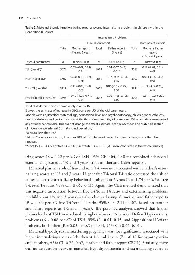
112 Chapter 2.5
izing scores (B = 0.22 per SD of TSH, 95% CI: 0.04, 0.40 for combined behavioral externalizing scores at 1½ and 3 years, from mother and father reports).
Maternal plasma levels of free and total T4 were not associated with children’s exter-nalizing scores at 1½ and 3 years. Higher free T4/total T4 ratio decreased the risk of father reported externalizing behavioral problems at 3 years (B = -1.74 per SD of free T4/total T4 ratio, 95% CI: -3.06, -0.41). Again, the GEE method demonstrated that this negative association between free T4/total T4 ratio and externalizing problems in children at 1½ and 3 years was also observed using all mother and father reports (B = -1.09 per SD free T4/total T4 ratio, 95% CI: -2.11, -0.07, based on mother and father reports at 1½ and 3 years). The post-hoc analyses showed that higher plasma levels of TSH were related to higher scores on Attention Deficit/Hyperactivity problems (B = 0.08 per SD of TSH, 95% CI: 0.01, 0.15) and Oppositional Defiant problems in children (B = 0.08 per SD of TSH, 95% CI: 0.02, 0.14).
Maternal hypothyroxinemia during pregnancy was not significantly associated with higher internalizing scores of children at 1½ and 3 years (B = -0.19 for hypothyroxin-emic mothers, 95% CI -0.75, 0.37, mother and father report CBCL). Similarly, there was no association between maternal hypothyroxinemia and externalizing scores at
Table 2. Maternal thyroid function during pregnancy and internalizing problems in children within the Generation R Cohort
Internalizing Problems
One parent report Both parents report
Total Mother reporta
(1 ½ and 3 years)Total Father report
(3 years)Total Mother & Father
report(1 ½ and 3 years)
Thyroid parameters n B (95% CI) p n B (95% CI) p n B (95% CI) p
TSH (per SD)b 36770.02 (-0.09, 0.11),
0.712618
0.24 (0.07, 0.42), 0.01*
36820.10 (-0.01, 0.21),
0.07
Free T4 (per SD)b 37020.03 (-0.11, 0.17),
0.702635
-0.07 (-0.25, 0.12), 0.47
37070.01 (-0.13, 0.15),
0.91
Total T4 (per SD)b 37190.11 (-0.02, 0.24),
0.092652
0.06 (-0.12, 0.25), 0.51
37240.09 (-0.04,0.22),
0.19
FreeT4/TotalT4 (per SD)b 3698-0.42 (-1.96, 0.71),
0.242632
-0.86 (-1.85, 0.13), 0.09
3703-0.51 (-1.22, 0.20),
0.16
Total of children in one or more analyses is 3736.B gives the estimate of increase in CBCL score per SD of thyroid parameters.Models were adjusted for maternal age, educational level and psychopathology, child’s gender, ethnicity, mode of delivery and gestational age at the time of maternal thyroid sampling. Other variables were tested as potential confounders but did not change the effect estimate (see the Methods and Materials section)CI = Confidence interval, SD = standard deviation,* p value less than 0.05a At the 1½ year assessment, less than 10% of the informants were the primary caregivers other than mothers.b SD of TSH = 1.43, SD of free T4 = 3.48, SD of total T4 = 31.31 (SDs were calculated in the whole sample)

Maternal prenatal thyroid function and behavioral problems 113
1½ and 3 years, based on mother and father reports (B = 0.17 for hypothyroxinemic mothers 95% CI -0.53, 0.87). With the alternative cut-off for the definition of hypo-thyroxinemia, we obtained very similar results.
Maternal psychopathology and mode of delivery of the child were not intermediates in the association between maternal thyroid function and behavioral outcome of children. Adding quadratic terms of thyroid parameters to the model did not significantly im-prove the model’s fit and did not support the notion of a non-linear association between maternal thyroid parameters and behavioral problems of children (data not shown).
Discussion
In the present study, higher plasma levels of maternal TSH in the first half of preg-nancy predicted externalizing problems in the offspring. Plasma levels of free and total T4 in the mothers were not associated with internalizing and externalizing problems in their children. Lower levels of maternal Free T4/total T4 ratio though increased the risk of externalizing problems in the offspring.
Table 3. Maternal thyroid function during pregnancy and externalizing problems in children within the Generation R Cohort
Externalizing Problems
One parent report Both parents report
Total Mother reporta
(1 ½ and 3 years)Total Father report
(3 years)Total Mother & Father
report(1 ½ and 3 years)
Thyroid parameters n B (95% CI) p n B (95% CI) p n B (95% CI) p
TSH (per SD)b 36810.15 (-0.03, 0.33),
0.102616
0.26 (0.02, 0.50), 0.03*
36870.22 (0.04, 0.40),
0.02*
Free T4 (per SD)b 37060.02 (-0.16, 0.20),
0.812633
-0.14 (-0.39, 0.11), 0.27
3712-0.02 (-0.16, 0.20),
0.80
Total T4 (per SD)b 37230.15 (-0.04, 0.34),
0.132650
0.08 (-0.17, 0.33), 0.55
37290.12 (-0.06, 0.30),
0.19
FreeT4/TotalT4 (per SD)b 3702-0.74 (-1.78, 0.30),
0.172630
-1.74 (-3.06, -0.41), 0.01*
3708-1.09 (-2.11, -0.07),
0.03*
Total of children in one or more analyses is 3736.B gives the estimate of increase in CBCL score per SD of thyroid parameters.Models were adjusted for maternal age, educational level and psychopathology, child’s gender, ethnicity, mode of delivery and gestational age at the time of maternal thyroid sampling. Other variables were tested as potential confounders but did not change the effect estimate (see the Methods and Materials section)CI = Confidence interval, SD = standard deviation,* p value less than 0.05a At the 1½ year assessment, less than 10% of the informants were the primary caregivers other than mothers.b SD of TSH = 1.43, SD of free T4 = 3.48, SD of total T4 = 31.31 (SDs were calculated in the whole sample)

114 Chapter 2.5
Results from molecular and clinical observations provide evidence for a prominent role of thyroid hormones in brain development de Escobar et al., 2004, 2007; Had-dow et al., 1999). Insufficient thyroid hormone levels during the critical period of fetal corticogenesis lead to impairments in migration, differentiation and distribution of neural cells in the neocortex and the hippocampus (Auso et al., 2004; Lavado-Autric et al., 2003; Zoeller & Rovet, 2004). Vermiglio and colleagues (2004) showed that maternal hypothyroxinemia and plasma levels of TSH during pregnancy are associated with ADHD in children. But the retrospective design of the study and the small sample size makes it difficult to infer a causal relation from their results. In another study, Kooistra et al. (2006) demonstrated that maternal free T4 but not TSH levels is a predictor for the orientation of neonates at a very young age. They assessed the behavior of the child at the age of 3 weeks, which is too early to interpret straight forwardly the outcome as behavioral problems (Kooistra et al., 2006). In the present study, we showed that maternal TSH levels and the ratio of freeT4/total T4 can predict externalizing problems in the offspring. Further analyses confirmed this by showing an association between maternal thyroid parameters and Attention Deficit/Hyperactivity and Oppositional Deviant problems in children. Our results support the evidence that TSH is a good indicator of thyroid function due to the delicate feedback mechanism of the pituitary. Mild increases in TSH levels (as a stimulatory mechanism for thyroid hormone secretion) can signal low levels of maternal thyroid hormones (Lavado-Autric et al., 2003). This may lead to impaired fetal brain develop-ment and subsequent cognitive and behavioral problems. However, we have to be cautious not to overinterpret the difference in the relations of TSH with internalizing and externalizing problems. Firstly, the effect estimates for internalizing and exter-nalizing problems were very similar with largely overlapping confidence intervals. Secondly, the overall association between TSH and internalizing problems just failed to reach the significant level. On the other hand, the role of thyroid hormones in normal development of neural structures in the cerebral cortex makes an effect on the externalizing problems particularly plausible since cortical structures are responsible for the regulation of inhibitory processes (Auso et al., 2004; Campbell, Shaw, & Gil-liom, 2000; Zoeller & Rovet, 2004). The impairment in the control system may lead to deficits in executive functions as seen in externalizing problems such as ADHD (Sergeant, Geurts, & Oosterlaan, 2002).
Despite some evidence from previous epidemiological studies (Vermiglio et al. 2004; Pop et al., 2003), it remains a challenge to investigate the role of thyroid hormones during pregnancy with valid indicators of maternal thyroid function. While maternal thyroid insufficiency during pregnancy is mostly defined by high plasma levels of TSH, Pop and colleagues (1999, 2003) presented free T4 as an important predictor for neurodevelopment of the offspring. The studies examining maternal high levels of

Maternal prenatal thyroid function and behavioral problems 115
TSH in early pregnancy as a risk factor for abnormal neurodevelopment in children suggest that TSH, although not the biologically active hormone in the brain, is a more sensitive indicator of maternal thyroid dysfunction (Haddow et al. 1999; Vermiglio et al. 2004). This notion is consistent with the results of the present study, as we showed that maternal TSH levels were associated with problem behavior of the child. In con-trast, we failed to confirm the hypothesis that low maternal free and total T4 during pregnancy predict behavioral problems in the offspring. The results of the present study may imply that the best surrogate indicator for maternal thyroid dysfunction in the general population can be different from clinical samples. As T3 in the brain of the fetus is directly derived from T4 (Lavado-Autric et al. 2003), its measurement in maternal plasma has limited value in the diagnosis of thyroid dysfunction. Therefore, the active biologic agent may not be the best marker of the underlying dysfunction.
Free T4/total T4 ratio is inversely related to serum TBG concentrations (Passath, Leb, & Ollinger, 1984). Older studies referred to TBG as “part of a control system” in maternal blood that helps fetal tissue to receive an optimal level of thyroid hormones for development (Ekins et al., 1994; Sinha, Pickard, & Ekins, 1992). The authors argued that such binding protein (TBG) potentially enhances hormone transport to target organs. Our results do not support this theory, since we demonstrated a protective effect of free T4/total T4 ratio for externalizing problems of children. This effect was consistent at 1½ and 3 years and for behavioral problems reported by both informants. It is also known that TBG plasma levels increase during pregnancy under the influence of estrogens (Glinoer, 1997). But, in normal pregnancy, T3 and T4 increase parallel with the elevation of maternal TBG levels and a new equilibrium occurs to provide an adequate amount of thyroid hormones to the fetus (de Escobar et al., 2007). Since we used the ratio of freeT4/total T4 as a proxy for TBG levels in plasma, our findings must be interpreted cautiously. In contrast, some other studies suggested that TBG may selectively release T4 under specific circumstances (Zoeller & Crofton, 2000). Our results indicate that the fraction of thyroid hormone bound to binding proteins is more important than the absolute value of free and total T4. Perhaps this influences the equilibrium of thyroid parameters in blood and their availability to the fetus.
Both parents are valuable informants for the child’s behavior (Achenbach, McCo-naughy, & Howell, 1987). But, it is well known that the interparental correlation of the rating on emotion and behavior of the child is only moderate (Christensen, Margolin, & Sullaway, 1992). Mothers may be more aware of internalizing behavior of the offspring probably because they spend more time with their children compared to fathers (Treutler & Epkins, 2003), whereas fathers may be more sensitive to exter-nalizing behavior (Dave, Nazareth, Senior, & Sherr, 2008). In the present study, we thus used ratings by both parents to reduce the informant effect on the report of the child’s behavior and emotion (Rowe & Kandel, 1997).

116 Chapter 2.5
Our study has several strengths. To our knowledge, this is the first longitudinal population-based study to investigate the effect of maternal thyroid function on be-havioral problems as an indicator of neurodevelopment in the offspring. We measured maternal thyroid parameters before the 18th week of gestation, as recommended in the literature (Haddow et al., 1999; Pop, Kuijpens et al., 1999), and assessed the behavior of the child on two occasions. Since the association between maternal thyroid function and children’s problem behavior can potentially be explained by extraneous factors, we considered several confounders and intermediates in the relationship.
Some limitations of our study need to be discussed. The participation rate in the present study was high but the possibility of selection bias remains, as the non-respon-dents differed from participants. We did not measure the iodine levels in maternal blood and in the diet, a factor which has been shown to have effects on the brain structure of the child and thyroid hormones (Vermiglio et al., 2004). We suggest that future studies measure maternal thyroid parameters repeatedly in the first trimester and later in pregnancy to determine the period in which the brain development is sensitive to maternal thyroid dysfunction.
Numerous causes of poor development in children have been identified but it is not easy to define the contribution of any etiologic factor to the occurrence of behavioral problems in the general population (Rose, 2001). Nevertheless, it is established that a subtle change in a physiologic function as a result of a public health intervention may lead to a considerable effect on the health of children. Therefore, any small change of maternal thyroid function during pregnancy may have substantial implications on problem behavior of children (Rose, 2001). Yet, it is premature to speculate about possible intervention and their impact before the relation observed is confirmed.
Conclusion
In summary, there was a positive linear association between maternal plasma TSH levels and behavioral scores of children. This suggests that subtle variation of maternal thyroid function in the general population impacts on fetal brain development which determines behavioral and emotional problems later in life.

Maternal prenatal thyroid function and behavioral problems 117
References
Abalovich, M., Amino, N., Barbour, L. A., Cobin, R. H., De Groot, L. J., Glinoer, D., et al. (2007). Management of thyroid dysfunction during pregnancy and postpartum: an Endocrine Society Clinical Practice Guideline. Journal of Clinical Endocrinology and Metabolism, 92(8 Suppl), S1-47.
Achenbach, T. M., McConaughy, S. H., & Howell, C. T. (1987). Child/adolescent behavioral and emotional problems: implications of cross-informant correlations for situational specificity. Psychological Bulletin, 101(2), 213-232.
Achenbach, T. M., & Rescorla, L. A. (2000). Manual for ASEBA preschool forms & profiles. Burling-ton, VT: University of Vermont, Research Center for Children, Youth and Families.
Auso, E., Lavado-Autric, R., Cuevas, E., Del Rey, F. E., Morreale De Escobar, G., & Berbel, P. (2004). A moderate and transient deficiency of maternal thyroid function at the beginning of fetal neocorticogenesis alters neuronal migration. Endocrinology, 145(9), 4037-4047.
Bernal, J. (2007). Thyroid hormone receptors in brain development and function. Nature Clinical Practice, 3(3), 249-259.
Campbell, S. B., Shaw, D. S., & Gilliom, M. (2000). Early externalizing behavior problems: tod-dlers and preschoolers at risk for later maladjustment. Development and Psychopathology, 12(3), 467-488.
Christensen, A., Margolin, G., & Sullaway, M. (1992). Interparental agreement on child behavior problems. Psychological Assessment, 4, 419-425.
Cleary-Goldman, J., Malone, F. D., Lambert-Messerlian, G., Sullivan, L., Canick, J., Porter, T. F., et al. (2008). Maternal thyroid hypofunction and pregnancy outcome. Obstetrics and Gynecology, 112(1), 85-92.
Dave, S., Nazareth, I., Senior, R., & Sherr, L. (2008). A comparison of father and mother report of child behaviour on the Strengths and Difficulties Questionnaire. Child Psychiatry and Human Development, 39(4), 399-413.
De Beurs, E. (2004). Brief Symptom Inventory, handleiding [Dutch manual]. Leiden, The Netherlands.de Escobar, G. M., Obregon, M. J., & del Rey, F. E. (2004). Maternal thyroid hormones early in
pregnancy and fetal brain development. Best Practice & Research, 18(2), 225-248.de Escobar, G. M., Obregon, M. J., & del Rey, F. E. (2007). Iodine deficiency and brain develop-
ment in the first half of pregnancy. Public Health Nutrition, 10(12A), 1554-1570.Derogatis, L. R. (1993). Brief Symptom Inventory (BSI): Administration, scoring and procedures.
Manual, third edition. Minneapolis, MN: National Computer Systems Inc.Ekins, R. P., Sinha, A. K., Pickard, M. R., Evans, I. M., & al Yatama, F. (1994). Transport of thyroid
hormones to target tissues. Acta Medica Austriaca, 21(2), 26-34.Evans, I. M., Pickard, M. R., Sinha, A. K., Leonard, A. J., Sampson, D. C., & Ekins, R. P. (2002).
Influence of maternal hyperthyroidism in the rat on the expression of neuronal and astrocytic cytoskeletal proteins in fetal brain. Journal of Endocrinology, 175(3), 597-604.
Glinoer, D. (1997). The regulation of thyroid function in pregnancy: pathways of endocrine adapta-tion from physiology to pathology. Endocrine Reviews, 18(3), 404-433.
Haddow, J. E., Palomaki, G. E., Allan, W. C., Williams, J. R., Knight, G. J., Gagnon, J., et al. (1999). Maternal thyroid deficiency during pregnancy and subsequent neuropsychological development of the child. New England Journal of Medicine, 341(8), 549-555.
Jaddoe, V. W., Mackenbach, J. P., Moll, H. A., Steegers, E. A., Tiemeier, H., Verhulst, F. C., et al. (2006). The Generation R Study: Design and cohort profile. European Journal of Epidemiology, 21(6), 475-484.
118 Chapter 2.5
Jaddoe, V. W., van Duijn, C. M., van der Heijden, A. J., Mackenbach, J. P., Moll, H. A., Steegers, E. A., et al. (2008). The Generation R Study: design and cohort update until the age of 4 years. European Journal of Epidemiology, 23(12), 801-811.
Klein, R. Z., Sargent, J. D., Larsen, P. R., Waisbren, S. E., Haddow, J. E., & Mitchell, M. L. (2001). Relation of severity of maternal hypothyroidism to cognitive development of offspring. Journal of Medical Screening, 8(1), 18-20.
Kooistra, L., Crawford, S., van Baar, A. L., Brouwers, E. P., & Pop, V. J. (2006). Neonatal effects of maternal hypothyroxinemia during early pregnancy. Pediatrics, 117(1), 161-167.
Kuntsi, J., Rijsdijk, F., Ronald, A., Asherson, P., & Plomin, R. (2005). Genetic influences on the stability of attention-deficit/hyperactivity disorder symptoms from early to middle childhood. Biological Psychiatry, 57(6), 647-654.
Lavado-Autric, R., Auso, E., Garcia-Velasco, J. V., Arufe Mdel, C., Escobar del Rey, F., Berbel, P., et al. (2003). Early maternal hypothyroxinemia alters histogenesis and cerebral cortex cytoarchi-tecture of the progeny. Journal of Clinical Investigation, 111(7), 1073-1082.
Man, E. B., & Jones, W. S. (1969). Thyroid function in human pregnancy. V. Incidence of maternal serum low butanol-extractable iodines and of normal gestational TBG and TBPA capacities; retardation of 8-month-old infants. American Journal of Obstetrics and Gynecology, 104(6), 898-908.
Man, E. B., & Serunian, S. A. (1976). Thyroid function in human pregnancy. IX. Development or retardation of 7-year-old progeny of hypothyroxinemic women. Am J Obstet Gynecol, 125(7), 949.
Mickey, R. M., & Greenland, S. (1989). The impact of confounder selection criteria on effect estimation. American Journal of Epidemiology, 129(1), 125-137.
Pagliacci, M. C., Pelicci, G., Grignani, F., Giammartino, C., Fedeli, L., Carobi, C., et al. (1987). Thyroid function tests in patients undergoing maintenance dialysis: characterization of the ‘low-T4 syndrome’ in subjects on regular hemodialysis and continuous ambulatory peritoneal dialysis. Nephron, 46(3), 225-230.
Passath, A., Leb, G., & Ollinger, P. (1984). [Hypotheses on the validity of the radioimmunologic measurement of FT4]. Klin Wochenschr, 62(13), 640-650.
Pop, V. J., Brouwers, E. P., Vader, H. L., Vulsma, T., van Baar, A. L., & de Vijlder, J. J. (2003). Maternal hypothyroxinaemia during early pregnancy and subsequent child development: a 3-year follow-up study. Clinical Endocrinology, 59(3), 282-288.
Pop, V. J., de Vries, E., van Baar, A. L., Waelkens, J. J., de Rooy, H. A., Horsten, M., et al. (1995). Maternal thyroid peroxidase antibodies during pregnancy: a marker of impaired child develop-ment? Journal of Clinical Endocrinology and Metabolism, 80(12), 3561-3566.
Pop, V. J., Kuijpens, J. L., van Baar, A. L., Verkerk, G., van Son, M. M., de Vijlder, J. J., et al. (1999). Low maternal free thyroxine concentrations during early pregnancy are associated with impaired psychomotor development in infancy. Clinical Endocrinology, 50(2), 149-155.
Pop, V. J., van Baar, A. L., & Vulsma, T. (1999). Should all pregnant women be screened for hypothyroidism? Lancet, 354(9186), 1224-1225.
Rose, G. (2001). Sick individuals and sick populations. International Journal of Epidemiology, 30(3), 427-432; discussion 433-424.
Rothman, K. J. (1990). No adjustments are needed for multiple comparisons. Epidemiology, 1(1), 43-46.
Rothman, K. J., Greenland, S., & Lash, T. L. (2008). Modern Epidemiology (3rd ed.). Philadelphia: Lipponcott-Raven publishers.

Maternal prenatal thyroid function and behavioral problems 119
Rowe, D. C., & Kandel, D. (1997). In the eye of the beholder? Parental ratings of externalizing and internalizing symptoms. Journal of Abnorm Child Psychology, 25(4), 265-275.
Sergeant, J. A., Geurts, H., & Oosterlaan, J. (2002). How specific is a deficit of executive function-ing for attention-deficit/hyperactivity disorder? Behavioural Brain Research, 130(1-2), 3-28.
Simko, J., & Horacek, J. (2007). Carbamazepine and risk of hypothyroidism: a prospective study. Acta Neurologica Scandinavica, 116(5), 317-321.
Sinha, A. K., Pickard, M. R., & Ekins, R. P. (1992). Maternal hypothyroxinemia and brain develop-ment: I. A hypothetical control system governing fetal exposure to maternal thyroid hormones. Acta Medica Austriaca, 19 Suppl 1, 40-48.
Treutler, C. M., & Epkins, C. C. (2003). Are discrepancies among child, mother, and father reports on children’s behavior related to parents’ psychological symptoms and aspects of parent-child relationships? Journal of Abnormal Child Psychology, 31(1), 13-27.
Vermiglio, F., Lo Presti, V. P., Moleti, M., Sidoti, M., Tortorella, G., Scaffidi, G., et al. (2004). At-tention deficit and hyperactivity disorders in the offspring of mothers exposed to mild-moderate iodine deficiency: a possible novel iodine deficiency disorder in developed countries. Journal of Clinical Endocrinology and Metabolism, 89(12), 6054-6060.
Zoeller, R. T., & Crofton, K. M. (2000). Thyroid hormone action in fetal brain development and potential for disruption by environmental chemicals. Neurotoxicology, 21(6), 935-945.
Zoeller, R. T., & Rovet, J. (2004). Timing of thyroid hormone action in the developing brain: clinical observations and experimental findings. Journal of Neuroendocrinology, 16(10), 809-818.


Chapter 3Fetal growth as determinant
of behavior and cognition


3 .1Fetal size in mid- and late pregnancy
is related to infant alertness

124 Chapter 3.1
Abstract
The vulnerability for behavioral problems is partly shaped in fetal life. Numerous studies have related indicators of intrauterine growth, e.g. birth weight and body size, to behavioral development. We investigated whether fetal size in mid- and late pregnancy is related to infant irritability and alertness. In a population-based birth cohort of 4,255 singleton full term infants ultrasound measurements of fetal head and abdominal circumference in mid- and late pregnancy were performed. Infant irritabil-ity and alertness scores were obtained by the Mother and Baby Scales at 3 months and z-standardized. Multiple linear regression analyses revealed curvilinear associations (inverted J-shape) of measures of fetal size in both mid- and late pregnancy with infant alertness. Fetal size characteristics were not associated with infant irritability. These results suggest that alterations of intrauterine growth affecting infant alertness are already detectable from mid-pregnancy onwards.

Fetal size and infant alertness 125
Introduction
An increasing number of epidemiological and clinical studies have demonstrated the significance of indicators of fetal growth, e.g. birth weight and body size, with regard to subsequent psychological and behavioral child development. Associations of low birth weight (<2,500 g) and very low birth weight (<1,500 g) with psychiatric symptoms and disorders and cognitive and educational functioning in childhood have been reported (Breslau & Chilcoat, 2000; Gray, Indurkhya, & McCormick, 2004; Hack, Taylor, Klein, Eiben, Schatschneider, & Mercuri-Minich, 1994; Horwood, Mogridge, & Darlow, 1998). In addition, low birth weight has been related to infant temperament (Hughes, Shults, McGrath, & Medoff-Cooper, 2002; Langkamp, Kim, & Pascoe, 1998). Little is known, however, about possible associations of the nor-mal variation in fetal growth and size with early behavioral development in infancy. Prematurity and low birth weight can lead to a variety of medical interventions and complications such as breathing difficulties that could influence outcome. Further-more, postmaturity (i.e. being delivered after 42 completed weeks of gestation) is also known to be related with a number of medical and developmental risks, e.g. labor complications, macrosomia and behavioral and developmental abnormalities (Alexander, McIntire, & Leveno, 2000; Shea, Wilcox, & Little, 1998). Thus, it is crucial to also focus on full term infants when the impact of fetal conditions on subsequent psychological development is studied.
The ‘fetal programming’ hypothesis by Barker (1995, 1998) stresses the importance of human fetal experience in determining developmental patterns. Since the prenatal period is a time of enormous growth and change, in which tissues develop in a specific sequence from conception to maturity, the fetus is vulnerable to both organizing and disorganizing influences on organ development. Fetal programming is a process by which a prenatal event during a sensitive developmental period has a long-lasting or permanent influence on the development of organs and associated physiological and metabolic systems. Most support for the fetal programming hypothesis stems from research relating measures of low size at birth to alterations in endocrine metabolic functions (Godfrey & Barker, 2000). Studies that investigated the association of size at birth within the normal range with medical disorders in adulthood, such as non-insulin dependent diabetic mellitus and ischemic heart disease (Barker, Winter, Os-mond, Margetts, & Simmonds, 1989; Lithell, McKeigue, Berglund, Mohsen, Lithell, & Leon, 1996), also support the hypothesis.
Studies of mental health showed that body size at birth within the normal range also predicts behavioral functioning in adulthood, such as susceptibility to stress, psychologi-cal distress and psychopathology in adulthood (Cheung, Khoo, Karlberg, & Machin, 2002; Nilsson, Nyberg, & Ostergren, 2001; Wiles, Peters, Leon, & Lewis, 2005).

126 Chapter 3.1
Although it is likely that the association of size at birth within the normal range with behavioral functioning observed in adults is already present in childhood, only few studies demonstrated an association of measures of body size at birth with behav-ioral development among infants or children (Lahti, Raikkonen, Kajantie, Heinonen, Pesonen, Jarvenpaa, & Strandberg, 2006; Pesonen, Raikkonen, Kajantie, Heinonen, Strandberg, & Jarvenpaa, 2006; van Os, Wichers, Danckaerts, Van Gestel, Derom, & Vlietinck, 2001; Wiles, Peters, Heron, Gunnell, Emond, & Lewis, 2006). For example, a recent study by Wiles et al. (2006) reported a weak association of fetal growth indexed by birth length with childhood behavioral problems at age 7 years. In addition, Pesonen et al. (2006) observed that thinness at birth and birth length was linked to temperamental negative affectivity of healthy 5.5-year-old terms. Fur-thermore, a study of children aged 5 to 6 years, who were born healthy and at term, demonstrated associations of lower ponderal index, a smaller head circumference, and a smaller head circumference-to-length ratio at birth with behavioral symptoms of the attention deficit hyperactivity disorder (Lahti et al., 2006). Van Os et al. (2001) demonstrated in a twin study that birth weight was inversely associated with child-hood behavioral problems and that the degree of birth weight discordance was linearly related to the discordance for childhood behavioral problems in 10 year-old children. This finding suggests that the influence of birth weight on subsequent psychological functioning is not reduced to shared genetic and environmental factors.
Earlier studies that investigated the relation between fetal development within the normal range of gestation and psychological outcomes in childhood suggests that the association between size at birth and subsequent cognitive and behavioral functioning may not necessarily be linear (Pesonen et al., 2006; Shenkin, Starr, & Deary, 2004; Wiles et al., 2006). Pesonen et al. (2006) observed a u-shaped relation between length at birth and child fearfulness. Similarly, Wiles et al. (2006) reported some evidence for nonlinear associations of birth weight with prosocial behavior at age 47 months and with emotional problems at age 81 months.
In the current study, we examined the effects of fetal development within the normal range of gestation on infant irritability and alertness at the age of 3 months. Both infant irritability and alertness are considered to be crucial for the early de-velopment of the infant and to shape mother-infant interactions (Crockenberg & Smith, 1982). Infant irritability represents aspects of early negative emotionality, which have been shown to be predictive of externalizing disorders at age 5.5 years (Shaw, Owens, Giovannelli, & Winslow, 2001). Infant alertness is an indicator of early neurobehavioral development and represents the infant’s abilities to attend and respond to novel stimuli. It is important in regulating the communicative exchanges with the mother and in facilitating the development of parent-neonate interactions, e.g. during feeding. Moreover, infant alertness is involved in the development of the

Fetal size and infant alertness 127
infant’s visual attention (Colombo, 2001) and maturation of the visual system (Roy, Gosselin, Hanna, Orquin, & Chemtob, 2004) and has been reported to be predictive of early mental development (Feldman, Eidelman, Sirota, & Weller, 2002).
The rationale for conducting the current study is twofold: First, we aimed to focus on subjects born healthy at term (i.e. > 37 weeks of gestation and ≤ 42 weeks of gesta-tion) in order to examine the influence of fetal growth within the normal range of gestation on infant irritability and alertness. Second, earlier studies used birth weight, length or thinness as indices of fetal growth, which are considered to be rather unspe-cific and crude summary measures of intrauterine growth, since they do not provide information on specific periods of fetal development and fetal growth patterns. In the current study, in addition to birth weight direct markers of fetal size in mid- and late pregnancy, such as head and abdominal circumference, were used. Furthermore, in the present study we also used the ratio of abdominal and head circumference, which measures levels of symmetry of the fetal growth pattern. Moreover, markers of fetal size were used to investigate the relation between fetal growth from mid- to late pregnancy with infant alertness and irritability.
We hypothesized that a larger fetal size and increased fetal growth rates from mid- to late pregnancy are associated with more infant alertness and less infant irritability. As earlier studies reported evidence suggesting that the association between body size at birth and behavioral functioning in childhood may not necessarily be linear we also tested nonlinear association of fetal size with infant behavior.
Methods
Design
The current study was embedded in the Generation R Study, a population-based multi-ethnic cohort study from fetal life until young adulthood in Rotterdam, The Netherlands. The Generation R Study has previously been described in detail (Jaddoe, Mackenbach, Moll, Steegers, Tiemeier, Verhulst, Witteman, & Hofman, 2006). As-sessments in pregnant women consisted of physical examinations, fetal ultrasounds, biological samples and questionnaires. All children were born between April 2002 and January 2006. Of all eligible children in the study area, 61% participated in the Generation R Study (Jaddoe et al., 2006).
The study has been approved by the Medical Ethics Committee of the Erasmus Medical Center, Rotterdam (numbers: prenatal, MEC 198.782/2001/31 and postna-tal, MEC 217.595/2002/202). Written informed consent was obtained from all adult participants.

128 Chapter 3.1
Population for analysis
In the postnatal follow-up of the Generation R cohort, 7,654 live born children and their mothers, who were prenatally recruited, were asked to participate. In the first few months, 34 children deceased. As the current study focused on normal gestation, 393 prematurely born children (gestational age < 37 weeks) and 349 postmaturely born children (gestational age > 42 weeks) were excluded. Moreover, 70 children were excluded because they were twins and, therefore, prenatal ultrasound data could not unambiguously be assigned to these children. The remaining 6,808 children were eligible for the current study. Mothers of 651 children withdrew written consent for postnatal participation. Furthermore, 1902 mothers did not provide information on infant alertness and irritability. This left information on birth weight and infant ir-ritability and alertness for 4,255 infants (62.5% of the eligible subjects) in the total sample of the current study. These 4,255 infants were included in one or more of the analyses. Due to different amounts of missing data on fetal ultrasound measure-ments the number of subjects included in our analyses differed. The analysis of mid-pregnancy fetal size was based on 4030 observations and the analysis of late pregnancy fetal size on 4130 observations. In addition, analyses on the association between fetal growth from mid- to late pregnancy and infant behavior were conducted in 3,985 subjects of the total sample.
Fetal ultrasound measurements and birth weight
Trained sonographers conducted fetal ultrasound examinations during the visits to research centers in early (gestational age < 18 weeks), mid-pregnancy (gestational age 18-25 weeks) and late pregnancy (gestational age ≥ 25 weeks). Early pregnancy data were not included in the analysis since these fetal ultrasound examinations were used to establish gestational age.
Online measurements included head and abdominal circumference, which were measured using standardized techniques. Head circumference in mid- and late pregnancy indicate fetal head growth and abdominal circumference represents gen-eral intrauterine growth. The ratio of abdominal and head circumference, which was calculated by dividing the abdominal circumference through the head circumference, assesses the symmetry of fetal growth and is an indicator of brain sparing. Intra- and inter-observer reliability of fetal biometry in early pregnancy within the Generation R Study was high. Intra-observer intraclass correlation coefficients varied from 0.985 for abdominal circumference to 0.995 for head circumference, inter-observer intraclass correlation coefficients varied from 0.980 for abdominal circumference to 0.988 for head circumference, with coefficients of variation between 2.2 and 3.8%. Child birth weight was obtained from medical records completed by midwives and gynecologists.

Fetal size and infant alertness 129
Infant irritability and alertness
Three months after delivery, the ‘Mother and Baby Scales’ (MABS) were used to assess early infant behavior. This parent-report measure by St James-Roberts and Wolke (1983), includes a number of subscales that assess infant negative emotionality and alertness during and outside feeding (St James-Roberts & Wolke, 1983, 1988). We used the ‘Your Baby and Your Feelings’ part of the MABS, in which mothers were asked to report on scales of infant irritability and alertness. The ‘Unsettled-Irregular’ scale contains 15 items, e.g. “My baby has fussed before settling down”, and the ‘Alertness-Responsiveness’ scale consists of 8 items; e.g. “When I talk to my baby s/he seems to take notice”. Each item was rated on 6-point uni-dimensional rating scales ranging from ‘0’ (not at all) to ‘5’ (very much/often). Unsettled-Irregular scores range from 0-75 and alertness-responsiveness scores range from 0-40. The internal consistencies for the ‘Unsettled-Irregular’ scale and the ‘Alertness-Responsiveness’ scale in the current study were: α = 0.75 and α = 0.64, respectively.
A study by St James-Roberts and Wolke (1988) addressed convergences among mothers’ and professionals’ assessment of difficult infant behavior using the well-known Neonatal Behavioral Assessment Scale (NBAS), a nurse scale of mother and baby behavior and the MABS. In this study by St James-Roberts and Wolke (1988) maternal reports using the MABS included a diary of specific behaviors, such as feed-ing, ratings of specific behaviors, i.e. infant irritability during and outside feeding, and temperament impressions ratings comprising the ‘Infant Easiness’ and ‘Infant Regularity’ scale. In general, moderate convergences between infant behavior assessed by the MABS and objective assessments by external raters were found demonstrating correlation coefficients between maternal and professional assessments that ranged from 0.27 to 0.52 (St James-Roberts & Wolke, 1988).
Covariates
Information on maternal age, pre-pregnancy body mass index, education, ethnicity and parity (0, 1, or ≥ 2) was obtained by questionnaire at enrolment. Education was divided in five categories: Ranging from primary education only to higher education with a university degree. Ethnicity of the mother was based on the country of birth of the grandparents. Maternal height was measured during the first visit to the research centre. Use of alcohol (‘no alcohol use’ and ‘alcohol use’) during pregnancy, prenatal smoking (‘non-smoking’ and ‘smoking’) and other drug use during pregnancy (‘no other drug use’ and ‘other drug use’), e.g. marihuana, were asked for at inclusion and again at 30 weeks of pregnancy. During pregnancy and at age 3 months of the children maternal symptoms of depression and anxiety in pregnancy were assessed with the Brief Symptom Inventory, a validated self-report questionnaire consisting of 53 items (Derogatis, 1993). For the current study, the 6-item anxiety scale, e.g.

130 Chapter 3.1
“nervousness or shaking inside”, and the 6-item depression scale, e.g. “feeling lonely” were used. Information on child gender, Apgar scores 1 minute after birth and pH values in the umbilical cord, mode of delivery (‘spontaneous vaginal’, ‘instrumental vaginal’ and ‘Caesarean section’), gestational diabetes, preeclampsia and maternal hy-pertension during pregnancy was obtained from medical records. Gestational age was established by the fetal ultrasound assessments since using the last menstrual period has several limitations, including the large number of women who do not remember the exact date of their last menstrual period or have irregular menstrual cycles (Altman & Chitty, 1997; Robinson, Sweet, & Adam, 1979). Child age was obtained when mothers reported infant irritability and alertness.
Statistical analyses
Multiple linear regression analysis was used to examine whether fetal size in mid- and late pregnancy and birth weight predict infant irritability and alertness. Fetal size characteristics were expressed in centimeters and birth weight in 100 grams. Infant irritability and alertness were the dependent variables in our main analyses. These variables were z-standardized across the sample of the current study. The betas of the fetal size characteristics or birth weight and their 95 percent confidence intervals are directly interpretable as in- or decrease of the z-standardized infant irritability and alertness scores per 1 centimeter (or per 100 grams) increase of the respective fetal size characteristic (or birth weight).
In additional analyses, we investigated whether fetal growth from mid- to late preg-nancy affects infant irritability and alertness by calculating a difference score (fetal size in late pregnancy - fetal size in mid-pregnancy) and conducting multiple linear regression analyses.
Regression models investigating the association of fetal size characteristics and fetal growth from mid-pregnancy to late pregnancy with infant irritability and alertness were adjusted for potential confounders. First, we considered socio-economic status related variables (maternal educational level, maternal smoking in pregnancy and ethnicity), obstetric and neonatal variables (gestational age and parity) and other known predictors of mother-reported behavioral outcomes in infancy (infant gender, infant age, maternal anxiety and depression) as possible confounders, because they have been depicted as confounding variables in the Alspac study, which related length and weight at birth to childhood behavioral problems (Wiles et al., 2006). Further-more, we regarded maternal size (height/weight) as a possible genetic confounding factor in line with the study by Wiles et al. (2006). Several perinatal and obstetric complications were also considered as confounding variables since these factors have been demonstrated to predict behavioral outcome in preterm born infants (Larroque, N’Guyen The Tich, Guedeney, Marchand, Burguet, & Epipage Study, 2005). Models

Fetal size and infant alertness 131
examining the association of fetal growth from mid- to late pregnancy with infant behavior were additionally adjusted for the respective fetal size characteristic and ges-tational age in mid-pregnancy, and the time interval between mid- and late pregnancy. Although controversy exists on how to identify confounding variables, Mickey and Greenland (1989) suggest that the change-in-estimate criterion tends to be superior compared to other methods for selecting confounders. The conventional change-in-estimate criterion is defined as a change of 10% or more (Mickey & Greenland, 1989; Rothman & Greenland, 1998). As observational studies, like our study, are very sensitive to residual confounding, we used a more conservative change-in-estimate criterion of 5%. Thus, covariates were selected when they changed the main effect estimates by more than 5% after adding them to the basic model, which included the respective fetal size or growth parameter and included gestational age at the time of fetal ultrasound assessment and gender. Apgar scores and pH values, gestational diabetes, maternal hypertension during pregnancy, preeclampsia and other maternal drug use during pregnancy, did not confound the relations between fetal size charac-teristics or birth weight and infant irritability and alertness and were, therefore, not included in the final models.
Furthermore, a quadratic term of the different fetal size characteristics or birth weight was tested because previous studies found nonlinear associations between birth weight or length and various outcomes (Graafmans, Richardus, Borsboom, Bakketeig, Langhoff-Roos, Bergsjo, Macfarlane, Verloove-Vanhorick, & Mackenbach, 2002; Nilsson et al., 2001; Pesonen et al., 2006; Wiles et al., 2006). To test for an interac-tion effect between gender and fetal size, a multiplicative interaction term of gender and fetal size or birth weight was added to the analyses. With all tests, p values < 0.05 were considered statistically significant.
Results
Sample characteristics
Table 1 presents the baseline characteristics of the mothers and their children. Head and abdominal circumference were strongly and significantly correlated with each other (r = 0.60, p < 0.001) in late pregnancy. Correlations between the ultrasound measurements in mid-pregnancy were similar (data not shown). All correlations between measurements of fetal size in mid-pregnancy with measurements of fetal size in late pregnancy were significant but weak (i.e., r < 0.30). In addition, infant alertness and irritability were inversely but weakly correlated with each other (Pearson correlation, r = -0.11, p < 0.001).

132 Chapter 3.1
The non-response analysis showed that head circumference (M = 17.9 cm (SD = 1.4) vs. M = 17.9 cm (SD = 1.6), t = 1.40, p = 0.161) and abdominal circumference (M = 15.7 cm (SD = 1.4) vs. M = 15.7 cm (SD = 1.6), t = 0.62, p = 0.532) in mid-pregnancy of responders and non-responders did not significantly differ. However, responders had a slightly larger head circumference (M = 28.6 cm (SD = 1.2) vs. M = 28.5 (SD = 1.3), t = 3.24, p = 0.001) and abdominal circumference (M = 26.5 cm (SD = 1.6) vs. M = 26.4 cm (SD = 1.7), t = 2.71, p = 0.007) in late pregnancy than non-responders. In addition, responders had a higher birth weight (M = 3.48 kg (SD = .48) vs. M = 3.42 kg (SD = .50), t = 4.47, p < 0.001) and gestational age (M = 40.0 weeks (SD = 1.14) vs. M = 39.9 weeks (SD = 1.17), t = 2.06, p = 0.039) when compared to non-responders. Mothers of children included in the study were more likely to be Dutch (61.4% vs. 34.8%, χ2 = 495.65, df = 7, p < 0.001) to smoke less
Table 1. Maternal and child characteristics (n = 4255)
Maternal characteristics M (SD)a
Age, y 30.8 (4.78)
Height, cm 168.4 (7.33)
Pre-pregnancy BMI, kg/m2 23.3 (4.03)
Parity (%)
0 57.5
1 30.9
≥ 2 11.6
Education (%)
Primary education 7.4
Secondary education 1st phase 11.2
Secondary education 2nd phase 27.7
Higher education 1st phase 23.4
Higher education 2nd phase 30.3
Ethnicity (%)
Dutch 61.4
Cape Verdian 2.6
Moroccan 3.8
Dutch Antilles 2.2
Surinamese 6.6
Turkish 5.8
Other Western 12.7
Other non-western 4.9
Smoking during pregnancy (%) 22.2
Alcohol use in pregnancy (%) 61.1
Other drug use in pregnancy (%) 2.4
Note. aUnless otherwise indicated

Fetal size and infant alertness 133
(22.2% vs. 26.4%, χ2 = 11.70, df = 1, p = 0.001) and to be higher educated (% higher education with a university degree 30.3 % vs. 16.8%, χ2 = 351.72, df = 4, p < 0.001).
Fetal size in mid- and late pregnancy and infant irritability and alertness
First, analyses showed that fetal size in mid- and late pregnancy was not related to infant irritability and alertness when modeled with linear terms only (data not shown). Several significant relations were found, however, when quadratic terms of fetal size characteristics were introduced. Table 2 presents the associations of fetal size measured in mid-pregnancy and late pregnancy with infant irritability and alertness. In mid-pregnancy, abdominal circumference and the ratio of abdominal and head cir-cumference were curvilinearly related to infant alertness, whereas head circumference was not associated with infant alertness. The fully adjusted model of the association
Table 1. Maternal and child characteristics (n = 4255) (continued)
Maternal characteristics M (SD)a
Mode of delivery (%)
Spontaneous vaginal 70.1
Instrumental vaginal 17.9
Caesarean section 12.0
Gestational diabetes (%) 0.6
Preeclampsia (%) 1.2
Maternal hypertension during pregnancy (%) 3.4
Prenatal anxiety, score 0.24 (0.40)
Prenatal depression, score 0.19 (0.43)
Depression at age 3 months, score 0.21 (0.46)
Child characteristics M (SD)a
Gender (girls, %) 51.1
Birth weight, kg 3.48 (0.48)
Gestational age at birth, wk 40.0 (1.14)
Gestational age in mid-pregnancy, wk 20.6 (1.09)
Head circumference in mid-pregnancy, cm 17.9 (1.4)
Abdominal circumference in mid-pregnancy, cm 15.7 (1.4)
Ratio of abdominal and head circumference in mid-pregnancy 0.88 (0.04)
Gestational age in late pregnancy, wk 30.4 (1.08)
Head circumference in late pregnancy, cm 28.6 (1.2)
Abdominal circumference in late pregnancy, cm 26.5 (1.6)
Ratio of abdominal and head circumference in late pregnancy 0.93 (0.04)
pH value in the umbilical cord 7.26 (0.16)
Apgar score 1 minute after birth 8.65 (1.13)
Child age, months 3.23 (1.55)
Note. aUnless otherwise indicated

134 Chapter 3.1
of abdominal circumference in mid-pregnancy with infant alertness explained 3.4% (i.e. R2 = 0.034) of the total variance, the respective model of the abdominal-to-head circumference ratio explained 3.5%. None of the fetal size characteristics in mid-pregnancy were linked to infant irritability.
In late pregnancy, head circumference was curvilinearly associated with infant alert-ness, while the abdominal circumference and the abdominal-to-head ratio were not related to infant alertness (Table 2). The fully adjusted model of the association be-tween head circumference in late pregnancy and infant alertness accounted for 3.8% of the total variance. Figure 1 illustrates the distribution of fetal head circumference in late pregnancy, it also shows the curvilinear association between head circumference and infant alertness as estimated by multiple linear regression. Like in mid-pregnancy, none of the fetal size characteristics in late pregnancy were related to infant irritability.
Table 2. Associations of fetal size characteristics in mid-pregnancy and late pregnancy and birth weight with infant irritability and alertness
Unsettled-Irregular Alertness-Responsiveness
Fetal size characteristics B 95% CI B 95% CI
Mid-pregnancy (n = 4030) Head circumference, cm -0.11 (-0.49; 0.27) 0.26 (-0.13; 0.64)
Head circumference, cm2 0.00 (-0.01; 0.01) -0.01 (-0.02; 0.00)
Abdominal circumference, cm -0.22 (-0.52; 0.09) 0.38 (0.07; 0.69)*
Abdominal circumference, cm2 0.01 (-0.00; 0.02) -0.01 (-0.02; -0.00)*
Ratio of abdominal and head circumference
2.63 (-18.9; 24.2) 25.1 (3.48; 46.8)*
Ratio of abdominal and head circumference2 -1.30 (-13.6; 11.0) -13.9 (-26.3; -1.63)*
Late pregnancy (n = 4130)
Head circumference, cm 0.00 (-0.71; 0.72) 1.20 (0.48; 1.91)***
Head circumference, cm2 -0.00 (-0.01; 0.01) -0.02 (-0.03; -0.01)***
Abdominal circumference, cm 0.04 (-0.33; 0.40) 0.33 (-0.04; 0.69)
Abdominal circumference, cm2 0.00 (-0.01; 0.01) -0.01 (-0.13; 0.00)
Ratio of abdominal and head circumference 2.92 (-19.5; 25.3) 15.6 (-6.93; 38.1)
Ratio of abdominal and head circumference2 -1.29 (-13.3; 10.8) -8.71 (-20.8; 3.38)
Size at birth (n = 4255)
Birth weight per 100 grams -0.003 (-0.009; 0.003) -0.002 (-0.008; 0.004)
Birth weight per 100 grams2 0.000 (-0.000; 0.000) 0.000 (-0.000; 0.000)
Note. All models included the linear and quadratic term of the respective (fetal) size characteristic and were adjusted for gender, gestational age in mid-pregnancy or in late pregnancy and/or for gestational age at birth, parity, maternal age, maternal height, pre-pregnancy body mass index, maternal education, maternal smoking and alcohol use during pregnancy, maternal ethnicity, mode of delivery, maternal prenatal depression and anxiety, maternal depression at age 3 months and child age.* p-value < 0.05, ** p-value < 0.01, *** p-value < 0.001

Fetal size and infant alertness 135
Head circumference in late pregnancy (cm), Mean = 28.6 cm (S.D. = 1.2 cm)
31.030.530.029.529.028.528.027.527.026.526.0
Freq
uenc
y
500
400
300
200
100
0
-0,15
-0,1
-0,05
0
0,05
0,1
26,0 26,5 27,0 27,5 28,0 28,5 29,0 29,5 30,0 30,5 31,0
Head circumference in late pregnancy (cm)
z-st
anda
rdiz
ed s
core
s of
Ale
rtne
ss-
Res
pons
iven
ess
Figure 1. Fetal head circumference and infant alertness. The upper part of the figure depicts a frequency plot of head circumference in late pregnancy representing the 95% range of 4130 fetal measurements. It also shows a fitted curve to illustrate the distribution. The lower part shows the curvilinear association of head circumference with infant alertness at age 3 months as estimated by multiple linear regression. This analysis was adjusted for gender, gestational age in late pregnancy and at birth, parity, maternal age, maternal height, pre-pregnancy body mass index, maternal education, maternal smoking and alcohol use during pregnancy, maternal ethnicity, mode of delivery, maternal prenatal depression and anxiety, maternal depression at age 3 months and child age.

136 Chapter 3.1
Whether or not adjusted for gestational age, birth weight was not related to infant irritability and alertness (data not shown). Furthermore, in the fully adjusted model birth weight was neither linked to infant irritability nor to alertness (Table 2).
Fetal growth from mid- to late pregnancy and infant irritability and alertness
Analyses of the relation between fetal growth from mid- to late pregnancy and infant behavior demonstrated little evidence for associations of difference scores based on the markers of fetal size in mid- and late pregnancy with infant behavior. Tests of linear and quadratic trends revealed no associations of fetal head and abdominal growth from mid- to late pregnancy with infant irritability and alertness (Table 3). The change in the abdominal-to-head ratio from mid- to late pregnancy was also not related to infant irritability. However, the difference score of the abdominal-to-head circumfer-ence ratio in mid- and late pregnancy was curvilinearly associated with infant alertness (Table 3). This association had an inverted u-shape, which indicates that children whose had grew too slow and too fast in comparison to the growth rate of their abdo-men in the period between mid- and late pregnancy have lower z-standardized infant alertness scores. The fully adjusted model of the association between the difference score of the abdominal-to-head circumference ratio in mid- and late pregnancy with infant alertness explained 4.1% of the total variance.
Table 3. Associations of fetal growth from mid- to late pregnancy with infant irritability and alertness (n = 3985)
Unsettled-Irregular Alertness-Responsiveness
Fetal growth characteristics B 95% CI B 95% CI
Head circumference, cm -0.15 (-0.31; 0.01) 0.02 (-0.14; 0.19)
Head circumference, cm2 0.01 (-0.00; 0.01) -0.00 (-0.01; 0.01)
Abdominal circumference, cm -0.05 (-0.18; 0.08) 0.08 (-0.04; 0.21)
Abdominal circumference, cm2 0.00 (-0.00; 0.01) -0.01 (-0.01; 0.00)
Ratio of abdominal and head circumference 0.44 (-0.78; 1.64) 0.78 (-0.41; 1.97)
Ratio of abdominal and head circumference2 -0.06 (-8.38; 8.26) -15.6 (-23.8; -7.31)***
Note. All models included the linear and quadratic term of the respective fetal growth characteristic and were adjusted for the respective fetal size characteristic in mid-pregnancy, gestational age in mid-pregnancy and at birth, the time interval between mid- and late pregnancy, gender, parity, maternal age, maternal height, pre-pregnancy body mass index, maternal education, maternal smoking and alcohol use during pregnancy, maternal ethnicity, mode of delivery, maternal prenatal depression and anxiety, maternal depression at age 3 months and child age*** p-value < .001

Fetal size and infant alertness 137
Discussion
In the current study, we found evidence for an association between fetal develop-ment from mid-pregnancy onwards and infant behavior. We observed curvilinear associations (inverted J-shape) of different measures of fetal size in both mid- and late pregnancy with infant alertness. Levels of symmetric fetal growth from mid- to late pregnancy were also curvilinearly related to infant alertness. However, birth weight, with or without correction for gestational age, was neither related to infant alertness nor to irritability.
So far, studies relating birth weight and behavioral development in childhood and infancy showed inconsistent findings. While some studies found that low birth weight is associated with behavioral development in infancy (Hughes et al., 2002; Langkamp et al., 1998) and childhood (Gray et al., 2004; Horwood et al., 1998) our findings as regards birth weight within the normal range are in line with the negative results from earlier studies of behavioral development (Lahti et al., 2006; Pesonen et al., 2006; Wiles et al., 2006). Birth weight is the endpoint of fetal growth and, consequently, a rather crude summary measure of intrauterine growth since it may not reflect fetal development in a valid manner. The ultrasound measurements in the current study are indicative of fetal head growth, the level of symmetry of intrauterine growth and general intrauterine growth at different time points in pregnancy. In the current study, these selected prenatal measures predicted subsequent psychological functioning bet-ter than birth weight did. The apparent lack of concordance among measures, e.g. birth weight, gestational age, body size, etc. suggests that using multiple outcome measures should possibly be considered by other researchers, since it appears that results from research that is based on using disparate measures, e.g. birth weight, body size, or aspects of intrauterine growth are probably not directly comparable.
In the current study, fetal head circumference was strongly correlated with abdomi-nal circumference when assessed at the same time. This suggests that both assessments of fetal abdominal and head circumference reflect the general intrauterine growth process and that specific organ growth patterns in utero are similar to organ growth later in life, i.e. strongly related to general growth. It also suggests that the associa-tions between measures of fetal size with infant alertness can mainly be explained by general intrauterine growth processes and only partly by specific fetal organ growth. Nevertheless, when cautiously interpreting head size as an indirect and rough measure of structural brain growth, it appears that alterations in brain size in late pregnancy have lasting effects.
Our findings demonstrate that measures of fetal size and growth in mid- and late pregnancy are associated with infant alertness but not with infant irritability. Infant irritability and alertness are distinct behavioral features, which were found to be

138 Chapter 3.1
inversely though only weakly correlated with each other. On the one hand, infant irritability indicates an aspect of negative emotionality whereas infant alertness, on the other hand, represents the infant’s abilities to attend and respond to novel stimuli and towards its environment and an aspect of early neurobehavioral development, which is assumed to contribute to the quality of mother-infant interaction, e.g. during feeding or play (Arco, DeMeis, Self, & Gutrecht, 1984; Field, 1978). Furthermore, infant alertness is involved in the development of infant visual attention (Colombo, 2001) and the maturation of the infant’s visual system (Roy et al., 2004) and has been shown to be predictive of early cognitive development (Feldman et al., 2002). Therefore, it can be speculated that the observed association between fetal size within the normal range and infant alertness stresses the significance of fetal development for early neurobehavioral development.
The results of the current study showed a curvilinear association between fetal size and infant alertness, which suggests that those infants who had a small fetal size and a very large fetal size had lower scores on infant alertness. However, alternatively it is likely that once this curvilinear association reaches a certain peak for infant alertness further discrimination of the infant alertness scores may no longer be possible in the upper range of the fetal size distribution. Earlier research also showed curvilinear associations between measures of intrauterine growth and somatic and psychologi-cal outcome measures (Graafmans et al., 2002; Nilsson et al., 2001; Pesonen et al., 2006; Shenkin et al., 2004; Wiles et al., 2006). For example, Graafmans et al. (2002) reported a curvilinear association between birth weight and perinatal mortality in seven Western European countries. Studies on the influence of intrauterine growth as indexed by birth weight on general psychological and cognitive functioning also found curvilinear associations (Nilsson et al., 2001; Shenkin et al., 2004). In addition, earlier studies of behavioral development observed curvilinear relations between fetal development and child behavior and temperament (Pesonen et al., 2006; Wiles et al., 2006). Although these studies investigated the effects of intrauterine growth on differ-ent outcomes they suggest a common feature, namely a two-tailed curvilinear pattern of size in the prenatal and perinatal period. Small fetal size might be caused by factors underlying intrauterine growth restriction but the specific mechanisms that may explain the effects of very large fetal size on infant alertness remain to be characterized.
We also observed that the change in the abdominal-to-head circumference ratio from mid- to late pregnancy was curvilinearly associated to infant alertness. This association supports two interpretations. On the one hand, children, whose head grew too slow from mid- to late pregnancy in comparison to the growth rate of their abdomen in this period of time, are less alert in infancy. This finding suggests that a relatively slower head growth, probably accompanied by slower brain growth, leads to impaired infant alertness. On the other hand, a very fast relative head growth, i.e.

Fetal size and infant alertness 139
asymmetric fetal growth restriction, also seems to negatively influence infant alertness. This suggests that despite a ‘brain sparing effect’, which refers to a relative protection of the brain as compared to other fetal organs, the brain is not completely protected by this effect. The ‘brain sparing effect’ is well documented in animal studies (Cohn, Sacks, Heymann, & Rudolph, 1974; Sheldon, Peeters, Jones, Makowski, & Meschia, 1979). These studies showed that in the presence of fetal growth restriction, which is most commonly caused by placental insufficiency (Kingdom, Huppertz, Seaward, & Kaufmann, 2000), the central nervous system is preferentially perfused. This adaptive mechanism has the purpose to maintain oxygen supply to the brain as much as pos-sible. Nevertheless, the term ‘brain-sparing’ may be somewhat misleading, because it may be an indicator of a compromised placental function and does not guarantee normal development after birth. In a clinical sample of preterms, many of whom were intrauterine growth restricted, it was shown that brain-sparing is associated with cognitive impairment at the age of 5 years (Scherjon, Briet, Oosting, & Kok, 2000).
Epidemiological studies estimate that 25% birth weight variance arises from envi-ronmental influences and 38-80% birth weight variance arises from genetic influences (Johnston, Clark, & Savage, 2002). In addition, earlier research and theory clearly suggest that behavioral development and tendencies are genetically grounded and highly heritable (Plomin, Owen, & McGuffin, 1994). This raises the possibility that a common genetic factor underlies both fetal development and infant behavioral char-acteristics such as infant alertness. We controlled for some shared genetic influences on fetal development and infant behavioral characteristics by adjusting for maternal height and pre-pregnancy body mass index. But residual genetic influences on fetal growth and infant behavior remain likely. In addition, fetal programming might ex-plain the observed association between fetal size patterns and infant alertness (Barker, 1995, 1998). As the prenatal period is a time of enormous growth, the developing fetal nervous system is particularly vulnerable to adverse prenatal influences, such as alterations in thyroid function and hypothalamic-pituitary-adrenal axis, i.e. elevated levels of glucocorticoid hormones during pregnancy. Glucocorticoid hormones are increased in response to prenatal environmental events, such as maternal undernutri-tion, placental insufficiency and restriction of placental blood flow (Challis, Sloboda, Matthews, Holloway, Alfaidy, Patel, Whittle, Fraser, Moss, & Newnham, 2001). Ear-lier studies showed that elevated levels of stress hormones are associated with reduced fetal growth and an adverse behavioral development in both humans and non-humans (Davis, Glynn, Schetter, Hobel, Chicz-Demet, & Sandman, 2007; Reinisch, Simon, Karow, & Gandelman, 1978; Weinstock, 2005).
The main strengths of this population-based birth cohort are fetal ultrasound measurements during pregnancy and information on a large number of potential confounders.

140 Chapter 3.1
Nevertheless, several potential limitations of the present study must also be discussed. First, reporter bias may have influenced our results, because data of the outcome measures were based on maternal reports and only moderate convergences between infant behavior assessed by the MABS and objective assessments by external raters have previously been reported (St James-Roberts & Wolke, 1988). However, since mothers were blind to the fetal size and growth patterns of their infants, es-pecially because we focused on infants born at term, we assume that this type of misclassification does not depend on the determinant. It has also been pointed out that maternal report draws on the extensive knowledge a primary caregiver has of her infants’ behavior (Rothbart & Goldsmith, 1985). Furthermore, our adjustment for maternal symptoms of anxiety and depression may capture part of a possible reporter bias, but it must be emphasized that only studies using multi-method measurements of infant behavior can fully clarify this issue. Second, although we controlled for a large number of confounders, including maternal smoking, maternal anxiety dur-ing pregnancy and postnatal maternal depression, residual confounding, e.g. due to malnutrition, may still have influenced our results. Third, we also cannot rule out that selective non-response influenced our findings since data on infant irritability and alertness were more complete in healthier, Dutch-speaking, and higher-educated mothers, whose children had smaller fetal sizes in late pregnancy. The smaller fetal sizes in the children of less healthy and less educated mothers could have led to lower infant alertness. Finally, although the size of the association between fetal size in mid- and late pregnancy and infant alertness was small, such effects may be significant in public health terms. It may, therefore, be important to identify the sources of variation in intrauterine growth that are both amendable to intervention and linked to improved subsequent behavioral functioning.
In conclusion, the present study showed that fetal size in mid- and late pregnancy may predict infant alertness. Infant alertness has been shown to be important for early mother-infant interaction and to predict early mental development (Crockenberg & Smith, 1982; Feldman et al., 2002). Future research should address possible biologi-cal mechanisms, e.g. hypothalamic-pituitary-adrenal activity during pregnancy, and their effect on fetal growth characteristics in different periods of pregnancy, brain development and subsequent behavioral development of the child. Ideally, this should be investigated in prospective studies with a longer follow-up of the psychological development of the child in order to shed more light on the consequences of adverse fetal growth on subsequent child functioning and development.

Fetal size and infant alertness 141
References
Alexander, J. M., McIntire, D. D., & Leveno, K. J. (2000). Forty weeks and beyond: pregnancy outcomes by week of gestation. Obstetrics and Gynecology, 96(2), 291-294.
Altman, D. G., & Chitty, L. S. (1997). New charts for ultrasound dating of pregnancy. Ultrasounds in Obstetrics and Gynecology, 10(3), 174-191.
Arco, C. M., DeMeis, D. K., Self, P. A., & Gutrecht, N. (1984). Interrelationships among maternal and infant characteristics during the neonatal period. Journal of Pediatric Psychology, 9(2), 131-147.
Barker, D. J. (1995). Fetal origins of coronary heart disease. British Medical Journal, 311(6998), 171-174.
Barker, D. J. (1998). Mothers, babies and health in later life. Edinburgh: Churchill Livingstone.Barker, D. J., Winter, P. D., Osmond, C., Margetts, B., & Simmonds, S. J. (1989). Weight in
infancy and death from ischaemic heart disease. Lancet, 2(8663), 577-580.Breslau, N., & Chilcoat, H. D. (2000). Psychiatric sequelae of low birth weight at 11 years of age.
Biological Psychiatry, 47(11), 1005-1011.Challis, J. R., Sloboda, D., Matthews, S. G., Holloway, A., Alfaidy, N., Patel, F. A., et al. (2001). The
fetal placental hypothalamic-pituitary-adrenal (HPA) axis, parturition and post natal health. Molecular and Cell Endocrinology, 185(1-2), 135-144.
Cheung, Y. B., Khoo, K. S., Karlberg, J., & Machin, D. (2002). Association between psychologi-cal symptoms in adults and growth in early life: longitudinal follow up study. British Medical Journal, 325(7367), 749.
Cohn, H. E., Sacks, E. J., Heymann, M. A., & Rudolph, A. M. (1974). Cardiovascular responses to hypoxemia and acidemia in fetal lambs. American Journal of Obstetrics and Gynecology, 120(6), 817-824.
Colombo, J. (2001). The development of visual attention in infancy. Annual Review of Psychology, 52, 337-367.
Crockenberg, S. B., & Smith, P. (1982). Antecedents of mother-infant interaction and infant ir-ritability in the first 3 months of life. Infant Behavior & Development, 5(3), 105-119.
Davis, E. P., Glynn, L. M., Schetter, C. D., Hobel, C., Chicz-Demet, A., & Sandman, C. A. (2007). Prenatal exposure to maternal depression and cortisol influences infant temperament. Journal of the American Academy of Child and Adolescent Psychiatry, 46(6), 737-746.
Derogatis, L. R. (1993). Brief Symptom Inventory (BSI): Administration, scoring and procedures. Manual, third edition. Minneapolis, MN.
Feldman, R., Eidelman, A. I., Sirota, L., & Weller, A. (2002). Comparison of skin-to-skin (kan-garoo) and traditional care: parenting outcomes and preterm infant development. Pediatrics, 110(1 Pt 1), 16-26.
Field, T. (1978). The Three Rs of Infant-Adult Interactions: Rhythms, Repertoires, and Responsitiv-ity. Journal of Pediatric Psychology, 3(3), 131-136.
Godfrey, K. M., & Barker, D. J. (2000). Fetal nutrition and adult disease. American Journal of Clinical Nutrition, 71(5 Suppl), 1344S-1352S.
Graafmans, W. C., Richardus, J. H., Borsboom, G. J., Bakketeig, L., Langhoff-Roos, J., Bergsjo, P., et al. (2002). Birth weight and perinatal mortality: a comparison of “optimal” birth weight in seven Western European countries. Epidemiology, 13(5), 569-574.
Gray, R. F., Indurkhya, A., & McCormick, M. C. (2004). Prevalence, stability, and predictors of clinically significant behavior problems in low birth weight children at 3, 5, and 8 years of age. Pediatrics, 114(3), 736-743.

142 Chapter 3.1
Hack, M., Taylor, H. G., Klein, N., Eiben, R., Schatschneider, C., & Mercuri-Minich, N. (1994). School-age outcomes in children with birth weights under 750 g. New England Journal of Medicine, 331(12), 753-759.
Horwood, L. J., Mogridge, N., & Darlow, B. A. (1998). Cognitive, educational, and behavioural outcomes at 7 to 8 years in a national very low birthweight cohort. Archives of Disease in Child-hood, Fetal and Neonatal Edition, 79(1), F12-20.
Hughes, M. B., Shults, J., McGrath, J., & Medoff-Cooper, B. (2002). Temperament characteristics of premature infants in the first year of life. Journal of Developmental and Behavioral Pediatrics, 23(6), 430-435.
Jaddoe, V. W., Mackenbach, J. P., Moll, H. A., Steegers, E. A., Tiemeier, H., Verhulst, F. C., et al. (2006). The Generation R Study: Design and cohort profile. European Journal of Epidemiology, 21(6), 475-484.
Johnston, L. B., Clark, A. J., & Savage, M. O. (2002). Genetic factors contributing to birth weight. Archives of Disease in Childhood, Fetal and Neonatal Edition, 86(1), F2-3.
Kingdom, J., Huppertz, B., Seaward, G., & Kaufmann, P. (2000). Development of the placental villous tree and its consequences for fetal growth. European Journal of Obstetrics, Gynecology and Reproductive Biology, 92(1), 35-43.
Lahti, J., Raikkonen, K., Kajantie, E., Heinonen, K., Pesonen, A. K., Jarvenpaa, A. L., et al. (2006). Small body size at birth and behavioural symptoms of ADHD in children aged five to six years. Journal of Child Psychology and Psychiatry, 47(11), 1167-1174.
Langkamp, D. L., Kim, Y., & Pascoe, J. M. (1998). Temperament of preterm infants at 4 months of age: maternal ratings and perceptions. Journal of Developmental and Behavioral Pediatrics, 19(6), 391-396.
Larroque, B., N’Guyen The Tich, S., Guedeney, A., Marchand, L., Burguet, A., & Epipage Study, G. (2005). Temperament at 9 months of very preterm infants born at less than 29 weeks’ gesta-tion: the Epipage study. Journal of Developmental and Behavioral Pediatrics, 26(1), 48-55.
Lithell, H. O., McKeigue, P. M., Berglund, L., Mohsen, R., Lithell, U. B., & Leon, D. A. (1996). Relation of size at birth to non-insulin dependent diabetes and insulin concentrations in men aged 50-60 years. British Medical Journal, 312(7028), 406-410.
Mickey, R. M., & Greenland, S. (1989). The impact of confounder selection criteria on effect estimation. American Journal of Epidemiology, 129(1), 125-137.
Nilsson, P. M., Nyberg, P., & Ostergren, P. O. (2001). Increased susceptibility to stress at a psy-chological assessment of stress tolerance is associated with impaired fetal growth. International Journal of Epidemiology, 30(1), 75-80.
Pesonen, A. K., Raikkonen, K., Kajantie, E., Heinonen, K., Strandberg, T. E., & Jarvenpaa, A. L. (2006). Fetal programming of temperamental negative affectivity among children born healthy at term. Developmental Psychobiology, 48(8), 633-643.
Plomin, R., Owen, M. J., & McGuffin, P. (1994). The genetic basis of complex human behaviors. Science, 264(5166), 1733-1739.
Reinisch, J. M., Simon, N. G., Karow, W. G., & Gandelman, R. (1978). Prenatal exposure to prednisone in humans and animals retards intrauterine growth. Science, 202(4366), 436-438.
Robinson, H. P., Sweet, E. M., & Adam, A. H. (1979). The accuracy of radiological estimates of gestational age using early fetal crown-rump length measurements by ultrasound as a basis for comparison. British Journal of Obstetrics and Gynaecology, 86(7), 525-528.
Rothbart, M. K., & Goldsmith, H. H. (1985). Three approaches to the study of infant tempera-ment. Developmental Review, 5(3), 237-260.
Fetal size and infant alertness 143
Rothman, K. J., & Greenland, S. (1998). Modern Epidemiology (2nd ed.). Philadelphia: Lippincott-Raven Publishers.
Roy, M. S., Gosselin, J., Hanna, N., Orquin, J., & Chemtob, S. (2004). Influence of the state of alertness on the pattern visual evoked potentials (PVEP) in very young infant. Brain & Develop-ment, 26(3), 197-202.
Scherjon, S., Briet, J., Oosting, H., & Kok, J. (2000). The discrepancy between maturation of visual-evoked potentials and cognitive outcome at five years in very preterm infants with and without hemodynamic signs of fetal brain-sparing. Pediatrics, 105(2), 385-391.
Shaw, D. S., Owens, E. B., Giovannelli, J., & Winslow, E. B. (2001). Infant and toddler pathways leading to early externalizing disorders. Journal of the American Academy of Child and Adolescent Psychiatry, 40(1), 36-43.
Shea, K. M., Wilcox, A. J., & Little, R. E. (1998). Postterm delivery: a challenge for epidemiologic research. Epidemiology, 9(2), 199-204.
Sheldon, R. E., Peeters, L. L., Jones, M. D., Jr., Makowski, E. L., & Meschia, G. (1979). Redistribu-tion of cardiac output and oxygen delivery in the hypoxemic fetal lamb. American Journal of Obstetrics and Gynecology, 135(8), 1071-1078.
Shenkin, S. D., Starr, J. M., & Deary, I. J. (2004). Birth weight and cognitive ability in childhood: a systematic review. Psychological Bulletin, 130(6), 989-1013.
St James-Roberts, I., & Wolke, D. (1983). Differences between maternal and objective ratings of difficult neonatal behavioural style: implications for temperament research and clinical perspec-tives. Journal of Reproductive and Infant Psychology(1), 53-60.
St James-Roberts, I., & Wolke, D. (1988). Convergences and discrepancies, among mothers’ and professionals’ assessments of difficult neonatal behaviour. Journal of Child Psychology and Psy-chiatry, 29(1), 21-42.
van Os, J., Wichers, M., Danckaerts, M., Van Gestel, S., Derom, C., & Vlietinck, R. (2001). A prospective twin study of birth weight discordance and child problem behavior. Biological Psychiatry, 50(8), 593-599.
Weinstock, M. (2005). The potential influence of maternal stress hormones on development and mental health of the offspring. Brain, Behavior and Immunity, 19(4), 296-308.
Wiles, N. J., Peters, T. J., Heron, J., Gunnell, D., Emond, A., & Lewis, G. (2006). Fetal growth and childhood behavioral problems: results from the ALSPAC cohort. American Journal of Epidemiology, 163(9), 829-837.
Wiles, N. J., Peters, T. J., Leon, D. A., & Lewis, G. (2005). Birth weight and psychological distress at age 45-51 years: results from the Aberdeen Children of the 1950s cohort study. British Journal of Psychiatry, 187, 21-28.


3 .2Fetal growth from mid- to
late pregnancy is associated with infant development

146 Chapter 3.2
Abstract
Aim The aim of this study was to investigate within a population-based cohort of 4384 infants (2182 males, 2202 females) whether fetal growth from early pregnancy onwards is related to infant development and whether this potential relationship is independent of postnatal growth.Method Ultrasound measurements were performed in early, mid-, and late pregnancy. Estimated fetal weight was calculated using head and abdominal circumference and femur length. Infant development was measured with the Minnesota Infant Develop-ment Inventory at 12 months (SD 1.1 months, range 10–17 months). Information on postnatal head size and body weight at 7 months was obtained from medical records.Results After adjusting for potential confounders and for postnatal growth, faster fetal weight gain from mid- to late pregnancy predicted a reduced risk of delayed social development (odds ratio [OR] 0.82; 95% CI 0.71–0.95, p = 0.008), self-help abilities (OR 0.84; 95% CI 0.73–0.98, p = 0.023), and overall infant development (OR 0.65; 95% CI 0.49–0.87, p=0.003). Similar findings were observed for fetal head growth from mid- to late pregnancy.Interpretation Faster fetal growth predicts a lower risk of delayed infant development independent of postnatal growth. These results suggest that reduced fetal growth be-tween mid- and late pregnancy may determine subsequent developmental outcomes.

Fetal growth and infant development 147
Introduction
The ‘fetal programming’ hypothesis postulates that human fetal experience deter-mines developmental patterns (Barker, Winter, Osmond, Margetts, & Simmonds, 1989). Fetal programming is a process during a sensitive developmental period with a long-lasting effect on the maturation of organs and associated physiological systems. Support for the fetal programming hypothesis stems from research investigating the relation between low birth weight (<2,500 g) and adverse somatic health outcomes, such as heart disease (Barker et al., 1989). Low birth weight also negatively influences psychological outcomes, including cognitive and motor functioning in childhood (Leitner et al., 2007).
Little is known, however, about the relation between fetal growth within the entire range of gestation and infant development. Most population-based studies examining the association between fetal growth and development used birth weight or length as indicators of intrauterine growth. These studies reported that higher birth weight or length predicted better cognitive and motor functioning in childhood (Cheung, Yip, & Karlberg, 2001; Shenkin, Starr, & Deary, 2004). Birth weight or length are crude summary measures at the endpoint of intrauterine growth, since they do not provide information on specific periods of fetal growth or on different body parts. The same birth weight can be obtained by different fetal growth patterns (Bloomfield, Oliver, & Harding, 2006). While experiencing fetal growth restriction due to environmental influences, an individual fetus may still attain a normal birth weight because of its high genetic growth potential.
Therefore, in this study birth weight and serial fetal ultrasound assessments from early pregnancy onwards were used as indicators of fetal development. Serial mea-surements offer the opportunity to address the effect of fetal growth rate on infant development. Moreover, as head circumference correlates with brain volume (Cooke, Lucas, Yudkin, & Pryse-Davies, 1977), repeated measures of fetal head growth can indicate fetal brain development.
A few studies with small sample sizes (n < 200) have investigated the relation between repeated fetal ultrasound measurements and development found evidence suggesting that fetal growth in early and mid-pregnancy rather than in late pregnancy may pre-dict developmental outcome (Harvey, Prince, Bunton, Parkinson, & Campbell, 1982; Walker, Thame, Chang, Bennett, & Forrester, 2007). Head circumference at 14 weeks gestation but not at 25 and 35 weeks gestation was associated with reasoning ability at age 6-8 years (Walker et al., 2007). Slow head growth before 26 weeks gestation predicted poorer motor and cognitive development at age 5 years but reduced head growth after mid-pregnancy did not affect developmental outcome (Harvey et al., 1982).

148 Chapter 3.2
Postnatal growth in infancy is also positively associated with cognitive function-ing in childhood (Emond, Blair, Emmett, & Drewett, 2007). As fetal and postnatal growth are also related, postnatal growth may account for the association between fetal growth and cognitive development.
In the current population-based study, the effect of serial fetal ultrasound mea-surements from early pregnancy onwards on infant development was examined and whether a possible effect was independent of early postnatal size. We hypothesized that faster fetal growth, particularly head growth, is related to a reduced risk of de-layed infant development.
Methods
Design
This study was embedded in Generation R, a population-based cohort from fetal life onwards in Rotterdam, The Netherlands. The Generation R Study has previously been described in detail (Jaddoe et al., 2008). All children were born between April 2002 and January 2006.
The study has been approved by the Medical Ethics Committee of the Erasmus Medical Center, Rotterdam (numbers: prenatal, MEC 198.782/2001/31 and post-natal, MEC 217.595/2002/202). Written informed consent was obtained from the parents.
Participants
In the postnatal follow-up of the Generation R cohort, 7,654 live born children and their prenatally recruited mothers participated. Postnatally, 34 children died. Moreover, 35 twins were excluded because prenatal ultrasound data could not unambiguously be assigned. The remaining 7,550 children were eligible for the study. Mothers of 651 children withdrew consent. Furthermore, 2515 mothers provided no information on infant development. Thus, 4,384 (58.1% of 7,550) participants were included in one or more of the analyses. Due to missing data on fetal ultrasound measurements the number of participants included in our analyses differed. Analyses of early pregnancy fetal size were based on 3,045 observations, those of mid-pregnancy fetal size on up to 4,194 observations and those of late pregnancy fetal size on up to 4,311 observations. Data on one or the other fetal growth measure and neuromotor assessments at 14 months were available in a subgroup of 755 children.

Fetal growth and infant development 149
Fetal ultrasound measurements and birth weight
Trained sonographers conducted fetal ultrasound examinations at the visits to the research centers in early (gestational age < 18 weeks), mid-pregnancy (gestational age 18-25 weeks) and late pregnancy (gestational age ≥ 25 weeks). Early pregnancy ultrasound measurements were primarily used to calculate gestational age (Verburg et al., 2008).
Head circumference, abdominal circumference and femur length were all measured to the nearest millimeter using standardized techniques. Estimated fetal weight, calculated using the Hadlock formula including head and abdominal circumference, and femur length (Hadlock, Harrist, Carpenter, Deter, & Park, 1984), represented general fetal growth. Before 18 weeks gestation estimated fetal weight cannot be es-tablished (Hadlock et al., 1984). Therefore, with regard to early pregnancy data only head circumference was included in our analysis. Child birth weight was obtained from medical records completed by midwives and gynecologists. The intra- and inter-observer reliability of fetal biometry in early pregnancy was high. Intra-observer intraclass correlation coefficients varied from 0.985 to 0.995 inter-observer intraclass correlation coefficients varied from 0.980 to 0.988, with coefficients of variation between 2.2 and 3.8%.
Infant development
A Dutch translation of the Minnesota Infant Development Inventory (MIDI) was used to assess developmental milestone attainment of 12-month-old infants by maternal report (Ireton, 1997). We used 57 age-appropriate items for children aged 6-18 months according to the MIDI manual’s instruction (Ireton, 1997). These items comprise statements that each describe one developmental milestone and depict five cognitive and motor developmental domains: social development (10 items), (2) self-help skills, including eating, toileting, and dressing (9 items), (3) gross motor development (13 items), (4) fine motor development (11 items) and (5) language development (14 items).
Parents were asked to indicate the milestones their child is able to perform. By totaling the yes-responses, sum scores were obtained for each scale. The MIDI was scored according to age-appropriate standards. The accessory age level was defined by the age when at least 75% of children in an American norm sample had mastered this skill (Ireton, 1997), as there are no European norms. Results for each scale were interpreted as delayed if scores fell 25% below age-cut-off, reflecting a deviance of the developmental age of at least 3 months below the actual child age at milestone assessment in line with the age-cut-off used previously (Creighton & Sauve, 1988).
Previous research addressing the construct validity of MIDI scores judged a one-factor solution best (Reilly & Eaves, 2000). Therefore, we also addressed overall infant

150 Chapter 3.2
development by totaling the sum scores of the five recommended developmental scales. Again, overall developmental delay was defined as scores falling 25% below age-cut-off.
In a previous study, the MIDI demonstrated good sensitivity (85%) in detecting delays and fair specificity (77%) in identifying normal development (Creighton & Sauve, 1988). In our study, internal consistencies ranged from α = 0.62 for social development to α = 0.88 for overall infant development.
To test the validity of the parent report in our study and consistency of results, a sec-ond developmental measure was used. In a subgroup of 755 children of Dutch origin, trained research assistants, who were blinded to the ultrasound findings, performed age-adapted neurodevelopmental assessments at 14 months (SD 0.9 months, range 12-20 months). These assessments were based on an adapted version of Touwen’s examination of infant neurodevelopment including 22 muscle tone items (de Groot, Hopkins, & Touwen, 1992). Most tone items were scored as normal, low or high, e.g. during ventral or vertical suspension, during the traction test, or when lying or sitting. Per individual, all deviant low and high muscle tone items were summed. We defined high or low muscle tone as non-optimal if children were rated as deviant on one or more of the low or high muscle tone items. Overall, muscle tone was defined as deviant if children had high or low muscle tone.
Covariates
Information on maternal age, pre-pregnancy body mass index, education, parity (0, ≥ 1), and ethnicity was obtained by questionnaires at enrolment. Use of alcohol and smoking during pregnancy (‘no smoking’, ‘quit smoking when pregnancy was known’, continued to smoke < 5, 5-9 or > 9 cigarettes a day’) was asked for at 12 and 30 weeks pregnancy. Maternal education was divided into five categories, ranging from primary education to higher education. We based infant ethnicity on the country of birth of the grandparents and parents. Mothers reported about the duration of breastfeeding at 2, 6, and 12 months. We assessed maternal depressive symptoms two months post-partum with the Brief Symptom Inventory. Information on head circumference and body weight at 7 months postpartum was obtained from medical records completed by general practitioners at child health clinics.
Statistical analysis
To examine whether non-response was selective, we compared core data of infants in-cluded to eligible infants who were not included in the analyses because of missing data.
To investigate whether parent-reported milestone attainment predicts tester-admin-istered muscle tone, logistic regression analyses adjusted for maternal age, education, child age and gender were conducted.

Fetal growth and infant development 151
Logistic regression was used to examine whether fetal size, fetal growth and birth weight are associated with developmental delay and neuromotor functioning in infan-cy. Measures of fetal size and birth weight were expressed as gestational-age-adjusted standard deviation scores that were constructed using reference growth curves from the Generation R Study (Verburg et al., 2008). To investigate fetal growth, difference scores were calculated (SD score of fetal size in mid-pregnancy - SD score of fetal size in early pregnancy; SD score of fetal size in late pregnancy - SD score of fetal size in mid-pregnancy). Multiple logistic regression analyses were adjusted for infant gender, age at follow-up and ethnicity, maternal age, pre-pregnancy body mass index, education, use of alcohol and smoking during pregnancy, and maternal depression two months postpartum. Models investigating head growth or weight gain from early pregnancy onwards were additionally adjusted for the respective fetal size character-istic in mid- or early pregnancy. The choice of confounders was determined a priori and based on earlier literature (Cheung et al., 2001; Gale, O’Callaghan, Bredow, Martyn, & Avon Longitudinal Study of Parents and Children Study, 2006; Shenkin et al., 2004; Walker et al., 2007). Additional covariates were included in the analyses if the effect estimates of the fetal growth parameters changed meaningfully (> 5%). Parity and breastfeeding did not pass this threshold. In a final step, postnatal weight or head circumference measured at 7 months was added to the model to investigate whether the association of fetal size or growth with developmental outcome was inde-pendent of postnatal growth. This final step was only performed if fetal size or growth characteristics significantly predicted infant development. If applicable, this model was additionally adjusted for child age at the assessment of postnatal weight or head circumference. In accordance with good statistical practice we did not adjust p-values for multiple comparisons (Rothman, 1990). To avoid the bias of a complete case analysis, we accounted for missing information on confounders by using a missing dummy category for categorical variables or imputing the mean.
Non-response analysis
Non-response analysis showed that responders had a higher birth weight (M = 3,464 grams (SD = 541) vs. 3,366 grams (SD = 556), t = 6.85, p < 0.001) and gestational age (M = 40.0 (SD = 1.6) vs. 39.8 (SD = 1.8), t =3.97, p < 0.001) than non-responders. Children included in the study were also more likely to be Dutch (66.2% vs. 34.1%, χ2 = 709.89, df = 7, p < 0.001). Participating mothers were more likely to be non-smoking (78.8% vs. 72.3%, χ2 = 87.73, df = 4, p < 0.001) and to be higher educated (% higher education with a university degree: 31.8 % vs. 15.0%, χ2 = 703.04, df = 4, p < 0.001).
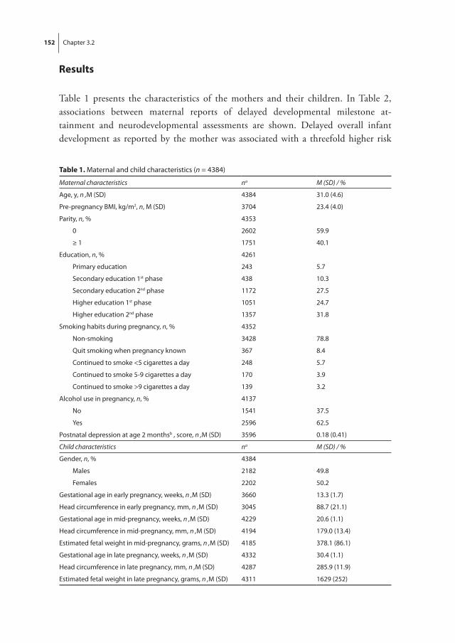
152 Chapter 3.2
Results
Table 1 presents the characteristics of the mothers and their children. In Table 2, associations between maternal reports of delayed developmental milestone at-tainment and neurodevelopmental assessments are shown. Delayed overall infant development as reported by the mother was associated with a threefold higher risk
Table 1. Maternal and child characteristics (n = 4384)
Maternal characteristics na M (SD) / %
Age, y, n ,M (SD) 4384 31.0 (4.6)
Pre-pregnancy BMI, kg/m2, n, M (SD) 3704 23.4 (4.0)
Parity, n, %
0
≥ 1
4353
2602
1751
59.9
40.1
Education, n, %
Primary education
Secondary education 1st phase
Secondary education 2nd phase
Higher education 1st phase
Higher education 2nd phase
4261
243
438
1172
1051
1357
5.7
10.3
27.5
24.7
31.8
Smoking habits during pregnancy, n, % 4352
Non-smoking 3428 78.8
Quit smoking when pregnancy known 367 8.4
Continued to smoke <5 cigarettes a day 248 5.7
Continued to smoke 5-9 cigarettes a day 170 3.9
Continued to smoke >9 cigarettes a day 139 3.2
Alcohol use in pregnancy, n, % 4137
No 1541 37.5
Yes 2596 62.5
Postnatal depression at age 2 monthsb , score, n ,M (SD) 3596 0.18 (0.41)
Child characteristics na M (SD) / %
Gender, n, % 4384
Males 2182 49.8
Females 2202 50.2
Gestational age in early pregnancy, weeks, n ,M (SD) 3660 13.3 (1.7)
Head circumference in early pregnancy, mm, n ,M (SD) 3045 88.7 (21.1)
Gestational age in mid-pregnancy, weeks, n ,M (SD) 4229 20.6 (1.1)
Head circumference in mid-pregnancy, mm, n ,M (SD) 4194 179.0 (13.4)
Estimated fetal weight in mid-pregnancy, grams, n ,M (SD) 4185 378.1 (86.1)
Gestational age in late pregnancy, weeks, n ,M (SD) 4332 30.4 (1.1)
Head circumference in late pregnancy, mm, n ,M (SD) 4287 285.9 (11.9)
Estimated fetal weight in late pregnancy, grams, n ,M (SD) 4311 1629 (252)

Fetal growth and infant development 153
Table 1. Maternal and child characteristics (n = 4384) (continued)
Child characteristics na M (SD) / %
Gestational age at birth, weeks, n ,M (SD) 4384 40.0 (1.6)
Birth weight, grams, n ,M (SD) 4384 3464 (536)
Age at assessment of postnatal size, mo, n ,M (SD) 4142 7.1 (1.9)
Body weight at 7 months, kg, n ,M (SD) 8.2 (1.2) 8.2 (1.2)
Head circumference at 7 months, cm, n ,M (SD) 4096 44.0 (1.7)
Ethnicity, n, %
Dutch
Cape Verdean
Moroccan
Dutch Antilles
Surinamese
Turkish
Other Western
Other non-western
4321
2859
74
158
93
229
224
401
263
66.2
1.7
3.7
2.2
5.3
5.6
9.3
6.1
Breastfeeding patterns, n, % 3819
Not breastfed 323 8.5
Breastfed <3 months 859 22.5
Breastfed 3-6 months 1310 34.3
Breastfed >6 months 1327 34.7
Age at parent-report of developmental milestone
attainment, mo, n, M (SD)
4384 12.4 (1.1)
Delayed social development at 12 months, n, % 333 7.6
Delayed self-help development at 12 months, n, % 373 8.6
Delayed gross motor development at 12 months, n, % 762 17.5
Delayed fine motor development at 12 months, n, % 345 8.4
Delayed language development at 12 months, n, % 426 9.8
Delayed overall infant development at 12 months, n, % 89 2.0
Age at neurodevelopmental assessmentc, mo, n, M (SD) 755c 14.6 (0.9)
Low muscle tone symptoms at 14 monthsc, n, % 30 5.3
High muscle tone symptoms at 14 monthsc, n, % 130 17.2
Overall deviant muscle tone at 14 monthsc, n, % 155 20.4
aNumbers differ due to missing values. Data was incomplete in the following covariates: Parity (0.7%), pre-pregnancy BMI (15.5%), maternal education (2.8%), prenatal smoking (0.7%), prenatal alcohol use (5.6%), maternal depression (18.0%), child ethnicity (1.4%), breastfeeding (12.4%), weight at 7 months (5.5%), head circumference at 7 months (6.6%), and age at assessment of postnatal growth (5.5%).bPostnatal depression was assessed with the depression scale of the Brief Symptom Inventory. The depression scale consists of 6 items, e.g. “feeling lonely”. Each item was rated on a 5-point rating scale ranging from ‘0’ (not at all) to ‘4’ (extremely). The total scores for symptoms of depression were calculated by summing the item scores and divided by the number of endorsed items.cNeuromotor assessments were carried out by research nurses in a subgroup of 755 Dutch children

154 Chapter 3.2
of tester-administered low muscle tone (OR 2.98; 95% CI 1.18-7.53, p = 0.021) and a similarly increased the risk of observed high muscle tone and overall deviant muscle tone (Table 2). Delayed gross motor attainment was related to a higher risk of low and overall deviant muscle tone but not to an increased risk of high muscle tone. Similar associations were observed between delayed fine motor development and neurodevelopmental assessments (Table 2).
Table 3 shows associations of fetal size in early, mid- and late pregnancy and birth weight with different aspects of infant development. We only report associations between fetal size and infant development that were adjusted for confounders. In early pregnancy, fetal size, i.e. head circumference, was generally not related to infant development. Only larger fetal head circumference predicted a reduced risk of delayed self-help abilities. In mid-pregnancy, higher fetal head circumference and higher estimated fetal weight were related to a lower likelihood of delayed self-help abilities. In late pregnancy, higher fetal head circumference predicted a lower risk of a delay in social, self-help, fine motor, and overall infant development. Similarly, higher fetal weight in this period was related to a reduced risk of delayed social, self-help, gross motor, and overall infant development.
Next, significant associations were additionally adjusted for postnatal growth (see also Table 3). All of the observed associations described above were attenuated but remained significant after additional adjustment for postnatal growth with one exception: head circumference in late pregnancy no longer predicted overall infant development.
Higher birth weight was linked to a reduced likelihood of delayed self-help abilities and overall infant development but not to the other developmental domains (Table 3). After adjustment for postnatal growth, these associations were no longer significant.
Table 4 presents associations between fetal growth from early pregnancy onwards and infant development. Faster fetal head growth from early to mid-pregnancy pre-dicted a reduced risk of delayed fine motor development but not any other aspect of infant development. Faster fetal head growth from mid- to late pregnancy was associ-
Table 2. Associations between maternal reports of delayed developmental milestone attainment at 12 months and neurodevelopmental assessments by research nurses at 14 months (n = 755)
Non-optimal neuromotor development
Domains of delayed infant development
Low muscle toneOR (95% CI) p
High muscle toneOR (95% CI) p
Overall deviant muscle toneOR (95% CI) p
Fine motor delay 1.82 (0.97-3.41) 0.061 2.63 (1.07-6.49) 0.035 1.89 (1.04-3.43) 0.036
Gross motor delay 1.76 (1.14-2.71) 0.011 1.90 (0.94-3.83) 0.072 1.78 (1.19-2.67) 0.005
Delayed overall infant development 2.98 (1.18-7.53) 0.021 3.54 (1.07-11.75) 0.039 3.76 (1.58-8.95) 0.003
OR = Odd ratio, CI: Confidence interval.Models were adjusted for gender, maternal age, maternal education, and child age at follow-up.

Fetal growth and infant development 155
Tabl
e 3.
Ass
ocia
tions
of f
etal
siz
e in
ear
ly, m
id- a
nd la
te p
regn
ancy
and
siz
e at
birt
h w
ith d
elay
in in
fant
dev
elop
men
t at 1
2 m
onth
s
Del
ay in
(in
dom
ains
of)
infa
nt d
evel
opm
ent
Feta
l siz
e ch
arac
teris
tics
Soci
alO
R (
95%
CI)
pSe
lf-he
lpO
R (
95%
CI)
pG
ross
mot
orO
R (
95%
CI)
pFi
ne m
otor
OR
(95
% C
I) p
Lang
uage
OR
(95
% C
I) p
Ove
rall
infa
nt d
evel
opm
ent
OR
(95
% C
I) p
Size
in e
arly
pre
gnan
cyH
ead
circ
umfe
renc
e, p
er S
D (n
=304
5)Ad
just
ed m
odel
a
Addi
tiona
lly a
djus
ted
for p
ostn
atal
hea
dci
rcum
fere
nce
1.07
(0.9
5-1.
22) 0
.276
n.a.
0.84
(0.7
4-0.
98) 0
.012
0.85
(0.7
4-0.
97) 0
.018
0.96
(0.8
8-1.
05) 0
.364
n.a.
1.05
(0.9
3-1.
19) 0
.432
n.a.
1.03
(0.9
1-1.
15) 0
.663
n.a.
0.79
(0.6
1-1.
02) 0
.066
n.a.
Size
in m
id-p
regn
ancy
Hea
d ci
rcum
fere
nce,
per
SD
(n =
419
4)Ad
just
ed m
odel
a
Addi
tiona
lly a
djus
ted
for p
ostn
atal
hea
dci
rcum
fere
nce
0.97
(0.8
6-1.
09) 0
.618
n.a.
0.85
(0.7
6-0.
95) 0
.007
0.87
(0.7
7-0.
99) 0
.028
0.99
(0.9
1-1.
07) 0
.717
n.a.
0.90
(0.8
0-1.
01) 0
.062
n.a.
1.10
(0.9
9; 1
.22)
0.0
90
n.a.
0.84
(0.
68-1
.05)
0.1
29
n.a.
Estim
ated
feta
l wei
ght,
per S
D (n
= 4
185)
Adju
sted
mod
el a
Addi
tiona
lly a
djus
ted
for p
ostn
atal
wei
ght
0.95
(0.8
4-1.
07) 0
.366
n.a.
0.82
(0.7
3-0.
93) 0
.002
0.84
(0.7
4-0.
96) 0
.008
0.96
(0.8
8-1.
04) 0
.312
n.a.
0.98
(0.8
7-1.
10) 0
.719
n.a.
1.03
(0.9
2-1.
15) 0
.638
n.a.
0.92
(0.7
3-1.
15) 0
.460
n.a.
Size
in la
te p
regn
ancy
Hea
d ci
rcum
fere
nce,
per
SD
(n =
428
7)Ad
just
ed m
odel
a
Addi
tiona
lly a
djus
ted
for p
ostn
atal
hea
dci
rcum
fere
nce
0.82
(0.7
3-0.
93) 0
.002
0.87
(0.7
6-0.
99) 0
.036
0.81
(0.7
2-0.
92) 0
.001
0.84
(0.7
4-0.
96) 0
.012
1.01
(0.9
2-1.
10) 0
.873
n.a.
0.78
(0.7
0-0.
88) <
0.00
1
0.80
(0.7
1-0.
91) 0
.001
0.93
(0.8
5-1.
05) 0
.305
n.a.
0.67
(0.5
3-0.
85) 0
.001
0.80
(0.6
2-1.
04) 0
.091
Estim
ated
feta
l wei
ght,
per S
D (n
= 4
311)
Adju
sted
mod
el a
Addi
tiona
lly a
djus
ted
for p
ostn
atal
wei
ght
0.83
(0.7
3-0.
93) 0
.002
0.84
(0.7
4-0.
95) 0
.004
0.78
(0.6
9-0.
88) <
0.00
10.
81 (0
.72-
0.92
) 0.0
010.
89 (0
.82-
0.98
) 0.0
110.
90 (0
.83-
0.99
) 0.0
240.
90 (0
.80-
1.00
) 0.0
60n.
a.0.
92 (0
.83-
1.02
) 0.1
22n.
a.0.
63 (0
.49-
0.79
) <0.
001
0.66
(0.5
1-0.
84) 0
.001
Size
at b
irth
Birt
h w
eigh
t, pe
r SD
(n =
438
4)Ad
just
ed m
odel
a
Addi
tiona
lly a
djus
ted
for p
ostn
atal
wei
ght
0.92
(0.8
2-1.
03) 0
.154
n.a.
0.83
(0.7
3-0.
93) 0
.001
0.88
(0.7
8-1.
00) 0
.051
0.96
(0.8
8-1.
04) 0
.290
n.a.
0.92
(0.8
2-1.
03) 0
.133
n.a.
0.93
(0.8
3-1.
03) 0
.160
n.a.
0.76
(0.6
1-0.
92) 0
.013
0.88
(0.6
9-1.
11) 0
.275
OR
= O
dd ra
tio, w
hich
repr
esen
ts th
e od
ds o
f del
ay in
infa
nt d
evel
opm
ent p
er o
ne s
tand
ard
devi
atio
n sc
ore
incr
ease
of t
he re
spec
tive
(feta
l) si
ze c
hara
cter
istic
in e
arly
, m
id- a
nd la
te p
regn
ancy
and
at b
irth
and
was
der
ived
from
mul
tiple
logi
stic
regr
essi
on m
odel
s. CI
: Con
fiden
ce in
terv
al. n
.a.:
not a
pplic
able
.a M
odel
s w
ere
adju
sted
for g
ende
r, m
ater
nal a
ge, m
ater
nal p
re-p
regn
ancy
bod
y m
ass
inde
x, m
ater
nal e
duca
tion,
mat
erna
l sm
okin
g an
d al
coho
l use
dur
ing
preg
nanc
y,
child
eth
nici
ty, m
ater
nal d
epre
ssio
n at
age
2 m
onth
s an
d ch
ild a
ge a
t fol
low
-up.

156 Chapter 3.2
Tabl
e 4.
Ass
ocia
tions
of f
etal
gro
wth
from
ear
ly p
regn
ancy
onw
ards
with
del
ay in
infa
nt d
evel
opm
ent a
t 12
mon
ths
Del
ay in
(in
dom
ains
of)
infa
nt d
evel
opm
ent
Feta
l gro
wth
cha
ract
eris
tics
Soci
alO
R (
95%
CI)
pSe
lf-he
lpO
R (
95%
CI)
pG
ross
mot
orO
R (
95%
CI)
pFi
ne m
otor
OR
(95
% C
I) p
Lang
uage
OR
(95
% C
I) p
Ove
rall
infa
nt
deve
lopm
ent
OR
(95
% C
I) p
Gro
wth
from
ear
ly to
mid
-pre
gnan
cyH
ead
circ
umfe
renc
e, p
er S
D (n
= 2
992)
Adju
sted
mod
el a
Addi
tiona
lly a
djus
ted
for p
ostn
atal
hea
d ci
rcum
fere
nce
0.98
(0.8
5-1.
13) 0
.757
n.a.
0.92
(0.7
9-1.
07) 0
.260
n.a.
0.99
(0.8
9-1.
10) 0
.827
n.a.
0.82
(0.7
1-0.
95) 0
.008
0.85
(0.7
3-0.
98) 0
.030
1.05
(0.9
2; 1
.20)
0.4
92
n.a.
0.84
(0.6
2-1.
12) 0
.229
n.a.
Gro
wth
from
mid
- to
late
pre
gnan
cyH
ead
circ
umfe
renc
e, p
er S
D (n
= 4
112)
Adju
sted
mod
el a
Addi
tiona
lly a
djus
ted
for p
ostn
atal
hea
d ci
rcum
fere
nce
0.79
(0.6
9-0.
92) 0
.001
0.84
(0.7
3-0.
97) 0
.018
0.82
(0.7
2-0.
95) 0
.006
0.86
(0.7
4-0.
99) 0
.045
1.02
(0.9
3-1.
13) 0
.652
n.a.
0.79
(0.6
9-0.
90) <
0.00
1
0.81
(0.7
0-0.
93) 0
.003
0.92
(0.8
1-1.
05) 0
.210
n.a.
0.71
(0.5
4-0.
92) 0
.011
0.85
(0.6
4-1.
13) 0
.265
Estim
ated
feta
l wei
ght,
per S
D (
n =
4118
)Ad
just
ed m
odel
a
Addi
tiona
lly a
djus
ted
for p
ostn
atal
wei
ght
0.81
(0.7
0-0.
93) 0
.003
0.82
(0.7
1-0.
95) 0
.008
0.81
(0.7
0-0.
93) 0
.004
0.84
(0.7
3-0.
98) 0
.023
0.90
(0.8
2-0.
99) 0
.037
0.91
(0.8
2-1.
01) 0
.068
0.90
(0.7
9-1.
03) 0
.138
n.a.
0.90
(0.7
9-1.
03) 0
.121
n.a.
0.62
(0.4
7-0.
81) 0
.001
0.65
(0.4
9-0.
87) 0
.003
OR
= O
dd ra
tio, w
hich
repr
esen
ts th
e od
ds o
f del
ay in
infa
nt d
evel
opm
ent p
er o
ne s
tand
ard
devi
atio
n sc
ore
incr
ease
of t
he re
spec
tive
feta
l gro
wth
cha
ract
eris
tics
from
ea
rly p
regn
ancy
onw
ards
and
was
der
ived
from
mul
tiple
logi
stic
regr
essi
on m
odel
s. CI
: Con
fiden
ce in
terv
al. n
.a.:
not a
pplic
able
.a M
odel
s w
ere
adju
sted
for g
ende
r, m
ater
nal a
ge, m
ater
nal p
re-p
regn
ancy
bod
y m
ass
inde
x, m
ater
nal e
duca
tion,
mat
erna
l sm
okin
g an
d al
coho
l use
dur
ing
preg
nanc
y,
child
eth
nici
ty, m
ater
nal d
epre
ssio
n at
age
2 m
onth
s, ch
ild a
ge a
t fol
low
-up
and
the
resp
ectiv
e fe
tal s
ize
char
acte
ristic
in e
arly
or m
id-p
regn
ancy
.

Fetal growth and infant development 157
ated with a reduced risk of a delay in social, self-help, fine motor, and overall infant development. Likewise, increased fetal weight gain in this period was related to a reduced risk of delayed social, self-help, and overall infant development. Again, most of the observed associations described above were attenuated but remained significant after additionally controlling for postnatal growth. Only the relation between fetal head growth from mid- to late pregnancy and overall infant development was no longer significant and faster fetal weight gain from mid- to late pregnancy no longer predicted gross motor development (Table 4).
Regarding research nurse assessments of neuromotor development similar patterns emerged, but frequently did not reach significance. In this subset of children, a higher head circumference in late pregnancy was significantly related to a reduced risk of overall deviant muscle tone development (OR 0.82; 95% CI 0.67-0.99, p = 0.044). Faster fetal head growth from mid- to late pregnancy predicted a lower risk of low muscle tone (OR 0.79; 95% CI 0.63-0.99, p = 0.038) and overall deviant muscle tone (OR 0.77; 95% CI 0.62-0.95, p = 0.014) after controlling for covariates. However, all these associations were no longer significant after additionally adjusting for postnatal head size (data not shown).
Discussion
This population-based study showed that faster overall growth from mid- to late preg-nancy reduces the risk of delayed infant development at age 12 months. In particular, social and fine motor development and self-help abilities were affected. Fetal size in late pregnancy more consistently predicted infant development than fetal size in early and mid-pregnancy. After controlling for early postnatal growth these associations were attenuated but most remained significant.
A few studies with small sample sizes (n < 200) addressed the relation between fetal growth and cognitive development. Our findings are in line with a study of small-for-gestational-age born children (n = 10) showing that slowing of head growth before 26 weeks gestation predicted poorer developmental outcome at age 5 years (Harvey et al., 1982). Walker et al. (2007) observed that fetal head circumference at 14 weeks gestation predicts reasoning abilities at age 6-8 years in 186 children but found no association of fetal ultrasound measurements in mid- and late pregnancy with cognitive development. There are two possible reasons for this contrast with our findings. First, our study was based on a large population-based sample, and second, we examined the effect of fetal growth.
Our findings showed that faster fetal head growth reduces the risk of delayed infant development. Repeated measures of fetal head growth can be cautiously interpreted
158 Chapter 3.2
as indicators of fetal brain development, because head circumference correlates with brain volume (Cooke et al., 1977). Therefore, our results suggest that less optimal brain development from mid- to late pregnancy is associated with poorer cognitive and motor development in infancy.
Although previous population-based research showed that birth weight is related to cognitive functioning in childhood (Cheung et al., 2001; Shenkin et al., 2004), birth weight was not associated with infant development independent of early postnatal weight. The prenatal measures used in our study predicted infant development better than birth weight. Birth weight is a rather crude summary measure at the endpoint of fetal growth whereas fetal ultrasound measurements depict fetal growth patterns and the fetal growth velocity timing during pregnancy.
The observed associations between fetal growth and developmental milestone at-tainment can only partly be explained by postnatal growth. This suggests that fetal growth affects infant development independent of postnatal growth. Similar findings were reported by a previous study determining the influence of head growth at dif-ferent developmental periods on child IQ at 4 and 8 years (Gale et al., 2006). An increase in head circumference at birth, interpreted as prenatal head growth by Gale et al. (2006), was related to a higher IQ at 4 years independent of postnatal head growth but not to IQ at 8 years.
Infant developmental milestone attainment at 12 months was strongly associated with non-optimal muscle tone development as assessed by research nurses at 14 months. This association of the parent assessment with systematic observation follow-ing a research protocol further supports the validity of the measure. We also found similar relations of fetal growth with deviant muscle tonus, although not independent of postnatal growth. However, this neuromotor functioning measure was obtained only in a subgroup. Thus, the lack of an association can easily be explained by low statistical power.
Fetal growth characteristics, in this study, did not predict language development. Children aged 12 months display little variation in language development. Children at this age may even be in the prelinguistic stage, whereas many have developed vari-ous fine motor skills. The low variation of language development at age 12 months may not allow detecting subtle effects.
Genetic factors largely determine neuropsychological functioning (Fox, Hersh-berger, & Bouchard, 1996). Epidemiological studies estimate that 38-80% of the birth weight variance arises from genetic influences (Johnston, Clark, & Savage, 2002). Possibly, a common genetic factor underlies intrauterine growth and infant developmental outcome. To account for shared genetic influences we controlled for maternal size, i.e. pre-pregnancy body mass index. But residual genetic influences on intrauterine growth and infant developmental outcome remain likely. Alternatively,

Fetal growth and infant development 159
the observed relation between fetal growth and infant development could be explained by fetal programming (Barker et al., 1989). Since the prenatal period is a time of rapid growth, the developing fetus is particularly vulnerable to adverse prenatal environ-mental events, such as placental insufficiency and prenatal alterations in maternal thyroid function, which are related to both reduced fetal growth and subsequent poor cognitive and motor functioning (Gagnon, 2003; Haddow et al., 1999; Scherjon, Briet, Oosting, & Kok, 2000).
Strengths of this population-based birth cohort study involved serial fetal ultrasound measurements, information on a large number of confounders and a large sample size.
Potential limitations of this study must also be discussed. First, we may have over-adjusted our analyses by controlling for factors possibly preceding intrauterine growth in the causal pathway to infant development. Maternal smoking and low educational level are known factors that affect fetal growth. However, disentangling the impact of epiphenomena of smoking, e.g. poor prenatal care, dietary restriction, and low socioeconomic status, from the true biological effect of prenatal cigarette exposure on reduced intrauterine growth and adverse brain development remains a challenge. Second, we cannot rule out that selective non-response influenced our findings since data on infant development were more complete in healthier, Dutch children of higher-educated mothers. This can lead to underestimation of the true association but it is less likely that the selective non-response led to spurious associations. Third, data on developmental outcome measures were partly based on maternal reports. This raises the concern that mothers might over- or misreport their infants’ developmental milestone achievement. However, previous research demonstrated moderate to good overall agreement between MIDI scores and the tester-administered Bayley Scales (Creighton & Sauve, 1988; Doig, Macias, Saylor, Craver, & Ingram, 1999). Within our study, delayed overall infant development as measured by the MIDI strongly pre-dicted non-optimal neuromotor development as observed by research nurses. Fourth, early pregnancy data were primarily used to calculate gestational age. Therefore using gestational age-adjusted SD scores of fetal head circumference in early pregnancy as a main determinant in our analyses is probably less valid. These data show less variation and less discriminating power than data later in pregnancy as early pregnancy data were used as a starting point for the pregnancy dating process and the calculation of the gestational-age-specific reference growth curves.
In conclusion, this study showed that fetal head and general growth from mid- to late pregnancy affects developmental outcomes in infancy independent of early postnatal growth. While the size of the association between fetal growth from mid- to late pregnancy and infant development was moderate, such effects may be significant in public health terms, in particular because earlier studies showed that infant devel-opment predicts cognitive and academic functioning in adolescence and adulthood

160 Chapter 3.2
(Taanila, Murray, Jokelainen, Isohanni, & Rantakallio, 2005). It may, therefore, be important to identify the sources of variation in fetal growth that are both amendable to intervention and linked to improved subsequent cognitive and motor functioning. Future research should address possible biological mechanisms, e.g. alterations in ma-ternal prenatal thyroid function, and their effect on fetal growth, brain development and cognitive and motor development of the child.

Fetal growth and infant development 161
References
Barker, D. J., Winter, P. D., Osmond, C., Margetts, B., & Simmonds, S. J. (1989). Weight in infancy and death from ischaemic heart disease. Lancet, 2(8663), 577-580.
Bloomfield, F. H., Oliver, M. H., & Harding, J. E. (2006). The late effects of fetal growth patterns. Archives of Disease in Childhood. Fetal and Neonatal Edition, 91(4), F299-304.
Cheung, Y. B., Yip, P. S., & Karlberg, J. P. (2001). Fetal growth, early postnatal growth and motor development in Pakistani infants. International Journal of Epidemiology, 30(1), 66-72.
Cooke, R. W., Lucas, A., Yudkin, P. L., & Pryse-Davies, J. (1977). Head circumference as an index of brain weight in the fetus and newborn. Early Human Development, 1(2), 145-149.
Creighton, D. E., & Sauve, R. S. (1988). The Minnesota Infant Development Inventory in the Developmental Screening of High-Risk Infants at 8 Months. Canadian Journal of Behavioural Science-Revue Canadienne Des Sciences Du Comportement, 20(4), 424-433.
de Groot, L., Hopkins, B., & Touwen, B. C. (1992). A method to assess the development of muscle power in preterms after term age. Neuropediatrics, 23(4), 172-179.
Doig, K. B., Macias, M. M., Saylor, C. F., Craver, J. R., & Ingram, P. E. (1999). The Child Devel-opment Inventory: A developmental outcome measure for follow-up of the high-risk infant. Journal of Pediatrics, 135(3), 358-362.
Emond, A. M., Blair, P. S., Emmett, P. M., & Drewett, R. F. (2007). Weight faltering in infancy and IQ levels at 8 years in the Avon Longitudinal Study of Parents and Children. Pediatrics, 120(4), e1051-1058.
Fox, P. W., Hershberger, S. L., & Bouchard, T. J., Jr. (1996). Genetic and environmental contribu-tions to the acquisition of a motor skill. Nature, 384(6607), 356-358.
Gagnon, R. (2003). Placental insufficiency and its consequences. European Journal of Obstetrics, Gynecology and Reproductive Biology, 110 Suppl 1, S99-107.
Gale, C. R., O’Callaghan, F. J., Bredow, M., Martyn, C. N., & Avon Longitudinal Study of Parents and Children Study, T. (2006). The influence of head growth in fetal life, infancy, and childhood on intelligence at the ages of 4 and 8 years. Pediatrics, 118(4), 1486-1492.
Haddow, J. E., Palomaki, G. E., Allan, W. C., Williams, J. R., Knight, G. J., Gagnon, J., et al. (1999). Maternal thyroid deficiency during pregnancy and subsequent neuropsychological development of the child. New England Journal of Medicine, 341(8), 549-555.
Hadlock, F. P., Harrist, R. B., Carpenter, R. J., Deter, R. L., & Park, S. K. (1984). Sonographic estimation of fetal weight. The value of femur length in addition to head and abdomen measure-ments. Radiology, 150(2), 535-540.
Harvey, D., Prince, J., Bunton, J., Parkinson, C., & Campbell, S. (1982). Abilities of children who were small-for-gestational-age babies. Pediatrics, 69(3), 296-300.
Ireton, H. R. (1997). Child Development Review and the Child Development Inventories. In H. R. Ireton (Ed.), Child Development Inventories in Education and Health Care: Screening and Assessing Young Children (pp. 3-15). Minneapolis, MN: Behavior Science Systems.
Jaddoe, V. W., van Duijn, C. M., van der Heijden, A. J., Mackenbach, J. P., Moll, H. A., Steegers, E. A., et al. (2008). The Generation R Study: design and cohort update until the age of 4 years. European Journal of Epidemiology, 23(12), 801-811.
Johnston, L. B., Clark, A. J., & Savage, M. O. (2002). Genetic factors contributing to birth weight. Archives of Disease in Childhood. Fetal and Neonatal Edition, 86(1), F2-3.
Kuntsi, J., Rijsdijk, F., Ronald, A., Asherson, P., & Plomin, R. (2005). Genetic influences on the stability of attention-deficit/hyperactivity disorder symptoms from early to middle childhood. Biological Psychiatry, 57(6), 647-654.


Chapter 4Early language functioning


4 .1Predictors of continuity
and discontinuity of early language functioning

166 Chapter 4.1
Abstract
Purpose: We investigated continuity and discontinuity of language functioning in a population-based cohort in the Netherlands (n = 3,609).Method: The criterion for expressive language delay at 18 months was vocabulary score < 10th percentile on the MacArthur Communicative Development Inventory (MCDI) and at 30 months it was vocabulary score < 10th percentile or no word com-binations on the Language Development Survey (LDS).Results: Most children (84.9%) had normal language development at both ages, 6.2% were “late bloomers,” 6.3% had a late onset expressive language delay, and 2.5% had a persistent expressive language delay. MCDI production and comprehen-sion at 18 months explained 11.9% of the variance in 30-month vocabulary scores, with low birth weight, child age and gender, ethnicity, maternal age and education, and parenting stress explaining an additional 6.0%. Multinomial logistic regression analyses identified biological, demographic, and psychological factors associated with outcomes.Conclusions: Although multiple perinatal, demographic and maternal psychosocial factors significantly predicted language functioning at 30 months, positive predictive value and sensitivity were low. Future studies are needed to address to what extent additional factors, such as brain maturation and genetic influences, can improve the prediction and our understanding of continuity and discontinuity of language delay.

Predictors of continuity of early language functioning 167
Introduction
Most children start to speak their first words at around 12 months of age and rapidly accelerate their use of words by 18 months, but there are huge individual differences in rate of early expressive vocabulary development. For example, Rescorla and Achenbach (2002) reported that for children age 18 to 23 months in the normative sample of the Language Development Survey (LDS), the mean of 105 words and standard deviation (SD) of 83 yielded a “normal” range of 20 words to 188 words. However, even with this wide range in what is considered normal vocabulary development in toddlers, it is possible to reliably identify late talkers. Normed and empirically validated parent-report measures of early expressive language development, such as the LDS (Rescorla, 1989) and the MacArthur Communicative Development Inventory (MCDI; Fenson, Dale, Reznick Bates, et al. 1994), can be used to achieve this aim. For some toddlers, delayed language acquisition is the first indication of a language impairment that may persist throughout childhood. As delays in language development are associated with poor social competence (Gertner, Rice, & Hadley, 1994), emotional and behavioral problems (Plomin, Price, Eley, Dale, & Stevenson, 2002; Rescorla, Ross, & McClure, 2007), attention deficit/hyperactivity (Giddan & Milling, 1999), and cognitive de-lays/reading problems (Oliver, Dale, & Plomin, 2004; Rescorla, 2002, 2005, 2009; Snowling, Adams, Bishop, & Stothard, 2001), identification of early language delay and provision of timely intervention would appear to be important public health goals (Law, Boyle, Harris, Harkness, & Nye, 2000).
However, intervention for many late talkers may be unnecessary, as most toddlers displaying early language delay appear to catch up in language development during the preschool period (Ellis Weismer, 2007; Paul, 1996; Rescorla, 2002; Thal, Tobias, & Morrison, 1991; Whitehurst & Fischel, 1994). Longer-term follow-ups showed that late talkers generally perform in the average range once they enter school, although they continue to have weaker language skills than peers with typical language histories (Rescorla, 2009). Previous longitudinal studies yielded few predictors with sufficient validity to determine which late-talking toddlers would outgrow their language delay (i.e., late bloomers) and which would continue to be delayed in expressive language. Stronger predictive validity data have been reported by Ellis Weismer (2007), who found that nonverbal IQ, expressive language functioning, and word comprehen-sion at 2 ½ were able to differentiate well between late bloomers and children with persistent delay at 3 ½. Yet, like most studies of late talkers the Ellis Weismer sample was quite small and homogeneous (n = 40).
One study that examined the outcome of late talkers identified in a large population is the British TEDS study (Dale, Price, Bishop, & Plomin, 2003), which reported on age 3 and age 4 outcomes for twins identified with vocabulary scores < 10th percentile

168 Chapter 4.1
on the MCDI at age 2. It can be calculated from the findings reported by Dale et al. (2003) that 44% of the 740 late talkers identified at age 2 still manifested expressive language delays at age 3 (i.e., 326), whereas only 7% of the 7,068 toddlers with typi-cal language development at age 2 were delayed by age 3 (i.e., 509). These numbers suggest that 835 children had an expressive language delay at 3, 61% of whom had not been delayed at 2. Age 2 vocabulary scores and nonverbal cognitive ability were significant predictors of age 3 outcomes, but effect sizes were small, and adding ma-ternal education, the child’s history of ear infection and gender to the model did not substantially improve prediction. The findings by Dale et al. (2003) thus illustrated that language delay at age 2 and a number of additional factors only poorly predicted language delay at a later age and that most children with language delay at age 3 had normal language development at age 2.
A study by Feldman et al. (2005) addressing the concurrent and predictive validity of the MCDI among 113 children at ages 2 and 3 years found a significant and moderate correlation of .58 between MCDI expressive vocabulary scores at age 2 and 3. When defining language delay at age 2 as MCDI vocabulary scores below the 10th percentile and language delay at age 3 as parent reported vocabulary scores more than 1 SD below the mean, modest sensitivity (50%) and positive predictive value (64%) were reported. These findings also suggest that many children with early language delay appear to catch up in language development at later ages, and that many children with apparently normal development at age 2 show delays later in life.
Similar results were reported by Westerlund, Berglund, and Eriksson (2006), who evaluated the effectiveness of the Swedish screening version of the MCDI at 18 months in identifying language delay at age 3 years in an unselected Swedish popula-tion of 2,080 children. Although MCDI word production at 18 months significantly predicted language delay at age 3, only 17.6% of the 108 children with a language delay at 18 months based on the Swedish version of the MCDI had a language delay at age 3 identified on the basis of formalized observations by research nurses, indicat-ing very low positive predictive value. Sensitivity was also modest (50%), indicating that only half the children with language delay at 3 had been delayed on the MCDI at 18 months.
Language outcomes from age 3 to age 4 ½ for children from the NICHD Early Child Care Study have also been reported (La Paro, Justice, Skibbe, & Pianta, 2004). Of the 73 children who appeared to have a specific language impairment (SLI) at age 3, 40 (55%) still had language impairment at 4 ½ years, whereas 33 (45%) had language skills in the normal range. Maternal sensitivity and maternal depression were significant predictors of language outcome.
As recently noted by Leonard (2009) and Ellis Weismer (2007), the percentage of late talkers with delay that is persistent enough to warrant a diagnosis of SLI at age

Predictors of continuity of early language functioning 169
4 or 5 is too low to account for the documented prevalence of 7% of SLI at age 5 (Tomblin, et al., 1997). As Ellis Weismer (2007) has noted, “Given the relatively low proportion of late talkers who display clinical language impairment at school entry, we must continue to ask where those 7% of kindergarten children with SLI come from if not from the ranks of late talkers.” (p. 95). This conundrum suggests that children with SLI are a heterogeneous group, with children arriving at a SLI diagnosis by different routes, a phenomenon known as equifinality. That is, some percentage of preschoolers with SLI represents late talkers who did not recover, and some other percentage represents children who were not apparently late talkers. However, few studies indicate the magnitude of these two complementary percentages.
Some factors associated with language delay have been identified in previous stud-ies. For example, genetic factors partly determine verbal abilities (Plomin & DeFries, 1998). Furthermore, slower intrauterine growth as indexed by low birth weight predicts language problems later in life (Ortiz-Mantilla, Choudhury, Leevers, & Be-nasich, 2008). Previous studies have also demonstrated that children who grow up in socially disadvantaged families have a higher risk of early language delay (Hoff, 2003). Mothers with less education use less language and lower quality language while inter-acting with their children than mothers with more education (Hoff, 2003), resulting in lower rates of expressive language development. Additionally, some data suggest that children from immigrant families tend to have slower early language develop-ment than children from non-immigrant families (Rescorla & Achenbach, 2002), and immigrant families are often also socially disadvantaged. Among the strongest predictors of expressive language delay in a sample of 1,189 children in Connecticut assessed using MCDI short forms (prevalence of delay = 14% at 18 to 23 months and 18% at 30 to 36 months) were low maternal education, low maternal expressiveness, and high parenting stress (Horwitz, et al., 2003). Other studies have also reported that parenting stress is associated with language problems and that this association is mediated by maladaptive parent-child interactions (Magill-Evans & Harrison, 1999; Noel, Peterson, & Jesso, 2008). Although the studies mentioned above have already shown that maternal education, child ethnicity, parenting stress, and perinatal factors predict children’s language functioning, studies that consider multiple factors at the same time for the prediction of continuous and discontinuous language delay are sparse, particularly within the setting of large-scale population-based studies.
Rationale for the Study
The present study used a large and diverse general population sample to examine the biological and demographic factors associated with continuity versus discontinuity in early language skills. To our knowledge, no previous study has examined the factors that differentiate the four possible outcome groups obtained from cross-tabulating

170 Chapter 4.1
delay status at two time points (no delay, early delay only, later delay only, and per-sistent delay). The time points used in this study are 18 months and 30 months. The current study used a large population-based sample of Dutch children that was highly diverse in ethnicity and maternal educational level, allowing us to examine the effect of demographic factors on the continuity and discontinuity of expressive language delay. Based on expressive language skills measured at 18 and 30 months, four major questions were addressed in the research: (a) what percentage of the sample have normal language at both ages, expressive delay at 18 months only, expressive delay at 30 months only, and persistent expressive delay; (b) what predictive and concurrent factors significantly differentiate these four language outcome groups; (c) when all significant predictors are tested together using linear regression, which ac-count for unique variance in vocabulary score at 30 months; (d) when all significant predictors are entered in multinomial logistic regression, which yield significant odds ratios (ORs) for the three delay outcome groups relative to the reference children with normal language at both ages.
Method
Participants
This study was embedded in the Generation R Study, a population-based cohort study from fetal life onwards in Rotterdam, The Netherlands. The Generation R Study has previously been described in detail (Jaddoe, et al., 2008). All children were born between April 2002 and January 2006. The study has been approved by the Medical Ethics Committee of the Erasmus Medical Center, Rotterdam, and written informed consent was obtained from all adult participants.
Language development data around 18 months were complete for 5,281 children in the Generation R cohort. When these children were 30 months of age, 1,011 of their mothers did not report information on language development. Furthermore, we excluded 661 children for whom data were missing for one or more of the predictors used in our main analyses or whose ages were out of range for our language measures. This left 3,609 children (68.3% of the 5,281 eligible subjects) in our main analyses.
Information about child ethnicity, maternal age, and maternal educational level was obtained by questionnaires during pregnancy. In the current study, ethnicity of the child was based on the country of birth of the parents and grandparents using the cat-egorization employed by Statistics Netherlands (Statistics Netherlands, 2004). In this categorization, individuals from any European country, from the U.S., Canada, and Australia, and from Japan are classified as “Other Western,” whereas individuals from countries such as Turkey, Morocco, Surinam, the Dutch Antilles, and Cape Verde are

Predictors of continuity of early language functioning 171
classified as “non-Western.” As can be seen in Table 1, the sample was very diverse in ethnicity (71.5% Dutch, 9.0% “Other Western,” and 19.5% “non-Western”).
Following the definitions of Statistics Netherlands (2004), we divided maternal education into three categories: Low education (no education, primary school or ≤3 years secondary school), Medium education (> 3 years secondary school, intermediate vocational training), and High education (higher vocational training or university de-gree). As can be seen in Table 1, there was considerable diversity in maternal education: 61.7% High, 26.3% Medium, and 12.0% Low. Cross-tabulation of maternal educa-tion by child ethnicity indicated that Dutch and Other Western children had mothers with higher levels of education (67.0% and 74.3% High, respectively), whereas non-Western children had mothers with lower levels of education (36.5% High).
Table 1. Participant characteristics (n = 3,609)
Maternal characteristics M (SD)a
Age, years 31.7 (4.4)
Education (%)
High 61.7
Medium 26.3
Low 12.0
Parenting stress, score 0.30 (0.29)
Child characteristics M (SD)a
Gestational age at birth, weeks 39.9 (1.7)
Low birth weight, Yes (%) 4.5
Gender, boys, Yes (%) 49.6
Child ethnicity (%)
Dutch 71.5
Other Western 9.0
Non-Western 19.5
Age at the ‘18’ months assessment 18.2 (0.6)
16-17 months (%) 43.3
18-19 months (%) 54.1
20 months (%) 2.6
Age at the ‘30’ months assessment 30.9 (1.2)
28-29 months (%) 21.1
30-31 months (%) 63.5
32-35 months (%) 15.4
Word production at ‘18’ months, score 17.5 (16.6)
Word comprehension at ‘18’ months, score 55.8 (24.9)
Word production at ‘30’ months, score 238.9 (58.6)
Note. aUnless otherwise indicated.

172 Chapter 4.1
Language data for this study were all obtained by questionnaires completed by par-ents. Although 18 months and 30 months were the targeted ages for the two language assessments, parents varied in how promptly they returned the questionnaires. Thus, child age for the 18-month assessment ranged from 16 to 20 months, with 54.1% in the 18-to-19 month bracket (M = 18.2 months, SD = 0.62). Similarly, child age at the 30-month assessment ranged from 28 to 35 months, with 63.5% in the 30-to-31 month age bracket (M = 30.9 months, SD = 1.2).
Measures
At 18 and 30 months, parents completed parent reports on their children’s language skills. Parents whose native languages were English or Turkish completed the Dutch version of the language measures that were translated into those languages. Moroccan parents who spoke only Arabic were interviewed at home by Arabic-speaking research assistants to obtain language data.
Language skills at 18 months were assessed using the Dutch version of the Ma-cArthur Short Form Vocabulary Checklist (N-CDI 2A), which is appropriate for measuring the word production and comprehension of children aged 16 to 30 months (Zink & Lejaegere, 2003). The MCDI contains a list of 112 words and is based on the original MacArthur Communicative Development Inventory of 680 words (Fenson, et al., 1994; Fenson, et al., 2000). Parents reported on their children’s comprehension and production of the same set of 112 monomorphemic root words. The number of positive responses was summed for both receptive and expressive vocabulary. The expressive vocabulary scores were positively skewed, as many children had very low scores. The raw expressive vocabulary scores were log-transformed to improve the normality of the distribution. For statistical analyses, the log-transformed expressive vocabulary scores and the normally distributed raw receptive vocabulary scores were z-standardized across the study sample. To identify language delay at 18 months, we converted the expressive and receptive vocabulary raw scores into age- and gender-specific percentile scores based on the whole Generation R sample using one month age brackets. Expressive and receptive language delay at 18 months was defined as scores below the 10th percentile, in line with a previous definition of expressive language delay based on the MCDI (Dale, 1996). As our primary interest was to address continuity and discontinuity of expressive language functioning, we used the expressive vocabulary scale of the MCDI as our main baseline measure.
The Dutch short form of the MCDI has excellent internal consistency and test-retest reliability, as well as concurrent validity (Zink & Lejaegere, 2003). Furthermore, validity results revealed that both language production and comprehension scores on the short form correlated highly with the respective scores on the original form of the MCDI, i.e. r = 0.97 and r = 0.99, respectively (Zink & Lejaegere, 2003). Fenson et
Predictors of continuity of early language functioning 173
al. (1994) reported a correlation of 0.73 between the original MCDI and a standard test-administered measure of expressive vocabulary. In the current sample, internal consistencies of word production and comprehension were 0.97 and 0.98, which is very similar to the internal consistencies reported by Zink & Lejaegere (2003).
Expressive language at 30 months was assessed using parent report on a Dutch translation of the Language Development Survey (LDS; Rescorla, 1989). The LDS contains a 310-word vocabulary checklist, with words arranged alphabetically within 14 semantic categories (e.g. animals, foods, modifiers, vehicles etc.). The parent was asked to identify each word that her child uses spontaneously, yielding a total vocabu-lary score. For statistical analyses, LDS total vocabulary scores were z-standardized across the study sample after log transformation to improve the normality of the distribution. To determine language delay at 30 months, we converted raw total vo-cabulary scores into age- and gender-specific percentile scores based on the complete Generation R sample, again using one month age brackets. To be consistent with the cutpoint used for 18 months, we defined a vocabulary delay at 30 months as word production scores below the 10th percentile. The LDS also asks the parent to indicate whether the child has begun to combine words into phrases and, if so, to write down up to five of the child’s best sentences. The parent wrote down these sentences in the child’s native language. Due to the difficulty in determining the number of words in the listed sentences for some languages, the only information about sentences used in this study was whether the child was reported to produce word combinations. Thus, expressive language delay at 30 months was operationalized as an LDS vocabulary score below the 10th percentile or no word combinations.
Test-retest reliability of the LDS total vocabulary score over one week was 0.99 in a sample of 30 children (Rescorla, 1989), and 0.97 over one month in a sample of 66 children (Rescorla & Alley, 2001). Correlations between LDS vocabulary score and tested expressive vocabulary using various measures have ranged from 0.66 to 0.87, indicating strong concurrent validity (Achenbach & Rescorla, 2000). LDS vocabulary score at 24 to 31 months has been reported to significantly predict expressive language outcomes up to age 17 (Rescorla, 2009). In this study, internal consistency of the LDS was 0.99, which is very similar to the internal consistency of the LDS reported in previous research (Achenbach & Rescorla 2000; Rescorla, 1989).
It has previously been reported that total scores on the complete 680-word MCDI (Fenson, et al., 1994) and total scores on the LDS were correlated at .95 for a sample of 239 children age 23 to 25 months (Rescorla, Ratner, Jusczyk, & Jusczyk, 2005). Correlations across different semantic categories ranged from 0.84 to 0.94. Thus, when used concurrently, the full MCDI and the LDS yield very comparable informa-tion about rank ordering of children in terms of their vocabulary scores. Although no studies have reported correlations between the short form of the MCDI (Fenson, et

174 Chapter 4.1
al., 2000) and the LDS, the very high correlations between the short form and the complete MCDI in English (r = 0.98) (Fenson, et al., 2000) suggest that the short form MCDI and the LDS would be highly correlated when used concurrently.
In the current study, MCDI expressive language score at 18 months was used as the primary predictor of LDS outcome at 30 months. However, based on findings from previous research (Dale, Price, Bishop, & Plomin, 2003), several other predictors were chosen from the Generation R measures obtained at or prior to 18 months. First, among these additional predictors was MCDI receptive language score, as delays in receptive language have been found to predict continuing expressive language delay (Ellis Weismer, 2007; Leonard, 2009). Additionally, the three demographic variables of maternal age, maternal education, and child ethnicity were used as predictors, based on the widely reported association between familial factors and language development (Hart & Risley, 1995). Additional predictors tested were child birth weight and gestational age. This choice was based on the assumption that biologically vulnerable children might be more likely to manifest persistent language delay. Data on child birth weight were obtained from medical records completed by midwives and gynecologists. Gestational age was established by fetal ultrasound examinations within the Generation R Study (Verburg, et al., 2008).
Finally, mothers reported information on parenting stress using the Dutch short version of the Nijmegen Parenting Stress Index (NOSIK; De Brock, Vermulst, Gerris, & Abidin, 1992) in the 18-month questionnaire. We used an adapted version of a NOSIK subscale assessing maternal perceptions of caregiving, which consisted of 12 items, e.g. “I have much more trouble raising children than I thought”. The items could be rated on a 4-point scale ranging from 0 (totally disagree) to 3 (totally agree). A total score was computed by taking the mean of the 12 item ratings. In the current study, the internal consistency of this caregiving subscale was α = 0.74. Parenting stress was included as a predictor because previous studies have indicated an associa-tion between parenting stress and slower language development.
To show whether delays in language are associated with nonverbal cognitive ability, we included among our measures the Dutch version of the Parent Report of Children’s Abilities (PARCA; Saudino, et al., 1998), which was used at 30 months. The PARCA is a nonverbal cognitive development measure obtained from parents. The parent-administered portion of the PARCA comprises three subtests: (a) matching-to-sample; (b) block building and (c) imitation. The parent-report part of the PARCA comprises 26 questions assessing quantitative skills, spatial abilities, symbolic play, planning and organizing, adaptive behaviors, and memory. The questions are formulated in terms of specific “activities,” with mothers asked to report whether or not they have seen their child perform the particular activity. Overall PARCA score was calculated by adding the sum scores of the parent-administered part and the parent-report part. PARCA

Predictors of continuity of early language functioning 175
scores were normally distributed and were z-standardized across the sample of the present study. Previous research with the PARCA has indicated a significant and large correlation with the Mental Development Index (MDI) of the Bayley Scales of Infant Development-II (r = 0.55) (Saudino, et al., 1998). In a validation study of the original PARCA based on a sample of 107 2-year-old children, internal consistencies of the parent-administered and parent-report part were good (0.83 and 0.74, respectively) (Saudino, et al., 1998).
Attrition Analysis
To examine whether non-response was selective, we compared the 3,609 included children (who had language scores at both ages) with the 1,672 excluded children (who had missing language scores at one or both ages or missing data on the possible predictors). These analyses revealed that included children were less likely to have a lower birth weight (4.5% vs. 6.7%, χ2 = 11.85, df = 1, p < 0.001) and had a higher gestational age (M = 39.9 weeks (SD, 1.7) vs. 39.7 weeks (SD, 1.9), t = 3.07, p = 0.002) compared to excluded children. Included children were more likely to be Dutch (71.5% vs. 54.2%, χ2 = 158.2, df = 2, p < 0.001) than excluded children. Mothers of included children were more likely to have high levels of education (% High education 61.7 % vs. 42.2%, χ2 = 197.5, df = 2, p < 0.001), and to be older (M = 31.7 years (SD, 4.4) vs. 30.4 years (SD, 5.3), t = 8.83, p < 0.001) than mothers of excluded children.
Statistical Analyses
Using the categorical assignment of expressive language delay at 18 months (word production scores below the age- and gender specific 10th percentile) and at 30 months (word production scores below the age- and gender specific 10th percentile or no word combinations), we cross-tabulated expressive delay status at 18 and 30 months. This cross-tabulation yielded four groups: (a) children with no expressive language delay; (b) late bloomers, i.e. children with expressive language delay at 18 months but normal language functioning at 30 months; (c) children with late onset expressive language delay, i.e. children with normal language functioning at 18 months but expressive language delay at 30 months; and (d) children with persistent expressive language delay. We first compared these four groups on all predictors, using one-way ANOVAs with Student-Newman-Keuls (S-N-K) post-hoc tests for continuous variables and Chi-square tests for categorical variables. All predictors that significantly differentiated among the groups were then used in a multiple linear regression analysis to determine the percentage of unique variance in LDS vocabulary at 30 months accounted for by these predictors. MCDI receptive and expressive z-scores were used in this analysis. We also included gender and child age as additional predictors in the
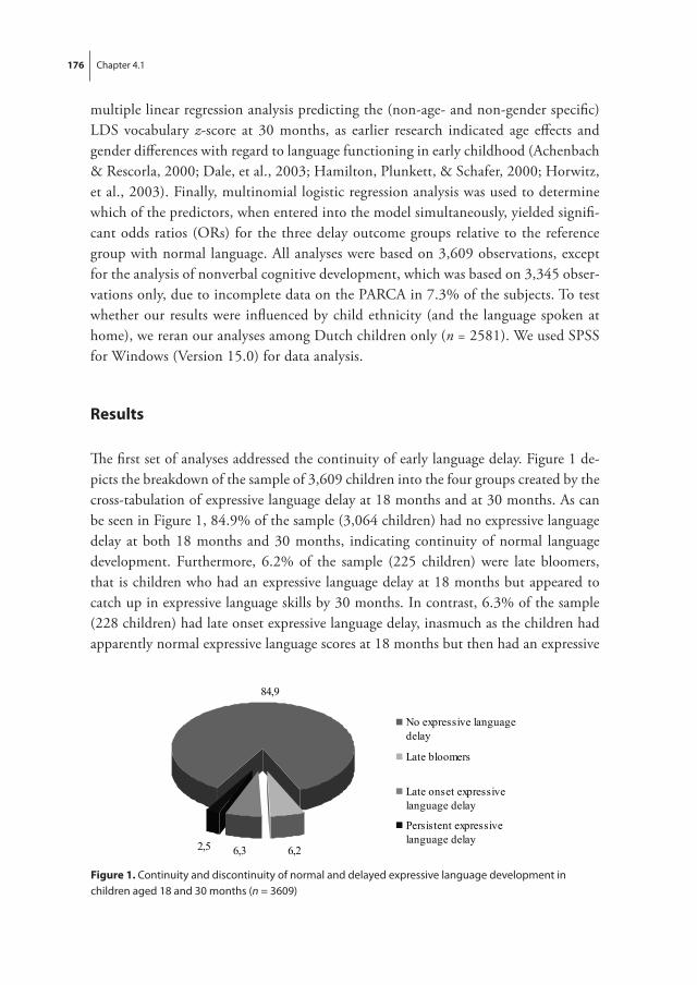
176 Chapter 4.1
multiple linear regression analysis predicting the (non-age- and non-gender specific) LDS vocabulary z-score at 30 months, as earlier research indicated age effects and gender differences with regard to language functioning in early childhood (Achenbach & Rescorla, 2000; Dale, et al., 2003; Hamilton, Plunkett, & Schafer, 2000; Horwitz, et al., 2003). Finally, multinomial logistic regression analysis was used to determine which of the predictors, when entered into the model simultaneously, yielded signifi-cant odds ratios (ORs) for the three delay outcome groups relative to the reference group with normal language. All analyses were based on 3,609 observations, except for the analysis of nonverbal cognitive development, which was based on 3,345 obser-vations only, due to incomplete data on the PARCA in 7.3% of the subjects. To test whether our results were influenced by child ethnicity (and the language spoken at home), we reran our analyses among Dutch children only (n = 2581). We used SPSS for Windows (Version 15.0) for data analysis.
Results
The first set of analyses addressed the continuity of early language delay. Figure 1 de-picts the breakdown of the sample of 3,609 children into the four groups created by the cross-tabulation of expressive language delay at 18 months and at 30 months. As can be seen in Figure 1, 84.9% of the sample (3,064 children) had no expressive language delay at both 18 months and 30 months, indicating continuity of normal language development. Furthermore, 6.2% of the sample (225 children) were late bloomers, that is children who had an expressive language delay at 18 months but appeared to catch up in expressive language skills by 30 months. In contrast, 6.3% of the sample (228 children) had late onset expressive language delay, inasmuch as the children had apparently normal expressive language scores at 18 months but then had an expressive Chapter 4.1 (Figure 1):
84,9
6,26,32,5
No expressive languagedelay
Late bloomers
Late onset expressivelanguage delay
Persistent expressivelanguage delay
Figure 1. Continuity and discontinuity of normal and delayed expressive language development in children aged 18 and 30 months (n = 3609)

Predictors of continuity of early language functioning 177
language delay. Finally, 2.5% of the sample (92 children) had persistent expressive language delay, with delayed expressive language skills at both 18 and 30 months.
The majority of children delayed at 18 months scored in the normal range by 30 months, which is reflected in the low positive predictive value of the MCDI at 18 months (92/92 + 225 = 29%). However, this high false positive rate was not achieved by minimizing the false negative rate, as indicated by sensitivity of only 29% (92/92 + 228). Thus, the majority of children identified with expressive language delay at 30 months on the LDS had not scored below the 10th percentile on the MCDI at 18 months. Furthermore, most of the children in this large sample who scored in the normal range at 18 months continued to score in the normal range at 30 months, reflected in the very high negative predictive value of 93% (3,064/3,064 + 228). Fi-nally, most of the children with normal language skills at 30 months also had normal language skills at 18 months, reflected in the 93% specificity (3,064/3,064 + 225). To further examine the degree of accuracy with which MCDI word production scores at 18 months discriminated children with and without expressive language delay at 30 months, we calculated a ROC curve, as shown in Figure 2. The area under the ROC curve (AUC) was 0.74 (95% CI 0.71; 0.77), p < 0.001), indicating only fair accuracy in predicting expressive language delay at 30 months based on MCDI word production scores at 18 months.
Figure 2. ROC curve for MCDI word production at 18 months on expressive language delay at 30 months (the area under the ROC curve was 0.74 (95%CI 0.71; 0.77), p < 0.001))

178 Chapter 4.1 Predictors of continuity of early language functioning 179
Previous research has often used a uniform cutpoint for the identification of early language delay, which has typically yielded higher rates of early language delay in boys than in girls (Dale, et al., 2003; Rescorla, 1989). This is because young boys generally have smaller reported vocabularies than young girls (Achenbach & Rescorla, 2000; Rescorla & Alley, 2001). In the current study, girls had significantly higher MCDI z-scores (0.13 vs. -0.13, F (1, 3608) = 58.81, p < 0.001), and LDS z-scores (0.10 vs. -0.10, F (1, 3608) = 35.19, p < 0.001) than boys. Therefore, we tested whether use of a gender-neutral, age-specific 10th percentile cutpoint would yield more boys than girls in the current study. Chi-square tests using a uniform cutpoint showed that at both 18 months and 30 months, boys were more likely to have a delay in expressive language development than girls (18 months: 10.3% vs. 7.5%, χ2 = 8.77, df = 1, p < 0.001; at 30 months: 10.9% vs. 5.6%, χ2 = 29.8, df = 1, p < 0.001). Our findings regarding gender differences in expressive language delay are very similar to the findings of the study by Dale et al. (2003), although they reported slightly higher rates of language delay in boys (i.e. 12.8%).
Table 2 presents the status of each of the four outcome groups with respect to all of the measures used in this study. The overall pattern of results presented in Table 2 suggests important demographic and birth history differences between children in the four outcome groups. Specifically, among late bloomers, girls were somewhat overrepresented (53.3%). Furthermore, late bloomers were more likely to have a low birth weight (9.3%) but to come from families with Dutch parents (77.8%) and high educational levels (59.1% high education). In contrast, children with late onset expressive language delay were more likely to be boys (54.4%) and to have a normal birth weight (94.7%), but to come from families with non-Western parents (41.7%) and with low maternal educational levels (28.5% low education). Children with no expressive language delay came from families with the highest rate of maternal educa-tion (63.9% high education), had high rates of Dutch ethnicity (73.3%), and had the lowest percentage of low birth weights (3.9%). Finally, the group of children with persistent delay had the highest percentage of low birth weights (10.9%) and was intermediate between the other groups in terms of Dutch ethnicity (60.9%) and maternal high education (51.1%).
Table 2 also shows significant differences in verbal and nonverbal cognitive func-tioning among the four outcome groups. As would be expected given how the four outcome groups were defined, the groups differed significantly in MCDI expressive language score at 18 months (Table 2). The late bloomers and children with persistent expressive language delay had the lowest comprehension z-scores at 18 months (-0.48, -0.59, respectively), although their z-scores for comprehension were less extreme than their z-scores for expressive vocabulary at 18 months (-2.09, -2.11). LDS z-scores at age 30 months differed significantly across the four outcome groups, as would

178 Chapter 4.1 Predictors of continuity of early language functioning 179
Tabl
e 2.
Par
ticip
ant c
hara
cter
istic
s by
leve
l of e
xpre
ssiv
e la
ngua
ge fu
nctio
ning
at 1
8 an
d 30
mon
ths
of a
ge (n
= 3
609)
No
expr
essi
ve la
ngua
ge
dela
ya
(n =
306
4)M
(SD
)b
Late
blo
omer
sa
(n =
225
)
M (S
D)b
Late
ons
et e
xpre
ssiv
e la
ngua
ge d
elay
a
(n =
228
)M
(SD
)b
Pers
iste
nt e
xpre
ssiv
e la
ngua
ge d
elay
a
(n =
92)
M (S
D)b
Sign
ifica
nce
test
ing
and
effec
t siz
e fo
r co
ntin
uous
mea
sure
s
Mot
her
Mat
erna
l age
, yea
rsM
ater
nal e
duca
tion
(%)
H
igh
M
ediu
m
Low
Pare
ntin
g st
ress
, sco
reCh
ildG
ende
r (%
)
boys
gi
rlsEt
hnic
ity (%
)
Dut
ch
Oth
er W
este
rn
Non
-Wes
tern
Ges
tatio
nal a
ge, w
eeks
Low
birt
h w
eigh
t (%
)W
ord
prod
uctio
n at
18
mon
ths,
z-sc
ore
Wor
d co
mpr
ehen
sion
at 1
8 m
onth
s, z-
scor
eW
ord
prod
uctio
n at
30
mon
ths,
z-sc
ore
Non
verb
al c
ogni
tive
deve
lopm
ent
at 3
0 m
onth
s (P
ARC
A),
z-sc
ore
31.7
b (4.3
)
63.9
25.7
10.4
0.29
b (.28
)
49.4
50.6
73.3
8.7
18.0
40.0
a (1.6
)3.
90.
24a (0
.77)
0.06
a (0.9
5)
0.24
a (0.3
9)
0.07
a (0.9
5)
32.6
a (4.3
)
59.1
26.2
14.7
0.31
b (.28
)
46.7
53.3
77.8
7.6
14.7
39.4
b (2.2
)9.
3-2
.09 c (0
.47)
-0.4
8 c (1.0
5)
-0.0
2 b (0.4
6)
-0.3
0 b (1.0
6)
29.7
c (5.1
)
38.6
32.9
28.5
0.41
a (.38
)
54.4
45.6
46.1
12.3
41.7
39.8
a (1.9
)5.
3-0
.27 b (0
.76)
-0.1
8 b (1.1
8)
-2.0
1 c (1.7
8)
-0.4
9 b (1.1
1)
31.5
b (5.5
)
51.1
30.4
18.5
0.38
a (.41
)
50.0
40.0
60.9
13.0
26.1
39.5
b (2.3
)10
.9-2
.11 c
(0.4
6)
-0.5
9 c (1.3
0)
-2.9
4 d (2.2
9)
-0.8
0 c (1.
53)
F(3,
360
6) =
19.
6 , p
< 0
.001
, η2 =
0.0
2
χ2 (6) =
91.
2, p
< 0
.001
F(3,
360
6) =
14.
5 , p
< 0
.001
, η2 =
0.0
1
χ2 (3) =
2.9
0, p
= 0
.407
χ2 (6) =
101
.3,
p <
0.00
1
F(3,
360
6) =
9.8
3 , p
< 0
.001
, η2 =
0.0
1χ2 (3
) = 2
4.4,
p <
.001
F(3,
360
6) =
946
.3 ,
p <
0.00
1, η
2 = 0
.44
F(3,
360
6) =
34.
4 , p
< 0
.001
, η2 =
0.0
3
F(3,
360
6) =
133
9.5
, p <
0.0
01, η
2 = 0
.53
F(3,
334
2) =
44.
2 , p
< 0
.001
, η2 =
0.0
4
Not
e. a L
angu
age
deve
lopm
ent w
as re
port
ed b
y m
othe
rs a
t 18
mon
ths
usin
g th
e D
utch
sho
rt fo
rm v
ersi
on o
f the
MCD
I (Zi
nk &
Lej
aege
re, 2
003)
. Exp
ress
ive
lang
uage
de
lay
at 1
8 m
onth
s w
as d
efine
d as
a w
ord
prod
uctio
n sc
ore
belo
w th
e 10
th p
erce
ntile
. A
t 30
mon
ths
mot
hers
repo
rted
lang
uage
dev
elop
men
t usi
ng th
e LD
S (R
esco
rla,
1989
). Ex
pres
sive
del
ay a
t 30
mon
ths
was
defi
ned
as a
wor
d pr
oduc
tion
scor
e be
low
the
10th
per
cent
ile o
r no
com
bina
tion
of w
ords
. Fo
r mat
erna
l edu
catio
n, c
hild
ge
nder
, eth
nici
ty a
nd lo
w b
irth
wei
ght p
erce
ntag
es re
pres
ent t
he p
ropo
rtio
n of
chi
ldre
n (o
r mot
hers
) in
the
defin
ed g
roup
who
fall
into
the
resp
ectiv
e ca
tego
ry o
f la
ngua
ge fu
nctio
ning
at 1
8 an
d 30
mon
ths
of a
ge. P
ARC
A =
Par
ent R
epor
t of C
hild
ren’
s A
bilit
ies.
p-va
lues
wer
e de
rived
from
AN
OVA
s fo
r con
tinuo
us v
aria
bles
and
from
ch
i-squ
are
test
s fo
r cat
egor
ical
var
iabl
es. M
eans
with
diff
eren
t sub
scrip
ts a
re s
igni
fican
tly d
iffer
ent a
t p <
.05
(S-N
-K).
b Unl
ess
othe
rwis
e in
dica
ted.

180 Chapter 4.1
be expected (Table 2). Children with no expressive language delay had significantly larger vocabularies than children in the “late bloomer” group. The effect size of 0.53 indicates very large group differences in LDS vocabulary, which are further indicated by z-scores of -2.01 and -2.94 for the late onset expressive language delay group and the persistent expressive language delay group, respectively. The difference in mean LDS score for the persistent delay group relative to the no delay group was more than three SDs (-2.94 vs. 0.24). The four groups also differed in nonverbal cognitive ability (Table 2). Children with no expressive delay at either age had significantly higher nonverbal cognitive ability scores than the late bloomers. PARCA scores did not differ between the late bloomers and children with late onset expressive language delay, but the persistent delay group showed the lowest PARCA scores.
Prior to conducting multiple regression analyses, we calculated Pearson correlations among the three language scores. There was a statistically significant and moderate correlation between MCDI word comprehension and word production at 18 months (r = 0.44, p < 0.001). The correlation of word production at 18 months with LDS word production at 30 months was also statistically significant and moderate (r = 0.34, p < 0.001). Furthermore, the correlation of word comprehension at 18 months and LDS word production at 30 months was statistically significant and weak (r = 0.19, p < 0.001)
Table 3 presents results of a stepwise multiple linear regression analysis examining the percentage of unique variance in LDS vocabulary at 30 months accounted for by all predictors that significantly differentiated among the four language outcome groups. Non-gender specific z-standardized LDS vocabulary scores at 30 months were used as the dependent variable. In Step 1, word production at 18 months was entered into the linear regression model to show the unique variance explained by our main predictor (Table 3). Word production at 18 months was positively related to LDS vocabulary at 30 months, accounting for 11.7% of the variance. In Step 2, word comprehension at 18 months significantly predicted a higher LDS vocabulary at 30 months, but it only added 0.2% to the earlier proportion of explained variance. In Step 3, all other predic-tors including child gender were added to the regression model. We also included child age at both language assessment time periods to account for age effects on language functioning. All predictors were significant except for gestational age, with the final model explaining 17.9% of the variance. This illustrates that the additional demographic, perinatal, maternal psychosocial and child factors only explained 6% of the variance. In summary, higher word production and comprehension at 18 months, older child age at the second language assessment and older maternal age predicted larger LDS vocabulary at 30 months, whereas medium and low maternal education, higher levels of parenting stress, other Western and non-Western ethnicity, low birth weight, older child age during the first language assessment and being a boy predicted
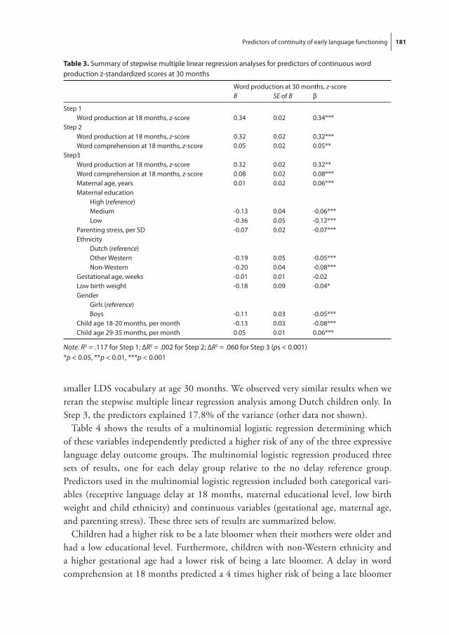
Predictors of continuity of early language functioning 181
smaller LDS vocabulary at age 30 months. We observed very similar results when we reran the stepwise multiple linear regression analysis among Dutch children only. In Step 3, the predictors explained 17.8% of the variance (other data not shown).
Table 4 shows the results of a multinomial logistic regression determining which of these variables independently predicted a higher risk of any of the three expressive language delay outcome groups. The multinomial logistic regression produced three sets of results, one for each delay group relative to the no delay reference group. Predictors used in the multinomial logistic regression included both categorical vari-ables (receptive language delay at 18 months, maternal educational level, low birth weight and child ethnicity) and continuous variables (gestational age, maternal age, and parenting stress). These three sets of results are summarized below.
Children had a higher risk to be a late bloomer when their mothers were older and had a low educational level. Furthermore, children with non-Western ethnicity and a higher gestational age had a lower risk of being a late bloomer. A delay in word comprehension at 18 months predicted a 4 times higher risk of being a late bloomer
Table 3. Summary of stepwise multiple linear regression analyses for predictors of continuous word production z-standardized scores at 30 months
Word production at 30 months, z-scoreB SE of B β
Step 1 Word production at 18 months, z-scoreStep 2 Word production at 18 months, z-score Word comprehension at 18 months, z-scoreStep3 Word production at 18 months, z-score Word comprehension at 18 months, z-score Maternal age, years Maternal education High (reference) Medium Low Parenting stress, per SD Ethnicity Dutch (reference) Other Western Non-Western Gestational age, weeks Low birth weight Gender Girls (reference) Boys Child age 18-20 months, per month Child age 29-35 months, per month
0.34 0.02 0.34***
0.32 0.02 0.32***0.05 0.02 0.05**
0.32 0.02 0.32**0.08 0.02 0.08***0.01 0.02 0.06***
-0.13 0.04 -0.06***-0.36 0.05 -0.12***-0.07 0.02 -0.07***
-0.19 0.05 -0.05***-0.20 0.04 -0.08***-0.01 0.01 -0.02-0.18 0.09 -0.04*
-0.11 0.03 -0.05***-0.13 0.03 -0.08***0.05 0.01 0.06***
Note. R2 = .117 for Step 1; ∆R2 = .002 for Step 2; ∆R2 = .060 for Step 3 (ps < 0.001)*p < 0.05, **p < 0.01, ***p < 0.001

182 Chapter 4.1
(OR = 4.43, 95% CI = 3.12; 6.29, p < 0.001). All other predictors were not associated with being a late bloomer (Table 4).
As can be seen in Table 4, children of older mothers had a lower risk of late onset expressive language delay. Both low and medium maternal education were related to a higher risk of late onset expressive language delay. Moreover, other Western and non-Western child ethnicity and higher levels of parenting stress were related to a higher risk of late onset expressive language delay. Again, a delay in word comprehension at 18 months was associated with an approximately 4 times higher risk of late onset expressive language delay (OR = 3.74, 95% CI = 2.57; 5.44, p < 0.001). All other predictors were not related to late onset expressive language delay (Table 4).
In the last group of children, we found that low maternal education and higher lev-els of parenting stress were associated with a higher likelihood of persistent expressive language delay. In this group of children, delay in word comprehension at 18 months was associated with a 9 times higher risk of persistent expressive language delay (OR = 9.13, 95% CI = 5.80; 14.37, p < 0.001). None of the other predictors was linked to persistent expressive language delay.
Finally, we reran the multinomial logistic regression analysis among Dutch children only to test whether child ethnicity influenced our results. In this analysis, very similar results emerged (data not shown).
Discussion
The current population-based study showed that only a small proportion of children, i.e. 2.5%, had persistent expressive language delay at 18 and 30 months. Most children had normal language development (84.9%) at both ages, 6.2% were late bloomers, and 6.3% had late onset expressive language delay. Moreover, the current study identified predictors of continuity and discontinuity of language delay from 18 to 30 months. Receptive language delay at 18 months predicted all three expressive language delay outcomes and was particularly strongly related to persistent expressive language delay. Late onset expressive delay was primarily associated with environmental risk factors, including low maternal education, non-Dutch child ethnicity and parenting stress.
The MCDI at 18 months had both low positive predictive value (29%) and low sensitivity (29%) when predicting LDS scores at 30 months. That is, most of the children delayed on the 18-month MCDI were not still delayed at 30 months, and most of the children delayed at 30 months had not been delayed at 18 months. The current study thus corroborates the findings reported by Westerlund et al. (2006) for Swedish children from age 18 months to age 3 years. In that study, positive predictive

Predictors of continuity of early language functioning 183
Tabl
e 4.
Pre
dict
ors
of te
mpo
rary
or p
ersi
sten
t exp
ress
ive
lang
uage
del
ay in
ear
ly c
hild
hood
No
expr
essi
ve la
ngua
ge d
elay
OR
(95%
CI)
Late
blo
omer
s
OR
(95%
CI)
Late
ons
et e
xpre
ssiv
e la
ngua
ge
dela
yO
R (9
5% C
I)
Pers
iste
nt e
xpre
ssiv
e la
ngua
ge
dela
yO
R (
95%
CI)
Mat
erna
l age
, yea
rsM
ater
nal e
duca
tion
H
igh
(refe
renc
e)
Med
ium
Lo
wPa
rent
ing
stre
ss, p
er S
DEt
hnic
ity
Dut
ch (r
efer
ence
)
Oth
er W
este
rn
Non
-Wes
tern
Ges
tatio
nal a
ge, w
eeks
Low
birt
h w
eigh
tD
elay
ed w
ord
com
preh
ensi
on a
t 18
mon
ths
refe
renc
e- - re
fere
nce
refe
renc
ere
fere
nce
- - refe
renc
ere
fere
nce
refe
renc
ere
fere
nce
- refe
renc
e
1.05
(1.0
2; 1
.09)
**- - 1.
26 (0
.90;
1.7
5)1.
96 (1
.27;
3.0
0)**
1.05
(0.9
2; 1
.21)
- - 0.77
(0.4
5; 1
.30)
0.66
(0.4
4; 0
.99)
*0.
89 (0
.82;
0.9
7)**
1.27
(0.6
6; 2
.45)
4.43
(3.1
2; 6
.29)
***
0.95
(0.
92; 0
.98)
**- - 1.
65 (1
.17;
2.3
2)**
2.99
(2.0
3; 4
.40)
***
1.22
(1.0
9; 1
.35)
***
- - 2.18
(1.3
9; 3
.41)
**2.
16 (1
.56;
2.9
8)**
*1.
00 (0
.91;
1.1
0)1.
11 (0
.53;
2.3
7)
3.74
(2.5
7; 5
.44)
***
1.00
(0.9
5; 1
.05)
- - 1.49
(0.9
0; 2
.48)
2.27
(1.2
1; 4
.27)
*1.
22 (1
.03;
1.4
3)*
- - 1.59
(0.8
3; 3
.07)
1.22
(0.7
2; 2
.06)
0.98
(0.8
6; 1
.12)
2.29
(0.
90; 5
.84)
9.13
(5.8
0; 1
4.37
)***
Not
e. O
R =
Odd
ratio
; CI =
Con
fiden
ce In
terv
al.
The
mod
el w
as b
ased
on
mul
tinom
ial l
ogis
tic re
gres
sion
ana
lysi
s. Th
e di
ffere
nt e
xpre
ssiv
e la
ngua
ge d
elay
cat
egor
ies
wer
e co
mpa
red
to th
e re
fere
nce
grou
p, i.
e. n
o ex
pres
sive
lang
uage
del
ay.
*p <
0.0
5, *
*p <
0.0
1, *
**p
< 0.
001

184 Chapter 4.1
value from the MCDI at 18 months was only 17.6%, and half the children delayed at 3 had not been delayed at 18 months.
Somewhat better prediction has been reported using the MCDI at 24 months than has been found using the MCDI at 18 months. For example, Dale et al. (2003) reported that 44% of the 740 late talkers identified at age 2 still manifested expres-sive language delays at age 3 (i.e., 326). However, 61% of the 835 children with an expressive language delay at 3 had not been delayed at 2. Furthermore, Feldman et al. (2005) found that half of the children with a parent reported language delay at age 3 had not been delayed at age 2 and reported that the positive predictive value from the MCDI at age 2 was 64%.
To some extent, the poor prediction observed in the current study derives from the decision statistics paradigm itself, whereby a fixed cutpoint is imposed on an underlying continuum to identify delay. This means that children just missing the cutpoint (i.e., at the 11th percentile) are classified as normal. An indication of how dichotomization yields to prediction errors can be seen in the fact that children delayed at 30 months only had quite low word production and comprehension scores at 18 months, but their scores were just not quite low enough to be below the 10th percentile cutpoint. However, the poor prediction is not only attributable to dichotomization, as the ROC curve for MCDI word production scores at 18 months on expressive language delay only demonstrated fair accuracy. Furthermore, the correlation between MCDI word production at 18 months and LDS word production at 30 months was 0.34, which indicates only a moderate degree of association between these two measures. Modest ROC results were also reported by Feldman et al. (2005), but the correlation reported by Feldman et al. (2005) between MCDI scores at 24 and 36 months was slightly higher than our correlation of 0.34 (i.e., 0.58 for Feldman). Thus our results are in line with previous research showing that MCDI scores at 18 months even less accurately predict subsequent language delay than MCDI scores at 24 months (Dale et al., 2003; Feldman et al, 2005; Westerlund et al., 2006). This pattern of findings therefore suggests that the earlier language delay is detected, the less accurately it seems to predict later language functioning.
In this study, we identified a number of predictors of expressive language function-ing at 30 months of age. In the full sample, both expressive and receptive language functioning at 18 months were significant predictors of expressive language function-ing at 30 months, consistent with previous work by Ellis Weismer (2007), and Thal et al. (1991). Lower maternal education, younger maternal age, and higher levels of parenting stress also predicted lower expressive language functioning at 30 months, as reported in other studies (Hart & Risley, 1995; Hoff, 2003; Horwitz, et al., 2003). In accordance with previous findings, we observed that being a boy, member of an ethnic minority group, and of low birth weight predicted poorer expressive language

Predictors of continuity of early language functioning 185
functioning at 30 months (Achenbach & Rescorla, 2000; Dale, et al., 2003; Ortiz-Mantilla, et al., 2008; Rescorla & Achenbach, 2002). It is important to underscore, however, that this replication occurred in a sample that is very different from the samples used in these previous studies. That is, the current study utilized a very large population-based sample in the Netherlands that was very diverse in maternal educa-tion, ethnicity, national origin, and language spoken in the home.
Our findings that children’s expressive language functioning depends on multiple demographic, psychosocial, perinatal and child developmental factors may have important public health implications in terms of prevention efforts. However, taken together all predictors explained no more than 17.9% of the variance. This indicates that many other factors not measured in the present study may also contribute to language outcomes at 30 months.
The particular focus of our study was to examine continuity and discontinuity of language delay from 18 to 30 months. We were able to identify some factors that differentiated the four outcome groups obtained from cross-tabulating expressive language delay status at 18 and 30 months. Our results showed that, not surprisingly, children with no expressive language delay, i.e. normal language development at both ages, were most advantaged with regard to perinatal and demographic factors and had the highest level of cognitive functioning. Children with continuously normal language development are born healthy and grow up in a healthy, stimulating, and protective environment. Previous research has shown that high maternal SES is re-lated to better fetal development, as indexed by higher birth weights, possibly due to a healthy life-style of the mother, including healthy diet (James, Nelson, Ralph, & Leather, 1997). Hoff (2003) showed that mothers with higher education use more language and higher quality language while interacting with their children than moth-ers with low education.
Among the three groups of children with expressive language delay, the late bloom-ers, who apparently recovered from early language delay, were more likely to have a low birth weight, to be girls and of Dutch ethnicity, and to have mothers with high educational levels and older age. Late bloomers also had delayed word comprehension at 18 months. Overall, these findings suggest that late bloomers may have manifested early receptive and expressive language delay due to some mild developmental lag, which was associated with lower birth weight in some of the children. As late bloom-ers tended to be girls from Dutch families whose mothers had higher educational levels and older ages, they may have enjoyed stimulating home environments between 18 and 30 months that helped them to catch up in language by 30 months. This group may have had a slightly later onset of a normal brain maturation, as suggested by research demonstrating that myelination of language-related brain areas in early childhood coincides with early language development (Pujol, et al., 2006). Neverthe-

186 Chapter 4.1
less, although late bloomers were no longer delayed in language development at 30 months, we cannot rule out that they may still be at risk to display developmental problems later in life, as previous research showed that late talkers performed in the average range on most language tasks by age 5 but had significantly poorer scores on a number of language measures at age 6 to 9 years (Rescorla 2002) and continued to have weaker language scores than comparison children through age 17 (Rescorla, 2009).
Children with late onset expressive language delay were more likely to come from socially disadvantaged families, to have non-Dutch ethnicity, and to have mothers with medium and low education. Parenting stress and delayed word comprehension at 18 months raised the risk of expressive language delay at 30 months. This pattern of results suggests that the children in this group manifested a language delay by 30 months because they received less stimulation from their socially disadvantaged and stressed mothers. Previous studies have demonstrated that the association of low ma-ternal education and parenting stress with poor language functioning was mediated by maladaptive parent-child interactions and low levels of stimulation (Hoff, 2003; Magill-Evans & Harrison, 1999; Noel, et al., 2008).
Finally, the group with persistent delay had the highest percentage of low birth weight children and the lowest verbal and nonverbal cognitive scores. Low mater-nal education and higher levels of parenting stress significantly predicted persistent expressive language delay. Strikingly, delayed word comprehension at 18 months predicted a 9-fold higher risk of persistent delay. These findings suggest that children with a persistent expressive language delay from 18 to 30 months had a significant and marked language impairment, in particular, because of the strong association with receptive language delay. Most likely, the chronic language problems of this group of children can be explained by biological vulnerabilities, as these children were less socially disadvantaged than children with late onset expressive language delay and had the highest percentage of low birth weight. Although the association between low birth weight and persistent language delay was non-significant, this is probably due to low statistical power arising from the small number of cases with persistent language delay. Previous studies have shown that genetic effects play an important role in SLI (Bishop, North, & Donlan, 1995). It may be, therefore, that persistent language delay in this group was also partly due to a genetic predisposition.
An important finding of this study is that receptive language delay at 18 months yielded significant and very high odds ratios in predicting the three expressive lan-guage delay outcome groups, in particular with regard to the prediction of persistent expressive language delay. These findings are in line with previous small-scale studies showing that delays in receptive language predict continuing expressive language delay (Ellis Weismer, 2007; Leonard, 2009; Thal et al., 1991). This suggests that receptive

Predictors of continuity of early language functioning 187
language delay plays an important role in predicting persistent expressive language de-lay, but also that deficits in language expression and in language comprehension and processing are closely related. The idea of a close relation between language expression and comprehension is supported by Ullman and Pierpoint (2005), who proposed that many children with a language impairment have a deficit in the neural circuitry responsible for procedural memory that affects both language comprehension and production.
Strengths of this population-based birth cohort study included the assessment of language development at two time points in toddlerhood, information on a large number of possible predictors of language development, and a large and highly diverse sample. However, a limitation of this study is that data on verbal and non-verbal cognitive development were completely based on maternal report. Although the parent-based measures used in this study have been shown to be reliable and valid (Fenson, et al., 1994; Rescorla, 1989; Rescorla & Alley, 2001; Saudino, et al., 1998) and to significantly predict tester-administered measures both concurrently and predictively (Oliver, Dale, & Plomin, 2004; Rescorla, 2002), structured testing and/or observation would have been a valuable addition to parent report in this study. An additional limitation of this study is that different language measures were used at 18 and 30 months. Although previous research (Rescorla et al., 2005) has shown high correlations between the MCDI and the LDS, measure variance may have somewhat attenuated prediction results in this study. Another limitation is that our results may have been influenced by something of a floor effect on the MCDI at 18 months (mean = 17.5 in the whole sample, out of maximum score of 112) and a slight ceiling effect on the LDS at 30 months (mean = 238.9 in the whole sample, out of maximum score of 310). These psychometric limitations may have led to a misclassification of expressive language delay, thus reducing the sensitivity of the 18-month MCDI to predict delay as defined with the 30-month LDS assessment. Furthermore, the language measures were not translated into all languages that the study participants spoke, as some of the participating ethnic minorities only received a Dutch version of the language measures. Yet, when our analyses were restricted to Dutch children, we observed very similar results. Finally, selective attrition is another limitation of our study, in that data on language development were more complete in healthier, Dutch children of higher-educated mothers.
This study, which showed that multiple perinatal, demographic and maternal psy-chosocial factors as well as language functioning at 18 months significantly predicted language functioning at 30 months, has several practical implications. First, the low sensitivity and low positive predictive value indicated that the 18-month MCDI has limited clinical utility for predicting 30-month LDS outcomes for individual children. However, findings suggested that late bloomers may be children with mild

188 Chapter 4.1
developmental lags who recover from early language delay because of growing up in families with favorable sociodemographic backgrounds. Our findings also showed that late onset expressive language delay was primarily related to unfavorable envi-ronmental influences, including sociodemographic risk factors and parenting stress. Furthermore, this study suggests that persistent expressive language delay is most likely determined by biological vulnerabilities. Additionally, receptive language delay at 18 months predicted all three language delay outcomes, and played a particularly important role in predicting persistent expressive language delay. This suggests that interventions aimed at language impairments should address both expressive and receptive language skills. Interventions should also incorporate parenting stress reduc-tion programs and encourage parents, in particular those with social disadvantages, to stimulate the language development of their children. To improve the identification of persistent language delay, both expressive and receptive language skills should be assessed. Our results indicate that further longitudinal research is needed to identify more predictors of temporary versus persistent language delay in toddlers. Future research should address to what extent biological factors, including brain maturation and genetic influences, and cognitive factors, such as working memory, attention and phonological awareness, can improve the prediction and our understanding of continuity and discontinuity of early language delay.

Predictors of continuity of early language functioning 189
References
Achenbach, T. M., & Rescorla, L. A. (2000). Manual for ASEBA preschool forms & profiles. Burling-ton, VT: University of Vermont, Research Center for Children, Youth and Families.
Bishop, D. V., North, T., & Donlan, C. (1995). Genetic basis of specific language impairment: evidence from a twin study. Developmental Medicine and Child Neurology, 37(1), 56-71.
Cohen, J. (1988). Statistical power analysis for the behavioral sciences (2nd ed.). Hillsdale, NJ: Law-rence Earlbaum Associates.
Dale, P. S. (1996). Parent report assessment of language and communication. In K. N. Cole, P. S. Dale & D. J. Thal (Eds.), Assessment of communication and language. Baltimore: Paul H. Brookes.
Dale, P. S., Price, T. S., Bishop, D. V., & Plomin, R. (2003). Outcomes of early language delay: I. Predicting persistent and transient language difficulties at 3 and 4 years. Journal of Speech, Language and Hearing Research, 46(3), 544-560.
De Brock, A., Vermulst, A. A., Gerris, J. R. M., & Abidin, R. R. (Eds.). (1992). Nijmeegse Ouderlijke Stress Index, handleiding experimentele versie [NOSIK - Nijmegen Parenting Stress Index, manual experimental version] Lisse, The Netherlands: Swets en Zeitlinger.
Ellis Weismer, S. (2007). Typical talkers, late talkers, and children with specific language impairment: A language endowment spectrum? In R. Paul (Ed.), The influence of developmental perspectives on research and practice in communication disorders: A festschrift for Robin S. Chapman. Mahwah, NJ: Lawrence Erlbaum Associates.
Feldman, H. M., Dale, P. S., Campbell, T. F., Colborn, D. K., Kurs-Lasky, M., Rockette, H. E., et al. (2005). Concurrent and predictive validity of parent reports of child language at ages 2 and 3 years. Child Development, 76(4), 856-868.
Fenson, L., Dale, P. S., Reznick, J. S., Bates, E., Thal, D. J., & Pethick, S. J. (1994). Variability in early communicative development. Monographs of the Society for Research in Child Development, 59(5), 1-173; discussion 174-185.
Fenson, L., Pethick, S., Renda, C., Cox, J. L., Dale, P. S., & Reznick, J. S. (2000). Short-form versions of the MacArthur Communicative Development Inventories. Applied Psycholinguistics, 21(1), 95-115.
Gertner, B. L., Rice, M. L., & Hadley, P. A. (1994). Influence of communicative competence on peer preferences in a preschool classroom. Journal of Speech and Hearing Research, 37(4), 913-923.
Giddan, J. J., & Milling, L. (1999). Comorbidity of psychiatric and communication disorders in children. Child and Adolescent Psychiatric Clinics of North America, 8(1), 19-36, v.
Hamilton, A., Plunkett, K., & Schafer, G. (2000). Infant vocabulary development assessed with a British communicative development inventory. Journal of Child Language, 27(3), 689-705.
Hart, B., & Risley, T. (Eds.). (1995). Meaningful differences in the everyday experiences of young American children. Baltimore: Paul H. Brookes.
Hoff, E. (2003). The specificity of environmental influence: socioeconomic status affects early vocabulary development via maternal speech. Child Development, 74(5), 1368-1378.
Horwitz, S. M., Irwin, J. R., Briggs-Gowan, M. J., Bosson Heenan, J. M., Mendoza, J., & Carter, A. S. (2003). Language delay in a community cohort of young children. Journal of American Academy of Child and Adolescent Psychiatry, 42(8), 932-940.
Jaddoe, V. W., van Duijn, C. M., van der Heijden, A. J., Mackenbach, J. P., Moll, H. A., Steegers, E. A., et al. (2008). The Generation R Study: design and cohort update until the age of 4 years. European Journal of Epidemiology, 23(12), 801-811.

190 Chapter 4.1
James, W. P., Nelson, M., Ralph, A., & Leather, S. (1997). Socioeconomic determinants of health. The contribution of nutrition to inequalities in health. British Medical Journal, 314(7093), 1545-1549.
La Paro, K. M., Justice, L., Skibbe, L. E., & Pianta, R. C. (2004). Relations among maternal, child, and demographic factors and the persistence of preschool language impairment. American Journal of Speech and Language Pathology, 13(4), 291-303.
Law, J., Boyle, J., Harris, F., Harkness, A., & Nye, C. (2000). The feasibility of universal screening for primary speech and language delay: findings from a systematic review of the literature. Developmental Medicine and Child Neurology, 42(3), 190-200.
Leonard, L. B. (2009). Is expressive language disorder an accurate diagnostic category? American Journal of Speech-Language Pathology, 18(2), 115-123.
Magill-Evans, J., & Harrison, M. J. (1999). Parent-child interactions and development of toddlers born preterm. West Journal of Nursing Research, 21(3), 292-307; discussion 308-212.
Noel, M., Peterson, C., & Jesso, B. (2008). The relationship of parenting stress and child tem-perament to language development among economically disadvantaged preschoolers. Journal of Child Language, 35(4), 823-843.
Oliver, B., Dale, P. S., & Plomin, R. (2004). Verbal and nonverbal predictors of early language problems: an analysis of twins in early childhood back to infancy. Journal of Child Language, 31(3), 609-631.
Ortiz-Mantilla, S., Choudhury, N., Leevers, H., & Benasich, A. A. (2008). Understanding language and cognitive deficits in very low birth weight children. Developmental Psychobiology, 50(2), 107-126.
Paul, R. (1996). Clinical implications of the natural history of slow expressive language develop-ment. Journal of Speech-Language Pathology, 5(2), 5-30.
Plomin, R., & DeFries, J. C. (1998). The genetics of cognitive abilities and disabilities. Scientific American, 278(5), 62-69.
Plomin, R., Price, T. S., Eley, T. C., Dale, P. S., & Stevenson, J. (2002). Associations between behaviour problems and verbal and nonverbal cognitive abilities and disabilities in early child-hood. Journal of Child Psychology and Psychiatry, 43(5), 619-633.
Pujol, J., Soriano-Mas, C., Ortiz, H., Sebastian-Galles, N., Losilla, J. M., & Deus, J. (2006). My-elination of language-related areas in the developing brain. Neurology, 66(3), 339-343.
Rescorla, L. (1989). The Language Development Survey: a screening tool for delayed language in toddlers. Journal of Speech and Hearing Disorders, 54(4), 587-599.
Rescorla, L. (2002). Language and reading outcomes to age 9 in late-talking toddlers. Journal of Speech, Language and Hearing Research, 45(2), 360-371.
Rescorla, L. (2005). Age 13 language and reading outcomes in late-talking toddlers. Journal of Speech, Language and Hearing Research, 48(2), 459-472.
Rescorla, L. (2009). Age 17 language and reading outcomes in late-talking toddlers: support for a dimensional perspective on language delay. Journal of Speech, Language and Hearing Research, 52(1), 16-30.
Rescorla, L., & Achenbach, T. M. (2002). Use of the language development survey (LDS) in a national probability sample of children 18 to 35 months old. Journal of Speech, Language and Hearing Research, 45(4), 733-743.
Rescorla, L., & Alley, A. (2001). Validation of the language development survey (LDS): a parent report tool for identifying language delay in toddlers. Journal of Speech, Language and Hearing Research, 44(2), 434-445.

Predictors of continuity of early language functioning 191
Rescorla, L., Ratner, N. B., Jusczyk, P., & Jusczyk, A. M. (2005). Concurrent validity of the language development survey: associations with the MacArthur-Bates communicative development in-ventories: words and sentences. American Journal of Speech-Language Pathology, 14(2), 156-163.
Rescorla, L., Ross, G. S., & McClure, S. (2007). Language delay and behavioral/emotional problems in toddlers: findings from two developmental clinics. Journal of Speech, Language and Hearing Research, 50(4), 1063-1078.
Saudino, K. J., Dale, P. S., Oliver, B., Petrill, S. A., Richardson, V., Rutter, M., et al. (1998). The validity of parent-based assessment of the cognitive abilities of 2-year-olds. British Journal of Developmental Psychology, 16, 349-363.
Snowling, M. J., Adams, J. W., Bishop, D. V. M., & Stothard, S. E. (2001). Educational attainments of school leavers with a preschool history of speech-language impairments. International Journal of Language & Communication Disorders, 36(2), 173-183.
Statistics Netherlands (2004). Standaard Onderwijsindeling 2003 [Categorization of the Dutch educa-tional system 2003]. Voorburg/Heerlen.
Thal, D., Tobias, S., & Morrison, D. (1991). Language and gesture in late talkers: a 1-year follow-up. Journal of Speech and Hearing Research, 34(3), 604-612.
Tomblin, J. B., Records, N. L., Buckwalter, P., Zhang, X., Smith, E., & O’Brien, M. (1997). Preva-lence of specific language impairment in kindergarten children. Journal of Speech, Language and Hearing Research, 40(6), 1245-1260.
Verburg, B. O., Steegers, E. A., De Ridder, M., Snijders, R. J., Smith, E., Hofman, A., et al. (2008). New charts for ultrasound dating of pregnancy and assessment of fetal growth: longitudinal data from a population-based cohort study. Ultrasound in Obstetrics & Gynecology, 31(4), 388-396.
Westerlund, M., Berglund, E., & Eriksson, M. (2006). Can severely language delayed 3-year-olds be identified at 18 months? Evaluation of a screening version of the MacArthur-Bates Com-municative Development Inventories. Journal of Speech, Language and Hearing Research, 49(2), 237-247.
Whitehurst, G. J., & Fischel, J. E. (1994). Practitioner review: early developmental language delay: what, if anything, should the clinician do about it? Journal of Child Psychology and Psychiatry, 35(4), 613-648.
Zink, I., & Lejaegere, M. (2003). N-CDIs: Korte vormen, aanpassing en hernormering van de MacAr-thur Short Form Vocabulary Checklists van Fenson et al. Leuuven, Belgium: Acco.


C hapter 5General discussion


General discussion 195
The main aims of this thesis were: 1) to examine whether adverse prenatal environ-mental factors are associated with poor fetal growth or less optimal early behavioral and cognitive functioning, 2) to investigate whether reduced fetal growth negatively affects early behavioral, cognitive and motor development, and 3) to explore which perinatal, socio-demographic and maternal psychological factors predict the continu-ity and discontinuity of early language functioning.
All studies in this thesis were embedded in the Generation R Study, a multi-ethnic population-based cohort study among 9,778 pregnant women and their children in Rotterdam, the Netherlands. In the current chapter, I will address methodological considerations, discuss the main findings in a broader context, and conclude with implications for clinical practice and future research.
Methodological considerations
The strengths and limitations of the different studies in this dissertation have been depicted in the respective chapters. In the current chapter, I will address more general methodological considerations that concern observational studies. To be precise, I will discuss methodological considerations with regard to the design of the present observational studies, possible selection effects and methodological aspects that are related to the information about determinants and outcomes. I will also consider aspects related to confounding and causality.
Study design
The Generation R Study is a prospective birth cohort study that consists of a number of subcohorts following participants from fetal life onwards. The prospective design of the Generation R Study enables the researcher to control data collection, to assess temporal relationships and to identify factors affecting health, developmental changes and deviant developmental patterns. The Generation R Study was designed to follow children from the general population, instead of following a high-risk or exposure-specific group, e.g. very low birth weight children or intrauterine drug-exposed chil-dren. Population-based studies usually address the potential effects of exposures that a considerable proportion of the general population has experienced (Rothman, 2002). In the current dissertation, this concerns the studies examining the effects of maternal psychological distress and thyroid functioning during pregnancy. High-risk cohorts select study participants on the basis of the same or very similar value for a certain vari-able that could be a potential confounder. Previous studies that investigated the effects of variations in fetal growth on neurodevelopmental outcomes, including behavioral and cognitive functioning in childhood, most often used clinical samples of preterm,

196 Chapter 5
small-for-gestational-age or very low birth weight children (Fattal-Valevski et al., 1999; Gray, Indurkhya, & McCormick, 2004; Hack et al., 1992; Hack et al., 2002; Hack et al., 1994; Hack et al., 2004; Horwood, Mogridge, & Darlow, 1998; Leitner et al., 2007). These studies have the advantage of restriction. It is difficult, however, to generalize the results of these studies to full-term and normal birth weight children, in particular because prematurity and low birth weight can lead to a variety of medical interventions and complications such as breathing difficulties that could influence outcome. The epidemiological studies included in the current dissertation had the aim to provide support for hypotheses that are applicable to the general population. Nevertheless, the generalizability of the association between an adverse prenatal fac-tor and subsequent behavioral and cognitive functioning observed in a non-clinical sample relies on the biological, sociodemographic and statistical representativeness of the respective study sample.
Furthermore, the best way to achieve and enable correct scientific inferences is to warrant the validity of a study. However, different types of bias can have negative consequences for the validity of a study. Among these types of bias are selection bias, information bias, and confounding bias (Rothman & Greenland, 1998b). Arguably, all studies depicted in this dissertation might have been influenced by these three forms of bias.
Selection bias
Selection bias is a distortion that occurs when there is a difference in the relation between determinant and outcome among those who participate and among those who should have been theoretically eligible for the study, including those who do not participate in the study (Rothman & Greenland, 1998b). The initial response rate of the Generation R Study was 61% (Jaddoe et al., 2008). Non-response because of non-participation and missing values at baseline among participants was not random. The percentages of mothers with a lower socio-economic status and from ethnic minorities and of mothers or children with medical complications are smaller among the partici-pants than expected from the population figures in Rotterdam (van Lith, 2004). This indicates a selection towards are more affluent and healthy study population, which could have resulted in a bias in the association studies presented in this dissertation if the selection mechanisms are associated with both the determinant and the outcome. Due to selective non-response the association between the respective determinant and outcome could thus differ within the study population and the eligible population. It is, however, likely that this is not generally the case. During the enrollment phase of the Generation R Study, there was an increase in the baseline response rates from approximately 51% in 2002 and 2003 to 68% in 2004 and 2005 and there was a decrease in the percentage of women reporting to ever have smoked from 43% in
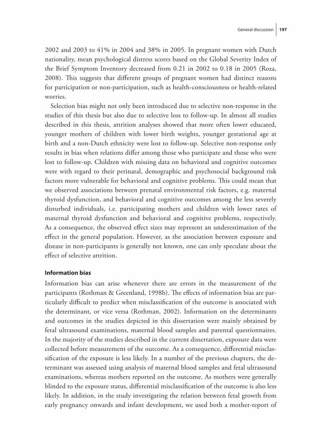
General discussion 197
2002 and 2003 to 41% in 2004 and 38% in 2005. In pregnant women with Dutch nationality, mean psychological distress scores based on the Global Severity Index of the Brief Symptom Inventory decreased from 0.21 in 2002 to 0.18 in 2005 (Roza, 2008). This suggests that different groups of pregnant women had distinct reasons for participation or non-participation, such as health-consciousness or health-related worries.
Selection bias might not only been introduced due to selective non-response in the studies of this thesis but also due to selective loss to follow-up. In almost all studies described in this thesis, attrition analyses showed that more often lower educated, younger mothers of children with lower birth weights, younger gestational age at birth and a non-Dutch ethnicity were lost to follow-up. Selective non-response only results in bias when relations differ among those who participate and those who were lost to follow-up. Children with missing data on behavioral and cognitive outcomes were with regard to their perinatal, demographic and psychosocial background risk factors more vulnerable for behavioral and cognitive problems. This could mean that we observed associations between prenatal environmental risk factors, e.g. maternal thyroid dysfunction, and behavioral and cognitive outcomes among the less severely disturbed individuals, i.e. participating mothers and children with lower rates of maternal thyroid dysfunction and behavioral and cognitive problems, respectively. As a consequence, the observed effect sizes may represent an underestimation of the effect in the general population. However, as the association between exposure and disease in non-participants is generally not known, one can only speculate about the effect of selective attrition.
Information bias
Information bias can arise whenever there are errors in the measurement of the participants (Rothman & Greenland, 1998b). The effects of information bias are par-ticularly difficult to predict when misclassification of the outcome is associated with the determinant, or vice versa (Rothman, 2002). Information on the determinants and outcomes in the studies depicted in this dissertation were mainly obtained by fetal ultrasound examinations, maternal blood samples and parental questionnaires. In the majority of the studies described in the current dissertation, exposure data were collected before measurement of the outcome. As a consequence, differential misclas-sification of the exposure is less likely. In a number of the previous chapters, the de-terminant was assessed using analysis of maternal blood samples and fetal ultrasound examinations, whereas mothers reported on the outcome. As mothers were generally blinded to the exposure status, differential misclassification of the outcome is also less likely. In addition, in the study investigating the relation between fetal growth from early pregnancy onwards and infant development, we used both a mother-report of

198 Chapter 5
infant developmental milestone attainment and research nurse assessments of neuro-motor development (Chapter 3.2). Fetal head growth from mid- to late pregnancy was related to both maternal reports and research nurse assessments of developmental outcome in infancy before adjustments were made. However, after adjustments were made fetal head growth from mid- to late pregnancy was only associated with mother-reported infant development but no longer to tester-administered developmental outcome in infancy. Nevertheless, maternal reports of infant developmental mile-stone attainment were strongly related to research nurse assessments of neuromotor development in infancy providing evidence for the validity of the mother-reported infant developmental outcome measure. In addition, in Chapter 2.5 addressing the association between maternal thyroid function in early pregnancy and behavioral problems in the offspring we were able to demonstrate consistency of results across different informants. Higher maternal TSH levels during early pregnancy were related to maternal and paternal reports of childhood behavioral problems at age 1½ and 3 years. In chapter 2.2 and 2.3, both information on the exposure (maternal anxiety and family stress) and information on behavioral and cognitive outcome were based on mother-reports. It is likely that reports of the mother on her own psychological experience of stress and anxiety and her child’s developmental outcomes reflect a com-mon negative perceptual bias (Fergusson, Lynskey, & Horwood, 1993). However, there is doubt whether mothers experiencing adverse psychological functioning really have distorted perceptions of their children’s problems (Richters, 1992). Nonetheless, future studies are needed to clarify whether reports on child developmental outcome by mothers with psychological problems are indeed biased or not. Within the context of longitudinal research assessment of cognitive development, structured testing or observation could be a valuable addition to parent report. In particular, standard-ized cognitive testing is broadly accepted as a highly accurate measure of children’s cognitive abilities. Despite this, it has been argued that standardized tester-based assessment is limited in the information it can provide about the typicality of the child’s performance (Bornstein & Haynes, 1998). Standardized cognitive testing requires children to perform at their best in the presence of a stranger. Most examiners will have heard a parent complaining “his or her youngster’s not saying (or doing) something with a stranger that the same child says (or does) often when with the parent alone” (Bornstein & Haynes, 1998, p.655). This suggests a possible limit to the validity of standardized tester-based assessments of the child’s daily functioning and developmental level. Furthermore, previous research demonstrated that shyness in children is indeed related to underperforming on cognitive tests. Shy children particularly underperform when being individually tested by an examiner (Crozier & Hostettler, 2003; Evans, 1993). Ideally future research should combine standardized tester-based assessment and parents’ assessments of children’s cognitive abilities. The

General discussion 199
latter benefits from the fact that parents observe their child across time and in many situations and, as a consequence, may have access to unique information on the child, in contrast to the examiner who is a stranger (Fenson et al., 1994). Moreover, the ad-ditional use of standardized assessments of children’s school performances, including reading, writing and mathematic skills, would contribute to an even more complete picture of children’s cognitive abilities and performances later in life. In conclusion, a multiple informant approach would significantly improve the accuracy and validity of the assessment of children’s cognitive abilities. The assessment of child behavior also requires a multiple informant approach, because each informant, e.g. parents, teachers and clinicians, contributes to the validity of the information (Achenbach & Edelbrock, 1984; Kraemer et al., 2003). To draw definite conclusions on the effects of prenatal and postnatal environmental factors on child development, future longitudi-nal studies should integrate multiple sources of information about children’s cognitive and behavioral functioning. More valid conclusions about the potential intrauterine effects of maternal distress, i.e. stress, anxiety or depression, during pregnancy can be drawn if future studies not only use self-report but also information derived from standardized observations or maternal endocrine measures of maternal distress, such as cortisol.
Confounding bias
Due to its multidisciplinary setting and ongoing data collection, the Generation R Study has a very important strength, i.e. information on a large number of potential confounding variables. Confounding is often regarded as a confusion of effects, in which the apparent effect of the exposure under study is distorted due to the ef-fect of an extraneous factor that is mistaken for or mixed with the actual exposure effect (Rothman & Greenland, 1998b). So that a certain extraneous factor can be considered as a confounding factor, two conditions must be met. First, a certain third variable is related to the exposure without being the consequence of the exposure. Second, the same third variable is related to the outcome independent of the expo-sure. A confounding factor is thus not an intermediate in the causal pathway between exposure and outcome (Rothman & Greenland, 1998b). In the studies examining the association of maternal prenatal stress and anxiety with infant temperament and cognitive development (Chapters 2.2 and 2.3), many confounders of the associa-tion were included. For example, in Chapter 2.2 we demonstrated that confounders like maternal education, family income and function, maternal prenatal smoking and alcohol use, perinatal factors, infant age, gender and ethnicity substantively at-tenuated the relation between maternal prenatal anxiety and infant temperamental difficulties. This clearly shows that, in studies addressing intrauterine environmental effects on cognitive and behavioral development, control for confounding includes

200 Chapter 5
multiple biological, socio-demographic, and psychological factors. Controlling for confounders, however, may also introduce overadjustment bias (VanderWeele, 2009). Overadjustment can result from controlling for an intermediate in the causal chain between a negative intrauterine exposure and developmental outcome, or from con-trolling for a variable that is causally related to the exposure but only correlated to the outcome. In the studies investigating the relation between fetal growth and infant behavior and development (Chapters 3.1 and 3.2) controlling for factors possibly preceding fetal growth in the causal pathway to infant behavior and development may have resulted in overadjustment. Maternal smoking and low educational level are both known factors that affect fetal growth (Jaddoe et al., 2007; Kleinman & Madans, 1985; Kramer, 1987). However, disentangling the impact of epiphenomena of smok-ing, e.g. poor prenatal care, dietary restriction and low socioeconomic status, from the true biological effect of prenatal cigarette exposure on reduced intrauterine growth and adverse brain development remains a challenge (Ernst, Moolchan, & Robinson, 2001). Furthermore, it is rather unlikely that socio-demographic and life style related factors, such as maternal educational level, ethnicity, maternal smoking and alcohol use during pregnancy are intermediates in the association of fetal growth with infant behavior and development.
Although we were able to adjust for a large number of confounders in our analyses, we also may have missed potential confounders. Within epidemiological studies the issue of residual confounding always remains likely. In particular, in the studies ad-dressing prenatal determinants of cognitive development, i.e. maternal family stress, maternal thyroid functioning and fetal growth (Chapters 2.3, 2.4 and 3.2) we were not able to control for maternal IQ, which has been shown to be an important con-founder in studies investigating the association between prenatal factors and children’s cognitive functioning (Breslau, Paneth, Lucia, & Paneth-Pollak, 2005). Future studies are needed to show whether these determinants are related to cognitive outcome in childhood independent of maternal IQ. By controlling for maternal education, how-ever, we already partly took the cognitive abilities of the mother into account (Breslau et al., 2005; Richards, Hardy, Kuh, & Wadsworth, 2001).
Causality
A final methodological issue that must be considered is causal inference. Hill (1965) formulated nine criteria, with which one can determine whether an observed associa-tion is causal. These ‘causal criteria’ are temporality, strength, consistency, specificity, biological gradient, plausibility, coherence, experimental evidence, and analogy. These causal criteria can be considered as viewpoints or standards that may provide positive support to inferences about causality (Hill, 1965). Each causal criterion, however, has its own problems and as consistency, plausibility, coherence, and analogy are rather

General discussion 201
vague and subjective criteria (Rothman, 2002), these criteria will not be addressed here.
With regard to observational studies to determine whether an observed association is causal, temporality is possibly the most important criterion (Rothman, 2002). In other words, a certain observed relation between determinant and outcome only al-lows causal inference if the cause precedes the effect in time (Rothman & Greenland, 1998a). The majority of the studies presented in the current thesis had a longitudinal design. However, in Chapter 2.2, the observed association between maternal postnatal anxiety and infant temperamental difficulties defies the temporality criterion, as the analyses of this observation were based on cross-sectional data. Thus, regarding the observed association between maternal postnatal anxiety and infant temperamental difficulties the direction of the effect is not clear. In other words, due to the cross-sectional design one cannot conclude whether maternal postnatal anxiety may have lead to infant temperamental difficulties or vice versa.
Strength of an association relies on the prevalence of other causal factors and on the effects of confounders (Rothman, 2002). In Chapter 4.1, low maternal educational level was strongly related to expressive language delay in toddlerhood. The effect sizes of variations in fetal growth from mid- to late pregnancy on infant behavior and developmental outcome, however, were only small-to-moderate (Chapters 3.1 and 3.2). Very often causes of disease are only small-to-moderate, but this does not neces-sarily reflect a weak causal association. With regard to the prediction of behavioral and cognitive development, other factors, such as genetic effects, may be more prevalent in the ‘causal pie’. Small-to-moderate effect sizes suggest that, on population level, the total burden of behavioral and cognitive problems is not caused by variations in fetal growth. In a small proportion of cases, however, alterations in fetal growth may trigger or may be a necessary but in itself insufficient causal component of the occurrence of behavioral and cognitive problems.
Specificity suggests that an association is more likely to be causal if the exposure is associated to a single outcome rather than to a number of different outcomes (Roth-man, 2002). This criterion is misleading, however, as it implies that the more out-comes, e. g. fetal growth trajectories and infant temperamental difficulties (Chapters 2.1 and 2.2), are related to a certain exposure, e.g. maternal anxiety during pregnancy, the greater the evidence that the exposure is not causally associated with any of them.
The availability of experimental evidence is limited as human experiments are unethical within the context of the currently reported research. For example, it is unethical to put pregnant women on a low-iodine diet to test the effects of maternal hypothyroxinaemia. Nevertheless, the observed association between maternal prenatal distress, i.e. anxiety and family stress, and neurodevelopmental outcomes in early childhood (Chapters 2.2 and 2.3), have also been reported by a previous study based

202 Chapter 5
on a natural quasi experiment. This study showed that children of mothers who had been prenatally stressed due to a natural disaster, i.e. an ice storm, had lower general intellectual and language functioning at age two years (Laplante et al., 2004). How-ever, within the setting of such a natural quasi experiment, conditions, e.g. the degree of exposure, cannot be manipulated.
Finally, the criterion of biological gradient reflects the presence of an exposure-response curve with an expected shape, i.e. a curve representing a linear or non-linear increasing or decreasing pattern (Rothman & Greenland, 1998a). With regard to our studies, for example, we observed a linear association between the exposure to increasing levels of maternal FT4 levels during early pregnancy and better language functioning in toddlerhood. However, although the biological gradient is often taken as a sign of a causal relation, it can also be the result of confounding or other biases (Rothman, 2002).
In general, one can conclude that it is not easy to determine whether an observed association is causal. As mentioned before the addressed causal criteria by Hill (1965) only represent viewpoints or standards, which may provide support for causal infer-ences. Furthermore, Hill himself stated about his criteria that not any “hard-and-fast rules of evidence” existed by which to judge causation: “None of my nine viewpoints can bring indisputable evidence for or against the cause-and-effect hypothesis and none can be required as a sine qua non.“ (Hill, 1965). Actually, the earlier described temporality criterion is a sine qua non for causal inference about an observed as-sociation; at least as long as the temporal relation between cause and effect is not reversed (Rothman & Greenland, 1998a). Thus, for determining whether an observed association is causal there is no criterion other than temporality.
In addition, whereas causation is a crucial concept in the practice of epidemiol-ogy, there is no single definition of causation (Parascandola & Weed, 2001). Existing definitions refer to, for example, necessary causes, sufficient component-causes, and causal inference based on a probabilistic approach. Necessary causes reflect condi-tions without which a certain effect cannot occur (Rothman & Greenland, 1998a). Sufficient-component causes represent a number of components, of which each on its own is not sufficient to result in an effect but taken together the components make up a cause that warrants that the effect will occur (Rothman & Greenland, 1998a). Possibly, within (social) epidemiological research the most practical definition is the probabilistic one, in which a cause increases the probability of an effect to occur. Most importantly, one should always bear in mind that causal models are constructed within the limits that were defined by the researcher (Parascandola & Weed, 2001).
Explanatory knowledge about epidemiologic hypotheses is often limited and insuf-ficient and, as a consequence, epidemiologic hypotheses are sometimes little more than vague statements about a relation between exposure and outcome (Rothman

General discussion 203
& Greenland, 1998a). To deal with this issue, researchers in the different fields of epidemiology usually concentrate on testing the negation of the null hypothesis that the exposure does not imply a causal effect on the outcome. Any observed effect, however, may refute the null hypothesis. As a consequence, it is important to for-mulate hypotheses that assume underlying biological and psychosocial mechanisms. For example, with regard to the findings in Chapter 2.4, it is tempting to speculate that prenatal exposure to maternal hypothyroxinaemia is causally related to cognitive delays in the offspring. However, it is more likely that maternal hypothyroxinaemia is an intermediate in the relation between low-iodine diet of the mother during pregnancy and cognitive problems in their children or that it is a biological marker for maternal auto-immune deficiencies during pregnancy that result in childhood cognitive problems. Therefore, future studies are needed addressing the question whether the relation between maternal thyroid dysfunction during pregnancy and adverse cognitive development can be explained by factors such as maternal iodine intake and TPO-antibodies during pregnancy.
Main findings
Prenatal environmental determinants of child developmental outcome
The vulnerability for behavioral and cognitive problems is partly shaped in fetal life (Breslau & Chilcoat, 2000; Brown, van Os, Driessens, Hoek, & Susser, 2000; Gray et al., 2004; Hack et al., 1994; Horwood et al., 1998; Laplante et al., 2004; Laplante, Brunet, Schmitz, Ciampi, & King, 2008; Wiles et al., 2006). In previous research, several prenatal environmental factors have been examined in relation to intrauterine growth and behavioral and cognitive outcomes. In the current thesis, we addressed two main determinants, i.e. maternal prenatal distress and maternal thyroid function in early pregnancy, and we showed that these determinants affect fetal growth or subsequent cognitive and behavioral development.
Animal studies suggest that exposure to maternal prenatal stress negatively influ-ences offspring development; it can affect both physical development, e.g. birth weight and brain development (Lesage et al., 2004; Weinstock, 2001), and psychological outcomes, e.g. cognitive and behavioral functioning (Weinstock, 1997, 2001). In non-human primates, experimentally-induced prenatal stress predicted neuromotor delays, shorter attention spans, and more infant irritability (Schneider & Coe, 1993; Schneider, Coe, & Lubach, 1992). Studies in humans showed that psychological distress in pregnant women (i.e. maternal anxiety, depression and perceived stress) is associated with an increased risk of spontaneous abortion, preterm delivery, and low birth weight (Hedegaard, Henriksen, Sabroe, & Secher, 1993; Nakano et al.,

204 Chapter 5
2004; Paarlberg et al., 1999). Moreover, previous human studies demonstrated that maternal psychological distress is related to adverse neurodevelopmental outcome in the offspring, i.e. temperamental difficulties and behavioral and cognitive problems (Austin, Hadzi-Pavlovic, Leader, Saint, & Parker, 2005; Huizink, de Medina, Mulder, Visser, & Buitelaar, 2002; O’Connor, Heron, Golding, Beveridge, & Glover, 2002). In addition, the relationship with the partner may partly explain the association be-tween maternal prenatal distress and subsequent child neurodevelopmental outcomes (Bergman, Sarkar, O’Connor, Modi, & Glover, 2007; Stott, 1973).
We found that maternal symptoms of anxiety or depression were associated with impaired fetal weight gain and reduced fetal head and abdominal growth. Only ma-ternal symptoms of anxiety were related to a smaller fetal size in late pregnancy and a lower birth weight.
In the current thesis, we also investigated whether maternal prenatal and postnatal anxiety are related to temperamental difficulties in infancy (Chapter 2.2). We found that pregnancy-specific and general anxiety during the pre- and postnatal period were independently associated with infant temperamental difficulties. Chronically high ma-ternal anxiety predicted a particularly high infant activity level and negative affectivity.
We also studied the association of maternal and paternal experiences of family stress during pregnancy with early verbal and nonverbal cognitive development (Chapter 2.3). Maternal family stress was related to low word comprehension and low non-verbal cognitive functioning in the offspring independent of paternal experiences of family stress. Paternal prenatal family stress only predicted poorer nonverbal cognitive development independent of the mother. Furthermore, children of parents who both reported high levels of family stress had a particularly increased risk of low verbal and nonverbal cognitive functioning.
Maternal thyroid function during pregnancy is crucial for neurodevelopment from early pregnancy onwards (Morreale de Escobar, Obregon, & Escobar del Rey, 2000). Evidence from animal studies suggests that thyroid hormones are involved in neo-corticogenesis and in the formation of the hippocampus and cytoarchitecture of the somatosensory cortex (Auso et al., 2004; Lavado-Autric et al., 2003). In humans, it is well recognized that low levels of thyroid hormones during pregnancy caused by io-dine deficiency lead to mental retardation in the offspring (Gardner, 1975). Maternal gestational hypothyroidism has been associated with neurodevelopmental deficits in children aged 7-9 years (Haddow et al., 1999). Furthermore, recent findings suggest that maternal hypothyroxinaemia, i.e. low maternal prenatal FT4 levels accompanied by maternal prenatal TSH levels within the normal range, can negatively affect child health outcomes, such as neonatal behavior and infant psychomotor and cognitive functioning (Kooistra, Crawford, van Baar, Brouwers, & Pop, 2006; Pop et al., 2003; Pop et al., 1999).

General discussion 205
In the current thesis, we addressed the effects of maternal thyroid function during early pregnancy on both early cognitive and behavioral development in toddlerhood. We found that maternal mild and severe hypothyroxinaemia, i.e. FT4 levels < 10th or < 5th percentile in pregnant women with normal TSH levels, respectively, predicted a higher risk of expressive language delay at 18 months and at 30 months. Maternal severe hypothyroxinaemia was also associated with a higher risk of delayed nonverbal cognitive development at 30 months. However, maternal hypothyroxinaemia was not associated with behavioral problems. In contrast, higher plasma levels of maternal TSH in early pregnancy predicted externalizing behavioral problems in the offspring, but they were not related to cognitive outcome. Furthermore, low levels of maternal free T4/total T4 ratio during early pregnancy increased the risk of behavioral problems in the offspring. As different indicators of maternal prenatal thyroid function, e.g. TSH and FT4 levels, were either related to behavioral problems or cognitive functioning but not to both outcomes in early childhood, our findings suggest that there is no single and specific indicator of deficient maternal thyroid dysfunction during early pregnancy that may be predictive of neurodevelopmental outcome later in life.
Our findings suggest that the two examined prenatal environmental factors, i.e. maternal distress during pregnancy and maternal thyroid function during early pregnancy affect offspring development. Our results showed that maternal distress during pregnancy negatively influences fetal general growth and fetal head growth, and children’s cognitive and behavioral functioning. Furthermore, we found that non-optimal maternal thyroid function in early pregnancy affects behavioral and cognitive development in the offspring. We propose the following explanatory mechanisms.
Environmental effects on fetal brain developmentBoth prenatal environmental factors may have a direct and negative impact on the de-veloping fetal brain resulting in subsequent adverse behavioral and cognitive develop-ment. Animal research suggests that the effect of maternal prenatal stress on fetal brain development and later neurodevelopmental problems is mediated by an increased maternal hypothalamic-pituitary-adrenal (HPA) axis activity (Huizink, Mulder, & Buitelaar, 2004; Talge, Neal, Glover, & Early Stress, 2007). Increased maternal HPA axis activity is characterized by elevated stress hormone levels (Barbazanges, Piazza, Le Moal, & Maccari, 1996). These maternal stress hormones can enter the fetal circula-tion by transplacental transport or by stress-induced release of placental hormones (Huizink et al., 2004). Prenatal glucocorticoid exposure can affect fetal brain develop-ment (Antonow-Schlorke, Schwab, Li, & Nathanielsz, 2003) because cortisol crosses the blood-brain barrier (Zarrow, Philpott, & Denenberg, 1970). For example, prenatal cortisol influences regions of the limbic system, e.g. the amygdala, which are involved in the regulation of behavior, such as fearfulness and behavioral inhibition (Zarrow

206 Chapter 5
et al., 1970). Furthermore, prenatal glucocorticoids permanently affect developing monoaminergic and other neurotransmitter systems (Muneoka et al., 1997; Slotkin, Barnes, McCook, & Seidler, 1996). Maternal prenatal thyroid hormones also seem to affect fetal brain development. Evidence from animal studies suggests that deficient maternal thyroid functioning negatively affects neocorticogenesis and the formation of the hippocampus and cytoarchitecture of the somatosensory cortex (Auso et al., 2004; Lavado-Autric et al., 2003).
Environmental effects on the uteroplacental and fetoplacental circulationSecond, adverse prenatal environmental factors, such as maternal prenatal stress or thyroid function, could negatively affect the uteroplacental and fetoplacental circula-tion, which results in fetal hypoxia. Subsequently, fetal hypoxia may in turn negatively affect fetal brain development. Maternal prenatal stress may impair uteroplacental blood flow because cortisol and catecholamines are particularly known to affect vessel tone (Huizink et al., 2004). It has been shown that exposure to maternal stress is related to intrauterine growth restriction (Lou et al., 1994; Rahman, Bunn, Lovel, & Creed, 2007; Rondo et al., 2003), which is one of the signs of chronic fetal hypoxia. We found evidence for a more general, symmetrical growth restriction including reduced fetal head growth due to maternal psychological distress during pregnancy. Maternal thyroid dysfunction may also lead to placental insufficiency (Morreale de Escobar et al., 2000). Indeed, previous research indicated that deficient maternal thy-roid function is also related to intrauterine growth restriction (Phoojaroenchanachai et al., 2001).
Epigenetic modificationsThird, the effects of prenatal environmental factors on neurodevelopment and subsequent neuropsychological functioning may be accounted for by epigenetic dysregulation of genes. Epigenetic modifications reflect changes in gene expression by DNA methylation and altered chromatin structure (Gluckman, Hanson, Cooper, & Thornburg, 2008). Animal research suggests that exposure to prenatal stress hormones alters the expression of hippocampal genes that play an important role in regulating the rate of pre-synaptic maturation and induce permanent changes in neurodevelop-mental outcome of rats (Bogoch, Biala, Linial, & Weinstock, 2007; Darnaudery & Maccari, 2008). Previous animal studies also showed that acute changes in maternal thyroid hormone exert a direct action on the expression of genes in the fetal brain that are important for neurological development (Dowling, Martz, Leonard, & Zoeller, 2000; Dowling & Zoeller, 2000).

General discussion 207
Epiphenomena of lifestyle factors during pregnancyFourth, epiphenomena of lifestyle factors during pregnancy may also account for the association of prenatal environmental factors with offspring developmental outcome. The effects of maternal prenatal distress and maternal thyroid function during early pregnancy on child behavior and cognition were attenuated but remained significant after control for sociodemographic and lifestyle-related factors, such as maternal smoking and alcohol use during pregnancy. However, with regard to maternal pre-natal distress it is also possible that its association with child developmental outcome might also be partly accounted for by a general reduced food intake of the mother or by a low intake of essential fatty acids or vitamins, such as Vitamin B 12. Further-more, low-iodine diet of the mother during pregnancy may explain the association of maternal thyroid function during early pregnancy with behavioral and cognitive functioning in early childhood. Previous research showed that low levels of thyroid hormones during pregnancy caused by iodine deficiency lead to mental retardation in the offspring (Gardner, 1975).
Genetic effectsFifth, genetic influences may partly account for our findings. Twin and molecular research demonstrated that genetic factors largely determine both behavioral and cog-nitive development (Cyphers, Phillips, Fulker, & Mrazek, 1990; Plomin & DeFries, 1998; Plomin, Owen, & McGuffin, 1994). Furthermore, stress sensitivity and anxiety have a genetic basis (Clement, Calatayud, & Belzung, 2002; Wust et al., 2004) as does the thyroid function (Panicker et al. 2008). Therefore, it is tempting to speculate that common genetic factors underlie the respective associations of maternal psychological distress and thyroid function during pregnancy with offspring behavioral and cogni-tive development.
Maladaptive parenting and parent-child interactions due to distressFinally, the association of maternal anxiety during the pre- and postnatal period with infant temperament and the association of maternal and paternal family stress during pregnancy with cognitive development can also be explained by behavioral mechanisms. It has been proposed that parental distress affects parenting behavior and parent-child interactions (Goodman & Gotlib, 1999; Pauli-Pott, Mertesacker, Bade, Bauer, & Beckmann, 2000; Ramchandani, Stein, Evans, O’Connor, & team, 2005; Stein et al., 2001). Poor parenting behaviors, due to parental distress, may lead to inadequate and less stimulating parent-child interactions. These maladaptive parent-child interactions negatively influence subsequent child development (Feld-man, Greenbaum, Yirmiya, & Mayes, 1996; Mantymaa, Puura, Luoma, Salmelin, & Tamminen, 2006; Pauli-Pott et al., 2000; Puckering et al., 1995).
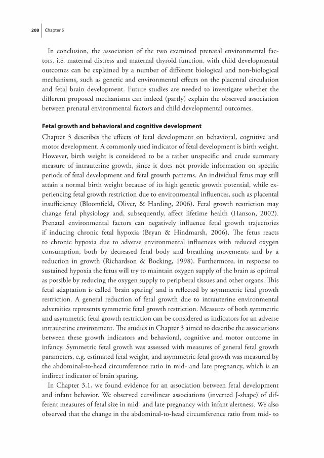
208 Chapter 5
In conclusion, the association of the two examined prenatal environmental fac-tors, i.e. maternal distress and maternal thyroid function, with child developmental outcomes can be explained by a number of different biological and non-biological mechanisms, such as genetic and environmental effects on the placental circulation and fetal brain development. Future studies are needed to investigate whether the different proposed mechanisms can indeed (partly) explain the observed association between prenatal environmental factors and child developmental outcomes.
Fetal growth and behavioral and cognitive development
Chapter 3 describes the effects of fetal development on behavioral, cognitive and motor development. A commonly used indicator of fetal development is birth weight. However, birth weight is considered to be a rather unspecific and crude summary measure of intrauterine growth, since it does not provide information on specific periods of fetal development and fetal growth patterns. An individual fetus may still attain a normal birth weight because of its high genetic growth potential, while ex-periencing fetal growth restriction due to environmental influences, such as placental insufficiency (Bloomfield, Oliver, & Harding, 2006). Fetal growth restriction may change fetal physiology and, subsequently, affect lifetime health (Hanson, 2002). Prenatal environmental factors can negatively influence fetal growth trajectories if inducing chronic fetal hypoxia (Bryan & Hindmarsh, 2006). The fetus reacts to chronic hypoxia due to adverse environmental influences with reduced oxygen consumption, both by decreased fetal body and breathing movements and by a reduction in growth (Richardson & Bocking, 1998). Furthermore, in response to sustained hypoxia the fetus will try to maintain oxygen supply of the brain as optimal as possible by reducing the oxygen supply to peripheral tissues and other organs. This fetal adaptation is called ‘brain sparing’ and is reflected by asymmetric fetal growth restriction. A general reduction of fetal growth due to intrauterine environmental adversities represents symmetric fetal growth restriction. Measures of both symmetric and asymmetric fetal growth restriction can be considered as indicators for an adverse intrauterine environment. The studies in Chapter 3 aimed to describe the associations between these growth indicators and behavioral, cognitive and motor outcome in infancy. Symmetric fetal growth was assessed with measures of general fetal growth parameters, e.g. estimated fetal weight, and asymmetric fetal growth was measured by the abdominal-to-head circumference ratio in mid- and late pregnancy, which is an indirect indicator of brain sparing.
In Chapter 3.1, we found evidence for an association between fetal development and infant behavior. We observed curvilinear associations (inverted J-shape) of dif-ferent measures of fetal size in mid- and late pregnancy with infant alertness. We also observed that the change in the abdominal-to-head circumference ratio from mid- to

General discussion 209
late pregnancy was curvilinearly associated to infant alertness. In Chapter 3.2, we show that faster overall fetal growth from mid- to late pregnancy reduces the risk of delayed infant developmental milestone attainment at age 12 months. In particular, social and fine motor development and self-help abilities were affected. Fetal size in late pregnancy was more consistently related to infant development than fetal size in early and mid-pregnancy. In general, these findings suggest that reduced fetal growth from mid- to late pregnancy negatively affects infant developmental outcome. In the following we will address potential mechanisms that may explain the observed as-sociation of fetal growth with infant behavior and development.
First, the observed relation between fetal growth and infant behavioral and devel-opmental outcome could be explained by the ‘fetal programming’ hypothesis, which stresses the importance of human fetal experience in determining developmental pat-terns (Barker, 1995, 1998; Barker, Winter, Osmond, Margetts, & Simmonds, 1989). Since the prenatal period is a time of enormous growth and change, in which tissues develop in a specific sequence from conception to maturity, the fetus is vulnerable to both organizing and disorganizing influences on organ development. Fetal program-ming is a process by which a prenatal event, such as placental insufficiency, during a sensitive developmental period has a long-lasting or permanent influence on the de-velopment of organs, including the brain, and associated physiological and metabolic systems (Barker, 1998). Previous studies showed that placental insufficiency does not only negatively affect fetal growth but also subsequent behavioral and neuropsycho-logical development (Gagnon, 2003; Roza et al., 2008; Scherjon, Briet, Oosting, & Kok, 2000).
Second, our observation that the change in the abdominal-to-head circumference ratio from mid- to late pregnancy was curvilinearly associated to infant alertness al-lows for two interpretations. On the one hand, children, whose head grew too slow from mid- to late pregnancy in comparison to the growth rate of their abdomen in this period of time, are less alert in infancy. This finding suggests that a relatively slower head growth, probably accompanied by slower brain growth, leads to impaired infant alertness. On the other hand, a very fast relative head growth, i.e. asymmetric fetal growth restriction, also seems to negatively influence infant alertness. This sug-gests that despite a ‘brain sparing effect’, which refers to a relative protection of the brain as compared to other fetal organs, the brain is not completely protected by this effect. The ‘brain sparing effect’ is well documented in animal studies (Cohn, Sacks, Heymann, & Rudolph, 1974; Sheldon, Peeters, Jones, Makowski, & Meschia, 1979). These studies showed that in the presence of fetal growth restriction, which is most commonly caused by placental insufficiency (Kingdom, Huppertz, Seaward, & Kaufmann, 2000), the central nervous system is preferentially perfused. Nevertheless, the term ‘brain-sparing’ may be somewhat misleading, because it may be an indicator

210 Chapter 5
of a compromised placental function and does not guarantee normal development after birth. Indeed, previous studies showed that brain-sparing is associated with behavioral problems at age 18 months and cognitive impairment at the age of 5 years (Roza et al., 2008; Scherjon et al., 2000).
Fourth, genetic factors largely determine both behavioral and neuropsychological development (Cyphers et al., 1990; Fox, Hershberger, & Bouchard, 1996; Plomin & DeFries, 1998; Plomin et al., 1994). In addition, 38-80% birth weight variance arises from genetic influences (Johnston et al., 2002). Possibly, common genetic factors underlie the respective associations of intrauterine growth and infant behavioral or developmental outcome in infancy.
Finally, although the effects of small fetal size and reduced fetal growth on subse-quent developmental outcomes might be caused by factors underlying intrauterine growth restriction, the specific mechanisms that explain the negative effects of very large fetal size on infant alertness remain to be characterized.
Predictors of continuity and discontinuity of early language functioning
In chapter 4, we addressed to what extent multiple perinatal, demographic and post-natal psychosocial factors predict the continuity and discontinuity of early language functioning.
Although most children start to speak their first words at around 12 months of age and rapidly accelerate their use of words by 18 months, there are huge individual differences in rate of early expressive vocabulary development. For some toddlers, de-layed language acquisition, i.e. being a late talker, is the first indication of a language impairment that may persist throughout childhood. Furthermore, delays in language development are related to a number of other developmental problems, including behavioral disorders, cognitive delays, and reading problems (Giddan & Milling, 1999; Harlaar, Hayiou-Thomas, Dale, & Plomin, 2008; Oliver, Dale, & Plomin, 2004; Rescorla, 2002, 2005, 2009; Snowling, Adams, Bishop, & Stothard, 2001). However, intervention for many late talkers may be unnecessary as most toddlers displaying early language delay appear to catch up in language development during the preschool period (Ellis Weismer, 2007; Paul, 1996; Thal, Tobias, & Morrison, 1991; Whitehurst & Fischel, 1994). For example, a previous study by Westerlund, Berglund, and Eriksson (2006) addressing to what extent language delay at 18 months predicts language delay at age 3 years in an unselected Swedish population of 2,080 children showed that only 17.6% of the 108 children with a language delay at 18 months had a language delay at age 3.
As recently noted by Leonard (2009) and Ellis Weismer (2007), the percentage of late talkers with delay that is persistent enough to warrant a diagnosis of specific language impairment (SLI) at age 4 or 5 is too low to account for the documented

General discussion 211
prevalence of 7% of SLI at age 5 (Tomblin, et al., 1997). Furthermore, as Ellis Weismer (2007) has noted, “Given the relatively low proportion of late talkers who display clinical language impairment at school entry, we must continue to ask where those 7% of kindergarten children with SLI come from if not from the ranks of late talkers.”(Ellis Weismer, 2007, p. 95). This conundrum suggests that children with SLI are a heterogeneous group, with children arriving at a SLI diagnosis by different routes, a phenomenon known as equifinality. That is, some percentage of preschool-ers with SLI represents late talkers who did not recover, and some other percentage represents children who were not apparently late talkers. To extend our knowledge about the continuity and discontinuity of early language abilities, we examined which perinatal, demographic and, maternal psychosocial factors predict temporary and persistent language delay in toddlerhood. In other words, we addressed the factors that differentiate the four possible outcome groups obtained from cross-tabulating language delay status at 18 and 30 months of age (no delay, early delay only, later delay only, and persistent delay).
We found that most children had normal language development (84.9%) at both ages, 6.2% were “late bloomers,” 6.3% had a late onset expressive language delay at 30 months only and 2.5% had a persistent expressive language delay. This finding indeed indicates that most toddlers who display early language delay appear to catch up in language development later in toddlerhood. Although word production and comprehension at 18 months and the other perinatal, demographic and maternal psychosocial factors significantly predicted language functioning at 30 months, the MacArthur Communicative Development Inventory (MCDI) word production scores at 18 months had both low positive predictive value (29%) and low sensitivity (29%) in predicting expressive language outcome at 30 months.
Furthermore, our results showed that children with normal language development at both ages were most advantaged with regard to perinatal and demographic factors and had the highest level of cognitive functioning. Thus, not surprisingly, this suggests that children with continuously normal language development are born healthy and grow up in a healthy, stimulating, and protective environment. Previous research has shown that high maternal SES is related to better fetal development, as indexed by higher birth weights, possibly due to a healthy life-style of the mother, including healthy diet (James, Nelson, Ralph, & Leather, 1997). In addition, Hoff (2003) showed that mothers with higher education use more language and higher quality language while interacting with their children than mothers with low education.
In the study presented in Chapter 4.1, we also observed that among the three groups of children with expressive language delay, the “late bloomers,” who apparently recov-ered from early language delay, were more likely to have a low birth weight, to be girls, to be of Dutch ethnicity, and to have mothers with high educational levels and high

212 Chapter 5
maternal age. Late bloomers also had delayed word comprehension at 18 months. Overall, these findings suggest that late bloomers may have manifested early receptive and expressive language delay due to some mild developmental lag, which was associ-ated with lower birth weight in some of the children. As late bloomers tended to be girls from Dutch families whose mothers had higher educational levels and older ages, they may have enjoyed stimulating home environments between 18 and 30 months that helped them to catch up in language by 30 months. This group may have had a slightly later onset of a normal brain maturation, as suggested by research demonstrat-ing that myelination of language-related brain areas in early childhood coincides with early language development (Pujol et al., 2006). Nevertheless, although late bloomers were no longer delayed in language development at 30 months, we cannot rule that they may still be at risk to display developmental problems later in life. as previous research showed that late talkers performed in the average range on most language tasks by age 5 but had significantly poorer scores on a number of language measures at age 6 to 9 years (Rescorla, 2002) and continued to heave weaker language scores than comparison children through age 17 (Rescorla, 2009).
Children with late onset expressive language delay were more likely to come from socially disadvantaged families, to have non-Dutch ethnicity, and to have mothers with medium and low education. Parenting stress and delayed word comprehension at 18 months raised the risk of expressive language delay at 30 months. This pattern of results suggests that the children in this group manifested a language delay by 30 months because they received less stimulation from their socially disadvantaged and stressed mothers. Previous studies have demonstrated that the association of low ma-ternal education and parenting stress with poor language functioning was mediated by maladaptive parent-child interactions and low levels of stimulation (Hoff, 2003; Magill-Evans & Harrison, 1999; Noel, et al., 2008).
Finally, the group with persistent delay had the highest percentage of low birth weight children and the lowest verbal and nonverbal cognitive scores. Low maternal education and higher levels of parenting stress significantly predicted persistent ex-pressive language delay. Strikingly, receptive language delay at 18 months predicted a 9-fold higher risk of persistent delay. These findings suggest that children with a persistent expressive language delay from 18 to 30 months had a significant and marked language impairment, in particular, because of the strong association with receptive language delay. Most likely, the chronic language problems of this group of children can be explained by biological vulnerabilities, as these children were less socially disadvantaged than children with late onset expressive language delay and had the highest percentage of low birth weight. Although the association between low birth weight and persistent language delay was non-significant, this was probably due to low statistical power arising from the small number of cases with persistent

General discussion 213
language delay. Previous studies have shown that genetic effects play an important role in SLI (Bishop, North, & Donlan, 1995; Tomblin & Buckwalter, 1998). It may be, therefore, that persistent language delay in this group was also partly due to a genetic predisposition.
An important finding of the study presented in Chapter 4.1 was that receptive language delay at 18 months yielded significant and very high odds ratios in predict-ing the three expressive language delay outcome groups, in particular with regard to the prediction of persistent expressive language delay. This suggests that receptive language delay plays an important role in predicting persistent expressive language delay, but also that deficits in language expression and in language comprehension and processing are closely related. The idea of a close relation between language expression and comprehension is supported by Ullman and Pierpoint (2005), who proposed that many children with a language impairment have a deficit in the neural circuitry responsible for procedural memory that affects both language comprehension and production.
Clinical implications
The main outcomes of the studies in this dissertation were infant and toddler behavioral and cognitive functioning. The prevalence of problem behavior in general population studies of both toddlers and children ranges from 12% to 18% (Koot & Verhulst, 1991; Pallapies, 2006; Richman, Stevenson, & Graham, 1975; Roberts, Attkisson, & Rosenblatt, 1998; Skovgaard et al., 2005). Furthermore, the prevalence of cognitive problems ranges from 3.9% for general mental delay to 17.5% for language delay (Horwitz et al., 2003; Simpson, Colpe, & Greenspan, 2003). Both behavioral and cognitive problems in infancy and toddlerhood continue into childhood, adolescence, and adulthood and negatively affect academic achievement (Caspi, Henry, McGee, Moffitt, & Silva, 1995; Hertzig, & Farber, 1997; Hinshaw, 1992; Kuntsi, Rijsdijk, Ronald, Asherson, & Plomin, 2005; Muris & Ollendick, 2005; Nigg, 2006; Oliver et al., 2004; Price et al., 2005; Rescorla, 2002, 2005, 2009; Shaw, Owens, Giovannelli, & Winslow, 2001; Taanila, Murray, Jokelainen, Isohanni, & Rantakallio, 2005). Fur-thermore, behavioral and cognitive problems constitute a major burden for children, parents, teachers, and society. However, there is still a large amount of uncertainty about the mechanisms underlying the development and continuity of behavioral and cognitive problems in children.
The majority of the findings described in this dissertation extended our knowledge about the etiology of behavioral and cognitive problems. We also addressed the value of a number of child, perinatal, demographic and maternal psychosocial factors in pre-

214 Chapter 5
dicting the continuity and discontinuity of language functioning in toddlerhood. This knowledge can guide the development of appropriate intervention and prevention programs, such as primary prevention, secondary prevention, and, therapeutic inter-vention. In this section, I will address the possible implications of the research findings presented in this thesis. I want to emphasize that with regard to the development of therapeutic interventions, replication and elaboration of our results are required.
This thesis showed that maternal distress during pregnancy is a risk factor for re-duced fetal growth. We also found that chronic maternal distress during the pre- and postnatal period negatively affects infant temperament. It is crucial that midwives and gynecologists are well aware of the possible negative effects of maternal distress during pregnancy on the developing child. Information about maternal distress can easily be obtained by questionnaires. Pregnant women at elevated risk should be referred to further psychodiagnostic assessment. Moreover, pregnant women with diagnosed mental health problems could then be invited to participate, for example, in distress reduction programs. In addition, mothers with diagnosed mental health problems may need additional counseling, interventions, or extended follow-up during the pre- and postnatal period by child psychiatrists and/or psychologists. These mental health professionals could monitor and treat maternal psychopathology and warrant the de-velopment of adaptive parenting skills and healthy parent-child interactions. We also recommend that parents-to be are informed about the potentially harmful effects of maternal prenatal distress via preconception prevention and intervention programs. The attitudes and knowledge of parents-to be with regard to maternal prenatal distress can be improved by increasing public awareness (via schools and media) and offering a pre-pregnancy visit for couples and persons planning pregnancy as part of standard maternity care (Johnson et al., 2006; Misra, Guyer, & Allston, 2003).
We also found evidence for an adverse effect of maternal and paternal family stress during pregnancy on toddler’s cognitive development. When cautiously interpreting family stress as indirect indicator of relationship strain and marital conflict, our find-ings suggest that relationship strain and low marital quality during pregnancy may extend to the development of the child. Therefore, preconception and intervention programs should also aim to address parental family stress during pregnancy and the improvement of prenatal marital quality.
Extended follow-up or specific interventions should be considered and tested for children with signs of symmetric and asymmetric intrauterine growth restriction. These factors may be added to screening protocols addressing risk factors of behavioral and cognitive problems that can be used in primary child health care settings. In addition, neonatologists and pediatricians should be aware of and informed about the possible long-term negative effects of intrauterine growth restriction on subsequent behavioral and cognitive development.

General discussion 215
We also found that higher maternal TSH levels during early pregnancy are related to externalizing problems in early childhood. Furthermore, we showed that maternal hypothyroxinaemia, i.e. low maternal FT4 concentrations in pregnant women with normal TSH levels, is a risk factor for verbal and nonverbal cognitive delay. It is tempt-ing to recommend thyroid function screening including FT4 measures of women in early pregnancy and that affected women should be given adequate amounts of iodine or T4 supplements as soon as possible in an attempt to counteract adverse effects on fetal brain development. Yet, first clinical trials addressing the potentially beneficial effects of iodine treatment or T4 supplementation in early pregnancy are needed, before the implementation of FT4 screening programs in early gestation can be justified. Moreover, public health programs should promote adequate iodine diet during pregnancy. In other words, public health programs should promote the adequate intake of food likely to be high in iodine, such as fish or iodine-containing vitamin, during pregnancy.
Finally, we found that the MacArthur Communicative Development Inventory (MCDI) word production scores at 18 months had both low positive predictive value (29%) and low sensitivity (29%) in predicting expressive language outcome at 30 months. This suggests that 18 months MCDI scores have limited clinical utility for the prediction of 30 months expressive language outcomes for individual children. While it is generally assumed that intervention has more positive effects when pro-vided earlier rather than later in life, it has also been stated that it is inefficient, if not unethical, to provide language treatment to toddlers whose language problems probably resolve spontaneously (Paul, 2000). The development of a parent-report screening measure that can distinguish between transient and persistent language delay in early childhood would improve the efficacy of preschool language treatment and intervention programs. An alternative strategy seems to be the use of a two-stage process, in which the MCDI is used to identify a high-risk group of toddlers who are further screened and diagnosed by professionals. Toddlers with diagnosed lan-guage problems could then be monitored, followed up and treated by professionals. Furthermore, when interpreting persistent expressive language delay from 18 to 30 months as an indicator of permanent language impairments in early childhood, our results that receptive language delay at 18 months, parenting stress and low maternal education was related to persistent expressive language delay in toddlerhood may have practical implications. Preschool interventions aimed at language impairments should address both expressive and receptive language abilities and also incorporate parent-ing stress reduction programs and encourage parents, in particular those with social disadvantages, to stimulate the language development of their children.

216 Chapter 5
Future research
This dissertation revealed a number of findings which may generate future studies. First, there are several other prenatal environmental factors, such as maternal diet and medication use during pregnancy, which may negatively influence human fetal and child developmental outcomes, such as fetal growth and brain development and behavioral and cognitive functioning later in life. With regard to nutritional factors, prospective population-based studies are needed to examine the association of maternal intake of essential vitamins, minerals, and fatty acids with pre- and postnatal growth and brain development and with subsequent cognitive and behavioral functioning. Furthermore, clinical trials should address the effects of dietary changes in pregnant women. Apart from addressing additional prenatal biological factors, future studies should also address other prenatal psychosocial factors, such as attitudes towards child rearing and parenting and their effects on postnatal parenting stress and skills and behavioral and cognitive development of the child.
Second, to better understand the association between prenatal environmental fac-tors and cognitive and behavioral functioning later in life, future studies are needed that address whether alterations in brain development are an intermediate in the association between prenatal environmental factors and behavioral and cognitive functioning in childhood and adolescence. Advanced imaging techniques, both structural and functional, should be used to elaborate by which alterations of specific brain regions the adverse effects of prenatal environmental factors on behavioral and cognitive development are mediated. Future studies that address the effect of prenatal environmental factors, such as maternal thyroid dysfunction during early pregnancy, on brain development itself are also needed, to improve our understanding of the pos-sible impact of prenatal environmental factors and as, so far, prenatal environmental influences on brain development have been understudied. Ideally, magnetic resonance imaging should be used in large samples of young children and adolescents from the general population that have been prospectively followed from fetal life onwards to identify brain abnormalities due to prenatal environmental influences.
Third, prospective population-based future studies addressing the impact of prenatal gene-environment interactions on subsequent behavioral and cognitive development could shed more light on the mechanisms that are involved in determining behavioral and cognitive development from prenatal life onwards. For example, population-based future studies that investigate the interaction between maternal prenatal stress and polymorphisms of the glucocorticoid receptor gene could increase our understanding of the impact of maternal prenatal stress on offspring development.
Fourth, future studies are needed that address the distinction between short and long term effects of prenatal environmental factors on child development. On the one

General discussion 217
hand, it is possible that some adverse prenatal environmental factors only have short term effects. It would be interesting to address which postnatal environmental factors have the potential to compensate for adverse prenatal environmental influences. In other words, prospective population-based studies are needed that investigate which protective factors can explain the recovery from early developmental problems due to adverse prenatal environmental influences. On the other hand, some negative effects of adverse prenatal environmental factors may persist into behavioral, cognitive and learning difficulties in childhood and adolescence. Future studies should therefore also examine which prenatal factors determine persistent behavioral and cognitive problems later in life. Furthermore, future studies are needed that investigate which postnatal environmental factors possibly even exacerbate the effects of prenatal en-vironmental risk factors on behavioral and cognitive functioning in childhood and adolescence.
Fifth, more research is needed that addresses the effects of paternal prenatal stress or distress, e.g. paternal family stress or depression, on children’s behavioral and cogni-tive development, as the mechanisms underlying the association of paternal prenatal stress or distress and child developmental outcomes have been poorly understood. Furthermore, the possible impact of paternal psychological influences on child devel-opment has been understudied.
Finally, in prospective population-based studies addressing the long-term conse-quences of prenatal and early postnatal factors on developmental outcome adequate assessment of behavioral and cognitive problems is one of the key challenges. As discussed before, next to parent reports on behavioral and cognitive functioning, researchers should use multiple sources of information, such as standardized observa-tions, tester-administered assessments and teacher and child reports.
Conclusion
Behavioral and cognitive abilities and disabilities are assumed to be the result of genetic influences and the accumulation of biological, psychological, and social influences. From conception to birth, interactions with the biological environment have the greatest impact on development, in particular during critical periods, but from birth onwards, interactions with the psychosocial environment become more important.
The studies described in this dissertation particularly emphasize the effects of both prenatal and postnatal environmental factors on child development. Our findings demonstrate that maternal prenatal distress and maternal thyroid dysfunction during early pregnancy may increase the likelihood of deviant developmental outcome, such as behavioral problems and cognitive delay. Furthermore, our results suggest that

218 Chapter 5
relationship patterns with the partner during pregnancy influence maternal prenatal experiences of family stress and that both maternal and paternal prenatal experiences of family stress negatively influence cognitive development in the offspring. Reduced fetal general and head growth have been shown to affect infant behavior and develop-ment. In addition, our findings suggest that adverse socio-demographic background factors negatively affect the language development of the child. Also maternal postna-tal psychological factors, such as maternal anxiety and parenting stress, are negatively related to behavioral and cognitive development in early childhood.
The research findings presented in this dissertation demonstrate the complex rela-tions between adverse prenatal environmental influences, fetal development, post-natal environmental factors and child behavioral and cognitive outcomes. Although prenatal developmental patterns are fundamental for development in postnatal life, the effects of intrauterine experiences and development on child and adult mental health and cognitive functioning cannot be studied without taking into account the influence of the postnatal psychological and social environment. To better understand why mental health and cognitive functioning partly originate in utero, future research addressing the effects of fetal programming should integrate and elucidate the differ-ent biological and environmental mechanisms that influence human behavioral and cognitive development.

General discussion 219
References
Achenbach, T. M., & Edelbrock, C. S. (1984). Psychopathology of childhood. Annual Review of Psychology, 35, 227-256.
Antonow-Schlorke, I., Schwab, M., Li, C., & Nathanielsz, P. W. (2003). Glucocorticoid exposure at the dose used clinically alters cytoskeletal proteins and presynaptic terminals in the fetal baboon brain. Journal of Physiology, 547(Pt 1), 117-123.
Auso, E., Lavado-Autric, R., Cuevas, E., Del Rey, F. E., Morreale De Escobar, G., & Berbel, P. (2004). A moderate and transient deficiency of maternal thyroid function at the beginning of fetal neocorticogenesis alters neuronal migration. Endocrinology, 145(9), 4037-4047.
Barbazanges, A., Piazza, P. V., Le Moal, M., & Maccari, S. (1996). Maternal glucocorticoid secretion mediates long-term effects of prenatal stress. Journal of Neuroscience, 16(12), 3943-3949.
Barker, D. J. (1995). Fetal origins of coronary heart disease. British Medical Journal, 311(6998), 171-174.
Barker, D. J. (1998). Mothers, babies and health in later life. Edinburgh: Churchill Livingstone.Barker, D. J., Winter, P. D., Osmond, C., Margetts, B., & Simmonds, S. J. (1989). Weight in
infancy and death from ischaemic heart disease. Lancet, 2(8663), 577-580.Bergman, K., Sarkar, P., O’Connor, T. G., Modi, N., & Glover, V. (2007). Maternal stress during
pregnancy predicts cognitive ability and fearfulness in infancy. Journal of the American Academy of Child and Adolescent Psychiatry, 46(11), 1454-1463.
Bloomfield, F. H., Oliver, M. H., & Harding, J. E. (2006). The late effects of fetal growth patterns. Archives of Disease in Childhood. Fetal and Neonatal Edition, 91(4), F299-304.
Bogoch, Y., Biala, Y. N., Linial, M., & Weinstock, M. (2007). Anxiety induced by prenatal stress is associated with suppression of hippocampal genes involved in synaptic function. Journal of Neurochemistry, 101(4), 1018-1030.
Bornstein, M. H., & Haynes, O. M. (1998). Vocabulary competence in early childhood: measure-ment, latent construct, and predictive validity. Child Development, 69(3), 654-671.
Breslau, N., & Chilcoat, H. D. (2000). Psychiatric sequelae of low birth weight at 11 years of age. Biological Psychiatry, 47(11), 1005-1011.
Breslau, N., Paneth, N., Lucia, V. C., & Paneth-Pollak, R. (2005). Maternal smoking during preg-nancy and offspring IQ. International Journal of Epidemiology, 34(5), 1047-1053.
Brown, A. S., van Os, J., Driessens, C., Hoek, H. W., & Susser, E. S. (2000). Further evidence of relation between prenatal famine and major affective disorder. American Journal of Psychiatry, 157(2), 190-195.
Bryan, S. M., & Hindmarsh, P. C. (2006). Normal and abnormal fetal growth. Hormone Research, 65 Supplement 3, 19-27.
Caspi, A., Henry, B., McGee, R. O., Moffitt, T. E., & Silva, P. A. (1995). Temperamental origins of child and adolescent behavior problems: from age three to age fifteen. Child Development, 66(1), 55-68.
Clement, Y., Calatayud, F., & Belzung, C. (2002). Genetic basis of anxiety-like behaviour: a critical review. Brain Research Bulletin, 57(1), 57-71.
Cohn, H. E., Sacks, E. J., Heymann, M. A., & Rudolph, A. M. (1974). Cardiovascular responses to hypoxemia and acidemia in fetal lambs. American Journal of Obstetrics and Gynecology, 120(6), 817-824.
Crozier, W. R., & Hostettler, K. (2003). The influence of shyness on children’s test performance. British Journal of Educational Psychology, 73(Pt 3), 317-328.

220 Chapter 5
Cyphers, L. H., Phillips, K., Fulker, D. W., & Mrazek, D. A. (1990). Twin temperament during the transition from infancy to early childhood. Journal of the American Academy of Child and Adolescent Psychiatry, 29(3), 392-397.
Darnaudery, M., & Maccari, S. (2008). Epigenetic programming of the stress response in male and female rats by prenatal restraint stress. Brain Research Review, 57(2), 571-585.
Dowling, A. L., Martz, G. U., Leonard, J. L., & Zoeller, R. T. (2000). Acute changes in maternal thyroid hormone induce rapid and transient changes in gene expression in fetal rat brain. Journal of Neuroscience, 20(6), 2255-2265.
Dowling, A. L., & Zoeller, R. T. (2000). Thyroid hormone of maternal origin regulates the expres-sion of RC3/neurogranin mRNA in the fetal rat brain. Brain Research, 82(1-2), 126-132.
Ellis Weismer, S. (2007). Typical talkers, late talkers, and children with specific language impairment: A language endowment spectrum? In R. Paul (Ed.), The influence of developmental perspectives on research and practice in communication disorders: A festschrift for Robin S. Chapman. Mahwah, NJ: Lawrence Erlbaum Associates.
Ernst, M., Moolchan, E. T., & Robinson, M. L. (2001). Behavioral and neural consequences of prenatal exposure to nicotine. Journal of the American Academy of Child and Adolescent Psychiatry, 40(6), 630-641.
Evans, M. A. (1993). Communicative competence as a dimension of shyness. In K. H. Rubin & J. B. Asendorpf (Eds.), Social withdrawal, inhibition and shyness in childhood. Hillsdale, NJ: Erlbaum.
Fattal-Valevski, A., Leitner, Y., Kutai, M., Tal-Posener, E., Tomer, A., Lieberman, D., et al. (1999). Neurodevelopmental outcome in children with intrauterine growth retardation: a 3-year follow-up. Journal of Child Neurology, 14(11), 724-727.
Feldman, R., Greenbaum, C. W., Yirmiya, N., & Mayes, L. C. (1996). Relations between cyclicity and regulation in mother-infant interaction at 3 and 9 months and cognition at 2 years. Journal of Applied Developmental Psychology, 17(3), 347-365.
Fenson, L., Dale, P. S., Reznick, J. S., Bates, E., Thal, D. J., & Pethick, S. J. (1994). Variability in early communicative development. Monographs of the Society for Research in Child Development, 59(5), 1-173; discussion 174-185.
Fergusson, D. M., Lynskey, M. T., & Horwood, L. J. (1993). The effect of maternal depression on maternal ratings of child behavior. Journal of Abnormal Child Psychology, 21(3), 245-269.
Fox, P. W., Hershberger, S. L., & Bouchard, T. J., Jr. (1996). Genetic and environmental contribu-tions to the acquisition of a motor skill. Nature, 384(6607), 356-358.
Gagnon, R. (2003). Placental insufficiency and its consequences. European Journal of Obstetrics, Gynecology and Reproductive Biology, 110 Suppl 1, S99-107.
Gardner, L. I. (1975). Historical notes on cretinism. In L. I. Gardner (Ed.), Endocrine and genetic diseases of childhood and adolescence (2nd ed.). Philadelphia: W. B. Saunders.
Giddan, J. J., & Milling, L. (1999). Comorbidity of psychiatric and communication disorders in children. Child and Adolescent Psychiatric Clinics of North America, 8(1), 19-36, v.
Gluckman, P. D., Hanson, M. A., Cooper, C., & Thornburg, K. L. (2008). Effect of in utero and early-life conditions on adult health and disease. New England Journal of Medicine, 359(1), 61-73.
Goodman, S. H., & Gotlib, I. H. (1999). Risk for psychopathology in the children of depressed mothers: A developmental model for understanding mechanisms of transmission. Psychological Review, 106(3), 458-490.

General discussion 221
Gray, R. F., Indurkhya, A., & McCormick, M. C. (2004). Prevalence, stability, and predictors of clinically significant behavior problems in low birth weight children at 3, 5, and 8 years of age. Pediatrics, 114(3), 736-743.
Hack, M., Breslau, N., Aram, D., Weissman, B., Klein, N., & Borawski-Clark, E. (1992). The effect of very low birth weight and social risk on neurocognitive abilities at school age. Journal of Developmental and Behavioral Pediatrics, 13(6), 412-420.
Hack, M., Flannery, D. J., Schluchter, M., Cartar, L., Borawski, E., & Klein, N. (2002). Outcomes in young adulthood for very-low-birth-weight infants. New England Journal of Medicine, 346(3), 149-157.
Hack, M., Taylor, H. G., Klein, N., Eiben, R., Schatschneider, C., & Mercuri-Minich, N. (1994). School-age outcomes in children with birth weights under 750 g. New England Journal of Medicine, 331(12), 753-759.
Hack, M., Youngstrom, E. A., Cartar, L., Schluchter, M., Taylor, H. G., Flannery, D., et al. (2004). Behavioral outcomes and evidence of psychopathology among very low birth weight infants at age 20 years. Pediatrics, 114(4), 932-940.
Haddow, J. E., Palomaki, G. E., Allan, W. C., Williams, J. R., Knight, G. J., Gagnon, J., et al. (1999). Maternal thyroid deficiency during pregnancy and subsequent neuropsychological development of the child. New England Journal of Medicine, 341(8), 549-555.
Hanson, M. (2002). Birth weight and the fetal origins of adult disease. Pediatric Research, 52(4), 473-474.
Harlaar, N., Hayiou-Thomas, M. E., Dale, P. S., & Plomin, R. (2008). Why do preschool language abilities correlate with later reading? A twin study. Journal of Speech Language and Hearing Research, 51(3), 688-705.
Hedegaard, M., Henriksen, T. B., Sabroe, S., & Secher, N. J. (1993). Psychological distress in pregnancy and preterm delivery. Bmj, 307(6898), 234-239.
Hertzig, M. E., & Farber, E. A. (1997). Annual Progress in Child Psychiatry and Child Development. Bristol, UK: Brunner/Mazel.
Hill, A. B. (1965). The Environment and Disease: Association or Causation? Proceedings of the Royal Society of Medicine, 58, 295-300.
Hinshaw, S. P. (1992). Externalizing behavior problems and academic underachievement inchildhood and adolescence: Causal relationships and underlying mechanisms. Psychological Bulletin,
111(1), 127-155.Horwitz, S. M., Irwin, J. R., Briggs-Gowan, M. J., Bosson Heenan, J. M., Mendoza, J., & Carter,
A. S. (2003). Language delay in a community cohort of young children. Journal of American Academy of Child and Adolescent Psychiatry, 42(8), 932-940.
Horwood, L. J., Mogridge, N., & Darlow, B. A. (1998). Cognitive, educational, and behavioural outcomes at 7 to 8 years in a national very low birthweight cohort. Archives of Disease in Child-hood. Fetal and Neonatal Edition, 79(1), F12-20.
Huizink, A. C., Mulder, E. J., & Buitelaar, J. K. (2004). Prenatal stress and risk for psychopathology: specific effects or induction of general susceptibility? Psychological Bulletin, 130(1), 115-142.
Jaddoe, V. W., van Duijn, C. M., van der Heijden, A. J., Mackenbach, J. P., Moll, H. A., Steegers, E. A., et al. (2008). The Generation R Study: design and cohort update until the age of 4 years. European Journal of Epidemiology, 23(12), 801-811.
Jaddoe, V. W., Verburg, B. O., de Ridder, M. A., Hofman, A., Mackenbach, J. P., Moll, H. A., et al. (2007). Maternal smoking and fetal growth characteristics in different periods of pregnancy: the generation R study. American Journal of Epidemiology, 165(10), 1207-1215.

222 Chapter 5
Johnson, K., Posner, S. F., Biermann, J., Cordero, J. F., Atrash, H. K., Parker, C. S., et al. (2006). Recommendations to improve preconception health and health care--United States. A report of the CDC/ATSDR Preconception Care Work Group and the Select Panel on Preconception Care. Morbidity and Mortality Weekly Reports Recommendations and Reortsp, 55(RR-6), 1-23.
Johnston, L. B., Clark, A. J., & Savage, M. O. (2002). Genetic factors contributing to birth weight. Archives of Disease in Childhood Fetal and Neonatal Edition, 86(1), F2-3.
Kingdom, J., Huppertz, B., Seaward, G., & Kaufmann, P. (2000). Development of the placental villous tree and its consequences for fetal growth. European Journal of Obstetrics, Gynecology and Reproductive Biology, 92(1), 35-43.
Kleinman, J. C., & Madans, J. H. (1985). The effects of maternal smoking, physical stature, and educational attainment on the incidence of low birth weight. American Journal of Epidemiology, 121(6), 843-855.
Kooistra, L., Crawford, S., van Baar, A. L., Brouwers, E. P., & Pop, V. J. (2006). Neonatal effects of maternal hypothyroxinemia during early pregnancy. Pediatrics, 117(1), 161-167.
Koot, H. M., & Verhulst, F. C. (1991). Prevalence of problem behavior in Dutch children aged 2-3. Acta Psychiatrica Scandinavica Supplement, 367, 1-37.
Kopp, P., Kitajima, K., & Jameson, J. L. (1996). Syndrome of resistance to thyroid hormone: insights into thyroid hormone action. Proceedings of the Society of Experimental Biology and Medicine, 211(1), 49-61.
Kraemer, H. C., Measelle, J. R., Ablow, J. C., Essex, M. J., Boyce, W. T., & Kupfer, D. J. (2003). A new approach to integrating data from multiple informants in psychiatric assessment and research: mixing and matching contexts and perspectives. American Journal of Psychiatry, 160(9), 1566-1577.
Kramer, M. S. (1987). Determinants of low birth weight: methodological assessment and meta-analysis. Bulletin of the World Health Organization, 65(5), 663-737.
Kuntsi, J., Rijsdijk, F., Ronald, A., Asherson, P., & Plomin, R. (2005). Genetic influences on the stability of attention-deficit/hyperactivity disorder symptoms from early to middle childhood. Biological Psychiatry, 57(6), 647-654.
Laplante, D. P., Barr, R. G., Brunet, A., Galbaud du Fort, G., Meaney, M. L., Saucier, J. F., et al. (2004). Stress during pregnancy affects general intellectual and language functioning in human toddlers. Pediatric Research, 56(3), 400-410.
Laplante, D. P., Brunet, A., Schmitz, N., Ciampi, A., & King, S. (2008). Project Ice Storm: prenatal maternal stress affects cognitive and linguistic functioning in 5 1/2-year-old children. Journal of the American Academy of Child and Adolescent Psychiatry, 47(9), 1063-1072.
Lavado-Autric, R., Auso, E., Garcia-Velasco, J. V., Arufe Mdel, C., Escobar del Rey, F., Berbel, P., et al. (2003). Early maternal hypothyroxinemia alters histogenesis and cerebral cortex cytoarchi-tecture of the progeny. Journal of Clinical Investigation, 111(7), 1073-1082.
Leitner, Y., Fattal-Valevski, A., Geva, R., Eshel, R., Toledano-Alhadef, H., Rotstein, M., et al. (2007). Neurodevelopmental outcome of children with intrauterine growth retardation: a longitudinal, 10-year prospective study. Journal of Child Neurology, 22(5), 580-587.
Lesage, J., Del-Favero, F., Leonhardt, M., Louvart, H., Maccari, S., Vieau, D., et al. (2004). Prenatal stress induces intrauterine growth restriction and programmes glucose intolerance and feeding behaviour disturbances in the aged rat. Journal of Endocrinology, 181(2), 291-296.
Lou, H. C., Hansen, D., Nordentoft, M., Pryds, O., Jensen, F., Nim, J., et al. (1994). Prenatal stressors of human life affect fetal brain development. Developmental Medicine and Child Neurol-ogy, 36(9), 826-832.

General discussion 223
Mantymaa, M., Puura, K., Luoma, I., Salmelin, R. K., & Tamminen, T. (2006). Mother’s early perception of her infant’s difficult temperament, parenting stress and early mother-infant inter-action. Nordic Journal of Psychiatry, 60(5), 379-386.
Misra, D. P., Guyer, B., & Allston, A. (2003). Integrated perinatal health framework. A multiple determinants model with a life span approach. American Journal of Preventive Medicine, 25(1), 65-75.
Morreale de Escobar, G., Obregon, M. J., & Escobar del Rey, F. (2000). Is neuropsychological development related to maternal hypothyroidism or to maternal hypothyroxinemia? Journal of Clinical Endocrinology and Metabolism, 85(11), 3975-3987.
Muneoka, K., Mikuni, M., Ogawa, T., Kitera, K., Kamei, K., Takigawa, M., et al. (1997). Prenatal dexamethasone exposure alters brain monoamine metabolism and adrenocortical response in rat offspring. American Journal of Physiology, 273(5 Pt 2), R1669-1675.
Muris, P., & Ollendick, T. H. (2005). The role of temperament in the etiology of child psychopa-thology. Clinical Child and Family Psychology Review, 8(4), 271-289.
Nakano, Y., Oshima, M., Sugiura-Ogasawara, M., Aoki, K., Kitamura, T., & Furukawa, T. A. (2004). Psychosocial predictors of successful delivery after unexplained recurrent spontaneous abortions: a cohort study. Acta Psychiatrica Scandinavicaq, 109(6), 440-446.
Nigg, J. T. (2006). Temperament and developmental psychopathology. Journal of Child Psychology and Psychiatry, 47(3-4), 395-422.
Oliver, B., Dale, P. S., & Plomin, R. (2004). Verbal and nonverbal predictors of early language problems: an analysis of twins in early childhood back to infancy. Journal of Child Language, 31(3), 609-631.
Paarlberg, K. M., Vingerhoets, A. J., Passchier, J., Dekker, G. A., Heinen, A. G., & van Geijn, H. P. (1999). Psychosocial predictors of low birthweight: a prospective study. British Journal of Obstetrica and Gynaecology, 106(8), 834-841.
Panicker V, Wilson SG, Spector TD, Brown SJ, Falchi M, Richards JB, Surdulescu GL, Lim EM, Fletcher SJ, Walsh JP (2008). Heritability of serum TSH, free T4 and free T3 concentrations: a study of a large UK twin cohort. Clinical Endocrinology, 68, 652-659.
Pallapies, D. (2006). Trends in childhood disease. Mutation Research, 608(2), 100-111.Parascandola, M., & Weed, D. L. (2001). Causation in epidemiology. Journal of Epidemiology and
Community Health, 55(12), 905-912.Paul, R. (1996). Clinical implications of the natural history of slow expressive language develop-
ment. Journal of Speech-Language Pathology, 5(2), 5-30.Paul, R. (2000). Predicting outcomes of early expressive language delay: Ethical implications. InD. V. M. Bishop & L. B. Leonard (Eds.), Speech and language impairments in children: Causes,
characteristics, intervention and outcome (pp. 195–209). Hove, U.K.: Psychology Press.Pauli-Pott, U., Mertesacker, B., Bade, U., Bauer, C., & Beckmann, D. (2000). Contexts of relations
of infant negative emotionality to caregiver’s reactivity/sensitivity. Infant Behavior & Develop-ment, 23(1), 23-39.
Phoojaroenchanachai, M., Sriussadaporn, S., Peerapatdit, T., Vannasaeng, S., Nitiyanant, W., Boon-namsiri, V., et al. (2001). Effect of maternal hyperthyroidism during late pregnancy on the risk of neonatal low birth weight. Clininical Endocrinology, 54(3), 365-370.
Plomin, R., & DeFries, J. C. (1998). The genetics of cognitive abilities and disabilities. Scientific American, 278(5), 62-69.
Plomin, R., Owen, M. J., & McGuffin, P. (1994). The genetic basis of complex human behaviors. Science, 264(5166), 1733-1739.

224 Chapter 5
Pop, V. J., Brouwers, E. P., Vader, H. L., Vulsma, T., van Baar, A. L., & de Vijlder, J. J. (2003). Maternal hypothyroxinaemia during early pregnancy and subsequent child development: a 3-year follow-up study. Clinical Endocrinology, 59(3), 282-288.
Pop, V. J., Kuijpens, J. L., van Baar, A. L., Verkerk, G., van Son, M. M., de Vijlder, J. J., et al. (1999). Low maternal free thyroxine concentrations during early pregnancy are associated with impaired psychomotor development in infancy. Clinical Endocrinology, 50(2), 149-155.
Price, T. S., Simonoff, E., Asherson, P., Curran, S., Kuntsi, J., Waldman, I., et al. (2005). Continuity and change in preschool ADHD symptoms: longitudinal genetic analysis with contrast effects. Behavior Genetics, 35(2), 121-132.
Puckering, C., Pickles, A., Skuse, D., Heptinstall, E., Dowdney, L., & Zur-Szpiro, S. (1995). Mother-child interaction and the cognitive and behavioural development of four-year-old children with poor growth. Journal of Child Psychology and Psychiatry, 36(4), 573-595.
Pujol, J., Soriano-Mas, C., Ortiz, H., Sebastian-Galles, N., Losilla, J. M., & Deus, J. (2006). My-elination of language-related areas in the developing brain. Neurology, 66(3), 339-343.
Rahman, A., Bunn, J., Lovel, H., & Creed, F. (2007). Association between antenatal depression and low birthweight in a developing country. Acta Psychiatrica Scandinavica, 115(6), 481-486.
Ramchandani, P., Stein, A., Evans, J., O’Connor, T. G., & team, A. s. (2005). Paternal depression in the postnatal period and child development: a prospective population study. Lancet, 365(9478), 2201-2205.
Rescorla, L. (2002). Language and reading outcomes to age 9 in late-talking toddlers. Journal of Speech, Language and Hearing Research, 45(2), 360-371.
Rescorla, L. (2005). Age 13 language and reading outcomes in late-talking toddlers. Journal of Speech, Language and Hearing Research, 48(2), 459-472.
Rescorla, L. (2009). Age 17 language and reading outcomes in late-talking toddlers: support for a dimensional perspective on language delay. Journal of Speech, Language and Hearing Research, 52(1), 16-30.
Richards, M., Hardy, R., Kuh, D., & Wadsworth, M. E. (2001). Birth weight and cognitive function in the British 1946 birth cohort: longitudinal population based study. British Medical Journal, 322(7280), 199-203.
Richardson, B. S., & Bocking, A. D. (1998). Metabolic and circulatory adaptations to chronic hypoxia in the fetus. Comparative Biochemistry and Physiology Part A: Molecular & Integrative Physiology, 119(3), 717-723.
Richman, N., Stevenson, J. E., & Graham, P. J. (1975). Prevalence of behaviour problems in 3-year-old children: an epidemiological study in a London borough. Journal of Child Psychology and Psychiatry, 16(4), 277-287.
Richters, J. E. (1992). Depressed mothers as informants about their children: a critical review of the evidence for distortion. Psychological Bulletin, 112(3), 485-499.
Roberts, R. E., Attkisson, C. C., & Rosenblatt, A. (1998). Prevalence of psychopathology among children and adolescents. American Journal of Psychiatry, 155(6), 715-725.
Rondo, P. H., Ferreira, R. F., Nogueira, F., Ribeiro, M. C., Lobert, H., & Artes, R. (2003). Maternal psychological stress and distress as predictors of low birth weight, prematurity and intrauterine growth retardation. European Journal of Clinical Nutrition, 57(2), 266-272.
Rothman, K. J. (2002). Epidemiology: an introduction. New York: Oxford University Press, Inc.Rothman, K. J., & Greenland, S. (1998a). Causation and causal inference. In K. J. Rothman &
S. Greenland (Eds.), Modern Epidemiology, 2nd Edition (pp. 7 - 28). Philadelphia: Lippincott Williams & Wilkins.

General discussion 225
Rothman, K. J., & Greenland, S. (1998b). Precision and validity in epidemiologic studies. In K. J. Rothman & S. Greenland (Eds.), Modern Epidemiology, 2nd Edition (pp. 115 - 134). Philadel-phia: Liipincott Williams & Wilkins.
Roza, S. J. (2008). Prenatal and early postnatal brain development. Unpublished Ph.D. thesis, Eras-mus Medical University Center Rotterdam.
Roza, S. J., Steegers, E. A., Verburg, B. O., Jaddoe, V. W., Moll, H. A., Hofman, A., et al. (2008). What is spared by fetal brain-sparing? Fetal circulatory redistribution and behavioral problems in the general population. American Journal of Epidemiology, 168(10), 1145-1152.
Scherjon, S., Briet, J., Oosting, H., & Kok, J. (2000). The discrepancy between maturation of visual-evoked potentials and cognitive outcome at five years in very preterm infants with and without hemodynamic signs of fetal brain-sparing. Pediatrics, 105(2), 385-391.
Schneider, M. L., & Coe, C. L. (1993). Repeated social stress during pregnancy impairs neuromotor development of the primate infant. Journal of Developmental and Behavioral Pediatrics, 14(2), 81-87.
Schneider, M. L., Coe, C. L., & Lubach, G. R. (1992). Endocrine activation mimics the adverse effects of prenatal stress on the neuromotor development of the infant primate. Developmental Psychobiology, 25(6), 427-439.
Shaw, D. S., Owens, E. B., Giovannelli, J., & Winslow, E. B. (2001). Infant and toddler pathways leading to early externalizing disorders. Journal of the American Academy of Child and Adolescent Psychiatry, 40(1), 36-43.
Sheldon, R. E., Peeters, L. L., Jones, M. D., Jr., Makowski, E. L., & Meschia, G. (1979). Redistribu-tion of cardiac output and oxygen delivery in the hypoxemic fetal lamb. American Journal of Obstetrics and Gynecology, 135(8), 1071-1078.
Simpson, G. A., Colpe, L., & Greenspan, S. (2003). Measuring functional developmental delay in infants and young children: prevalence rates from the NHIS-D. Paediatric and Perinatal Epidemiology, 17(1), 68-80.
Skovgaard, A. M., Olsen, E. M., Houmann, T., Christiansen, E., Samberg, V., Lichtenberg, A., et al. (2005). The Copenhagen County child cohort: design of a longitudinal study of child mental health. Scand J Public Health, 33(3), 197-202.
Slotkin, T. A., Barnes, G. A., McCook, E. C., & Seidler, F. J. (1996). Programming of brainstem serotonin transporter development by prenatal glucocorticoids. Brain Research, Developmental Brain Research, 93(1-2), 155-161.
Snowling, M. J., Adams, J. W., Bishop, D. V. M., & Stothard, S. E. (2001). Educational attainments of school leavers with a preschool history of speech-language impairments. International Journal of Language & Communication Disorders, 36(2), 173-183.
Stein, A., Woolley, H., Murray, L., Cooper, P., Cooper, S., Noble, F., et al. (2001). Influence of psychiatric disorder on the controlling behaviour of mothers with 1-year-old infants. A study of women with maternal eating disorder, postnatal depression and a healthy comparison group. British Journal of Psychiatry, 179, 157-162.
Stott, D. H. (1973). Follow-up study from birth of the effects of prenatal stresses. Developmental Medicine and Child Neurology, 15(6), 770-787.
Taanila, A., Murray, G. K., Jokelainen, J., Isohanni, M., & Rantakallio, P. (2005). Infant devel-opmental milestones: a 31-year follow-up. Developmental Medicine & Child Neurology, 47(9), 581-586.

226 Chapter 5
Talge, N. M., Neal, C., Glover, V., & Early Stress, Translational Research and Prevention Science Network: Fetal and Neonatal Experience on Child and Adolescent Mental Health (2007). An-tenatal maternal stress and long-term effects on child neurodevelopment: how and why? Journal of Child Psychology and Psychiatry, 48(3-4), 245-261.
Thal, D., Tobias, S., & Morrison, D. (1991). Language and gesture in late talkers: a 1-year follow-up. Journal of Speech and Hearing Research, 34(3), 604-612.
van Lith, H. (2004). Demografische gegevens (Publication., from Center for Research and Statistics, Rotterdam (COS): http:/www.cos.rotterdam.nl
VanderWeele, T. J. (2009). On the relative nature of overadjustment and unnecessary adjustment. Epidemiology, 20(4), 496-499.
Weinstock, M. (1997). Does prenatal stress impair coping and regulation of hypothalamic-pituitary-adrenal axis? Neuroscience and Biobehavioral Reviews, 21(1), 1-10.
Weinstock, M. (2001). Alterations induced by gestational stress in brain morphology and behaviour of the offspring. Progress in Neurobiology, 65(5), 427-451.
Whitehurst, G. J., & Fischel, J. E. (1994). Practitioner review: early developmental language delay: what, if anything, should the clinician do about it? Journal of Child Psychology and Psychiatry, 35(4), 613-648.
Wiles, N. J., Peters, T. J., Heron, J., Gunnell, D., Emond, A., & Lewis, G. (2006). Fetal growth and childhood behavioral problems: results from the ALSPAC cohort. American Journal of Epidemiology, 163(9), 829-837.
Wust, S., Federenko, I. S., van Rossum, E. F., Koper, J. W., Kumsta, R., Entringer, S., et al. (2004). A psychobiological perspective on genetic determinants of hypothalamus-pituitary-adrenal axis activity. Annals of the New York Academy of Sciences, 1032, 52-62.
Zarrow, M. X., Philpott, J. E., & Denenberg, V. H. (1970). Passage of 14C-4-corticosterone from the rat mother to the foetus and neonate. Nature, 226(5250), 1058-1059.

C hapter 6Summary


Summary / Samenvatting 229
Behavioral and cognitive development represent two major domains of child develop-ment. Traditionally, the study of child development from a psychological perspective, has taken birth as its starting point. However, current insight suggests that prenatal influences explain a significant part of variation in later behavioral and cognitive development. Small variations in the fetal physiological environment induced by internal or external factors can be of critical and long-lasting importance, given an increased sensitivity of the rapidly developing brain.
As a consequence, the general aim of this dissertation was to extend existing knowl-edge on prenatal determinants of behavioral and cognitive development in infancy and toddlerhood. The studies presented in this dissertation were carried out within the Generation R Study, a prospective multi-ethnic population-based cohort from fetal life onwards in Rotterdam, the Netherlands.
In Chapter 2, the effects of prenatal environmental factors on fetal growth and on behavioral and cognitive development were studied. These environmental fac-tors included maternal prenatal psychological distress, i.e. anxiety, depression and stress, and maternal thyroid function during early pregnancy. In Chapter 2.1, we investigated whether maternal psychological distress affects fetal growth during the period of mid-pregnancy until birth. We found that maternal symptoms of anxiety or depression were associated with impaired fetal weight gain and impaired fetal head and abdominal growth. This suggests that fetal growth can be affected by different aspects of maternal distress.
In Chapter 2.2 it was examined whether maternal anxiety that is temporary or chronic during the pre- and postnatal period predicts infant temperament. We observed that maternal pregnancy-specific and general anxiety during the pre- and postnatal period were all independently associated with perceived infant temperamen-tal difficulties. In particular, chronically high maternal anxiety predicted the highest activity level and negative affectivity of the infant. These findings show that different forms of maternal anxiety during both the pre- and postnatal period are indepen-dently related to perceived temperamental problems in infancy. They also emphasize the significance of chronic maternal anxiety for infant mental health.
The study presented in Chapter 2.3 addressed whether parental experiences of family stress during pregnancy predict verbal and nonverbal cognitive functioning of toddlers. We found that maternal prenatal family stress was associated with low word comprehen-sion and poorer nonverbal cognitive functioning independent of paternal prenatal fam-ily stress. In contrast, paternal experience of family stress during pregnancy predicted only poorer nonverbal cognitive development independent of the mother. Furthermore, children of parents who both reported high levels of family stress had a particularly increased risk of low verbal and nonverbal cognitive functioning. These findings show that parental family stress during pregnancy affects toddlers’ cognitive outcomes.

230 Chapter 6
In Chapter 2.4 we investigated associations of maternal prenatal FT4 and TSH levels, and mild and severe maternal hypothyroxinaemia in early pregnancy with verbal and nonverbal cognitive functioning in early childhood. Continuous measures of thyroid parameters were not consistently related to cognitive outcomes. Both mild and severe maternal hypothyroxinaemia were associated with a higher risk of expres-sive language delay at 18 and 30 months. Severe maternal hypothyroxinaemia also predicted a higher risk of nonverbal cognitive delay at 30 months. This suggests that maternal hypothyroxinaemia is a risk factor for cognitive delay in early childhood.
In the study described in Chapter 2.5 the association of parameters of maternal thyroid function in the non-clinical range during early pregnancy with behavioral and emotional problems of the offspring was examined. We found that higher levels of maternal TSH in early pregnancy predicted higher externalizing scores in children aged 1 ½ and 3 years. Free T4 and total T4 of mothers were not associated with internalizing or externalizing scores of children. The linear relation between higher levels of TSH in the normal range and more behavioral problems implies that subtle impairments of the maternal thyroid function may affect the child. These results provide further evidence that the maternal thyroid function during early pregnancy is crucial for fetal brain development and thereby determine problem behavior later in life.
In Chapter 3 we investigated whether reduced fetal growth, affects infant behavior and developmental milestone attainment. The study presented in Chapter 3.1 exam-ined whether fetal size in mid- and late pregnancy is related to infant irritability and alertness. We observed curvilinear associations (inverted J-shape) of measures of fetal size in both mid- and late pregnancy with infant alertness. Fetal size characteristics were not associated with infant irritability. These results suggest that alterations of in-trauterine growth affecting infant alertness are already detectable from mid-pregnancy onwards. In Chapter 3.2 we investigated whether fetal growth from early pregnancy onwards is related to infant development and whether this potential relationship is independent of postnatal growth. We found that faster fetal growth reduces the risk of delayed infant development at age 12 months. In particular, social and fine motor development and self-help abilities were affected. Fetal size in late pregnancy predicted infant development more consistently than fetal size in early and mid-preg-nancy. These associations were attenuated by controlling for early postnatal growth but largely remained significant. These results suggest that reduced fetal growth may determine subsequent developmental outcomes.
In Chapter 4, we examined to what extent multiple perinatal, demographic and maternal postnatal psychosocial factors can explain the continuity and discontinuity of language functioning in toddlerhood. Only a small proportion of children, i.e. 2.5%, manifested persistent expressive language delay from 18 to 30 months. Most
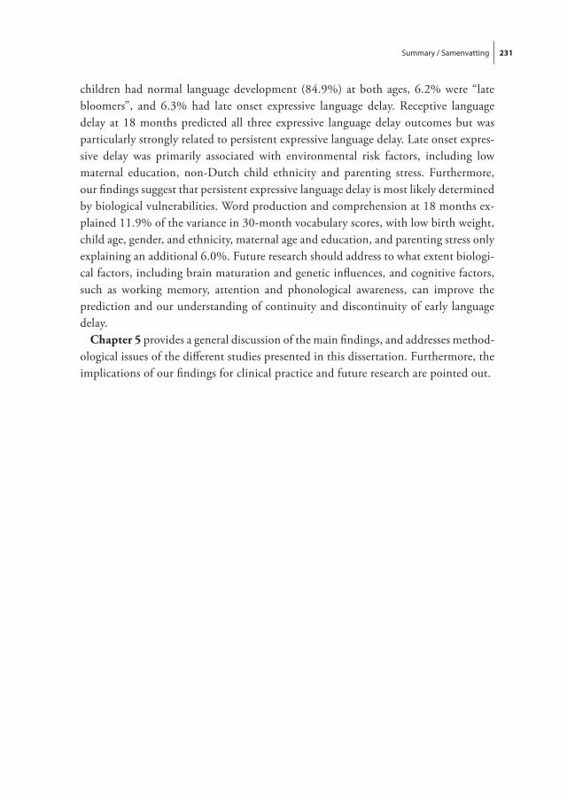
Summary / Samenvatting 231
children had normal language development (84.9%) at both ages, 6.2% were “late bloomers”, and 6.3% had late onset expressive language delay. Receptive language delay at 18 months predicted all three expressive language delay outcomes but was particularly strongly related to persistent expressive language delay. Late onset expres-sive delay was primarily associated with environmental risk factors, including low maternal education, non-Dutch child ethnicity and parenting stress. Furthermore, our findings suggest that persistent expressive language delay is most likely determined by biological vulnerabilities. Word production and comprehension at 18 months ex-plained 11.9% of the variance in 30-month vocabulary scores, with low birth weight, child age, gender, and ethnicity, maternal age and education, and parenting stress only explaining an additional 6.0%. Future research should address to what extent biologi-cal factors, including brain maturation and genetic influences, and cognitive factors, such as working memory, attention and phonological awareness, can improve the prediction and our understanding of continuity and discontinuity of early language delay.
Chapter 5 provides a general discussion of the main findings, and addresses method-ological issues of the different studies presented in this dissertation. Furthermore, the implications of our findings for clinical practice and future research are pointed out.

Samenvatting
Gedrags- en cognitieve ontwikkeling zijn twee zeer belangrijke domeinen van de ont-wikkeling van het kind. In het verleden werd de ontwikkeling van het kind vanuit een psychologisch perspectief vanaf de geboorte bestudeerd. Recente wetenschappelijke inzichten suggereren echter dat prenatale invloeden een significant deel van de variatie van de gedrags- en cognitieve ontwikkeling van kinderen en volwassenen kunnen verklaren. Kleine veranderingen in de fysiologische omgeving van de foetus, veroor-zaakt door ongunstige externe en interne factoren, kunnen de gedrags- en cognitieve ontwikkeling blijvend negatief beïnvloeden, gezien de verhoogde kwetsbaarheid van het zich ontwikkelende brein.
Het doel van dit proefschrift is de bestaande kennis over prenatale determinanten van de gedrags- en cognitieve ontwikkeling van zeer jonge kinderen uit te breiden. De in dit proefschrift beschreven studies werden uitgevoerd in het kader van het Ge-neration R onderzoek, een prospectieve multi-etnische cohortstudie vanaf het foetale leven in Rotterdam, Nederland.
In hoofdstuk 2 werden de effecten van prenatale omgevingsfactoren op de foetale groei en op de gedrags- en cognitieve ontwikkeling van zeer jonge kinderen onder-zocht. Daarbij werd ingegaan op de effecten van omgevingsfactoren zoals maternale prenatale angst, depressie en stress en maternaal prenataal schildklierdisfunctioneren. In hoofdstuk 2.1 gingen we na of maternale prenatale stress en symptomen van angst en depressie de foetale groei vanaf de tweede helft van de zwangerschap tot de geboorte negatief beïnvloeden. Maternale symptomen van angst en depressie tijdens de zwangerschap waren geassocieerd met een verminderde foetale gewichtstoename en een gereduceerde foetale hoofd- en buikgroei. Deze resultaten suggereren dat foetale groei negatief wordt beïnvloed door symptomen van angst en depressie van moeder.
In hoofdstuk 2.2 beschreven we de samenhang van tijdelijke en chronische angst van moeder tijdens de prenatale en postnatale periode met het temperament van kinderen van zes maanden oud. Uit de resultaten bleek dat zwangerschaps-specifieke angsten en algemene angst van moeder tijdens de prenatale en postnatale periode onafhankelijk waren gerelateerd aan een door moeder moeilijker ervaren tempera-ment van hun kinderen van zes maanden oud. Chronische angst van moeder tijdens de prenatale en postnatale periode voorspelden de hoogste mate van door moeder waargenomen negatieve affectiviteit en activiteit bij kinderen van zes maanden oud.

Samenvatting 233
Deze bevindingen laten zien dat verschillende vormen van maternale angst tijdens de prenatale en postnatale periode onafhankelijk gerelateerd zijn aan percepties van moeilijker temperament bij zeer jonge kinderen. Verder benadrukken deze bevin-dingen dat chronische maternale angsten een negatieve invloed uitoefenen op de geestelijke gezondheid van zeer jonge kinderen.
In hoofdstuk 2.3 gingen we in op de vraagstelling of maternale en paternale ervaringen van stress in het gezin tijdens de zwangerschap de verbale en non-verbale cognitieve ontwikkeling van peuters beïnvloeden. Uit de resultaten bleek dat prena-tale ervaringen van stress in het gezin door moeder geassocieerd waren met een laag taalbegrip en een langzamere non-verbale cognitieve ontwikkeling in de peutertijd onafhankelijk van paternale prenatale ervaringen van stress in het gezin. Ervaringen van stress in het gezin door vader tijdens de zwangerschap voorspelden alleen een langzamere non-verbale cognitieve ontwikkeling in de peutertijd onafhankelijk van de waarneming van stress in het gezin door moeder. Bovendien hadden peuters van ouders die beiden stress in het gezin tijdens de zwangerschap rapporteerden een bijzonder verhoogd risico op een vertraagde verbale en non-verbale cognitieve ontwikkeling. Deze bevindingen suggereren dat ouderlijke stress in het gezin tijdens de zwangerschap de cognitieve ontwikkeling van peuters negatief beïnvloedt.
In hoofdstuk 2.4 onderzochten we het verband van maternale vrije T4 en TSH concentraties, en milde en ernstige hypothyroxinemie tijdens de eerste helft van de zwangerschap met het verbaal en non-verbaal cognitief functioneren van jonge kinderen. Vrije T4 en TSH concentraties tijdens de eerste helft van de zwangerschap waren niet consistent geassocieerd met de verschillende cognitieve uitkomstmaten. Zowel milde als ook ernstige maternale hypothyroxinemie waren geassocieerd met een achterstand in de taalproductie van kinderen van 18 en 30 maanden oud. Ernstige maternale hypothyroxinemie voorspelde ook een non-verbale cognitieve achterstand bij kinderen van 30 maanden oud. Dit suggereert dat maternale hypothyroxinemie een risicofactor is voor cognitieve achterstanden in de vroege kindertijd. In hoofd-stuk 2.5 werd de samenhang tussen de schildklierfunctie van moeder tijdens de eerste helft van de zwangerschap en gedragsproblemen in het nageslacht onderzocht. Een toename in maternale TSH concentraties tijdens de eerste helft van de zwangerschap voorspelde een hogere mate van externaliserend gedrag bij kinderen van anderhalf en drie jaar oud. Vrije T4 en totale T4 van moeder tijdens de eerste helft van de zwan-gerschap waren niet gerelateerd aan internaliserend en externaliserend gedrag van de kinderen. De lineaire relatie tussen stijgende maternale TSH concentraties tijdens de eerste helft van de zwangerschap en een toename in gedragsproblemen bij het nage-slacht impliceert dat subtiele beperkingen in het prenatale schildklierfunctioneren van moeder het gedrag van het kind negatief beïnvloeden. Deze resultaten suggereren dat de maternale schildklierfunctie tijdens de eerste helft van de zwangerschap cruci-

234 Chapter 6
aal is voor de foetale hersenenontwikkeling en daarbij ook gedragsproblemen in het latere leven bepaalt.
In hoofdstuk 3 richtten we ons op de vraag of vertraagde foetale groei het gedrag en de ontwikkeling van zeer jonge kinderen negatief beïnvloedt. Het in hoofdstuk 3.1 beschreven onderzoek ging in op de vraag of foetale grootte vanaf de tweede helft van de zwangerschap gerelateerd is aan de alertheid en prikkelbaarheid van kinderen van drie maanden oud. Er werden curvilineaire associaties (omgedraaide J-vorm) tussen verschillende maten van foetale grootte vanaf de tweede helft van de zwangerschap en de alertheid van kinderen van drie maanden oud geobserveerd. De verschillende maten van foetale grootte waren niet gerelateerd aan de prikkelbaarheid van kinderen van drie maanden oud. Deze resultaten suggereren dat veranderingen in de foetale groei die de alertheid van zeer jonge kinderen beïnvloeden al vanaf de tweede helft van de zwangerschap vastgesteld kunnen worden. In hoofdstuk 3.2 werd onderzocht of foetale groei vanaf het tweede zwangerschapstrimester gerelateerd is aan de ontwik-keling van kinderen van 12 maanden oud en of deze mogelijke relatie onafhankelijk is van postnatale groei. Snellere foetale groei voorspelde een gereduceerd risico op ontwikkelingsachterstanden van kinderen van 12 maanden oud. Met name werden de sociale en fijn-motorische ontwikkeling en de zelfredzaamheid van de kinderen negatief beïnvloed. Foetale grootte tijdens de 30ste zwangerschapsweek voorspelde de ontwikkeling van de kinderen van 12 maanden oud consistenter dan foetale grootte tijdens eerdere fasen van de zwangerschap. Na controle voor postnatale groei nam de sterkte van de geobserveerde associaties af, maar deze associaties waren nog steeds significant. Deze bevindingen maken duidelijk dat een snellere foetale groei, onaf-hankelijk van de postnatale groei, een verlaagd risico op ontwikkelingsachterstanden voorspelt. Deze resultaten suggereren dat een vertraagde foetale groei latere ontwik-kelingsuitkomsten bepaalt.
In hoofdstuk 4 werd onderzocht in welke mate multipele perinatale, demografi-sche en maternaal psychosociale factoren de continuïteit en discontinuïteit van het verbale functioneren in de peutertijd kunnen verklaren. Alleen een kleine fractie van de kinderen, d.w.z. 2,5%, vertoonden een persisterende expressieve taalachterstand tussen een leeftijd van 18 en 30 maanden. In dezelfde leeftijdsperiode hadden de meeste kinderen (84,9%) een normale taalontwikkeling, 6,2 % van de kinderen wa-ren zogenaamde ‘laatbloeiers’ en 6.3% van de kinderen hadden een laat beginnende expressieve taalachterstand, d.w.z. deze kinderen hadden alleen op een leeftijd van 30 maanden een expressieve taalachterstand. Een achterstand in taalbegrip op 18 maan-den voorspelde alle drie vormen van expressieve taalachterstand en was bijzonder sterk gerelateerd aan een persisterende expressieve taalachterstand. Een laat beginnende ex-pressieve taalachterstand was primair geassocieerd met omgevings-gerelateerde risico-factoren, zoals een laag opleidingsniveau van moeder, een niet-Nederlandse etniciteit

Samenvatting 235
en ouderlijke stress. Verder suggereren onze bevindingen dat persisterende expressieve taalachterstand hoogstwaarschijnlijk bepaald wordt door biologische kwetsbaarheden. Taalproductie en taalbegrip op 18 maanden verklaarden 11,9% van de variantie van de taalproductie scores op 30 maanden. Een laag geboortegewicht, het geslacht, de leeftijd en de etniciteit van de kinderen, de leeftijd en het opleidingsniveau van moeder en ouderlijke stress verklaarden alleen maar additionele 6% van de variantie van de taalproductie scores op 30 maanden. Toekomstig onderzoek is nodig om uit te zoeken in hoeverre biologische factoren (zoals genetische invloeden en hersenenrijping) en cognitieve factoren (zoals het werkgeheugen, aandacht en fonologisch bewustzijn) de voorspelling en ons begrip van de continuïteit en discontinuïteit van een taalachter-stand in de peutertijd kunnen verbeteren.
Hoofdstuk 5 bestaat uit een algemene discussie, waarin de belangrijkste bevindingen en methodologische aspecten van de verschillende onderzoeken worden besproken. In het laatste deel van dit hoofdstuk gaan we in op mogelijke implicaties van onze bevindingen voor de praktijk en op mogelijkheden voor toekomstig onderzoek.

Dankwoord
Aan het eind van dit proefschrift wil ik mijn dankbaarheid aan iedereen uiten die hieraan direct of indirect heeft bijgedragen.
Allereerst gaat mijn dank uit naar de deelnemers van het Generation R onderzoek. Beste ouders en kinderen, zonder jullie vrijwillige bijdrage en trouwe medewerking had dit proefschrift niet bestaan. Hartelijk dank voor alle inzet, geduld en moeite.
Mijn promotoren: Prof. dr. H.G. Schmidt en Prof. dr. F.C. Verhulst, wil ik van harte danken. Beste Henk, hartelijk dank voor al jouw ondersteuning, kritisch meedenken, en motiverende, inspirerende en wijze adviezen. Beste Frank, dankjewel dat ik zo veel van jouw expertise en enorme onderzoekservaring heb mogen en kunnen leren en vooral wil ik je danken voor de fijne samenwerking, je steun en je vertrouwen.
Mijn co-promotoren: Mijn bijzondere dank gaat uit naar mijn co-promotoren dr. Jacqueline Schenk en dr. Henning Tiemeier. Lieve Jacqueline, hartelijk dank dat je mij van het begin af aan zo enorm hebt ondersteund, altijd een luisterend oor voor mij hebt gehad en altijd alle vertrouwen in mij had. Je hebt mij enorm goed opge-leid en je bent een fantastische begeleider, die zowel menselijk als inhoudelijk sterk is. Lieber Henning, vielen Dank für die außergewöhnlich gute Ausbildung, Deine Unterstützung und Dein Wohlwollen. Ich möchte Dir auch sehr danken für all die Möglichkeiten die Du mir erschaffen hast. Durch Deine wissenschaftliche Brillianz und Präzision habe ich sehr viel gelernt.
Mijn leescommissie, en overige commissieleden: Prof dr. H.J. de Koning, beste Harry, hartelijk dank voor je bereidheid om als secretaris van de leescommissie op te treden. Prof. dr. B.R.H.M. van den Bergh en Prof. dr. R.A. Zwaan wil ik danken voor hun bereidheid om mijn proefschrift te lezen en deel uit te maken van mijn leescommissie. Prof. dr. E.A.P. Steegers en Prof. dr. T.J. Visser, beste Eric en beste Theo, hartelijk dank voor jullie bereidheid om plaats te nemen in mijn commissie en jullie nuttige commentaren op de diverse artikelen.
De projectleiders van Generation R, dr. Rosalinde Snijders en dr. Vincent Jaddoe, wil ik van harte danken. Beste Rosalinde, hartelijk dank voor je enorme inzet bij het opstarten van Generation R en de fijne samenwerking. Beste Vincent, dankjewel voor al je enthousiasme en inspanning voor Generation R en je helder commentaar op mijn artikelen.
Mijn co-auteurs, Prof. dr. H. Hooijkaas, Prof. dr. J.P. Mackenbach, Prof. dr. H.A. Moll, dr. J.J. Bongers-Schokkin, dr. S.M.P.F. de Muinck Keizer-Schrama, dr. H. Raat,

Dankwoord 237
dr. Y.B. de Rijke, dr. W. Visser en de overige coauteurs, die elders in dit dankwoord genoemd worden, wil ik hartelijk danken voor de fijne samenwerking en hun com-mentaren en suggesties die tot verbetering van de diverse artikelen hebben geleid.
Mijn overige begeleiders / mentoren: Mijn dank gaat ook uit naar Prof. dr. L. Rescorla en Prof. dr. A. Hofman. Dear Leslie, thank you very much for the great time that I have had in Burlington due to your hospitality and great support. I have learned very much from you and I really enjoyed working with you. Beste Bert, ik wil jou, het “master brein” van Generation R van harte danken voor alle kennis die ik door jouw enorme onderzoekservaring, expertise en uitstekende didactische colleges tijdens de Nihes opleiding heb kunnen opdoen.
Mijn dank gaat ook uit naar mijn leidinggevenden bij het Instituut voor Psychologie, Prof. dr. Marise Born, Prof. dr. Henk van der Molen, Prof. dr. Remy Rikers, Prof. dr. Fred Paas en dr. Guus Smeets. Beste Marise, Henk, Remy, Fred en Guus hartelijk dank voor jullie ondersteuning, begeleiding en fijne samenwerking bij het Instituut voor Psychologie.
Dr. Lidia Arends wil ik van harte danken voor al haar statistische ondersteuning en geduld en voor de vele leuke gesprekken. Beste Lidia, ik heb enorm veel van je geleerd en werk graag met je samen.
Mijn scriptiestudenten, waarvan sommige ook coauteurs zijn (in chronologische volgorde): Yara Tjoa, Charlotte Barendregt, Bouchra Ftitache, Isil Sincer, Fatima Khan, Maureen Khan, Marije Verhage, Nicky Molhoek, Akghar Ghassabian (toen nog Nihes-studente), Cootje Donkersloot en Lysanne van de Velde wil ik van harte danken voor de fijne en gezellige samenwerking en voor alle dataverzamelingsactivi-teiten. Door jullie goede vragen, en door de begeleiding van jullie schrijfproces heb ik veel geleerd.
Mijn collega’s en oud-collega’s bij Generation R, Alwin, Anne, Annemarie P., Anne-marie V., Anne Marie V., Anneke, Akghar, Alwin, Bero, Busra, Carmelo, Christy, Claudia K., Claudia C., Dennis, Edith, Edward, Elianne, Elizia, Ernst-Jan, Esther, Eszter, Fleur, Hanan, Herman, Ilse, Janneke, Jeanine, Jessica, Jitske, Jolien de G., Jolien R., Joost, Jos, Katja, Lamise, Layla, Lenie, Lies, Liesbeth, Lindsay, Maaike, Maartje, Marcel, Margot, Marina, Mijke, Miranda G., Miranda L., Nathalie, Ni-cole, Nienke, Patricia, Pauline, Pascual, Petra, Rachel, Ralf, Rianne, Rineke Rob, Rolieke, Rose, Rukiye, Sabah, Sabine, Tonie, Yvonne en de vele anderen die ik nu eventueel ben vergeten (neem me niet kwalijk), wil ik van harte danken voor de inzet en enthousiasme voor Generation R, de fijne samenwerking, de gezelligheid, fijne gesprekken, leuke borrels, bruisende feesten en uitjes en het samen kunnen lachen. Aan sommigen van jullie wil ik nog een paar persoonlijke woorden richten. Lieve Patricia, hartelijk dank voor je ondersteuning, behulpzaamheid en humor, je bent een geweldige secretaresse. Lieve Alwin, dankjewel voor alle hulp, gezelligheid en grappen

238 Dankwoord
op de gang. Lieve Anne en Lenie, jullie waren fijne kamergenoten, hartelijk dank voor alle gezelligheid (= Gemütlichkeit), fijne tijd en vermaak. Lieve Hanan, ik ben heel dankbaar voor de fijne pauzes en gesprekken met jou op weg naar de koffiecorner, ik leef zeker een aantal jaren langer doordat we samen zo veel hebben gelachen. Dear Carmelo, I am very grateful that I already got to know you in the beginning of my PhD-project. You were a great support in my first years in Rotterdam. I very much appreciate your truthful friendship and I wish you a wonderful life with Maria-Luisa and Camilla. Mijn lieve vriend Joost, hartelijk dank voor de fijne tijd, enorme lol en je oprechte interesse. Ik vind het heel bijzonder dat ik nog op “volwassen” leeftijd zo’n dierbare vriend heb leren kennen.
Mijn collega’s bij het Instituut voor Psychologie, Anique, Bjoern, Bruno, Catrien, Colin, Esther, Eveline, Hanny, Huib, Ilona, Jeanine, Katinka, Liselotte, Marianne, Marike, Mario, Marissa, Marja, Mirella, Nadja, Nathanja, Peter, Samantha, Sofie, Susan, Suzanne, Stephanie, Wendy en de vele andere fijne collega’s, wil ik van harte danken voor de fijne samenwerking en werksfeer, de leuke gesprekken bij de koffie, ondersteuning en hulp bij het organiseren en bewerkstelligen van de vele onder-wijstaken. Ook aan sommige van jullie wil nog enkele persoonlijke woorden richten. Lieve Hanny en Mirella, jullie zijn het hart van ons Instituut en zorgen met jullie behulpzaamheid en ondersteuning voor een fantastisch functionerende afdeling. Lieve Ilona, Jeanine, Marissa, Nadja, Nathanja, Wendy en Marja, hartelijk dank voor jullie onmisbare steun bij de diverse onderwijsblokken. Lieve Annemarie, Esther, Marlies, Stephanie, en Colin, jullie waren / zijn fijne kamergenoten, hartelijk dank voor alle ondersteuning, fijne gesprekken en gezelligheid. Lieve Eveline, hartelijk dank voor de fijne hulp bij de lay-out en vormgeving.
Mijn paranimfen, Tamara van Batenburg en Paul Span, wil ik danken voor hun trouwe en diepe vriendschap en enorme steun tijdens mijn eerste jaren in Rotterdam.
Mijn vrienden Silvia, Tina, Thanos, Albert, Carolien en Andrew wil ik danken voor hun trouw en ondersteuning en voor hun diepe vriendschap.
Meinen Schulfreunden, Cigdem, Karim, Ozan, Baris, Barbara, Monica, Daniel und Jan will ich auch herzlich danken. Manche von Euch kenne ich jetzt schon über 30 Jahre, allein das ist schon sehr besonders. Ich bin sehr froh, daß es Euch gibt.
Mijn schoonouders, Josée van Oers en Dick van der Moolen wil ik danken dat ze altijd achter Elise en mij staan.
Meinen Eltern und meiner Schwester möchte ich für ihre ständige Unterstützung danken. Liebe Mama und lieber Papa, danke, daß ihr mir so viel Liebe und Stär-ke geschenkt habt. Liebe Mama, ich bin unsagbar froh, daß Du diese Zeilen lesen kannst. Liebe Antje, ich bin sehr glücklich und stolz, daß ich solch eine liebe und tolle Schwester habe.

Dankwoord 239
Mijn laatste woorden van dit dankwoord wil ik richten aan Elise:
vertrouwen en rouwenalles kan
zoals in de kelk van het levenen het eeuwig schitterend wederzijdse beleven
groeit ons gezamenlijk bestaanwant geven en nemen
gaan vanzelf oog in oog staan
Bedankt voor alles en dat je er altijd voor me bent lieve Lies. Ik hou van jou.

About the author
Jens Henrichs, son of Christel and Manfred Henrichs, was born on December 22nd, 1976 in Wesel, Germany. He grew up in Voerde, Germany, where he attended secondary school at the “Gymnasium Voerde”, which he finished in 1996 with a degree in pre-university secondary education. After one year of community service, he started his studies in Psychology at the Catholic University of Nijmegen, the Netherlands. He received his Master of Arts in Developmental Psychology in August 2003. From October 2003 to April 2004, he completed his clinical internship as developmental psychologist at the Department of Child & Adolescent Psychiatry, University Medical Center - Rheinisch-Westfaelische Technische Hochschule Aachen, Germany. In May 2004, he started working on his PhD research project, of which the results are described in the current dissertation. His research project was embedded in the Generation R Study, Erasmus University Medical Center, Rotterdam, and was made possible through a collaboration between the Institute of Psychology, Erasmus University Rotterdam, and the Department of Child & Adolescent Psychiatry, Sophia Children’s Hospital - Erasmus Medical Center, Rotterdam. As part of his PhD train-ing, he obtained a Master of Science in Epidemiology from the Netherlands Institute for Health Sciences (Nihes) in August 2007. Since February 2008, he is working as a lecturer at the Institute of Psychology, Erasmus University Rotterdam, teaching Bachelor and Master courses in Educational & Developmental Psychology.
Jens Henrichs lives happily together with Elise van der Moolen.