prenatal exposure to inflammatory conditions increases ... · prenatal exposure to inflammatory...
TRANSCRIPT

RESEARCH ARTICLE
Prenatal Exposure to InflammatoryConditions Increases Cx43 and Panx1
Unopposed Channel Opening and Activationof Astrocytes in the Offspring
Effect on Neuronal Survival
Beatriz C. Avenda~no, Trinidad D. Montero, Carolina E. Ch�avez, Rommy von Bernhardi,
and Juan A. Orellana
Several epidemiological studies indicate that children born from mothers exposed to infections during gestation, have anincreased risk to develop neurological disorders, including schizophrenia, autism and cerebral palsy. Given that it is unknownif astrocytes and their crosstalk with neurons participate in the above mentioned brain pathologies, the aim of this work wasto address if astroglial paracrine signaling mediated by Cx43 and Panx1 unopposed channels could be affected in the off-spring of LPS-exposed dams during pregnancy. Ethidium uptake experiments showed that prenatal LPS-exposure increasesthe activity of astroglial Cx43 and Panx1 unopposed channels in the offspring. Induction of unopposed channel opening byprenatal LPS exposure depended on intracellular Ca21 levels, cytokine production and activation of p38 MAP kinase/iNOSpathway. Biochemical assays and Fura-2AM/DAF-FM time-lapse fluorescence images revealed that astrocytes from the off-spring of LPS-exposed dams displayed increased spontaneous Ca21 dynamics and NO production, whereas iNOS levels andrelease of IL-1b/TNF-a were also increased. Interestingly, we found that prenatal LPS exposure enhanced the release of ATPthrough astroglial Cx43 and Panx1 unopposed channels in the offspring, resulting in an increased neuronal death mediatedby the activation of neuronal P2X7 receptors and Panx1 channels. Altogether, this evidence suggests that astroglial Cx43 andPanx1 unopposed channel opening induced by prenatal LPS exposure depended on the inflammatory activation profile andthe activation pattern of astrocytes. The understanding of the mechanism underlying astrocyte-neuron crosstalk could contrib-ute to the development of new strategies to ameliorate the brain abnormalities induced in the offspring by prenatalinflammation.
GLIA 2015;63:2058–2072Key words: neuroinflammation, hemichannel, connexin, glia, pannexin
Introduction
Different epidemiological studies indicate that children
born from mothers exposed to infections during gesta-
tion (e.g., sinusitis, pneumonia or pyelonephritis), have an
increased risk to develop neurological disorders including
schizophrenia, autism and cerebral palsy (Boksa, 2010).These
epidemiological data have been supported by a large body of
experimental evidence in rodent models mostly based in the
administration of lipopolysaccharide (LPS) during pregnancy
(Boksa, 2010). Offspring from LPS-exposed pregnant rats
exhibit brain cell apoptosis, morphological and structural
brain damage, neuronal proliferation deficit, memory and
learning impairments and increased anxiety-like behaviors
(Golan et al., 2005; Meyer et al., 2006; Rousset et al., 2006).
Although it is clear that chronic brain inflammation contrib-
utes to the offspring’s neurological abnormalities induced by
View this article online at wileyonlinelibrary.com. DOI: 10.1002/glia.22877
Published online June 19, 2015 in Wiley Online Library (wileyonlinelibrary.com). Received Apr 13, 2015, Accepted for publication June 4, 2015.
Address correspondence to Juan A. Orellana, Departamento de Neurolog�ıa, Escuela de Medicina, Pontificia Universidad Cat�olica de Chile, Marcoleta 391,
Santiago, Chile. E-mail: [email protected]
From the Departamento de Neurolog�ıa, Escuela de Medicina, Pontificia Universidad Cat�olica de Chile, Santiago, Chile
2058 VC 2015 Wiley Periodicals, Inc.
prenatal inflammation, involved mechanisms are unknown
(Gilmore and Jarskog, 1997).
For a long time, astrocytes were considered to be equiv-
alent to connective tissue or simple support cells in the cen-
tral nervous system (CNS). However, with the emerging
concept of the tripartite synapse, astrocytes are now recognized
as essential protagonists in brain processing, learning and
memory (Araque et al., 2014). To accomplish their regulatory
functions on synaptic transmission, most astrocytes express a
large repertoire of neurotransmitter receptors, allowing them
to sense the neuronal activity and respond locally by the
release of “gliotransmitters,” such as glutamate, D-serine and
ATP (Araque et al., 2014). In addition to their trophic and
synaptic role in the CNS, astrocytes are key players in the
maintenance of homeostatic balance of pH, neurotransmitters
and ions, as well as in the control of cell-to-cell Ca21 signal-
ing and communication (Volterra et al., 2014). The fact that
gliotransmitter release by astrocytes could be governed by
pro-inflammatory molecules, including cytokines and prosta-
glandins, indicates that astrocyte-to-neuron signaling could be
sensitive to changes in the production of these mediators
occurring in pathological conditions (Agulhon et al., 2012).
In fact, a CNS inflammatory reaction characterized by astro-
glial and microglial cell activation has been described in vari-
ous brain pathologies, including stroke, Alzheimer’s disease
and meningitis (Verkhratsky et al., 2014). In this process,
astrocytes undergo molecular, functional and morphological
changes that result in the consequent impairment of the inter-
cellular communication normally occurring among these cells
and with neurons (Rossi and Volterra, 2009). Among altera-
tions, changes in intracellular free Ca21 ([Ca21]i) dynamics,
cytokine release and production of nitric oxide (NO) have
been reported (Agulhon et al., 2012).
In the CNS, intercellular communication and gliotrans-
mitter release is mediated in part through unopposed plasma
membrane channels formed by connexins or pannexins (Mon-
tero and Orellana, 2015; Orellana and Stehberg, 2014).
These channels, also known as “hemichannels” in the case of
those formed by connexins, enable diffusion exchange
between the intra- and extracellular compartments under
physiological conditions, allowing cellular release of relevant
quantities of autocrine/paracrine signaling molecules (MacVi-
car and Thompson, 2010; Wang et al., 2013b). Nevertheless,
it has been proposed that dysregulation of hemichannel and
pannexin channel properties could be critical in the genesis
and maintenance of homeostatic imbalances observed in sev-
eral diseases (Bosch and Kielian, 2014; Orellana et al.,
2012b; Penuela et al., 2014). Until now, there is no evidence
whether glial cell activation and glia-to-neuron communica-
tion are affected by prenatal exposure to inflammation.
Therefore, our aim was to address if hemichannel and pan-
nexin channel opening and activation of astrocytes was modu-
lated in the offspring from LPS-exposed dams. Here, we
show that prenatal LPS exposure induced Cx43 and Panx1
unopposed channel opening in astrocytes by affecting [Ca21]i
dynamics, cytokine release and NO production. Moreover,
Cx43 and Panx1-unopposed channel dependent release of
ATP by astrocytes resulted on increased neuronal death by a
mechanism involving P2X7 receptors.
Materials and Methods
Reagents and AntibodiesGap26, Gap19; TAT-L2 and 10panx1 peptides were obtained from
Genscript (New Jersey). HEPES, water (W3500), DMEM, DNAse
I, poly-L-lysine, LN-6, A740003, SB203580, MRS2179, polyclonal
anti-Cx43 antibody, Anti-GFAP monoclonal antibody, Brilliant blue
G (BBG), oATP, Lucifer yellow (LY), ethidium (Etd) bromide, and
probenecid (Prob) were purchased from Sigma-Aldrich (St. Louis,
MO). Fetal bovine serum (FBS) was obtained from Hyclone (Logan,
UT). Penicillin, streptomycin, BAPTA, polyclonal anti-Panx1 anti-
body (PI488000), FURA-2AM, goat anti-mouse Alexa Fluor 488/
555 and goat anti-rabbit Alexa Fluor 488/555 were obtained from
Invitrogen (Carlsbad, CA). Anti-NeuN monoclonal antibody and
Fluoro-Jade C (F-Jade) were obtained from Chemicon (Martinsried/
Munich, Germany). Normal goat serum (NGS) was purchased from
Zymed (San Francisco, CA). Anti-Cx43 monoclonal antibody
(610061) was obtained from BD Biosciences (Franklin Lakes, NJ).
A soluble form of the TNF-a receptor (sTNF-aR1) and a recombi-
nant receptor antagonist for IL-1b (IL-1ra) were from R&D Systems
(Minneapolis, MN). Horseradish peroxidase (HRP)-conjugated anti-
rabbit IgG was purchased from Pierce (Rockford, IL).
AnimalsAnimal experimentation was conducted in accordance with the
guideline for care and use of experimental animals of the US
National Institutes of Health (NIH), guidelines generated by the ad
hoc committee of the Chilean government (CONICYT) and the
Bioethics Committee of the Pontificia Universidad Cat�olica de Chile
School of Medicine. Mice of 8–9 weeks of age were housed in cages
in a temperature-controlled (248C) and humidity-controlled
Abbreviations
BBG Brilliant blue GCNS Central nervous systemDIV Days in vitroDT Dye transferECL Enhanced chemiluminescenceFBS Fetal bovine serumHBSS Hank’s Balanced Salt SolutionHRP Horseradish peroxidaseLPS LipopolysaccharideLY Lucifer yellowNGS Normal goat serumNIH National Institutes of HealthNO Nitric oxidePAGE Polyacrylamide gel electrophoresisSDS Sodium dodecyl sulfateSL Scrape-loading
Avenda~no et al.: Prenatal LPS Open Astroglial Hemichannels
November 2015 2059

vivarium under a 12 h light/dark cycle (lights on 8:00 AM), with ad
libitum access to food and water.
Prenatal LPS Exposure ProtocolThe protocol of inflammatory stimulation was applied on gestation
day 17. Pregnant mice were randomly assigned to one of two
groups: (1) control (PBS, i.p injection) and (2) prenatal LPS (0.01
mg/g E. Coli LPS, i.p injection). Following full term delivery, off-
spring were used to prepare astroglial cell primary cultures.
Cell Cultures
Astroglial Cell Cultures. Astroglial cell primary cultures were
prepared from cortex of postnatal day 2 (P2) mice as previously
described (Orellana et al., 2011b). Briefly, brains were removed, and
cortices were dissected. Meninges were carefully peeled off and tissue
was mechanically dissociated in Ca21 and Mg21 free Hank’s Bal-
anced Salt Solution (CM-HBSS) with 0.25% trypsin and 1%
DNase. Cells were seeded onto 60-mm plastic dishes (Nunclon) or
onto glass coverslips (Gassalem, Limeil-Brevannes, France) placed
inside 16-mm 24-well plastic plates (Nunclon) at the density of
2x10⁶ cells/dish or 1 3 105 cells/well, respectively, in DMEM, sup-
plemented with penicillin (5 U/mL), streptomycin (5 mg/mL), and
10% FBS. After 8–10 days in vitro (DIV), 1 mM cytosine-
arabinoside was added for 3 days to eliminate proliferating micro-
glia. Medium was changed twice a week and cultures were used after
3 weeks. At that stage, these cultures contained >97% GFAP1 cells.
No neurons were detected as judged by MAP2 and NeuN staining.
Neuron Cultures. Neurons were obtained by plating cell suspen-
sions obtained from dissociated E16 mice cerebral cortex (5x10⁴
cells/coverslip). Briefly, brains were removed, and cortices were dis-
sected. Meninges were carefully peeled off and tissue was mechani-
cally dissociated in CM-HBSS with 0.25% trypsin and gently
triturated. Dissociated cells were plated onto poly-D-lysine-coated
coverslips in DMEM media with 5% horse serum, 5 U/mL penicil-
lin, and 5 lg/mL streptomycin. After 5 h, medium was replaced
with neurobasal medium supplemented with glutamax and B27.
After 3 DIV, 1 mM cytosine-arabinoside was added for 3 days to
eliminate proliferating astrocytes One fourth of the culture medium
was changed twice a week.
Cell TreatmentsTo obtain conditioned media (CM) from astrocytes, cells were
seeded (2 3 106 cells in 35-mm dishes) in DMEM containing 10%
FBS for 96 h. Afterwards, CM was collected, filtered (0.22 mm
pore), and stored at 2208C until use. Neuronal death was evaluated
by F-Jade in neuron cultures exposed to the astrocyte’s CM for 3 h
with and without co-treatment with blockers of purinergic receptors
(oATP, BBG, A740003, MRS2179) and Panx1 hemichannels
(10panx1, Prob). To evaluate the involvement of factors released via
hemichannels, molecular (siRNAs), and pharmacological (mimetic
peptides) inhibition of hemichannels were applied to astroglial cell
cultures for 96 h before the CM was collected.
Neuronal Death QuantificationNeuron cultures were fixed in 40% ethanol at 48C for 5 min, treated
with 0.1% Triton X-100 in PBS for 10 min and rinsed twice with dis-
tilled water. Preparations were incubated with 0.001% F-Jade in dis-
tilled water and gently shaken for 30 min in the dark as described
(Schmuck and Kahl, 2009). Later, F-Jade was removed and cells were
incubated with anti-NeuN monoclonal antibody (Millipore, 1:500) or
anti-MAP2 monoclonal antibody (Sigma, 1:500) diluted in 0.1%
PBS-Triton X-100 with 2% NGS at 48C overnight. After five rinses in
0.1% PBS-Triton X-100, cells were incubated with goat anti-mouse
IgG Alexa Fluor 355 (1:1000) at room temperature for 50 min. After
several washes, coverslips were mounted in DAKO fluorescent mount-
ing medium and examined with a confocal laser-scanning microscope
(Olympus, Fluoview FV1000, Tokyo, Japan).
Dye Uptake and Time-Lapse Fluorescence ImagingFor time-lapse fluorescence imaging, astrocytes plated on glass cover-
slips were washed twice in HBSS. Then, cells were incubated with
Locke’s solution with 5 mM Etd. Fluorescence intensity of selected
cells was recorded (ROIs, regions of interest) using an Olympus BX
51W1I upright microscope with a 40x water immersion objective.
Changes were monitored using an imaging system equipped with a
Retga 1300I fast-cooled monochromatic digital camera (12-bit)
(Qimaging, Burnaby, BC, Canada), monochromator for fluorophore
excitation, and Metafluor software (Universal Imaging, Downing-
town, PA) for image acquisition and analysis. To assess for changes
in slope, regression lines were fitted to points before and after the
various experimental conditions using Excel program, and mean val-
ues of slopes were compared using GraphPad Prism software and
expressed as AU/min. In some experiments, astrocytes were pre-
incubated with synthetic mimetic peptides, Gap26 (100 mM;
VCYDKSFPISHVR, first extracellular loop domain of Cx43),
Gap19 (100 mM; KQIEIKKFK, intracellular loop domain of Cx43),
TAT-L2 (100 mM; YGRKKRRQRRRDGANVDMHLKQIEIKKF
KYGIEEHGK, second intracellular loop domain of Cx43), and10panx1 (100 mM, WRQAAFVDSY, first extracellular loop domain
of Panx1), for 15 min before experiments were performed. Similarly,
in another set of experiments, astrocyte cultures were incubated with
sTNF-aR1 (soluble form of the receptor that binds TNF-a), IL-1ra
(IL-1b receptor endogenous blocker), L-N6 (iNOS inhibitor), or
SB203580 (p38 MAP kinase inhibitor) for 24 h.
Scrape Loading/Dye Diffusion TechniqueGap junction permeability was evaluated at room temperature using
the scrape-loading/dye transfer (SL/DT) technique. Briefly, astroglial
cultures were washed for 10 min in HEPES-buffered salt solution
containing the following (in mM): 140 NaCl, 5.5 KCl, 1.8 CaCl2,
1 MgCl2, 5 glucose, 10 HEPES, pH 7.4 followed by washing in a
Ca21-free HEPES solution for 1 min. Then, a razor blade cut was
made in the monolayer in a HEPES-buffered salt solution with nor-
mal Ca21 concentration containing the fluorescent dye LY. After 1
min, LY (100 lM) was washed out several times with HEPES-
buffered salt solution. At five minutes after scraping, fluorescent
images were captured using an Olympus BX 51W1I upright micro-
scope with a 40x water immersion objective. Changes were
2060 Volume 63, No. 11
monitored using an imaging system equipped with a Retga 1300I
fast-cooled monochromatic digital camera (12-bit) (Qimaging, Bur-
naby, BC, Canada), monochromator for fluorophore excitation, and
Metafluor software (Universal Imaging, Downingtown, PA) for
image acquisition and analysis. For each trial, data were quantified
by measuring fluorescence areas in three representative fields. Quan-
tification of changes in gap junctional communication induced by
different treatments was performed by measuring the fluorescence
area, expressed as arbitrary units (AU).
siRNA TransfectionsiRNA duplexes against mouse Cx43 or Panx1 were predesigned and
obtained from Origene (Rockville, MD). siRNA (10 nM) was trans-
fected with Oligofectamine (Invitrogen) according to the Origene appli-
cation guide for Trilencer-27 siRNA. Negligible cell death was detected
after transfection (data not shown). Sequences for siRNAs against
mouse Cx43 and Panx1 were siRNA-Cx43: rGrCrArGrUrGrCrArCrAr
UrGrUrArArCrUrArArUrUrUrATT and siRNA-Panx1: rArGrArArCrAr
UrArArGrUrGrArGrCrUrCrArArArUrCrGTA, respectively.
Immunofluorescence and Confocal MicroscopyAstrocytes grown on coverslips were fixed at room temperature with
2% paraformaldehyde for 30 min and then washed three times with
PBS. They were incubated three times for 5 min in 0.1 M PBS-
glycine, and then in 0.1% PBS-Triton X-100 containing 10% NGS
for 30 min. Cells were incubated with anti-GFAP monoclonal anti-
body (Sigma, 1:400) and anti-Cx43 polyclonal antibody (SIGMA,
1:400) diluted in 0.1% PBS-Triton X-100 with 2% NGS at 48C
overnight. After five rinses in 0.1% PBS-Triton X-100, cells were
incubated with goat anti-mouse IgG Alexa Fluor 355 (1:1000) or
goat anti-rabbit IgG Alexa Fluor 488 (1:1000) at room temperature
for 50 min. After several rinses, coverslips were mounted in DAKO
fluorescent mounting medium and examined with a confocal laser-
scanning microscope (Olympus, Fluoview FV1000, Tokyo, Japan).
Intracellular Ca21 and NO ImagingCells plated on glass coverslips were loaded with 5 mM Fura-2-AM
or 5 mM DAF-FM in DMEM without serum at 378C for 45 min
and then washed three times in Locke’s solution (154 mM NaCl,
5.4 mM KCl, 2.3 mM CaCl2, 5 mM HEPES, pH 7.4) followed by
de-esterification at 378C for 15 min. The experimental protocol for
[Ca21]i and NO imaging involved data acquisition every 5 s (emis-
sion at 510 and 515 nm, respectively) at 340/380-nm and 495 exci-
tation wavelengths, respectively, using an Olympus BX 51W1I
upright microscope with a 403 water immersion objective. Changes
were monitored using an imaging system equipped with a Retga
1300I fast-cooled monochromatic digital camera (12-bit) (Qimaging,
Burnaby, BC, Canada), monochromator for fluorophore excitation,
and METAFLUOR software (Universal Imaging, Downingtown, PA)
for image acquisition and analysis. Analysis involved determination
of pixels assigned to each cell. The average pixel value allocated to
each cell was obtained with excitation at each wavelength and cor-
rected for background. Due to the low excitation intensity, no
bleaching was observed even when cells were illuminated for a few
minutes. The FURA-2AM ratio was obtained after dividing the 340-
nm by the 380-nm fluorescence image on a pixel-by-pixel base
(R 5 F340 nm/F380 nm). Fura-2AM in situ calibration to obtain the
intracellular Ca21 concentration ([Ca21]i) for single cells was per-
formed according to the equation of Grynkiewicz and colleagues
(Grynkiewicz et al., 1985): [Ca21]i 5 Kd b(R 2 Rmin)/(Rmax 2 R),
where Kd 5 224 nM is the dissociation constant of Fura-2AM and R
is the measured F340/F380 ratio. The Rmin was determined by using
10 mM EGTA 1 10 mM A23187 in Ca21-free Locke’s solution and
Rmax was obtained by using 5 mM ionomycin and 10 mM CaCl2. b
was calculated as the ratio Fmin/Fmax at 380 nm.
IL-1b and TNF-a Determination AssayIL-1b and TNF-a were determined in the astrocyte CM. Samples
were centrifuged at 14.000 g for 40 min. Supernatants were col-
lected and protein content assayed by the BCA method. IL-1b and
TNF-a levels were determined by sandwich ELISA, according to the
manufacturer’s protocol (eBioscience, San Diego, CA). For the assay,
100 mL of samples were added per ELISA plate well and incubated
at 48C overnight. A calibration curve with recombinant cytokine was
included. Detection antibody was incubated at room temperature for
1 h and the reaction developed with avidin–HRP and substrate solu-
tion. Absorbance was measured at 450 nm with reference to 570 nm
with the microplate reader Synergy HT (Biotek Instruments).
Western Blot AnalysisAstrocytes were rinsed twice with PBS (pH 7.4) and harvested by
scraping with a rubber policeman in ice-cold PBS containing 5 mM
EDTA, Halt (78440) and M-PER protein extraction cocktail
(78501) according to the manufacturer instructions (Pierce, Rock-
ford, IL). The cell suspension was sonicated on ice. Proteins were
measured using the Bio-Rad protein assay. Aliquots of cell lysates
(100 lg of protein) were resuspended in Laemli’s sample buffer, sep-
arated in an 8% sodium dodecyl sulfate polyacrylamide gel electro-
phoresis (SDS-PAGE) and electro-transferred to nitrocellulose sheets.
Nonspecific protein binding was blocked by incubation of nitrocellu-
lose sheets in PBS-BLOTTO (5% nonfat milk in PBS) for 30 min.
Blots were then incubated with primary antibody at 48C overnight,
followed by four 15 min washes with PBS. Then, blots were incu-
bated with HRP- conjugated goat anti-rabbit antibody at room tem-
perature for 1 h and then rinsed four times with PBS for 15 min.
Immunoreactivity was detected by enhanced chemiluminescence
(ECL) detection using the SuperSignal kit (Pierce, Rockford, IL)
according to the manufacture�rs instructions.
Cell Surface Biotinylation and QuantitationCells cultured on 90-mm dishes were washed three times with ice-cold
Hank’s saline solution (pH 8.0), and 3 mL of sulfo-NHS-SS-biotin
solution (0.5 mg/mL) was added followed by incubation for 30 min at
48C. Cells were washed three times with ice-cold saline containing
15 mM glycine (pH 8.0) to block unreacted biotin. The cells were har-
vested and incubated with an excess of immobilized NeutrAvidin
(1 mL of NeutrAvidin per 3 mg of biotinylated protein) for 1 h at 48C
after which 1 mL of wash buffer (saline solution, pH 7.2 containing
0.1% SDS and 1% Nonidet P-40) was added. The mixture was centri-
fuged for 2 min at 14.000 r.p.m. at 48C. The supernatant was removed
Avenda~no et al.: Prenatal LPS Open Astroglial Hemichannels
November 2015 2061
and discarded, and the pellet was resuspended in 40 mL of saline solu-
tion, pH 2.8 containing 0.1 M glycine, to release the proteins from
the biotin. After the mixture was centrifuged at 14,000 r.p.m. at 48C
for 2 min, the supernatant was collected, and the pH was adjusted
immediately by adding 10 mL of 1 M Tris, pH 7.5. Relative protein
levels were measured using Western blot analysis as described above.
Resulting immunoblot signals were scanned, and the densitometric
analysis was performed with IMAGEJ software. Densitometric arbi-
trary units were normalized to the signal obtained from total protein
measured with Ponceau red.
Measurement of Extracellular ATP ConcentrationATP concentration was determined in 100 lL of the CM from
astrocytes using luciferin/luciferase bioluminescence assay (Sigma-
Aldrich). Baseline measurements were performed on separate cultures
using standard solutions. The amount of ATP in each sample was
calculated from standard curves and normalized for the protein con-
centration as determined by the BCA assay (Pierce).
Data analysis and StatisticsFor each data group, results were expressed as mean 6 standard error
(SEM); n refers to the number of independent experiments. For statis-
tical analysis, each experimental condition was compared with its cor-
responding control, and significance was determined using a one-way
ANOVA followed, in case of significance, by a Tukey post-hoc test.
Results
Astrocytes Obtained From the Offspring ofLPS-Exposed Dams Exhibit an Increased Openingof Cx43 and Panx1 Unopposed ChannelsIt has become increasingly recognized that hemichannels and
pannexin channels play a crucial role in the physiology and
pathophysiology of the CNS (Montero and Orellana, 2015;
Shestopalov and Slepak, 2014). To examine whether astro-
cytes obtained from the offspring of LPS-exposed dams
exhibit an increased hemichannel and pannexin channel activ-
ity, the functional state of these channels was assessed by
recording the rate of ethidium (Etd) uptake. Etd only crosses
the plasma membrane in healthy cells by passing through spe-
cific large-pore channels, including hemichannels and pan-
nexin channels. Upon binding to intracellular nucleic acids,
Etd becomes fluorescent, indicating channel opening when
appropriate blockers are employed (Schalper et al., 2008).
Cultured astrocytes from the offspring of LPS-exposed
dams exhibited a �3-fold increase in Etd uptake compared with
those under control conditions (Fig. 1A–E). Astrocytes express
functional hemichannels formed by Cx43 (Contreras et al.,
2002) and Panx1 channels (Iglesias et al., 2009). The possible
role of Cx43 hemichannels in prenatal LPS exposure-induced
Etd uptake was studied using mimetic peptides with sequences
homologous to the first extracellular (Gap26) or intracellular
(Gap 19 and TAT-L2) loop domains of Cx43 (Wang et al.,
2013a). Gap26 (100 mM), Gap19 (100 mM) and TAT-L2 (100
mM) strongly reduced prenatal LPS exposure-induced Etd
uptake in astrocytes to �126%, �177% and �137%, respec-
tively (Fig. 1C,D). Supporting these findings, downregulation of
Cx43 with siRNACx43, but not scrambled siRNA completely
inhibited the Etd uptake induced by prenatal LPS exposure
(Fig. 1D). To scrutinize the involvement of Panx1 channels in
the prenatal LPS exposure-induced Etd uptake, we employed a
siRNAPanx1, probenecid and the mimetic peptide 10panx1 with
an amino acid sequence homologous to the first extracellular
loop domain of Panx1 (Pelegrin and Surprenant, 2006). Down-
regulation of Panx1, probenecid (500 mM) and 10panx1 (100
mM) partially inhibited the prenatal LPS exposure-induced Etd
uptake in astrocytes (Fig. 1D). These results strongly suggest
that prenatal LPS exposure increases the opening of Cx43 and
Panx1 unopposed channels in the offspring astrocytes.
Given that previous studies have demonstrated that
increased [Ca21]i, TNF-a/IL-1b release, iNOS/NO and p38
MAPk pathways participate in the opening of glial cell hemi-
channels (De Bock et al., 2014; Orellana et al., 2013; Retamal
et al., 2007), we examined whether these factors were involved
in the prenatal LPS exposure-induced unopposed channel
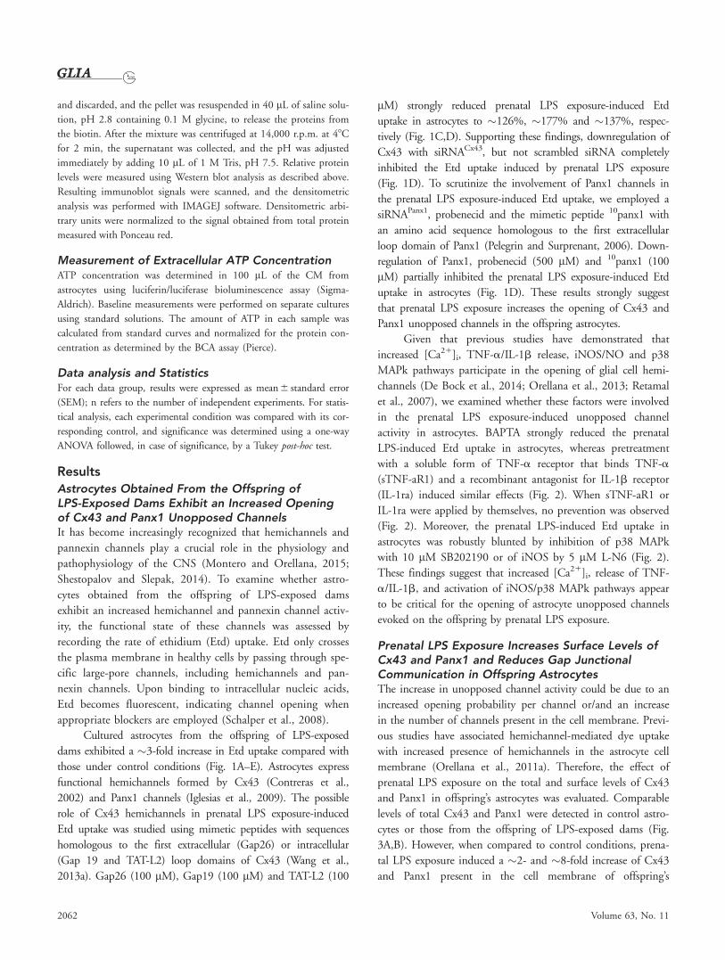
activity in astrocytes. BAPTA strongly reduced the prenatal
LPS-induced Etd uptake in astrocytes, whereas pretreatment
with a soluble form of TNF-a receptor that binds TNF-a
(sTNF-aR1) and a recombinant antagonist for IL-1b receptor
(IL-1ra) induced similar effects (Fig. 2). When sTNF-aR1 or
IL-1ra were applied by themselves, no prevention was observed
(Fig. 2). Moreover, the prenatal LPS-induced Etd uptake in
astrocytes was robustly blunted by inhibition of p38 MAPk
with 10 lM SB202190 or of iNOS by 5 lM L-N6 (Fig. 2).
These findings suggest that increased [Ca21]i, release of TNF-
a/IL-1b, and activation of iNOS/p38 MAPk pathways appear
to be critical for the opening of astrocyte unopposed channels
evoked on the offspring by prenatal LPS exposure.
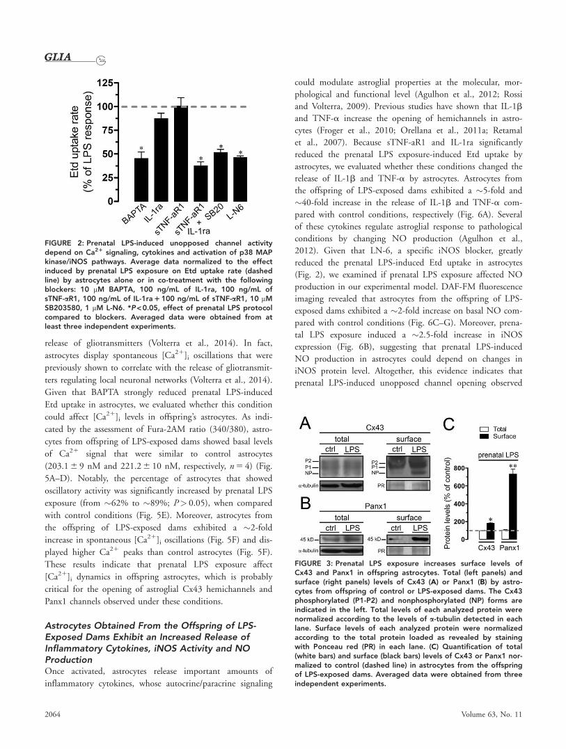
Prenatal LPS Exposure Increases Surface Levels ofCx43 and Panx1 and Reduces Gap JunctionalCommunication in Offspring AstrocytesThe increase in unopposed channel activity could be due to an
increased opening probability per channel or/and an increase
in the number of channels present in the cell membrane. Previ-
ous studies have associated hemichannel-mediated dye uptake
with increased presence of hemichannels in the astrocyte cell
membrane (Orellana et al., 2011a). Therefore, the effect of
prenatal LPS exposure on the total and surface levels of Cx43
and Panx1 in offspring’s astrocytes was evaluated. Comparable
levels of total Cx43 and Panx1 were detected in control astro-
cytes or those from the offspring of LPS-exposed dams (Fig.
3A,B). However, when compared to control conditions, prena-
tal LPS exposure induced a �2- and �8-fold increase of Cx43
and Panx1 present in the cell membrane of offspring’s
2062 Volume 63, No. 11

astrocytes, respectively (Fig. 3A–C). These findings support the
idea that part of the increased Etd uptake evoked by prenatal
LPS exposure in offspring’s astrocytes could be linked to an
increase of membrane unopposed channels.
In the CNS, gap junctional coupling mediated by Cx43
plays important roles in the propagation of intercellular Ca21
waves, and neurotransmitter and ionic homeostasis, thus help-
ing to ensure proper neuronal function (De Bock et al., 2014).
In view of previous studies that have shown that increased
hemichannel activity evoked by inflammatory conditions is
accompanied with a reduction in dye coupling of astrocytes
(Retamal et al., 2007), we investigated whether astroglial cell
coupling was affected by prenatal LPS exposure. As previously
reported (Giaume et al., 1991; Dermietzel et al., 1991), con-
trol astrocytes exhibit a high LY intercellular diffusion (Fig.
4A). However, astrocytes from the offspring of LPS-exposed
dams showed a �50% decrease in LY diffusion when com-
pared with those under control conditions (Fig. 4B,C). Given
that retrieval of gap junction channels from the plasma mem-
brane is a mechanism that can lead to cellular uncoupling, we
investigated whether LPS exposure-induced astroglial uncou-
pling was associated with changes in the cell distribution of
Cx43. In control astrocytes, Cx43 was detected as fine granules
dispersed in cellular interfaces (Fig. 4D,E), whereas in those
from offspring of LPS-exposed dams, it was localized mostly at
intracellular vesicle-like structures of irregular size, and signifi-
cantly reduced and disorganized at cell membrane appositions
(Fig. 4F,G). These results suggest that cell-to-cell uncoupling
evoked by prenatal LPS exposure may be related to a change
in the cell distribution of Cx43 in astrocytes.
Astrocytes Obtained From the Offspring of LPS-Exposed Dams Display Enhanced Spontaneous[Ca21]i OscillationsRecent evidence indicate that a moderate rise (>500 nM) of
[Ca21]i promotes Cx43 hemichannel opening (Wang et al.,
2013a), and a similar effect has been described for Panx1
hemichannels (Locovei et al., 2006). Different studies have
shown that [Ca21]i dynamics play a critical role on astroglial
activation, serving as a highly sensitive system to mediate
FIGURE 1: Increased Etd uptake induced by prenatal LPS exposure is mediated by Cx43 and Panx1 unopposed channels in offspring’sastrocytes. (A–C) Representative fluorescence images depicting nuclear Etd staining (red) in dye uptake experiments (10 min exposure)by astrocytes from offspring of control (A) and LPS-exposed dams alone (B) or with Gap19 (mimetic peptide that blocks Cx43 hemichan-nels, added during recordings) (C). (D) Time-lapse measurements of Etd uptake by astrocytes from offspring of control (white circles)and LPS-exposed dams alone (black circles) or plus Gap19 added during recordings (gray circles). (E) Averaged Etd uptake rate normal-ized with control condition (dashed line) by astrocytes from offspring of LPS-exposed dams alone or in combination with the followingblockers: 100 mM Gap26, 100 mM Gap19, 100 mM TAT-L2, 100 mM 10panx1, 500 mM Probenecid (Prob), siRNACx43, siRNAPanx1; and siR-NAscrb. ***P < 0.001, prenatal LPS protocol compared to control; #P < 0.05, ##P < 0.005, effect of prenatal LPS protocol compared toblockers. Data correspond to at least three independent experiments. Scale bar 5 100 mm. [Color figure can be viewed in the onlineissue, which is available at wileyonlinelibrary.com.]
Avenda~no et al.: Prenatal LPS Open Astroglial Hemichannels
November 2015 2063
release of gliotransmitters (Volterra et al., 2014). In fact,
astrocytes display spontaneous [Ca21]i oscillations that were
previously shown to correlate with the release of gliotransmit-
ters regulating local neuronal networks (Volterra et al., 2014).
Given that BAPTA strongly reduced prenatal LPS-induced
Etd uptake in astrocytes, we evaluated whether this condition
could affect [Ca21]i levels in offspring’s astrocytes. As indi-
cated by the assessment of Fura-2AM ratio (340/380), astro-
cytes from offspring of LPS-exposed dams showed basal levels
of Ca21 signal that were similar to control astrocytes
(203.1 6 9 nM and 221.2 6 10 nM, respectively, n 5 4) (Fig.
5A–D). Notably, the percentage of astrocytes that showed
oscillatory activity was significantly increased by prenatal LPS
exposure (from �62% to �89%; P> 0.05), when compared
with control conditions (Fig. 5E). Moreover, astrocytes from
the offspring of LPS-exposed dams exhibited a �2-fold
increase in spontaneous [Ca21]i oscillations (Fig. 5F) and dis-
played higher Ca21 peaks than control astrocytes (Fig. 5F).
These results indicate that prenatal LPS exposure affect
[Ca21]i dynamics in offspring astrocytes, which is probably
critical for the opening of astroglial Cx43 hemichannels and
Panx1 channels observed under these conditions.
Astrocytes Obtained From the Offspring of LPS-Exposed Dams Exhibit an Increased Release ofInflammatory Cytokines, iNOS Activity and NOProductionOnce activated, astrocytes release important amounts of
inflammatory cytokines, whose autocrine/paracrine signaling
could modulate astroglial properties at the molecular, mor-
phological and functional level (Agulhon et al., 2012; Rossi
and Volterra, 2009). Previous studies have shown that IL-1b
and TNF-a increase the opening of hemichannels in astro-
cytes (Froger et al., 2010; Orellana et al., 2011a; Retamal
et al., 2007). Because sTNF-aR1 and IL-1ra significantly
reduced the prenatal LPS exposure-induced Etd uptake by
astrocytes, we evaluated whether these conditions changed the
release of IL-1b and TNF-a by astrocytes. Astrocytes from
the offspring of LPS-exposed dams exhibited a �5-fold and
�40-fold increase in the release of IL-1b and TNF-a com-
pared with control conditions, respectively (Fig. 6A). Several
of these cytokines regulate astroglial response to pathological
conditions by changing NO production (Agulhon et al.,
2012). Given that LN-6, a specific iNOS blocker, greatly
reduced the prenatal LPS-induced Etd uptake in astrocytes
(Fig. 2), we examined if prenatal LPS exposure affected NO
production in our experimental model. DAF-FM fluorescence
imaging revealed that astrocytes from the offspring of LPS-
exposed dams exhibited a �2-fold increase on basal NO com-
pared with control conditions (Fig. 6C–G). Moreover, prena-
tal LPS exposure induced a �2.5-fold increase in iNOS
expression (Fig. 6B), suggesting that prenatal LPS-induced
NO production in astrocytes could depend on changes in
iNOS protein level. Altogether, this evidence indicates that
prenatal LPS-induced unopposed channel opening observed
FIGURE 2: Prenatal LPS-induced unopposed channel activitydepend on Ca21 signaling, cytokines and activation of p38 MAPkinase/iNOS pathways. Average data normalized to the effectinduced by prenatal LPS exposure on Etd uptake rate (dashedline) by astrocytes alone or in co-treatment with the followingblockers: 10 mM BAPTA, 100 ng/mL of IL-1ra, 100 ng/mL ofsTNF-aR1, 100 ng/mL of IL-1ra 1 100 ng/mL of sTNF-aR1, 10 mMSB203580, 1 mM L-N6. *P < 0.05, effect of prenatal LPS protocolcompared to blockers. Averaged data were obtained from atleast three independent experiments.
FIGURE 3: Prenatal LPS exposure increases surface levels ofCx43 and Panx1 in offspring astrocytes. Total (left panels) andsurface (right panels) levels of Cx43 (A) or Panx1 (B) by astro-cytes from offspring of control or LPS-exposed dams. The Cx43phosphorylated (P1-P2) and nonphosphorylated (NP) forms areindicated in the left. Total levels of each analyzed protein werenormalized according to the levels of a-tubulin detected in eachlane. Surface levels of each analyzed protein were normalizedaccording to the total protein loaded as revealed by stainingwith Ponceau red (PR) in each lane. (C) Quantification of total(white bars) and surface (black bars) levels of Cx43 or Panx1 nor-malized to control (dashed line) in astrocytes from the offspringof LPS-exposed dams. Averaged data were obtained from threeindependent experiments.
2064 Volume 63, No. 11

in astrocytes could be triggered through an autocrine/para-
crine effect evoked by inflammatory cytokines and NO.
Prenatal LPS Exposure Induces Cx43 and Panx1-Dependent Release of ATP in Offspring AstrocytesAstrocytes exposed to inflammatory conditions show an
increased hemichannel-dependent release of the gliotransmitter
ATP (Orellana et al., 2011a,2011b; Wei et al., 2014). Accord-
ingly, we examined whether prenatal LPS-induced unopposed
channel activity observed in astrocytes could be associated with
an increased release of ATP. As shown by ATP measurement
with the luciferin/luciferase bioluminescence assay, astrocytes
from the offspring of LPS-exposed dams exhibited a �4.5-fold
increase on ATP release compared with the control condition
(Fig. 7). Gap19 (100 mM) and TAT-L2 (100 mM) abolished
the prenatal LPS-induced ATP release in astrocytes (Fig. 7). In
agreement with these findings, downregulation of Cx43 by siR-
NACx43 completely inhibited the ATP release induced by pre-
natal LPS exposure (Fig. 7). In addition, the mimetic peptide10panx1 (100 mM), probenecid (500 mM) and siRNAPanx1 par-
tially inhibited prenatal LPS-induced ATP release by astrocytes
(Fig. 7). The evidence indicates that prenatal LPS exposure
increased the release of ATP by opening of Cx43 and Panx1
unopposed channels in offspring astrocytes.
Recently, it has been demonstrated that ATP elicits its
own release in an autocrine manner via P2 receptors and acti-
vation of Cx43 and Panx1 unopposed channels (Orellana
et al., 2012a; Orellana et al., 2013). We found that 10 mM
MRS2179, a P2Y1 receptor blocker, fully abolished the release
of ATP induced by prenatal LPS exposure (Fig. 7). The
FIGURE 4: Prenatal LPS exposure reduces gap junctional communication and affect cellular distribution of Cx43 in offspring astrocytes. (A–B) Representative fluorescence micrographs of SL/DT with LY by offspring’s astrocytes of control (A) or LPS-exposed (B) dams. (C) Averageddata normalized to control (dashed line) of SL/DT with LY by astrocytes from offspring of LPS-exposed dams. * P < 0.05, prenatal LPS proto-col compared to control. Averaged data were obtained from three independent experiments. Yellow scale bar 5 250 mm. (D–G)Representative confocal images depicting Cx43 (green), GFAP (red) and DAPI (blue, epifluorescence) immunolabeling by astrocytes fromthe offspring of control (D–E) or LPS-exposed (F–G) dams. Insets: 2.5X magnification of the indicated area of panels E and G. CalibrationBars: white 5 70 mm and green= 15 mm. [Color figure can be viewed in the online issue, which is available at wileyonlinelibrary.com.]
Avenda~no et al.: Prenatal LPS Open Astroglial Hemichannels
November 2015 2065

involvement of P2X7 receptors on the prenatal LPS-induced
ATP release was confirmed by the inhibitory effect induced
by 200 mM oATP, a general P2X receptor blocker, or 10 mM
A740003 and 10 mM BBG, both P2X7 receptor blockers
(Fig. 7). These results suggest that activation of both P2Y1
and P2X7 receptors lead to the opening of Cx43 and Panx1
unopposed channels, resulting in further release of ATP.
Astroglial Cx43 and Panx1 Unopposed ChannelOpening Induced by Prenatal LPS Exposure Lead toNeuronal DeathBecause hemichannel-mediated release of ATP occurs in acti-
vated astrocytes, promoting neuronal damage (Orellana et al.,
2011a,2011b), we examined whether increased astroglial
unopposed channel activity induced by prenatal LPS exposure
could affect neurons through a paracrine pathway. Therefore,
enriched neuron cultures were incubated for 3 h with condi-
tioned medium (CM) of astrocytes from the offspring of
LPS-exposed or control dams. Under control conditions, less
than �2% of 12 day-old MAP-2 and NeuN positive neurons
were labeled with F-Jade (Fig. 8A,D,G), a marker of neurode-
generation and neuronal death (Noraberg et al., 1999;
Schmuck and Kahl, 2009). After treatment with CM of astro-
cytes from prenatal LPS-exposed dams, neuronal death
showed a 15-fold increase, affecting �43% of neurons (Fig.
8B,E,G). In contrast, CM of astrocytes from control dams
did not affect neuronal survival, supporting the idea that
FIGURE 5: Prenatal LPS exposure increases spontaneous Ca21
dynamics by astrocytes from the offspring. (A–D) Representativefluorescence micrographs and plots of basal Fura-2AM ratio(pseudo-colored scale) over time by astrocytes from the offspringof control (A and B) and LPS-exposed dams (C and D). Representa-tive plots of relative changes in [Ca21]i over time of cells depictedin panel A and C are shown. (E) Averaged data of the percentageof oscillatory astrocytes from the offspring of control (white bar)and LPS-exposed dams (black bar). (F) Averaged data of oscillationfrequency by astrocytes from the offspring of control (white bar)and LPS-exposed dams (black bar). (G) Averaged data of ampli-tude peak normalized to basal Fura-2AM ratio by astrocytes fromthe offspring of control (white bar) and LPS-exposed dams (blackbar). *P < 0.05, prenatal LPS protocol compared to control.Averaged data were obtained from at least three independentexperiments. Scale bar 5 120 mm. [Color figure can be viewed inthe online issue, which is available at wileyonlinelibrary.com.]
FIGURE 6: Prenatal LPS exposure increases the release of IL-1band TNF-a and NO production by astrocytes from the offspring.(A) Averaged data of levels of IL-1b and TNF-a released by astro-cytes from the offspring of control (white bar) and LPS-exposeddams (black bar). (B–E) Representative fluorescence micrographsof basal NO production (DAF-FM, green and pseudo-coloredscale) by astrocytes from the offspring of control (B and D) andLPS-exposed dams (C and E). (F) Average of DAF-FM fluorescenceby astrocytes from the offspring of control (white bar) and LPS-exposed dams (black bar). (G) Quantification of protein level ofiNOS by astrocytes from the offspring of control (white bar, lane1)and LPS-exposed dams (black bar, lane 2). Protein expression wasnormalized by the corresponding level of a-tubulin. *P < 0.05,**P < 0.005; prenatal LPS protocol compared to control. Averageddata were obtained from at least three independent experiments.Scale bar 5 150 mm. [Color figure can be viewed in the onlineissue, which is available at wileyonlinelibrary.com.]
2066 Volume 63, No. 11

prenatal LPS-exposure could potentiate neurotoxic activation
of astrocytes (Fig. 8G). Neuronal death was associated with
focal beadlike swellings of dendrites and axons (neuritic bead-
ing), which has been proposed as an early pathological feature
of neuronal cell dysfunction (Fig. 8B) (Takeuchi et al., 2005).
To elucidate the contribution of ATP released from astrocytes
in the CM-induced neuronal death, we employed blockers of
P2Y1 (MRS2179) and P2X7 receptors (BBG, oATP,
A740003). MRS2179 (10 mM) failed on inhibiting CM-
induced neuronal death (Fig. 8G), whereas BBG (10 mM),
oATP (200 mM) and A740003 (10 mM) abolished neurotox-
icity, reducing neuronal death from �43% to �2% in all
cases (Fig. 8C,F,G). These findings imply that ATP released
by astrocytes into the CM and its action on neuronal P2X7
receptors is part of the mechanism that decrease neuronal sur-
vival in our experimental model. Because activation of neuro-
nal purinergic receptors and further activation of Panx1
channels lead to neuronal death (Orellana et al.,
2011a,2011b), we inhibited neuronal Panx1 channels with10panx1 and probenecid to examine their contribution to the
CM-induced neuronal death. 10panx1 and probenecid also
abolished CM-induced neuronal death (F-Jade1 cells were
reduced from �43% to �2%, Fig. 8G), indicating that ATP
released from astrocytes acted on P2X7 receptor and induced
further opening of neuronal Panx1 channels. Notably, when
CM were obtained from astrocytes exposed to TAT-L2 or
Gap19, the induction of neuronal death was abolished (Fig.
8G). Similar findings were obtained in neurons treated with
CM from siRNACx43-treated astrocytes (Fig. 8G), whereas
only a partial reduction in death was observed with CM from
astrocytes treated with 10panx1, probenecid or siRNAPanx1
(from �43% to �15%, �14% and �14.5%, respectively)
(Fig. 8G). The above data suggest that unopposed channel
opening and neuronal death was due to the Cx43 and Panx1-
dependent release of ATP by astrocytes obtained from the off-
spring of LPS-exposed dams.
Discussion
Despite that previous studies have demonstrated that prenatal
inflammation increases astrogliosis and GFAP expression in
the offspring (Hao et al., 2010; Samuelsson et al., 2006),
whether astrocytes contribute to brain dysfunction induced by
the latter condition has remained unknown (Gilmore and Jar-
skog, 1997). Our model, consisting in a single LPS injection
during pregnancy, increased the production of inflammatory
mediators (cytokines and NO) and altered intracellular Ca21
dynamics in offspring astrocytes. Due to these functional
changes, astrocytes exhibited an enhanced p38MAPK/iNOS-
dependent release of ATP via astroglial cell Cx43 and Panx1
unopposed channels, which in consequence caused the
impairment of neuronal survival.
As assayed by Etd uptake experiments, prenatal LPS-
induced unopposed channel activity was due to Cx43 and
Panx1, since it did not occur when astrocytes were treated
with siRNAs that downregulated both proteins. Second, drugs
and mimetic peptides known to block Cx43 or Panx1 unop-
posed channels significantly inhibited the above response.
How does LPS exposure induce the opening of Cx43 and
Panx1 unopposed channels in astrocytes? It is broadly known
that maternal environment and physiological status during
pregnancy could be crucial in the inflammatory balance and
immunity response in the offspring (Boksa, 2010). At one
end, prenatal LPS-induced inflammatory conditions may lead
to a long-lasting activation of glial cells and thereof a long-
term production of inflammatory mediators, including IL-1b
and TNF-a (Fig. 9). Indeed, both cytokines are upregulated
in the offspring’s brain of LPS-exposed dams (Boksa, 2010)
and their production has been involved in the opening of
astroglial hemichannels (Retamal et al., 2007). Supporting
this evidence, we found that prenatal LPS exposure increases
the astroglial production of IL-1b and TNF-a, whereas inhi-
bition of IL-1b/TNF-a signaling significantly blunted the
prenatal LPS-induced unopposed channel opening in off-
spring astrocytes. These inflammatory cytokines likely trig-
gered by a p38 MAPk/iNOS-dependent S-nitrosylation of
Cx43, resulting in further opening of astroglial hemichannels,
as has been previously described (Froger et al., 2010; Orellana
et al., 2011a; Retamal et al., 2006, 2007) (Fig. 9). In agree-
ment with this idea, we observed that prenatal LPS-induced
FIGURE 7: Prenatal LPS exposure increases the unopposedchannel-dependent release of ATP by astrocytes from the off-spring. Averaged data of ATP release by astrocytes from the off-spring of control (white bar) and LPS-exposed dams alone (blackbars) or in combination with the following blockers: 100 mM TAT-L2, 100 mM Gap19, siRNACx43, 100 mM 10panx1, 500 mM Probene-cid (Prob), siRNAPanx1; and siRNAscrb, 10 mM MRS2179, 10 mM(BBG), 200 mM oxidized ATP (oATP) and 10 mM A740003. *P < 0.05, prenatal LPS protocol compared to control; # P < 0.05; ##
P < 0.005, effect of prenatal LPS protocol compared to blockers.Averaged data were obtained from at least three independentexperiments.
Avenda~no et al.: Prenatal LPS Open Astroglial Hemichannels
November 2015 2067

astroglial Etd uptake was strongly reduced by inhibiting p38
MAPk or iNOS. Moreover, prenatal LPS exposure increased
protein levels of iNOS and NO production in offspring
astrocytes, revealing their activated state. Given that S-
nitrosylation inhibits the opening of Panx1 channels (Lohman
et al., 2012), their activation possibly took place by a differ-
ent manner.
How does unopposed channel opening evoked by pre-
natal LPS exposure affect intercellular communication among
astrocytes? ATP is considered to be an essential transmitter
for the communication among astrocytes, and can be released
through membrane channels and vesicles (Fields and Burn-
stock, 2006). Here, we demonstrated that prenatal LPS expo-
sure triggered the release of ATP from astrocytes via Cx43
FIGURE 8: Prenatal LPS-induced release of ATP via astroglial Cx43 and Panx1 unopposed channels trigger neuronal death by activationof neuronal P2X7 receptors and Panx1 channels. (A–F) Representative immunofluorescence images depicting MAP-2 (red), NeuN (red) orF-Jade (green) labeling of neurons under control conditions (A and D), treated for 3 h with CM harvested from astrocytes (CM-Ast) ofprenatal LPS protocol alone (B and E) or with CM-Ast of prenatal LPS protocol plus 10 lM A740003 (C and F). The respective bottominsets of representative with staining for MAP2, NeuN and F-Jade are also shown in B and E. (G) Averaged data of neuronal death aspercent of neurons positive to F-Jade staining under control conditions (white bar), treated for 3 h with CM-Ast of control conditions(red bar) or prenatal LPS protocol alone (blue bars) or in combination with the following blockers: 10 mM MRS2179, 10 mM (BBG), 200mM oxidized ATP (oATP), 10 mM A740003, 100 mM 10panx1, 500 mM Probenecid. In some experiments, the effect on neuronal death ofCM-Ast of prenatal LPS protocol made in the presence of 100 mM TAT-L2, 100 mM Gap19, siRNACx43, 100 mM 10panx1, 500 mM Probene-cid (Prob), or siRNAPanx1 was studied (*, green bars). **P < 0.005, CM-Ast of prenatal LPS protocol compared to control; #P < 0.05;##P < 0.005, effect of CM-Ast prenatal LPS protocol compared to blockers. Averaged data were obtained from at least three independ-ent experiments. Scale bar 5 150 mm. [Color figure can be viewed in the online issue, which is available at wileyonlinelibrary.com.]
2068 Volume 63, No. 11

and Panx1 unopposed channels and depended on P2Y1 and
P2X7 receptors (Fig. 9). In fact, prenatal LPS exposure-
induced ATP release was not detected after pharmacological
(e.g., drugs, mimetic peptides) or molecular (siRNAs) inhibi-
tion of Cx43/Panx1 unopposed channels and P2Y1/P2X7
receptors. In agreement with our observations, recent reports
have shown that ATP induces its own release via hemichan-
nels and subsequent activation of purinergic receptors (Orel-
lana et al., 2012a; Orellana et al., 2013). In our system,
autocrine/paracrine release of ATP could control unopposed
channel opening as a secondary mechanism to that exerted by
p38 MAPk and NO production (Fig. 9). The opening of
unopposed channels could take place by protein–protein
interactions with activated P2X7 receptors (Iglesias et al.,
2008) or through increases in [Ca21]i caused by activation of
P2Y1 receptors (Locovei et al., 2006; Orellana et al., 2012a;
Saez et al., 2013). Indeed, previous studies have shown that a
moderate rise in [Ca21]i (>500 nM) evokes Cx43 (Wang
et al., 2013a), as well as Panx1 (Locovei et al., 2006) unop-
posed channel opening. In line with the latter, we observed
that chelation of [Ca21]i blunted the prenatal LPS-induced
unopposed channel opening in offspring astrocytes. Previous
evidence indicates that rises on [Ca21]i increases levels of sur-
face Cx43 hemichannels, enhancing their contribution to the
release of paracrine mediators (Schalper et al., 2008). Rele-
vant to this point, we found that prenatal LPS exposure
increased the amplitude and number of spontaneous [Ca21]i
oscillations in astrocytes, which could explain the increased
levels of surface Cx43 and Panx1 unopposed channels
observed under these conditions. Impaired transmission of
FIGURE 9: Modulation of astroglial Cx43 hemichannels and Panx1 channels by prenatal LPS exposure. Prenatal LPS exposure increasethe release of IL-1b and TNF-a in astrocytes (1), leading to the activation of a p38MAPK/iNOS-dependent pathway and further produc-tion of NO (2). By an unknown mechanism NO evoke the activation of Cx43 and/or Panx1 unopposed channels (3). ATP released viaunopposed channels activates P2X7 and P2Y1 receptors (4 and 5), triggering a self-perpetuating mechanism, in which high levels of[Ca21]i (6 and 7) could reactivate Cx43 and Panx1 unopposed channels in astrocytes (8). In addition, paracrine release of ATP from astro-cytes could act on neighboring or distant neurons, resulting in the activation of P2X7 (9) receptors. The latter increase levels of [Ca21]i,and thereof the activity of neuronal Panx1 channels (10), resulting in neuronal function impairment and cell death. Contrary to the effecton unopposed channel function, prenatal LPS exposure additionally decreased the functional coupling between offspring astrocytes(11). [Color figure can be viewed in the online issue, which is available at wileyonlinelibrary.com.]
Avenda~no et al.: Prenatal LPS Open Astroglial Hemichannels
November 2015 2069

intercellular Ca21 waves and change of Ca21 dynamics often
have been related with altered gliotransmission evoked by
inflammatory conditions in astrocytes (Agulhon et al., 2012).
Part of this communication is mediated by cell-to-cell cou-
pling mediated by gap junction channels. Here, we observed
that in addition to increase unopposed channel opening, pre-
natal LPS exposure also reduced astroglial coupling as meas-
ured by LY diffusion. These data are in agreement with the
proposed idea that hemichannels and gap junction channels
are oppositely regulated during inflammatory conditions
(Orellana et al., 2009). Nevertheless, this phenomenon often
lacks correlate between in vivo and ex vivo models, as previ-
ously shown for the opposite regulation of astroglial hemi-
channels and gap junction channels induced by LPS-
stimulated microglia (Retamal et al., 2007; Abudara et al.,
2015). Whether this could be also the case in our experimen-
tal model is unknown and thereof, further studies are
required to elucidate the biological relevance of this phenom-
enon and the possible impact of other cell types (e.g., neu-
rons) in vivo.
How does unopposed channel opening evoked by prena-
tal LPS exposure affect astrocyte-neuron crosstalk? Having
described that CM harvested from activated astrocytes reduces
neuronal survival by activating Panx1 channels and P2X7
receptors in neurons (Orellana et al., 2011a; Orellana et al.,
2011b); here, we show that ATP released through Cx43 and
Panx1 unopposed channels activated P2X7 receptors and Panx1
channels in neurons, increasing neuronal death. Since activa-
tion of P2X7 receptors lead to opening of Panx1 channels
(Iglesias et al., 2008), it is plausible that CM-induced neuronal
death could be associated with an ionic, osmotic and intracel-
lular Ca21 imbalance evoked by P2X7 receptor and Panx1
channel activation. We propose that ATP released from astro-
cytes could stimulate distant astrocytes and neurons in a para-
crine manner, causing Ca21 responses that could depend on
the inflammatory profile of astrocytes (Fig. 9). If that is cor-
rect, the activation of purinergic receptors could be turned off
in part by diffusion of ATP to distal regions as well as by
desensitization of P2Y1 receptors and degradation of extracellu-
lar ATP by exonucleases. In parallel, an alternative negative
feedback loop is the inhibitory effect that could be exerted by
ATP on Panx1 channels (Qiu and Dahl, 2009). Alternatively,
we cannot rule out that astroglial hemichannel-dependent neu-
ronal death in vivo, could occurs in addition with other direct
effects evoked by astrocytes (e.g., high release of cytokines) that
impact neuronal survival (Boksa, 2010).
Whereas hemichannel and pannexin channel-dependent
release of gliotransmitters underlies crucial functions in the
physiology of the CNS (Montero and Orellana, 2015; Orel-
lana and Stehberg, 2014), their uncontrolled opening could
lead to excitotoxicity (Orellana et al., 2011a,2011b; Takeuchi
et al., 2006). Here, we showed for the first time that prenatal
LPS exposure increases Cx43 and Panx1 unopposed channel
opening in the offspring’s astrocytes. These findings are in
agreement with recent reports showing that pro-inflammatory
conditions increase the opening of astroglial Cx43 hemichan-
nels (Orellana et al., 2011b; Orellana et al., 2014b; Retamal
et al., 2007). In addition, although previous studies have
found that LPS indirectly open Cx43 hemichannels but not
Panx1 channels in astrocytes (Abudara et al., 2015; Retamal
et al., 2007), this study, in agreement with recent reports
(Beckel et al., 2014; Iwabuchi and Kawahara, 2011; Pan
et al., 2015; Santiago et al., 2011; Wei et al., 2014), reveals
that Panx1 can form functional channels in these cells. Addi-
tional studies will be required to determine whether inflam-
matory profile of astrocytes and activity of their unopposed
channels could determine neuronal survival in vivo. Under-
standing of the mechanisms underlying astrocyte-neuron
crosstalk can contribute to the knowledge on the pathways
implicated on neuroinflammation and open novel therapeutic
avenues for ameliorate the brain abnormalities induced by
prenatal inflammation in the offspring.
Acknowledgment
Grant sponsor: FONDECYT 11121133 (to J.A.O.), the
Committee for Aid and Education in Neurochemistry from
the International Society for Neurochemistry (to J.A.O.) and
FONDECYT 1131025 (to R.v.B.).
References
Abudara V, Roux L, Dall�erac G, Matias I, Dulong J, Mothet JP, Rouach N,Giaume C. 2015. Activated microglia impairs neuroglial interaction byopening Cx43 hemichannels in hippocampal astrocytes. Glia 63:795–811.
Agulhon C, Sun MY Murphy T, Myers T, Lauderdale K Fiacco TA. 2012.Calcium Signaling and Gliotransmission in Normal vs. Reactive Astrocytes.Front Pharmacol 3:139–
Araque A, Carmignoto G, Haydon PG, Oliet SH, Robitaille R, Volterra A.2014. Gliotransmitters travel in time and space. Neuron 81:728–739.
Beckel JM, Argall AJ, Lim JC, Xia J, Lu W, Coffey EE, Macarak EJ,Shahidullah M, Delamere NA Zode GS, et al. 2014. Mechanosensitive releaseof adenosine 5’-triphosphate through pannexin channels andmechanosensitive upregulation of pannexin channels in optic nerve headastrocytes: A mechanism for purinergic involvement in chronic strain. Glia 62:1486–1501.
Boksa P. 2010. Effects of prenatal infection on brain development andbehavior: A review of findings from animal models. Brain Behav Immun 24:881–897.
Bosch M, Kielian T. 2014. Hemichannels in neurodegenerative diseases: Isthere a link to pathology? Front Cell Neurosci 8:242-
Contreras JE, S�anchez HA, Eugenin EA, Speidel D, Theis M, Willecke K,Bukauskas FF, Bennett MVL, S�aez JC. 2002. Metabolic inhibition inducesopening of unapposed connexin 43 gap junction hemichannels and reducesgap junctional communication in cortical astrocytes in culture. Proc Natl AcadSci U S A 99:495–500.
2070 Volume 63, No. 11

De Bock M, Decrock E, Wang N, Bol M, Vinken M, Bultynck G, Leybaert L.2014. The dual face of connexin-based astroglial Ca(21) communication: Akey player in brain physiology and a prime target in pathology. Biochim Bio-phys Acta 1843:2211–2232.
Dermietzel R, Hertberg EL, Kessler JA Spray DC. 1991. Gap junctionsbetween cultured astrocytes: Immunocytochemical, molecular, andelectrophysiological analysis. J Neurosci 11:1421–1432.
Fields RD, Burnstock G. 2006. Purinergic signalling in neuron-glia interac-tions. Nat Rev Neurosci 7:423–436.
Froger N, Orellana JA, Calvo CF, Amigou E, Kozoriz MG, Naus CC, Saez JC,Giaume C. 2010. Inhibition of cytokine-induced connexin43 hemichannelactivity in astrocytes is neuroprotective. Mol Cell Neurosci 45:37–46.
Giaume C, Fromaget C, el Aoumari A, Cordier J, Glowinski J, Gros D. 1991.Gap junctions in cultured astrocytes: Single-channel currents and characteri-zation of channel-forming protein. Neuron 6:133–143.
Gilmore JH, Jarskog LF. 1997. Exposure to infection and brain development:Cytokines in the pathogenesis of schizophrenia. Schizophr Res 24:365–367.
Golan HM, Lev V, Hallak M, Sorokin Y, Huleihel M. 2005. Specificneurodevelopmental damage in mice offspring following maternalinflammation during pregnancy. Neuropharmacology 48:903–917.
Grynkiewicz G, Poenie M, Tsien RY. 1985. A new generation of Ca21
indicators with greatly improved fluorescence properties. J Biol Chem 260:3440–3450.
Hao LY, Hao XQ, Li SH, Li XH. 2010. Prenatal exposure to lipopolysaccharideresults in cognitive deficits in age-increasing offspring rats. Neuroscience166:763–770.
Iglesias R, Dahl G, Qiu F, Spray DC, Scemes E. 2009. Pannexin 1: Themolecular substrate of astrocyte "hemichannels". J Neurosci 29:7092–7097.
Iglesias R, Locovei S, Roque A, Alberto AP, Dahl G, Spray DC, Scemes E.2008. P2X7 receptor-Pannexin1 complex: Pharmacology and signaling. Am JPhysiol Cell Physiol 295:C752–C760.
Iwabuchi S, Kawahara K. 2011. Functional significance of the negative-feedback regulation of ATP release via pannexin-1 hemichannels under ische-mic stress in astrocytes. Neurochem Int 58:376–384.
Locovei S, Wang J, Dahl G. 2006. Activation of pannexin 1 channels by ATPthrough P2Y receptors and by cytoplasmic calcium. FEBS Lett 580:239–244.
Lohman AW, Weaver JL, Billaud M, Sandilos JK, Griffiths R, Straub AC,Penuela S, Leitinger N, Laird DW Bayliss DA, et al. 2012. S-nitrosylationinhibits pannexin 1 channel function. J Biol Chem 287:39602–39612.
MacVicar BA, Thompson RJ. 2010. Non-junction functions of pannexin-1channels. Trend Neurosci 33:93–102.
Meyer U, Nyffeler M, Engler A, Urwyler A, Schedlowski M, Knuesel I, Yee BK,Feldon J. 2006. The time of prenatal immune challenge determines thespecificity of inflammation-mediated brain and behavioral pathology.J Neurosci 26:4752–4762.
Montero TD, Orellana JA. 2015. Hemichannels: New pathways forgliotransmitter release. Neuroscience 286C:45–59.
Noraberg J, Kristensen BW, Zimmer J. 1999. Markers for neuronaldegeneration in organotypic slice cultures. Brain Res Brain Res Protoc 3:278–290.
Orellana JA, Busso D, Ramirez G, Campos M, Rigotti A, Eugenin J, vonBernhardi R. 2014a. Prenatal nicotine exposure enhances Cx43 and Panx1unopposed channel activity in brain cells of adult offspring mice fed a high-fat/cholesterol diet. Front Cell Neurosci 8:403-
Orellana JA, Froger N, Ezan P, Jiang JX, Bennett MV, Naus CC, Giaume C,Saez JC. 2011a. ATP and glutamate released via astroglial connexin 43hemichannels mediate neuronal death through activation of pannexin 1hemichannels. J Neurochem 118:826–840.
Orellana JA, Montero TD, von Bernhardi R. 2013. Astrocytes inhibit nitricoxide-dependent Ca(21) dynamics in activated microglia: Involvement ofATP released via pannexin 1 channels. Glia 61:2023–2037.
Orellana JA, Saez JC, Bennett MV, Berman JW, Morgello S, Eugenin EA.2014b. HIV increases the release of dickkopf-1 protein from human astrocytesby a Cx43 hemichannel-dependent mechanism. J Neurochem 128:752–763.
Orellana JA, S�aez PJ, Cort�es-campos C, Elizondo RJ, Shoji KF, Contreras-Duarte S, Figueroa V, Velarde V, Jiang JX Nualart F, et al. 2012a. Glucoseincreases intracellular free Ca(21) in tanycytes via ATP released throughconnexin 43 hemichannels. Glia 60:53–68.
Orellana JA, S�aez PJ, Shoji KF, Schalper KA, Palacios-Prado N, Velarde V,Giaume C, Bennett MV, S�aez JC. 2009. Modulation of brain hemichannelsand gap junction channels by pro-inflammatory agents and their possiblerole in neurodegeneration. Antioxid Redox Signal 11:369–399.
Orellana JA, Shoji KF, Abudara V, Ezan P, Amigou E, Saez PJ, Jiang JX,Naus CC, Saez JC, Giaume C. 2011b. Amyloid beta-induced death in neu-rons involves glial and neuronal hemichannels. J Neurosci 31:4962–4977.
Orellana JA, Stehberg J. 2014. Hemichannels: New roles in astroglialfunction. Front Physiol 5:193-
Orellana JA, von Bernhardi R, Giaume C, Saez JC. 2012b. Glial hemichannelsand their involvement in aging and neurodegenerative diseases. RevNeurosci 23:163–177.
Pan HC, Chou YC, Sun SH. 2015. P2X7 R-mediated Ca(21) -independent d-serine release via pannexin-1 of the P2X7 R-pannexin-1 complex in astro-cytes. Glia 63:877–893.
Pelegrin P, Surprenant A. 2006. Pannexin-1 mediates large pore formationand interleukin-1beta release by the ATP-gated P2X7 receptor. EMBO J 25:5071–5082.
Penuela S, Harland L, Simek J, Laird DW. 2014. Pannexin channels and theirlinks to human disease. Biochem J 461:371–381.
Qiu F, Dahl G. 2009. A permeant regulating its permeation pore: Inhibitionof pannexin 1 channels by ATP. Am J Physiol Cell Physiol 296:C250–C255.
Retamal MA, Cortes CJ, Reuss L, Bennett MV, Saez JC. 2006. S-nitrosylationand permeation through connexin 43 hemichannels in astrocytes: Inductionby oxidant stress and reversal by reducing agents. Proc Natl Acad Sci U S A103:4475–4480.
Retamal MA, Froger N, Palacios-Prado N, Ezan P, Saez PJ, Saez JC, GiaumeC. 2007. Cx43 hemichannels and gap junction channels in astrocytes areregulated oppositely by proinflammatory cytokines released from activatedmicroglia. J Neurosci 27:13781–13792.
Rossi D, Volterra A. 2009. Astrocytic dysfunction: Insights on the role inneurodegeneration. Brain Res Bull 80:224–232.
Rousset CI, Chalon S, Cantagrel S, Bodard S, Andres C, Gressens P, Saliba E.2006. Maternal exposure to LPS induces hypomyelination in the internalcapsule and programmed cell death in the deep gray matter in newborn rats.Pediatr Res 59:428–433.
Saez PJ, Shoji KF, Retamal MA, Harcha PA, Ramirez G, Jiang JX, vonBernhardi R, Saez JC. 2013. ATP is required and advances cytokine-inducedgap junction formation in microglia in vitro. Mediators Inflamm 2013:216402-
Santiago MF, Veliskova J, Patel NK, Lutz SE, Caille D, Charollais A, Meda P,Scemes E. 2011. Targeting pannexin1 improves seizure outcome. PLoS One6:e25178-
Samuelsson AM, Jennische E, Hansson HA Holmang A. 2006. Prenatalexposure to interleukin-6 results in inflammatory neurodegeneration in hippo-campus with NMDA/GABA(A) dysregulation and impaired spatial learning.Am J Physiol Regul Integr Comp Physiol 290:R1345–R1356.
Schalper KA, Palacios-Prado N, Orellana JA, Saez JC. 2008. Currently usedmethods for identification and characterization of hemichannels. CellCommun Adhes 15:207–218.
Schmuck G, Kahl R. 2009. The use of Fluoro-Jade in primary neuronal cellcultures. Arch Toxicol 83:397–403.
Shestopalov VI, Slepak VZ. 2014. Molecular pathways of pannexin1-mediatedneurotoxicity. Front Physiol 5:23-
Takeuchi H, Jin S, Wang J, Zhang G, Kawanokuchi J, Kuno R, Sonobe Y,Mizuno T, Suzumura A. 2006. Tumor necrosis factor-alpha induces
Avenda~no et al.: Prenatal LPS Open Astroglial Hemichannels
November 2015 2071

neurotoxicity via glutamate release from hemichannels of activated microgliain an autocrine manner. J Biol Chem 281:21362–21368.
Takeuchi H, Mizuno T, Zhang G, Wang J, Kawanokuchi J, Kuno R, SuzumuraA. 2005. Neuritic beading induced by activated microglia is an early featureof neuronal dysfunction toward neuronal death by inhibition of mitochondrialrespiration and axonal transport. J Biol Chem 280:10444–10454.
Verkhratsky A, Parpura V, Pekna M, Pekny M, Sofroniew M. 2014. Glia in thepathogenesis of neurodegenerative diseases. Biochem Soc Trans 42:1291–1301.
Volterra A, Liaudet N, Savtchouk I. 2014. Astrocyte Ca(2)(1) signalling: Anunexpected complexity. Nat Rev Neurosci 15:327–335.
Wang N, De Bock M, Decrock E, Bol M, Gadicherla A, Bultynck G, LeybaertL. 2013a. Connexin targeting peptides as inhibitors of voltage- andintracellular Ca21-triggered Cx43 hemichannel opening. Neuropharmacology75:506–516.
Wang N, De Bock M, Decrock E, Bol M, Gadicherla A, Vinken M, RogiersV, Bukauskas FF, Bultynck G, Leybaert L. 2013b. Paracrine signalingthrough plasma membrane hemichannels. Biochim Biophys Acta 1828:35–50.
Wei H, Deng F, Chen Y, Qin Y, Hao Y, Guo X. 2014. Ultrafine carbon blackinduces glutamate and ATP release by activating connexin and pannexinhemichannels in cultured astrocytes. Toxicology 323:32–41.
2072 Volume 63, No. 11