presenilin-1 acts via id1 to regulate the function of
TRANSCRIPT
4427Research Article
IntroductionMuscle satellite cells are myogenic stem cells that are locatedbetween the basal lamina and the plasmalemma of myofibers(Mauro, 1961). They are necessary for postnatal muscle growth,and are responsible for maintenance, hypertrophy and repair of adultskeletal muscle. It has been shown that satellite cells are able toself-renew to maintain their population (Collins et al., 2005) andmuch work is currently directed at understanding how self-renewalis regulated (reviewed by Zammit, 2008).
The paired-box transcription factor Pax7 is expressed byquiescent satellite cells and is implicated in the generation ofcommitted myogenic progenitors, but its role in the regulation ofsatellite cell self-renewal is in debate (Lepper et al., 2009;McKinnell et al., 2008; Olguin et al., 2007; Seale et al., 2000;Zammit et al., 2006). Following activation, satellite cells co-expressPax7 with MyoD [a member of the myogenic regulatory factorfamily, together with Myf5, Mrf4 and Myog (myogenin)] andproliferate. Later, satellite-cell-derived myoblasts eitherdownregulate Pax7, maintain MyoD and induce Myog as theyundergo myogenic differentiation, or they downregulate MyoD andmaintain Pax7, returning to a quiescent-like state (Halevy et al.,2004; Zammit et al., 2004). Interestingly, the total number ofsatellite cells in adult muscles remains relatively constant afterrepeated muscle injury and regeneration, indicating that the self-renewal system of satellite cells is carefully coordinated (Collinset al., 2005; Yoshida et al., 1998; Zammit et al., 2004). However,the molecular mechanism of satellite cell self-renewal remainspoorly understood, although recent advances have shown thatNotch and canonical Wnt signalling play a role (Brack et al., 2008;
Conboy and Rando, 2002; Kitzmann et al., 2006; Kuang et al.,2007; Perez-Ruiz et al., 2008).
Notch signalling controls many events, including differentiation,proliferation and apoptosis in various tissues (Hansson et al., 2004).In skeletal muscle, the Notch-signalling pathway is involved inactivation, and proliferation of muscle satellite cells, and has beenimplicated in their self-renewal (Conboy et al., 2003; Conboy andRando, 2002; Kitzmann et al., 2006; Kopan et al., 1994; Kuang etal., 2007; Nofziger et al., 1999; Ono et al., 2007). For example,inhibition of Notch activity enhances myogenic differentiation ofmurine and human myoblasts (Kitzmann et al., 2006; Kuang et al.,2007). Notch is activated by binding of members of the delta-likeand Jagged families (in mammals) to its extracellular domain, whichresults in -secretase-mediated cleavage to release the Notchintracellular domain (Notch ICD) (Herreman et al., 2000; Struhland Greenwald, 1999; Zhang et al., 2000). The Notch ICDtranslocates into the nucleus, where it interacts with the DNA-binding protein CSL/RBP-J (RBP-J is a member of CSL family ofproteins) to regulate the transcription of target genes such as Hes1(Jarriault et al., 1995).
Together with nicastrin, Pen-2 and Aph-1, the other crucialcomponent of the -secretase complex is presenilin (reviewed byDe Strooper, 2003). Presenilin-1 (PS1) and presenilin-2 (PS2) aremembrane proteins that function as the catalytic subunit of the -secretase complex, an intramembrane protease with a number ofsubstrates of the type I membrane protein family (De Strooper etal., 1999) (reviewed by Parks and Curtis, 2007; Vetrivel et al., 2006).For example, in addition to cleavage of activated Notch, -secretasetargets also include, but are not limited to, amyloid precursor protein
Muscle satellite cells are the resident stem cells of adult skeletalmuscle. Here, we have examined the role of the multifunctionalprotein presenilin-1 (PS1) in satellite cell function. PS1 acts asa crucial component of the -secretase complex, which isrequired to cleave single-pass transmembrane proteins such asNotch and amyloid- precursor protein. PS1, however, alsofunctions through -secretase-independent pathways. Activationof satellite cells was accompanied by induction of PS1, with PS1knockdown enhancing their myogenic differentiation, butreducing their self-renewal. Transfection with siRNA againstPS1 led to accelerated myogenic differentiation during muscleregeneration in vivo. Conversely, constitutive expression of PS1resulted in the suppression of myogenic differentiation and
promotion of the self-renewal phenotype. Importantly, wefound that PS1 also acts independently of its role in -secretaseactivity in controlling myogenesis, which is mediated in part byId1 (inhibitor of DNA binding 1), a negative regulator of themyogenic regulatory factor MyoD. PS1 can control Id1, whichaffects satellite cell fate by regulating the transcriptional activityof MyoD. Taken together, our observations show that PS1 is akey player in the choice of satellite cell fate, acting through both-secretase-dependent and -secretase-independent mechanisms.
Key words: Satellite cell, Myoblast, Presenilin-1, Id1, Pax7, MyoD,-secretase, Self-renewal, Skeletal muscle, Myogenic differentiation,Stem cell, Cell fate choice
Summary
Presenilin-1 acts via Id1 to regulate the function ofmuscle satellite cells in a -secretase-independentmannerYusuke Ono1,*, Viola F. Gnocchi1, Peter S. Zammit1,‡ and Ryoichi Nagatomi2,‡
1Kingʼs College London, Randall Division of Cell and Molecular Biophysics, Guyʼs Campus, London, SE1 1UL, UK2Department of Medicine & Science in Sports & Exercise, Tohoku University Graduate School of Medicine, Sendai 980-8575, Japan*Author for correspondence ([email protected])‡These authors contributed equally
Accepted 20 September 2009Journal of Cell Science 122, 4427-4438 Published by The Company of Biologists 2009doi:10.1242/jcs.049742
Jour
nal o
f Cel
l Sci
ence

4428
(APP), Delta, Jagged, CD44, CD43, Erb4, E-cadherin, N-cadherinand syndecan (De Strooper et al., 1999) (reviewed by Parks andCurtis, 2007; Vetrivel et al., 2006). Importantly, PS1 also functionsvia -secretase-independent pathways (Akbari et al., 2004; Esselenset al., 2004; Huppert et al., 2005; Meredith et al., 2002; Repetto etal., 2007; Tu et al., 2006; Wilson et al., 2004). For example,somitogenesis is abrogated in PS1-null mice, yet embryos lackingother essential components of the -secretase complex, such asnicastrin, Pen-2 and Aph-1, or null for the Notch pathwaycomponent CSL/RBP-J, still develop anterior somites in thecomplete absence of Notch signalling (Huppert et al., 2005).Furthermore, the roles of PS1 in Ca2+ homeostasis (Akbari et al.,2004; Tu et al., 2006); autophagy and protein degradation (Esselenset al., 2004; Repetto et al., 2007; Wilson et al., 2004); and Wnt–-catenin signalling (Meredith et al., 2002) have all also been shownto be via -secretase-independent mechanisms.
Here, we sought to explore the role of PS1 in satellite cellfunction. We found that PS1 is strongly expressed in activated andproliferating satellite cells, where PS1 knockdown acceleratesmyogenic differentiation and reduces the number of cells undergoingself-renewal. By contrast, constitutive PS1 expression led to adownregulation of MyoD expression and decreased differentiation.PS1 acts via Id1 (inhibitor of DNA binding 1), a potent negativeregulator of the ability of MyoD to activate its transcriptional targets.Because PS1-null mice die during embryogenesis, we used PS1–/–
and/or PS2–/– murine embryonic fibroblasts (MEFs) and found thatId1 levels were low, but significantly upregulated by addition ofPS1 or a mutant PS1 that lacks -secretase activity, but not by PS2.Taken together, these observations show that PS1 has an essentialrole in satellite cell function, and can act independently of -secretaseactivity by regulating Id proteins to control MyoD transcriptionalactivity.
ResultsPS1 is induced during activation of satellite cellsTo investigate the expression dynamics of PS1 during myogenicprogression, immunostaining was performed on satellite cells retainedin their niche on isolated myofibres. PS1 was not detectable in Pax7+
quiescent satellite cells, analysed immediately after isolation (T0).However, culture of myofibres in plating medium for 24 hours (T24)showed that activated satellite cells induced robust PS1 expression,located in the membrane compartment (Fig. 1A,E), which remainedhigh in proliferating Ki67+ or undifferentiated Pax7+ satellite cells atT48 (Fig. 1A,B). PS1 was then downregulated in satellite cell progenycommitted to myogenic differentiation after 72 hours (T72), as shownby the presence of Myog (Fig. 1C). PS1 was clearly expressed inKi67+ proliferating plated satellite-cell-derived myoblasts, but not indifferentiating Myog+ cells, 5 days after isolation (Fig. 1D). Usingwestern blotting, we also confirmed that PS1 is highly expressed inplated proliferating satellite-cell-derived myoblasts, and decreasedin cells induced to differentiate by switching to low-serum medium(Fig. 1F).
PS1 knockdown inhibits satellite cell self-renewalHaving shown that PS1 is expressed in activated and proliferatingsatellite cells, we next examined its function. In order to determinethe effect of knockdown of PS1 on lineage progression in satellite-cell-derived myoblasts, cells were transfected with either of twosiRNA duplexes against PS1 (PS1si-1 or PS1si-2) and controlsiRNA. Both siRNA species targeting PS1 effectively reduced PS1protein levels in plated satellite-cell-derived myoblasts, whenassayed 48 hours after transfection using western blot (Fig. 2A).At 72 hours after transfection, reduced PS1 levels resulted in amarked promotion of differentiation, as indicated by the higherfusion index for cells transfected with PS1 siRNA than for cells
Journal of Cell Science 122 (24)
Fig. 1. PS1 is expressed by activated andproliferating satellite cells. Myofibres andtheir associated satellite cells were isolatedand either immediately fixed (T0) orcultured in activation medium for either 24hours (T24), 48 hours (T48) or 72 hours(T72) before fixation. (A)Immunostainingshowed that Pax7+ satellite cells on freshlyisolated myofibres (T0) did not expressPS1. (B)PS1 could be detected afteractivation and during proliferation, asshown by the co-expression of PS1 andKi67. (C)PS1 was downregulated assatellite-cell-derived myoblasts committedto myogenic differentiation, asdemonstrated by the presence of Myog(arrows). (D)Immunocytochemistry onplated satellite-cell-derived myoblastsconfirmed that expression of PS1 wasassociated with proliferating Ki67+ cellsbut not differentiating Myog+ cells(arrows). (E)High magnificationimmunofluorecent image to show thelocalisation of PS1 in plated satellite-cell-derived cells. (F)Western blot to illustratethat PS1 expression decreases aftermyogenic differentiation in plated satellite-cell-derived cells. Representative data of atleast three independent experiments areshown. Scale bars: 60m (A-C), 30m (D)and 5m (E).
Jour
nal o
f Cel
l Sci
ence
4429PS1 regulates satellite cell fate
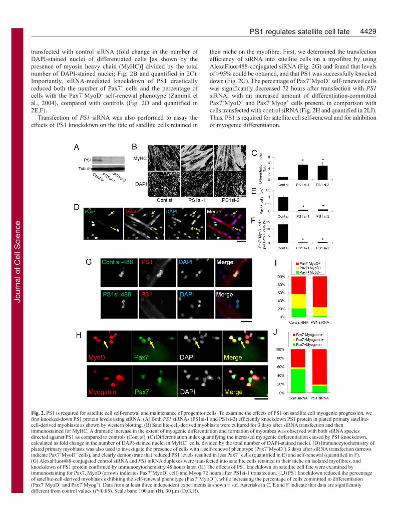
transfected with control siRNA (fold change in the number ofDAPI-stained nuclei of differentiated cells [as shown by thepresence of myosin heavy chain (MyHC)] divided by the totalnumber of DAPI-stained nuclei; Fig. 2B and quantified in 2C).Importantly, siRNA-mediated knockdown of PS1 drasticallyreduced both the number of Pax7+ cells and the percentage ofcells with the Pax7+MyoD– self-renewal phenotype (Zammit etal., 2004), compared with controls (Fig. 2D and quantified in2E,F).
Transfection of PS1 siRNA was also performed to assay theeffects of PS1 knockdown on the fate of satellite cells retained in
their niche on the myofibre. First, we determined the transfectionefficiency of siRNA into satellite cells on a myofibre by usingAlexaFluor488-conjugated siRNA (Fig. 2G) and found that levelsof >95% could be obtained, and that PS1 was successfully knockeddown (Fig. 2G). The percentage of Pax7+MyoD– self-renewed cellswas significantly decreased 72 hours after transfection with PS1siRNA, with an increased amount of differentiation-committedPax7–MyoD+ and Pax7–Myog+ cells present, in comparison withcells transfected with control siRNA (Fig. 2H and quantified in 2I,J).Thus, PS1 is required for satellite cell self-renewal and for inhibitionof myogenic differentiation.
Fig. 2. PS1 is required for satellite cell self-renewal and maintenance of progenitor cells. To examine the effects of PS1 on satellite cell myogenic progression, wefirst knocked-down PS1 protein levels using siRNA. (A)Both PS1 siRNAs (PS1si-1 and PS1si-2) efficiently knockdown PS1 protein in plated primary satellite-cell-derived myoblasts as shown by western blotting. (B)Satellite-cell-derived myoblasts were cultured for 3 days after siRNA transfection and thenimmunostained for MyHC. A dramatic increase in the extent of myogenic differentiation and formation of myotubes was observed with both siRNA speciesdirected against PS1 as compared to controls (Cont si). (C)Differentiation index quantifying the increased myogenic differentiation caused by PS1 knockdown,calculated as fold change in the number of DAPI-stained nuclei in MyHC+ cells, divided by the total number of DAPI-stained nuclei. (D)Immunocytochemistry ofplated primary myoblasts was also used to investigate the presence of cells with a self-renewal phenotype (Pax7+MyoD–) 3 days after siRNA transfection (arrowsindicate Pax7+MyoD– cells), and clearly demonstrate that reduced PS1 levels resulted in less Pax7+ cells (quantified in E) and self-renewal (quantified in F).(G)AlexaFluor488-conjugated control siRNA and PS1 siRNA duplexes were transfected into satellite cells retained in their niche on isolated myofibres, andknockdown of PS1 protein confirmed by immunocytochemistry 48 hours later. (H)The effects of PS1 knockdown on satellite cell fate were examined byimmunostaining for Pax7, MyoD (arrows indicates Pax7+MyoD– cell) and Myog 72 hours after PS1si-1 transfection. (I,J)PS1 knockdown reduced the percentageof satellite-cell-derived myoblasts exhibiting the self-renewal phenotype (Pax7+MyoD–), while increasing the percentage of cells committed to differentiation(Pax7–MyoD+ and Pax7–Myog+). Data from at least three independent experiments is shown ± s.d. Asterisks in C, E and F indicate that data are significantlydifferent from control values (P<0.05). Scale bars: 100m (B), 30m (D,G,H).
Jour
nal o
f Cel
l Sci
ence
4430
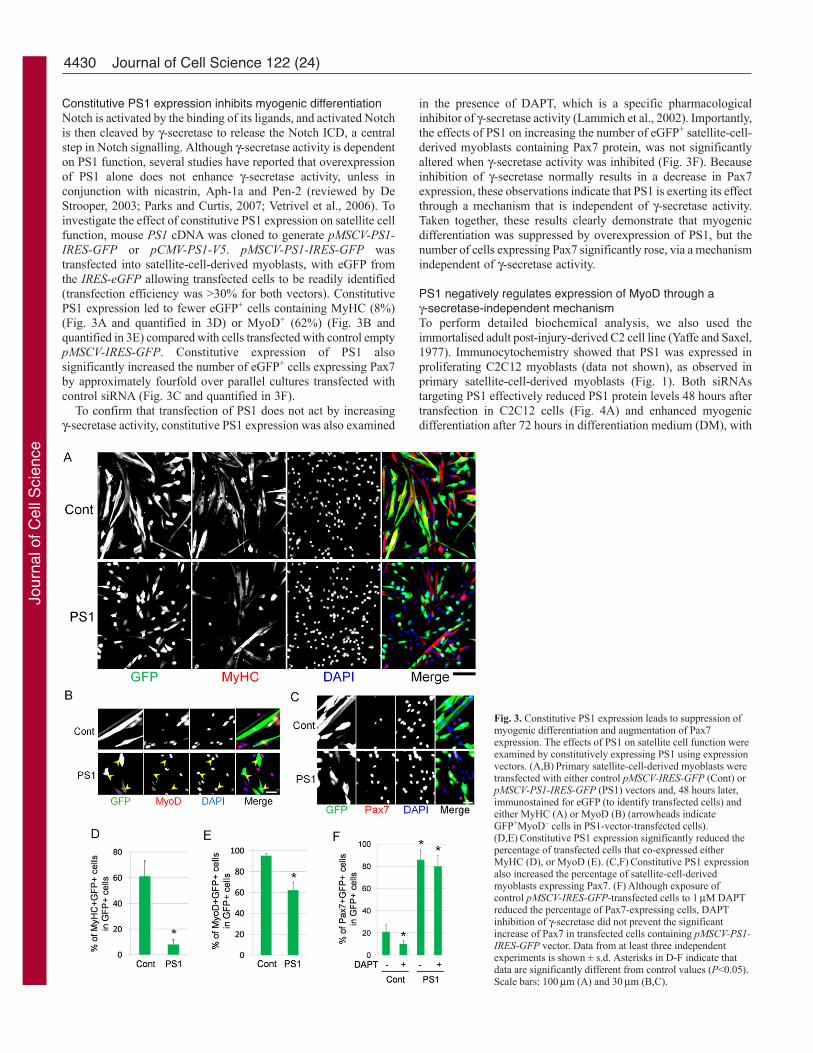
Constitutive PS1 expression inhibits myogenic differentiationNotch is activated by the binding of its ligands, and activated Notchis then cleaved by -secretase to release the Notch ICD, a centralstep in Notch signalling. Although -secretase activity is dependenton PS1 function, several studies have reported that overexpressionof PS1 alone does not enhance -secretase activity, unless inconjunction with nicastrin, Aph-1a and Pen-2 (reviewed by DeStrooper, 2003; Parks and Curtis, 2007; Vetrivel et al., 2006). Toinvestigate the effect of constitutive PS1 expression on satellite cellfunction, mouse PS1 cDNA was cloned to generate pMSCV-PS1-IRES-GFP or pCMV-PS1-V5. pMSCV-PS1-IRES-GFP wastransfected into satellite-cell-derived myoblasts, with eGFP fromthe IRES-eGFP allowing transfected cells to be readily identified(transfection efficiency was >30% for both vectors). ConstitutivePS1 expression led to fewer eGFP+ cells containing MyHC (8%)(Fig. 3A and quantified in 3D) or MyoD+ (62%) (Fig. 3B andquantified in 3E) compared with cells transfected with control emptypMSCV-IRES-GFP. Constitutive expression of PS1 alsosignificantly increased the number of eGFP+ cells expressing Pax7by approximately fourfold over parallel cultures transfected withcontrol siRNA (Fig. 3C and quantified in 3F).
To confirm that transfection of PS1 does not act by increasing-secretase activity, constitutive PS1 expression was also examined
in the presence of DAPT, which is a specific pharmacologicalinhibitor of -secretase activity (Lammich et al., 2002). Importantly,the effects of PS1 on increasing the number of eGFP+ satellite-cell-derived myoblasts containing Pax7 protein, was not significantlyaltered when -secretase activity was inhibited (Fig. 3F). Becauseinhibition of -secretase normally results in a decrease in Pax7expression, these observations indicate that PS1 is exerting its effectthrough a mechanism that is independent of -secretase activity.Taken together, these results clearly demonstrate that myogenicdifferentiation was suppressed by overexpression of PS1, but thenumber of cells expressing Pax7 significantly rose, via a mechanismindependent of -secretase activity.
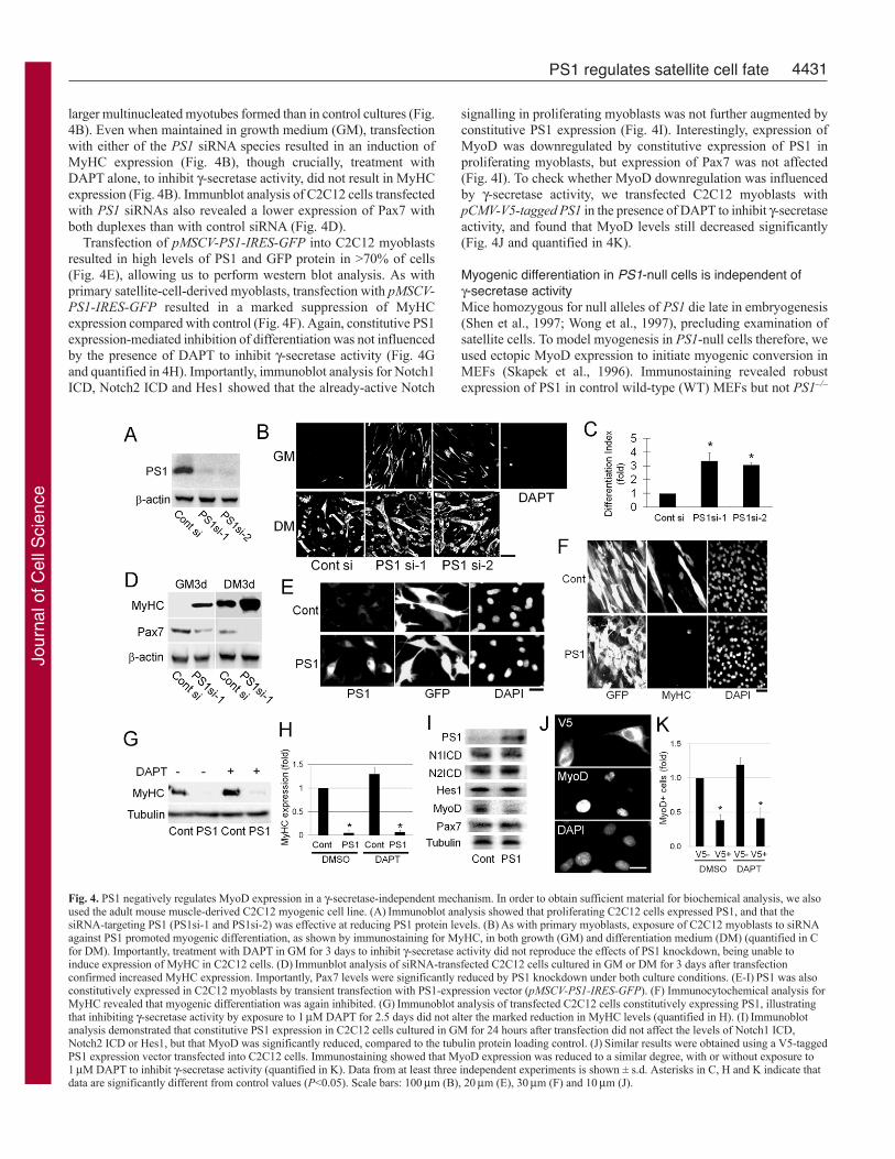
PS1 negatively regulates expression of MyoD through a-secretase-independent mechanismTo perform detailed biochemical analysis, we also used theimmortalised adult post-injury-derived C2 cell line (Yaffe and Saxel,1977). Immunocytochemistry showed that PS1 was expressed inproliferating C2C12 myoblasts (data not shown), as observed inprimary satellite-cell-derived myoblasts (Fig. 1). Both siRNAstargeting PS1 effectively reduced PS1 protein levels 48 hours aftertransfection in C2C12 cells (Fig. 4A) and enhanced myogenicdifferentiation after 72 hours in differentiation medium (DM), with
Journal of Cell Science 122 (24)
Fig. 3. Constitutive PS1 expression leads to suppression ofmyogenic differentiation and augmentation of Pax7expression. The effects of PS1 on satellite cell function wereexamined by constitutively expressing PS1 using expressionvectors. (A,B)Primary satellite-cell-derived myoblasts weretransfected with either control pMSCV-IRES-GFP (Cont) orpMSCV-PS1-IRES-GFP (PS1) vectors and, 48 hours later,immunostained for eGFP (to identify transfected cells) andeither MyHC (A) or MyoD (B) (arrowheads indicateGFP+MyoD– cells in PS1-vector-transfected cells).(D,E)Constitutive PS1 expression significantly reduced thepercentage of transfected cells that co-expressed eitherMyHC (D), or MyoD (E). (C,F)Constitutive PS1 expressionalso increased the percentage of satellite-cell-derivedmyoblasts expressing Pax7. (F)Although exposure ofcontrol pMSCV-IRES-GFP-transfected cells to 1M DAPTreduced the percentage of Pax7-expressing cells, DAPTinhibition of -secretase did not prevent the significantincrease of Pax7 in transfected cells containing pMSCV-PS1-IRES-GFP vector. Data from at least three independentexperiments is shown ± s.d. Asterisks in D-F indicate thatdata are significantly different from control values (P<0.05).Scale bars: 100m (A) and 30m (B,C).
Jour
nal o
f Cel
l Sci
ence
4431PS1 regulates satellite cell fate
larger multinucleated myotubes formed than in control cultures (Fig.4B). Even when maintained in growth medium (GM), transfectionwith either of the PS1 siRNA species resulted in an induction ofMyHC expression (Fig. 4B), though crucially, treatment withDAPT alone, to inhibit -secretase activity, did not result in MyHCexpression (Fig. 4B). Immunblot analysis of C2C12 cells transfectedwith PS1 siRNAs also revealed a lower expression of Pax7 withboth duplexes than with control siRNA (Fig. 4D).
Transfection of pMSCV-PS1-IRES-GFP into C2C12 myoblastsresulted in high levels of PS1 and GFP protein in >70% of cells(Fig. 4E), allowing us to perform western blot analysis. As withprimary satellite-cell-derived myoblasts, transfection with pMSCV-PS1-IRES-GFP resulted in a marked suppression of MyHCexpression compared with control (Fig. 4F). Again, constitutive PS1expression-mediated inhibition of differentiation was not influencedby the presence of DAPT to inhibit -secretase activity (Fig. 4Gand quantified in 4H). Importantly, immunoblot analysis for Notch1ICD, Notch2 ICD and Hes1 showed that the already-active Notch
signalling in proliferating myoblasts was not further augmented byconstitutive PS1 expression (Fig. 4I). Interestingly, expression ofMyoD was downregulated by constitutive expression of PS1 inproliferating myoblasts, but expression of Pax7 was not affected(Fig. 4I). To check whether MyoD downregulation was influencedby -secretase activity, we transfected C2C12 myoblasts withpCMV-V5-tagged PS1 in the presence of DAPT to inhibit -secretaseactivity, and found that MyoD levels still decreased significantly(Fig. 4J and quantified in 4K).
Myogenic differentiation in PS1-null cells is independent of-secretase activityMice homozygous for null alleles of PS1 die late in embryogenesis(Shen et al., 1997; Wong et al., 1997), precluding examination ofsatellite cells. To model myogenesis in PS1-null cells therefore, weused ectopic MyoD expression to initiate myogenic conversion inMEFs (Skapek et al., 1996). Immunostaining revealed robustexpression of PS1 in control wild-type (WT) MEFs but not PS1–/–
Fig. 4. PS1 negatively regulates MyoD expression in a -secretase-independent mechanism. In order to obtain sufficient material for biochemical analysis, we alsoused the adult mouse muscle-derived C2C12 myogenic cell line. (A)Immunoblot analysis showed that proliferating C2C12 cells expressed PS1, and that thesiRNA-targeting PS1 (PS1si-1 and PS1si-2) was effective at reducing PS1 protein levels. (B)As with primary myoblasts, exposure of C2C12 myoblasts to siRNAagainst PS1 promoted myogenic differentiation, as shown by immunostaining for MyHC, in both growth (GM) and differentiation medium (DM) (quantified in Cfor DM). Importantly, treatment with DAPT in GM for 3 days to inhibit -secretase activity did not reproduce the effects of PS1 knockdown, being unable toinduce expression of MyHC in C2C12 cells. (D)Immunblot analysis of siRNA-transfected C2C12 cells cultured in GM or DM for 3 days after transfectionconfirmed increased MyHC expression. Importantly, Pax7 levels were significantly reduced by PS1 knockdown under both culture conditions. (E-I)PS1 was alsoconstitutively expressed in C2C12 myoblasts by transient transfection with PS1-expression vector (pMSCV-PS1-IRES-GFP). (F) Immunocytochemical analysis forMyHC revealed that myogenic differentiation was again inhibited. (G)Immunoblot analysis of transfected C2C12 cells constitutively expressing PS1, illustratingthat inhibiting -secretase activity by exposure to 1M DAPT for 2.5 days did not alter the marked reduction in MyHC levels (quantified in H). (I)Immunoblotanalysis demonstrated that constitutive PS1 expression in C2C12 cells cultured in GM for 24 hours after transfection did not affect the levels of Notch1 ICD,Notch2 ICD or Hes1, but that MyoD was significantly reduced, compared to the tubulin protein loading control. (J)Similar results were obtained using a V5-taggedPS1 expression vector transfected into C2C12 cells. Immunostaining showed that MyoD expression was reduced to a similar degree, with or without exposure to1M DAPT to inhibit -secretase activity (quantified in K). Data from at least three independent experiments is shown ± s.d. Asterisks in C, H and K indicate thatdata are significantly different from control values (P<0.05). Scale bars: 100m (B), 20m (E), 30m (F) and 10m (J).
Jour
nal o
f Cel
l Sci
ence
4432
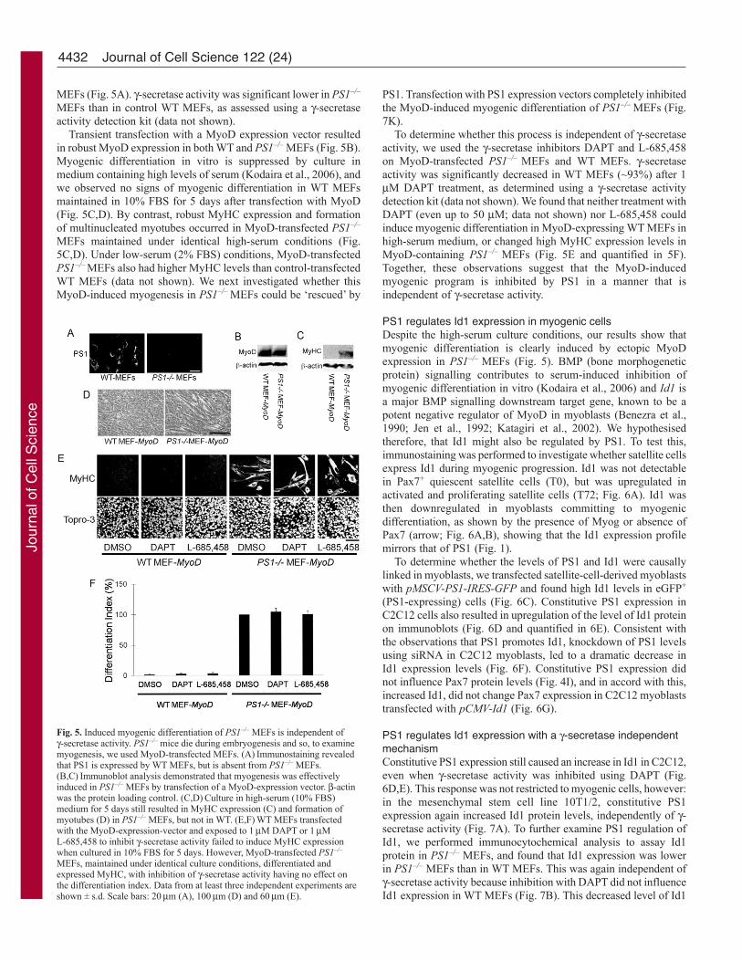
MEFs (Fig. 5A). -secretase activity was significant lower in PS1–/–
MEFs than in control WT MEFs, as assessed using a -secretaseactivity detection kit (data not shown).
Transient transfection with a MyoD expression vector resultedin robust MyoD expression in both WT and PS1–/– MEFs (Fig. 5B).Myogenic differentiation in vitro is suppressed by culture inmedium containing high levels of serum (Kodaira et al., 2006), andwe observed no signs of myogenic differentiation in WT MEFsmaintained in 10% FBS for 5 days after transfection with MyoD(Fig. 5C,D). By contrast, robust MyHC expression and formationof multinucleated myotubes occurred in MyoD-transfected PS1–/–
MEFs maintained under identical high-serum conditions (Fig.5C,D). Under low-serum (2% FBS) conditions, MyoD-transfectedPS1–/– MEFs also had higher MyHC levels than control-transfectedWT MEFs (data not shown). We next investigated whether thisMyoD-induced myogenesis in PS1–/– MEFs could be ‘rescued’ by
PS1. Transfection with PS1 expression vectors completely inhibitedthe MyoD-induced myogenic differentiation of PS1–/– MEFs (Fig.7K).
To determine whether this process is independent of -secretaseactivity, we used the -secretase inhibitors DAPT and L-685,458on MyoD-transfected PS1–/– MEFs and WT MEFs. -secretaseactivity was significantly decreased in WT MEFs (~93%) after 1M DAPT treatment, as determined using a -secretase activitydetection kit (data not shown). We found that neither treatment withDAPT (even up to 50 M; data not shown) nor L-685,458 couldinduce myogenic differentiation in MyoD-expressing WT MEFs inhigh-serum medium, or changed high MyHC expression levels inMyoD-containing PS1–/– MEFs (Fig. 5E and quantified in 5F).Together, these observations suggest that the MyoD-inducedmyogenic program is inhibited by PS1 in a manner that isindependent of -secretase activity.
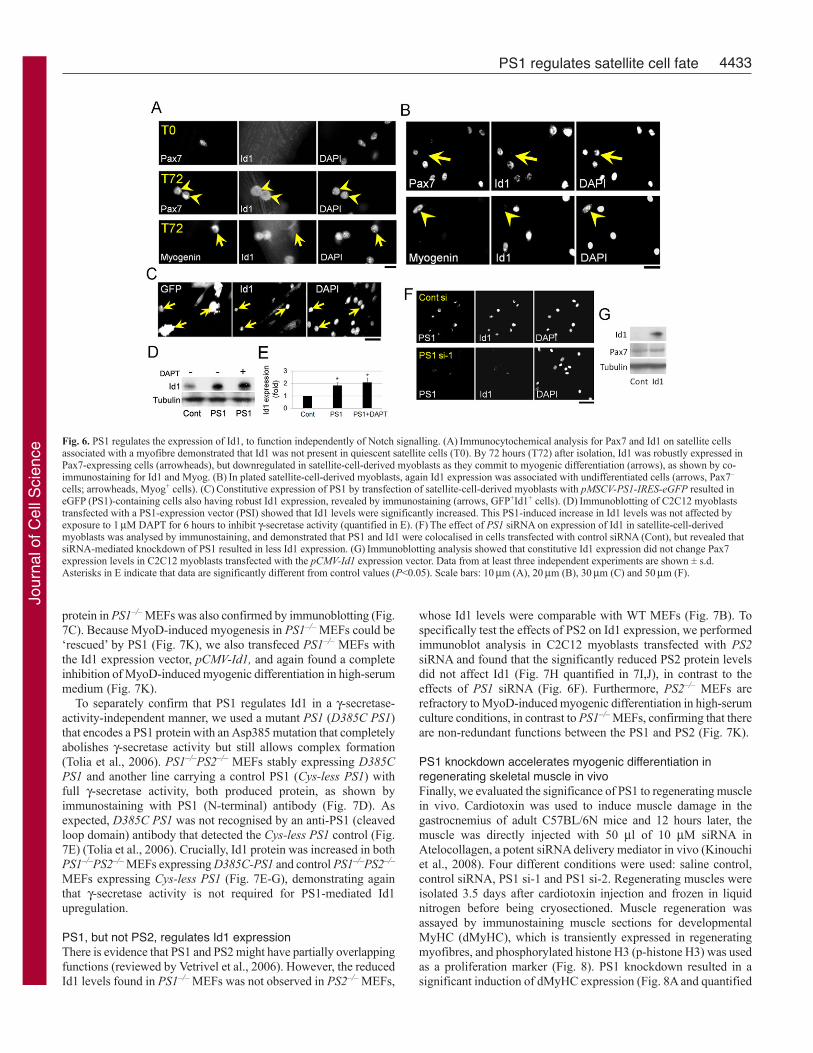
PS1 regulates Id1 expression in myogenic cellsDespite the high-serum culture conditions, our results show thatmyogenic differentiation is clearly induced by ectopic MyoDexpression in PS1–/– MEFs (Fig. 5). BMP (bone morphogeneticprotein) signalling contributes to serum-induced inhibition ofmyogenic differentiation in vitro (Kodaira et al., 2006) and Id1 isa major BMP signalling downstream target gene, known to be apotent negative regulator of MyoD in myoblasts (Benezra et al.,1990; Jen et al., 1992; Katagiri et al., 2002). We hypothesisedtherefore, that Id1 might also be regulated by PS1. To test this,immunostaining was performed to investigate whether satellite cellsexpress Id1 during myogenic progression. Id1 was not detectablein Pax7+ quiescent satellite cells (T0), but was upregulated inactivated and proliferating satellite cells (T72; Fig. 6A). Id1 wasthen downregulated in myoblasts committing to myogenicdifferentiation, as shown by the presence of Myog or absence ofPax7 (arrow; Fig. 6A,B), showing that the Id1 expression profilemirrors that of PS1 (Fig. 1).
To determine whether the levels of PS1 and Id1 were causallylinked in myoblasts, we transfected satellite-cell-derived myoblastswith pMSCV-PS1-IRES-GFP and found high Id1 levels in eGFP+
(PS1-expressing) cells (Fig. 6C). Constitutive PS1 expression inC2C12 cells also resulted in upregulation of the level of Id1 proteinon immunoblots (Fig. 6D and quantified in 6E). Consistent withthe observations that PS1 promotes Id1, knockdown of PS1 levelsusing siRNA in C2C12 myoblasts, led to a dramatic decrease inId1 expression levels (Fig. 6F). Constitutive PS1 expression didnot influence Pax7 protein levels (Fig. 4I), and in accord with this,increased Id1, did not change Pax7 expression in C2C12 myoblaststransfected with pCMV-Id1 (Fig. 6G).
PS1 regulates Id1 expression with a -secretase independentmechanismConstitutive PS1 expression still caused an increase in Id1 in C2C12,even when -secretase activity was inhibited using DAPT (Fig.6D,E). This response was not restricted to myogenic cells, however:in the mesenchymal stem cell line 10T1/2, constitutive PS1expression again increased Id1 protein levels, independently of -secretase activity (Fig. 7A). To further examine PS1 regulation ofId1, we performed immunocytochemical analysis to assay Id1protein in PS1–/– MEFs, and found that Id1 expression was lowerin PS1–/– MEFs than in WT MEFs. This was again independent of-secretase activity because inhibition with DAPT did not influenceId1 expression in WT MEFs (Fig. 7B). This decreased level of Id1
Journal of Cell Science 122 (24)
Fig. 5. Induced myogenic differentiation of PS1–/– MEFs is independent of-secretase activity. PS1–/– mice die during embryogenesis and so, to examinemyogenesis, we used MyoD-transfected MEFs. (A)Immunostaining revealedthat PS1 is expressed by WT MEFs, but is absent from PS1–/– MEFs.(B,C)Immunoblot analysis demonstrated that myogenesis was effectivelyinduced in PS1–/– MEFs by transfection of a MyoD-expression vector. -actinwas the protein loading control. (C,D)Culture in high-serum (10% FBS)medium for 5 days still resulted in MyHC expression (C) and formation ofmyotubes (D) in PS1–/– MEFs, but not in WT. (E,F)WT MEFs transfectedwith the MyoD-expression-vector and exposed to 1M DAPT or 1ML-685,458 to inhibit -secretase activity failed to induce MyHC expressionwhen cultured in 10% FBS for 5 days. However, MyoD-transfected PS1–/–
MEFs, maintained under identical culture conditions, differentiated andexpressed MyHC, with inhibition of -secretase activity having no effect onthe differentiation index. Data from at least three independent experiments areshown ± s.d. Scale bars: 20m (A), 100m (D) and 60m (E).
Jour
nal o
f Cel
l Sci
ence
4433PS1 regulates satellite cell fate
protein in PS1–/– MEFs was also confirmed by immunoblotting (Fig.7C). Because MyoD-induced myogenesis in PS1–/– MEFs could be‘rescued’ by PS1 (Fig. 7K), we also transfeced PS1–/– MEFs withthe Id1 expression vector, pCMV-Id1, and again found a completeinhibition of MyoD-induced myogenic differentiation in high-serummedium (Fig. 7K).
To separately confirm that PS1 regulates Id1 in a -secretase-activity-independent manner, we used a mutant PS1 (D385C PS1)that encodes a PS1 protein with an Asp385 mutation that completelyabolishes -secretase activity but still allows complex formation(Tolia et al., 2006). PS1–/–PS2–/– MEFs stably expressing D385CPS1 and another line carrying a control PS1 (Cys-less PS1) withfull -secretase activity, both produced protein, as shown byimmunostaining with PS1 (N-terminal) antibody (Fig. 7D). Asexpected, D385C PS1 was not recognised by an anti-PS1 (cleavedloop domain) antibody that detected the Cys-less PS1 control (Fig.7E) (Tolia et al., 2006). Crucially, Id1 protein was increased in bothPS1–/–PS2–/– MEFs expressing D385C-PS1 and control PS1–/–PS2–/–
MEFs expressing Cys-less PS1 (Fig. 7E-G), demonstrating againthat -secretase activity is not required for PS1-mediated Id1upregulation.
PS1, but not PS2, regulates Id1 expressionThere is evidence that PS1 and PS2 might have partially overlappingfunctions (reviewed by Vetrivel et al., 2006). However, the reducedId1 levels found in PS1–/– MEFs was not observed in PS2–/– MEFs,
whose Id1 levels were comparable with WT MEFs (Fig. 7B). Tospecifically test the effects of PS2 on Id1 expression, we performedimmunoblot analysis in C2C12 myoblasts transfected with PS2siRNA and found that the significantly reduced PS2 protein levelsdid not affect Id1 (Fig. 7H quantified in 7I,J), in contrast to theeffects of PS1 siRNA (Fig. 6F). Furthermore, PS2–/– MEFs arerefractory to MyoD-induced myogenic differentiation in high-serumculture conditions, in contrast to PS1–/– MEFs, confirming that thereare non-redundant functions between the PS1 and PS2 (Fig. 7K).
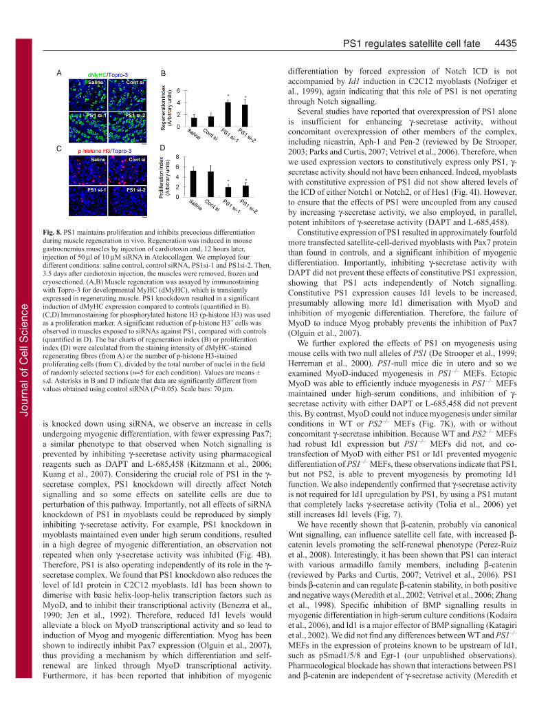
PS1 knockdown accelerates myogenic differentiation inregenerating skeletal muscle in vivoFinally, we evaluated the significance of PS1 to regenerating musclein vivo. Cardiotoxin was used to induce muscle damage in thegastrocnemius of adult C57BL/6N mice and 12 hours later, themuscle was directly injected with 50 l of 10 M siRNA inAtelocollagen, a potent siRNA delivery mediator in vivo (Kinouchiet al., 2008). Four different conditions were used: saline control,control siRNA, PS1 si-1 and PS1 si-2. Regenerating muscles wereisolated 3.5 days after cardiotoxin injection and frozen in liquidnitrogen before being cryosectioned. Muscle regeneration wasassayed by immunostaining muscle sections for developmentalMyHC (dMyHC), which is transiently expressed in regeneratingmyofibres, and phosphorylated histone H3 (p-histone H3) was usedas a proliferation marker (Fig. 8). PS1 knockdown resulted in asignificant induction of dMyHC expression (Fig. 8A and quantified
Fig. 6. PS1 regulates the expression of Id1, to function independently of Notch signalling. (A)Immunocytochemical analysis for Pax7 and Id1 on satellite cellsassociated with a myofibre demonstrated that Id1 was not present in quiescent satellite cells (T0). By 72 hours (T72) after isolation, Id1 was robustly expressed inPax7-expressing cells (arrowheads), but downregulated in satellite-cell-derived myoblasts as they commit to myogenic differentiation (arrows), as shown by co-immunostaining for Id1 and Myog. (B)In plated satellite-cell-derived myoblasts, again Id1 expression was associated with undifferentiated cells (arrows, Pax7–
cells; arrowheads, Myog+ cells). (C)Constitutive expression of PS1 by transfection of satellite-cell-derived myoblasts with pMSCV-PS1-IRES-eGFP resulted ineGFP (PS1)-containing cells also having robust Id1 expression, revealed by immunostaining (arrows, GFP+Id1+ cells). (D)Immunoblotting of C2C12 myoblaststransfected with a PS1-expression vector (PSI) showed that Id1 levels were significantly increased. This PS1-induced increase in Id1 levels was not affected byexposure to 1M DAPT for 6 hours to inhibit -secretase activity (quantified in E). (F)The effect of PS1 siRNA on expression of Id1 in satellite-cell-derivedmyoblasts was analysed by immunostaining, and demonstrated that PS1 and Id1 were colocalised in cells transfected with control siRNA (Cont), but revealed thatsiRNA-mediated knockdown of PS1 resulted in less Id1 expression. (G)Immunoblotting analysis showed that constitutive Id1 expression did not change Pax7expression levels in C2C12 myoblasts transfected with the pCMV-Id1 expression vector. Data from at least three independent experiments are shown ± s.d.Asterisks in E indicate that data are significantly different from control values (P<0.05). Scale bars: 10m (A), 20m (B), 30m (C) and 50m (F).
Jour
nal o
f Cel
l Sci
ence

4434
in 8B) and a significant reduction in the number of cells containingp-histone H3 (Fig. 8C and quantified in 8D) compared with controlsiRNA. These observations indicate that PS1 is required formaintenance of proliferating cells and inhibition of myogenicdifferentiation during muscle regeneration in vivo as well as in vitro.
DiscussionUnderstanding how the decision between self-renewal anddifferentiation is regulated in satellite cells is central tounderstanding how skeletal muscle maintains a viable stem cellcompartment. Here, we show that the multifunctional protein PS1is able to direct muscle satellite cells away from myogenicdifferentiation and towards the self-renewal phenotype. Although
PS1 is a key component of the -secretase complex, we found thatPS1 also acts through -secretase-independent mechanisms to affectsatellite cell fate, by controlling Id1 protein.
Satellite cells are normally mitotically quiescent in maturemuscle, so must first be activated to enter the cell cycle and generatemyoblasts (Zammit, 2008). Important pathways associated withactivation include sphingolipid (Nagata et al., 2006b; Nagata et al.,2006a), p38MAPK (Jones et al., 2005) and Notch signalling(Conboy and Rando, 2002). Notch signalling in proliferatingsatellite cells plays a role in expanding the satellite cell pool afteractivation, while preventing precocious differentiation (Conboy etal., 2003; Conboy and Rando, 2002; Kitzmann et al., 2006; Kopanet al., 1994; Kuang et al., 2007; Nofziger et al., 1999). When PS1
Journal of Cell Science 122 (24)
Fig. 7. PS1 controls Id1 expression in non-muscle cells via a -secretase-activity-independent mechanism. (A)PS1-regulated Id1 expression was not restricted tomyogenic cells: in the mesenchymal stem cell line 10T1/2, immunostaining showed that constitutive PS1 expression increased Id1 protein levels, which was alsoindependent of -secretase activity, as shown by exposure to 1M DAPT for 6 hours. (B)Immunostaining showed that reduction in Id1 levels was specific toPS1–/– MEFs because PS2–/– MEFs had robust Id1 levels, comparable to WT MEFs. Exposure of WT MEFs to 1M DAPT for 12 hours did not result in areduction in Id1 levels. (C)Although Id1 was clearly present in WT MEFs, immunoblot analysis demonstrated that Id1 was expressed at very low levels fromPS1–/– MEFs. -actin was used as a control for protein loading. (D)Immunostaining showed that anti-PS1 (N-terminal) antibody recognised both stably insertedD385C PS1 (PS1 mutant with no -secretase activity) and control Cys-less PS1 in PS1–/–PS2–/– MEFs. (E)Immunoblot analysis revealed that PS1 protein inD385C-PS1-inserted PS1–/–PS2–/– MEFs was not detected by anti-PS1 (cleaved loop domain) antibody, which recognised the Cys-less PS1-inserted PS1–/–PS2–/–
MEFs. (E,F). Id1 protein was increased in both Cys-less PS1 and D385C-PS1 expressing PS1–/–PS2–/– MEFs (quantified in F). (G)Immunostaining confirmed thatD385C PS1 inserted into PS1–/–PS2–/– MEFs can upregulate Id1 protein, as does the control Cys-less PS1-inserted PS1–/–PS2–/– MEFs. (H)Immunoblot analysisillustrates that specific siRNA-mediated knockdown of PS2 does not affect the level of Id1 protein in C2C12 myoblasts (quantified in I,J). (K)Immunoblotting 5days after transfection illustrated that the presence of a MyoD-expression vector resulted in myogenic differentiation in PS1–/– MEFs maintained in high-serumculture conditions, as shown by the presence of MyHC. By contrast, WT or PS2–/– MEFs did not undergo myogenic conversion. Importantly, co-transfection of theMyoD expression vector with either a PS1-expression vector, or an Id1-expression vector, largely blocked this induction of MyHC in PS1–/– MEFs. Tubulin wasused as an internal control for protein loading. Data from at least three independent experiments are shown ± s.d. Asterisks in F, I and J indicate whether data aresignificantly different from control values (P<0.05). Scale bars: 30m (A), 10m (B) and 100m (D,G).
Jour
nal o
f Cel
l Sci
ence
4435PS1 regulates satellite cell fate
is knocked down using siRNA, we observe an increase in cellsundergoing myogenic differentiation, with fewer expressing Pax7;a similar phenotype to that observed when Notch signalling isprevented by inhibiting -secretase activity using pharmacogicalreagents such as DAPT and L-685,458 (Kitzmann et al., 2006;Kuang et al., 2007). Considering the crucial role of PS1 in the -secretase complex, PS1 knockdown will directly affect Notchsignalling and so some effects on satellite cells are due toperturbation of this pathway. Importantly, not all effects of siRNAknockdown of PS1 in myoblasts could be reproduced by simplyinhibiting -secretase activity. For example, PS1 knockdown inmyoblasts maintained even under high serum conditions, resultedin a high degree of myogenic differentiation, an observation notrepeated when only -secretase activity was inhibited (Fig. 4B).Therefore, PS1 is also operating independently of its role in the -secretase complex. We found that PS1 knockdown also reduces thelevel of Id1 protein in C2C12 myoblasts. Id1 has been shown todimerise with basic helix-loop-helix transcription factors such asMyoD, and to inhibit their transcriptional activity (Benezra et al.,1990; Jen et al., 1992). Therefore, reduced Id1 levels wouldalleviate a block on MyoD transcriptional activity and so lead toinduction of Myog and myogenic differentiation. Myog has beenshown to indirectly inhibit Pax7 expression (Olguin et al., 2007),thus providing a mechanism by which differentiation and self-renewal are linked through MyoD transcriptional activity.Furthermore, it has been reported that inhibition of myogenic
differentiation by forced expression of Notch ICD is notaccompanied by Id1 induction in C2C12 myoblasts (Nofziger etal., 1999), again indicating that this role of PS1 is not operatingthrough Notch signalling.
Several studies have reported that overexpression of PS1 aloneis insufficient for enhancing -secretase activity, withoutconcomitant overexpression of other members of the complex,including nicastrin, Aph-1 and Pen-2 (reviewed by De Strooper,2003; Parks and Curtis, 2007; Vetrivel et al., 2006). Therefore, whenwe used expression vectors to constitutively express only PS1, -secretase activity should not have been enhanced. Indeed, myoblastswith constitutive expression of PS1 did not show altered levels ofthe ICD of either Notch1 or Notch2, or of Hes1 (Fig. 4I). However,to ensure that the effects of PS1 were uncoupled from any causedby increasing -secretase activity, we also employed, in parallel,potent inhibitors of -secretase activity (DAPT and L-685,458).
Constitutive expression of PS1 resulted in approximately fourfoldmore transfected satellite-cell-derived myoblasts with Pax7 proteinthan found in controls, and a significant inhibition of myogenicdifferentiation. Importantly, inhibiting -secretase activity withDAPT did not prevent these effects of constitutive PS1 expression,showing that PS1 acts independently of Notch signalling.Constitutive PS1 expression causes Id1 levels to be increased,presumably allowing more Id1 dimerisation with MyoD andinhibition of myogenic differentiation. Therefore, the failure ofMyoD to induce Myog probably prevents the inhibition of Pax7(Olguin et al., 2007).
We further explored the effects of PS1 on myogenesis usingmouse cells with two null alleles of PS1 (De Strooper et al., 1999;Herreman et al., 2000). PS1-null mice die in utero and so weexamined MyoD-induced myogenesis in PS1–/– MEFs. EctopicMyoD was able to efficiently induce myogenesis in PS1–/– MEFsmaintained under high-serum conditions, and inhibition of -secretase activity with either DAPT or L-685,458 did not preventthis. By contrast, MyoD could not induce myogenesis under similarconditions in WT or PS2–/– MEFs (Fig. 7K), with or withoutconcomitant -secretase inhibition. Because WT and PS2–/– MEFshad robust Id1 expression but PS1–/– MEFs did not, and co-transfection of MyoD with either PS1 or Id1 prevented myogenicdifferentiation of PS1–/– MEFs, these observations indicate that PS1,but not PS2, is able to prevent myogenesis by promoting Id1function. We also independently confirmed that -secretase activityis not required for Id1 upregulation by PS1, by using a PS1 mutantthat completely lacks -secretase activity (Tolia et al., 2006) yetstill increases Id1 levels (Fig. 7).
We have recently shown that -catenin, probably via canonicalWnt signalling, can influence satellite cell fate, with increased -catenin levels promoting the self-renewal phenotype (Perez-Ruizet al., 2008). Interestingly, it has been shown that PS1 can interactwith various armadillo family members, including -catenin(reviewed by Parks and Curtis, 2007; Vetrivel et al., 2006). PS1binds -catenin and can regulate -catenin stability, in both positiveand negative ways (Meredith et al., 2002; Vetrivel et al., 2006; Zhanget al., 1998). Specific inhibition of BMP signalling results inmyogenic differentiation in high-serum culture conditions (Kodairaet al., 2006), and Id1 is a major effector of BMP signalling (Katagiriet al., 2002). We did not find any differences between WT and PS1–/–
MEFs in the expression of proteins known to be upstream of Id1,such as pSmad1/5/8 and Egr-1 (our unpublished observations).Pharmacological blockade has shown that interactions between PS1and -catenin are independent of -secretase activity (Meredith et
Fig. 8. PS1 maintains proliferation and inhibits precocious differentiationduring muscle regeneration in vivo. Regeneration was induced in mousegastrocnemius muscles by injection of cardiotoxin and, 12 hours later,injection of 50l of 10M siRNA in Atelocollagen. We employed fourdifferent conditions: saline control, control siRNA, PS1si-1 and PS1si-2. Then,3.5 days after cardiotoxin injection, the muscles were removed, frozen andcryosectioned. (A,B)Muscle regeneration was assayed by immunostainingwith Topro-3 for developmental MyHC (dMyHC), which is transientlyexpressed in regenerating muscle. PS1 knockdown resulted in a significantinduction of dMyHC expression compared to controls (quantified in B).(C,D)Immunostaining for phosphorylated histone H3 (p-histone H3) was usedas a proliferation marker. A significant reduction of p-histone H3+ cells wasobserved in muscles exposed to siRNAs against PS1, compared with controls(quantified in D). The bar charts of regeneration index (B) or proliferationindex (D) were calculated from the staining intensity of dMyHC-stainedregenerating fibres (from A) or the number of p-histone H3-stainedproliferating cells (from C), divided by the total number of nuclei in the fieldof randomly selected sections (n5 for each condition). Values are means ±s.d. Asterisks in B and D indicate that data are significantly different fromvalues obtained using control siRNA (P<0.05). Scale bars: 70m.
Jour
nal o
f Cel
l Sci
ence
4436
al., 2002) and, as mentioned above, Notch ICD is not accompaniedby Id1 induction in C2C12 myoblasts (Nofziger et al., 1999). Thus,speculatively, PS1 might operate through its effects on -cateninstability to affect canonical Wnt signalling to control Id1,independently of its -secretase activity.
PS1 is also known to regulate Ca2+ homeostasis through -secretase-independent mechanisms (Akbari et al., 2004; Tu et al.,2006). In regenerating muscles, Id1 can be negatively regulated byCalcineurin, a Ca2+-calmodulin-dependent serine/threonine proteinphosphatase (Sakuma et al., 2005), so PS1 might also control Id1protein through the regulation of Ca2+ homeostasis. Id1 protein isalso known to be controlled by protein stabilisation (Bounpheng etal., 1999), so PS1 might also more directly control Id1 expressionthrough such a mechanism.
Taken together, our data provide evidence of a novel mechanismoperating in stem cells, whereby PS1 controls Id1 to regulate thetranscriptional activity of bHLH transcription factors, operatingindependently of -secretase activity. In the neural system forexample, PS1 is essential for maintenance of the neural progenitorcell pool and for preventing neural differentiation in the developingbrain (Hitoshi et al., 2002). In adults, proliferating neural progenitorcells strongly express PS1, but mature neurones do not (Wen et al.,2002). Importantly, Id1 negatively regulates neurogenic transcriptionfactors such as Mash1 in brain (Nakashima et al., 2001; Vinals etal., 2004), paralleling its actions on MyoD in muscle. Speculativelytherefore, PS1 might have a common role in maintaining theprogenitor cell pool via regulation of Id1 in both muscle and brain.The demonstration of a PS1-Id1 signalling network might alsoprovide new insight into the pathogenesis of mutated PS1-relatedearly-onset familial Alzheimer’s disease (reviewed by Parks andCurtis, 2007; Vetrivel et al., 2006).
In conclusion, our study shows that PS1 acts as a potent regulatorof fate choice in muscle satellite cells. Undoubtedly, some of theeffects of PS1 on satellite cell function are due to its role as a crucialcomponent of the -secretase complex, central to Notch signalling.However, PS1 also operates in a -secretase-independent mannerto control MyoD, and our results show that this is probablyachieved through regulation of Id1. The mechanisms that promotesatellite cell self-renewal for the maintenance of the stem cell pooland those that prevent precocious myogenic differentiation must belinked and feedback on each other to carefully regulate the extentof differentiation for repair, versus maintenance, of a viable stemcell pool, able to respond to future needs. PS1 control of MyoDtranscriptional activity would appear to be one of these links betweendifferentiation and self-renewal mechanisms.
Materials and MethodsIsolation and culture of primary satellite cells and myoblastsAdult (8-12 weeks old) C57BL10 mice were killed by cervical dislocation, and theextensor digitorum longus (EDL) muscles isolated and digested in collagenase aspreviously described (Beauchamp et al., 2000). Myofibres and associated satellitecells were isolated and cultured in plating medium (DMEM supplemented with 10%horse serum, 0.5% chicken embryonic extract, 4 mM L-glutamine and 1% penicillin-streptomycin) at 37°C in 5% CO2, as described previously (Perez-Ruiz et al., 2008).Satellite cells were removed from myofibres by enzymatic treatment with 0.125%trypsin-EDTA solution for 10 minutes at 37°C and maintained in high-serum-containing medium (DMEM supplemented with 20% FBS, 1% chicken embryoextract, 10 ng/ml FGF, 4 mM L-glutamine and 1% penicillin-streptomycin). Thismedium supported both proliferation and differentiation of satellite cell progeny whenbFGF was removed. The C2 (Yaffe and Saxel, 1977) subclone C2C12 and 10T1/2cell lines were obtained from Riken Cell Bank (Tsukuba, Japan). C2C12 cells weremaintained in growth medium (GM; F-10 medium supplemented with 20% FBS andantibiotics). For myogenic differentiation (both C2 and satellite cells), the culturemedium was replaced with differentiation medium (DM; DMEM containing 2% horseserum and antibiotics) for 72 hours at 37°C. 10T1/2 cells, WT, PS1–/–, PS2–/–,
PS1–/–PS2–/– MEFs and PS1–/–PS2–/– MEFs expressing Cys-less PS1 or D385C PS1(Herreman et al., 1999; Herreman et al., 2003) were maintained in DMEM containing10% FBS and antibiotics.
Antibodies and ReagentsAntibodies were obtained from the following sources: mouse and rabbit anti-PS1antibodies from Millipore (Bedford, MA); mouse anti-GFP from Roche (Basel,Switzerland); rabbit anti-PS2 antibody from Abcam; mouse anti-Notch1 antibodyfrom BD Biosciences; rat anti-Ki67 from DAKO; goat anti-PS1 antibody, rabbit anti-Id1 antibody, rabbit anti-Hes1 antibody goat anti-Notch2 antibody and rabbit anti-MyoD antibody were obtained from Santa Cruz (Santa Cruz, CA); mouse anti-developmental myosin heavy chain (dMyHC) antibody from Novocastra (Newcastle,UK); mouse anti-MyHC (MF20), anti-Pax7 antibody, anti-Myog antibody (F5D) andanti-tubulin antibody (E7) were obtained from the DSHB (Iowa City, IA); rabbit anti-p-histone H3 antibody from Cell Signaling Technology (Beverly, MA) and Topro-3,rabbit anti-GFP antibody and mouse anti-V5 antibody from Invitrogen (Carlsbad,CA). Mounting medium containing DAPI was purchased from Vector Laboratories(Burlingame, CA). Nuclei were counterstained with either DAPI or Topro-3. DAPTand L-685,468 were purchased from Peptide Institute (Osaka, Japan) and dissolvedand applied in DMSO. -secretase activity was measured using a -secretase activitydetection kit according to the manufacturer’s instructions (R&D Systems).
Immunoblot analysisImmunoblot analysis was performed as previously described (Ono et al., 2006). Rabbitor mouse anti-PS1 (recognise loop domain), anti-PS2, anti-MyHC, anti-tubulin, anti-Pax7, anti-Notch1, anti-Notch2, anti-Hes1, anti-MyoD, anti-Id1 or anti--actinantibody were applied at 4°C overnight. Horseradish-peroxidase-conjugated secondaryantibodies were used for visualisation by chemiluminescence with a digitalluminescent image analyser LAS-1000 (Fujifilm, Tokyo, Japan).
ImmunostainingImmunocytochemistry was performed as previously described (Ono et al., 2007).Primary antibodies were used in PBS as follows: goat anti-PS1 (recognises N-terminal), anti-Id1, anti-Ki67, anti-MyHC, anti-Pax7, anti-Myog, mouse anti-GFP,rabbit anti-GFP, anti-V5 or anti-MyoD antibody at 4°C overnight. Forimmunohistochemistry, frozen muscle cross-sections were fixed with cold acetone,blocked with M.O.M kit (Vector Laboratories) and incubated with either anti-dMyHCor p-histone H3 antibody. Immunostained myofibres and plated cells were viewedon a Zeiss Axiophot 200M using Plan-Neofluar lenses, or on a Nikon C1si confocalusing Plan-Fluor lenses. Digital images were acquired with a Zeiss AxioCam HRmCharge-Coupled Device using AxioVision software version 4.4. Images wereoptimised globally and assembled into figures using Adobe Photoshop.
RNA interference in vitroThe transfection of siRNA (Stealth siRNA; Invitrogen) into C2C12 myoblasts andprimary muscle progenitors cells was performed using Lipofectamine 2000 reagent(Invitrogen) as previously described (Ono et al., 2007). Transfection of siRNA intosingle myofibres was carried out 20-24 hours after isolation. All samples wereexamined 72 hours after the transfection. The following siRNA sequences were used:PS1 siRNA-1, 5�-ACTCTCTTTCCAGCTCTTATCTATT-3�; PS1 siRNA-2, 5�-GCACCTTTGTCCTACTTCCAGAATG-3�; and PS2 siRNA, 5�-CCACUAUCAA -GU CUGUGCGUUUCUA-3�. The control siRNA sequence and AlexaFlour488-conjugated siRNA were purchased from Invitrogen.
Plasmid construction and transfectionPS1 cDNA was cloned into pMSCV-IRES-GFP (Zammit et al., 2006) or pCMV-V5-expression vectors to generate pMSCV-PS1-IRES-GFP or pCMV-PS1-V5 respectively.Transfection was performed once or twice (10 hours after the first transfection) usingLipofectamine 2000 (Invitrogen) or Lipofectamine LTX (Invitrogen) with PlusReagents (Invitrogen) in accordance with the manufacturer’s instructions.
Muscle injury and in vivo siRNA transfectionMale 8-week old C57BL/6N mice were used according to the Guidelines andRegulations for Laboratory Animal Care of Tohoku University Graduate School ofMedicine. Muscle damage was induced by direct intramuscular injection of 50 l of10 M cardiotoxin (Sigma) into the belly of gastrocnemius muscle using a 29G 1/2insulin syringe. For in vivo siRNA transfection, siRNA duplexes were incubated withAtelocollagen (Koken, Japan) according to the manufacturer’s instructions.
Statistical analysisData are presented as mean ± standard deviation. Comparisons among groups weredetermined by the Student’s t-test. P values of <0.05 were considered to bestatistically significant.
We would like to thank: Bart De Strooper (Center for HumanGenetics, Katholieke Universiteit Leuven) for kindly providing theCys-less PS1 and D385C PS1 constructs, and WT, PS1–/–, PS2–/–,
Journal of Cell Science 122 (24)
Jour
nal o
f Cel
l Sci
ence
4437PS1 regulates satellite cell fate
PS1–/–PS2–/– MEFs and PS1–/–PS2–/– MEFs expressing Cys-less PS1or D385C PS1; Douglas Melton and Robert Benezra for generouslysharing constructs pCMV-MyoD and pcDNA3-mId1, respectivelythrough Addgene; the Pax7, Myog, tubulin and MF20 antibodies,developed by A. Kawakami, W. E. Wright, M. Klymkowsky and D.A. Fischman, respectively, were obtained from the DevelopmentalStudies Hybridoma Bank developed under the auspices of the NICHDand maintained by the University of Iowa; and Frederico Calhabeu andPaul Knopp for much help. Y.O. received support from TohokuUniversity research fellowships and is now funded by the MuscularDystrophy Campaign (grant number RA3/737). V.F.G. is supported bythe Medical Research Council (grant number G0700307). This workwas supported in part by a research grant from the Uehara MemorialFoundation. The laboratory of P.S.Z. is also supported by theAssociation of International Cancer Research and the Wellcome Trust.Deposited in PMC for release after 6 months.
ReferencesAkbari, Y., Hitt, B. D., Murphy, M. P., Dagher, N. N., Tseng, B. P., Green, K. N., Golde,
T. E. and LaFerla, F. M. (2004). Presenilin regulates capacitative calcium entrydependently and independently of gamma-secretase activity. Biochem. Biophys. Res.Commun. 322, 1145-1152.
Beauchamp, J. R., Heslop, L., Yu, D. S., Tajbakhsh, S., Kelly, R. G., Wernig, A.,Buckingham, M. E., Partridge, T. A. and Zammit, P. S. (2000). Expression of CD34and Myf5 defines the majority of quiescent adult skeletal muscle satellite cells. J. CellBiol. 151, 1221-1234.
Benezra, R., Davis, R. L., Lockshon, D., Turner, D. L. and Weintraub, H. (1990). Theprotein Id: a negative regulator of helix-loop-helix DNA binding proteins. Cell 61, 49-59.
Bounpheng, M. A., Dimas, J. J., Dodds, S. G. and Christy, B. A. (1999). Degradationof Id proteins by the ubiquitin-proteasome pathway. FASEB J. 13, 2257-2264.
Brack, A. S., Conboy, I. M., Conboy, M. J., Shen, J. and Rando, T. A. (2008). A temporalswitch from notch to Wnt signaling in muscle stem cells is necessary for normal adultmyogenesis. Cell Stem Cell 2, 50-59.
Collins, C. A., Olsen, I., Zammit, P. S., Heslop, L., Petrie, A., Partridge, T. A. andMorgan, J. E. (2005). Stem cell function, self-renewal, and behavioral heterogeneityof cells from the adult muscle satellite cell niche. Cell 122, 289-301.
Conboy, I. M. and Rando, T. A. (2002). The regulation of Notch signaling controls satellitecell activation and cell fate determination in postnatal myogenesis. Dev. Cell 3, 397-409.
Conboy, I. M., Conboy, M. J., Smythe, G. M. and Rando, T. A. (2003). Notch-mediatedrestoration of regenerative potential to aged muscle. Science 302, 1575-1577.
De Strooper, B. (2003). Aph-1, Pen-2, and Nicastrin with Presenilin generate an activegamma-Secretase complex. Neuron 38, 9-12.
De Strooper, B., Annaert, W., Cupers, P., Saftig, P., Craessaerts, K., Mumm, J. S.,Schroeter, E. H., Schrijvers, V., Wolfe, M. S., Ray, W. J. et al. (1999). A presenilin-1-dependent gamma-secretase-like protease mediates release of Notch intracellulardomain. Nature 398, 518-522.
Esselens, C., Oorschot, V., Baert, V., Raemaekers, T., Spittaels, K., Serneels, L., Zheng,H., Saftig, P., De Strooper, B., Klumperman, J. et al. (2004). Presenilin 1 mediatesthe turnover of telencephalin in hippocampal neurons via an autophagic degradativepathway. J. Cell Biol. 166, 1041-1054.
Halevy, O., Piestun, Y., Allouh, M. Z., Rosser, B. W., Rinkevich, Y., Reshef, R.,Rozenboim, I., Wleklinski-Lee, M. and Yablonka-Reuveni, Z. (2004). Pattern of Pax7expression during myogenesis in the posthatch chicken establishes a model for satellitecell differentiation and renewal. Dev. Dyn. 231, 489-502.
Hansson, E. M., Lendahl, U. and Chapman, G. (2004). Notch signaling in developmentand disease. Semin. Cancer Biol. 14, 320-328.
Herreman, A., Hartmann, D., Annaert, W., Saftig, P., Craessaerts, K., Serneels, L.,Umans, L., Schrijvers, V., Checler, F., Vanderstichele, H. et al. (1999). Presenilin 2deficiency causes a mild pulmonary phenotype and no changes in amyloid precursorprotein processing but enhances the embryonic lethal phenotype of presenilin 1deficiency. Proc. Natl. Acad. Sci. USA 21, 11872-11877.
Herreman, A., Serneels, L., Annaert, W., Collen, D., Schoonjans, L. and De Strooper,B. (2000). Total inactivation of gamma-secretase activity in presenilin-deficientembryonic stem cells. Nat. Cell Biol. 2, 461-462.
Herreman, A., Van Gassen, G., Bentahir, M., Nyabi, O., Craessaerts, K., Mueller, U.,Annaert, W. and De Strooper, B. (2003). gamma-Secretase activity requires thepresenilin-dependent trafficking of nicastrin through the Golgi apparatus but not itscomplex glycosylation. J. Cell Sci. 116, 1127-1136.
Hitoshi, S., Alexson, T., Tropepe, V., Donoviel, D., Elia, A. J., Nye, J. S., Conlon, R.A., Mak, T. W., Bernstein, A. and van der Kooy, D. (2002). Notch pathway moleculesare essential for the maintenance, but not the generation, of mammalian neural stemcells. Genes Dev. 16, 846-858.
Huppert, S. S., Ilagan, M. X., De Strooper, B. and Kopan, R. (2005). Analysis of Notchfunction in presomitic mesoderm suggests a gamma-secretase-independent role forpresenilins in somite differentiation. Dev. Cell 8, 677-688.
Jarriault, S., Brou, C., Logeat, F., Schroeter, E. H., Kopan, R. and Israel, A. (1995).Signalling downstream of activated mammalian Notch. Nature 377, 355-358.
Jen, Y., Weintraub, H. and Benezra, R. (1992). Overexpression of Id protein inhibits themuscle differentiation program: in vivo association of Id with E2A proteins. Genes Dev.6, 1466-1479.
Jones, N. C., Tyner, K. J., Nibarger, L., Stanley, H. M., Cornelison, D. D., Fedorov,Y. V. and Olwin, B. B. (2005). The p38alpha/beta MAPK functions as a molecularswitch to activate the quiescent satellite cell. J. Cell Biol. 169, 105-116.
Katagiri, T., Imada, M., Yanai, T., Suda, T., Takahashi, N. and Kamijo, R. (2002).Identification of a BMP-responsive element in Id1, the gene for inhibition of myogenesis.Genes Cells 7, 949-960.
Kinouchi, N., Ohsawa, Y., Ishimaru, N., Ohuchi, H., Sunada, Y., Hayashi, Y.,Tanimoto, Y., Moriyama, K. and Noji, S. (2008). Atelocollagen-mediated local andsystemic applications of myostatin-targeting siRNA increase skeletal muscle mass. GeneTher. 15, 1126-1130.
Kitzmann, M., Bonnieu, A., Duret, C., Vernus, B., Barro, M., Laoudj-Chenivesse, D.,Verdi, J. M. and Carnac, G. (2006). Inhibition of Notch signaling induces myotubehypertrophy by recruiting a subpopulation of reserve cells. J. Cell Physiol. 208, 538-548.
Kodaira, K., Imada, M., Goto, M., Tomoyasu, A., Fukuda, T., Kamijo, R., Suda, T.,Higashio, K. and Katagiri, T. (2006). Purification and identification of a BMP-likefactor from bovine serum. Biochem. Biophys. Res. Commun. 345, 1224-1231.
Kopan, R., Nye, J. S. and Weintraub, H. (1994). The intracellular domain of mouseNotch: a constitutively activated repressor of myogenesis directed at the basic helix-loop-helix region of MyoD. Development 120, 2385-2396.
Kuang, S., Kuroda, K., Le Grand, F. and Rudnicki, M. A. (2007). Asymmetric self-renewal and commitment of satellite stem cells in muscle. Cell 129, 999-1010.
Lammich, S., Okochi, M., Takeda, M., Kaether, C., Capell, A., Zimmer, A. K., Edbauer,D., Walter, J., Steiner, H. and Haass, C. (2002). Presenilin-dependent intramembraneproteolysis of CD44 leads to the liberation of its intracellular domain and the secretionof an Abeta-like peptide. J. Biol. Chem. 277, 44754-44759.
Lepper, C., Conway, S. J. and Fan, C. M. (2009). Adult satellite cells and embryonicmuscle progenitors have distinct genetic requirements. Nature 460, 627-631.
Mauro, A. (1961). Satellite cell of skeletal muscle fibers. J. Biophys. Biochem. Cytol. 9,493-495.
McKinnell, I. W., Ishibashi, J., Le Grand, F., Punch, V. G., Addicks, G. C., Greenblatt,J. F., Dilworth, F. J. and Rudnicki, M. A. (2008). Pax7 activates myogenic genes byrecruitment of a histone methyltransferase complex. Nat. Cell Biol. 10, 77-84.
Meredith, J. E., Jr., Wang, Q., Mitchell, T. J., Olson, R. E., Zaczek, R., Stern, A. M.and Seiffert, D. (2002). Gamma-secretase activity is not involved in presenilin-mediatedregulation of beta-catenin. Biochem. Biophys. Res. Commun. 299, 744-750.
Nagata, Y., Kobayashi, H., Umeda, M., Ohta, N., Kawashima, S., Zammit, P. S. andMatsuda, R. (2006a). Sphingomyelin levels in the plasma membrane correlate with theactivation state of muscle satellite cells. J. Histochem. Cytochem. 54, 375-384.
Nagata, Y., Partridge, T. A., Matsuda, R. and Zammit, P. S. (2006b). Entry of musclesatellite cells into the cell cycle requires sphingolipid signaling. J. Cell Biol. 174, 245-253.
Nakashima, K., Takizawa, T., Ochiai, W., Yanagisawa, M., Hisatsune, T., Nakafuku,M., Miyazono, K., Kishimoto, T., Kageyama, R. and Taga, T. (2001). BMP2-mediatedalteration in the developmental pathway of fetal mouse brain cells from neurogenesisto astrocytogenesis. Proc. Natl. Acad. Sci. USA 98, 5868-5873.
Nofziger, D., Miyamoto, A., Lyons, K. M. and Weinmaster, G. (1999). Notch signalingimposes two distinct blocks in the differentiation of C2C12 myoblasts. Development126, 1689-1702.
Olguin, H. C., Yang, Z., Tapscott, S. J. and Olwin, B. B. (2007). Reciprocal inhibitionbetween Pax7 and muscle regulatory factors modulates myogenic cell fate determination.J. Cell Biol. 177, 769-779.
Ono, Y., Sensui, H., Sakamoto, Y. and Nagatomi, R. (2006). Knockdown of hypoxia-inducible factor-1alpha by siRNA inhibits C2C12 myoblast differentiation. J. CellBiochem. 98, 642-649.
Ono, Y., Sensui, H., Okutsu, S. and Nagatomi, R. (2007). Notch2 negatively regulatesmyofibroblastic differentiation of myoblasts. J. Cell Physiol. 210, 358-369.
Parks, A. L. and Curtis, D. (2007). Presenilin diversifies its portfolio. Trends Genet. 23,140-150.
Perez-Ruiz, A., Ono, Y., Gnocchi, V. F. and Zammit, P. S. (2008). beta-Catenin promotesself-renewal of skeletal-muscle satellite cells. J. Cell Sci. 121, 1373-1382.
Repetto, E., Yoon, I. S., Zheng, H. and Kang, D. E. (2007). Presenilin 1 regulates epidermalgrowth factor receptor turnover and signaling in the endosomal-lysosomal pathway. J.Biol. Chem. 282, 31504-31516.
Sakuma, K., Nakao, R., Aoi, W., Inashima, S., Fujikawa, T., Hirata, M., Sano, M. andYasuhara, M. (2005). Cyclosporin A treatment upregulates Id1 and Smad3 expressionand delays skeletal muscle regeneration. Acta Neuropathol. 110, 269-280.
Seale, P., Sabourin, L. A., Girgis-Gabardo, A., Mansouri, A., Gruss, P. and Rudnicki,M. A. (2000). Pax7 is required for the specification of myogenic satellite cells. Cell102, 777-786.
Shen, J., Bronson, R. T., Chen, D. F., Xia, W., Selkoe, D. J. and Tonegawa, S. (1997).Skeletal and CNS defects in Presenilin-1-deficient mice. Cell 89, 629-639.
Skapek, S. X., Rhee, J., Kim, P. S., Novitch, B. G. and Lassar, A. B. (1996). Cyclin-mediated inhibition of muscle gene expression via a mechanism that is independent ofpRB hyperphosphorylation. Mol. Cell. Biol. 16, 7043-7053.
Struhl, G. and Greenwald, I. (1999). Presenilin is required for activity and nuclear accessof Notch in Drosophila. Nature 398, 522-525.
Tolia, A., Chavez-Gutierrez, L. and De Strooper, B. (2006). Contribution of presenilintransmembrane domains 6 and 7 to a water-containing cavity in the gamma-secretasecomplex. J. Biol. Chem. 281, 27633-27642.
Jour
nal o
f Cel
l Sci
ence
4438
Tu, H., Nelson, O., Bezprozvanny, A., Wang, Z., Lee, S. F., Hao, Y. H., Serneels, L.,De Strooper, B., Yu, G. and Bezprozvanny, I. (2006). Presenilins form ER Ca2+ leakchannels, a function disrupted by familial Alzheimer’s disease-linked mutations. Cell126, 981-993.
Vetrivel, K. S., Zhang, Y. W., Xu, H. and Thinakaran, G. (2006). Pathological andphysiological functions of presenilins. Mol Neurodegener. 1, 4.
Vinals, F., Reiriz, J., Ambrosio, S., Bartrons, R., Rosa, J. L. and Ventura, F. (2004).BMP-2 decreases Mash1 stability by increasing Id1 expression. EMBO J. 23, 3527-3537.
Wen, P. H., Friedrich, V. L., Jr., Shioi, J., Robakis, N. K. and Elder, G. A. (2002).Presenilin-1 is expressed in neural progenitor cells in the hippocampus of adult mice.Neurosci. Lett. 318, 53-56.
Wilson, C. A., Murphy, D. D., Giasson, B. I., Zhang, B., Trojanowski, J. Q. and Lee,V. M. (2004). Degradative organelles containing mislocalized alpha-and beta-synucleinproliferate in presenilin-1 null neurons. J. Cell Biol. 165, 335-346.
Wong, P. C., Zheng, H., Chen, H., Becher, M. W., Sirinathsinghji, D. J., Trumbauer,M. E., Chen, H. Y., Price, D. L., Van der Ploeg, L. H. and Sisodia, S. S. (1997).Presenilin 1 is required for Notch1 and DII1 expression in the paraxial mesoderm. Nature387, 288-292.
Yaffe, D. and Saxel, O. (1977). Serial passaging and differentiation of myogenic cellsisolated from dystrophic mouse muscle. Nature 270, 725-727.
Yoshida, N., Yoshida, S., Koishi, K., Masuda, K. and Nabeshima, Y. (1998). Cellheterogeneity upon myogenic differentiation: down-regulation of MyoD and Myf-5generates ‘reserve cells’. J. Cell Sci. 111, 769-779.
Zammit, P. S. (2008). All muscle satellite cells are equal, but are some more equal thanothers? J. Cell Sci. 121, 2975-2982.
Zammit, P. S., Golding, J. P., Nagata, Y., Hudon, V., Partridge, T. A. and Beauchamp,J. R. (2004). Muscle satellite cells adopt divergent fates: a mechanism for self-renewal?J. Cell Biol. 166, 347-357.
Zammit, P. S., Relaix, F., Nagata, Y., Ruiz, A. P., Collins, C. A., Partridge, T. A. andBeauchamp, J. R. (2006). Pax7 and myogenic progression in skeletal muscle satellitecells. J. Cell Sci. 119, 1824-1832.
Zhang, Z., Hartmann, H., Do, V. M., Abramowski, D., Sturchler-Pierrat, C.,Staufenbiel, M., Sommer, B., van de Wetering, M., Clevers, H., Saftig, P. et al. (1998).Destabilization of beta-catenin by mutations in presenilin-1 potentiates neuronalapoptosis. Nature 395, 698-702.
Zhang, Z., Nadeau, P., Song, W., Donoviel, D., Yuan, M., Bernstein, A. and Yankner,B. A. (2000). Presenilins are required for gamma-secretase cleavage of beta-APP andtransmembrane cleavage of Notch-1. Nat. Cell Biol. 2, 463-465.
Journal of Cell Science 122 (24)
Jour
nal o
f Cel
l Sci
ence