primary mantle cell lymphoma of the nasopharynx: …...case report primary mantle cell lymphoma of...
TRANSCRIPT
Braz J Otorhinolaryngol. 2015;81(4):447---450
www.bjorl.org
Brazilian Journal of
OTORHINOLARYNGOLOGY
CASE REPORT
Primary mantle cell lymphoma of the nasopharynx: arare clinical entity�
Linfoma primário de célula do manto da nasofaringe: uma entidade clínicarara
Ji-Hun Kanga, Young-Dae Parka, Chang-Hoon Leeb, Kyu-Sup Choa,∗
a Department of Otorhinolaryngology and Biomedical Research Institute, Pusan National University Hospital, Busan,Republic of Koreab Department of Pathology, Pusan National University School of Medicine, Pusan National University Hospital, Busan,Republic of Korea
Received 24 January 2015; accepted 19 February 2015
Available online 9 June 2015acabs
C
Afaogltmers
Introduction
Most non-Hodgkin’s lymphomas (NHL) in the head and neckregion develop in the extranodal lymphatic system of theWaldeyer ring.1 Within the Waldeyer ring, the nasopharynxis the second most common site of disease after the ton-sil. Primary nasopharyngeal lymphoma is much less common,occurring in only 8% of all NHL of the head and neck,2 anddiffuse large B-cell lymphoma (DLBCL) is the most commonhistologic type.3 Mantle cell lymphoma (MCL) is a distinctsubtype of B-cell lymphoma and comprises approximately5---10% of all lymphomas.4 MCL is characterized by an aggres-sive clinical course, and there is a pattern of frequentrelapse after conventional chemotherapy.4 MCLs involvingthe nasopharynx and oropharynx are extremely rare, andhave not been reported in the literature, to the best of the
� Please cite this article as: Kang J-H, Park Y-D, Lee C-H, Cho, K-S.
Primary mantle cell lymphoma of the nasopharynx: a rare clinicalentity. Braz J Otorhinolaryngol. 2015;81:447---50.∗ Corresponding author.E-mails: [email protected], [email protected] (K.S. Cho).
ieeH
http://dx.doi.org/10.1016/j.bjorl.2015.02.0021808-8694/© 2015 Associacão Brasileira de Otorrinolaringologia e Cirureserved.
uthors’ knowledge. This case report describes a rare clini-al presentation of primary MCL arising in the nasopharynxnd extending to the oropharynx. This study was approvedy the institutional review board of Pusan National Univer-ity Hospital.
ase report
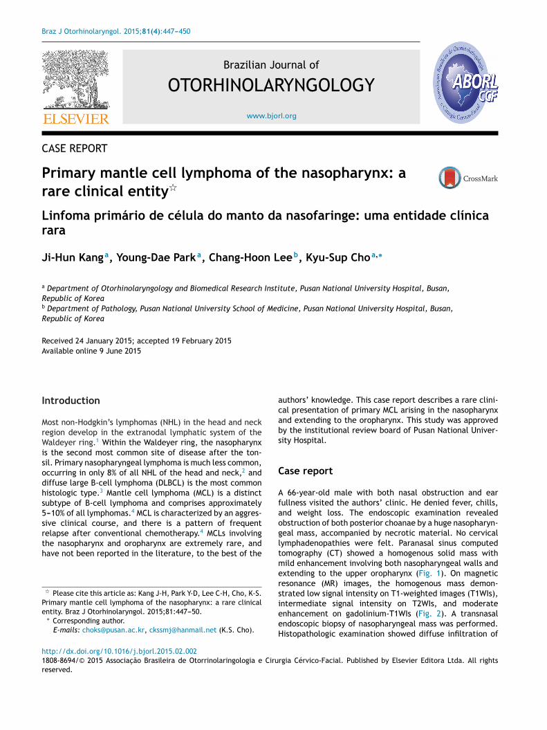
66-year-old male with both nasal obstruction and earullness visited the authors’ clinic. He denied fever, chills,nd weight loss. The endoscopic examination revealedbstruction of both posterior choanae by a huge nasopharyn-eal mass, accompanied by necrotic material. No cervicalymphadenopathies were felt. Paranasal sinus computedomography (CT) showed a homogenous solid mass withild enhancement involving both nasopharyngeal walls and
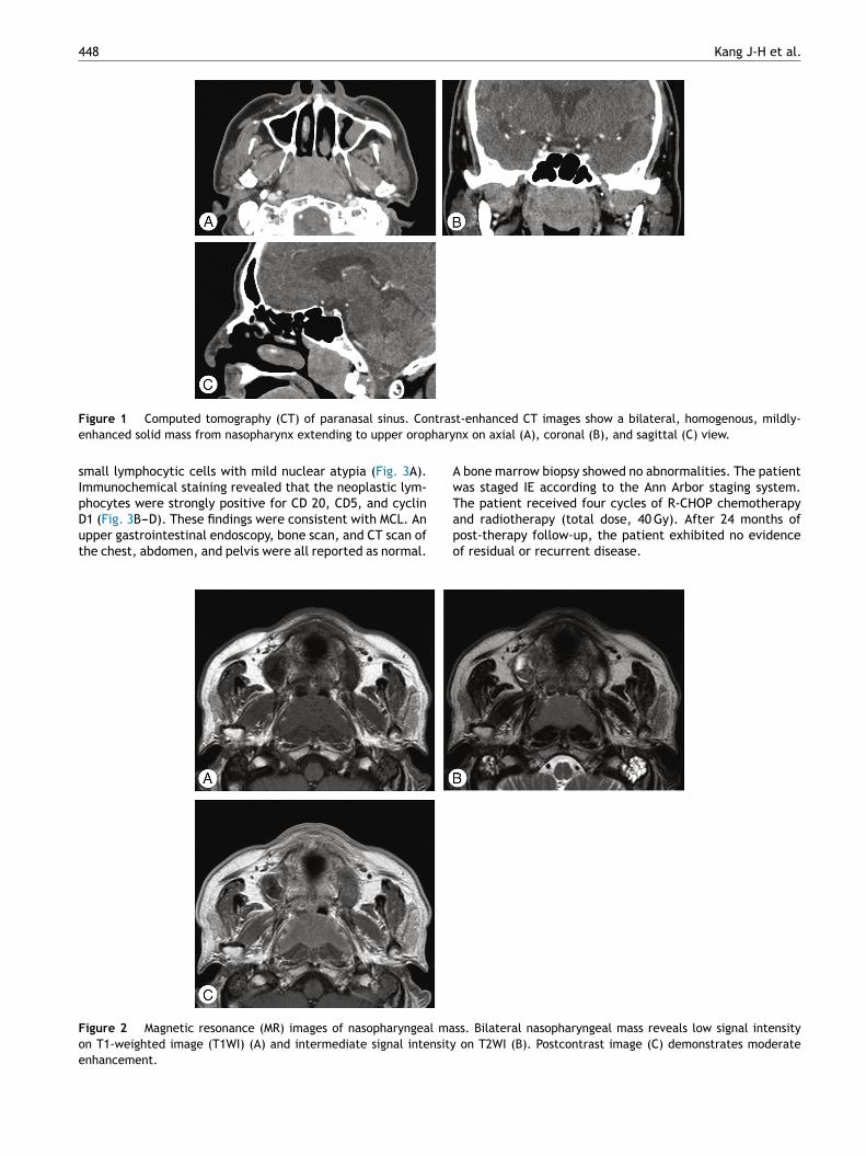
xtending to the upper oropharynx (Fig. 1). On magneticesonance (MR) images, the homogenous mass demon-trated low signal intensity on T1-weighted images (T1WIs),
ntermediate signal intensity on T2WIs, and moderatenhancement on gadolinium-T1WIs (Fig. 2). A transnasalndoscopic biopsy of nasopharyngeal mass was performed.istopathologic examination showed diffuse infiltration ofrgia Cérvico-Facial. Published by Elsevier Editora Ltda. All rights
448 Kang J-H et al.
F ntrase hary
sIpDut
AwT
Foe
igure 1 Computed tomography (CT) of paranasal sinus. Conhanced solid mass from nasopharynx extending to upper orop
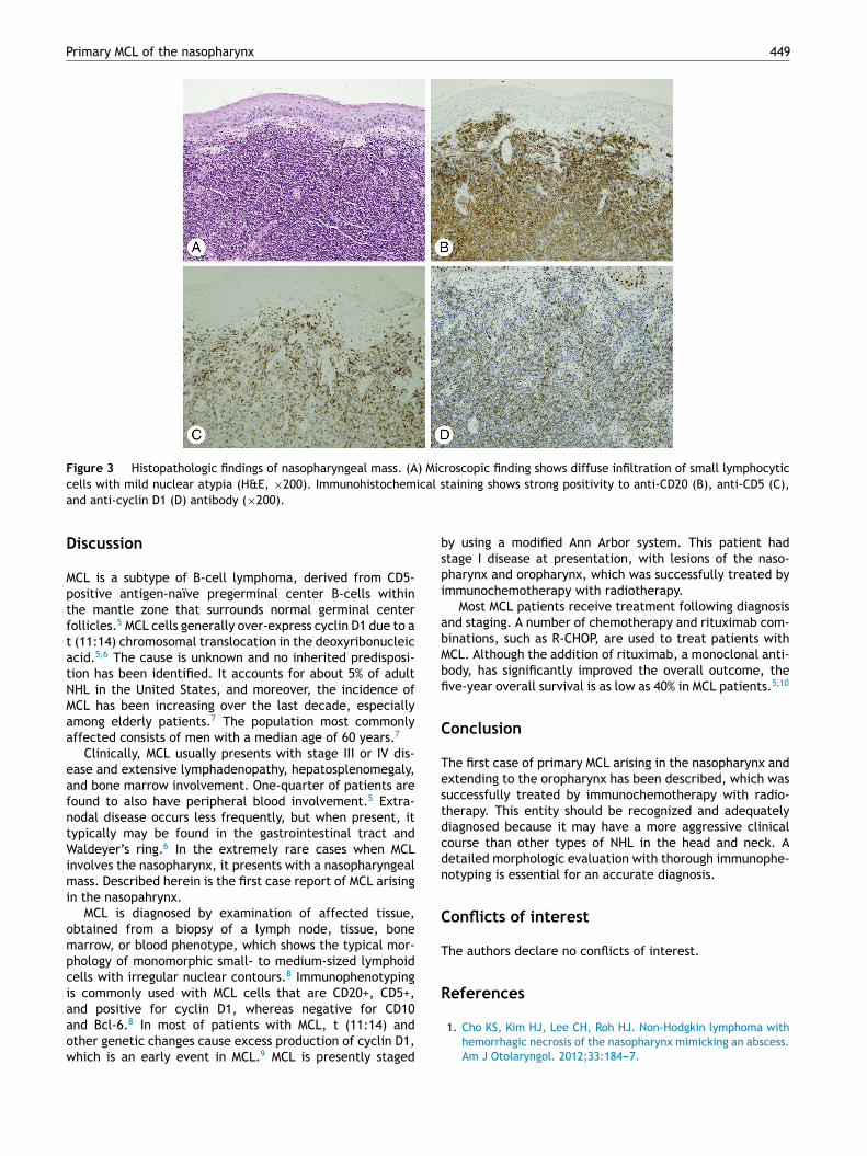
mall lymphocytic cells with mild nuclear atypia (Fig. 3A).mmunochemical staining revealed that the neoplastic lym-hocytes were strongly positive for CD 20, CD5, and cyclin
1 (Fig. 3B---D). These findings were consistent with MCL. Anpper gastrointestinal endoscopy, bone scan, and CT scan ofhe chest, abdomen, and pelvis were all reported as normal.apo
igure 2 Magnetic resonance (MR) images of nasopharyngeal man T1-weighted image (T1WI) (A) and intermediate signal intensitynhancement.
t-enhanced CT images show a bilateral, homogenous, mildly-nx on axial (A), coronal (B), and sagittal (C) view.
bone marrow biopsy showed no abnormalities. The patientas staged IE according to the Ann Arbor staging system.he patient received four cycles of R-CHOP chemotherapy
nd radiotherapy (total dose, 40 Gy). After 24 months ofost-therapy follow-up, the patient exhibited no evidencef residual or recurrent disease.ss. Bilateral nasopharyngeal mass reveals low signal intensity on T2WI (B). Postcontrast image (C) demonstrates moderate
Primary MCL of the nasopharynx 449
Figure 3 Histopathologic findings of nasopharyngeal mass. (A) Microscopic finding shows diffuse infiltration of small lymphocyticical s
bspi
abMbfi
C
Testdcdn
C
T
R
cells with mild nuclear atypia (H&E, ×200). Immunohistochemand anti-cyclin D1 (D) antibody (×200).
Discussion
MCL is a subtype of B-cell lymphoma, derived from CD5-positive antigen-naïve pregerminal center B-cells withinthe mantle zone that surrounds normal germinal centerfollicles.5 MCL cells generally over-express cyclin D1 due to at (11:14) chromosomal translocation in the deoxyribonucleicacid.5,6 The cause is unknown and no inherited predisposi-tion has been identified. It accounts for about 5% of adultNHL in the United States, and moreover, the incidence ofMCL has been increasing over the last decade, especiallyamong elderly patients.7 The population most commonlyaffected consists of men with a median age of 60 years.7
Clinically, MCL usually presents with stage III or IV dis-ease and extensive lymphadenopathy, hepatosplenomegaly,and bone marrow involvement. One-quarter of patients arefound to also have peripheral blood involvement.5 Extra-nodal disease occurs less frequently, but when present, ittypically may be found in the gastrointestinal tract andWaldeyer’s ring.6 In the extremely rare cases when MCLinvolves the nasopharynx, it presents with a nasopharyngealmass. Described herein is the first case report of MCL arisingin the nasopahrynx.
MCL is diagnosed by examination of affected tissue,obtained from a biopsy of a lymph node, tissue, bonemarrow, or blood phenotype, which shows the typical mor-phology of monomorphic small- to medium-sized lymphoidcells with irregular nuclear contours.8 Immunophenotypingis commonly used with MCL cells that are CD20+, CD5+,
and positive for cyclin D1, whereas negative for CD10and Bcl-6.8 In most of patients with MCL, t (11:14) andother genetic changes cause excess production of cyclin D1,which is an early event in MCL.9 MCL is presently stagedtaining shows strong positivity to anti-CD20 (B), anti-CD5 (C),
y using a modified Ann Arbor system. This patient hadtage I disease at presentation, with lesions of the naso-harynx and oropharynx, which was successfully treated bymmunochemotherapy with radiotherapy.
Most MCL patients receive treatment following diagnosisnd staging. A number of chemotherapy and rituximab com-inations, such as R-CHOP, are used to treat patients withCL. Although the addition of rituximab, a monoclonal anti-ody, has significantly improved the overall outcome, theve-year overall survival is as low as 40% in MCL patients.5,10
onclusion
he first case of primary MCL arising in the nasopharynx andxtending to the oropharynx has been described, which wasuccessfully treated by immunochemotherapy with radio-herapy. This entity should be recognized and adequatelyiagnosed because it may have a more aggressive clinicalourse than other types of NHL in the head and neck. Aetailed morphologic evaluation with thorough immunophe-otyping is essential for an accurate diagnosis.
onflicts of interest
he authors declare no conflicts of interest.
eferences
1. Cho KS, Kim HJ, Lee CH, Roh HJ. Non-Hodgkin lymphoma withhemorrhagic necrosis of the nasopharynx mimicking an abscess.Am J Otolaryngol. 2012;33:184---7.
4
10. Rasmussen PK. Diffuse large B-cell lymphoma and mantle cell
50
2. Cho KS, Kang DW, Kim HJ, Lee JK, Roh HJ. Differential diag-nosis of primary nasopharyngeal lymphoma and nasopharyngealcarcinoma focusing on CT, MRI, and PET/CT. Otolaryngol HeadNeck Surg. 2012;146:574---8.
3. Allam W, Ismaili N, Elmajjaoui S, Elgueddari BK, Ismaili M,Errihani H. Primary nasopharyngeal non-Hodgkin lymphomas: aretrospective review of 26 Moroccan patients. BMC Ear NoseThroat Disord. 2009;9:11.
4. Zhou Y, Wang H, Fang W, Romaguer JE, Zhang Y, Delasalle KB,et al. Incidence trends of mantle cell lymphoma in the UnitedStates between 1992 and 2004. Cancer. 2008;113:791---8.
5. Kang BW, Sohn SK, Moon JH, Chae YS, Kim JG, Lee SJ, et al. Clin-ical features and treatment outcomes in patients with mantle
cell lymphoma in Korea: study by the consortium for improvingsurvival of lymphoma. Blood Res. 2014;49:15---21.6. Chang CC, Rowe JJ, Hawkins P, Sadeghi EM. Mantle cell lym-phoma of the hard palate: a case report and review of
Kang J-H et al.
the differential diagnosis based on the histomorphology andimmunophenotyping pattern. Oral Surg Oral Med Oral PatholOral Endod. 2003;96:316---20.
7. Aschebrook-Kilfoy B, Caces DB, Ollberding NJ, Smith SM,Chiu BC. An upward trend in the age-specific incidence pat-terns for mantle cell lymphoma in the USA. Leuk Lymphoma.2013;54:1677---83.
8. Vose JM. Mantle cell lymphoma: 2013 update on diagnosis,risk stratification, and clinical management. Am J Hematol.2013;88:1082---8.
9. Pileri SA, Falini B. Mantle cell lymphoma. Haematologica.2009;94:1488---92.
lymphoma of the ocular adnexal region, and lymphoma of thelacrimal gland: an investigation of clinical and histopathologicalfeatures. Acta Ophthalmol. 2013;91:1---27.