printedin some properties of influenza virus...
TRANSCRIPT

JOURNAL OF VIROLOGY, Sept. 1969, p. 219-225Copyright © 1969 American Society for Microbiology
Some Properties of Influenza Virus NucleocapsidsD. W. KINGSBURY AND R. G. WEBSTER
Laboratory of Virology, St. Jude Children's Research Hospital, and the Uniiversity of Teniessee MedicalUnlits, Memphis, Tennessee 38101
Received for publication 23 April 1969
Nucleocapsids released from influenza virions by sodium deoxycholate sedi-mented heterogeneously in sucrose gradients. Highly infectious virus (complete)preparations yielded nucleocapsids with peak distributions at 64 and 56S; vonMagnus type virus (incomplete) lacked 64S nucleocapsids. Treatment of influenzavirus nucleocapsids with pancreatic ribonuclease rendered the associated viralribonucleic acid (RNA) molecules acid-soluble, indicating that capsid proteins donot completely surround the viral RNA's. However, the capsid proteins remainedassociated after enzymatic hydrolysis of the RNA, as judged by persistently highsedimentation rates. Sedimentation rates of viral nucleocapsids reflected the sedi-mentation rates of the associated RNA's: 64S nucleocapsids contained 18S RNA,whereas 56S nucleocapsids contained 15S RNA, although in both cases RNA'ssedimenting at 4 to 13S were also recovered. Furthermore, just as incomplete virionslacked 645 nucleocapsids, they also lacked 18S RNA. These findings support thehypothesis that the influenza virus genome is divided among several distinct piecesof RNA.
Many recent analyses of ribonucleic acid(RNA) extracted from influenza virions (3, 6, 7,19, 20, 22, 23, 27) indicate that the viral genome isnot a single large RNA molecule, but is dividedamong several small RNA molecules. If differentviral genes reside in different RNA molecules,they might simply reassort in mixed infections,explaining the extraordinarily high genetic re-combination frequencies observed in influenzavirus crosses (10).RNA is a very labile polymer, and it is possible
that suitable techniques have not yet been appliedto prevent a large influenza virus genome frombreaking during extraction. To obtain more in-formation on this problem, we have examinedproperties of influenza virus nucleocapsids, ex-
pecting that the viral RNA might be preserved inits native state if kept in association with capsidproteins, and that the true size of the viral RNAmight then be revealed upon removal of the capsidproteins.Thus, with another virus having rod-shaped
nucleocapsids enclosed in a lipid and proteinenvelope, Newcastle disease virus (NDV), disrup-tion of virions with sodium deoxycholate (DOC)released the large nucleocapsids apparently intact(16). NDV nucleocapsids were shown to sedimenthomogeneously and to contain homogeneous 50SRNA (16).Now, using DOC, we have found that several
sedimenting classes of nucleocapsids are released
from influenza virions and that different viralRNA is associated with each class. In furthercontrast to NDV nucleocapsids, which were ribo-nuclease resistant, influenza virus capsid proteinsdid not prevent the hydrolysis of the viral RNAby pancreatic ribonuclease.
Similar findings have recently been made by P.Duesberg (J. Mol. Biol., in press).
MATERIALS AND METHODS
Virus. The WSN strain of influenza virus (9) waspropagated by intra-allantoic inoculation of 10-dayembryonated hens' eggs with 10' to 103 EID6o (50%egg infectious doses) of virus, followed by incubationat 37 C for 40 hr. Allantoic fluid containing virus wascentrifuged at 1,500 X g for 20 min to remove debris,and virus was concentrated by centrifugation at 40,000X g for 30 min. Virus pellets were suspended in phos-phate-buffered saline (PBS) containing 1% gelatinand stored at -70 C. The final preparation had 109EID,5 and 1.8 X 10³ hemagglutinating units (HAU)per ml.
Radioisotopically labeled virus. To produce labeledvirus with a low ratio of EIDr, to HAU [incomplete orvon Magnus (28) virus], primary chick embryofibroblast monolayer cultures in plastic dishes (150mm in diameter) were infected with 3 X 10⁸ ElI15, ofWSN in 3 ml of PBS, an input multiplicity of approxi-mately 3 EID5o/cell. After 30 min at 30 C, the inoc-ulum was removed and replaced by 10 ml of one ofseveral labeling media described below. Incubation at37 C in an atmosphere of5% CO2 in air was continuedfor 24 hr when virus was collected from the fluid.
219
Vol. 4, No. 3Printed in U.S.A.
on May 26, 2018 by guest
http://jvi.asm.org/
Dow
nloaded from
KINGSBURY AND WEBSTER
Ratios of EID50 to HAU for such preparations wereapproximately 103.
Labeled virus with a high ratio of EID50 to HAUwas obtained similarly, but an infecting input multi-plicity of approximately 3 X 10⁻³ EID5o/cell wasused, and labeling media were added after preliminaryincubation for 24 hr at 37 C in bicarbonate-bufferedEarle's saline containing 0.5% lactalbumin hydroly-sate. Scant virus was produced during the first 24 hrafter infection at this low multiplicity, but high titersaccrued in the next 24 hr. Preparations with EID50 toHAU ratios of 7 X 10⁴ to 2 X 10⁵ were obtained.These EID50 to HAU ratios were significantly higherthan the ratio of virus produced after infection at highmultiplicity, but were lower than the ratio of ouregg-grown virus. However, we believe our results arerepresentative of infectious virus, since nucleocapsidsfrom these virions sedimented like those described byPaucker et al. (21), and P. Duesberg (J. Mol. Biol.,in press), and the RNA sedimented as described byothers (3, 7, 19, 20, 22).
Labeling media. The medium for labeling viral pro-teins contained Eagle's salts (8), dextrose, vitamins, andglutamine, with 10% of the recommended concentra-tions of amino acids other than glutamine. Either 5/,c/ml of a mixture of 15 14C-amino acids (NewEngland Nuclear Corp., Boston, Mass.; averagespecific activity approximately 100 mc/mmole) or25 Ac/ml of a mixture of 16 3H-amino acids (SchwarzBioResearch, Orangeburg, N.Y.; average specificactivity approximately 1,000 mc/mmole) were present.When virus with 14C-labeled proteins and 3H-la-beled RNA was desired, 100,uc/ml of 3H-5-uridine(Schwarz BioResearch, specific activity 20 c/mmole)was included with the "4C-amino acids.To label virus with 32P, 100 ,c/ml of carrier-free
32P-orthophosphate was included in phosphate-free,bicarbonate-buffered Earle's saline containing 0.5%lactalbumin hydrolysate.
Isolation of radioisotopically-labeled virus. This wasperformed as described for NDV (15), employingcentrifugation onto 30% (w/w) potassium tartrate andcentrifugation through 16% (w/w) potassium tartrate.The virus pellets were suspended in TE buffer [10-3 Msodium ethylenediaminetetraacetate, 5 X 10- Mtris(hydroxymethyl)aminomethane (Tris)-hydrochlo-ride, pH 7.4], and were kept at 4 C for up to 3 weekswith no change in properties studied in the presentwork.
Sucrose gradient rate zonal centrifugation. Forsedimentation analysis of viral nucleocapsids, 0.1 mlof 10% DOC (pH 7.4) was added to 2 ml of virus sus-pension in TE, and the mixture was immediatelylayered on a 29-ml linear 5 to 20% sucrose gradient inTE. After centrifugation at 18,000 rev/min 16 hr (orequivalent rev/min2 X time) in a Spinco model SW25.1 swinging bucket rotor at 4 C, 1-ml fractions werecollected with an ISCO density gradient fractionator(Instrumentation Specialties, Inc., Lincoln, Neb.). TEbuffer was used routinely in place of the phosphate-buffered saline used in the earlier study of NDVnucleocapsids (16), since aggregation of DOC occa-sionally occurred in the saline-containing buffer (24).However, the sedimentation properties and ribonu-
clease susceptibility of influenza virus nucleocapsidswere identical in both buffers.To prepare RNA from virions, virus was suspended
in phosphate-buffered saline containing 0.25%Pronase [Calbiochem, Los Angeles, Calif. (14)],incubated at 37 C for 3 hr, made 1% with respect tosodium dodecyl sulfate (SDS), and extracted withphenol at room temperature. The aqueous phase fromthe phenol extraction was directly layered on a 29-mllinear 5 to 20% sucrose gradient in TE and centrifugedat 4 C for 42 hr at 22,500 rev/min. Since ultravioletabsorbance was not monitored, the presence of phenolwas of no consequence
Determination of radioactivity. Acid-insoluble radio-activity was measured by precipitation of up to 1 ml ofsample with 3 ml of 5% trichloroacetic acid in thepresence of 500,.g of bovine serum albumin as carrier.When DOC was present in samples, it was precipitatedby the acid, but this did not affect the counting effici-encies of any of the isotopes employed. Precipitateswere collected by centrifugation, washed once with 3ml of 5% trichloroacetic acid, dissolved in 1 ml ofNCS (Amersham-Searle, Chicago, Ill.), mixed with10 ml of a toluene-based scintillant, and counted in aliquid scintillation spectrometer. Gain controls wereadjusted as indicated by Bush (4) for optimal resolu-tion of 3H-14C or 3H-32P combinations.
Precipitation of viral nucleocapsids with antiserum.The procedure was patterned according to the methodof Scharff et al. (26). After sucrose gradient centrif-ugation of DOC-treated 3H-amino acid-labeled virus,gradient fractions were dialyzed against TE. To 0.25ml of each fraction was added 0.05 ml of 1/100 dilu-tion of guinea pig serum containing antibodies toinfluenza A (PR 8) soluble antigen. This serum, a giftfrom H. G. Pereira, had been prepared according tothe method of Lief and Henle (18), and did not inhibitWSN hemagglutination when diluted more than 1 :10.The mixture was incubated at 37 C for 2 hr. To formsedimentable precipitates, 0.05 ml of rabbit antiserumto guinea pig gamma globulin was added to eachsample at optimal concentration, as determined byequivalence tests, and incubation was continued for 2hr at 37 C and 18 hr at 4 C. The precipitates werecollected by centrifugation at 1,000 X g for 20 min,washed twice with 3 ml of 0.14 M NaCl, precipitatedwith trichloroacetic acid, and were processed forcounting as described above. To correct for volumechanges during dialysis, a portion of each dialyzedsample was precipitated directly with acid and thecounts were compared with acid-precipitable countsin each sample before dialysis, as described by Scharffet al. (26).
RESULTSSedimentation properties of influenza virus nu-
cleocapsids. In view of evidence that incomplete(28) influenza virions contain less RNA andnucleocapsid protein ("soluble antigen") thancomplete virions (1, 17), both typeis of virionswere prepared in labeled form to examine thesedimentation behavior of subviral structures con-taining RNA. After disruption of virions with
220 J. VIROL.
on May 26, 2018 by guest
http://jvi.asm.org/
Dow
nloaded from
INFLUENZA VIRUS NUCLEOCAPSIDS
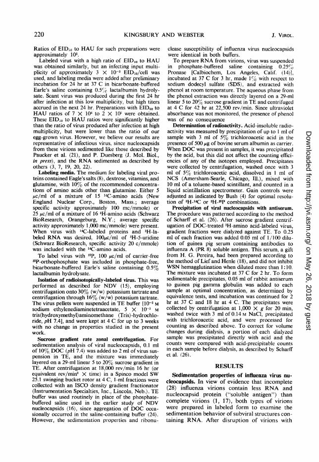
DOC, some of the labeled RNA sedimented morerapidly than viral RNA isolated with SDS andphenol (see below). The most rapidly sedimentingradioactive component of DOC-treated 3H-uri-dine-labeled complete virus was approximately64S (Fig. 1, fraction 20), with another componentdiscernible at 56S (Fig. 1, fraction 17), relative to74S ribosomes and 60S and 45S ribosomal sub-units from chick embryo fibroblasts centrifugedin 0.01 M Tris-hydrochloride, 0.01 M KCI, 0.0015M MgCl2, and 0.5%O DOC (pH 7.4). In contrast,32P-labeled incomplete virus yielded little 64Smaterial relative to material sedimenting at 56S(Fig. 1, fraction 17). Occasionally, minor compo-nents were seen at about 45S (Fig. 1, fraction 14),and at about 35S (Fig. 3A, fraction 11) in incom-plete virus preparations. Radioactivity remainingin the upper 10 ml of such gradients varied inamount from preparation to preparation (see alsoFig. 2, 3, and 6); although slow sedimenting,virus-specific components may be present there, itis likely that obscuring contaminants were presentas well.Some labeled proteins sedimented with the viial
RNA's (Fig. 2A, 3A, and 4, fractions 14 to 30) asexpected if the RNA's were associated with viralcapsid proteins in nucleocapsids.
12L . 25455 6OS 74
10 20 30
5 X
z o~~~I
labeled 20 30
FciG.ty 1. dsucrsegrdient raterzoalscentrifugtiondof
virions:,laee3wtH-curid/ine an(icomplete virios,;
Symbols:0,t/ H ontcomplete virus) ;
T'Ounsm (inomleeTirs)
1LA l45S 60SG 74S B 4S 62S 74S
\ll f_ ,L\i \
3V- 3
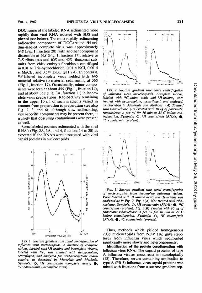
FIG 2 Sucrose gradient rate zonal centrifugationof influenza virus nucleocapsids. Complete virions,labeled with '4C-amino acids and 3H-uridine, weretreated with deoxycholate, centrifuged, and analyzed,as described in Materials and Methods. (A) Treatedwith ribonuclease. (B) Treated with JO ,utg ofpancreaticribonuclease A per ml for 10 min at 23 C before cen-trifugation. Symbols: 0, 3H counts/mmn (RNA);-*14C counts/jmmn (protein).
., ,0
TO BOTOTO| OT
o! 1 .! g 0 -
7P EFFLUENT VOLJME [m!-) TM O EFFLUENJT VOLUMVE -Im) BfO
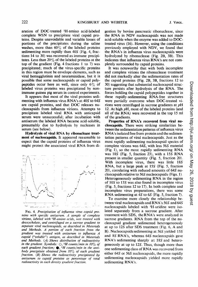
FIG. 3. Sucrose gradient rate zonal centrifugationof nucleocapsids from incomplete influenza virions.Virus labeled with 4C-amino acids and3H-uridine wasanalyzed as in Fig. 2. Fig. 3(A) Not treated with ribo-nuclease. Symbols: 0, 'H counts/mmn (RNA);*@, 14Ccounts/mi (protein). Fig. 3(B) Treated with 10Mfg ofpancreaticribonuckease A per ml for 10min at 23 Cbefore centrifugation. Symbols: 0, 'H counts/mmn(RNA)'; * 4C counts/min(protein ).
Thus, methods which yielded homogeneous200S nucleocapsids from NDV (16) gave struc-tures from influenza virus which sedimentedsignificantly more slowly and heterogeneously.
Identification of the protein cosedimenting withinfluenza virus RNA. The capsid proteins of typeA influenza viruses cross-react immunologically(18). Therefore, serum containing antibodies totype A (PR 8) influenza virus capsid proteins wasmixed with fractions from a sucrose gradient sep-
VOL. 4, 1969 221
on May 26, 2018 by guest
http://jvi.asm.org/
Dow
nloaded from
KINGSBURY AND WEBSTER
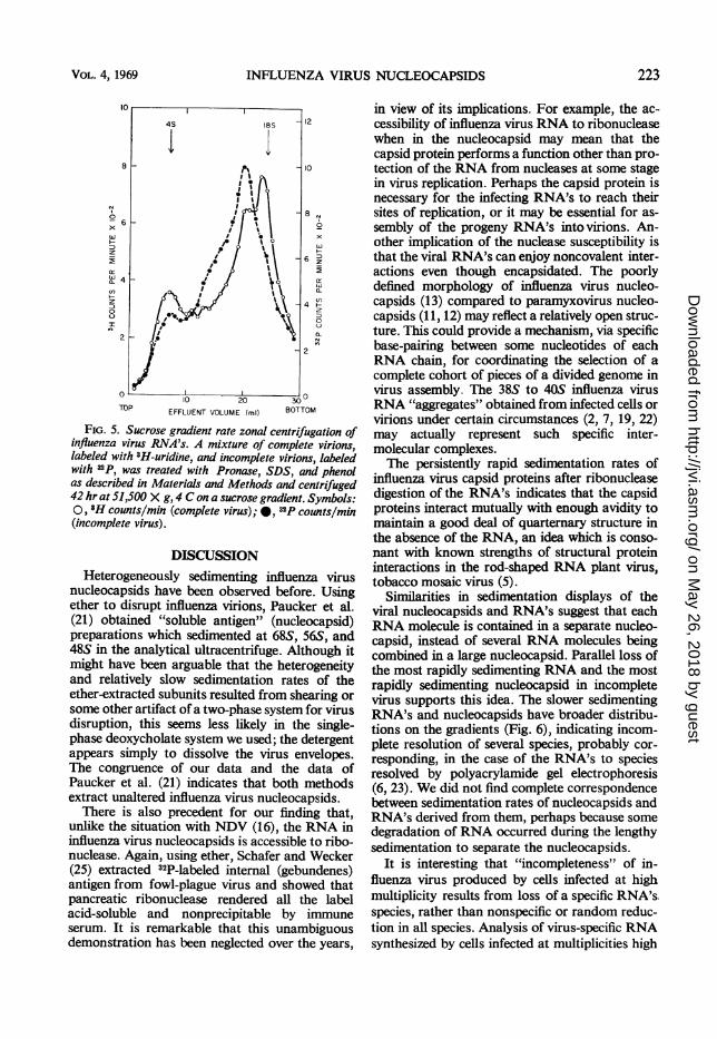
aration of DOC-treated 3H-amino acid-labeledcomplete WSN to precipitate viral capsid pro-teins. Despite unavoidable and variable losses ofportions of the precipitates during the salinewashes, more than 60%o of the labeled proteinssedimenting more rapidly than 405 (Fig. 4, frac-tions 14 to 30) was recovered as immune precipi-tates. Less than 20% of the labeled proteins at thetop of the gradient (Fig. 4 fractions 1 to 7) wasprecipitated; much of the virus-specific proteinsin this region must be envelope elements, such asviral hemagglutinin and neuraminidase, but it ispossible that some nucleocapsids or capsid poly-peptides occur here as well, since only 6%Yo oflabeled virus proteins was precipitated by non-immune guinea pig serum in control experiments.
It appears that most of the viral proteins sedi-menting with influenza virus RNA's at 40S to 64Sare capsid proteins, and that DOC releases nu-cleocapsids from influenza virions. Attempts toprecipitate labeled viral RNA with anticapsidserum were unsuccessful; after incubation withantiserum the labeled RNA became acid-soluble,presumably due to hydrolysis by nucleases inserum (see below).
Hydrolysis of viral RNA by ribonuclease treat-ment of nucleocapsids. It appeared reasonable toexpect that the capsid proteins of influenza virusmight protect the associated viral RNA from di-
r4 -
2o
TO \FLE ` L V
FIG. 4. Precipitation of influenza virus capsid pro-teins with specific antiserum. A sample of completevirions, labeled with 3H-amino acids, was treated withdeoxycholate, and centrifuged on a sucrose gradient toseparate viral nucleocapsids, as described in Materialsand Methods. A portion of each fraction from thegradient was treated with antiserum to influenza Acapsid ("soluble") antigen, as described in Materialsand Methods. (A) Shows distribution of radioactivityin the gradient. Symbols: 0, 3H counts/min in 10% ofeach gradient fraction; 0, 3H counts/mill in the anti-body precipitate obtained from 10%0 of each gradientfraction. (B) Shows the radioactivity precipitated byantiserum to capsid proteins as percentage of totalradioactivity in each density gradient fraction.
gestion by bovine pancreatic ribonuclease, sincethe RNA in NDV nucleocapsids was not madeacid-soluble when the enzyme was added to DOC-treated virus (16). However, using the conditionspreviously employed with NDV, we found thatthe RNA's in influenza virus nucleocapsids werehydrolyzed by ribonuclease (Fig. 2B, 3B). Thisindicates that influenza virus RNA's are not com-pletely surrounded by capsid proteins.
It was noteworthy that with both incompleteand complete virions the ribonuclease treatmentdid not markedly alter the sedimentation rates ofthe capsid proteins (Fig. 2B, 3B, fractions 12 to30) suggesting that substantial nucleocapsid struc-ture persists after hydrolysis of the RNA. Theforces holding the capsid polypeptides together inthese rapidly-sedimenting RNA-free structureswere partially overcome when DOC-treated vi-rions were centrifuged in sucrose gradients at pH12. At high pH, most of the labeled proteins (andall of the RNA) were recovered in the top 15 mlof the gradients.
Properties of RNA's recovered from viral nu-cleocapsids. There were striking similarities be-tween the sedimentation patterns of influenza virusRNA's isolated free from protein and the sedimen-tation patterns of viral nucleocapsids. Just as themost rapidly sedimenting nucleocapsid species ofcomplete virions was 64S, with less 56S material(Fig. 1), so the most rapidly sedimenting RNAwas 18S (Fig. 5, fraction 23), with a 15S RNApresent in smaller quantity (Fig. 5, fraction 20).With incomplete virus, there was little 18SRNA, but a large peak at 155 (Fig. 5, fraction20), correlating with reduced amounts of 645 nu-cleocapsids relative to 565 nucleocapsids (Figs. 1).Heterogeneously sedimenting RNA in the regionof 10S to 13S was also found in incomplete virus(Fig. 5, fractions 12 to 17). In both complete andincomplete virus preparations, there was someRNA sedimenting at 4S to 65 (Fig. 5, fraction 7).To examine more closely the relationship be-
tween viral nucleocapsids and RNA's 56S and 64Snucleocapsids labeled with 3H-uridine were iso-lated separately from a sucrose gradient. Aftertreatment with SDS, the RNA's were analyzed insucrose gradients. RNA from the top of the nu-cleocapsid gradient sedimented heterogeneouslyat up to 12S after SDS treatment (Fig. 6, A andB). Nucleocapsids sedimenting at 56S yielded 15Sand 8S RNA's, whereas 64S nucleocapsids gaveRNA's sedimenting sharply at 185 and hetero-geneously at up to 12S. Thus, though more thanone sedimenting class ofRNA was recovered fromeither 64S or 56S nucleocapsids, the more rapidlysedimenting nucleocapsids yielded more rapidlysedimenting RNA's.
222 J. VIROL.
0. 00
00-------oo"".,V --
11r-, I,3.
.0
lpI0 4'! o
on May 26, 2018 by guest
http://jvi.asm.org/
Dow
nloaded from
INFLUENZA VIRUS NUCLEOCAPSIDS
8
cL 4
z
7-
I
2cj
O _T'P
10 20
EFFLUENT VOLUME (ml) 30BTBOTTOM
FIG. 5. Sucrose gradient rate zonal centrifugation ofinfluenza virus RNA's. A mixture of complete virions,labeled with 3H-uridine, and incomplete virions, labeledwith 32p, was treated with Pronase, SDS, and phenolas described in Materials and Methods and centrifuged42 hr at 51,500 X g, 4 C on a sucrose gradient. Symbols:0, 3H counts/min (complete virus);*, "2P counts/min(incomplete virus).
DISCUSSIONHeterogeneously sedimenting influenza virus
nucleocapsids have been observed before. Usingether to disrupt influenza virions, Paucker et al.(21) obtained "soluble antigen" (nucleocapsid)preparations which sedimented at 68S, 56S, and48S in the analytical ultracentrifuge. Although itmight have been arguable that the heterogeneityand relatively slow sedimentation rates of theether-extracted subunits resulted from shearing orsome other artifact of a two-phase system for virusdisruption, this seems less likely in the single-phase deoxycholate system we used; the detergentappears simply to dissolve the virus envelopes.The congruence of our data and the data ofPaucker et al. (21) indicates that both methodsextract unaltered influenza virus nucleocapsids.
There is also precedent for our finding that,unlike the situation with NDV (16), the RNA ininfluenza virus nucleocapsids is accessible to ribo-nuclease. Again, using ether, Schafer and Wecker(25) extracted 32P-labeled internal (gebundenes)antigen from fowl-plague virus and showed thatpancreatic ribonuclease rendered all the labelacid-soluble and nonprecipitable by immuneserum. It is remarkable that this unambiguousdemonstration has been neglected over the years,
in view of its implications. For example, the ac-cessibility of influenza virus RNA to ribonucleasewhen in the nucleocapsid may mean that thecapsid protein performs a function other than pro-tection of the RNA from nucleases at some stagein virus replication. Perhaps the capsid protein isnecessary for the infecting RNA's to reach theirsites of replication, or it may be essential for as-sembly of the progeny RNA's into virions. An-other implication of the nuclease susceptibility isthat the viral RNA's can enjoy noncovalent inter-actions even though encapsidated. The poorlydefined morphology of influenza virus nucleo-capsids (13) compared to paramyxovirus nucleo-capsids (11, 12) may reflect a relatively open struc-ture. This could provide a mechanism, via specificbase-pairing between some nucleotides of eachRNA chain, for coordinating the selection of acomplete cohort of pieces of a divided genome invirus assembly. The 38S to 40S influenza virusRNA "aggregates" obtained from infected cells orvirions under certain circumstances (2, 7, 19, 22)may actually represent such specific inter-molecular complexes.The persistently rapid sedimentation rates of
influenza virus capsid proteins after ribonucleasedigestion of the RNA's indicates that the capsidproteins interact mutually with enough avidity tomaintain a good deal of quarternary structure inthe absence of the RNA, an idea which is conso-nant with known strengths of structural proteininteractions in the rod-shaped RNA plant virus,tobacco mosaic virus (5).
Similarities in sedimentation displays of theviral nucleocapsids and RNA's suggest that eachRNA molecule is contained in a separate nucleo-capsid, instead of several RNA molecules beingcombined in a large nucleocapsid. Parallel loss ofthe most rapidly sedimenting RNA and the mostrapidly sedimenting nucleocapsid in incompletevirus supports this idea. The slower sedimentingRNA's and nucleocapsids have broader distribu-tions on the gradients (Fig. 6), indicating incom-plete resolution of several species, probably cor-responding, in the case of the RNA's to speciesresolved by polyacrylamide gel electrophoresis(6, 23). We did not find complete correspondencebetween sedimentation rates of nucleocapsids andRNA's derived from them, perhaps because somedegradation ofRNA occurred during the lengthysedimentation to separate the nucleocapsid-s.
It is interesting that "incompleteness" of in-fluenza virus produced by cells infected at highmultiplicity results from loss of a specific RNA's.species, rather than nonspecific or random reduc-tion in all species. Analysis of virus-specific RNAsynthesized by cells infected at multiplicities high
18S4S
223VOL. 4, 1969
on May 26, 2018 by guest
http://jvi.asm.org/
Dow
nloaded from

KINGSBURY AND WEBSTER
6x
a-C-)
BOTTOM TOPEFFLUENT VOLUME (ml)
FIG. 6. Sucrose gradient rate zonal centrifugation of influenza virus nucleocapsids and RNA's. A preparationof complete virus labeled with 3H-uridine was treated with deoxycholate and centrifuged on a sucrose gradient,as described in Materials and Methods. After fractionation of the gradient and analysis of a portion of each fr c-tion for acid precipitable radioactivity, fractions were pooled; RNA was extracted from each group offractionswith phenol and centrifuged on sucrose gradients at 51,500 X g for 24 hr, 4 C. RNA extractedfrom chick embryofibroblasts labeled with 14C-adenine was added to the viral RNA samples to provide sedimentation markers. (A)Deoxycholate-treated virus; (B) RNA from fractions I to 6 (b); (C) RNA from fractions 13 to 18 (c); (D) RNAfrom fractions 19 to 24 (d). Symbols: 0, 3H count/min (viral RNA); 0, 14C count/min (cell RNA).
enough to produce such incomplete virus revealsreduced amounts of this very species (6, 7), sug-
gesting that the basis of incompleteness in in-fluenza virus is a defect in RNA biosynthesisrather than in packaging of the RNA into virions.Future work may reveal whether the largest in-fluenza virus RNA's are simply synthesized in-smaller amounts or whether their synthesis isinitiated, but terminates prematurely, producingsmaller RNA fragments.
ACKNOWLEDGMENTS
This investigation was supported by Public Health Servicegrant Al 05343 from the National Institute of Allergy and Infec-tious Diseases, by a grant from the John A. Hartford Foundation,Inc., and by ALSAC. David W. Kingsbury was the recipient of aPublic Health Service Career Development Award HD 14,491.
R. A. Sproggs provided skilled technical assistance.
LITERATURE CITED
1. Ada, G. L., and B. T. Perry. 1956. Influenza virus nucleic acid:Relationship between biological characteristics of the virus
224
A
8
60
x
0J' 4
2
0
x
a-
MIn
J. VIROL.
0
C).Q)
Q)I
0x
a_
TOP
on May 26, 2018 by guest
http://jvi.asm.org/
Dow
nloaded from
INFLUENZA VIRUS NUCLEOCAPSIDS
particle and properties of the nucleic acid. J. Gen. Micro-biol. 14:623-633.
2. Agrawal, H. O., and G. Bruening. 1966. Isolation of high-molecular-weight, P32 labeled influenza virus ribonucleicacid. Proc. Nat. Acad. Sci. U.S.A. 55:818-825.
3. Barry, R. D., and P. Davies. 1968. The sedimentation of in-fluenza virus and its RNA in sucrose density gradients. J.Gen. Virol. 2:59-69.
4. Bush, E. T. 1964. Liquid scintillation counting of double-labeled samples. Choice of counting conditions for bestprecision in two-channel counting. Anal. Chem. 36:1082-1089.
5. Caspar, D. L. D. 1963. Assembly and stability of the tobaccomosaic virus particles. Advan. Protein Chem. 18:37-121.
6. Duesberg, P. H. 1968. The RNA's of influenza virus. Proc.Nat. Acad. Sci. U.S.A. 59:930-937.
7. Duesberg, P. H., and W. S. Robinson. 1967. On the structure
and replication of influenza virus. J. Mol. Biol. 25:383-405.8. Eagle, H. 1959. Amino acid metabolism in mammalian cell
culture. Science 130:432-437.9. Francis, T., Jr., and A. E. Moore. 1940. A study of the neuro-
tropic tendency in strains of the virus of epidemic influenza.J. Exp. Med. 72:717-728.
10. Hirst, G. K. 1962. Genetic recombination with Newcastledisease virus, polioviruses, and influenza. Cold SpringHarbor Symp. Quant. Biol. 27:303-308.
11. Home, R. W., A. P. Waterson, P. Wildy, and A. E. Farnham.1960. The structure and composition of the myxoviruses. L.
Electron microscope studies of the structure of myxovirusparticles by negative staining techniques. Virology 11:79-98.
12. Hosaka, Y., and K. Shimizu. 1968. Lengths of the nucleo-capsids of Newcastle disease and mumps viruses. J. Mol.Biol. 35:369-373.
13. Hoyle, L., R. W. Home, and A. P. Waterson. 1961. The struc-ture and composition of the myxoviruses. II. Componentsreleased from the influenza virus particle by ether. Virology13:448-459.
14. Huppert, J., and M. Semmel. 1965. Suppression de l'activiteribonucleasique par la pronase. Biochim. Biophys. Acta108:501-503.
15. Kingsbury, D. W. 1966. Newcastle disease virus RNA. I.Isolation and preliminary characterization of RNA fromvirus particles. J. Mol. Biol. 18:195-203.
16. Kingsbury, D. W., and R. W. Darlington. 1968. Isolation andproperties of Newcastle disease virus nucleocapsid. J.Virol. 2:248-255.
17. Lief, F. S., and W. Henle. 1956. Studies on the soluble antigenof influenza virus. III. The decreased incorporation of Santigen into elementary bodies of increasing incomplete-ness. Virology 2:782-797.
18. Lief, F. S., and W. Henle. 1959. Methods and procedures foruse of complement fixation technique in type- and strain-specific diagnosis of influenza. Bull. World Health Organ.20:411-420.
19. Nayak, D. P., and M. A. Baluda. 1967. Isolation and partialcharacterization of nucleic acid of influenza virus. J. Virol.1:1217-1223.
20. Noll, H., and E. Stutz. 1968. The use of sodium and lithiumdodecyl sulfate in nucleic acid isolation, p. 129-155. InL. Grossman and K. Moldave (ed.), Methods in enzy-mology, vol. 12B. Academic Press Inc., New York.
21. Paucker, K., A. Birch-Andersen, and P. von Magnus. 1959.Studies on the structure of influenza virus. L. Componentsof infectious and incomplete particles. Virology 8:1-20.
22. Pons, M. W. 1967. Studies on influenza virus ribonucleic acid.Virology 31:523-531.
23. Pons, M. W., and G. K. Hirst. 1968. Polyacrylamide gelelectrophoresis of influenza virus RNA. Virology 34:385-388.
24. Rich, A., and D. M. Blow. 1958. Formation of a helicalsteroid complex. Nature 182:423-426.
25. Schafer, W., and E. Wecker. 1958. Uber die Wirkung von
Ribonuclease auf 32P-markiertes, 'gebundenes Antigen', desVirus der Klassischen Geflugelpest. Arch. Exp. Veterinaer-med. 12:418-422.
26. Scharff, M. D., A. J. Shatkin, and L. Levintow. 1963. Asso-ciation of newly formed viral protein with specific poly-ribosomes. Proc. Nat. Acad. Sci. U.S.A. 50:686-694.
27. Sokol, F., and S. Schramek. 1963. Comparison of differentpreparations of influenza virus ribonucleic acid. Biochem.Biophys. Res. Commun. 12:21-26.
28. von Magnus, P. 1951. Propagation of the PR 8 strain of in-fluenza A virus in chick embryos. HI. The formation of"incomplete" virus following inoculation of large doses ofseed virus. Acta Pathol. Microbiol. Scand. 28:278-293.
VOL. 4, 1969 225
on May 26, 2018 by guest
http://jvi.asm.org/
Dow
nloaded from