projectionofanimmunological …mcb.berkeley.edu/courses/mcb150/admin/aire.pdf · sis–ectodermal...
TRANSCRIPT
32. N. M. Stano, S. S. Patel, J. Mol. Biol. 315, 1009 (2002).33. L. G. Brieba, R. Sousa, Biochemistry 40, 3882 (2001).34. P. A. Bullough, F. M. Hughson, J. J. Skehel, D. C. Wiley,
Nature 371, 37 (1994).35. J. Kuriyan, M. O’Donnell, J. Mol. Biol. 234, 915
(1993).36. V. A. Bloomfield, D. M. Crothers, I. Tinoco Jr., Nucleic
Acids: Structures, Properties, and Functions (Univer-sity Science Books, Sausalito, CA, 2000).
37. F. M. Richards, Annu. Rev. Biophys. Bioeng. 6, 151(1977).
38. G. A. Diaz, M. Rong, W. T. McAllister, R. K. Durbin,Biochemistry 35, 10837 (1996).
39. Y. Jia, A. Kumar, S. S. Patel, J. Biol. Chem. 271, 30451(1996).
40. A. Ujvari, C. T. Martin, J. Mol. Biol. 273, 775 (1997).41. V. Gopal, L. G. Brieba, R. Guajardo, W. T. McAllister, R.
Sousa, J. Mol. Biol. 290, 411 (1999).42. P. S. Freemont, J. M. Friedman, L. S. Beese, M. R.
Sanderson, T. A. Steitz, Proc. Natl. Acad. Sci. U.S.A.85, 8924 (1988).
43. S. Doublie, S. Tabor, A. M. Long, C. C. Richardson, T.Ellenberger, Nature 391, 251 (1998).
44. ���� , T. Ellenberger, Curr. Opin. Struct. Biol. 8,704 (1998).
45. A. Kornberg, T. Baker, DNA Replication (Freeman,New York, ed. 2, 1991).
46. U. K. Urs, R. Murali, H. M. Krishna Murthy, ActaCrystallogr. D 55, 1971 (1999).
47. J. Huang, J. Villemain, R. Padilla, R. Sousa, J. Mol. Biol.293, 457 (1999).
48. A. Kumar, S. S. Patel, Biochemistry 36, 13954 (1997).49. A. L. Gnatt, P. Cramer, J. Fu, D. A. Bushnell, R. D.
Kornberg, Science 292, 1876 (2001).50. P. Cramer, D. A. Bushnell, R. D. Kornberg, Science 292,
1863 (2001).51. M. C. Franklin, J. Wang, T. A. Steitz, Cell 105, 657
(2001).52. K. S. Murakami, S. Masuda, E. A. Campbell, O. Muzzin,
S. A. Darst, Science 296, 1285 (2002).53. D. G. Vassylyev et al., Nature 417, 712 (2002).54. S. S. Daube, P. H. von Hippel, Proc. Natl. Acad. Sci.
U.S.A. 96, 8390 (1999).55. S. Doublie, in Methods in Enzymology, C. W. Carter
and R. M. Sweet, Eds. (Academic Press, San Diego, CA,1997), vol. 277, pp. 523–530.
56. Z. Otwinowski, W. Minor, in Methods in EnzymologyC. W. Carter and R. M. Sweet, Eds. (Academic Press,San Diego, CA 1997), vol. 277, pp. 307–326.
57. M. Navarro, G. A. Cross, E. Wirtz, EMBO J. 18, 2265(1999).
58. A. T. Brunger et al., Acta Crystallogr. D54, 905(1998).
59. J. A. Christopher, SPOCK: The Structural PropertiesObservation and Calculation Kit (Program Manual)
(The Center for Macromolecular Design, Texas A&MUniversity, College Station, TX, 1998).
60. M. Carson, in Methods in Enzymology, C. W. Carterand R. M. Sweet, Eds. (Academic Press, San Diego, CA,1997), vol. 277, pp. 493–505.
61. Data were collected at two synchrotron sources for thiswork, ID-19 at Argonne National Labs (APS) and X25 atNational Synchrotron Light Source (NSLS). Use of theArgonne National Laboratory Structural Biology Centerbeamlines at the Advanced Photon Source was support-ed by the U.S. Department of Energy (DOE), Office ofBiological and Environmental Research, under ContractNo. W-31-109-ENG-38. Research carried out in part atthe NSLS, BNL, was supported by the DOE, Division ofMaterials Sciences and Division of Chemical Sciences,under Contract No. DE-AC02-98CH10886. We thankM. Becker (X25) and A. Joachimiak (ID-19) for theirsupport during beamline data collections; W. Kennedy,C. Joyce, and S. Kamtekar for many helpful discussionsand critical reading of the manuscript; and D. Crothersfor help with the thermodynamic calculations. The co-ordinates for the T7 RNAP elongation complex havebeen deposited in the PDB under accession code 1msw.Supported by NIH grant GM57510 to T.A.S.
16 August 2002; accepted 11 September 2002Published online 19 September 2002;10.1126/science.1077464Include this information when citing this paper.
Projection of an ImmunologicalSelf Shadow Within the Thymus
by the Aire ProteinMark S. Anderson,1 Emily S. Venanzi,1 Ludger Klein,2
Zhibin Chen,1 Stuart P. Berzins,1 Shannon J. Turley,1
Harald von Boehmer,2 Roderick Bronson,3 Andree Dierich,4
Christophe Benoist,1* Diane Mathis1*
Humans expressing a defective form of the transcription factor AIRE (autoim-mune regulator) develop multiorgan autoimmune disease. We used aire-deficient mice to test the hypothesis that this transcription factor regulatesautoimmunity by promoting the ectopic expression of peripheral tissue–restricted antigens in medullary epithelial cells of the thymus. This hypothesisproved correct. The mutant animals exhibited a defined profile of autoimmunediseases that depended on the absence of aire in stromal cells of the thymus.Aire-deficient thymic medullary epithelial cells showed a specific reduction inectopic transcription of genes encoding peripheral antigens. These findingshighlight the importance of thymically imposed “central” tolerance in con-trolling autoimmunity.
A problem that has intrigued immunologistsfor decades is how animals achieve immuno-logical tolerance to autoantigens (1). For ex-ample, T cells are generated in the thymus,
and because their antigen-specific receptorsare encoded by genes assembled through ran-dom somatic DNA rearrangement, the emer-gent repertoire of receptors inevitably in-cludes specificities capable of reacting to selfconstituents. To avoid the potentially patho-logical state of autoimmunity, it is necessaryto purge these self-reactive cells from therepertoire, either by removal or silencing.Some are removed in the thymus soon aftergeneration, but this raises the question of howthymocytes that are reactive to proteins ex-pressed only in nonthymic parenchymal tis-sues can be identified and dealt with. A com-monly held notion involves a dichotomy of“central” and “peripheral” mechanisms: Tol-
erance to ubiquitously expressed or blood-borne antigens is achieved in the thymus,whereas tolerance to tissue-restricted anti-gens is secured by means of diverse extrathy-mic processes.
Surprisingly, RNA transcripts encoding amultiplicity of proteins previously consideredto be synthesized only in particular peripheraltissues can be detected in the thymus (2, 3),specifically in very rare epithelial cells in themedulla (4, 5). Examples include transcriptsencoding transcription factors, structural pro-teins, membrane proteins, hormones, and se-creted proteins. Thymic medullary epithelialcells (MECs) have been increasingly implicatedin the clonal deletion or inactivation of semi-mature self-reactive thymocytes (6, 7), fuelinginterest in the precise function of these ectopi-cally expressed transcripts. Several transgenic(4, 8–17) and nontransgenic (18, 19) mousesystems have revealed a direct link betweenectopic synthesis of a designated protein inMECs and the absence of peripheral lympho-cyte reactivity to that protein. Many of theectopically expressed antigens (insulin, thyro-globulin, myelin basic protein, and retinal S-antigen) are associated with organ-specific au-toimmune diseases (type 1 diabetes, thyroiditis,multiple sclerosis, and uveitis, respectively). Inaddition, there are some very suggestive corre-lations between antigen expression levels in thethymus and disease susceptibility in humans(20, 21) and rodents (22). We sought to deter-mine what drives ectopic synthesis of peripher-al tissue–restricted proteins in MECs, and whatimpact this expression has on an animal’s stateof immunological self tolerance.
Following clues from the human system,we anticipated that mice lacking the aire genewould prove key to addressing these issues.Autoimmune polyendocrinopathy–candidia-
1Section on Immunology and Immunogenetics, JoslinDiabetes Center; Department of Medicine, Brighamand Women’s Hospital; Harvard Medical School, 1Joslin Place, Boston, MA 02215, USA. 2Dana FarberCancer Institute, 44 Binney Street, Boston, MA 02115,USA. 3Harvard Medical School, 200 Longwood Ave-nue, Boston, MA 02115, USA. 4Institut de Genetiqueet de Biologie Moleculaire et Cellulaire, CNRS/IN-SERM/ULP, 1 rue Laurent Fries, 67404 Strasbourg,France.
*To whom correspondence should be addressed. E-mail: [email protected]
R E S E A R C H A R T I C L E S
www.sciencemag.org SCIENCE VOL 298 15 NOVEMBER 2002 1395
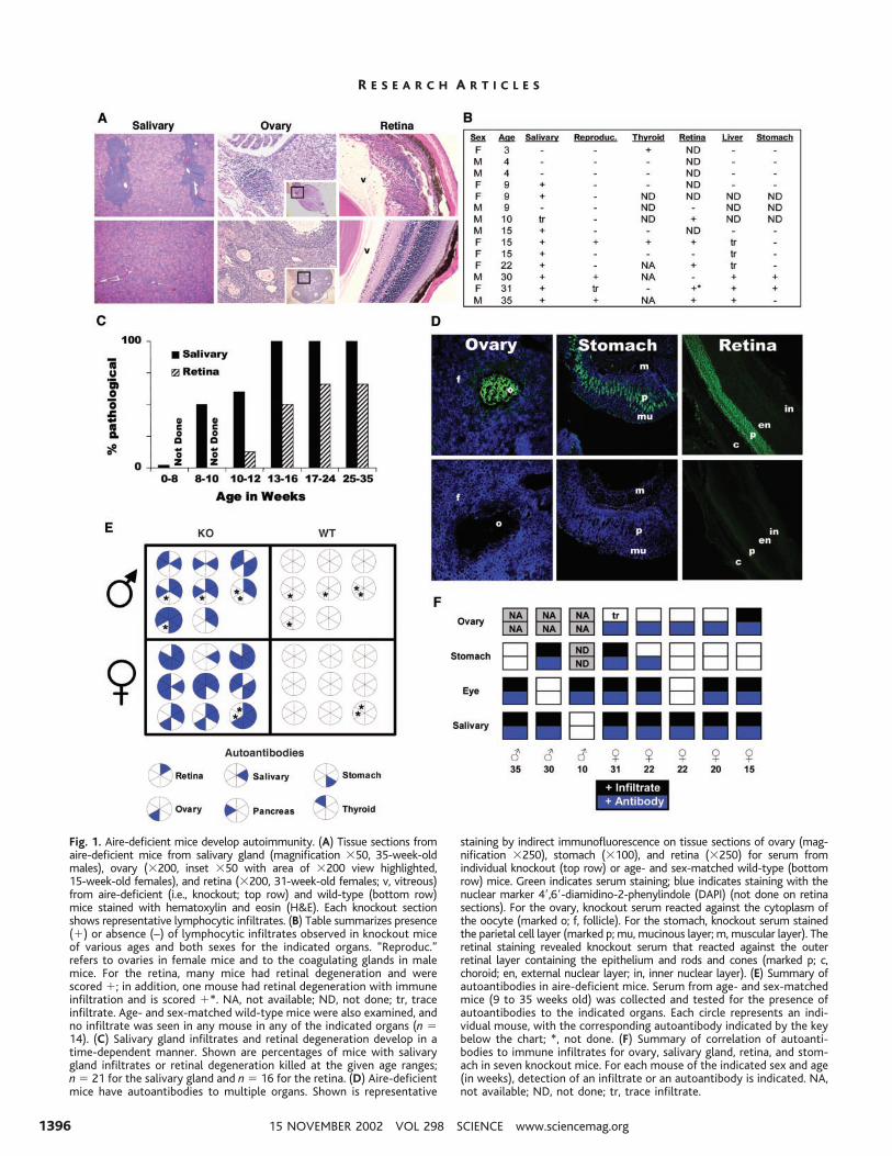
Fig. 1. Aire-deficient mice develop autoimmunity. (A) Tissue sections fromaire-deficient mice from salivary gland (magnification �50, 35-week-oldmales), ovary (�200, inset �50 with area of �200 view highlighted,15-week-old females), and retina (�200, 31-week-old females; v, vitreous)from aire-deficient (i.e., knockout; top row) and wild-type (bottom row)mice stained with hematoxylin and eosin (H&E). Each knockout sectionshows representative lymphocytic infiltrates. (B) Table summarizes presence(�) or absence (–) of lymphocytic infiltrates observed in knockout miceof various ages and both sexes for the indicated organs. “Reproduc.”refers to ovaries in female mice and to the coagulating glands in malemice. For the retina, many mice had retinal degeneration and werescored �; in addition, one mouse had retinal degeneration with immuneinfiltration and is scored �*. NA, not available; ND, not done; tr, traceinfiltrate. Age- and sex-matched wild-type mice were also examined, andno infiltrate was seen in any mouse in any of the indicated organs (n �14). (C) Salivary gland infiltrates and retinal degeneration develop in atime-dependent manner. Shown are percentages of mice with salivarygland infiltrates or retinal degeneration killed at the given age ranges;n � 21 for the salivary gland and n � 16 for the retina. (D) Aire-deficientmice have autoantibodies to multiple organs. Shown is representative
staining by indirect immunofluorescence on tissue sections of ovary (mag-nification �250), stomach (�100), and retina (�250) for serum fromindividual knockout (top row) or age- and sex-matched wild-type (bottomrow) mice. Green indicates serum staining; blue indicates staining with thenuclear marker 4�,6�-diamidino-2-phenylindole (DAPI) (not done on retinasections). For the ovary, knockout serum reacted against the cytoplasm ofthe oocyte (marked o; f, follicle). For the stomach, knockout serum stainedthe parietal cell layer (marked p; mu, mucinous layer; m, muscular layer). Theretinal staining revealed knockout serum that reacted against the outerretinal layer containing the epithelium and rods and cones (marked p; c,choroid; en, external nuclear layer; in, inner nuclear layer). (E) Summary ofautoantibodies in aire-deficient mice. Serum from age- and sex-matchedmice (9 to 35 weeks old) was collected and tested for the presence ofautoantibodies to the indicated organs. Each circle represents an indi-vidual mouse, with the corresponding autoantibody indicated by the keybelow the chart; *, not done. (F) Summary of correlation of autoanti-bodies to immune infiltrates for ovary, salivary gland, retina, and stom-ach in seven knockout mice. For each mouse of the indicated sex and age(in weeks), detection of an infiltrate or an autoantibody is indicated. NA,not available; ND, not done; tr, trace infiltrate.
R E S E A R C H A R T I C L E S
15 NOVEMBER 2002 VOL 298 SCIENCE www.sciencemag.org1396
sis–ectodermal dystrophy (APECED), alsoknown as autoimmune polyendocrine syn-drome–type 1 (APS-1), is a polyglandulardisorder that classically manifests as sponta-neous autoimmunity against the parathyroidand/or adrenal glands, and/or by a mucocuta-neous candidiasis infection [reviewed in (23,24)]. Other common ailments include auto-immune forms of premature ovarian failure,hepatitis, anemia, diabetes, alopecia, and viti-ligo. Disease is inherited in an autosomalrecessive manner as a result of loss of func-tion of a single susceptibility gene, whichencodes the AIRE protein. AIRE has shownDNA binding activity (25) and transcriptionaltransactivation potential (26, 27) in assays invitro. This protein and its murine homolog,aire, have a pattern of expression that sug-gests a potential role in shaping the T cell
repertoire: By far the highest amounts arefound in the thymus, specifically in MECs (4,28, 29). It seemed reasonable to hypothesizethat APECED patients develop multiorganautoimmunity because a defect in AIRE pre-vents or modifies ectopic transcription ofgenes encoding peripheral tissue–restrictedantigens in thymic MECs. A recent reportthat aire-deficient mice also exhibit autoim-mune manifestations is consistent with thisproposal, although that study concluded thatthe cause was a problem with peripheral lym-phocyte homeostasis (30). Here, we investi-gated an independently generated line of aire-deficient mice in an effort to learn whetherthe spectrum of autoimmunity manifested bythese mice is consistent with a broad defect incentral tolerance induction, and whether thisresults from an alteration in ectopic expres-
sion of tissue-restricted antigens by thymicMECs.
We engineered a line of mice carrying adefective aire gene by means of lox/cre-me-diated recombination in embryonic stem (ES)cells (fig. S1). These mice carry in homozy-gous state a mutant allele bearing a deletionof exon 2 and portions of the upstream anddownstream introns (fig. S1A). Mature airegene transcripts could be detected at near-normal levels in the thymus of mutant mice,but these were smaller than in the wild type,as expected given the absence of exon 2 (fig.S1B). Sequence analysis confirmed that thesetranscripts could not give rise to functionalprotein because a frame shift provoked by theabnormal juxtaposition of exons 1 and 3 re-sulted in premature truncation shortly afterexon 1 (fig. S1C). The aire gene mutationwas generated in ES cells derived from miceof the Sv/129 strain, and mice used in theexperiments described here were from F2 orF3 backcrosses onto the B6 geneticbackground.
Aire prevents autoimmunity. We firsttested whether the absence of aire in thesemice leads to broad defects in tolerance in-duction. Lymphocyte infiltrates were ob-served in several organs, usually confined toparticular structures rather than being distrib-uted indiscriminately throughout the organ.For example, lymphocytes were seen in thesalivary gland only in the perivascular re-gions, in the ovary only in the follicles, and inthe eye only in the retina (Fig. 1A). Theinfiltrates showed a pronounced age depen-dence by two criteria: (i) The number oforgans targeted increased in older aire-defi-cient mice, ranging from none or one at 3 to4 weeks of age to at least four or five after 30weeks (Fig. 1B). (ii) For any particular organ,the fraction of mutant mice with autoimmunemanifestations increased with age. For exam-ple, salivary gland infiltrates were rare inmice less than 8 weeks of age but wereconstant after 13 weeks (Fig. 1C). Autoanti-bodies had the same kind of target distribu-tion. Multiple organs were targeted, but onlyat specific substructures within each (e.g.,oocytes in the ovary, parietal cells in thestomach, the outer layer of the retina in theeye). All aire-deficient mice had serum auto-antibodies after 3 to 6 months of age, mostwith reactivity to several organs, as detectedby staining of tissues from wild-type mice(Fig. 1D). In the majority of aire-deficientmice, there was a good correlation betweenorgans targeted by infiltrates and those tar-geted by autoantibodies (Fig. 1F). In short,the phenotype of these aire-deficient mice iscertainly compatible with a broad defect intolerance induction, but one characterized byhighly selective, rather than indiscriminate,autoimmune attack.
We next sought to determine whether the
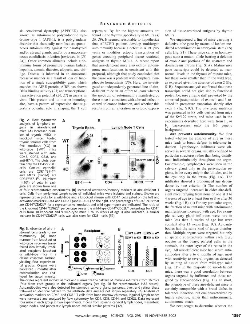
Fig. 2. Flow cytometricanalysis of lymphoid or-gans in aire-deficientmice. (A) Increased num-ber of thymic MECs inknockout mice. Pooledthymic stromal cells fromfive knockout (KO) orwild-type (WT ) micewere stained with anti-CD45, CDR1, G8.8, andanti-B7-1. The plots con-tain only the CD45–G8.8�
cells. Cortical epithelialcells are CDR1hiB7-1lo,and MECs (circled) areCDR1intB7-1hi. Numbers(�105) of cells in eachgate are shown from oneof four representative experiments. (B) Increased activation/memory markers in aire-deficient Tcells. Cells from peripheral lymph nodes of individual mice were isolated and stained. Shown arerepresentative plots for a wild-type and a knockout mouse with CD4� cells gated on the left andactivation markers CD44 and CD62 ligand (CD62L) on the right. The percentages of CD4� cells thatare CD44hiCD62Llo for a representative knockout and wild-type mouse are indicated. The ratio ofthe knockout CD44hiCD62Llo percentage versus the wild-type CD44hiCD62Llo percentage for CD4�
cells from 10 knockout and 9 wild-type mice 3 to 15 weeks of age is also indicated. A similarincrease in CD44hiCD62Llo cells was also seen for CD8� cells (32).
Fig. 3. Absence of aire instromal cells leads to au-toimmunity. (A) Bonemarrow from knockout orwild-type mice was trans-ferred into lethally irradi-ated recipient knockoutor wild-type mice in aclassic crisscross fashion,yielding four experimen-tal groups. Organs wereharvested 2 months afterreconstitution and ana-lyzed for autoimmunity.The circles represent individual mice and summarize the pattern of immune infiltrates from 16 mice(four from each group) in the indicated organs (see fig. S8 for representative H&E stains).Autoantibodies were also detected for stomach, salivary gland, pancreas, liver, and retina; thesefollowed an identical pattern to the infiltrate data and are not shown separately. (B) Analysis ofactivation markers on CD4� and CD8� T cells from bone marrow chimeras. Inguinal lymph nodeswere harvested and analyzed by flow cytometry for CD4, CD8, CD44, and CD62L. Data representfour mice in each group in two experiments. T cells from spleens, cervical lymph nodes, mesentericlymph nodes, and pancreatic lymph nodes exhibit similar patterns (32).
R E S E A R C H A R T I C L E S
www.sciencemag.org SCIENCE VOL 298 15 NOVEMBER 2002 1397

autoimmunity exhibited by aire-deficient micewas reflected in other immune system abnor-malities. Most of the features examined histo-logically, cytofluorimetrically, or via functionalassays appeared within the normal range (31)(figs. S2 to S7), in general agreement withprevious findings (30). These included thymo-cyte numbers, CD4/CD8 subset distributions,and differentiation marker profiles; bone mar-row cell numbers and immunoglobulin M(IgM)/B220 profiles; spleen and lymph nodecell numbers, IgM/TCR ( T cell receptor)–��,IgM/IgD, and CD4/CD8 profiles and
CD4�CD25� regulatory T cell compartments;dendritic cell numbers and subsets; T cell cyto-kine production after anti-CD3/CD28 stimula-tion; in vitro presentation of antigens or pep-tides; stimulatory and response capacity inmixed lymphocyte reactions; and thymus,spleen, and lymph node histology. Two featuresof the immune system in mutant mice wereabnormal, however. There was an average dou-bling of the number of MECs (Fig. 2A), al-though expression of major histocompatibilitycomplex (MHC) class II and costimulatorymolecules were normal (32) and there was a
near-doubling in the frequency of activated/memory CD44hiCD62Llo T cells in both theCD4� and CD8� compartments of the periph-eral lymphoid organs of most mice (Fig. 2B).This latter observation is consistent with thepossibility that aire somehow functions to re-duce the self reactivity of peripheral T cells.
Requirement for aire expression in thethymic stroma. To establish whether the au-toimmune manifestations in aire-deficientmice are attributable to its absence from ra-dioresistant or hematopoietic cells, we con-structed a set of radiation bone marrow chi-meras, which produced aire in radioresistantcells only, in hematopoietic cells only, inneither cell type, or in both. Signs of autoim-munity were detected only in those chimerasthat lacked aire in the radioresistant cells.Lymphocytic infiltrates were found in all ir-radiated aire-deficient recipients, whether thedonors of bone marrow cell precursors ex-pressed aire or not. The invasiveness of theinfiltrates was strikingly enhanced in thesechimeras vis-a-vis unmanipulated mutantmice according to multiple criteria: the frac-tion of young mice showing infiltrates, num-ber of organs infiltrated, and extent of theinfiltrate (Fig. 3A) (fig. S8). Likewise, irra-diated aire-deficient recipients of both wild-type and mutant bone marrow cell precursorsexhibited serum autoantibodies, and thesewere more prevalent and had a wider targetrange than was found with unmanipulatedmutant mice (Fig. 3A). Again, the profiles oforgans targeted by infiltrates and by autoan-tibodies were closely correlated (Fig. 3A).Finally, the abnormal accumulation of acti-vated/memory CD44hiCD62lo cells, bothCD4� and CD8�, also correlated with theabsence of aire in radioresistant cells (Fig.3B). Thus, the autoimmune manifestations inaire-deficient mice reflect this protein’s func-tion in radioresistant cells.
The next issue to resolve was whether aireexerts its influence on autoimmunity within thethymus or the periphery. Although aire and itshuman equivalent are predominantly synthe-sized in the thymus, expression in a number ofperipheral tissues has also been reported, includ-ing several of those targeted by lymphocyticinfiltrates or autoantibodies in aire-deficient in-dividuals (33, 34). However, expression in pe-ripheral parenchymal tissues has been contro-versial (35, 36), primarily because of its weak-ness and irreproducibility (35, 36). We carefullyexamined aire gene expression in normal miceby quantitative real-time polymerase chain reac-tion (PCR) (Fig. 4A). Transcripts were by farthe most abundant in the thymus. Althoughtranscripts were also detected in peripheral lym-phoid organs (at significantly lower levels), theywere essentially undetectable in the parenchy-mal tissues examined other than the ovary.These results imply that target tissues need notsynthesize significant levels of aire to avoid
Fig. 4. The thymus is critical for aire-associated autoimmunity. (A) Aire is expressed predominantlyin the thymus. Shown is relative expression of aire by quantitative real-time PCR using Taqmanprimers and probes on cDNA prepared from various whole organs from a 6-week-old B6 mouse.Normalization of cDNA content was done on cyclophilin; numbers represent a ratio of relative aireexpression to cyclophilin expression for each tissue. For tissues other than thymus, lymph node,spleen, and ovary, aire transcripts could not be detected. Given the normalization of cDNA contentby cyclophilin, the amount of aire in these tissues is at least two orders of magnitude less than thelevel seen in the thymus. Data shown are a representative experiment of several independentexperiments. (B) Transplantation of aire-deficient thymi leads to autoantibody production. Nudemice were transplanted with either aire-deficient (n � 3) or wild-type thymi (n � 6). Six weeksafter transplantation, serum was collected and tested for autoantibodies against stomach (�100)and retina (�250) by indirect immunofluorescence for recipients of aire-deficient (top panels) andwild-type control (bottom panels) thymi. Shown is representative staining from individual mice.Green indicates specific serum staining; blue (only on stomach sections) indicates staining with thenuclear marker DAPI. For the stomach, serum (3/3 knockout, 0/6 wild type) stained the parietal celllayer (p) of the stomach mucosa (mu, mucinous layer; m, muscular layer). For the retina, serum(2/2 knockout, 0/4 wild type) reacted against the outer layer containing the epithelium and rodsand cones (marked p; c, choroid; en, external nuclear layer; in, inner nuclear layer). (C) Mature aireknockout lymphocytes can transfer immune infiltrates. Table summarizes immune infiltratesobserved (12 weeks after transfer) in the salivary gland, retina, or stomach for Rag-deficientrecipients injected with aire knockout lymphocytes or wild-type lymphocytes. Numbers representindividual mice for each group. (D) Representative immune infiltrates in tissue sections stained withH&E from Rag-deficient mice injected with lymphocytes from aire knockout mice (top panels) orwild-type control mice (bottom panels) for retina (�200) and salivary gland (�100).
R E S E A R C H A R T I C L E S
15 NOVEMBER 2002 VOL 298 SCIENCE www.sciencemag.org1398
autoimmune attack and indicate the importanceof aire expression in lymphoid tissues.
To distinguish the influence of the thymusfrom that of peripheral lymphoid organs, and tofurther rule out any role for peripheral paren-chymal tissues, we performed a thymus graftexperiment. A thymus from either an aire-defi-cient mouse or a wild-type littermate was de-pleted of hematopoietic cells by incubation in2-deoxyguanosine (31) and was then trans-planted into an athymic B6nu/nu recipient. Sixweeks later, the generation of a peripheral T cellcompartment was confirmed, and serum auto-antibodies were assayed on stomach and eyesections from normal mice. Autoantibodieswere found in sera from each of three micehosting an aire-deficient thymus but in none ofsix hosts of a wild-type thymus, although the Tcell compartments were reconstituted to thesame level in the two cases. The pattern ofautoantibody staining was the same as that seenwith sera from unmanipulated aire-deficientmice (compare Figs. 4B and 1B). We thusconcluded that aire must be expressed in theradioresistant stromal cells of the thymus inorder to control autoimmunity in the periphery.
Influence of aire on T cell education.We also established that the autoimmune dis-ease that develops in aire-deficient mice islymphocyte-autonomous. Mature lympho-cytes were pooled from the spleen and lymphnodes of aire-deficient mice or wild-type lit-termates and were then transferred into alym-phoid Rago/o recipients. After 12 weeks, di-verse organs were assayed histologically.Lymphocytic infiltrates, similar to those ob-served in unmanipulated mice lacking aire,were found in the salivary gland, eye, andstomach of the mice that received aire-defi-cient, but not wild-type, lymphocytes (Fig. 4,C and D). These results confirm that aireexpression in peripheral parenchymal tissuesis not the factor controlling autoimmuneattack.
Aire regulates thymic gene expression.Given the association between aire deficiencyand autoimmunity, and the predominant ex-pression of this transcription factor (25–27)in thymic MECs (4, 5), we hypothesized thataire controls autoimmunity by regulating theectopic expression of peripheral tissue–re-stricted antigens in the medulla of the thy-mus. MECs were present (indeed, they wereoverrepresented) in aire-deficient mice anddisplayed normal levels of molecules associ-ated with antigen presentation (fig. S5). Thus,aire does not dictate differentiation of MECs,nor their ability to present antigens.
To obtain a broad perspective on the tran-scriptional profile of MECs, we prepared RNAfrom cells sorted from aire-deficient and controllittermates (sorting pooled thymocytes from fiveindividuals as described in Fig. 2A) and usedlabeled complementary RNA to probe 12,000mouse genes represented on Affymetrix
U74Av2 chips (31). Three independent experi-ments were performed to permit statistical treat-ment of the data, and three different statisticalassays were applied (31). The absence of aireresulted in loss of or reduction in expression ofa substantial number of genes (Fig. 5A), clearlymore than in the randomly permuted data set.When using a factor of 5 change as an arbitrarycutoff value, decreases in transcripts of 104genes were called significant by the Signifi-cance Analysis of Microarrays (SAM) programand 42 by the Quadrants program; decreasesranged from a factor of 5 to a factor of 70,frequently with complete extinction in mutantmice. In contrast, the number of genes inducedin mutant mice did not depart from that of therandom data set (14 significant by SAM, 6 byQuadrants). Thus, aire seems to function pre-dominantly as a transcriptional activator. Be-cause of statistical uncertainties linked to multi-ple sampling, the exact number of genes repre-sented on the U74Av2 chip and down-regulatedin the absence of aire cannot be ascertained, but
an estimate of 100 to 300 is a reasonable extrap-olation, on the basis of the data in Figs. 5A and6A and the statistical analysis. Similarly, it is notexactly certain what fraction of all murine tran-scripts are represented on the chip we used(likely 1⁄2 to 1⁄4). Therefore, the number of geneswhose expression is activated by aire probablyranges between 200 and 1200.
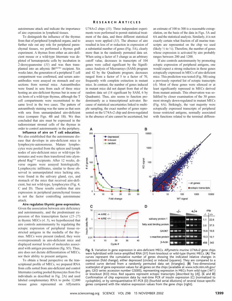
If aire controls autoimmunity by promotingectopic expression of peripheral antigens, onewould expect a strong reduction in those genesectopically expressed in MECs of aire-deficientmice. This prediction was tested (Fig. 5B) usinga previously reported list of ectopic transcripts(4). Most of these genes were silenced or atleast significantly repressed in MECs derivedfrom mutant animals. This observation was so-lidified by closer examination of the 30 genesmost strongly down-regulated in mutant MECs(Fig. 6A). Strikingly, the vast majority wereectopically expressed transcripts of peripheraltissue–restricted antigens, normally associatedwith functions related to the terminal differen-
Fig. 5. Variation in gene expression in aire-deficient MECs. Affymetrix murine U74Av2 gene chipswere hybridized with twice-amplified RNA (31) from knockout or wild-type thymic MECs. (A) Thecurves represent the cumulative number of genes showing the indicated relative changes inexpression (fold change), either depressed (circles) or induced (squares). They are compared to acontrol curve derived from a randomly permuted data set (triangles). (B) Two-dimensionalscatterplot of gene expression values for all genes on the chips (available at www.ncbi.nlm.nih.gov/geo, GEO series accession number GSE85), representing expression in MECs from wild-type (WT )or knockout (KO) mice. Red squares represent ectopic transcripts [described by (4)]. (C and D)Confirmation of chip expression data by real-time PCR of insulin expression (C) (normalized tocyclophilin), or by semiquantitative RT-PCR (D) (fourfold serial dilutions) of several tissue-specificgenes compared with the relative expression values from the gene chips (right).
R E S E A R C H A R T I C L E S
www.sciencemag.org SCIENCE VOL 298 15 NOVEMBER 2002 1399
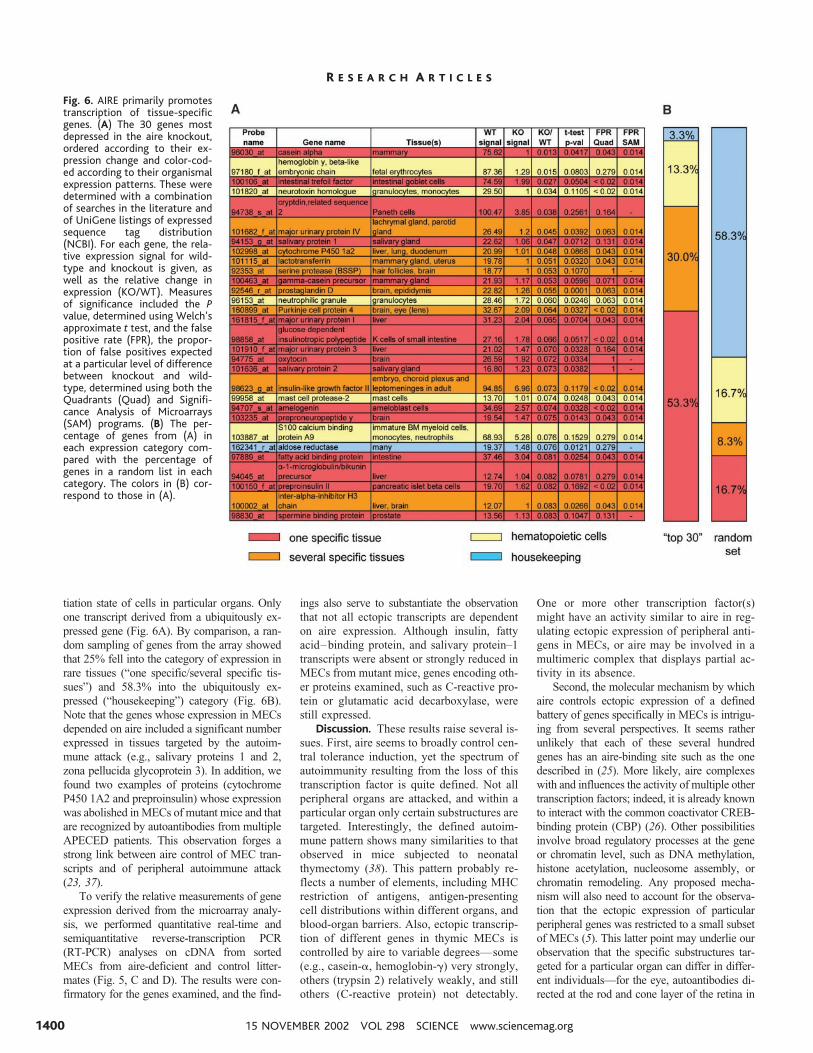
tiation state of cells in particular organs. Onlyone transcript derived from a ubiquitously ex-pressed gene (Fig. 6A). By comparison, a ran-dom sampling of genes from the array showedthat 25% fell into the category of expression inrare tissues (“one specific/several specific tis-sues”) and 58.3% into the ubiquitously ex-pressed (“housekeeping”) category (Fig. 6B).Note that the genes whose expression in MECsdepended on aire included a significant numberexpressed in tissues targeted by the autoim-mune attack (e.g., salivary proteins 1 and 2,zona pellucida glycoprotein 3). In addition, wefound two examples of proteins (cytochromeP450 1A2 and preproinsulin) whose expressionwas abolished in MECs of mutant mice and thatare recognized by autoantibodies from multipleAPECED patients. This observation forges astrong link between aire control of MEC tran-scripts and of peripheral autoimmune attack(23, 37).
To verify the relative measurements of geneexpression derived from the microarray analy-sis, we performed quantitative real-time andsemiquantitative reverse-transcription PCR(RT-PCR) analyses on cDNA from sortedMECs from aire-deficient and control litter-mates (Fig. 5, C and D). The results were con-firmatory for the genes examined, and the find-
ings also serve to substantiate the observationthat not all ectopic transcripts are dependenton aire expression. Although insulin, fattyacid–binding protein, and salivary protein–1transcripts were absent or strongly reduced inMECs from mutant mice, genes encoding oth-er proteins examined, such as C-reactive pro-tein or glutamatic acid decarboxylase, werestill expressed.
Discussion. These results raise several is-sues. First, aire seems to broadly control cen-tral tolerance induction, yet the spectrum ofautoimmunity resulting from the loss of thistranscription factor is quite defined. Not allperipheral organs are attacked, and within aparticular organ only certain substructures aretargeted. Interestingly, the defined autoim-mune pattern shows many similarities to thatobserved in mice subjected to neonatalthymectomy (38). This pattern probably re-flects a number of elements, including MHCrestriction of antigens, antigen-presentingcell distributions within different organs, andblood-organ barriers. Also, ectopic transcrip-tion of different genes in thymic MECs iscontrolled by aire to variable degrees—some(e.g., casein-�, hemoglobin-�) very strongly,others (trypsin 2) relatively weakly, and stillothers (C-reactive protein) not detectably.
One or more other transcription factor(s)might have an activity similar to aire in reg-ulating ectopic expression of peripheral anti-gens in MECs, or aire may be involved in amultimeric complex that displays partial ac-tivity in its absence.
Second, the molecular mechanism by whichaire controls ectopic expression of a definedbattery of genes specifically in MECs is intrigu-ing from several perspectives. It seems ratherunlikely that each of these several hundredgenes has an aire-binding site such as the onedescribed in (25). More likely, aire complexeswith and influences the activity of multiple othertranscription factors; indeed, it is already knownto interact with the common coactivator CREB-binding protein (CBP) (26). Other possibilitiesinvolve broad regulatory processes at the geneor chromatin level, such as DNA methylation,histone acetylation, nucleosome assembly, orchromatin remodeling. Any proposed mecha-nism will also need to account for the observa-tion that the ectopic expression of particularperipheral genes was restricted to a small subsetof MECs (5). This latter point may underlie ourobservation that the specific substructures tar-geted for a particular organ can differ in differ-ent individuals—for the eye, autoantibodies di-rected at the rod and cone layer of the retina in
Fig. 6. AIRE primarily promotestranscription of tissue-specificgenes. (A) The 30 genes mostdepressed in the aire knockout,ordered according to their ex-pression change and color-cod-ed according to their organismalexpression patterns. These weredetermined with a combinationof searches in the literature andof UniGene listings of expressedsequence tag distribution(NCBI). For each gene, the rela-tive expression signal for wild-type and knockout is given, aswell as the relative change inexpression (KO/WT). Measuresof significance included the Pvalue, determined using Welch’sapproximate t test, and the falsepositive rate (FPR), the propor-tion of false positives expectedat a particular level of differencebetween knockout and wild-type, determined using both theQuadrants (Quad) and Signifi-cance Analysis of Microarrays(SAM) programs. (B) The per-centage of genes from (A) ineach expression category com-pared with the percentage ofgenes in a random list in eachcategory. The colors in (B) cor-respond to those in (A).
R E S E A R C H A R T I C L E S
15 NOVEMBER 2002 VOL 298 SCIENCE www.sciencemag.org1400
one mouse (Fig. 1D), to other retinal layers inanother mouse (32).
Finally, it is worth considering aire-defi-cient mice as a model of APECED. Likehuman patients, the murine model showsmultiorgan attack by lymphocytic infiltratesand recognition by serum autoantibodies. Inboth cases, there is disease heterogeneity be-tween individuals and exacerbation with age.The differences between humans and mice inthe spectrum of organs targeted could well bedue to the influence of genetic modifiers, asrecently described for the HLA locus in hu-mans (39). Aire-deficient mice should proveinvaluable for dissecting the relative impor-tance of genetic, environmental, and stochas-tic processes in determining the target organspecificity of autoimmune destruction.
References and Notes1. K. M. Garza, V. S. Chan, P. S. Ohashi, Rev. Immuno-genet. 2, 2 (2000).
2. D. Hanahan, Curr. Opin. Immunol. 10, 656 (1998).3. L. Klein, B. Kyewski, Curr. Opin. Immunol. 12, 179
(2000).4. J. Derbinski, A. Schulte, B. Kyewski, L. Klein, NatureImmunol. 2, 1032 (2001).
5. L. Klein, B. Roettinger, B. Kyewski, Eur. J. Immunol. 31,2476 (2001).
6. P. Naquet, M. Naspetti, R. Boyd, Semin. Immunol. 11,47 (1999).
7. H. Kishimoto, J. Sprent, Clin. Immunol. 95, S3 (2000).
8. C. Jolicoeur, D. Hanahan, K. M. Smith, Proc. Natl.Acad. Sci. U.S.A. 91, 6707 (1994).
9. K. M. Smith, D. C. Olson, R. Hirose, D. Hanahan, Int.Immunol. 9, 1355 (1997).
10. M. W. Hoffmann, J. Allison, J. F. Miller, Proc. Natl.Acad. Sci. U.S.A. 89, 2526 (1992).
11. M. W. Hoffmann, W. R. Heath, D. Ruschmeyer,J. F. A. P. Miller, Proc. Natl. Acad. Sci. U.S.A. 92, 9851(1995).
12. M. G. Von Herrath, J. Dockter, M. B. A. Oldstone,Immunity 1, 231 (1994).
13. A. M. Sponaas et al., Int. Immunol. 6, 277 (1994).14. S. J. Antonia, T. Geiger, J. Miller, R. A. Flavell, Int.
Immunol. 7, 715 (1995).15. M. Oukka, M. Cohen-Tannoudji, Y. Tanaka, C. Babinet,
K. Kosmatopoulos, J. Immunol. 156, 968 (1996).16. L. Klein, T. Klein, U. Ruther, B. Kyewski, J. Exp. Med.
188, 5 (1998).17. M. Oukka et al., Immunity 4, 545 (1996).18. L. Klein, M. Klugmann, K.-A. Nave, V. K. Tuohy, B.
Kyewski, Nature Med. 6, 56 (2000).19. A. C. Anderson et al., J. Exp. Med. 5, 761 (2000).20. A. Pugliese et al., Nature Genet. 15, 293 (1997).21. P. Vafiadis et al., Nature Genet. 15, 289 (1997).22. C. E. Egwuagu, P. Charukamnoetkanok, I. Gery, J. Im-
munol. 159, 3109 (1997).23. P. Bjorses, J. Aaltonen, N. Horelli-Kuitunen, M. L.
Yaspo, L. Peltonen, Hum. Mol. Genet. 7, 1547 (1998).24. P. Peterson et al., Immunol. Today 19, 384 (1998).25. P. G. Kumar et al., J. Biol. Chem. 276, 41357 (2001).26. J. Pitkanen et al., J. Biol. Chem. 275, 16802 (2000).27. P. Bjorses et al., Am. J. Hum. Genet. 66, 378 (2000).28. M. C. Rosatelli et al., Hum. Genet. 103, 428 (1998).29. S. Zuklys et al., J. Immunol. 165, 1976 (2000).30. C. Ramsey et al., Hum. Mol. Genet. 11, 397 (2002).31. See supporting data on Science Online.32. M. S. Anderson et al., data not shown.
33. K. Nagamine et al., Nature Genet. 17, 399 (1997).34. M. Halonen et al., J. Histochem. Cytochem. 49, 197
(2001).35. M. Heino et al., Biochem. Biophys. Res. Commun.
257, 821 (1999).36. M. Heino et al., Eur. J. Immunol. 30, 1884 (2000).37. M. Gylling et al., J. Clin. Endocrinol. Metab. 85, 4434
(2000).38. A. Kojima, R. T. Prehn, Immunogenetics 14, 15 (1981).39. M. Halonen et al., J. Clin. Endocrinol. Metab. 87, 2568
(2002).40. We thank G. Losyev, Q.-M. Pham, J. Rasmussen, and R.
Saccone for help with flow cytometry, mice, microarraysoftware and chip analysis, respectively, and E. Smithfor assistance with the figures. Supported by the W. T.Young Chair fund, NIH grant RO1 DK60027-01 (D.M.and C.B.), Joslin Diabetes Center grant DERC 2 P30DK36836-16, a Howard Hughes Medical Institute Post-doctoral Fellowship for Physicians and NIH grant KO8-DK59958-01A1 (M.S.A.), an NSF Graduate ResearchFellowship and NIH training grant 2T32 DK07260-26 toJoslin Diabetes Center (E.S.V.), Irvington Institute forImmunological Research (L.K.), Juvenile Diabetes Re-search Foundation ( Z.C.), Human Frontier ScienceProgram (S.B.), and an NRSA fellowship in Cancer Im-munology ( T32CA70083-05) and the Cancer ResearchInstitute (S.J.T.).
Supporting Online Materialwww.sciencemag.org/cgi/content/full/1075958/DC1Materials and MethodsFigs. S1 to S8References
10 July 2002; accepted 19 September 2002Published online 10 October 2002;10.1126/science.1075958Include this information when citing this paper.
R E P O R T S
Broadband Modulation of Lightby Using an Electro-Optic
PolymerMark Lee,* Howard E. Katz, Christoph Erben, Douglas M. Gill,
Padma Gopalan, Joerg D. Heber, David J. McGee
A major challenge to increasing bandwidth in optical telecommunications is toencode electronic signals onto a lightwave carrier by modulating the light upto very fast rates. Polymer electro-opticmaterials have the necessary propertiesto function in photonic devices beyond the 40-GHz bandwidth currently avail-able. An appropriate choice of polymers is shown to effectively eliminate thefactors contributing to an optical modulator’s decay in the high-frequencyresponse. The resulting device modulates light with a bandwidth of 150 to 200GHz and produces detectable modulation signal at 1.6 THz. These rates arefaster than anticipated bandwidth requirements for the foreseeable future.
Transmitting signals by using infrared lightthrough optical fiber is the most effective wayto move large amounts of data rapidly over longdistances. Consequently, optical communica-tions form the backbone of the Internet andtelephone networks, and they are envisioned tocarry real-time multimedia content in the future.
Approaches to increasing optical bandwidth arebeing pursued to accommodate anticipatedgrowth in data traffic. At present, high-speedoptical networks use bandwidths of 10 GHz(�10 billion bits per second) per channel, and40-GHz products are being introduced. Basicresearch efforts are aiming to push bandwidthsto 80, 100, and even 160 GHz.
Expanding bandwidth beyond 100 GHz in-volves many scientific and engineering chal-lenges, among which is the encoding of elec-tronic data signals onto a lightwave carrier by
modulating the light in phase or amplitude. Thisis usually done with an electro-optic (E-O)modulator (1), where a signal voltage changesthe refractive index of an E-O dielectric opticalwaveguide, modulating the phase of a guidedlightwave. Amplitude modulation is obtainedby generating a phase difference betweenlight in two coherent waveguides and then com-bining to produce constructive or destructiveinterference. In present technology, lithium nio-bate is the E-O material most widely used inhigh-speed optical modulators.
An E-O device produces the strongestmodulation when � � radians. For aninput voltage Vin, � �(Vin/V�), where V�,the “half-wave” voltage that makes � �near zero frequency [direct current (dc)], is:
V� � 0d
2n3optr�L
(1)
where 0 is the carrier wavelength, d is the gapbetween voltage electrodes, nopt is the opticalrefraction index, r is the E-O coefficient of thedielectric, L is the device length, and � isdefined as the signal electric field in the opticalwaveguide normalized to the field that wouldbe there if air were the dielectric. The centralproblem for high-speed operation is that, for afixed Vin, decreases as modulation frequen-cy increases. This response deterioration hasthree physical origins. The first is dissipation ofVin due to resistance in the electrodes guid-
Bell Laboratories–Lucent Technologies, 600 MountainAvenue, Murray Hill, NJ 07974, USA.
*To whom correspondence should be addressed, E-mail: [email protected]
R E S E A R C H A R T I C L E S
www.sciencemag.org SCIENCE VOL 298 15 NOVEMBER 2002 1401