proliferative lesions in swimbladder of japanese medaka
TRANSCRIPT
Vol. 38: 135-142,1999 DISEASES OF AQUATIC ORGANISMS
Dis Aquat Org l Published November 8
Proliferative lesions in swimbladder of Japanese medaka Oryzias latipes and guppy Poecilia reticulata
John W. Fourniel.*, William E. ~ a w k i n s ~ , William W. walker1
'U.S. Environmental Protection Agency, National Health and Environmental Effects Research Laboratory, Gulf Ecology Division, 1 Sabine Island Drive, Gulf Breeze, Florida 32561, USA
* ~ e p a r t m e n t of Coastal Sciences, Institute of Marine Sciences, University of Southern Mississippi, PO Box 7000. Ocean Springs. Mississippi 39566, USA
ABSTRACT: Thlrteen cases of proliferative lesions of the swimbladder were encountered in Japanese medaka Oryzias latipes and guppy Poecilia reticulata from about 10 000 medaka and 5000 guppies used in carcinogenicity tests and histologically examined. Two of the 4 cases from medaka and 8 of the 9 from guppies occurred in untreated control specimens. The lesions affected the gas gland epithelium and included hyperplasia, adenoma, and adenocarcinoma. One medaka had hyperplasia of the gas gland epithelium and in 1 guppy the gland was enlarged with an increase in the number of epithelial layers. Gas gland adenomas, 3 cases in medaka and 1 in the guppy, were typically larger than the hyperplastic lesions, formed expansive masses up to 1 mm in greatest dimension, and exhibited a solid or glandular growth pattern and mild cellular pleomorphism. Adenocarcinoma was the most advanced lesion and all 7 cases occurred in guppies. Adenocarcinomas sometimes filled the entire swimbladder and measured up to 2.5 n m in diameter. Cells of adenocarcinomas were highly pleomorphic, with atyp- ical nuclei, and an elevated mitotic activity. Because most of these tumors occurred in fish from control groups or in tests with noncarcinogenic compounds, the lesions observed here are probably sponta- neous rather than chemically induced. Their rare occurrence, however, makes swimbladder prolifera- tive lesions in small-fish carcinogenesis models sensitive indcators of compounds that might target cells of the gas gland.
KEY WORDS: Fish - Swimbladder. Neoplasia . Carcinogenesis . Medaka . Guppy
INTRODUCTION
The swimbladder of fishes is generally not affected by histologically detectable lesions, especially non- infectious ones (Ferguson 1989), although some cases have been reported. Nonneoplastic lesions including hyperplasia and cellular swelling in the gas gland epi- thelium occurred in Japanese medaka Oryzias latipes exposed to bis(tri-n-butyltin) oxide (Wester et al. 1990), and hyperplasia of small basophilic epithelia1 cells on the lumenal side of the gas gland was reported in guppies Poecilia reticulata exposed to methyl mercury chloride (Wester & Canton 1992). Neoplastic lesions of the swimbladder similarly have not been reported fre- quently. Neoplastic lesions diagnosed as leiomyosarco- mas or fibrosarcomas associated with an infectious
agent, probably a retrovirus, were found in 4.6% of 500 pen-reared Atlantic salmon Salmo salar on a com- mercial marine fish farm in Scotland (Duncan 1978, McKnight 1978). High prevalences of neoplasms of the swimbladder secretory epithelium affected medaka ex- posed for 28 d to 4-chloroaniline and aniline (Johnson et al. 1989). In the rainbow trout Oncorhynchus my- kiss, papillary adenomas of the swirnbladder epithe- lium occurred after exposure to the carcinogens diethyl- nitrosamine (DEN) (Bailey et al. 1984), benzo(a)pyrene (Bap) (Hendricks et al. 1985), N-methyl-N1-nitro-N- nitrosoguanidine (MNNG) (Hendricks et al. 1980, Kirnura et al. 1981, Bailey et al. 1984), N-methyl-N- nitrosourea (MNU) (Kimura et al. 1981), and dimethyl- nitrosomorpholine (DMNM) (Hendricks et al. 1995).
This paper reports the occurrence of swirnbladder neoplasms in specimens of medaka and guppy, 2 small fish species that are used in carcinogenesis bioassays
Q Inter-Research 1999 Resale of full article not permitted
136 Dis Aquat Org 38: 135-142, 1999
(Johnson et al. 1989, Hawkins et al. 1995). Here we report and describe cases of proliferative lesions of the gas gland, including hyperplasia, adenoma, and ade- nocarcinoma.
MATERIALS AND METHODS
Medaka and guppy evaluated for this report came from stocks maintained at the Gulf Coast Research Laboratory (Ocean Springs, Mississippi, USA) and used in toxicological studies, primarily carcinogenesis tests, since about 1984. Carcinogenesis tests were carried out generally as follows: fish approximately 6 to 10 d old were exposed to test chemicals for periods that ranged from 1 h to 180 d under either static or flow- through conditions. Flow-through studies were con- ducted in enclosed systems similar to that described by Walker et al. (1985) and static exposures in containers within glove boxes. A typical test included equal num- bers of specimens assigned to an untreated control group, a solvent control group, and 3 or more exposure groups. Following exposure, fish were transferred to 38 1 grow-out aquaria maintained at 27°C under a 12 h:12 h photoperiod. Fish were fed dry flake food 3 times and brine shrimp nauplii once daily. The grow- out aquaria were monitored monthly for routine water- quality parameters. Samples for histopathology were
usually routinely taken at 24,36, and 52 wk after the be- ginning of exposures or when fish became moribund.
Prior to fixation, most fish were anesthetized in ice water or MS-222 (tricaine methanesulfonate), the ab- dominal cavity was opened and the whole fish placed into Lillie's fixative (Humason 1979). To insure decal- cification, specimens remained in fixative for 24 h to 1 wk, depending on their size. Some specimens were fixed in Bouin's fixative for 24 h and decalcified in a commercial decalcifying solution for 8 to 24 h. Speci- mens were rinsed in water, dehydrated in ethanol, cleared in xylene or a commercial xylene substitute, and embedded in paraffin. To survey most major organs, sections were taken from a mid-lateral and a median plane, mounted on glass slides, and stained with Harris' hematoxylin and eosin. In some cases, additional sections were cut and stained to verify the presence of a lesion or to confirm a diagnosis.
RESULTS
Normal anatomy and histology
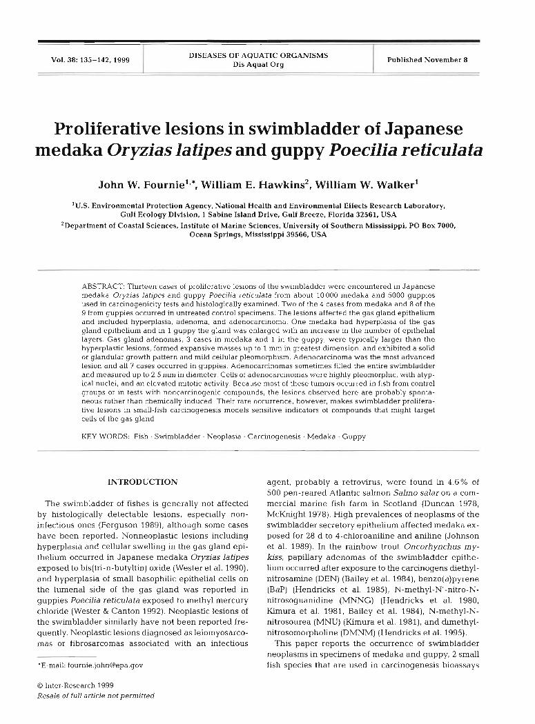
In the medaka and guppy, the swimbladder is located retroperitoneally in the dorsal abdominal cav- ity and is surrounded by a thin, fibrous tunica externa (Fig. 1). Both medaka and guppy appear to be physo-
Fig. 1 Oryzias latjpes. Low power view of a control Japanese medaka showing normal swimbladder structure. Note thin gas gland epithelium (arrowheads), rete mirabile (arrow), and lumen of swimbladder (asterisk). H&E. x22
Fournie et al.: Swimbladder proliferative lesions 137
clists, i.e. their swimbladders do not have Table 1 Features of swimbladder lesions in small fish from carcinogen- patent connections with the alimentary canal, esis tests. TCE = trichloroethylene; MAM-AC = methylazoxymethanol
The gas gland is situated at the cranial pole of acetate; CDBM = chlorodibrornomethane
the swimbladder and is composed of layers of large uniform epithelial cells with prominent eosinophilic cytoplasm. Vascularization of the gas gland is supplied by a rete mirabile, spe- cialized for transfer of gases by a counter- current multiplier system.
Lesion prevalence
The cases of swimbladder lesions in medaka and guppy are summarized in Table 1. Thirteen cases were diagnosed from about 10 000 me- daka and 5000 guppies older than 24 wk used in a variety of carcinogenesis tests, revealing an overall prevalence for both species of 0.09%. About three- fourths of the specimens were untreated control fish. The lesions arose from the gas gland epithelium and were diagnosed as hyperplasia, adenoma, or adenocarcinoma. Of the 4 medaka with lesions, 2 were males and 2 were females. One medaka with hyperplasia was from a trichloroethylene exposure, 1 with an adenoma was from a methylazoxymethanol acetate exposure, and 2 with adenomas were from control groups. Of the 9 guppies with lesions, 5 were males and 4 were females. Among
the guppies, single cases of hyperplasia or adenoma along with 6 of the 7 cases of adenocarcinoma occurred in control specimens. One guppy with adenocarcinoma had been exposed to chlorodibromomethane.
Hyperplasia
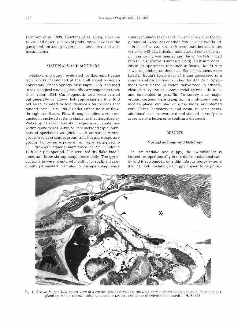
Hyperplasia was observed in only 2 specimens. The gas gland was enlarged with an increase in the num- ber of epithelial cell layers (Fig. 2). Cells were vacuo-
Case Species Sex Age (wk) Test group Lesion type
1 Medaka M 38 TCE Hyperplasia 2 Medaka M 24 Control Adenoma 3 Medaka F 27 MAM-Ac Adenoma 4 Medaka F 36 Control Adenoma 5 GUPPY F 51 Control Hyperplasia 6 GUPPY F 13 Control Adenoma 7 Guppy M 52 Control Adenocarcinoma 8 GUPPY F 24 Control Adenocarclnoma 9 GUPPY M 52 Control Adenocarcinoma
10 Guppy M 52 Control Adenocarcinoma 11 Guppy M 36 Control Adenocarcinoma 12 Guppy F 24 CDBM Adenocarcinoma 13 Guppy M 36 Control Adenocarcinoma
Fig. 2. Oryzias latipes. Gas gland of a rnedaka exposed to trichloroethylene showing distinct swelling and hyperplasia of glandular epithelium (arrow). H&E. x22
138 Dis Aquat Org 38: 135-142, 1999
lated and swollen but otherwise normal, and the architecture of the epithelium was not disrupted. The fibrous tunica externa was normal.
Adenoma
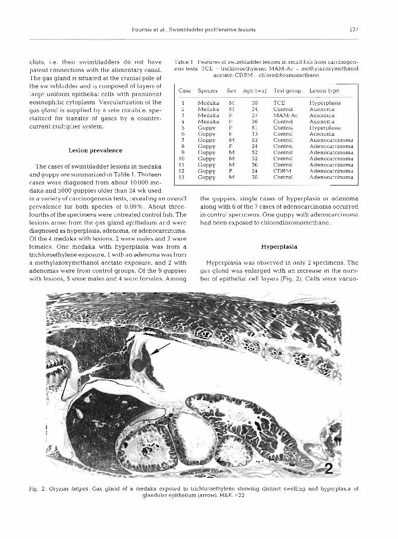
Adenomas occurred in 4 specimens, 3 in rnedaka and 1 in a guppy, and were composed of large, eosinophilic
Fig. 3. Oryzias latipes. Large adenoma from a control medaka showing
1 an expansive mass w t h a solid growth pattern.
H&E. x200
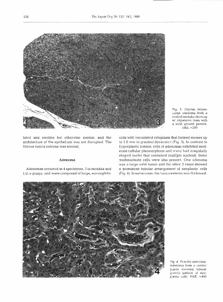
cells with vacuolated cytoplasm that formed masses up to 1.0 mm in greatest dimension (Fig. 3). In contrast to hyperplastic lesions, cells of adenomas exhibited mod- erate cellular pleomorphism and many had irregularly shaped nuclei that contained multiple nucleoli. Some multinucleate cells were also present. One adenoma was a large solid tumor and the other 3 cases showed a prominent tubular arrangement of neoplastic cells (Fig. 4). In some cases, the tu~llca externa was thickened.
I Fig. 4 . Poecilia reticulata. Adenoma from a control guppy showing tubular qrowth pattern of neo-
I . - I plastic c & ~ . H&E. x400

Fournie et al.: Swimbladder proliferative leslons 139
Fig. 5. Poecilia reticulata. Low-power view of a large adenocarcinoma (arrow) from a control guppy. Note the entire swimbladder is filled with tumor H&E. x22
Adenocarcinoma
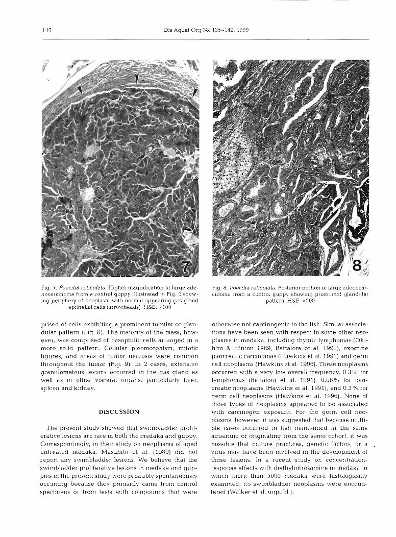
All 7 cases of adenocarcinoma occurred in guppies and the lesions ranged in size from very small to large, tumorous masses. Some of the tumors filled the swim- bladder lumen and measured up to 2.5 mm in greatest dimension (Fig. 5). The rete mirabile associated with adenocarcinomas usually appeared hyperplastic. Ade- nocarcinomas were generally chal-acterized by large, vacuolated, eosinophilic cells that exhibited the full spectrum of anaplastic features. The smallest carci- noma was histologically the most aggressive, with constituent cells exhibiting extreme cellular pleomor- phism, nuclear atypia, and elevated mitotic activity (Fig. 6). Neoplastic cells in 1 large carcinoma were arranged in a prominent glandular pattern, whereas other carcinomas exhibited a more solid pattern. The largest adenocarcinoma exhibited various cellular pat- terns (Fig. 5). Normal appearing gas gland epithelia1 cells with the typical eosinophilic cytoplasn~ were pre- sent at the periphery of this neoplasm (Fig. 7). An area in the posterior portion of the tumor mass was com-
Fig. 6. PoecLD'a ret~culata. High magnification of a small ade- nocarcinoma from a control guppy showing extensive cellular pleomorphlsm and nuclear atypia. Note mitotic figures (arrow-
heads). H&E. x600
140 Dis Aquat Org 38: 135-142, 1999
Fig. 7. Poecilia reticulala. Hlgher magnification of large ade- nocarcinoma from a control guppy illustrated in Fig. 5 show- ing periphery of neoplasm with normal appearing gas gland
epithelia1 cells (arrowheads). H&E. xlOO
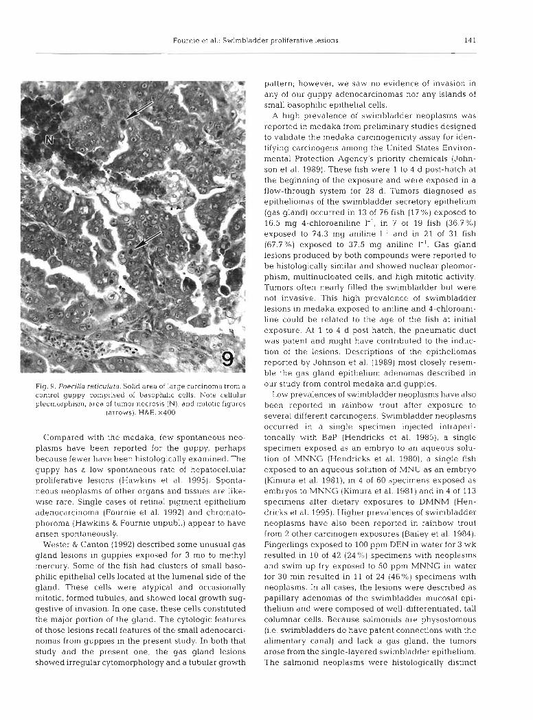
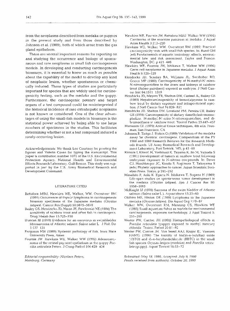
prised of cells exhibiting a prominent tubular or glan- dular pattern (Fig. 8). The majority of the mass, how- ever, was comprised of basophilic cells arranged in a more solid pattern. Cellular pleomorphism, mitotic figures, and areas of tumor necrosis were common throughout the tumor (Fig. 9). In 2 cases, extensive granulomatous lesions occurred in the gas gland as well as in other visceral organs, particularly liver, spleen and kidney.
DISCUSSION
The present study showed that swimbladder prolif- erative lesions are rare in both the medaka and guppy. Correspondingly, in their study on neoplasms of aged untreated medaka, Masahito et al. (1989) did not report any swimbladder lesions. We believe that the swimbladder proliferative lesions in rnedaka and gup- pies in the present study were probably spontaneously occurring because they primarily came from control specimens or from tests with compounds that were
Fig. 8. Poecilia reticulata. Posterior portion of large adenocar- cinoma from a control guppy showing prominent glandular
pattern. H&E. x200
otherwise not carcinogenic to the fish. Similar associa- tions have been seen with respect to some other neo- plasms in medaka, including thymic lymphomas (Oki- hiro & Hinton 1989, Battalora et al. 1991), exocrine pancreatic carcinomas (Hawkins et al. 1991) and germ cell neoplasms (Hawkins et al. 1996). These neoplasms occurred with a very low overall frequency, 0.3% for lymphomas (Battalora et al. 1991), 0.08% for pan- creatic neoplasms (Hawkins et al. 1991), and 0.3 % for germ cell neoplasms (Hawkins et al. 1996). None of those types of neoplasms appeared to be associated with carcinogen exposure. For the germ cell neo- plasms, however, it was suggested that because multi- ple cases occurred in fish maintained in the same aquarium or originating from the same cohort, it was possible that culture practices, genetic factors, or a virus may have been involved in the development of these lesions. In a recent study on concentration- response effects with diethylnitrosamine in medaka in which more than 5000 medaka were histologically examined, no swimbladder neoplasms were encoun- tered (Walker et al. unpubl.).
Fournie e t al.: Swimbladder proliferative lesions 141
Fig. 9. Poecilia reticulata. Solid area of large carcinoma from a control guppy comprised of basophilic cells. Note cellular pleomorphism, area of tumor necrosis (N), and mitotic flgures
(arrows). H&E. x400
Compared with the medaka, few spontaneous neo- plasms have been reported for the guppy, perhaps because fewer have been histologicaIly examined. The guppy has a low spontaneous rate of hepatocellular proliferative lesions (Hawkins et al. 1995). Sponta- neous neoplasms of other organs and tissues are like- wise rare. Single cases of retina1 pigment epithelium adenocarcinoma (Fournie et al. 1992) and chromato- phoroma (Hawkins & Fournie unpubl.) appear to have arisen spontaneously.
Wester & Canton (1992) described some unusual gas gland lesions in guppies exposed for 3 mo to methyl mercury. Some of the fish had clusters of small baso- philic epithelial cells located at the lumenal side of the gland. These cells were atypical and occasionally mitotic, formed tubules, and showed local growth sug- gestive of invasion. In one case, these cells constituted the major portion of the gland. The cytologic features of those lesions recall features of the small adenocarci- nomas from guppies in the present study. In both that study and the present one, the gas gland lesions showed irregular cytomorphology and a tubular growth
pattern; however, we saw no evidence of invasion in any of our guppy adenocarcinomas nor any islands of small basophilic epithelial cells.
A high prevalence of swimbladder neoplasms was reported in medaka from preliminary studies designed to validate the medaka carcinogenicity assay for iden- tifying carcinogens among the United States Environ- mental Protection Agency's priority chemicals (John- son et al. 1989). These fish were 1 to 4 d post-hatch at the beginning of the exposure and were exposed in a flow-through system for 28 d. Tumors diagnosed as epitheliomas of the swimbladder secretory epithelium (gas gland) occurred in 13 of 76 fish ( l ? %) exposed to 16.5 mg 4-chloroaniline I-', in 7 of 19 fish (36.7%) exposed to 74.3 mg aniline 1-' and in 21 of 31 fish (67.7%) exposed to 37.5 mg aniline 1-l. Gas gland lesions produced by both compounds were reported to be histologically similar and showed nuclear pleomor- phism, multinucleated cells, and high mitotic activity. Tumors often nearly filled the swimbladder but were not invasive. This high prevalence of swimbladder lesions in medaka exposed to aniline and 4-chloroani- line could be related to the age of the fish at initial exposure. At 1 to 4 d post-hatch, the pneumatic duct was patent and might have contributed to the induc- tion of the lesions. Descriptions of the epitheliomas reported by Johnson et al. (1989) most closely resem- ble the gas gland epithelium adenomas described in our study from control medaka and guppies.
Low prevalences of swirnbladder neoplasms have also been reported in rainbow trout after exposure to several different carcinogens. Swimbladder neoplasms occurred in a single specimen injected intraperi- toneally with Bap (Hendricks et al. 1985), a single specimen exposed as an embryo to an aqueous solu- tion of MNNG (Hendricks et al. 1980), a single fish exposed to an aqueous solution of MNU as an embryo (Kimura et al. 1981), in 4 of 60 specimens exposed as embryos to MNNG (Kimura et al. 1981) and in 4 of 113 specimens after dietary exposures to DMNM (Hen- dricks et al. 1995). Higher prevalences of swimbladder neoplasms have also been reported in rainbow trout from 2 other carcinogen exposures (Bailey et al. 1984). Fingerlings exposed to 100 ppm DEN in water for 3 wk resulted in 10 of 42 (24%) specimens with neoplasms and swim up fry exposed to 50 ppm MNNG in water for 30 min resulted in 11 of 24 (46 %) specimens with neoplasms. In all cases, the lesions were described as papillary adenomas of the swimbladder mucosal epi- thelium and were composed of well-differentiated, tall colun~nar cells. Because salmonids are physostomous (i.e. swimbladders do have patent connections with the alimentary canal) and lack a gas gland, the tumors arose from the single-layered swimbladder epithelium. The salmonid neoplasms were histologically distinct
142 Dis Aquat Org 38: 135-142, 1999
from the neoplasms described from medaka or guppies in the present study and from those described by Johnson et al. (1989). both of which arose from the gas gland epithelium.
There are several important reasons for reporting on and studying the occurrence and biology of sponta- neous and rare neoplasms in small fish carcinogenesis models. In developing and conducting carcinogenesis bioassays, it is essential to know as much as possible about the capability of the model to develop any kind of neoplastic lesion, whether spontaneous or chemi- cally induced. These types of studies are particularly important for species that are widely used for carcino- genicity testing, such as the medaka and the guppy. Furthermore, the carcinogenic potency and target organs of a test compound could be misinterpreted if the historical incidence of rare extrahepatic neoplasms is not known or considered. One of the clear advan- tages of using the small-fish models in bioassays is the statistical power achieved by being able to use large numbers of specimens in the studies. This facilitates determining whether or not a test compound induced a rarely-occurring lesion.
Acknowledgements. We thank Lee Courtney for printing the figures and Valerie Coseo for typing the manuscript. This paper is contribution number 1071 of the U.S. Environmental Protection Agency, National Health and Environmental Effects Research Laboratory, Gulf Breeze. This study was sup- ported in part by the U.S. Army Biomedical Research and Development Command.
LITERATURE CITED
Battalora MStJ, Hawkins WE, Walker, WW, Overstreet RM (1991) Occurrence of thyrmc lymphoma in carcinogenesis bioassay specimens of the Japanese medaka (Oryzias latipes). Cancer Res (Suppl) 50:5675-5678
Bailey GS, Hendricks JD, Nixon JE, Pawlowski NE (1984) The sensitivity of rainbow trout and other fish to carcinogens. Drug Metab Rev 15:725-750
Duncan IB (1978) Evidence for an oncovirus in swimbladder fibrosarcoma of Atlantic salmon S a h o salar L. J Fish Dis 1:127-131
Ferguson HW (1989) Systemic pathology of fish. Iowa State University Press, Ames
Fournie JW, Hawkins WE, Walker WW (1992) Adenocarci- noma of the retina1 pigment epithelium in the guppy Poe- cilia reticulata Peters. J Comp Pathol 106429-434
Editorial responsibllrty: Nicolaus Peters, Hamburg, Germany
Hawkins WE, Fournie JW, Battalora MSU, Walker WW (1991) Carcinoma of the exocrine pancreas in medaka. J Aquat Anim Health 3:213-220
Hawkins WE, Walker WW, Overstreet RM (1995) Practical carcinogenicity tests with small fish species. In: Rand GM (ed) Fundamentals of aquatic toxicology: effects, envlron- mental fate, and risk assessment. Taylor and Francis, Washington, DC, p 421-446
Hawkins WE, Fournie JW, Ishikawa T, Walker WW (1996) Germ cell neoplasms in Japanese medaka. J Aquat Anim Health 8:120-129
Hendricks JD, Scanlan RA, Williams JL, Sinnhuber RO, Grieco MP (1980) Carcinogenicity of N-methyl-Nr-nitro- N-nitrosoguanidine to the Livers and kidneys of rainbow trout (Salmo gairdnen') exposed as embryos. J Natl Can- cer Inst 64:1511-1519
Hendricks JD, Meyers TR, Shelton DW, Casteel JL, Bailey GS (1985) Hepatocarcinogenicity of benzo(a)pyrene to rain- bow trout by dietary exposure and intraperitoneal injec- tion. J Natl Cancer Inst 74:839-851
Hendricks JD, Shelton DW, Loveland PM. Pereira CB, Bailey GS (1995) Carcinogenicity of dietary dimethylnitrosomor- pholine, N-methyl-N'-nitro-N-nitrosoguanidine, and di- bromoethane in rainbow trout. Toxicol Pathol 23:447-457
Humason GL (1979) Animal tissue techniques, 4th edn. Free- man, San Francisco, CA
Johnson R, Tietge J , Stokes G (1989) Validation of the medaka assay for chemical carcinogens. Compendium of the FY 1988 and 1989 Research Reviews for the Research Meth- ods Branch. US Army Biomedical Research and Develop- ment Laboratory, Fort Detrick, MD, p 45-60
Kimura I, Kitaori H, Yoshizaki K, Tayama K, Ito M, Yamada S (1981) Development of tumors in rainbow trout following embryonic exposure to N-nitroso compounds. In: Dawe CJ, Harshbarger JC, Kondo S, Sugimura T, Takayama S (eds) Phyletic approaches to cancer. Japan Scientific Soci- eties Press, Tokyo, p 241-252
Masahito P, Aoki K, Egami N, Ishikawa T, Sugano H (1989) Life-span studies on spontaneous tumor development in the medaka (Oryzias lat~pes). Jpn J Cancer Res 80: 1058-1065
McKnight IJ (1978) Sarcoma of the swim bladder of Atlantic salmon (Salrno salar L.). Aquaculture 1355-60
Okihiro MS, Hinton DE (1989) Lymphoma in the Japanese medaka (Oryzias latipes). Dis Aquat Org 7:79-87
Walker WW, Overstreet RM, Manning CS, Hawkins WE (1985) Small aquarium fishes as models for environmental carcinogenesis: exposure methodology. J Appl Toxicol 5: 255-260
Wester PW. Canton JH (1992) Histopathological effects in PoecrLra reticulata (guppy) exposed to methyl mercury chloride Toxicoi Pathol 20:81-92
Wester PW, Canton JH, Van Ieserl AAJ, Krajnc El, Vaessen HAMG (1990) The toxicity of bis(tri-n-butyltin) oxide (TBTO) and di-n-butyltindichloride (BBTC) in the small fish species Oryzias Iatipes (medaka) and Poecilia reticu- lata (guppy). Aquat Toxicol 16:53-72
Submitted. May 18, 1999; Accepted: July 9, 1999 Proofs received from author(s). October 20, 1999