highly efficient generation of knock-in transgenic medaka
TRANSCRIPT

RESEARCH ARTICLE Open Access
Highly efficient generation of knock-intransgenic medaka by CRISPR/Cas9-mediated genome engineeringIkuko Watakabe1, Hisashi Hashimoto2, Yukiko Kimura1,4, Saori Yokoi3,5, Kiyoshi Naruse3,4 andShin-ichi Higashijima1,4*
Abstract
Background: Medaka (Oryzias latipes) is a popular animal model used in vertebrate genetic analysis. Recently, anefficient (~ 30%) knock-in system via non-homologous end joining (NHEJ) was established in zebrafish using theCRISPR/Cas9 system. If the same technique were applicable in medaka, it would greatly expand the usefulness ofthis model organism. The question of the applicability of CRISPR/Cas9 in medaka, however, has yet to be addressed.
Results: We report the highly efficient generation of knock-in transgenic medaka via non-homologous end joining(NHEJ). Donor plasmid containing a heat-shock promoter and a reporter gene was co-injected with a short guideRNA (sgRNA) targeted for genome digestion, an sgRNA targeted for donor plasmid digestion, and Cas9 mRNA.Broad transgene expression in the expression domain of a target gene was observed in approximately 25% ofinjected embryos. By raising these animals, we established stable knock-in transgenic fish with several differentconstructs for five genetic loci, obtaining transgenic founders at efficiencies of > 50% for all five loci. Further, weshow that the method is useful for obtaining mutant alleles. In the experiments where transgene integrationswere targeted between the transcription start site and the initiation methionine, the resultant transgenic fishbecame mutant alleles.
Conclusion: With its simplicity, design flexibility, and high efficiency, we propose that CRISPR/Cas9-mediatedknock-in via NHEJ will become a standard method for the generation of transgenic and mutant medaka.
Keywords: Medaka, Transgenic, CRISPR/Cas9, Knock-in
BackgroundMedaka (Oryzias latipes) is a small freshwater teleostspecies. Similar to zebrafish (Danio rerio), medaka is apopular animal model for vertebrate genetic analysis andoffers many advantages, including the availability ofhighly polymorphic inbred strains that can be effectivelyused for genetic mapping [1–3].Transgenic animals with reporter expression in specific
tissues or cell types are valuable tools, and many transgenicstrains have been generated in medaka [1, 4]. Traditional
methods for the generation of transgenic medaka, however,require promoter/enhancer hunting or bacterial artificialchromosome (BAC) modification, both of which involvetime-consuming steps. Recently, the targeted knock-in of areporter construct via a homology-dependent DNA repairwas shown to work well in medaka using the CRISPR/Cas9 system [5]. Although the technique is ideal for preciseknock-in, it also requires time-consuming molecularcloning steps for the construction of a donor plasmid. Inzebrafish, an efficient (~ 30%) knock-in system via non-ho-mologous end joining (NHEJ) has been established [6]. Inthis method, the co-injection of donor plasmid, short guideRNAs (sgRNAs), and Cas9 mRNA lead to the concurrentdigestion of the genomic DNA and the donor plasmid,resulting in the incorporation of the donor plasmid intothe genome. The technique does not require molecular
* Correspondence: [email protected] Institutes of Natural Sciences, Okazaki Institute for IntegrativeBioscience, National Institute for Basic Biology, Higashiyama 5-1, Myodaiji,Okazaki, Aichi 444-8787, Japan4Department of Basic Biology, School of Life Science, Graduate University forAdvanced Studies (SOKENDAI), Okazaki, Aichi 444-8787, JapanFull list of author information is available at the end of the article
© The Author(s). 2018 Open Access This article is distributed under the terms of the Creative Commons Attribution 4.0International License (http://creativecommons.org/licenses/by/4.0/), which permits unrestricted use, distribution, andreproduction in any medium, provided you give appropriate credit to the original author(s) and the source, provide a link tothe Creative Commons license, and indicate if changes were made. The Creative Commons Public Domain Dedication waiver(http://creativecommons.org/publicdomain/zero/1.0/) applies to the data made available in this article, unless otherwise stated.
Watakabe et al. Zoological Letters (2018) 4:3 DOI 10.1186/s40851-017-0086-3

cloning steps for the construction of a donor plasmid, andis now becoming standard for the generation of transgenicfish with reporter gene expression in a specific tissue. Thetechnique has also been shown to be useful for the gener-ation of mutant alleles [7]. If the same technique wereshown to be applicable in medaka, it would greatly expandthe usefulness of this model organism. To date, however,this question has remained unresolved.We show that reporter constructs consisting of a medaka
heat shock promoter (expected to work as a minimum pro-moter) and reporter genes integrated into the aimedgenomic loci with high frequency via CRISPR/Cas9-medi-ated NHEJ; more than 50% of raised animals became trans-genic founders. We further show that integrations can leadto the disruption of a gene when the integration wastargeted between the transcription start site and the initi-ation methionine. Given its simplicity, design flexibility, andhigh efficiency, we propose that CRISPR/Cas9-mediatedknock-in via NHEJ will become a standard method for thegeneration of transgenic and mutant medaka.
MethodsFish care and strainsMedaka adults, embryos, and larvae were maintained at25–28 °C. All procedures were performed in compliancewith the guidelines approved by the animal care and usecommittees of the National Institutes of Natural Sciencesand Nagoya University. Animals were staged by days postfertilization (dpf). The parental strain for the generationof all transgenic fish was d-rR. The genetic backgroundwas Nagoya for ml-3 (sox5), orange-red variety for lf-2(pax7a), and d-rR for pnp4a.
Construction of donor DNA for knock-inTbait (GGCTGCTGTCAGGGAGCTCATGG) sequence[6] was used as a bait sequence in donor plasmids. Twotypes of donor plasmids were used in this study: Tbait-hs-loxP-RFP-loxP-GFP and Tbait-hs-GFP. The hs repre-sents the 0.8 kb sequence from the medaka hsp70 pro-moter (the hsp70.1 gene; [8]). The sequence wasamplified by polymerase chain reaction (PCR) withprimers: AGCTGCGTCACGTGGTCCCG (forward) andTGCTTTGTGCTGTAAAGACGC (reverse). Except forthe usage of the medaka hs promoter, the constituentsof Tbait-hs-loxP-RFP-loxP-GFP and Tbait-hs-GFPplasmids are essentially as described in [6] and [7].
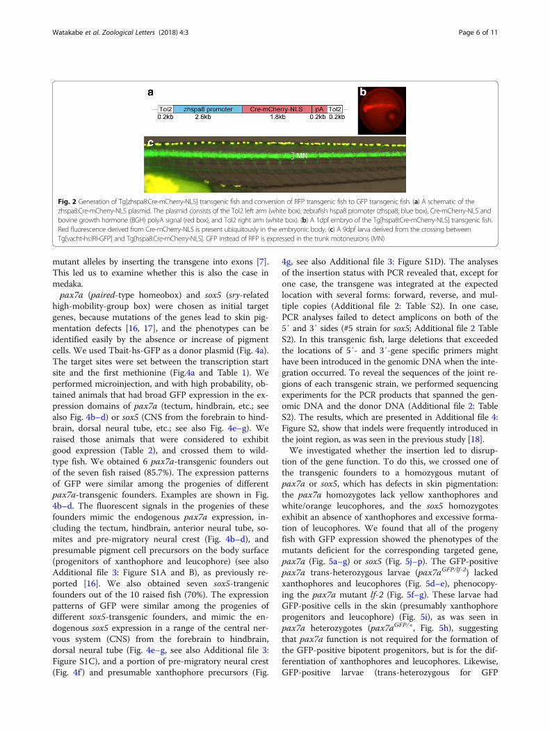
Construction of zhspa8:Cre and generationof Tg[zhspa8:Cre-mCherry-NLS] transgenic fishZebrafish hspa8 promoter, approximately 2.6 kb in length[9, 10], was used to express Cre-mCherry-NLS [11] ubiqui-tously in early embryos. The zhsp8 promoter, Cre-mCherry-NLS, and bovine growth hormone (BGH) polyAsequences were placed in this order in the Tol2-based
vector, pT2KXIGΔin [12]. Microinjection of Tol2-basedplasmid DNA into medaka embryos was performed as wasdone in zebrafish [12].
Preparation of sgRNAsTemplate DNA for sgRNA synthesize was PCR-amplifiedfrom pDR274 [13] with the forward primer, ATTTAGGT-GACACTATAgaxxxxxxxxxxxxxxxxxxGTTTTAGAGCTAGAAATAGC (for SP6 polymerase) or TAATACGACT-CACTATAggxxxxxxxxxxxxxxxxxxGTTTTAGAGCTA-GAAATAGC (for T7 polymerase), and the reverse primer,AAAAGCACCGACTCGGTGCC. The lowercase letterscorrespond to genome-targeting sequences (either 19 or 20mer) in sgRNAs. The genome-targeting sequences insgRNAs used in this study are shown in Table 1. After PCRamplification with Prime Star Taq polymerase (Takara,Otsu, Japan), PCR product was purified using a QIAquickPCR Purification Kit (Qiagen, Hilden, Germany). TemplateDNA thus obtained was used for the in vitro transcriptionof sgRNAs using a MAXIscript T7 kit (Life Technologies,Carlsbad, USA). pCS2-hSpCas9 (a gift from M. Kinoshitaand F. Zhang; [14]) was digested with NotI, and Cas9mRNA was transcribed using an mMESSAGEmMA-CHINE SP6 kit (Life Technologies). sgRNAs and Cas9mRNA were purified using an RNeasy Mini kit (Qiagen).For the vacht and nr5a1 genes, we tested two sgRNAs.
The sgRNA that yielded best results in F0 expressionassay was chosen to generate stable transgenic fish(Table 1). For the sox5, pax7a, and pnp4a genes, wetested just one sgRNA for each gene (Table 1).
Microinjection for knock-insgRNAs and Cas9 mRNA were co-injected into one-cellstage medaka embryos with Qiagen miniprep (Qiagen)purified donor DNA. Each embryo was injected with a so-lution containing ~ 9 ng/μl of sgRNA for digesting Tbait,~ 18 ng/μl of sgRNA for digesting genome DNA, ~200 ng/μl of Cas9 mRNA, and ~ 9 ng/μl of donor plasmid.Injection volume was adjusted such that approximately30–75% of injected embryos were dead within one weekafter injection (Table 2).
Insertion mappingFor insertion mapping, fluorescent F1 animals at 5–9dpf were collected, and genomic DNA was extractedwith standard protocols. The insertion status was ex-amined on either the 5′ side or the 3′ side of the in-sertion. For example, to examine the 5′ side of theinsertion, a PCR reaction was performed using a 5′primer that was specific to each gene (upstream ofthe expected insertion site) and a 3′ primer that wasspecific to the donor plasmid (sequence within thehsp70 promoter for detecting the forward insertion,and sequence within pBluescriptSK for detecting the
Watakabe et al. Zoological Letters (2018) 4:3 Page 2 of 11

inverse insertion). To examine whether the tandem-array insertion in the same direction occurred, a PCRreaction was performed with the two primers withinthe donor plasmid.For the sox5 and pax7a transgenic fish, nucleotide se-
quences of PCR products were determined to examinethe joint regions of the insertions.
ImagingImages were taken using an MVX10 microscope(Olympus, Tokyo, Japan), an MZ APO stereomicroscope(Leica, Wetzlar, Germany), and an LSM700 confocal laser-scanning microscope (Zeiss, Oberkochen, Germany).
ResultsStrategy for the generation of knock-in medaka andgeneration of Tg[vacht-hs:lRl-GFP] strainsFor NHEJ-mediated knock-in in medaka, we employed anexperimental scheme previously established by our groupin zebrafish [6]. Briefly, we co-injected sgRNA1 (for
genome digestion), sgRNA2 (for plasmid digestion), donorplasmid, and Cas9 mRNA into one-cell-stage medaka em-bryos (Fig. 1a). The vacht (vesicular acetylcholine trans-porter; also called slc18a3) gene, which is known to beexpressed in cholinergic neurons (motoneurons, primarily),was chosen as an initial target. The donor plasmid (Tbait-hs-lRl-GFP) contains a bait sequence (Tbait; [6]) upstreamof the insertion cassette for sgRNA2-guided DNA cleavage.This bait sequence was selected because the correspondingsgRNA (sgT) appears to have no off-target site in the me-daka genome (Additional file 1: Table S1). Tbait isfollowed by a medaka hsp70 promoter, which we expectto work as a minimal promoter (Fig. 1b). In this study, weextracted a 0.8 kb sequence of the medaka hsp70.1 pro-moter (see Methods) for this purpose. The hsp70 pro-moter is followed by the loxP-RFP-loxP-GFP (lRl-GFP)sequence (Fig. 1b). Without application of Cre recombin-ase, RFP will be expressed as a reporter gene. The targetsite for genome digestion was set upstream of the pro-spective transcriptional-start site of vacht (Fig. 1b; the se-quences used are shown in Table 1). Concurrent digestionof the genome (guided by sgRNA1) and the plasmid DNA(guided by sgRNA2) with Cas9 would result in the inte-gration of the donor plasmid into the genome via anNHEJ (Fig. 1b).We tested two sgRNAs (vacht-sg1 and vacht-sg2; Table
1). Injected animals were investigated for their RFP ex-pression around the hatching stage. In the case of vacht-sg1, we found animals that showed wide-spread RFP ex-pression in the trunk motoneurons (Fig. 1d; 1c is a con-trol animal) in 55% (11 of 20) of the animals (Table 2).The remaining 45% of the animals either showed sparseRFP expression in the motoneurons (Fig. 1e) or no
Table 1 DNA sequences for the corresponding sgRNAs
The protospacer-adjacent motif (PAM) sequence is labeled in green. The bold letters indicate that the corresponding sgRNAs were used to generate knock-in transgenicfish. The lowercase letter, g, at the beginning of the sequences indicates that the corresponding G is not present in the genome (mismatch). The mismatches derive fromthe demand that the first letter ought to be G for efficient in vitro transcription by T7 or SP6 polymerase. The numerals shown at the right indicate the locations of thecorresponding sequences with respect to the prospective transcription start sites (the 5’end of the longest cDNA) for each gene
Table 2 Results of microinjections and screening of transgenicfounders
injected survivedat 7dpf
goodexpression
survivedto adult
positivefounder
vacht sg1 84 20 11 10 5 / 10 (50%)
nr5a1 sg1 123 31 3 1 1 / 1 (100%)
sox5 sg1 52 25 11 10 7/ 10 (70%)
pax7a sg1 75 32 9 7 6 / 7 (85.7%)
pnp4a sg1 237 69* 13 6 3 / 6 (50%)
total 571 177 47 34 23 / 34 (67.6%)
The asterisk (*) indicates the number of surviving fish at 2 dpf
Watakabe et al. Zoological Letters (2018) 4:3 Page 3 of 11

Fig. 1 Strategy for the generation of knock-in medaka and generation of Tg[vacht-hs:lRl-GFP] strains. (a) For the generation of knock-in transgenic fish,sgRNA1 (for genome digestion), sgRNA2 (for plasmid digestion), donor plasmid with a bait sequence, and Cas9 mRNA are co-injected into one-cell-stagemedaka embryos. (b) A schematic representation of the vacht locus (grey box) and the sgRNA target sites (orange box), and the reporter gene constructconsisting of the Tbait (brown box), medaka hsp70 promoter (hsP, blue box), loxP, RFP-pA (red box), loxP, and GFP-pA (green box). After injection, theconcurrent cleavage of the targeted genomic locus and the Tbait-hs-lRl-GFP reporter plasmid results in the integration of the reporter by non-homologousend joining (NHEJ). The scheme shows the forward integration of the reporter. (c) Lateral view (red fluorescence) of a control larva at 9dpf. Red-yellowsignals in the dorsal region of the body are from the auto-fluorescence of pigment cells. (d) An example of an injected larva. RFP expression was presentbroadly in the motoneurons (MN). Animals with this kind of RFP expression were judged as having “good expression”, and raised to adulthood. (e) Anotherexample of an injected larva. In this animal, RFP expression was present in the motoneurons, but the number of RFP-expressing cells is much smaller thanthat in (d). Animals with this kind of RFP expression were judged as not having “good expression”, and were not raised. (f) Lateral view of a Tg[vacht-hs:lRl-GFP] larva. RFP expression was present broadly in the trunk motoneurons. All of the trunk motoneurons are likely to express RFP in this animal. (g) Maternalexpression of RFP in early embryos in the Tg[vacht-hs:lRl-GFP] line. The expression levels of RFP are variable among embryos. The embryos were obtainedfrom a single mother
Watakabe et al. Zoological Letters (2018) 4:3 Page 4 of 11

expression. In the zebrafish experiments, there was astrong correlation between the expression levels of a re-porter gene in injected animals and the probability ofbecoming transgenic founders. Animals that had goodreporter gene expression had a high probability of be-coming positive founders [6]. Thus, we raised only thoseanimals that were considered to have “good expression”(Fig. 1d; Table 2). The raised animals were crossed towild-type to examine if they would produce fluorescentoffspring. Among 10 fish screened, five produced larvaewith RFP expression in the motoneurons (Fig. 1f; Table 2).The expression patterns of RFP in these Tg [vacht-hs:lRl-GFP] transgenic fish were similar among progenies fromdifferent founders. We investigated the insertion sites ofeach line by PCR, and found that, in all cases, the trans-gene was integrated around the expected site in thegenome. As was seen in zebrafish, both forward-directionintegration and reverse-direction integration were ob-served. Moreover, in some cases multiple copies of donorplasmid were integrated (Additional file 2: Table S2).The medaka hsp70 promoter employed in this study
showed activity in female germ cells. Embryos producedfrom transgenic females showed ubiquitous red fluores-cence due to the maternal effect (Fig. 1g). Expression levelswere variable even among progenies from the same female(Fig. 1g). This maternally-derived fluorescence becamenegligible at around 3 dpf, and thus did not represent amajor problem for observation of RFP-labeled motoneu-rons at later stages. The maternally derived fluorescencewas also observed in other lines generated in this study.
Conversion of RFP transgenic fish to GFP transgenic fishby crossingThe reporter sequence (loxP-RFP-loxP-GFP) in theTg[vacht-hs:lRl-GFP] fish described above was aimed suchthat RFP expression could be converted to GFP expressionby the application of Cre. To conveniently change RFPtransgenic fish to GFP transgenic fish, we generated trans-genic medaka that ubiquitously express Cre-mCherry-NLSfusion protein [11] in early embryos. For this purpose, weused the zebrafish hspa8 promoter, which is known todrive gene expression ubiquitously in early zebrafishembryos [9, 10].Tg[zhsp8:Cre-mCherry-NLS] transgenic medaka was
generated by a Tol2-based transgenic method (Fig. 2a).Transgenic embryos of this line expressed Cre-mCherry-NLS proteins ubiquitously in the early stages (Fig. 2b).We crossed this Tg[zhsp8:Cre-mCherry-NLS] fish toTg[vacht-hs:lRl-GFP] transgenic fish. As expected, weobtained larvae that expressed GFP instead of RFP inthe motoneurons (Fig. 2c). The GFP-expressing animalswere raised to adulthoods, and crossed to wild-type fish.Approximately one-half of the fish expressed GFP in the
motoneurons; no RFP-expressing fish were obtained.These results indicate that RFP transgenic fish can begenetically transformed to GFP transgenic fish(Tg[vacht-hs:GFP]) by crossing alone.
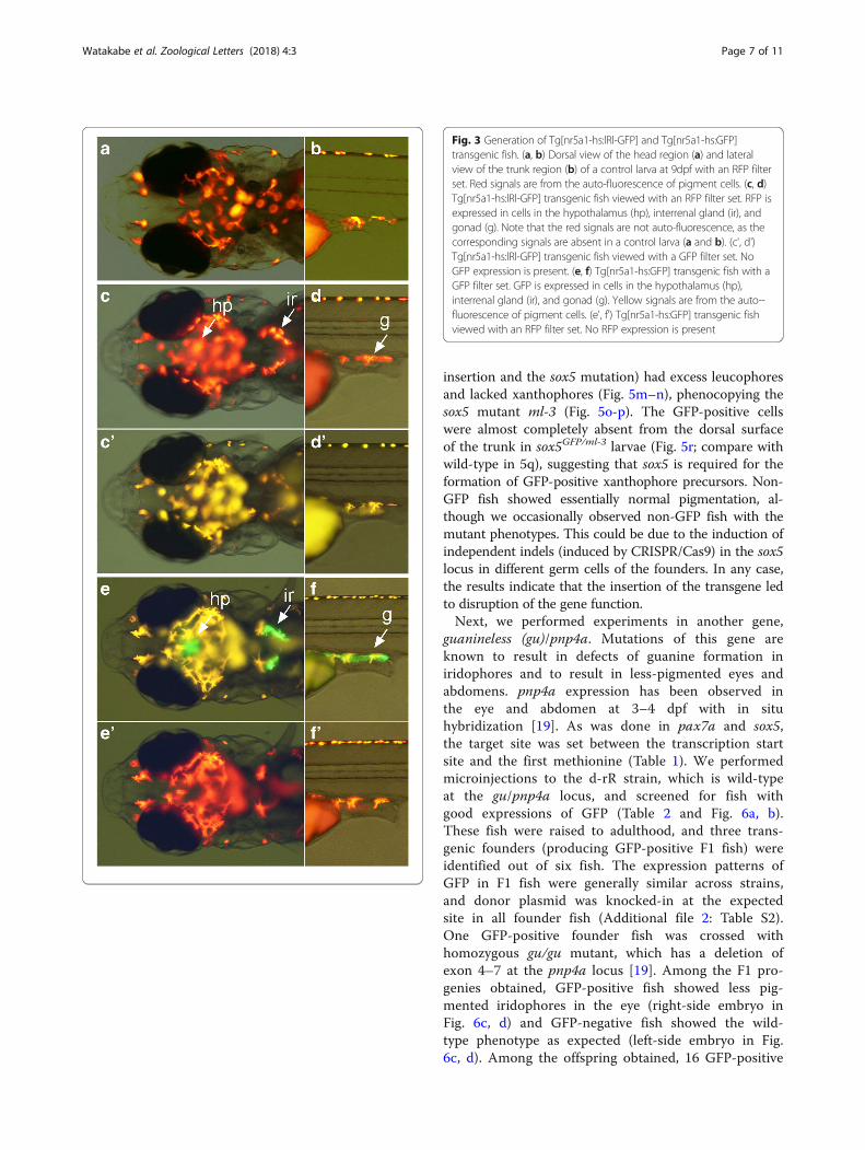
Generation of Tg[nr5a1-hs:lRl-GFP] and Tg[nr5a1-hs:GFP]strainsNext, we chose the nr5a1 (also called ftz-f1) gene, andexamined if the same technique worked for this gene.The nr5a1 gene is known to be expressed in cells in thehypothalamus (hp), interrenal gland (ir), and gonad (g)([15]; see also Figs. 3e and f). We tested two sgRNAs(nr5a1-sg1 and nr5a1-sg2). In both cases, the target siteswere set upstream of the prospective transcription startsite (Table 1), like the experiments with the vacht gene.We performed microinjections with the Tbait-hs-lRl-GFP (see, Fig. 1b). In the case of nr5a1-sg1, we observedanimals that had RFP expressions in the expression do-mains of nr5a1. Compared to the experiments with thevacht gene, the expression of RFP in these tissues in theinjected animals was more difficult to see, becausestrongly auto-fluorescent red pigment cells were locatednear the tissues where nr5a1 was expressed (see Figs.3a–d). We selected three animals (out of 31 survivors)that had good RFP expression in these tissues, and raisedthem (Table 2). Of these three fish, only one survived toadulthood. The survivor was crossed to a wild-type fish,and it turned out that the fish was a positive founder(Tg[nr5a1-hs:lRl-GFP]). RFP expression in the transgenicfish was observed in the expected tissues (hp, ir, and g inFigs. 3a–d). No GFP expression was observed in the trans-genic fish (Figs. 3c' and d'). We investigated the insertionsite of the line by PCR, and found that the transgene wasintegrated in the expected site in the forward direction asa single copy (Additional file 2: Table S2).We then examined if RFP fish could be converted to
GFP fish by crossing. A Tg[nr5a1-hs:lRl-GFP] transgenicfounder was crossed to Tg[zhsp8:Cre-mCherry-NLS] fish.This resulted in the production of animals in which GFPinstead of RFP was expressed in the hypothalamus (hp),interrenal gland (ir), and gonad (g) (Figs. 3e and f). Theanimals were raised to adulthood, and were then verifiedto produce GFP-expressing fish. These GFP transgenicfish did not express RFP (Figs. 3e' and f'). Thus, RFP trans-genic fish were converted to GFP transgenic fish, resultingin the establishment of Tg[nr5a1-hs:GFP].
Generation of mutant alleles for pax7a and sox5 genes byNHEJ-mediated knock-inIn the knock-in experiments for the vacht and nr5a1genes, we did not aim to disrupt gene functions. The in-sertion sites were set upstream of the genes, not in theexons. In zebrafish studies, the NHEJ-mediated knock-intechnique has also shown to be effective for obtaining
Watakabe et al. Zoological Letters (2018) 4:3 Page 5 of 11
mutant alleles by inserting the transgene into exons [7].This led us to examine whether this is also the case inmedaka.pax7a (paired-type homeobox) and sox5 (sry-related
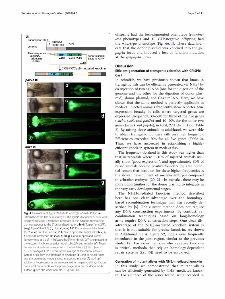
high-mobility-group box) were chosen as initial targetgenes, because mutations of the genes lead to skin pig-mentation defects [16, 17], and the phenotypes can beidentified easily by the absence or increase of pigmentcells. We used Tbait-hs-GFP as a donor plasmid (Fig. 4a).The target sites were set between the transcription startsite and the first methionine (Fig.4a and Table 1). Weperformed microinjection, and with high probability, ob-tained animals that had broad GFP expression in the ex-pression domains of pax7a (tectum, hindbrain, etc.; seealso Fig. 4b–d) or sox5 (CNS from the forebrain to hind-brain, dorsal neural tube, etc.; see also Fig. 4e–g). Weraised those animals that were considered to exhibitgood expression (Table 2), and crossed them to wild-type fish. We obtained 6 pax7a-transgenic founders outof the seven fish raised (85.7%). The expression patternsof GFP were similar among the progenies of differentpax7a-transgenic founders. Examples are shown in Fig.4b–d. The fluorescent signals in the progenies of thesefounders mimic the endogenous pax7a expression, in-cluding the tectum, hindbrain, anterior neural tube, so-mites and pre-migratory neural crest (Fig. 4b–d), andpresumable pigment cell precursors on the body surface(progenitors of xanthophore and leucophore) (see alsoAdditional file 3: Figure S1A and B), as previously re-ported [16]. We also obtained seven sox5-trangenicfounders out of the 10 raised fish (70%). The expressionpatterns of GFP were similar among the progenies ofdifferent sox5-transgenic founders, and mimic the en-dogenous sox5 expression in a range of the central ner-vous system (CNS) from the forebrain to hindbrain,dorsal neural tube (Fig. 4e–g, see also Additional file 3:Figure S1C), and a portion of pre-migratory neural crest(Fig. 4f') and presumable xanthophore precursors (Fig.
4g, see also Additional file 3: Figure S1D). The analysesof the insertion status with PCR revealed that, except forone case, the transgene was integrated at the expectedlocation with several forms: forward, reverse, and mul-tiple copies (Additional file 2: Table S2). In one case,PCR analyses failed to detect amplicons on both of the5′ and 3′ sides (#5 strain for sox5; Additional file 2 TableS2). In this transgenic fish, large deletions that exceededthe locations of 5′- and 3′-gene specific primers mighthave been introduced in the genomic DNA when the inte-gration occurred. To reveal the sequences of the joint re-gions of each transgenic strain, we performed sequencingexperiments for the PCR products that spanned the gen-omic DNA and the donor DNA (Additional file 2: TableS2). The results, which are presented in Additional file 4:Figure S2, show that indels were frequently introduced inthe joint region, as was seen in the previous study [18].We investigated whether the insertion led to disrup-
tion of the gene function. To do this, we crossed one ofthe transgenic founders to a homozygous mutant ofpax7a or sox5, which has defects in skin pigmentation:the pax7a homozygotes lack yellow xanthophores andwhite/orange leucophores, and the sox5 homozygotesexhibit an absence of xanthophores and excessive forma-tion of leucophores. We found that all of the progenyfish with GFP expression showed the phenotypes of themutants deficient for the corresponding targeted gene,pax7a (Fig. 5a–g) or sox5 (Fig. 5j–p). The GFP-positivepax7a trans-heterozygous larvae (pax7aGFP/lf-2) lackedxanthophores and leucophores (Fig. 5d–e), phenocopy-ing the pax7a mutant lf-2 (Fig. 5f–g). These larvae hadGFP-positive cells in the skin (presumably xanthophoreprogenitors and leucophore) (Fig. 5i), as was seen inpax7a heterozygotes (pax7aGFP/+, Fig. 5h), suggestingthat pax7a function is not required for the formation ofthe GFP-positive bipotent progenitors, but is for the dif-ferentiation of xanthophores and leucophores. Likewise,GFP-positive larvae (trans-heterozygous for GFP
Fig. 2 Generation of Tg[zhspa8:Cre-mCherry-NLS] transgenic fish and conversion of RFP transgenic fish to GFP transgenic fish. (a) A schematic of thezhspa8:Cre-mCherry-NLS plasmid. The plasmid consists of the Tol2 left arm (white box), zebrafish hspa8 promoter (zhspa8; blue box), Cre-mCherry-NLS andbovine growth hormone (BGH) polyA signal (red box), and Tol2 right arm (white box). (b) A 1dpf embryo of the Tg[hspa8:Cre-mCherry-NLS] transgenic fish.Red fluorescence derived from Cre-mCherry-NLS is present ubiquitously in the embryonic body. (c) A 9dpf larva derived from the crossing betweenTg[vacht-hs:lRl-GFP] and Tg[hspa8:Cre-mCherry-NLS]. GFP instead of RFP is expressed in the trunk motoneurons (MN)
Watakabe et al. Zoological Letters (2018) 4:3 Page 6 of 11
insertion and the sox5 mutation) had excess leucophoresand lacked xanthophores (Fig. 5m–n), phenocopying thesox5 mutant ml-3 (Fig. 5o-p). The GFP-positive cellswere almost completely absent from the dorsal surfaceof the trunk in sox5GFP/ml-3 larvae (Fig. 5r; compare withwild-type in 5q), suggesting that sox5 is required for theformation of GFP-positive xanthophore precursors. Non-GFP fish showed essentially normal pigmentation, al-though we occasionally observed non-GFP fish with themutant phenotypes. This could be due to the induction ofindependent indels (induced by CRISPR/Cas9) in the sox5locus in different germ cells of the founders. In any case,the results indicate that the insertion of the transgene ledto disruption of the gene function.Next, we performed experiments in another gene,
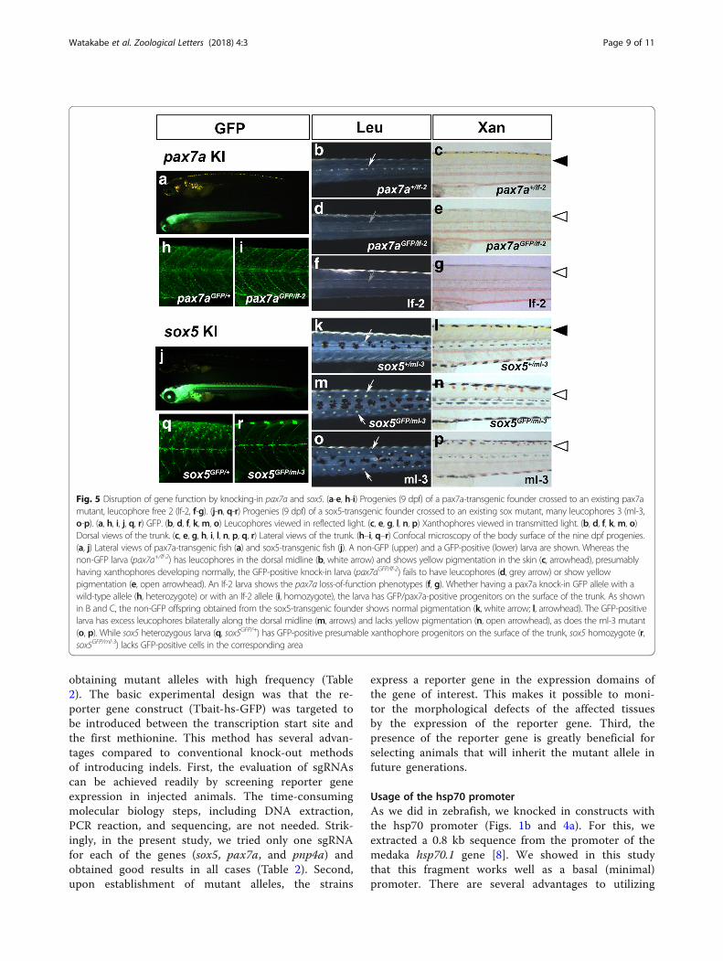
guanineless (gu)/pnp4a. Mutations of this gene areknown to result in defects of guanine formation iniridophores and to result in less-pigmented eyes andabdomens. pnp4a expression has been observed inthe eye and abdomen at 3–4 dpf with in situhybridization [19]. As was done in pax7a and sox5,the target site was set between the transcription startsite and the first methionine (Table 1). We performedmicroinjections to the d-rR strain, which is wild-typeat the gu/pnp4a locus, and screened for fish withgood expressions of GFP (Table 2 and Fig. 6a, b).These fish were raised to adulthood, and three trans-genic founders (producing GFP-positive F1 fish) wereidentified out of six fish. The expression patterns ofGFP in F1 fish were generally similar across strains,and donor plasmid was knocked-in at the expectedsite in all founder fish (Additional file 2: Table S2).One GFP-positive founder fish was crossed withhomozygous gu/gu mutant, which has a deletion ofexon 4–7 at the pnp4a locus [19]. Among the F1 pro-genies obtained, GFP-positive fish showed less pig-mented iridophores in the eye (right-side embryo inFig. 6c, d) and GFP-negative fish showed the wild-type phenotype as expected (left-side embryo in Fig.6c, d). Among the offspring obtained, 16 GFP-positive
Fig. 3 Generation of Tg[nr5a1-hs:lRl-GFP] and Tg[nr5a1-hs:GFP]transgenic fish. (a, b) Dorsal view of the head region (a) and lateralview of the trunk region (b) of a control larva at 9dpf with an RFP filterset. Red signals are from the auto-fluorescence of pigment cells. (c, d)Tg[nr5a1-hs:lRl-GFP] transgenic fish viewed with an RFP filter set. RFP isexpressed in cells in the hypothalamus (hp), interrenal gland (ir), andgonad (g). Note that the red signals are not auto-fluorescence, as thecorresponding signals are absent in a control larva (a and b). (c', d')Tg[nr5a1-hs:lRl-GFP] transgenic fish viewed with a GFP filter set. NoGFP expression is present. (e, f) Tg[nr5a1-hs:GFP] transgenic fish with aGFP filter set. GFP is expressed in cells in the hypothalamus (hp),interrenal gland (ir), and gonad (g). Yellow signals are from the auto--fluorescence of pigment cells. (e', f') Tg[nr5a1-hs:GFP] transgenic fishviewed with an RFP filter set. No RFP expression is present
Watakabe et al. Zoological Letters (2018) 4:3 Page 7 of 11
offspring had the less-pigmented phenotype (guanine-less phenotype) and 10 GFP-negative offspring hadthe wild-type phenotype (Fig. 6e, f ). These data indi-cate that the donor plasmid was knocked into the gu/pnp4a locus and induced a loss of function mutationat the gu/pnp4a locus.
DiscussionEfficient generation of transgenic zebrafish with CRISPR/Cas9In zebrafish, we have previously shown that knock-intransgenic fish can be efficiently generated via NHEJ byco-injection of two sgRNAs (one for the digestion of thegenome and the other for the digestion of donor plas-mid), donor plasmid, and Cas9 mRNA. Here, we haveshown that the same method is perfectly applicable inmedaka. Injected animals frequently show reporter geneexpression broadly in cells where targeted genes areexpressed (frequency, 30–50% for three of the five genes(vacht, sox5, and pax7a) and 10–20% for the other twogenes (nr5a1 and pnp4a); in total, 27% (47 of 177); Table2). By raising those animals to adulthood, we were ableto obtain transgenic founders with very high frequency.Efficiencies exceeded 50% for all five genes (Table 2).Thus, we have succeeded in establishing a highly-efficient knock-in system in medaka fish.The frequency obtained in this study was higher than
that in zebrafish where 5–10% of injected animals usu-ally show “good expression”, and approximately 30% ofraised animals became positive founders [6]. One poten-tial reason that accounts for these higher frequencies isthe slower development of medaka embryos comparedto zebrafish embryos [20, 21]: In medaka, there may bemore opportunities for the donor plasmid to integrate inthe very early developmental stages.The NHEJ-mediated knock-in method described
here has one clear advantage over the homology-based recombination technique that was recently de-scribed by [5]. The current method does not requireany DNA construction experiments. By contrast, re-combination techniques based on long-homologyarms require DNA construction steps. One clear dis-advantage of the NHEJ-mediated knock-in system isthat it is not suitable for precise knock-in. As shownin Additional file 4: Figure S2, indels were frequentlyintroduced in the joint region, similar to the previousstudy [18]. For experiments in which precise knock-inis critical, methods that rely on homology-dependentrepair systems (i.e., [5]) need to be employed.
Generation of mutant alleles with NHEJ-mediated knock-inIn this study, we demonstrated that mutant allelescan be efficiently generated by NHEJ-mediated knock-in. For all three of the genes tested, we succeeded in
Fig. 4 Generation of Tg[pax7a-hs:GFP] and Tg[sox5-hs:GFP] fish. (a)Schematic of the knock-in strategies. The sgRNAs for pax7a or sox5 weredesigned to target a sequence upstream of the initiation methioninethat corresponds to the 5′ untranslated region. (b–d) Tg[pax7a-hs:GFP].(e–g) Tg[sox5-hs:GFP]. (b, b', c, c', e, e', f, f') Dorsal views of the head(b, b', e, e') and the trunk (c, c', f, f') at 2 dpf in the bright field (b, c, e,f) and in fluorescence (b', c', e', f'). (d, g) Dorsal (upper) and lateral(lower) views at 9 dpf. In Tg[pax7a-hs:GFP] embryos, GFP is expressed inthe tectum, hindbrain, anterior neural tube (b'), and muscles (c'). Thesefluorescent signals are maintained in the hatchlings (d). In Tg[sox5-hs:GFP] embryos, GFP is expressed in a range of the central nervoussystem (CNS) from the forebrain to hindbrain (e'), and in neural tubesand the premigratory neural crest in a dotted manner (f'). At 9 dpf,additional fluorescent signals are observed in the pectoral fins, olfactorybulbs, and presumable xanthophore progenitors on the dorsal bodysurface (g; see also Additional file 3: Fig. S1C–D)
Watakabe et al. Zoological Letters (2018) 4:3 Page 8 of 11
obtaining mutant alleles with high frequency (Table2). The basic experimental design was that the re-porter gene construct (Tbait-hs-GFP) was targeted tobe introduced between the transcription start site andthe first methionine. This method has several advan-tages compared to conventional knock-out methodsof introducing indels. First, the evaluation of sgRNAscan be achieved readily by screening reporter geneexpression in injected animals. The time-consumingmolecular biology steps, including DNA extraction,PCR reaction, and sequencing, are not needed. Strik-ingly, in the present study, we tried only one sgRNAfor each of the genes (sox5, pax7a, and pnp4a) andobtained good results in all cases (Table 2). Second,upon establishment of mutant alleles, the strains
express a reporter gene in the expression domains ofthe gene of interest. This makes it possible to moni-tor the morphological defects of the affected tissuesby the expression of the reporter gene. Third, thepresence of the reporter gene is greatly beneficial forselecting animals that will inherit the mutant allele infuture generations.
Usage of the hsp70 promoterAs we did in zebrafish, we knocked in constructs withthe hsp70 promoter (Figs. 1b and 4a). For this, weextracted a 0.8 kb sequence from the promoter of themedaka hsp70.1 gene [8]. We showed in this studythat this fragment works well as a basal (minimal)promoter. There are several advantages to utilizing
Fig. 5 Disruption of gene function by knocking-in pax7a and sox5. (a-e, h-i) Progenies (9 dpf) of a pax7a-transgenic founder crossed to an existing pax7amutant, leucophore free 2 (lf-2, f-g). (j-n, q-r) Progenies (9 dpf) of a sox5-transgenic founder crossed to an existing sox mutant, many leucophores 3 (ml-3,o-p). (a, h, i, j, q, r) GFP. (b, d, f, k,m, o) Leucophores viewed in reflected light. (c, e, g, l, n, p) Xanthophores viewed in transmitted light. (b, d, f, k, m, o)Dorsal views of the trunk. (c, e, g, h, i, l, n, p, q, r) Lateral views of the trunk. (h–i, q–r) Confocal microscopy of the body surface of the nine dpf progenies.(a, j) Lateral views of pax7a-transgenic fish (a) and sox5-transgenic fish (j). A non-GFP (upper) and a GFP-positive (lower) larva are shown. Whereas thenon-GFP larva (pax7a+/lf-2) has leucophores in the dorsal midline (b, white arrow) and shows yellow pigmentation in the skin (c, arrowhead), presumablyhaving xanthophores developing normally, the GFP-positive knock-in larva (pax7aGFP/lf-2) fails to have leucophores (d, grey arrow) or show yellowpigmentation (e, open arrowhead). An lf-2 larva shows the pax7a loss-of-function phenotypes (f, g). Whether having a pax7a knock-in GFP allele with awild-type allele (h, heterozygote) or with an lf-2 allele (i, homozygote), the larva has GFP/pax7a-positive progenitors on the surface of the trunk. As shownin B and C, the non-GFP offspring obtained from the sox5-transgenic founder shows normal pigmentation (k, white arrow; l, arrowhead). The GFP-positivelarva has excess leucophores bilaterally along the dorsal midline (m, arrows) and lacks yellow pigmentation (n, open arrowhead), as does the ml-3 mutant(o, p). While sox5 heterozygous larva (q, sox5GFP/+) has GFP-positive presumable xanthophore progenitors on the surface of the trunk, sox5 homozygote (r,sox5GFP/ml-3) lacks GFP-positive cells in the corresponding area
Watakabe et al. Zoological Letters (2018) 4:3 Page 9 of 11

the hsp70 promoter in DNA constructs for knock-in.First, the efficiencies of obtaining transgenic foundersare likely to be increased, as reporter gene expressionoccurs irrespective of the direction of integration. In-deed, we found both types of integration (includingmulti-copy integrations) in the transgenic lines gener-ated in this study. Second, the reporter construct canbe targeted virtually anywhere near or within a geneof interest. This feature gives researchers flexibility indesigning sgRNAs.There are two potential disadvantages or concerns
regarding the usage of the hsp70 promoter. First, thepromoter has activity in female germ cells. We ob-served maternally derived fluorescence in many ofthe lines generated in this study. Although this didnot present major problems in the present study, itcould be a problem for monitoring the expressionpatterns of genes in the early stages. Nonetheless,this problem can likely be overcome, in many cases,by using transgenic males for mating. Second, geneexpression may not be completely recapitulated withthe usage of a heterologous promoter. Although re-porter gene expressions appeared to mimic the ex-pressions of the endogenous genes in the transgenicfish generated in the current study for all five of thegenes, the possibility of an occurrence of ectopic ex-pressions cannot be completely excluded. Indeed, wehave noted an ectopic expression in a Tg[evx2-hs:Gal4] transgenic fish strain in the case of zebra-fish [6]. Thus, researchers need to be aware of thepotential occurrence of an ectopic expression whenusing the hsp70 promoter.
Conversion of reporter gene expression by the Cre-loxPsystemIn this study, we showed that RFP expression can bechanged to GFP expression in transgenic fish with theloxP-RFP-loxP-GFP construct. To make this occurubiquitously, we established Tg[zhspa8:Cre-mCherry-NLS] in which the Cre-mCherry-NLS fusion proteinis expressed ubiquitously in early embryos. Using thisline, one reporter gene was easily converted to an-other reporter/driver gene by crossing alone. This is apowerful system for establishing transgenic fish thatexpress a reporter/driver gene that is itself not fluor-escent (without the aid of fluorescent reporters, pre-screening before raising animals is not possible). Forexample, we succeeded in establishing several Gal4driver lines using this system in zebrafish (our unpub-lished observation). In these studies, we first estab-lished loxP-RFP-loxP-Gal4 lines, and RFP was thenconverted to Gal4 by utilizing Tg[zhspa8:Cre-mCherry-NLS] transgenic zebrafish. The same strategycould be used to generate transgenic medaka that ex-press reporter/driver genes that are not fluorescentthemselves.
ConclusionWe report that the NHEJ-mediated knock-in system ishighly efficient in medaka, and is very useful for establish-ing mutant alleles. With its simplicity and high efficiency,we propose that the method described may become astandard technique for the generation of transgenic andmutant medaka.
Fig. 6 Disruption of gene function by knocking-in pnp4a. (a, b) The expression pattern of GFP in founder fish. GFP expression was observed inthe eye and abdomen. These fish were considered to have “good expression” of GFP, and were raised to adulthood. GFP expression was ob-served in the eye and abdomen. (c–f) The progenies (4 dpf) of a pnp4a-transgenic founder crossed to an existing gu mutant. All GFP-positive F1fish have less-pigmented eyes and all GFP-negative F1 fish have wild-type pigmentation. Asterisks indicate GFP-positive and less-pigmentedF1 fish
Watakabe et al. Zoological Letters (2018) 4:3 Page 10 of 11

Additional files
Additional file 1: Table S1. Genomic location of potential off-targetsites for sgT (sgRNA for Tbait) (DOCX 14 kb)
Additional file 2: Table S2. Status of the insertions in the transgenicfish generated in this study (DOCX 16 kb)
Additional file 3: Figure S1. GFP expression in Tg[pax7a-hs:GFP] andTg[sox5-hs:GFP]. (A, B) Tg[pax7a-hs:GFP]. (C, D) Tg[sox5-hs:GFP]. (A, C)Dorsal views of the head region magnified. (B, D) Lateral views of theanterior trunk region magnified. The signal in the tectum is especiallystrong in Tg[pax7a-hs:GFP] (A). Presumable pigment cell progenitors ofxanthophore and leucophore (B) and xanthophore (D) on the bodysurface are positive for GFP in Tg[pax7a-hs:GFP] and Tg[sox5-hs:GFP],respectively. (JPEG 1442 kb)
Additional file 4: Figure S2. Nucleotide sequences of the joint regionof the insertions for the sox5 and pax7a transgenic fish The PCR productsthat span the genomic DNA and the donor DNA (see Additional file 2:Table S2) were sequenced. The top two lines for each panel show thesequence of the genome and the donor. Underlined sequencescorrespond to sgRNA targets. The PAM sequences are indicated in green.The predicted digestion sites by CRISPR/Cas9 are indicated witharrowheads. Sequences in bold letters are expected to be present afterthe integrations. The predicted nucleotide sequence without an indel isshown in the third line as bold letters. (A) The 5′ side of the sox5transgenic fish with the forward integration (#1, #3, and #4 strains;Additional file 2:Table S2). (B) The 5′ side of the sox5 transgenic fish withthe reverse integration (#2 strains; Additional file 2: Table S2). (C) The 3′side of the sox5 transgenic fish with the forward integration (#1, #2, #3,and #7 strains; Additional file 2: Table S2). (D) The 3′ side of the sox5transgenic fish with the reverse integration (#6 strain; Additional file 2:Table S2). (E) The 5′ side of the pax7a transgenic fish with the forwardintegration (#1, #3, #4, and #6 strains; Additional file 2: Table S2). (F) The5′ side of the pax7a transgenic fish with the reverse integration (#2strains; Additional file 2: Table S2). (G) The 3′ side of the pax7a transgenicfish with the forward integration (#1and #6 strains; Additional file 2: TableS2). (H) The 3′ side of the pax7a transgenic fish with the reverse integration(#2, #3, #4, and #5 strains; Additional file 2: Table S2). (PDF 31 kb)
AcknowledgementsWe thank the Medaka National Bioresource Project (NBRP Medaka), which issupported by the Japan Agency for Medical Research and Development, forproviding the d-rR (MT837) and gu (MT827) strains. We also thank Ms. T.Yamazaki, H. Ito, and Y. Terasawa for their excellent technical assistance.
FundingThis work was supported in part by grants from the Ministry of Education,Science, Technology, Sports and Culture of Japan.
Availability of data and materialsDonor plasmids, as well as their sequence information, are available from theMedaka National Bioresource Project in Japan (NBRP Medaka).
Authors’ contributionsSH conceived and designed the study. IW performed the major parts of theexperiments. YK helped some of the experiments that IW performed. HHperformed the experiments related to the sox5 and pax7a genes. SY and KNperformed the experiments related to the pnp4a gene. All authors contributedto the writing of the manuscript. All authors read and approved the finalmanuscript.
Ethics approval and consent to participateFish were maintained and used in accordance with the guidelines approvedby the animal care and use committees of the National Institutes of NaturalSciences (approval number: 17A002) and Nagoya University.
Consent for publicationNot applicable.
Competing interestsThe authors declare that they have no competing interests.
Publisher’s NoteSpringer Nature remains neutral with regard to jurisdictional claims inpublished maps and institutional affiliations.
Author details1National Institutes of Natural Sciences, Okazaki Institute for IntegrativeBioscience, National Institute for Basic Biology, Higashiyama 5-1, Myodaiji,Okazaki, Aichi 444-8787, Japan. 2Bioscience and Biotechnology Center,Nagoya University, Nagoya, Aichi 464-8601, Japan. 3National Institutes ofNatural Sciences, National Institute for Basic Biology, Okazaki, Aichi 444-8585,Japan. 4Department of Basic Biology, School of Life Science, GraduateUniversity for Advanced Studies (SOKENDAI), Okazaki, Aichi 444-8787, Japan.5Present address: Faculty of Pharmaceutical Sciences, Hokkaido University,Sapporo, Hokkaido 060-0812, Japan.
Received: 12 September 2017 Accepted: 29 December 2017
References1. Kirchmaier S, et al. The genomic and genetic toolbox of the teleost medaka
(Oryzias latipes). Genetics. 2015;199(4):905–18.2. Naruse K, et al. Medaka genomics: a bridge between mutant phenotype and
gene function. Mech Dev. 2004;121(7-8):619–28.3. Takeda H, Shimada A. The art of medaka genetics and genomics: what makes
them so unique? Annu Rev Genet. 2010;44:217–41.4. Kinoshita M, et al. Transgenic medaka enables easy oocytes detection in live
fish. Mol Reprod Dev. 2009;76(2):202–7.5. Murakami Y, et al. An efficient system for homology-dependent targeted
gene integration in medaka (Oryzias latipes). Zoological Lett. 2017;3:10.6. Kimura Y, et al. Efficient generation of knock-in transgenic zebrafish carrying reporter/
driver genes by CRISPR/Cas9-mediated genome engineering. Sci Rep. 2014;47. Ota S, et al. Functional visualization and disruption of targeted genes using CRISPR/
Cas9-mediated eGFP reporter integration in zebrafish. Sci Rep. 2016;6:34991.8. Oda S, et al. Identification of a functional medaka heat shock promoter and
characterization of its ability to induce exogenous gene expression inmedaka in vitro and in vivo. Zool Sci. 2010;27(5):410–5.
9. Mizuno H, et al. Transgenic zebrafish for ratiometric imaging of cytosolic andmitochondrial Ca2+ response in teleost embryo. Cell Calcium. 2013;54(3):236–45.
10. Sugiyama M, et al. Illuminating cell-cycle progression in the developingzebrafish embryo. Proc Natl Acad Sci U S A. 2009;106(49):20812–7.
11. Satou C, Kimura Y, Higashijima S. Generation of multiple classes of V0neurons in zebrafish spinal cord: progenitor heterogeneity and temporalcontrol of neuronal diversity. J Neurosci. 2012;32(5):1771–83.
12. Urasaki A, Morvan G, Kawakami K. Functional dissection of the Tol2transposable element identified the minimal cis-sequence and a highlyrepetitive sequence in the subterminal region essential for transposition.Genetics. 2006;174(2):639–49.
13. Hwang WY, et al. Efficient genome editing in zebrafish using a CRISPR-Cassystem. Nat Biotechnol. 2013;31(3):227–9.
14. Cong L, et al. Multiplex genome engineering using CRISPR/Cas systems.Science. 2013;339(6121):819–23.
15. Morinaga C, et al. Mutations affecting gonadal development in Medaka, Oryziaslatipes. Mech Dev. 2004;121(7-8):829–39.
16. Kimura T, et al. Leucophores are similar to xanthophores in theirspecification and differentiation processes in medaka. Proc Natl Acad Sci US A. 2014;111(20):7343–8.
17. Nagao Y, et al. Sox5 functions as a fate switch in medaka pigment celldevelopment. PLoS Genet. 2014;10(4):e1004246.
18. Auer TO, et al. Highly efficient CRISPR/Cas9-mediated knock-in in zebrafishby homology-independent DNA repair. Genome Res. 2014;24(1):142–53.
19. Kimura T, Takehana Y, Naruse K. pnp4a is the causal gene of the MedakaIridophore mutant guanineless. G3 (Bethesda). 2017;7(4):1357–63.
20. Iwamatsu T. Stages of normal development in the medaka Oryzias latipes.Mech Dev. 2004;121(7-8):605–18.
21. Kimmel CB, et al. Stages of embryonic development of the zebrafish. Dev Dyn.1995;203(3):253–310.
Watakabe et al. Zoological Letters (2018) 4:3 Page 11 of 11