prosaposin inhibits tumor metastasis via paracrine and endocrine
TRANSCRIPT
Corrections
MEDICAL SCIENCESCorrection for ‘‘Prosaposin inhibits tumor metastasis via para-crine and endocrine stimulation of stromal p53 and Tsp-1,’’ bySoo-Young Kang, Ole J. Halvorsen, Karsten Gravdal, NanditaBhattacharya, Jung Min Lee, Nathan W. Liu, Brian T.Johnston, Adam B. Johnston, Svein A. Haukaas, KristieAamodt, Sun Yoo, Lars A. Akslen, and Randolph S. Watnick,which appeared in issue 29, July 21, 2009, of Proc Natl Acad SciUSA (106:12115–12120; first published July 6, 2009; 10.1073/pnas.0903120106).
The authors note that due to a printer’s error, on page 12120,right column, line 4, the reference citation is incorrect. Thecorrected text should instead read, ‘‘In relation to the prostate,the Psap-encoding gene is amplified in a subset of prostatecancers (31).’’ The related reference appears below. In addition,several panels in Fig. 5 were labeled incorrectly. The correctedfigure and its legend appear below.
31. Koochekpour S, et al. (2005) Amplification and overexpression of prosaposin in pros-tate cancer. Genes Chromosomes Cancer 44:351–364.
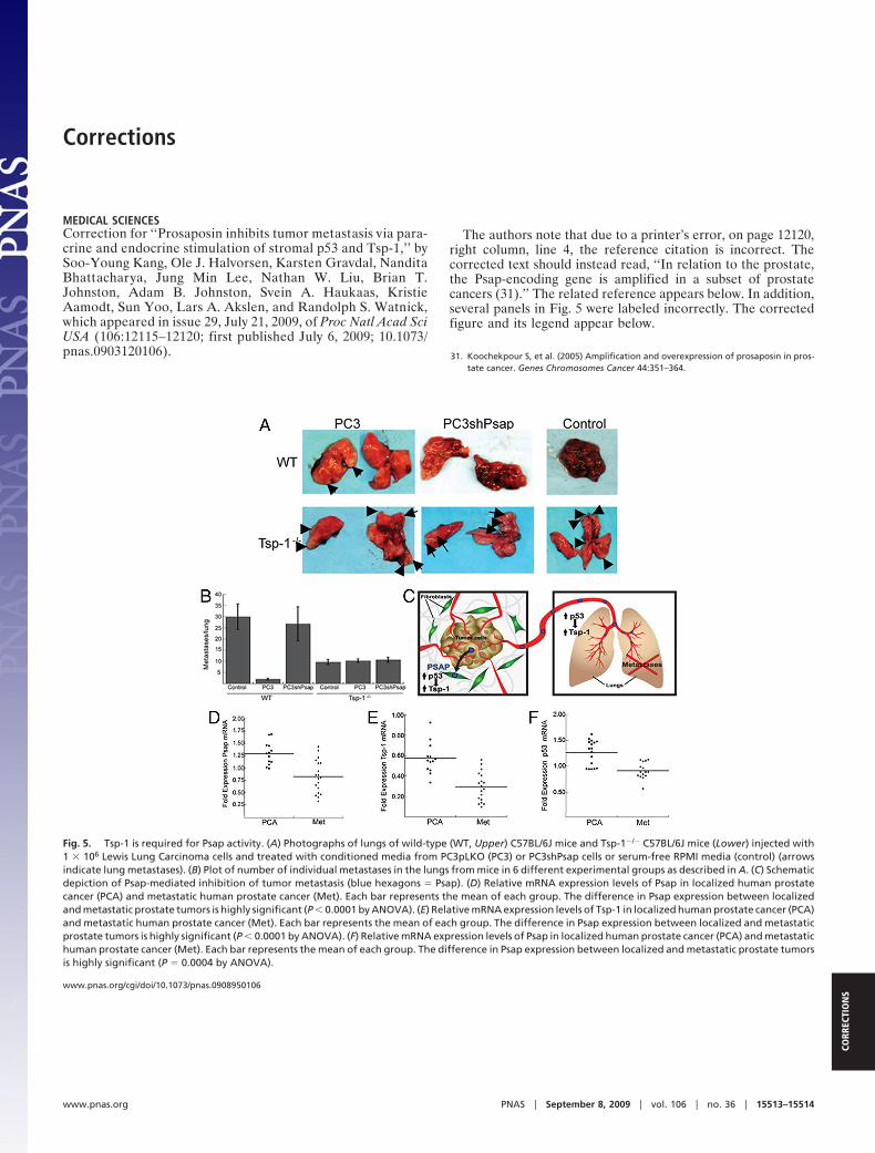
Fig. 5. Tsp-1 is required for Psap activity. (A) Photographs of lungs of wild-type (WT, Upper) C57BL/6J mice and Tsp-1�/� C57BL/6J mice (Lower) injected with1 � 106 Lewis Lung Carcinoma cells and treated with conditioned media from PC3pLKO (PC3) or PC3shPsap cells or serum-free RPMI media (control) (arrowsindicate lung metastases). (B) Plot of number of individual metastases in the lungs from mice in 6 different experimental groups as described in A. (C) Schematicdepiction of Psap-mediated inhibition of tumor metastasis (blue hexagons � Psap). (D) Relative mRNA expression levels of Psap in localized human prostatecancer (PCA) and metastatic human prostate cancer (Met). Each bar represents the mean of each group. The difference in Psap expression between localizedand metastatic prostate tumors is highly significant (P � 0.0001 by ANOVA). (E) Relative mRNA expression levels of Tsp-1 in localized human prostate cancer (PCA)and metastatic human prostate cancer (Met). Each bar represents the mean of each group. The difference in Psap expression between localized and metastaticprostate tumors is highly significant (P � 0.0001 by ANOVA). (F) Relative mRNA expression levels of Psap in localized human prostate cancer (PCA) and metastatichuman prostate cancer (Met). Each bar represents the mean of each group. The difference in Psap expression between localized and metastatic prostate tumorsis highly significant (P � 0.0004 by ANOVA).
www.pnas.org/cgi/doi/10.1073/pnas.0908950106
www.pnas.org PNAS � September 8, 2009 � vol. 106 � no. 36 � 15513–15514
CORR
ECTI
ON
S
Prosaposin inhibits tumor metastasis via paracrineand endocrine stimulation of stromal p53 and Tsp-1Soo-Young Kanga,b, Ole J. Halvorsenc, Karsten Gravdalc, Nandita Bhattacharyaa,b, Jung Min Leea, Nathan W. Liua,Brian T. Johnstona, Adam B. Johnstona,d, Svein A. Haukaase, Kristie Aamodta, Sun Yooa, Lars A. Akslenc,and Randolph S. Watnicka,b,1
aVascular Biology Program, Department of Surgery, Children’s Hospital Boston, Boston, MA 02115; bDepartment of Surgery, Harvard Medical School,Boston, MA 02115; cThe Gade Institute, Section for Pathology, University of Bergen, Haukeland University Hospital, N-5021 Bergen, Norway; dDepartment ofBiochemical Sciences, Harvard College, Cambridge, MA 02138; and eDepartment of Surgery, Section of Urology, University of Bergen, Haukeland UniversityHospital, N-5021 Bergen, Norway
Communicated by Robert A Weinberg, Whitehead Institute for Biomedical Research, Cambridge, MA, May 15, 2009 (received for review January 5, 2009)
Metastatic tumors can prepare a distant site for colonization viathe secretion of factors that act in a systemic manner. We hypoth-esized that non- or weakly metastatic human tumor cells may actin an opposite fashion by creating a microenvironment in distanttissues that is refractory to colonization. By comparing cell lineswith different metastatic potential, we have identified a tumor-secreted inhibitor of metastasis, prosaposin (Psap), which func-tions in a paracrine and endocrine fashion by stimulating theexpression of thrombospondin-1 (Tsp-1) in fibroblasts present inboth primary tumors and distant organs, doing so in a p53-dependent manner. Introduction of Psap in highly metastatic cellssignificantly reduced the occurrence of metastases, whereas inhi-bition of Psap production by tumor cells was associated withincreased metastatic frequency. In human prostate cancer, de-creased Psap expression was significantly associated with meta-static tumors. Our findings suggest that prosaposin, or otheragents that stimulate p53 activity in the tumor stroma, may be aneffective therapy by inhibition of the metastatic process.
metastasis � p53 � prosaposin � stroma � thrombospondin
The progression of human cancer to the metastatic stage is amajor contributing factor to its lethality. In order for a tumor to
form lethal metastases, it must gain access to the vasculature orlymphatic system (intravasation), survive during transit, exit thevascular or lymphatic channels (extravasation), and proliferate atthe metastatic site (1). Upon colonization of a distant tissue, tumorcells must induce neovascularization to grow beyond a microscopicsize. In this process, heterotypic tumor-stromal signaling can affecttumor growth by regulating the production and secretion of growth-promoting and growth-inhibitory proteins by the surrounding stro-mal fibroblasts and endothelial cells. Thus, it has been previouslydemonstrated that tumor cells can stimulate expression of thepro-angiogenic protein VEGF in the surrounding stroma (2, 3),whereas the regulation of thrombospondin-1 (Tsp-1) expression,one of the most potent endogenous anti-angiogenic proteins (4) inthe tumor-associated stroma, has not been as well-studied (5). Theexpression of thrombospondin-1 is positively regulated by p53 andnegatively regulated by Ras and Myc, thereby placing it at a criticalnexus of tumor suppressors and oncogenes (6, 7).
We report here a mechanism by which tumor cell secretion ofprosaposin (Psap), the precursor form of the lipid hydrolase acti-vators saposin A–D (8–11), inhibits the metastatic process. Theexpression of Psap was elevated in nonmetastatic prostate andbreast cancer cells compared to metastatic cell lines. Our findingsindicate that tumor cells at the primary site secrete Psap, which actsat distant sites to create a nonpermissive, or refractory, environ-ment with respect to colonization by tumor cells.
ResultsTsp-1 Expression in Tumor Cells is Inversely Related to MetastaticPotential. The initial step of metastasis is dependent on access to thevasculature or lymphatic system. We hypothesized that metastatic
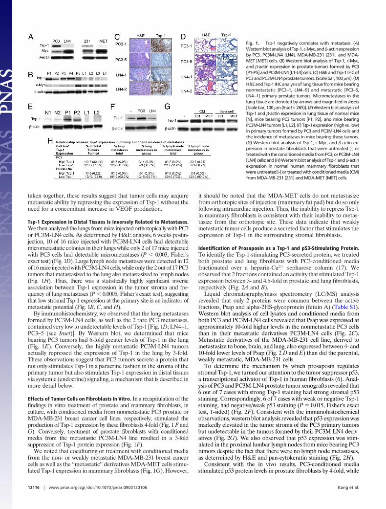
human tumors may differ in the relative expression of pro- andanti-angiogenic proteins compared to nonmetastatic tumor cell lines.Thus, we examined the expression of 2 potent angiogenic factors, Tsp-1and VEGF-A. We observed that nonmetastatic PC3 and MDA-MB-231 cells expressed high levels of Tsp-1 while their metastatic derivativesPC3M-LN4 and MDA-MET expressed no detectable Tsp-1 (as deter-mined by band volume density) (Fig. 1A). Interestingly, the weaklymetastatic PC3 (prostate) and MDA-MB-231 (breast) tumors cellsexpressed 10- and 3.5-fold higher levels of VEGF-A than their meta-static derivatives PC3M-LN4 and MDA-MET (Fig. S1).
It has been demonstrated that c-myc, a repressor of Tsp-1, is oftenamplified or overexpressed in human cancer (7, 12–16). Consistentwith these observations, we observed that levels of c-Myc weresignificantly lower (8-fold) in non- or weakly metastatic prostateand breast cancer cells than in their metastatic derivatives (Fig. 1A).
Tsp-1 Expression in Primary Tumor Stroma Is Inversely Related toMetastatic Potential. To determine whether levels of Tsp-1 ob-served in vitro are indicative of the expression levels in vivo and ofmetastatic frequency, we injected PC3 and PC3M-LN4 cells or-thotopically into prostate glands of SCID mice. Strong Tsp-1staining by immunohistochemistry was observed in the fibrousstroma surrounding invading tumor cells in 14 out of 17 PC3 tumors(Fig. 1 C and H) but in only 1 out of 16 highly metastatic PC3M-LN4prostate tumors (P � 0.0001, Fisher’s exact test) (Fig. 1 C and H).Significantly, the expression of 2 other antiangiogenic proteins,thrombospondin-2 (Tsp-2) and endostatin, revealed no elevation inPC3 primary tumors relative to normal tissue by western blotanalysis (Fig. S2). Consistent with the immunohistochemistry re-sults, 4 of 5 PC3 tumors expressed high levels of Tsp-1 andlow/undectectable levels of c-Myc by western blot (Fig. 1B). Con-versely, 4 out of 4 representative PC3M-LN4 tumors expressedbetween 5- and 8-fold lower levels of Tsp-1 and between 4- and7-fold higher levels of c-Myc (Fig. 1B). Strikingly, 1 of the PC3tumors that metastasized expressed 10-fold lower levels of Tsp-1and 8-fold higher levels of c-Myc by western blot (Fig. 1B; P5).
We also observed that 4 out of 5 PC3 tumors sampled containedbetween 10- and 20-fold higher levels of stromal (murine) VEGFthan the 4 sampled tumors formed by the PC3M-LN4 cells, asmeasured by ELISA (Fig. S3). Notably, the 1 metastatic PC3 tumorthat expressed levels of VEGF similar to the PC3M-LN4 tumorsalso expressed low levels of Tsp-1 as determined by immunohisto-chemistry and western blot (Fig. 1B and Fig. S1; LN4–1). When
Author contributions: S.-Y.K., L.A.A., and R.S.W. designed research; S.-Y.K., O.J.H., K.G.,N.B., J.M.L., N.W.L., B.T.J., A.B.J., S.A.H., K.I.A., S.M.Y., L.A.A., and R.S.W. performedresearch; S.-Y.K., O.J.H., K.G., N.B., L.A.A., and R.S.W. analyzed data; and S.-Y.K., L.A.A., andR.S.W. wrote the paper.
The authors declare no conflict of interest.
1To whom correspondence should be addressed. E-mail: [email protected].
This article contains supporting information online at www.pnas.org/cgi/content/full/0903120106/DCSupplemental.
www.pnas.org�cgi�doi�10.1073�pnas.0903120106 PNAS � July 21, 2009 � vol. 106 � no. 29 � 12115–12120
MED
ICA
LSC
IEN
CES
taken together, these results suggest that tumor cells may acquiremetastatic ability by repressing the expression of Tsp-1 without theneed for a concomitant increase in VEGF production.
Tsp-1 Expression in Distal Tissues Is Inversely Related to Metastases.We then analyzed the lungs from mice injected orthotopically with PC3or PC3M-LN4 cells. As determined by H&E analysis, 6 weeks postin-jection, 10 of 16 mice injected with PC3M-LN4 cells had detectablemicrometastatic colonies in their lungs while only 2 of 17 mice injectedwith PC3 cells had detectable micrometastases (P � 0.003, Fisher’sexact test) (Fig. 1D). Large lymph node metastases were detected in 12of 16 mice injected with PC3M-LN4 cells, while only the 2 out of 17 PC3tumors that metastasized to the lung also metastasized to lymph nodes(Fig. 1H). Thus, there was a statistically highly significant inverseassociation between Tsp-1 expression in the tumor stroma and fre-quency of lung metastases (P � 0.0005, Fisher’s exact test), suggestingthat low stromal Tsp-1 expression at the primary site is an indicator ofmetastatic potential (Fig. 1B, C, and H).
By immunohistochemistry, we observed that the lung metastasesformed by PC3M-LN4 cells, as well as the 2 rare PC3 metastases,contained very low to undetectable levels of Tsp-1 [Fig. 1D; LN4–1,PC3–5 (see Inset)]. By Western blot, we determined that micebearing PC3 tumors had 6-fold greater levels of Tsp-1 in the lung(Fig. 1E). Conversely, the highly metastatic PC3M-LN4 tumorsactually repressed the expression of Tsp-1 in the lung by 3-fold.These observations suggest that PC3 tumors secrete a protein thatnot only stimulates Tsp-1 in a paracrine fashion in the stroma of theprimary tumor but also stimulates Tsp-1 expression in distal tissuesvia systemic (endocrine) signaling, a mechanism that is described inmore detail below.
Effects of Tumor Cells on Fibroblasts In Vitro. In a recapitulation of thefindings in vitro treatment of prostate and mammary fibroblasts, inculture, with conditioned media from nonmetastatic PC3 prostate orMDA-MB-231 breast cancer cell lines, respectively, stimulated theproduction of Tsp-1 expression by these fibroblasts 4-fold (Fig. 1 F andG). Conversely, treatment of prostate fibroblasts with conditionedmedia from the metastastic PC3M-LN4 line resulted in a 3-foldsuppression of Tsp-1 protein expression (Fig. 1F).
We noted that coculturing or treatment with conditioned mediafrom the non- or weakly metastatic MDA-MB-231 breast cancercells as well as the ‘‘metastatic’’ derivatives MDA-MET cells stimu-lated Tsp-1 expression in mammary fibroblasts (Fig. 1G). However,
it should be noted that the MDA-MET cells do not metastasizefrom orthotopic sites of injection (mammary fat pad) but do so onlyfollowing intracardiac injection. Thus, the inability to repress Tsp-1in mammary fibroblasts is consistent with their inability to metas-tasize from the orthotopic site. These data indicate that weaklymetastatic tumor cells produce a secreted factor that stimulates theexpression of Tsp-1 in the surrounding stromal fibroblasts.
Identification of Prosaposin as a Tsp-1 and p53-Stimulating Protein.To identify the Tsp-1-stimulating PC3-secreted protein, we treatedboth prostate and lung fibroblasts with PC3-conditioned mediafractionated over a heparin-Cu2� sepharose column (17). Weobserved that 2 fractions contained an activity that stimulated Tsp-1expression between 3- and 4.5-fold in prostate and lung fibroblasts,respectively (Fig. 2A and B).
Liquid chromatography/mass spectrometry (LC/MS) analysisrevealed that only 2 proteins were common between the activefractions, Psap and alpha-2HS-glycoprotein (fetuin A) (Table S1).Western blot analysis of cell lysates and conditioned media fromboth PC3 and PC3M-LN4 cells revealed that Psap was expressed atapproximately 10-fold higher levels in the nonmetastatic PC3 cellsthan in their metastatic derivatives PC3M-LN4 cells (Fig. 2C).Metastatic derivatives of the MDA-MB-231 cell line, derived tometastasize to bone, brain, and lung, also expressed between 4- and10-fold lower levels of Psap (Fig. 2 D and E) than did the parental,weakly metastatic, MDA-MB-231 cells.
To determine the mechanism by which prosaposin regulatesstromal Tsp-1, we turned our attention to the tumor suppressor p53,a transcriptional activator of Tsp-1 in human fibroblasts (6). Anal-ysis of PC3 and PC3M-LN4 prostate tumor xenografts revealed that6 out of 7 cases with strong Tsp-1 staining had strong stromal p53staining. Correspondingly, 6 of 7 cases with weak or negative Tsp-1staining, had negative/weak p53 staining (P � 0.015, Fisher’s exacttest, 1-sided) (Fig. 2F). Consistent with the immunohistochemicalobservations, western blot analysis revealed that p53 expression wasmarkedly elevated in the tumor stroma of the PC3 primary tumorsbut undetectable in the tumors formed by their PC3M-LN4 deriv-atives (Fig. 2G). We also observed that p53 expression was stim-ulated in the proximal lumbar lymph nodes from mice bearing PC3tumors despite the fact that there were no lymph node metastases,as determined by H&E and pan-cytokeratin staining (Fig. 2H).
Consistent with the in vivo results, PC3-conditioned mediastimulated p53 protein levels in prostate fibroblasts by 4-fold, while
Fig. 1. Tsp-1 negatively correlates with metastasis. (A)WesternblotanalysisofTsp-1,c-Myc,and�-actinexpressionby PC3, PC3M-LN4 [LN4], MDA-MB-231 [231], and MDA-MET [MET] cells. (B) Western blot analysis of Tsp-1, c-Myc,and �-actin expression in prostate tumors formed by PC3[P1-P5]andPC3M-LN4[L1-L4]cells. (C)H&EandTsp-1 IHCofPC3andPC3M-LN4prostatetumors. (Scalebar,100�m).(D)H&EandTsp-1 IHCanalysisof lungtissuefrommicebearingnonmetastatic [PC3–1, LN4–9] and metastatic [PC3–5,LN4–1] primary prostate tumors. Micrometastases in thelung tissue are denoted by arrows and magnified in Insets[Scalebar,100�m(Inset�265)]. (E)WesternblotanalysisofTsp-1 and �-actin expression in lung tissue of normal mice[N], mice bearing PC3 tumors [P1, P2], and mice bearingPC3M-LN4tumors[L1,L2]. (F)Tsp-1expression(highvs. low)in primary tumors formed by PC3 and PC3M-LN4 cells andthe incidence of metastases in mice bearing these tumors.(G) Western blot analysis of Tsp-1, c-Myc, and �-actin ex-pression in prostate fibroblasts that were untreated [-] ortreatedwiththeconditionedmediafromPC3,orPC3M-LN4[LN4]cells;and(H)WesternblotanalysisofTsp-1and�-actinexpression in normal human mammary fibroblasts thatwere untreated [-] or treated with conditioned media (CM)from MDA-MB-231 [231] and MDA-MET [MET] cells.
12116 � www.pnas.org�cgi�doi�10.1073�pnas.0903120106 Kang et al.
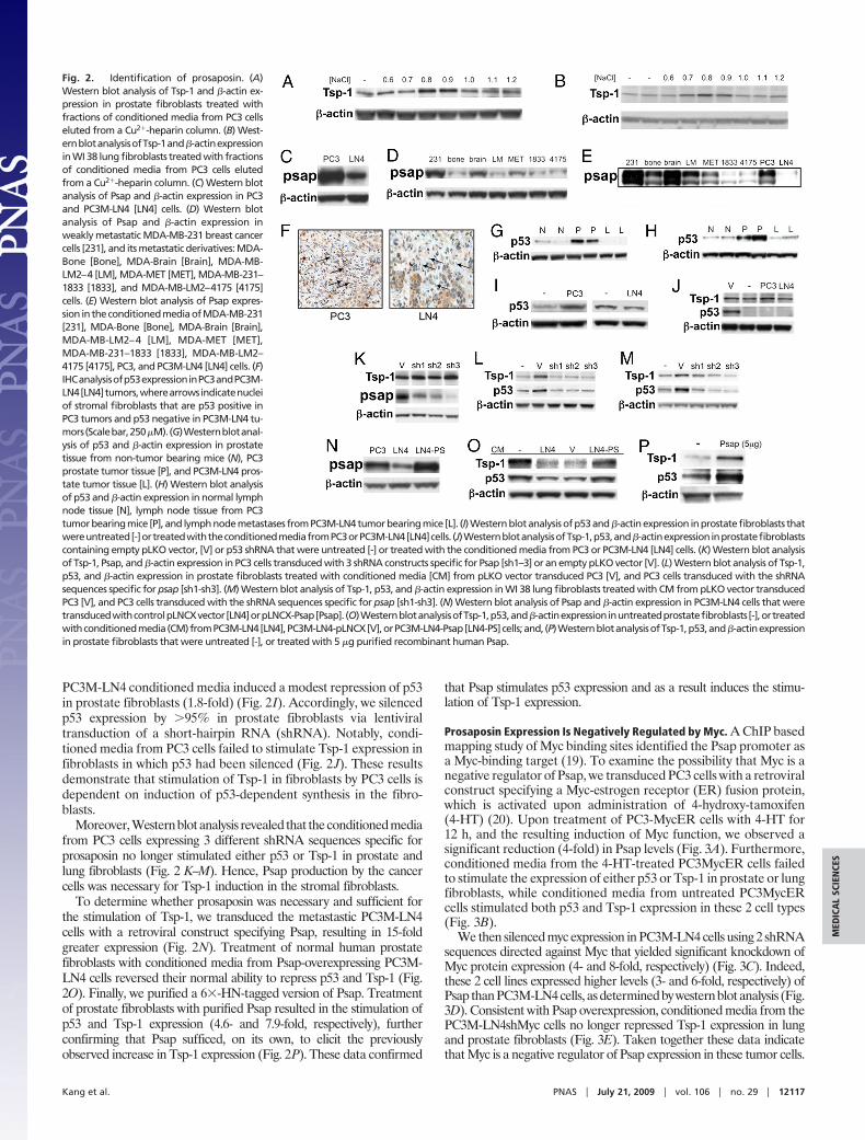
PC3M-LN4 conditioned media induced a modest repression of p53in prostate fibroblasts (1.8-fold) (Fig. 2I). Accordingly, we silencedp53 expression by �95% in prostate fibroblasts via lentiviraltransduction of a short-hairpin RNA (shRNA). Notably, condi-tioned media from PC3 cells failed to stimulate Tsp-1 expression infibroblasts in which p53 had been silenced (Fig. 2 J). These resultsdemonstrate that stimulation of Tsp-1 in fibroblasts by PC3 cells isdependent on induction of p53-dependent synthesis in the fibro-blasts.
Moreover, Western blot analysis revealed that the conditioned mediafrom PC3 cells expressing 3 different shRNA sequences specific forprosaposin no longer stimulated either p53 or Tsp-1 in prostate andlung fibroblasts (Fig. 2 K–M). Hence, Psap production by the cancercells was necessary for Tsp-1 induction in the stromal fibroblasts.
To determine whether prosaposin was necessary and sufficient forthe stimulation of Tsp-1, we transduced the metastastic PC3M-LN4cells with a retroviral construct specifying Psap, resulting in 15-foldgreater expression (Fig. 2N). Treatment of normal human prostatefibroblasts with conditioned media from Psap-overexpressing PC3M-LN4 cells reversed their normal ability to repress p53 and Tsp-1 (Fig.2O). Finally, we purified a 6�-HN-tagged version of Psap. Treatmentof prostate fibroblasts with purified Psap resulted in the stimulation ofp53 and Tsp-1 expression (4.6- and 7.9-fold, respectively), furtherconfirming that Psap sufficed, on its own, to elicit the previouslyobserved increase in Tsp-1 expression (Fig. 2P). These data confirmed
that Psap stimulates p53 expression and as a result induces the stimu-lation of Tsp-1 expression.
Prosaposin Expression Is Negatively Regulated by Myc. A ChIP basedmapping study of Myc binding sites identified the Psap promoter asa Myc-binding target (19). To examine the possibility that Myc is anegative regulator of Psap, we transduced PC3 cells with a retroviralconstruct specifying a Myc-estrogen receptor (ER) fusion protein,which is activated upon administration of 4-hydroxy-tamoxifen(4-HT) (20). Upon treatment of PC3-MycER cells with 4-HT for12 h, and the resulting induction of Myc function, we observed asignificant reduction (4-fold) in Psap levels (Fig. 3A). Furthermore,conditioned media from the 4-HT-treated PC3MycER cells failedto stimulate the expression of either p53 or Tsp-1 in prostate or lungfibroblasts, while conditioned media from untreated PC3MycERcells stimulated both p53 and Tsp-1 expression in these 2 cell types(Fig. 3B).
We then silenced myc expression in PC3M-LN4 cells using 2 shRNAsequences directed against Myc that yielded significant knockdown ofMyc protein expression (4- and 8-fold, respectively) (Fig. 3C). Indeed,these 2 cell lines expressed higher levels (3- and 6-fold, respectively) ofPsap than PC3M-LN4 cells, as determined by western blot analysis (Fig.3D). Consistent with Psap overexpression, conditioned media from thePC3M-LN4shMyc cells no longer repressed Tsp-1 expression in lungand prostate fibroblasts (Fig. 3E). Taken together these data indicatethat Myc is a negative regulator of Psap expression in these tumor cells.
Fig. 2. Identification of prosaposin. (A)Western blot analysis of Tsp-1 and �-actin ex-pression in prostate fibroblasts treated withfractions of conditioned media from PC3 cellseluted from a Cu2�-heparin column. (B) West-ernblotanalysisofTsp-1and�-actinexpressionin WI 38 lung fibroblasts treated with fractionsof conditioned media from PC3 cells elutedfrom a Cu2�-heparin column. (C) Western blotanalysis of Psap and �-actin expression in PC3and PC3M-LN4 [LN4] cells. (D) Western blotanalysis of Psap and �-actin expression inweakly metastatic MDA-MB-231 breast cancercells [231], and its metastatic derivatives: MDA-Bone [Bone], MDA-Brain [Brain], MDA-MB-LM2–4 [LM], MDA-MET [MET], MDA-MB-231–1833 [1833], and MDA-MB-LM2–4175 [4175]cells. (E) Western blot analysis of Psap expres-sion intheconditionedmediaofMDA-MB-231[231], MDA-Bone [Bone], MDA-Brain [Brain],MDA-MB-LM2–4 [LM], MDA-MET [MET],MDA-MB-231–1833 [1833], MDA-MB-LM2–4175 [4175], PC3, and PC3M-LN4 [LN4] cells. (F)IHCanalysisofp53expressioninPC3andPC3M-LN4[LN4]tumors,wherearrowsindicatenucleiof stromal fibroblasts that are p53 positive inPC3 tumors and p53 negative in PC3M-LN4 tu-mors(Scalebar,250�M).(G)Westernblotanal-ysis of p53 and �-actin expression in prostatetissue from non-tumor bearing mice (N), PC3prostate tumor tissue [P], and PC3M-LN4 pros-tate tumor tissue [L]. (H) Western blot analysisof p53 and �-actin expression in normal lymphnode tissue [N], lymph node tissue from PC3tumor bearing mice [P], and lymph node metastases from PC3M-LN4 tumor bearing mice [L]. (I) Western blot analysis of p53 and �-actin expression in prostate fibroblasts thatwereuntreated[-]ortreatedwiththeconditionedmediafromPC3orPC3M-LN4[LN4]cells. (J)WesternblotanalysisofTsp-1,p53,and�-actinexpressioninprostatefibroblastscontaining empty pLKO vector, [V] or p53 shRNA that were untreated [-] or treated with the conditioned media from PC3 or PC3M-LN4 [LN4] cells. (K) Western blot analysisof Tsp-1, Psap, and �-actin expression in PC3 cells transduced with 3 shRNA constructs specific for Psap [sh1–3] or an empty pLKO vector [V]. (L) Western blot analysis of Tsp-1,p53, and �-actin expression in prostate fibroblasts treated with conditioned media [CM] from pLKO vector transduced PC3 [V], and PC3 cells transduced with the shRNAsequences specific for psap [sh1-sh3]. (M) Western blot analysis of Tsp-1, p53, and �-actin expression in WI 38 lung fibroblasts treated with CM from pLKO vector transducedPC3 [V], and PC3 cells transduced with the shRNA sequences specific for psap [sh1-sh3]. (N) Western blot analysis of Psap and �-actin expression in PC3M-LN4 cells that weretransducedwithcontrolpLNCXvector[LN4]orpLNCX-Psap[Psap]. (O)WesternblotanalysisofTsp-1,p53,and�-actinexpressioninuntreatedprostatefibroblasts [-],ortreatedwithconditionedmedia(CM)fromPC3M-LN4[LN4],PC3M-LN4-pLNCX[V],orPC3M-LN4-Psap[LN4-PS]cells;and, (P)WesternblotanalysisofTsp-1,p53,and�-actinexpressionin prostate fibroblasts that were untreated [-], or treated with 5 �g purified recombinant human Psap.
Kang et al. PNAS � July 21, 2009 � vol. 106 � no. 29 � 12117
MED
ICA
LSC
IEN
CES
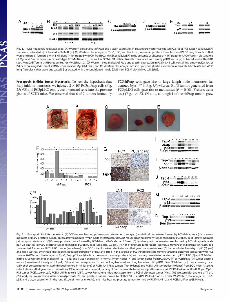
Prosaposin Inhibits Tumor Metastasis. To test the hypothesis thatPsap inhibits metastasis, we injected 2 � 106 PC3shPsap cells (Fig.2 J; #3) and PC3pLKO empty vector control cells, into the prostateglands of SCID mice. We observed that 6 of 7 tumors formed by
PC3shPsap cells gave rise to large lymph node metastases (asdenoted by a ‘‘*’’ in Fig. 5F) whereas 0 of 8 tumors generated fromPC3pLKO cells gave rise to metastases (P � 0.001, Fisher’s exacttest) (Fig. 4 A–E). Of note, although 1 of the shPsap tumors grew
Fig. 3. Myc negatively regulates psap. (A) Western blot analysis of Psap and �-actin expression in pBabepuro vector transduced PC3 [V] or PC3-MycER cells [MycER]that were untreated [-] or treated with 4-HT [�]. (B) Western blot analysis of Tsp-1, p53, and �-actin expression in prostate fibroblasts and WI 38 lung fibroblasts thatwereuntreated [-], treatedwith4-HTalone [�]or treatedwithCMfromPC3-MycERcells [MycER] in thepresenceorabsenceof4-HTtreatment. (C)Westernblotanalysisof Myc and �-actin expression in wild-type PC3M-LN4 cells [-], as well as PC3M-LN4 cells lentivirally transduced with empty pLKO vector [V] or transduced with pLKOspecifying 2 different shRNA sequences for Myc [sh1, sh2]. (D) Western blot analysis of Psap and �-actin expression in PC3M-LN4 cells containing empty pLKO vector[V] or expressing 2 different shRNA sequences for Myc [sh1, sh2]; and (E) Western blot analysis of Tsp-1, p53, and �-actin expression in prostate fibroblasts and WI38lung fibroblasts that were untreated [-] or treated with the conditioned media [CM] from PC3M-LN4-shMyc cells [sh1].
Fig. 4. Prosaposin inhibits metastasis. (A) SCID mouse bearing primary prostate tumor (xenograft) and distal metastases formed by PC3-shPsap cells (black arrowindicates primary prostate tumor, green arrows indicate lymph node metastases). (B) SCID mouse bearing primary tumor formed by PC3pLKO cells (arrow indicatesprimary prostate tumor). (C) Primary prostate tumor formed by PC3shPsap cells (Scale bar, 0.5 cm). (D) Lumbar lymph node metastases formed by PC3shPsap cells (scalebar, 0.5 cm). (E) Primary prostate tumor formed by PC3pLKO cells (Scale bar, 0.5 cm). (F) Plot of prostate tumor mass (individual tumors, in milligrams) of PC3shPsaptumors (first 7 lanes) and PC3pLKO tumors (last 4 lanes) from SCID mice. Asterisks refer to tumors that gave rise to metastases. (G) Immunohistochemistry of p53 (Upper)and Tsp-1 (Lower) after Psap knockdown. Reduced expression of p53 and Tsp-1 in the stroma of PC3shPsap prostate tumors (Right) is observed compared with PC3tumors. (H) Western blot analysis of Tsp-1, Psap, p53, and �-actin expression in normal prostate [N] and primary prostate tumors formed by PC3pLKO [P] and PC3shPsap[sh] cells. (I) Western blot analysis of Tsp-1, p53, and �-actin expression in normal lymph nodes [N] and lymph nodes from PC3pLKO [P] or PC3shPsap [sh] tumor bearingmice. (J) Western blot analysis of Tsp-1, p53, and �-actin expression in normal lung tissue (N) and lung tissue from PC3pLKO (P) or PC3shPsap [sh] tumor bearing mice.(K) Plotofprostate tumormass (individual tumors, inmilligrams)ofPC3M-LN4-Psaptumors (first10 lanes)andPC3M-LN4tumors (last10 lanes) fromSCID mice.Asterisksrefer to tumors that gave rise to metastases. (L) Immuno-histochemical staining of Psap in prostate tumor xenografts. Upper Left, PC3M-LN4 tumor [LN4]; Upper Right,PC3 tumor [PC3]. Lower Left, PC3M-LN4-Psap cells [LN4]. Lower Right, lung micrometastases from a PC3M-LN4-psap tumor [Met]. (M) Western blot analysis of Tsp-1,p53, and �-actin expression in the normal prostate [N], and prostate tumors formed by PC3M-LN4 [L] and PC3M-LN4-psap [L-P] cells. (N) Western blot analysis of Tsp-1,p53, and b-actin expression in the lung tissue of normal mice [N], and mice bearing prostate tumors formed by PC3M-LN4 [L] and PC3M-LN4-psap [L-P] cells.
12118 � www.pnas.org�cgi�doi�10.1073�pnas.0903120106 Kang et al.
significantly larger than the PC3pLKO tumors, 3 of the smallershPsap tumors (Fig. 4F, #4, 5, and 7), which grew to approximatelythe same size as the PC3pLKO control tumors, formed visiblelymph node metastases (Fig. 4D). Lung metastases were alsoincreased with 2 out of 7 PC3shPsap tumors forming detectablemetastases by histological examination, compared with 2/17 for PC3(P � 0.006, by Fisher’s exact test). These data indicate thatreduction of Psap expression significantly potentiated the ability ofotherwise nonmetastatic tumor cells to form lymph node metas-tases.
Immunohistochemical analysis of p53 and Tsp-1 expression inprimary tumors formed by PC3 and PC3shPsap tumors revealedthat p53 expression in the tumor-associated stroma of PC3shPsaptumors was completely negative (n � 4), in contrast to the stromaof control PC3 tumors in which p53 expression was strongly positive(Fig. 4G). In contrast to the strong staining in PC3 tumors, mostPC3shPsap tumors were weak or negative for Tsp-1 staining withinthe central parts of the tumors (Fig. 4G). Western blot analyses ofprimary tumor, lymph nodes, and lungs confirmed the IHC results(Fig. 4 H–J).
We then injected 2 � 106 PC3M-LN4-psap cells orthotopically inthe prostate gland of SCID mice. After 5–6 weeks, the mice wereexamined visually for the presence of macrometastases as well ashistologically and immunohistochemically (by staining for pan-cytokeratin expression) for the presence of micrometastases. Thesizes of the Psap-expressing tumors were similar to those of thePC3M-LN4 tumors (Fig. 4K). We detected lung metastases (de-noted by ‘‘*’’ in Fig. 4K) in only 2 out of 10 mice injected withPC3M-LN4-Psap cells, compared with 8 out of 10 injected withPC3M-LN4 cells (P � 0.042, Fisher’s exact test).
By IHC and Western blot analysis we observed that PC3M-LN4-Psap tumors expressed 3–6-fold higher levels of Psap than parental
PC3M-LN4 tumors and even higher levels than the PC3 tumors(Fig. 4L–N). Strikingly the 2 lung metastases formed by PC3M-LN4-Psap cells also expressed moderate levels of Psap (Fig. 4L).Notably, despite the moderate levels of Psap expression, the 2tumors that formed metastases expressed similarly low levels ofTsp-1 and p53 in both the primary tumors and lung tissue as thePC3M-LN4 tumors (Fig. 4 M and N). These findings suggest thatthese tumors were able to compensate for the increased expressionof Psap, perhaps by increasing the expression of a protein thatrepresses Tsp-1 and p53 in the stroma. The fact that Psap did notdramatically affect the sizes of the primary tumors suggests that itmay affect dissemination of tumor cells or colonization of distalsites.
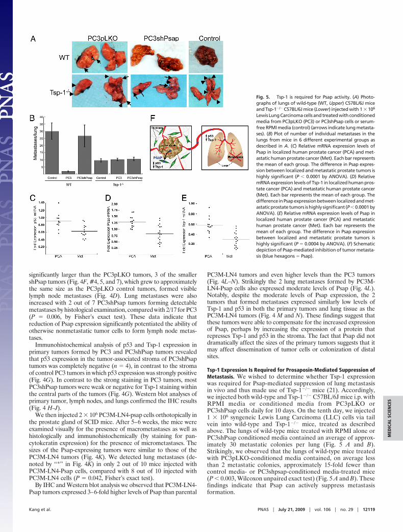
Tsp-1 Expression Is Required for Prosaposin-Mediated Suppression ofMetastasis. We wished to determine whether Tsp-1 expressionwas required for Psap-mediated suppression of lung metastasisin vivo and thus made use of Tsp-1�/� mice (21). Accordingly,we injected both wild-type and Tsp-1�/� C57BL/6J mice i.p. withRPMI media or conditioned media from PC3pLKO orPC3shPsap cells daily for 10 days. On the tenth day, we injected1 � 106 syngeneic Lewis Lung Carcinoma (LLC) cells via tailvein into wild-type and Tsp-1�/� mice, treated as describedabove. The lungs of wild-type mice treated with RPMI alone orPC3shPsap conditioned media contained an average of approx-imately 30 metastatic colonies per lung (Fig. 5 A and B).Strikingly, we observed that the lungs of wild-type mice treatedwith PC3pLKO-conditioned media contained, on average lessthan 2 metastatic colonies, approximately 15-fold fewer thancontrol media- or PC3shpsap-conditioned media-treated mice(P � 0.003, Wilcoxon unpaired exact test) (Fig. 5 A and B). Thesefindings indicate that Psap can actively suppress metastasisformation.
Fig. 5. Tsp-1 is required for Psap activity. (A) Photo-graphs of lungs of wild-type (WT, Upper) C57BL/6J miceand Tsp-1�/� C57BL/6J mice (Lower) injected with 1 � 106
Lewis Lung Carcinoma cells and treated with conditionedmedia from PC3pLKO (PC3) or PC3shPsap cells or serum-free RPMI media (control) (arrows indicate lung metasta-ses). (B) Plot of number of individual metastases in thelungs from mice in 6 different experimental groups asdescribed in A. (C) Relative mRNA expression levels ofPsap in localized human prostate cancer (PCA) and met-astatic human prostate cancer (Met). Each bar representsthe mean of each group. The difference in Psap expres-sion between localized and metastatic prostate tumors ishighly significant (P � 0.0001 by ANOVA). (D) RelativemRNA expression levels of Tsp-1 in localized human pros-tate cancer (PCA) and metastatic human prostate cancer(Met). Each bar represents the mean of each group. Thedifference inPsapexpressionbetween localizedandmet-astatic prostate tumors is highly significant (P � 0.0001 byANOVA). (E) Relative mRNA expression levels of Psap inlocalized human prostate cancer (PCA) and metastatichuman prostate cancer (Met). Each bar represents themean of each group. The difference in Psap expressionbetween localized and metastatic prostate tumors ishighly significant (P � 0.0004 by ANOVA). (F) Schematicdepiction of Psap-mediated inhibition of tumor metasta-sis (blue hexagons � Psap).
Kang et al. PNAS � July 21, 2009 � vol. 106 � no. 29 � 12119
MED
ICA
LSC
IEN
CES
We observed that Psap had no effect on the ability of LLC cellsto form metastases in the lungs of tsp-1�/� mice (Fig. 5 A and B).Hence, in the absence of Tsp-1 production in the lung, Psap failedto suppress metastasis formation by LLC cells. The observed lowernumbers of metastases in the Tsp-1 KO mice compared with theirwild-type counterparts is consistent with previously reported find-ings (22) and could be explained by a compensatory 4.5-foldincrease in Tsp-2 production induced by LLC cells in these mice, aphenomenon not observed in wild-type mice or human tumors(Figs. S5 and S6).
Prosaposin Correlation with Metastasis in Human Patients. We thenanalyzed a gene expression data set gathered from 55 patientsconsisting of normal prostate (5), benign prostatic hyperplasia(BPH) (13), localized primary prostate tumors (18), and primaryprostate tumors that formed metastases in the patients (19) (23).Consistent with the xenograft experiments, the relative psap,tsp-1. and p53 mRNA expression were approximately 40%, 50%,and 30% lower, on average, respectively in the metastatic tumorscompared with localized primary tumors (P � 0.0001 byANOVA) (Fig. 5 C–E). These data support the results obtainedexperimentally and demonstrate that in metastatic human pros-tate cancer, the expression of Psap is reduced compared tononmetastatic tumors.
DiscussionThe hypothesis that tumors metastasize to sites that arepermissive (24–28), or compatible, was first proposed byStephen Paget over 100 years ago. We demonstrate here thatweakly metastatic tumors create a refractory environment tocolonization in the stroma of primary tumors as well as indistant organs by stimulating the expression of Tsp-1. Thesefindings indicate that high levels of Tsp-1 in the primarytumors and in potential metastatic organs create a potentbarrier to metastasis. While Tsp-1 has been shown to be apotent anti-angiogenic factor it has recently been demon-strated that Tsp-1 can recruit macrophages to tumor tissue(29). Thus, it is possible that Psap inhibition of metastasis,while dependent on Tsp-1, is not solely mediated by alterationsto vessel permeability or angiogenesis.
Prosaposin (56 kDA) is proteolytically cleaved in the lysosomeinto saposins A, B, C, and D (30), which function as cofactors for
sphingolipid hydrolases (10, 11). Of interest, it has been demon-strated that tumor-secreted lipids are able to repress Tsp-1 indermal fibroblasts (5, 18). In relation to the prostate, the Psap-encoding gene is amplified in a subset of prostate cancers (33). Wehave determined that the expression of Psap is not only reduced inmetastatic prostate and breast cancer cell lines, but also in meta-static human prostate tumors (23). Significantly, these data suggestthat repression of Psap expression may be a mechanism to enhancetumor metastasis, and may, along with increased Myc expression,comprise a ‘‘metastatic switch.’’
Additionally, recent evidence suggests that there is an involvement inbone marrow derived cells in establishment of a premetastatic niche(26). These cells are composed, in part, of VEGFR1� cells, and theirrecruitment is dependent on VEGFR1 activity. Since Tsp-1 has beenshown to inhibit VEGF-mediated migration and recruitment, this is yetanother mechanism by which Psap may inhibit tumor metastasis.Further studies into the mechanism of Psap stimulation of stromal p53and Tsp-1 expression may provide therapeutic targets that couldprevent the metastatic spread of human prostate and breast tumors.Based on our observations, we propose that activation of stromal p53may also be a possible strategy to limit and perhaps cause regression oftumor growth and metastasis.
MethodsTail Vein Metastasis Assay. Wild-type and Tsp-1�/� C57BL/6J mice werepretreated with 500 �L serum-free conditioned media from PC3 orPC3shPsap cells or serum-free RPMI media for 10 days via i.p. (i.p.) injection.On the 10th day, we injected via tail vein mice with 1 � 106 LLC cells.Subsequently, i.p. injections of serum-free tumor cell conditioned media orcontrol RPMI were performed for 19 additional days. Following sacrifice,the lungs were photographed and the number of macrometastatic noduleswere counted.
ACKNOWLEDGMENTS. We thank J. Folkman†, R. Weinberg, B. Zetter, M.Klagsbrun, M. Moses, and R. Rodriguez for helpful discussions and insights.The authors also wish to thank Mrs. Gerd Lillian Hallseth and Mr. BendikNordanger for technical assistance. We also thank I. Fidler, L. Suva, M. Rosen-blatt, K. Polyak, and J. Massague for cell lines. We thank Judah Folkman andGeorge Naumov for supplying the Tsp-1�/� mice. We thank Arul Chinnaiyanand the Oncomine website for access to microarray expression data derivedfrom primary human prostate cancer samples. L.A.A. is supported by grantsfrom the Norwegian Research Council, the Norwegian Cancer Society, theHelse Vest Research Fund, and the Unger Vetlesen Research Fund. R.S.W issupported by the Gackstatter Foundation a grant from National Aeronauticsand Space Administration and a Breast Cancer Innovator Award from theDepartment of Defense.
1. Fidler IJ (2003) The pathogenesis of cancer metastasis: The ‘seed and soil’ hypothesisrevisited. Nat Rev Cancer 3:453–458.
2. Fukumura D, et al. (1998) Tumor induction of VEGF promoter activity in stromal cells. Cell94:715–725.
3. Dong J, et al. (2004) VEGF-null cells require PDGFR alpha signaling-mediated stromalfibroblast recruitment for tumorigenesis. EMBO J 23:2800–2810.
4. Lawler J (2002)Thrombospondin-1asanendogenous inhibitorofangiogenesisandtumorgrowth. J Cell Mol Med 6:1–12.
5. Kalas W, et al. (2005) Oncogenes and angiogenesis: Down-regulation of throm-bospondin-1 in normal fibroblasts exposed to factors from cancer cells harboringmutant ras. Cancer Res 65:8878 – 8886.
6. Dameron KM, Volpert OV, Tainsky MA, Bouck N (1994) Control of angiogenesis in fibro-blasts by p53 regulation of thrombospondin-1. Science 265:1582–1584.
7. Watnick RS, Cheng YN, Rangarajan A, Ince TA, Weinberg RA (2003) Ras modulates Mycactivity to repress thrombospondin-1 expression and increase tumor angiogenesis. CancerCell 3:219–231.
8. Sano A, et al. (1989) Sphingolipid hydrolase activator proteins and their precursors.Biochem Biophys Res Commun 165:1191–1197.
9. Sylvester SR, Morales C, Oko R, Griswold MD (1989) Sulfated glycoprotein-1(saposin precursor) in the reproductive tract of the male rat. Biol Reprod 41:941–948.
10. MorimotoS,etal. (1989)SaposinA:Secondcerebrosidaseactivatorprotein.ProcNatlAcadSci USA 86:3389–3393.
11. Morimoto S, Martin BM, Kishimoto Y, O’Brien JS (1988) Saposin D: A sphingomyelinaseactivator. Biochem Biophys Res Commun 156:403–410.
12. Nag A, Smith RG (1989) Amplification, rearrangement, and elevated expression of c-mycin the human prostatic carcinoma cell line LNCaP. The Prostate 15:115–122.
13. Escot C, et al. (1986) Genetic alteration of the c-myc protooncogene (MYC) in humanprimary breast carcinomas. Proc Natl Acad Sci USA 83:4834–4838.
14. JanzA,SevignaniC,KenyonK,NgoCV,Thomas-TikhonenkoA(2000)Activationofthemyconcoprotein leads to increased turnover of thrombospondin-1 mRNA. Nucleic Acids Res(Online) 28:2268–2275.
15. Ngo CV, et al. (2000) An in vivo function for the transforming Myc protein: Elicitation ofthe angiogenic phenotype. Cell Growth Differ 11:201–210
16. Tikhonenko AT, Black DJ, Linial ML (1996) Viral Myc oncoproteins in infected fibroblastsdown-modulate thrombospondin-1, a possible tumor suppressor gene. J Biol Chem271:30741–30747.
17. Shing Y (1988) Heparin-copper biaffinity chromatography of fibroblast growth factors.J Biol Chem 263:9059–9062.
18. Kalas W, Klement P, Rak J (2005) Downregulation of the angiogenesis inhibitor throm-bospondin 1 in fibroblasts exposed to platelets and their related phospholipids. BiochemBiophys Res Commun 334:549–554.
19. Fernandez PC, et al. (2003) Genomic targets of the human c-Myc protein. Genes Dev17:1115–1129.
20. LittlewoodTD,HancockDC,DanielianPS,ParkerMG,EvanGI (1995)Amodifiedoestrogenreceptor ligand-binding domain as an improved switch for the regulation of heterologousproteins. Nucleic Acids Res 23:1686–1690.
21. Lawler J, et al. (1998) Thrombospondin-1 is required for normal murine pulmonaryhomeostasis and its absence causes pneumonia. J Clin Invest 101:982–992.
22. Yee KO, et al. (2009) The effect of thrombospondin-1 on breast cancer metastasis. BreastCancer Res Treat 114:85–96.
23. Dhanasekaran SM, et al. (2001) Delineation of prognostic biomarkers in prostate cancer.Nature 412:822–826.
24. Gao D, et al. (2008) Endothelial progenitor cells control the angiogenic switch in mouselung metastasis. Science 319:195–198.
25. Gupta GP, et al. (2007) Mediators of vascular remodelling co-opted for sequential steps inlung metastasis. Nature 446:765–770.
26. Kaplan RN, et al. (2005) VEGFR1-positive haematopoietic bone marrow progenitors initi-ate the pre-metastatic niche. Nature 438:820–827.
27. McAllister SS, et al. (2008) Systemic endocrine instigation of indolent tumor growthrequires osteopontin. Cell 133:994–1005.
28. Padua D, et al. (2008) TGFbeta primes breast tumors for lung metastasis seeding throughangiopoietin-like 4. Cell 133:66–77.
29. Martin-Manso G, et al. (2008) Thrombospondin 1 promotes tumor macrophage recruit-ment and enhances tumor cell cytotoxicity of differentiated U937 cells. Cancer Res68:7090–7099.
30. O’Brien JS, Kishimoto Y (1991) Saposin proteins: Structure, function, and role in humanlysosomal storage disorders. FASEB J 5:301–308.
12120 � www.pnas.org�cgi�doi�10.1073�pnas.0903120106 Kang et al.