protein function - universidade federal de minas gerais
TRANSCRIPT

203
Protein Function
chapter
7Knowing the three-dimensional structure of a protein is an important partof understanding how the protein functions. However, the structure shownin two dimensions on a page is deceptively static. Proteins are dynamic mol-ecules whose functions almost invariably depend on interactions with othermolecules, and these interactions are affected in physiologically importantways by sometimes subtle, sometimes striking changes in protein confor-mation.
In this chapter, we explore how proteins interact with other moleculesand how their interactions are related to dynamic protein structure. The im-portance of molecular interactions to a protein’s function can hardly beoveremphasized. In Chapter 6, we saw that the function of fibrous proteinsas structural elements of cells and tissues depends on stable, long-termquaternary interactions between identical polypeptide chains. As we willsee in this chapter, the functions of many other proteins involve interac-tions with a variety of different molecules. Most of these interactions arefleeting, though they may be the basis of complex physiological processessuch as oxygen transport, immune function, and muscle contraction, thetopics we examine in detail in this chapter. The proteins that carry outthese processes illustrate the following key principles of protein function,some of which will be familiar from the previous chapter:
The functions of many proteins involve the reversible binding of othermolecules. A molecule bound reversibly by a protein is called a ligand.
A ligand may be any kind of molecule, including another protein. Thetransient nature of protein-ligand interactions is critical to life, allowingan organism to respond rapidly and reversibly to changing environ-mental and metabolic circumstances.
A ligand binds at a site on the protein called the binding site, whichis complementary to the ligand in size, shape, charge, and hydropho-bic or hydrophilic character. Furthermore, the interaction is specific:the protein can discriminate among the thousands of different mole-cules in its environment and selectively bind only one or a few. Agiven protein may have separate binding sites for several different lig-ands. These specific molecular interactions are crucial in maintainingthe high degree of order in a living system. (This discussion excludesthe binding of water, which may interact weakly and nonspecificallywith many parts of a protein. In Chapter 8, we consider water as aspecific ligand for many enzymes.)
Proteins are flexible. Changes in conformation may be subtle, reflect-ing molecular vibrations and small movements of amino acid residues

204 Part II Structure and Catalysis
throughout the protein. A protein flexing in this way is sometimes saidto “breathe.” Changes in conformation may also be quite dramatic,with major segments of the protein structure moving as much as sev-eral nanometers. Specific conformational changes are frequently es-sential to a protein’s function.
The binding of a protein and ligand is often coupled to a conforma-tional change in the protein that makes the binding site more comple-mentary to the ligand, permitting tighter binding. The structural adap-tation that occurs between protein and ligand is called induced fit.
In a multisubunit protein, a conformational change in one subunit often affects the conformation of other subunits.
Interactions between ligands and proteins may be regulated, usuallythrough specific interactions with one or more additional ligands.These other ligands may cause conformational changes in the proteinthat affect the binding of the first ligand.
Enzymes represent a special case of protein function. Enzymes bindand chemically transform other molecules—they catalyze reactions. Themolecules acted upon by enzymes are called reaction substrates ratherthan ligands, and the ligand-binding site is called the catalytic site or active site. In this chapter we emphasize the noncatalytic functions of pro-teins. In Chapter 8 we consider catalysis by enzymes, a central topic in bio-chemistry. You will see that the themes of this chapter—binding, specificity,and conformational change—are continued in the next chapter, with theadded element of proteins acting as reactants in chemical transformations.
Reversible Binding of a Protein to a Ligand:Oxygen-Binding ProteinsMyoglobin and hemoglobin may be the most-studied and best-understoodproteins. They were the first proteins for which three-dimensional struc-tures were determined, and our current understanding of myoglobin andhemoglobin is garnered from the work of thousands of biochemists overseveral decades. Most important, they illustrate almost every aspect of thatmost central of biochemical processes: the reversible binding of a ligand toa protein. This classic model of protein function will tell us a great dealabout how proteins work.
Oxygen Can Be Bound to a Heme Prosthetic GroupOxygen is poorly soluble in aqueous solutions (see Table 4–3) and cannot becarried to tissues in sufficient quantity if it is simply dissolved in blood serum.Diffusion of oxygen through tissues is also ineffective over distances greaterthan a few millimeters. The evolution of larger, multicellular animals dependedon the evolution of proteins that could transport and store oxygen. How-ever, none of the amino acid side chains in proteins is suited for the revers-ible binding of oxygen molecules. This role is filled by certain transition met-als, among them iron and copper, that have a strong tendency to bind oxygen.Multicellular organisms exploit the properties of metals, most commonly iron,for oxygen transport. However, free iron promotes the formation of highlyreactive oxygen species such as hydroxyl radicals that can damage DNAand other macromolecules. Iron used in cells is therefore bound in formsthat sequester it and/or make it less reactive. In multicellular organisms—especially those in which iron, in its oxygen-carrying capacity, must betransported over large distances—iron is often incorporated into a protein-bound prosthetic group called heme. (A prosthetic group is a compound per-manently associated with a protein that contributes to the protein’s function.)

Chapter 7 Protein Function 205
�O
CO O�
Fe�
CH3
CH
N�
CH2
CH2
CH2
CH2
CH2
CH3
CH3
CH3
CH
CH
CH CH
CH
OC
C CC
C
C
C C CC
C
C
C
CC
N
NN
CCH2
(b)
C
(a)
NH
X
N
HN
NX
X
XX
X
X
X
(d)
(c)
Fe
figure 7–1Heme. The heme group is present in myoglobin, hemo-globin, and many other proteins, designated heme pro-teins. Heme consists of a complex organic ring structure,protoporphyrin IX, to which is bound an iron atom in itsferrous (Fe2�) state. Porphyrins, of which protoporphyrinIX is only one example, consist of four pyrrole rings linkedby methene bridges (a), with substitutions at one or moreof the positions denoted X. Two representations of hemeare shown in (b) and (c). The iron atom of heme has sixcoordination bonds: four in the plane of, and bonded to, the flat porphyrin ring system, and two perpendicularto it (d).
N
CH2C
HC
Edge view
ring systemresidue
CN Fe O2
Histidine Plane ofporphyrin
H
Heme (or haem) consists of a complex organic ring structure, proto-
porphyrin, to which is bound a single iron atom in its ferrous (Fe2�) state(Fig. 7–1). The iron atom has six coordination bonds, four to nitrogenatoms that are part of the flat porphyrin ring system and two perpendic-ular to the porphyrin. The coordinated nitrogen atoms (which have an electron-donating character) help prevent conversion of the heme iron tothe ferric (Fe3�) state. Iron in the Fe2� state binds oxygen reversibly; in theFe3� state it does not bind oxygen. Heme is found in a number of oxygen-transporting proteins, as well as in some proteins, such as the cytochromes,that participate in oxidation-reduction (electron transfer) reactions (Chap-ter 19).
In free heme molecules, reaction of oxygen at one of the two “open” co-ordination bonds of iron (perpendicular to the plane of the porphyrin mol-ecule, above and below) can result in irreversible conversion of Fe2� toFe3�. In heme-containing proteins, this reaction is prevented by sequester-ing the heme deep within a protein structure where access to the two opencoordination bonds is restricted. One of these two coordination bonds is oc-cupied by a side-chain nitrogen of a His residue. The other is the bindingsite for molecular oxygen (O2) (Fig. 7–2). When oxygen binds, the elec-tronic properties of heme iron change; this accounts for the change in colorfrom the dark purple of oxygen-depleted venous blood to the bright red ofoxygen-rich arterial blood. Some small molecules, such as carbon monoxide(CO) and nitric oxide (NO), coordinate to heme iron with greater affinitythan does O2. When a molecule of CO is bound to heme, O2 is excluded,which is why CO is highly toxic to aerobic organisms. By surrounding andsequestering heme, oxygen-binding proteins regulate the access of CO andother small molecules to heme iron.
figure 7–2The heme group viewed from the side. This view showsthe two coordination bonds to Fe2� perpendicular to theporphyrin ring system. One of these two bonds is occu-pied by a His residue, sometimes called the proximal His.The other is the binding site for oxygen. The remainingfour coordination bonds are in the plane of, and bondedto, the flat porphyrin ring system.

206 Part II Structure and Catalysis
Myoglobin Has a Single Binding Site for OxygenMyoglobin (Mr 16,700; abbreviated Mb) is a relatively simple oxygen-binding protein found in almost all mammals, primarily in muscle tissue. Itis particularly abundant in the muscles of diving mammals such as seals andwhales that must store enough oxygen for prolonged excursions undersea.Proteins very similar to myoglobin are widely distributed, occurring even insome single-celled organisms. Myoglobin stores oxygen for periods whenenergy demands are high and facilitates its distribution to oxygen-starvedtissues.
Myoglobin is a single polypeptide of 153 amino acid residues with onemolecule of heme. It is typical of the family of proteins called globins,
which have similar primary and tertiary structures. The polypeptide is madeup of eight a-helical segments connected by bends (Fig. 7–3). About 78%of the amino acid residues in the protein are found in these a helices.
Any detailed discussion of protein function inevitably involves proteinstructure. Our treatment of myoglobin will be facilitated by introducingsome structural conventions peculiar to globins. As seen in Figure 7–3, thehelical segments are labeled A through H. An individual amino acid residuemay be designated either by its position in the amino acid sequence or byits location within the sequence of a particular a-helical segment. For ex-ample, the His residue coordinated to the heme in myoglobin, His93 (the93rd amino acid residue from the amino-terminal end of the myoglobinpolypeptide sequence), is also called His F8 (the 8th residue in a helix F).The bends in the structure are labeled AB, CD, EF, and so forth, reflectingthe a-helical segments they connect.
Protein-Ligand Interactions Can Be Described QuantitativelyThe function of myoglobin depends on the protein’s ability not only to bindoxygen, but also to release it when and where it is needed. Function in bio-chemistry often revolves around a reversible protein-ligand interaction ofthis type. A quantitative description of this interaction is therefore a centralpart of many biochemical investigations.
A
EF
F
H
FG
C
CD
DB
G
E
GHAB
figure 7–3The structure of myoglobin. The eight a-helical seg-ments (shown here as cylinders) are labeled A through H.Nonhelical residues in the bends that connect them arelabeled AB, CD, EF, and so forth, indicating the segmentsthey interconnect. A few bends, including BC and DE, areabrupt and do not contain any residues; these are notnormally labeled. (The short segment visible between Dand E is an artifact of the computer representation.) Theheme is bound in a pocket made up largely of the E andF helices, although amino acid residues from other seg-ments of the protein also participate.

Chapter 7 Protein Function 207
In general, the reversible binding of a protein (P) to a ligand (L) can bedescribed by a simple equilibrium expression:
(7–1)
The reaction is characterized by an equilibrium constant, Ka, such that
(7–2)
The term Ka is an association constant (not to be confused with the Ka
that denotes an acid dissociation constant; see p. 98). The association con-stant provides a measure of the affinity of the ligand L for the protein. Ka
has units of M�1; a higher value of Ka corresponds to a higher affinity of theligand for the protein. A rearrangement of Equation 7–2 shows that the ra-tio of bound to free protein is directly proportional to the concentration offree ligand:
(7–3)
When the concentration of the ligand is much greater than the concentrationof ligand-binding sites, the binding of the ligand by the protein does not ap-preciably change the concentration of free (unbound) ligand—that is, [L] re-mains constant. This condition is broadly applicable to most ligands that bindto proteins in cells and simplifies our description of the binding equilibrium.
Thus we can consider the binding equilibrium from the standpoint ofthe fraction, v (theta), of ligand-binding sites on the protein that are occu-pied by ligand:
(7–4)
Substituting Ka[L][P] for [PL] (see Eqn 7–3) and rearranging terms gives
(7–5)
The term Ka can be determined from a plot of v versus the concentration offree ligand, [L] (Fig. 7–4a). Any equation of the form x � y/(y � z) de-scribes a hyperbola, and v is thus found to be a hyperbolic function of [L].The fraction of ligand-binding sites occupied approaches saturation asymp-totically as [L] increases. The [L] at which half of the available ligand-bind-ing sites are occupied (at v � 0.5) corresponds to 1/Ka.
v �Ka[L][P]
Ka[L][P] � [P]�
Ka[L]Ka[L] � 1
�[L]
[L] �1Ka
v �binding sites occupied
total binding sites�
[PL][PL] � [P]
Ka[L] �[PL][P]
Ka �[PL]
[P][L]
P � L 34 PL
figure 7–4Graphical representations of ligand binding. The frac-tion of ligand-binding sites occupied, v, is plotted againstthe concentration of free ligand. Both curves are rectan-gular hyperbolas. (a) A hypothetical binding curve for aligand L. The [L] at which half of the available ligand-binding sites are occupied is equivalent to 1/Ka, or Kd.The curve has a horizontal asymptote at v � 1 and a ver-tical asymptote (not shown) at [L] � –1/Ka. (b) A curvedescribing the binding of oxygen to myoglobin. The partialpressure of O2 in the air above the solution is expressedin terms of kilopascals (kPa). Oxygen binds tightly to myo-globin with a P50 of only 0.26 kPa.
1.0
0.5
0
v
5
(a)
Kd 10[L] (arbitrary units)
1.0
0.5
0
v
5P50 10pO2 (kPa)
(b)

208 Part II Structure and Catalysis
It is sometimes intuitively simpler to consider the dissociation con-
stant, Kd, which is the reciprocal of Ka (Kd � 1/Ka) and is given in units ofmolar concentration (M). Kd is the equilibrium constant for the release ofligand. The relevant expressions change to
(7–6)
(7–7)
(7–8)
When [L] is equal to Kd, half of the ligand-binding sites are occupied.When [L] is lower than Kd, little ligand binds to the protein. In order for 90%of the available ligand-binding sites to be occupied, [L] must be nine timesgreater than Kd. In practice, Kd is used much more often than Ka to expressthe affinity of a protein for a ligand. Note that a lower value of Kd corre-sponds to a higher affinity of ligand for the protein. The mathematics can bereduced to simple statements: Kd is the molar concentration of ligand atwhich half of the available ligand-binding sites are occupied. At this point,the protein is said to have reached half saturation with respect to ligandbinding. The more tightly a protein binds a ligand, the lower the concentra-tion of ligand required for half the binding sites to be occupied, and thus thelower the value of Kd. Some representative dissociation constants are givenin Table 7–1.
The binding of oxygen to myoglobin follows the patterns discussedabove, but because oxygen is a gas, we must make some minor adjustmentsto the equations. We can simply substitute the concentration of dissolvedoxygen for [L] in Equation 7–8 to give
(7–9)
As for any ligand, Kd is equal to the [O2] at which half of the available ligand-binding sites are occupied, or [O2]0.5. Equation 7–9 becomes
(7–10)v �[O2]
[O2] � [O2]0.5
v �[O2]
[O2] � Kd
v �[L]
[L] � Kd
[PL] �[P][L]
Kd
Kd �[P][L][PL]
Some Protein Dissociation Constants
Protein Ligand Kd (M)*
Avidin (egg white)† Biotin 1 � 10�15
Insulin receptor (human) Insulin 1 � 10�10
Anti-HIV immunoglobulin gp41 (HIV-1 surface 4 � 10�10
(human)‡ protein)Nickel-binding protein (E. coli) Ni2� 1 � 10�7
Calmodulin (rat)§ Ca2� 3 � 10�6
2 � 10�5
*A reported dissociation constant is valid only for the particular solution conditions under whichit was measured. Kd values for a protein-ligand interaction can be altered, sometimes by severalorders of magnitude, by changes in solution salt concentration, pH, or other variables.†Interaction of avidin with the enzymatic cofactor biotin is among the strongest noncovalentbiochemical interactions known.‡This immunoglobulin was isolated as part of an effort to develop a vaccine against HIV.Immunoglobulins (described later in the chapter) are highly variable, and the Kd reported hereshould not be considered characteristic of all immunoglobulins.§Calmodulin has four binding sites for calcium. The values shown reflect the highest- andlowest-affinity binding sites observed in one set of measurements.
table 7–1

The concentration of a volatile substance in solution, however, is alwaysproportional to its partial pressure in the gas phase above the solution. Inexperiments using oxygen as a ligand, it is the partial pressure of oxygen,pO2, that is varied because this is easier to measure than the concentrationof dissolved oxygen. If we define the partial pressure of oxygen at [O2]0.5 asP50, substitution in Equation 7–10 gives
(7–11)
A binding curve for myoglobin that relates v to pO2 is shown in Figure 7–4b.
Protein Structure Affects How Ligands BindThe binding of a ligand to a protein is rarely as simple as the above equa-tions would suggest. The interaction is greatly affected by protein structureand is often accompanied by conformational changes. For example, thespecificity with which heme binds its various ligands is altered when theheme is a component of myoglobin. CO binds to free heme molecules over20,000 times better than does O2 (the Kd or P50 for CO binding is more than20,000 times lower than that for O2) but binds only about 200 times betterwhen the heme is bound in myoglobin. The difference is partly explained bysteric hindrance. When O2 binds to free heme, the axis of the oxygen mole-cule is positioned at an angle to the FeXO bond (Fig. 7–5a). In contrast,when CO binds to free heme, the Fe, C, and O atoms lie in a straight line(Fig. 7–5b). In both cases, the binding reflects the geometry of hybrid or-bitals in each ligand. In myoglobin, His64 (His E7), on the O2-binding side ofthe heme, is too far away to coordinate with the heme iron, but it does in-teract with a ligand bound to heme. This residue, called the distal His, doesnot affect the binding of O2 (Fig. 7–5c) but may preclude the linear bindingof CO, providing one explanation for the diminished binding of CO to hemein myoglobin (and hemoglobin). This effect on CO binding is physiologicallyimportant, because CO is a low-level byproduct of cellular metabolism.Other factors, not yet well-defined, also seem to modulate the interaction ofheme with CO in these proteins.
The binding of O2 to the heme in myoglobin alsodepends on molecular motions, or “breathing,” in theprotein structure. The heme molecule is deeplyburied in the folded polypeptide, with no direct pathfor oxygen to go from the surrounding solution to theligand-binding site. If the protein were rigid, O2 couldnot enter or leave the heme pocket at a measurablerate. However, rapid molecular flexing of the aminoacid side chains produces transient cavities in theprotein structure, and O2 evidently makes its way inand out by moving through these cavities. Computersimulations of rapid structural fluctuations in myo-globin suggest that there are many such pathways.One major route is provided by rotation of the sidechain of the distal His (His64), which occurs on ananosecond (10�9 s) time scale. Even subtle confor-mational changes can be critical for protein activity.
v �pO2
pO2 � P50
Chapter 7 Protein Function 209
(b)
O
X
AFeO OA
c
C
(a)
O
X
AFeO OA
OJ
figure 7–5Steric effects on the binding of ligands to the heme ofmyoglobin. (a) Oxygen binds to heme with the O2 axis atan angle, a binding conformation readily accommodatedby myoglobin. (b) Carbon monoxide binds to free hemewith the CO axis perpendicular to the plane of the por-phyrin ring. CO binding to the heme in myoglobin isforced to adopt a slight angle because the perpendiculararrangement is sterically blocked by His E7, the distalHis. This effect weakens the binding of CO to myoglobin.(c) Another view showing the arrangement of key aminoacid residues around the heme of myoglobin. The boundO2 is hydrogen-bonded to the distal His, His E7 (His64),further facilitating the binding of O2.
Phe CD1
His E7
His F8
(c)
Fe
H
O2
Val E11

210 Part II Structure and Catalysis
Oxygen Is Transported in Blood by HemoglobinNearly all the oxygen carried by whole blood in animals is bound and trans-ported by hemoglobin in erythrocytes (red blood cells). Normal human erythrocytes are small (6 to 9 mm in diameter), biconcave disks. They areformed from precursor stem cells called hemocytoblasts. In the matura-tion process, the stem cell produces daughter cells that form large amountsof hemoglobin and then lose their intracellular organelles—nucleus, mito-chondria, and endoplasmic reticulum. Erythrocytes are thus incomplete,vestigial cells, unable to reproduce and, in humans, destined to survive foronly about 120 days. Their main function is to carry hemoglobin, which isdissolved in the cytosol at a very high concentration (�34% by weight).
In arterial blood passing from the lungs through the heart to the pe-ripheral tissues, hemoglobin is about 96% saturated with oxygen. In the ve-nous blood returning to the heart, hemoglobin is only about 64% saturated.Thus, each 100 mL of blood passing through a tissue releases about one-third of the oxygen it carries, or 6.5 mL of O2 gas at atmospheric pressureand body temperature.
Myoglobin, with its hyperbolic binding curve for oxygen (Fig. 7–4b), is relatively insensitive to small changes in the concentration of dissolvedoxygen and so functions well as an oxygen-storage protein. Hemoglobin,with its multiple subunits and O2-binding sites, is better suited to oxygentransport. As we will see, interactions between the subunits of a multimericprotein can permit a highly sensitive response to small changes in ligandconcentration. Interactions among the subunits in hemoglobin cause conformational changes that alter the affinity of the protein for oxygen. Themodulation of oxygen binding allows the O2-transport protein to respond tochanges in oxygen demand by tissues.
Hemoglobin Subunits Are Structurally Similar to MyoglobinHemoglobin (Mr 64,500; abbreviated Hb) is roughly spherical, with a diam-eter of nearly 5.5 nm. It is a tetrameric protein containing four heme pros-thetic groups, one associated with each polypeptide chain. Adult hemoglo-bin contains two types of globin, two a chains (141 residues each) and twob chains (146 residues each). Although fewer than half of the amino acidresidues in the polypeptide sequences of the a and b subunits are identical,the three-dimensional structures of the two types of subunits are very sim-ilar. Furthermore, their structures are very similar to that of myoglobin(Fig. 7–6), even though the amino acid sequences of the three polypeptidesare identical at only 27 positions (Fig. 7–7). All three polypeptides are
Hemegroup
Myoglobin b subunit ofhemoglobin
figure 7–6A comparison of the structures of myoglobin and the bsubunit of hemoglobin.

figure 7–7The amino acid sequences of whale myoglobin and thea and b chains of human hemoglobin. Dashed linesmark helix boundaries. To align the sequences optimally,short breaks must be incorporated into both Hbsequences where a few amino acids are present in theother sequences. With the exception of the missing Dhelix in Hba, this alignment permits the use of the helixlettering convention that emphasizes the common posi-tioning of amino acid residues that are identical in allthree structures (shaded). Residues shaded in red areconserved in all known globins. Note that a common
211
L
A
T
V
L
Mb Hb� Hb� Mb Hb� Hb� Mb Hb� Hb�
onlyb�
Hb�
V V V E — P L F F— — H A — D E K RL L E — A F L LS S T M — V I L LE P P K — M S S GG A E D7 A G G E H NE D E E1 S S N A C VW K K E A P I L LQ T S D Q K I L VL N A L V V H V CV V K K K V T VL K T K G A L L LH A A H H HE7 H A AV A L G G G S A HW W W V K K G19 R H HA G G T K K H L FK K K V V V P P GV V V L A L G A K
A16 E G — T D G D E EA A — A A F F FD H N L L F H1 G T TV A V G T S A P PA G D A N D D A PG E E I A G A V VH Y V E19 L V L Q H QG G G K A A G A AQ A G K H H A S AD E E K V L M L YI A A G D D N D QL L L H D N K K KI E G H M L A F VR R R E P K L L VL M L A N G E A AF F L E A T L S GK L V F1 L L F F V V
B16 S S V K S A R S AC1 H F Y P A T K T N
P P P L L D V AE T W A S S I L LT T Q D E A T AL K Q S L L H21 A S HE T R H H HF8 HC1K K K
C7 K Y F F9 A A C HC2Y Y YF F F T H D HC3K R HD P E K K K ER H S H L L H26 LF F F K R H GK — G I V V YH D D G1 P D D QL L L I P P GK S S K V E
D1 T H T Y N N
1 11
20 20
20
40 40
40
60
60
60
80
80
80
100
100
100
120
120
120
140
140
140
141 146
153
A1
B1
NA1
H andProximal His
Distal His
letter-and-number designation for amino acids in two orthree different structures does not necessarily correspondto a common position in the linear sequence of aminoacids in the polypeptides. For example, the distal Hisresidue is His E7 in all three structures, but correspondsto His64, His58, and His63 in the linear sequences of Mb,Hba, and Hbb, respectively. Nonhelical residues at theamino and carboxyl termini, beyond the first (A) and last (H) a-helical segments, are labeled NA and HC,respectively.

212 Part II Structure and Catalysis
members of the globin family of proteins. The helix-naming convention de-scribed for myoglobin is also applied to the hemoglobin polypeptides, ex-cept that the a subunit lacks the short D helix. The heme-binding pocket ismade up largely of the E and F helices.
The quaternary structure of hemoglobin features strong interactionsbetween unlike subunits. The a1b1 interface (and its a2b2 counterpart) in-volves over 30 residues and is sufficiently strong that although mild treat-ment of hemoglobin with urea tends to cause the tetramer to disassembleinto ab dimers, the dimers remain intact. The a1b2 (and a2b1) interface in-volves 19 residues (Fig. 7–8). Hydrophobic interactions predominate at the interfaces, but there are also many hydrogen bonds and a few ion pairs(sometimes referred to as salt bridges), whose importance is discussed below.
Hemoglobin Undergoes a Structural Change on Binding OxygenX-ray analysis has revealed two major conformations of hemoglobin: the R state and the T state. Although oxygen binds to hemoglobin in eitherstate, it has a significantly higher affinity for hemoglobin in the R state. Oxy-gen binding stabilizes the R state. When oxygen is absent experimentally,the T state is more stable and is thus the predominant conformation of deoxyhemoglobin. T and R originally denoted “tense” and “relaxed,” re-spectively, because the T state is stabilized by a greater number of ion pairs,many of which lie at the a1b2 (and a2b1) interface (Fig. 7–9). The binding
figure 7–8Dominant interactions between hemoglobin subunits.In this representation, a subunits are light and b subunitsare dark. The strongest subunit interactions, highlighted,occur between unlike subunits. When oxygen binds, thea1b1 contact changes little, but there is a large change atthe a1b2 contact, with several ion pairs broken.
a1
b1a2
b2
figure 7–9Some ion pairs that stabilize the T state of deoxyhemo-globin. (a) A close-up view of a portion of a deoxyhemo-globin molecule in the T state. Interactions between theion pairs His HC3 and Asp FG1 of the b subunit (blue)and between Lys C5 of the a subunit (gray) and the a-carboxyl group of His HC3 of the b subunit are shownwith dashed lines. (Recall that HC3 is the carboxyl-terminal residue of the b subunit.) (b) The interactionsbetween these ion pairs and others not shown in (a) areschematized in this representation of the extendedpolypeptide chains of hemoglobin.
(a)
a subunit
b subunitAsp FG1
His HC3
Lys C5
NH�3 COO�
COO�
COO�
Arg�
Lys�Asp�Asp� Arg�
Asp� Lys�
His�
His�
Asp�
Asp�
HC3
HC3FG1
H9
HC3 FG1
HC3H9C5
C5
COO�
NH�3
NH�3
NH�3
b2
b1
a2
a1
(b)
Chapter 7 Protein Function 213
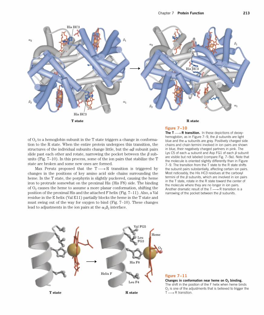
of O2 to a hemoglobin subunit in the T state triggers a change in conforma-tion to the R state. When the entire protein undergoes this transition, thestructures of the individual subunits change little, but the ab subunit pairsslide past each other and rotate, narrowing the pocket between the b sub-units (Fig. 7–10). In this process, some of the ion pairs that stabilize the Tstate are broken and some new ones are formed.
Max Perutz proposed that the T 88n R transition is triggered bychanges in the positions of key amino acid side chains surrounding theheme. In the T state, the porphyrin is slightly puckered, causing the hemeiron to protrude somewhat on the proximal His (His F8) side. The bindingof O2 causes the heme to assume a more planar conformation, shifting theposition of the proximal His and the attached F helix (Fig. 7–11). Also, a Valresidue in the E helix (Val E11) partially blocks the heme in the T state andmust swing out of the way for oxygen to bind (Fig. 7–10). These changeslead to adjustments in the ion pairs at the a1b2 interface.
figure 7–10The T 88n R transition. In these depictions of deoxy-hemoglobin, as in Figure 7–9, the b subunits are lightblue and the a subunits are gray. Positively charged sidechains and chain termini involved in ion pairs are shownin blue, their negatively charged partners in pink. The Lys C5 of each a subunit and Asp FG1 of each b subunitare visible but not labeled (compare Fig. 7–9a). Note thatthe molecule is oriented slightly differently than in Figure7–9. The transition from the T state to the R state shiftsthe subunit pairs substantially, affecting certain ion pairs.Most noticeably, the His HC3 residues at the carboxyltermini of the b subunits, which are involved in ion pairsin the T state, rotate in the R state toward the center ofthe molecule where they are no longer in ion pairs.Another dramatic result of the T 88n R transition is a narrowing of the pocket between the b subunits.
His HC3
His HC3
His HC3
a1
b1a2
b2a1
b1a2
b2
T state R state
T state R state
Val FG5
Heme
O2
LeuFG3
Helix F
Leu F4
His F8
figure 7–11Changes in conformation near heme on O2 binding.The shift in the position of the F helix when heme bindsO2 is one of the adjustments that is believed to trigger theT 88n R transition.

214 Part II Structure and Catalysis
Hemoglobin Binds Oxygen CooperativelyHemoglobin must bind oxygen efficiently in the lungs, where the pO2 isabout 13.3 kPa, and release oxygen in the tissues, where the pO2 is about 4kPa. Myoglobin, or any protein that binds oxygen with a hyperbolic bindingcurve, would be ill-suited to this function, for the reason illustrated in Fig-ure 7–12. A protein that bound O2 with high affinity would bind it efficientlyin the lungs but would not release much of it in the tissues. If the proteinbound oxygen with a sufficiently low affinity to release it in the tissues, itwould not pick up much oxygen in the lungs.
Hemoglobin solves the problem by undergoing a transition from a low-affinity state (the T state) to a high-affinity state (the R state) as more O2
molecules are bound. As a result, hemoglobin has a hybrid S-shaped, or sig-moid, binding curve for oxygen (Fig. 7–12). A single-subunit protein with asingle ligand-binding site cannot produce a sigmoid binding curve—even ifbinding elicits a conformational change—because each molecule of ligandbinds independently and cannot affect the binding of another molecule. Incontrast, O2 binding to individual subunits of hemoglobin can alter the affin-ity for O2 in adjacent subunits. The first molecule of O2 that interacts withdeoxyhemoglobin binds weakly, because it binds to a subunit in the T state.Its binding, however, leads to conformational changes that are communi-cated to adjacent subunits, making it easier for additional molecules of O2
to bind. In effect, the T 88n R transition occurs more readily in the secondsubunit once O2 is bound to the first subunit. The last (fourth) O2 moleculebinds to a heme in a subunit that is already in the R state, and hence it bindswith much higher affinity than the first molecule.
An allosteric protein is one in which the binding of a ligand to onesite affects the binding properties of another site on the same protein. Theterm allosteric derives from the Greek allos, “other,” and stereos, “solid” or“shape.” Allosteric proteins are those having “other shapes” or conforma-tions induced by the binding of ligands referred to as modulators. The con-formational changes induced by the modulator(s) interconvert more-activeand less-active forms of the protein. The modulators for allosteric proteinsmay be either inhibitors or activators. When the normal ligand and modula-tor are identical, the interaction is termed homotropic. When the modula-tor is a molecule other than the normal ligand the interaction is het-
erotropic. Some proteins have two or more modulators and therefore canhave both homotropic and heterotropic interactions.
Cooperative binding of a ligand to a multimeric protein, such as we ob-serve with the binding of O2 to hemoglobin, is a form of allosteric bindingoften observed in multimeric proteins. The binding of one ligand affects theaffinities of any remaining unfilled binding sites, and O2 can be consideredas both a normal ligand and an activating homotropic modulator. There isonly one binding site for O2 on each subunit, so the allosteric effects givingrise to cooperativity are mediated by conformational changes transmittedfrom one subunit to another by subunit-subunit interactions. A sigmoid
1.0
0.8
0.6
0.2
0.4
0
v
4 8 12 16pO2 (kPa)
pO2 intissues
pO2 inlungs
Transition fromlow- to high-affinity state
Low-affinitystate
High-affinitystate
figure 7–12A sigmoid (cooperative) binding curve. A sigmoidbinding curve can be viewed as a hybrid curve reflectinga transition from a low-affinity to a high-affinity state.Cooperative binding, as manifested by a sigmoid bindingcurve, renders hemoglobin more sensitive to the smalldifferences in O2 concentration between the tissues andthe lungs, allowing hemoglobin to bind oxygen in thelungs where pO2 is high and release it in the tissueswhere pO2 is low.

Chapter 7 Protein Function 215
bonding curve is diagnostic of cooperative binding. It permits a much moresensitive response to ligand concentration and is important to the functionof many multisubunit proteins. The principle of allostery extends readily toregulatory enzymes, as we will see in Chapter 8.
Cooperative Ligand Binding Can Be Described QuantitativelyCooperative binding of oxygen by hemoglobin was first analyzed byArchibald Hill in 1910. For a protein with n binding sites, the equilibrium ofEquation 7–1 becomes
(7–12)
and the expression for the association constant becomes
(7–13)
The expression for v (see Eqn 7–8) is
(7–14)
Rearranging, then taking the log of both sides, yields
(7–15)
(7–16)
Equation 7–16 is the Hill equation, and a plot of log [v/(1 � v)] versus log[L] is called a Hill plot. Based on the equation, the Hill plot should have aslope of n. However, the experimentally determined slope actually reflectsnot the number of binding sites, but the degree of interaction betweenthem. The slope of a Hill plot is therefore denoted nH, the Hill coefficient,
which is a measure of the degree of cooperativity. If nH equals 1, ligandbinding is not cooperative, a situation that can arise even in a multisubunitprotein if the subunits do not communicate. An nH of greater than 1 indi-cates positive cooperativity in ligand binding. This is the situation observedin hemoglobin, in which the binding of one molecule of ligand facilitates thebinding of others. The theoretical upper limit for nH is reached when nH �n. In this case the binding would be completely cooperative: all bindingsites on the protein would bind ligand simultaneously, and no protein mol-ecules partially saturated with ligand would be present under any condi-tions. This limit is never reached in practice, and the measured value of nH
is always less than the actual number of ligand-binding sites in the protein.An nH of less than 1 indicates negative cooperativity, in which the bind-
ing of one molecule of ligand impedes the binding of others. Well-documented cases of negative cooperativity are rare.
To adapt the Hill equation to the binding of oxygen to hemoglobin wemust again substitute pO2 for [L] and P50 for Kd:
(7–17)
Hill plots for myoglobin and hemoglobin are given in Figure 7–13.
Two Models Suggest Mechanisms for Cooperative BindingBiochemists now know a great deal about the T and R states of hemoglobin,but much remains to be learned about how the T 88n R transition occurs.Two models for the cooperative binding of ligands to proteins with multiplebinding sites have greatly influenced thinking about this problem.
log � v
1 � v� � n log pO2 � log P50
log � v
1 � v� � n log [L] � log Kd
v
1 � v�
[L]n
Kd
v �[L]n
[L]n � Kd
Ka �[PLn]
[P][L]n
P � nL 34 PLn
3
log pO2
�3
1 �
0 3�1 1
0
1
2
�2 2
�2
�1
log
��)
(
HemoglobinnH � 3
Hemoglobinhigh-affinity
statenH � 1
MyoglobinnH � 1 Hemoglobin
low-affinitystate
nH � 1
figure 7–13Hill plots for the binding of oxygen to myoglobin andhemoglobin. When nH � 1, there is no evident coopera-tivity. The maximum degree of cooperativity observed forhemoglobin corresponds approximately to nH � 3. Notethat while this indicates a high level of cooperativity, nH isless than n, the number of O2-binding sites in hemo-globin. This is normal for a protein that exhibits allostericbinding behavior.

216 Part II Structure and Catalysis
The first model was proposed by Jacques Monod, Jeffries Wyman, andJean-Pierre Changeux in 1965, and is called the MWC model or the con-
certed model (Fig. 7–14a). The concerted model assumes that the sub-units of a cooperatively binding protein are functionally identical, that eachsubunit can exist in (at least) two conformations, and that all subunits un-dergo the transition from one conformation to the other simultaneously. Inthis model, no protein has individual subunits in different conformations.The two conformations are in equilibrium. The ligand can bind to eitherconformation, but binds each with different affinity. Successive binding ofligand molecules to the low-affinity conformation (which is more stable inthe absence of ligand) makes a transition to the high-affinity conformationmore likely.
In the second model, the sequential model (Fig. 7–14b), proposed in1966 by Daniel Koshland and colleagues, ligand binding can induce achange of conformation in an individual subunit. A conformational changein one subunit makes a similar change in an adjacent subunit, as well as thebinding of a second ligand molecule, more likely. There are more potentialintermediate states in this model than in the concerted model. The twomodels are not mutually exclusive; the concerted model may be viewed asthe “all-or-none” limiting case of the sequential model. In Chapter 8 we willuse these models when we investigate allosteric enzymes.
Hemoglobin Also Transports H� and CO2
In addition to carrying nearly all the oxygen required by cells from the lungsto the tissues, hemoglobin carries two end products of cellular respira-tion—H� and CO2—from the tissues to the lungs and the kidneys, wherethey are excreted. The CO2, produced by oxidation of organic fuels in mitochondria, is hydrated to form bicarbonate:
This reaction is catalyzed by carbonic anhydrase, an enzyme particularlyabundant in erythrocytes. Carbon dioxide is not very soluble in aqueous so-lution, and bubbles of CO2 would form in the tissues and blood if it were notconverted to bicarbonate. As you can see from the equation, the hydrationof CO2 results in an increase in the H� concentration (a decrease in pH) in
CO2 � H2O 34 H� � HCO3�
figure 7–14Two general models for the interconversion of inactiveand active forms of cooperative ligand-binding proteins.Although the models may be applied to any protein—including any enzyme (Chapter 8)—that exhibits cooper-ative binding, four subunits are shown because the modelwas originally proposed for hemoglobin. In the concerted,or all-or-none, model (a) all the subunits are postulated tobe in the same conformation, either all (low affinity orinactive) or all (high affinity or active). Depending onthe equilibrium, K1, between and forms, thebinding of one or more ligand molecules (L) will pull theequilibrium toward the form. Subunits with bound Lare shaded. In the sequential model (b) each individualsubunit can be in either the or form. A very largenumber of conformations is thus possible.
□�
□
□�□
�
L
LL
LLL
LL
LL
L
L L
L LL
L LL L
L
LL
LLL
LL
LL
L
L
L
L
L
LL
LL L
L
L
L
L
L
LL
LL L
L
L
L
L
L
LL
L
L
L
L
L
L
LL
LL LL L
(a) (b)
All All

Chapter 7 Protein Function 217
the tissues. The binding of oxygen by hemoglobin is profoundly influencedby pH and CO2 concentration, so the interconversion of CO2 and bicarbon-ate is of great importance to the regulation of oxygen binding and release inthe blood.
Hemoglobin transports about 20% of the total H� and CO2 formed inthe tissues to the lungs and the kidneys. The binding of H� and CO2 is in-versely related to the binding of oxygen. At the relatively low pH and highCO2 concentration of peripheral tissues, the affinity of hemoglobin for oxy-gen decreases as H� and CO2 are bound, and O2 is released to the tissues.Conversely, in the capillaries of the lung, as CO2 is excreted and the bloodpH consequently rises, the affinity of hemoglobin for oxygen increases andthe protein binds more O2 for transport to the peripheral tissues. This effectof pH and CO2 concentration on the binding and release of oxygen by he-moglobin is called the Bohr effect, after Christian Bohr, the Danish physi-ologist (and father of physicist Niels Bohr) who discovered it in 1904.
The binding equilibrium for hemoglobin and one molecule of oxygencan be designated by the reaction
but this is not a complete statement. To account for the effect of H� con-centration on this binding equilibrium, we rewrite the reaction as
where HHb� denotes a protonated form of hemoglobin. This equation tellsus that the O2-saturation curve of hemoglobin is influenced by the H� con-centration (Fig. 7–15). Both O2 and H� are bound by hemoglobin, but withinverse affinity. When the oxygen concentration is high, as in the lungs, hemoglobin binds O2 and releases protons. When the oxygen concentrationis low, as in the peripheral tissues, H� is bound and O2 is released.
Oxygen and H� are not bound at the same sites in hemoglobin. Oxygenbinds to the iron atoms of the hemes, whereas H� binds to any of severalamino acid residues in the protein. A major contribution to the Bohr effectis made by His146 (His HC3) of the b subunits. When protonated, thisresidue forms one of the ion pairs—to Asp94 (Asp FG1)—that helps stabi-lize deoxyhemoglobin in the T state (Fig. 7–9). The ion pair stabilizes theprotonated form of His HC3, giving this residue an abnormally high pKa inthe T state. The pKa falls to its normal value of 6.0 in the R state becausethe ion pair cannot form, and this residue is largely unprotonated in oxyhe-moglobin at pH 7.6, the blood pH in the lungs. As the concentration of H�
rises, protonation of His HC3 promotes release of oxygen by favoring a tran-sition to the T state. Protonation of the amino-terminal residues of the asubunits, certain other His residues, and perhaps other groups has a similareffect.
Thus we see that the four polypeptide chains of hemoglobin communi-cate with each other not only about O2 binding to their heme groups, butalso about H� binding to specific amino acid residues. And there is stillmore to the story. Hemoglobin also binds CO2, again in a manner inverselyrelated to the binding of oxygen. Carbon dioxide binds as a carbamategroup to the a-amino group at the amino-terminal end of each globin chain,forming carbaminohemoglobin:
HHb� � O2 34 HbO2 � H�
Hb � O2 34 HbO2
figure 7–15Effect of pH on the binding of oxygen to hemoglobin.The pH of blood is 7.6 in the lungs and 7.2 in the tissues.Experimental measurements on hemoglobin binding areoften performed at pH 7.4.
1.0
0.5
0
v
420 6 8 10pO2 (kPa)
pH 7.4
pH 7.6
pH 7.2
NH2
O
�A
O
B
ABO
R
C C
O
H
Amino-terminalresidue
H�
OBC
AO
B
AO O
R
C C
O
H
O ON
H
OB
CG AO�
Carbamino-terminalresidue

218 Part II Structure and Catalysis
This reaction produces H�, contributing to the Bohr effect. The bound car-bamates also form additional salt bridges (not shown in Fig. 7–9) that helpto stabilize the T state and promote the release of oxygen.
When the concentration of carbon dioxide is high, as in peripheral tis-sues, some CO2 binds to hemoglobin and the affinity for O2 decreases, caus-ing its release. Conversely, when hemoglobin reaches the lungs, the highoxygen concentration promotes binding of O2 and release of CO2. It is thecapacity to communicate ligand-binding information from one polypeptidesubunit to the others that makes the hemoglobin molecule so beautifullyadapted to integrating the transport of O2, CO2, and H� by erythrocytes.
Oxygen Binding to Hemoglobin Is Regulated by 2,3-BisphosphoglycerateThe interaction of 2,3-bisphosphoglycerate (BPG) with hemoglobin pro-vides an example of heterotropic allosteric modulation. BPG is present inrelatively high concentrations in erythrocytes. When hemoglobin is isolated,it contains substantial amounts of bound BPG, which can be difficult to re-move completely. In fact, the O2-binding curves for hemoglobin that wehave examined to this point were obtained in the presence of bound BPG.2,3-Bisphosphoglycerate is known to greatly reduce the affinity of hemo-globin for oxygen—there is an inverse relationship between the binding ofO2 and the binding of BPG. We can therefore describe another bindingprocess for hemoglobin:
BPG binds at a site distant from the oxygen-binding site and regulatesthe O2-binding affinity of hemoglobin in relation to the pO2 in the lungs.BPG plays an important role in the physiological adaptation to the lowerpO2 available at high altitudes. For a healthy human strolling by the ocean,the binding of O2 to hemoglobin is regulated such that the amount of O2 de-livered to the tissues is equivalent to nearly 40% of the maximum that couldbe carried by the blood (Fig. 7–16). If the same person is quickly trans-ported to a mountainside at an altitude of 4,500 meters, where the pO2 isconsiderably lower, the delivery of O2 to the tissues is reduced. However,
HbBPG � O2 34 HbO2 � BPG
OB
H
�O
G
AO�
O�
J
O
O
OP
C
C
P
H
AP
A
A
A
A
CH OO O
O
O
O
O
�
O�
O
2,3-Bisphosphoglycerate
figure 7–16 Effect of BPG on the binding of oxygen to hemoglobin.The BPG concentration in normal human blood is about5 mM at sea level and about 8 mM at high altitudes. Notethat hemoglobin binds to oxygen quite tightly when BPGis entirely absent, and the binding curve appears to behyperbolic. In reality, the measured Hill coefficient for O2-binding cooperativity decreases only slightly (from 3 toabout 2.5) when BPG is removed from hemoglobin, butthe rising part of the sigmoid curve is confined to a verysmall region close to the origin. At sea level, hemoglobinis nearly saturated with O2 in the lungs, but only 60% sat-urated in the tissues, so that the amount of oxygenreleased in the tissues is close to 40% of the maximumthat can be carried in the blood. At high altitudes, O2
delivery declines by about one-fourth, to 30% ofmaximum. An increase in BPG concentration, however,decreases the affinity of hemoglobin for O2 so that nearly40% of what can be carried is again delivered to thetissues.
1.0
0.5
0
v
4 8 12 16pO2 (kPa)
BPG � 5 mM
pO2 intissues
pO2 inlungs
(4500 m)
pO2 inlungs
(sea level)
0.38
0.370.30
BPG = 0mM
BPG � 8 mM

Chapter 7 Protein Function 219
after just a few hours at the higher altitude, the BPG concentration in theblood has begun to rise, leading to a decrease in the affinity of hemoglobinfor oxygen. This adjustment in the BPG level has only a small effect on thebinding of O2 in the lungs but a considerable effect on the release of O2 inthe tissues. As a result, the delivery of oxygen to the tissues is restored tonearly 40% of that which can be transported by the blood. The situation isreversed when the person returns to sea level. The BPG concentration inerythrocytes also increases in people suffering from hypoxia, lowered oxy-genation of peripheral tissues due to inadequate function of the lungs or cir-culatory system.
BPG binds to hemoglobin in the cavity between the b subunits in the Tstate (Fig. 7–17). This cavity is lined with positively charged amino acidresidues that interact with the negatively charged groups of BPG. Unlike O2,only one molecule of BPG is bound to each hemoglobin tetramer. BPG low-ers hemoglobin’s affinity for oxygen by stabilizing the T state. The transitionto the R state narrows the binding pocket for BPG, precluding BPG binding.In the absence of BPG, hemoglobin is converted to the R state more easily.
Regulation of oxygen binding to hemoglobin by BPG has an importantrole in fetal development. Because a fetus must extract oxygen from itsmother’s blood, fetal hemoglobin must have greater affinity than the mater-nal hemoglobin for O2. In fetuses, g subunits are synthesized rather than bsubunits, and a2g2 hemoglobin is formed. This tetramer has a much loweraffinity for BPG than normal adult hemoglobin, and a correspondinglyhigher affinity for O2.
Sickle-Cell Anemia Is a Molecular Disease of HemoglobinThe great importance of the amino acid sequence in determining the sec-ondary, tertiary, and quaternary structures of globular proteins, and thustheir biological functions, is strikingly demonstrated by the hereditary hu-man disease sickle-cell anemia. More than 300 genetic variants of hemoglo-bin are known to occur in the human population. Most of these variationsconsist of differences in a single amino acid residue. The effects on hemo-globin structure and function are often minor but can sometimes be extra-ordinary. Each hemoglobin variation is the product of an altered gene. Thevariant genes are called alleles. Because humans generally have two copiesof each gene, an individual may have two copies of one allele (thus beinghomozygous for that gene) or one copy of each of two different alleles
(b)
(c)
(a)
figure 7–17 Binding of BPG to deoxyhemoglobin. (a) BPG bindingstabilizes the T state of deoxyhemoglobin, shown here asa mesh surface image. (b) The negative charges of BPGinteract with several positively charged groups (shown inblue in this GRASP surface image) that surround thepocket between the b subunits in the T state. (c) Thebinding pocket for BPG disappears on oxygenation, fol-lowing transition to the R state. (Compare (b) and (c) withFig. 7–10.)

220 Part II Structure and Catalysis
(heterozygous). Sickle-cell anemia is a genetic disease in which an individualhas inherited the allele for sickle-cell hemoglobin from both parents. Theerythrocytes of these individuals are fewer and also abnormal. In additionto an unusually large number of immature cells, the blood contains manylong, thin, crescent-shaped erythrocytes that look like the blade of a sickle(Fig. 7–18). When hemoglobin from sickle cells (called hemoglobin S) is de-oxygenated, it becomes insoluble and forms polymers that aggregate intotubular fibers (Fig. 7–19). Normal hemoglobin (hemoglobin A) remains sol-
figure 7–18A comparison of uniform, cup-shaped, normal erythro-cytes (a) with the variably shaped erythrocytes seen insickle-cell anemia (b). These cells range from normal tospiny or sickle-shaped.
(a) 2 m (b)
Interaction between molecules
Strand formation
Alignment and crystallization(fiber formation)
(b)
Hemoglobin A Hemoglobin S
a2
b1
b2
a1
(a)
figure 7–19Normal and sickle-cell hemoglobin. (a) Subtle differ-ences between the conformations of hemoglobin A andhemoglobin S result from a single amino acid change inthe b chains. (b) As a result of this change, deoxyhemo-globin S has a hydrophobic patch on its surface, whichcauses the molecules to aggregate into strands that aligninto insoluble fibers.

Chapter 7 Protein Function 221
uble on deoxygenation. The insoluble fibers of deoxygenated hemoglobin Sare responsible for the deformed sickle shape of the erythrocytes, and theproportion of sickled cells increases greatly as blood is deoxygenated.
The altered properties of hemoglobin S result from a single amino acidsubstitution, a Val instead of a Glu residue at position 6 in the two b chains.The R group of valine has no electric charge, whereas glutamate has a neg-ative charge at pH 7.4. Hemoglobin S therefore has two fewer negativecharges than hemoglobin A, one for each of the two b chains. Replacementof the Glu residue by Val creates a “sticky” hydrophobic contact point at po-sition 6 of the b chain, which is on the outer surface of the molecule. Thesesticky spots cause deoxyhemoglobin S molecules to associate abnormallywith each other, forming the long, fibrous aggregates characteristic of thisdisorder.
Sickle-cell anemia occurs in individuals homozygous for the sickle-cellallele of the gene encoding the b subunit of hemoglobin. Individuals who re-ceive the sickle-cell allele from only one parent and are thus heterozygousexperience a milder condition called sickle-cell trait; only about 1% of theirerythrocytes become sickled on deoxygenation. These individuals may livecompletely normal lives if they avoid vigorous exercise or other stresses onthe circulatory system.
People with sickle-cell anemia suffer from repeated crises brought onby physical exertion. They become weak, dizzy, and short of breath, andthey also experience heart murmurs and an increased pulse rate. The he-moglobin content of their blood is only about half the normal value of 15 to16 g/100 mL because sickled cells are very fragile and rupture easily; thisresults in anemia (“lack of blood”). An even more serious consequence isthat capillaries become blocked by the long, abnormally shaped cells, caus-ing severe pain and interfering with normal organ function—a major factorin the early death of many people with the disease.
Without medical treatment, people with sickle-cell anemia usually diein childhood. Nevertheless, the sickle-cell allele is surprisingly common incertain parts of Africa. Investigation into the persistence of an allele that is so obviously deleterious in homozygous individuals led to the finding that the allele confers a small but significant resistance to lethal forms ofmalaria in heterozygous individuals. Natural selection has resulted in an allele population that balances the deleterious effects of the homozygouscondition against the resistance to malaria afforded by the heterozygouscondition.
Complementary Interactions between Proteins andLigands: The Immune System and Immunoglobulins Our discussion of oxygen-binding proteins showed how the conformationsof these proteins affect and are affected by the binding of small ligands (O2
or CO) to the heme group. However, most protein-ligand interactions do notinvolve a prosthetic group. Instead, the binding site for a ligand is more of-ten like the hemoglobin binding site for BPG—a cleft in the protein linedwith amino acid residues, arranged to render the binding interaction highlyspecific. Effective discrimination between ligands is the norm at bindingsites, even when the ligands have only minor structural differences.
All vertebrates have an immune system capable of distinguishing mol-ecular “self” from “nonself” and then destroying those entities identified asnonself. In this way, the immune system eliminates viruses, bacteria, andother pathogens and molecules that may pose a threat to the organism. On aphysiological level, the response of the immune system to an invader is anintricate and coordinated set of interactions among many classes of proteins,

222 Part II Structure and Catalysis
molecules, and cell types. However, at the level of individual proteins, theimmune response demonstrates how an acutely sensitive and specific bio-chemical system is built upon the reversible binding of ligands to proteins.
The Immune Response Features a Specialized Array of Cells and ProteinsImmunity is brought about by a variety of leukocytes (white blood cells),including macrophages and lymphocytes, all arising from undifferenti-ated stem cells in the bone marrow. Leukocytes can leave the bloodstreamand patrol the tissues, each cell producing one or more proteins capable ofrecognizing and binding to molecules that might signal an infection.
The immune response consists of two complementary systems, the hu-moral and cellular immune systems. The humoral immune system (Latinhumor, “fluid”) is directed at bacterial infections and extracellular viruses(those found in the body fluids), but can also respond to individual proteinsintroduced into the organism. The cellular immune system destroys hostcells infected by viruses and also destroys some parasites and foreign tissues.
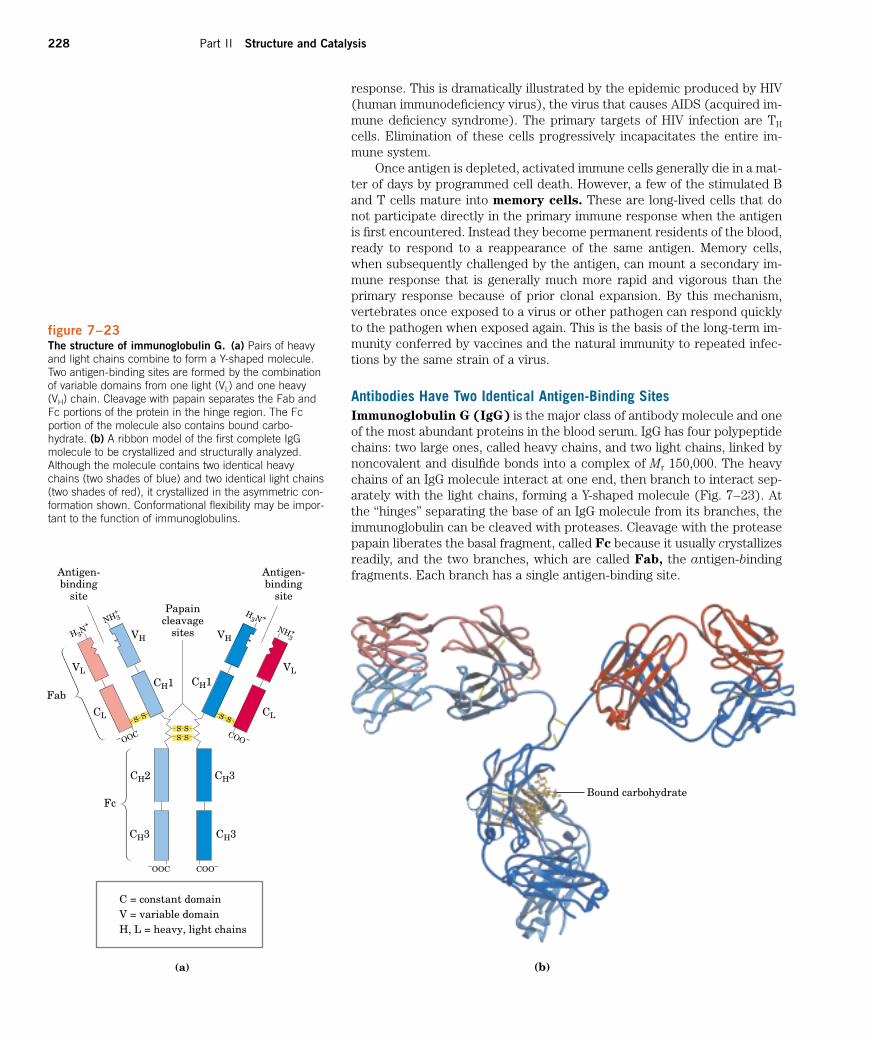
The proteins at the heart of the humoral immune response are solubleproteins called antibodies or immunoglobulins, often abbreviated Ig. Im-munoglobulins bind bacteria, viruses, or large molecules identified as for-eign and target them for destruction. Making up 20% of blood protein, theimmunoglobulins are produced by B lymphocytes or B cells, so namedbecause they complete their development in the bone marrow.
The agents at the heart of the cellular immune response are a class ofT lymphocytes or T cells (so called because the latter stages of their de-velopment occur in the thymus) known as cytotoxic T cells (TC cells,
also called killer T cells). Recognition of infected cells or parasites involvesproteins called T-cell receptors on the surface of TC cells. Recall fromChapter 2 (p. 30) that receptors are proteins, usually found on the outersurface of cells and extending through the plasma membrane; they recog-nize and bind extracellular ligands, triggering changes inside the cell.
In addition to cytotoxic T cells, there are helper T cells (TH cells),
whose function it is to produce soluble signaling proteins called cytokines,which include the interleukins. TH cells interact with macrophages. Table7–2 summarizes the functions of the various leukocytes of the immunesystem.
Each recognition protein of the immune system, either an antibody pro-duced by a B cell or a receptor on the surface of a T cell, specifically bindssome particular chemical structure, distinguishing it from virtually all oth-ers. Humans are capable of producing over 108 different antibodies with dis-tinct binding specificities. This extraordinary diversity makes it likely thatany chemical structure on the surface of a virus or invading cell will be rec-ognized and bound by one or more antibodies. Antibody diversity is derivedfrom random reassembly of a set of immunoglobulin gene segments via ge-netic recombination mechanisms that are discussed in Chapter 25.
Some properties of the interactions between antibodies or T-cell recep-tors and the molecules they bind are unique to the immune system, and aspecialized lexicon is used to describe them. Any molecule or pathogen ca-pable of eliciting an immune response is called an antigen. An antigen maybe a virus, a bacterial cell wall, or an individual protein or other macromole-cule. A complex antigen may be bound by a number of different antibodies.An individual antibody or T-cell receptor binds only a particular molecularstructure within the antigen, called its antigenic determinant or epitope.
It would be unproductive for the immune system to respond to small mol-ecules that are common intermediates and products of cellular metabolism.Molecules of Mr �5,000 are generally not antigenic. However, small moleculescan be covalently attached to large proteins in the laboratory, and in this
Some Types of Leukocytes Associated with the Immune System
Cell type Function
Macrophages Ingest large particlesand cells by phago-cytosis
B lymphocytes Produce and secrete(B cells) antibodies
T lymphocytes(T cells)Cytotoxic (killer) Interact with infected
T cells (TC) host cells through receptors on T-cellsurface
Helper T cells (TH) Interact with macro-phages and secretecytokines (inter-leukins) thatstimulate TC, TH,and B cells to proliferate.
table 7–2

Chapter 7 Protein Function 223
form they may elicit an immune response. These small molecules are calledhaptens. The antibodies produced in response to protein-linked haptenswill then bind to the same small molecules when they are free. Such anti-bodies are sometimes used in the development of analytical tests describedlater in this chapter or as catalytic antibodies (described in Box 8–3).
The interactions of antibody and antigen are much better understoodthan are the binding properties of T-cell receptors. However, before focus-ing on antibodies, we need to look at the humoral and cellular immune sys-tems in more detail to put the fundamental biochemical interactions intotheir proper context.
Self Is Distinguished from Nonself by the Display of Peptides on Cell SurfacesThe immune system must identify and destroy pathogens, but it must alsorecognize and not destroy the normal proteins and cells of the host organ-ism—the “self.” Detection of protein antigens in the host is mediated byMHC (major histocompatibility complex) proteins. MHC proteins bindpeptide fragments of proteins digested in the cell and present them on theoutside surface of the cell. These peptides normally come from the digestionof typical cellular proteins, but during a viral infection viral proteins are alsodigested and presented by MHC proteins. Peptide fragments from foreignproteins that are displayed by MHC proteins are the antigens the immunesystem recognizes as nonself. T-cell receptors bind these fragments andlaunch the subsequent steps of the immune response. There are two classesof MHC proteins (Fig. 7–20), which differ in their distribution among celltypes and in the source of digested proteins whose peptides they display.
figure 7–20MHC proteins These proteins consist of a and b chains.In class I MHC proteins (a), the small b chain is invariantbut the amino acid sequence of the a chain exhibits ahigh degree of variability, localized in specific domains ofthe protein that appear on the outside of the cell. Eachhuman produces up to six different a chains for class 1 MHC proteins. In class II MHC proteins (b), both the a and b chains have regions of relatively high variabilitynear their amino-terminal ends.
(a) Class I MHC protein (b) Class II MHC protein
Hypervariabledomains
+NH3
–OOCExtracellular
space
Plasmamembrane
Cytosol
H3N+
–OOC–OOC
+NH3
COO–
a chain
b chain
b chain a chain
+NH3
S
S
SS
SS
SS
SS
S S–– –

224 Part II Structure and Catalysis
Class I MHC proteins (Fig. 7–21) are found on the surface of virtuallyall vertebrate cells. There are countless variants in the human population,placing them among the most polymorphic of proteins. Because individualsproduce up to six class I MHC protein variants, any two individuals are unlikely to have the same set. Class I MHC proteins bind and display pep-tides derived from the proteolytic degradation and turnover of proteins thatoccurs randomly within the cell. These complexes of peptides and class IMHC proteins are the recognition targets of the T-cell receptors of the TC
cells in the cellular immune system. The general pattern of immune systemrecognition was first described by Rolf Zinkernagel and Peter Doherty in1974.
Each TC cell has many copies of only one T-cell receptor that is specificfor a particular class I MHC protein–peptide complex. To avoid creating a
Antigen
b chain
a chain
NH3�
�
NH3�
COO
Extracellularspace
Cytosol
(a)
–OOC
Plasmamembrane
Antigen
NH3�
(b)
figure 7–21Structure of a human class I MHC protein. (a) Thisimage is derived in part from the determined structure ofthe extracellular portion of the protein. The a chain ofMHC is shown in gray; the small b chain is blue; thedisulfide bonds are yellow. A bound ligand, a peptidederived from HIV, is shown in red. (b) Top view showing asurface contour image of the site where peptides arebound and displayed. The HIV peptide (red) occupies thesite. This part of the class I MHC protein interacts with T-cell receptors.

legion of TC cells that would set upon and destroy normal cells, the matu-ration of TC cells in the thymus includes a stringent selection process thateliminates more than 95% of the developing TC cells, including those thatmight recognize and bind class I MHC proteins displaying peptides from cel-lular proteins of the organism itself. The TC cells that survive and matureare those with T-cell receptors that do not bind to the organism’s own pro-teins. The result is a population of cells that bind foreign peptides bound toclass I MHC proteins of the host cell. These binding interactions lead to thedestruction of parasites and virus-infected cells. When an organ is trans-planted, its foreign class I MHC proteins are also bound by TC cells, leadingto tissue rejection.
Class II MHC proteins occur on the surfaces of a few types of spe-cialized cells that take up foreign antigens, including macrophages and Blymphocytes. Like class I MHC proteins, the class II proteins are highly poly-morphic, with many variants in the human population. Each human is ca-pable of producing up to 12 variants, and thus it is unlikely that any two in-dividuals have an identical set of variants. The class II MHC proteins bindand display peptides derived not from cellular proteins but from externalproteins ingested by the cells. The resulting class II MHC protein–peptidecomplexes are the binding targets of the T-cell receptors of the varioushelper T cells. TH cells, like TC cells, undergo a stringent selection processin the thymus, eliminating those that recognize the individual’s own cellularproteins.
Despite the elimination of most TC and TH cells during the selectionprocess in the thymus, a very large number survive, and these provide theimmune response. Each survivor has a single type of T-cell receptor thatcan bind to one particular chemical structure. The T cells patrolling thebloodstream and the tissues carry millions of different binding specificitiesin the T-cell receptors. Within the highly varied T-cell population there is al-most always a contingent of cells that can specifically bind any antigen thatmight appear. The vast majority of these cells never encounter a foreignantigen to which they can bind and typically die within a few days, replacedby new generations of T cells endlessly patrolling in search of the interac-tion that will launch the full immune response.
Molecular Interactions at Cell Surfaces Trigger the Immune ResponseA new antigen is often the harbinger of an infection—a signal to the im-mune system that a virus or other parasite may be rapidly growing in the or-ganism. Those few T cells and B cells possessing receptors or antibodiesthat can bind the antigen must be rapidly and selectively propagated toeliminate the infection. A hypothetical viral infection illustrates how this occurs.
When a virus invades a cell, it makes use of cellular functions and re-sources to replicate its nucleic acid and make viral proteins. Once inside thecell, viral macromolecules are relatively inaccessible to the antibodies of thehumoral immune system. However, some of the class I MHC proteins thatfind their way to the surface of an infected cell will generally display pep-tide fragments from viral proteins, which can then be recognized by TC lym-phocytes. Mature viruses become vulnerable to the humoral immune sys-tem when they are released from the infected cell and are present for a timein the extracellular environment. Some are then ingested by macrophages(which ingest only those antigens that are recognized by the antibodies pro-duced by a particular B cell). Viral peptide fragments will be displayed onthe surfaces of the macrophages and B cells, complexed to class II MHCproteins, and the peptide antigens will trigger a multi-pronged response in-volving B cells, TC cells, and TH cells (Fig. 7–22).
Chapter 7 Protein Function 225

226
MHC I
T-cellreceptor
Interleukinreceptor Interleukin 2, 4
MHC II CD4 CD8
Interleukinreceptor
Interleukin 2
CD8
IgG Fcreceptor
T-cell receptor
T-cell receptor
CD4
Interleukin 4
Infected cellwith MHC I
displayingnormal host
and viral peptides.
Lysed cell releasingvirus particles and antigens; somebecome bound byantibodies.
Viralpathogens
Soluble antibodies
B cell that producesantibodies to virus.
B cell with MHC II andviral peptides complexedwith TH cells via T-cellreceptor and CD4. B cellproliferates and producessoluble antibodies,stimulated by interleukins.
MHC class I proteindisplaying cellular peptides.
Antibody-boundvirus beingengulfed bymacrophage. Viralpeptides presented by MHC II.
Macrophagewith MHC IIcomplexed toTH cell viaT-cell receptorand CD4.TC and TH cells
bound to infectedcells mature andproliferate, stimulatedby interleukins. Memory cells
TC, TH, B
Host cell
Infected cellwith TC cell
bound toMHC I and
viral peptidesvia T-cell
receptor andCD8 receptor
protein.
Infected celldestroyed by
TC cell.
B cell
B cell
TC cell
TC cell
TH cellTH cell
figure 7–22Overview of the immune response to a viral infection. The individual steps are described in the text.

Chapter 7 Protein Function 227
The class I MHC protein–peptide complexes on infected cells are rec-ognized as foreign and bound by those TC cells with T-cell receptors havingthe appropriate binding specificity. The T-cell receptors respond only topeptide antigens that are complexed to class I MHC proteins. The TC cellshave an additional receptor, CD8, also called a coreceptor, that enhancesthe binding interactions of T-cell receptors and MHC proteins (Fig. 7–22,middle left). The TC cells live up to the name killer T cells by destroying thevirally infected cell to which they are complexed through their T-cell re-ceptors. Cell death is brought about by a number of mechanisms, not allwell understood. One mechanism involves the release of a protein calledperforin, which binds to and aggregates in the plasma membrane of thetarget cell, forming molecular pores that destroy the capacity of that cell to regulate its interior environment. TC cells also induce a process calledprogrammed cell death, or apoptosis (most commonly pronounced app -a-toe -sis), in which the cells complexed to TC cells undergo metabolicchanges that rapidly lead to the demise of the cell.
TC cells with the proper specificity must proliferate selectively if largenumbers of virus-infected cells are to be destroyed. To this end, TC cellscomplexed to an infected cell generate cell-surface receptors for signalingproteins called interleukins. Interleukins, secreted by a variety of cells,stimulate the proliferation of only those T and B cells bearing the requiredinterleukin receptors. Because T and B cells produce interleukin receptorsonly when they are complexed with an antigen, the only immune systemcells that proliferate are those few that can respond to the antigen. Theprocess of producing a population of cells by stimulated reproduction of aparticular ancestor cell is called clonal selection.
The peptides complexed to class II MHC proteins and displayed on thesurface of macrophages and B lymphocytes are similarly bound by the ap-propriate T-cell receptors of TH cells. The TH cells also have a coreceptor,called CD4, that enhances the binding interactions of the T-cell receptors.This overall binding interaction, in concert with secondary molecular sig-nals that are currently being identified, activates the TH cells. A subpopula-tion of activated TH cells secrete a small signal protein called interleukin-2(IL-2; Mr 15,000), which stimulates proliferation of nearby TC cells and TH
cells having the appropriate interleukin receptors. This greatly increasesthe number of available immune system cells capable of recognizing and re-sponding to the antigen. Another subpopulation of activated TH cells com-plexed to macrophages or B lymphocytes secrete interleukin-4 (IL-4; Mr
20,000), which stimulates the proliferation of B cells that recognize theantigen (Fig. 7–22, bottom right). Proliferation of the responding B, TC, andTH cells continues as long as the appropriate antigen is present.
The proliferating B cells promote the destruction of any extracellularviruses or bacterial cells. They first secrete large amounts of soluble anti-body that binds to the antigen. This bound antibody recruits a cellular sys-tem of about 20 proteins collectively called complement because they com-plement and enhance the action of the antibodies. The complement proteinsdisrupt the coats of many viruses or, in bacterial infections, produce holes inthe cell walls of bacteria, causing them to swell and burst by osmotic shock.
Unlike T cells, B cells do not undergo selection in the thymus to elimi-nate those producing antibodies that recognize host (self) proteins. How-ever, B cells do not contribute significantly to an immune response unlessthey are stimulated to proliferate by TH cells. The TH cells do undergo se-lection in the thymus, leaving no TH cells capable of stimulating B cells thatproduce antibodies potentially dangerous to the host.
The TH cells themselves participate only indirectly in the destruction ofinfected cells and pathogens, but their role is critical to the entire immune
��
228 Part II Structure and Catalysis
response. This is dramatically illustrated by the epidemic produced by HIV(human immunodeficiency virus), the virus that causes AIDS (acquired im-mune deficiency syndrome). The primary targets of HIV infection are TH
cells. Elimination of these cells progressively incapacitates the entire im-mune system.
Once antigen is depleted, activated immune cells generally die in a mat-ter of days by programmed cell death. However, a few of the stimulated Band T cells mature into memory cells. These are long-lived cells that donot participate directly in the primary immune response when the antigenis first encountered. Instead they become permanent residents of the blood,ready to respond to a reappearance of the same antigen. Memory cells,when subsequently challenged by the antigen, can mount a secondary im-mune response that is generally much more rapid and vigorous than theprimary response because of prior clonal expansion. By this mechanism,vertebrates once exposed to a virus or other pathogen can respond quicklyto the pathogen when exposed again. This is the basis of the long-term im-munity conferred by vaccines and the natural immunity to repeated infec-tions by the same strain of a virus.
Antibodies Have Two Identical Antigen-Binding SitesImmunoglobulin G (IgG) is the major class of antibody molecule and oneof the most abundant proteins in the blood serum. IgG has four polypeptidechains: two large ones, called heavy chains, and two light chains, linked bynoncovalent and disulfide bonds into a complex of Mr 150,000. The heavychains of an IgG molecule interact at one end, then branch to interact sep-arately with the light chains, forming a Y-shaped molecule (Fig. 7–23). Atthe “hinges” separating the base of an IgG molecule from its branches, theimmunoglobulin can be cleaved with proteases. Cleavage with the proteasepapain liberates the basal fragment, called Fc because it usually crystallizesreadily, and the two branches, which are called Fab, the antigen-bindingfragments. Each branch has a single antigen-binding site.
figure 7–23The structure of immunoglobulin G. (a) Pairs of heavyand light chains combine to form a Y-shaped molecule.Two antigen-binding sites are formed by the combinationof variable domains from one light (VL) and one heavy(VH) chain. Cleavage with papain separates the Fab andFc portions of the protein in the hinge region. The Fcportion of the molecule also contains bound carbo-hydrate. (b) A ribbon model of the first complete IgG molecule to be crystallized and structurally analyzed.Although the molecule contains two identical heavychains (two shades of blue) and two identical light chains(two shades of red), it crystallized in the asymmetric con-formation shown. Conformational flexibility may be impor-tant to the function of immunoglobulins.
SS
H3 NNH 3
H 3N+
COO –
NH3
S S
– OOC
COO––OOC
SSSS
(a)
+
+
+
VLVL
VHVH
CH1
CH3CH2
CH3CH3
Fc
Fab
Antigen-binding
site
CH1
CLCL
Antigen-binding
site
C = constant domainV = variable domainH, L = heavy, light chains
Papaincleavage
sites
Bound carbohydrate
(b)

Chapter 7 Protein Function 229
figure 7–24Binding of IgG to an antigen. To generate an optimal fit for the antigen, the binding sites of IgG often undergoslight conformational changes. Such induced fit iscommon to many protein-ligand interactions.
SS
H3 NNH 3
H 3N
+
COO –
NH3
S S
– OOC
COO––OOC
SSSS
Antibody
Antigen SSS S
SSSS
Antigen-antibody complex
+
+
+
The fundamental structure of immunoglobulins was first established byGerald Edelman and Rodney Porter. Each chain is made up of identifiabledomains; some are constant in sequence and structure from one IgG to thenext, others are variable. The constant domains have a characteristic struc-ture known as the immunoglobulin fold, a well-conserved structural mo-tif in the all-b class. There are three of these constant domains in eachheavy chain and one in each light chain. The heavy and light chains alsohave one variable domain each, in which most of the variability in aminoacid residue sequence is found. The variable domains associate to createthe antigen-binding site (Fig. 7–24).
figure 7–25IgM pentamer of immunoglobulin units. The pentameris cross-linked with disulfide bonds. The J chain is apolypeptide of Mr 20,000 found in both IgA and IgM.
J chain
Lightchains
m Heavychains
In many vertebrates, IgG is only one of five classes of immunoglobulins.Each class has a characteristic type of heavy chain, denoted a, d, e, g, andm for IgA, IgD, IgE, IgG, and IgM, respectively. Two types of light chain, k and l, occur in all classes of immunoglobulins. The overall structures ofIgD and IgE are similar to that of IgG. IgM occurs in either a monomeric, membrane-bound form or a secreted form that is a cross-linked pentamerof this basic structure (Fig. 7–25). IgA, found principally in secretions suchas saliva, tears, and milk, can be a monomer, dimer, or trimer. IgM is the firstantibody to be made by B lymphocytes and is the major antibody in theearly stages of a primary immune response. Some B cells soon begin to pro-duce IgD (with the same antigen-binding site as the IgM produced by thesame cell), but the unique function of IgD is less clear.

230 Part II Structure and Catalysis
The IgG described above is the major antibody in secondary immune re-sponses, which are initiated by memory B cells. As part of the organism’songoing immunity to antigens already encountered and dealt with, IgG isthe most abundant immunoglobulin in the blood. When IgG binds to an in-vading bacterium or virus, it not only activates the complement system, butalso activates certain leukocytes such as macrophages to engulf and destroythe invader. Yet another class of receptors on the cell surface of macrophagesrecognizes and binds the Fc region of IgG. When these Fc receptors bind anantibody-pathogen complex, the macrophage engulfs the complex byphagocytosis (Fig. 7–26).
Phagocytosis
IgG-coatedvirus
Fc regionof IgG
IgG Fcreceptor
Macrophage
figure 7–26Phagocytosis of an antibody-bound virus by amacrophage. The Fc regions of the antibodies bind to Fcreceptors on the surface of the macrophage, triggeringthe macrophage to engulf and destroy the virus.
IgE plays an important role in the allergic response, interacting with ba-sophils (phagocytic leukocytes) in the blood and histamine-secreting cellscalled mast cells that are widely distributed in tissues. This immunoglobu-lin binds, through its Fc region, to special Fc receptors on the basophils ormast cells. In this form, IgE serves as a kind of receptor for antigen. If anti-gen is bound, the cells are induced to secrete histamine and other biologi-cally active amines that cause dilation and increased permeability of bloodvessels. These effects on the blood vessels are thought to facilitate themovement of immune system cells and proteins to sites of inflammation.They also produce the symptoms normally associated with allergies. Pollenor other allergens are recognized as foreign, triggering an immune responsenormally reserved for pathogens.
Antibodies Bind Tightly and Specifically to AntigenThe binding specificity of an antibody is determined by the amino acidresidues in the variable domains of its heavy and light chains. Many residuesin these domains are variable, but not equally so. Some, particularly thoselining the antigen-binding site, are hypervariable—especially likely to differ.Specificity is conferred by chemical complementarity between the antigenand its specific binding site, in terms of shape and the location of charged,nonpolar, and hydrogen-bonding groups. For example, a binding site with anegatively charged group may bind an antigen with a positive charge in thecomplementary position. In many instances, complementarity is achievedinteractively as the structures of antigen and binding site are influenced byeach other during the approach of the ligand. Conformational changes inthe antibody and/or the antigen then occur that allow the complementarygroups to interact fully. This is an example of induced fit (Fig. 7–27).

Chapter 7 Protein Function 231
A typical antibody-antigen interaction is quite strong, characterized byKd values as low as 10�10 M (recall that a lower Kd corresponds to a strongerbinding interaction). The Kd reflects the energy derived from the variousionic, hydrogen-bonding, hydrophobic, and van der Waals interactions thatstabilize the binding. The binding energy required to produce a Kd of 10�10 M
is about 65 kJ/mol.A complex of a peptide derived from HIV (a model antigen) and an Fab
molecule illustrates some of these properties (Fig. 7–27). The changes instructure observed on antigen binding are particularly striking in this example.
The Antibody-Antigen Interaction Is the Basis for a Variety of Important Analytical ProceduresThe extraordinary binding affinity and specificity of antibodies makes themvaluable analytical reagents. Two types of antibody preparations are in use:polyclonal and monoclonal. Polyclonal antibodies are those produced bymany different B lymphocytes responding to one antigen, such as a proteininjected into an animal. Cells in the population of B lymphocytes produceantibodies that bind specific, different epitopes within the antigen. Thus,polyclonal preparations contain a mixture of antibodies that recognize dif-ferent parts of the protein. Monoclonal antibodies, in contrast, are syn-thesized by a population of identical B cells (a clone) grown in cell culture.These antibodies are homogeneous, all recognizing the same epitope. Thetechniques for producing monoclonal antibodies were developed byGeorges Köhler and Cesar Milstein.
The specificity of antibodies has practical uses. A selected antibody canbe covalently attached to a resin and used in a chromatography column ofthe type shown in Figure 5–18c. When a mixture of proteins is added to thecolumn, the antibody will specifically bind its target protein and retain it onthe column while other proteins are washed through. The target protein canthen be eluted from the resin by a salt solution or some other agent. This isa powerful tool for protein purification.
In another versatile analytical technique, an antibody is attached to aradioactive label or some other reagent that makes it easy to detect. Whenthe antibody binds the target protein, the label reveals the presence of theprotein in a solution or its location in a gel or even a living cell. Several variations of this procedure are illustrated in Figure 7–28.
figure 7–27Induced fit in the binding of an antigen to IgG. Themolecule, shown in surface contour, is the Fab fragmentof an IgG. The antigen this IgG binds is a small peptidederived from HIV. Two residues from the heavy chain(blue) and one from the light chain (pink) are colored toprovide visual points of reference. (a) View of the Fabfragment looking down on the antigen-binding site. (b) The same view, but here the Fab fragment is in the“bound” conformation; the antigen has been omittedfrom the image to provide an unobstructed view of thealtered binding site. Note how the binding cavity hasenlarged and several groups have shifted position. (c) The same view as in (b), but with the antigen picturedin the binding site as a red stick structure.
Conformation withno antigen bound
(a)
Antigen bound(hidden)
(b)
Antigen bound(shown)
(c)
Georges Köhler
Cesar Milstein

232 Part II Structure and Catalysis
An ELISA (enzyme-linked immunosorbent assay) allows for rapidscreening and quantification of the presence of an antigen in a sample (Fig.7–28b). Proteins in a sample are adsorbed to an inert surface, usually a 96-well polystyrene plate. The surface is washed with a solution of an inex-pensive nonspecific protein (often casein from nonfat dry milk powder) toblock proteins in subsequent steps from also adsorbing to these surfaces. Thesurface is then treated with a solution containing the primary antibody—an antibody against the protein of interest. Unbound antibody is washedaway and the surface is treated with a solution containing antibodies againstthe primary antibody. These secondary antibodies have been linked to anenzyme that catalyzes a reaction that forms a colored product. After un-bound secondary antibody is washed away, the substrate of the antibody-linked enzyme is added. Product formation (monitored as color intensity)is proportional to the concentration of the protein of interest in the sample.
In an immunoblot assay (Fig. 7–28c), proteins that have been sepa-rated by gel electrophoresis are transferred electrophoretically to a nitro-cellulose membrane. The membrane is blocked (as described above forELISA), then treated successively with primary antibody, secondary anti-body linked to enzyme, and substrate. A colored precipitate forms onlyalong the band containing the protein of interest. The immunoblot allows
97.4
66.2
45.0
31.0
21.5
14.4
(b) (c)ELISA assay SDS gel Immunoblot
1 2 3 654
figure 7–28Antibody techniques. The specific reaction of an anti-body with its antigen is the basis of several techniquesthat identify and quantify a specific protein in a complexsample. (a) A schematic representation of the generalmethod. (b) An ELISA testing for the presence of herpessimplex virus (HSV) antibodies in blood samples. Wellswere coated with an HSV antigen, to which antibodiesagainst HSV in a patient’s blood will bind. The secondantibody is anti–human IgG linked to horseradish peroxi-dase. Blood samples with greater amounts of HSV anti-body turn brighter yellow. (c) An immunoblot. Lanes 1 to3 are from an SDS gel; samples from successive stages inthe purification of a protein kinase have been separatedand stained with Coomassie blue. Lanes 4 to 6 show thesame samples, but these were electrophoretically trans-ferred to a nitrocellulose membrane after separation onan SDS gel. The membrane was then “probed” with anti-body against the protein kinase. The numbers betweenthe gel and the immunoblot indicate Mr (� 10�3).
1 Coat surface with sample(antigens).
2 Block unoccupied siteswith nonspecific protein.
3 Incubate with primaryantibody against specificantigen.
4 Incubate with antibody-enzyme complexthat binds primary antibody.
5 Add substrate.
6 Formation of coloredproduct indicates presenceof specific antigen.
(a)

Chapter 7 Protein Function 233
Myosin
Lightmeromyosin
Heavymeromyosin
S1
S1S2
+
trypsin
(b)
papain
(c)
(a)
20nm
Two supercoiledα helices
Carboxylterminus
2nm
Aminoterminus Light
chains
Tail
150 nm
Heads
17 nm
the detection of a minor component in a sample and provides an approxi-mation of its molecular weight.
We will encounter other aspects of antibodies in later chapters. Theyare extremely important in medicine and can tell us much about the struc-ture of proteins and the action of genes.
Protein Interactions Modulated by Chemical Energy:Actin, Myosin, and Molecular MotorsOrganisms move. Cells move. Organelles and macromolecules within cellsmove. Most of these movements arise from the activity of the fascinatingclass of protein-based molecular motors. Fueled by chemical energy, usuallyderived from ATP, large aggregates of motor proteins undergo cyclic con-formational changes that accumulate into a unified, directional force—thetiny force that pulls apart chromosomes in a dividing cell, and the immenseforce that levers a pouncing, quarter-ton jungle cat into the air.
The interactions among motor proteins, as you might predict, featurecomplementary arrangements of ionic, hydrogen-bonding, hydrophobic,and van der Waals interactions at protein binding sites. In motor proteins,however, these interactions achieve exceptionally high levels of spatial andtemporal organization.
Motor proteins underlie the contraction of muscles, the migration of or-ganelles along microtubules, the rotation of bacterial flagella, and the move-ment of some proteins along DNA. As we noted in Chapter 2, proteins calledkinesins and dyneins move along microtubules in cells, pulling along or-ganelles or reorganizing chromosomes during cell division (see Fig. 2–19).An interaction of dynein with microtubules brings about the motion of eu-karyotic flagella and cilia. Flagellar motion in bacteria involves a complexrotational motor at the base of the flagellum (see Fig. 19–32). Helicases,polymerases, and other proteins move along DNA as they carry out theirfunctions in DNA metabolism (Chapter 25). Here, we focus on the well-studied example of the contractile proteins of vertebrate skeletal muscle asa paradigm for how proteins translate chemical energy into motion.
The Major Proteins of Muscle Are Myosin and ActinThe contractile force of muscle is generated by the interaction of two proteins,myosin and actin. These proteins are arranged in filaments that undergotransient interactions and slide past each other to bring about contraction.Together, actin and myosin make up over 80% of the protein mass of muscle.
Myosin (Mr 540,000) has six subunits: two heavy chains (Mr 220,000)and four light chains (Mr 20,000). The heavy chains account for much of the overall structure. At their carboxyl termini, they are arranged as ex-tended a helices, wrapped around each other in a fibrous, left-handedcoiled coil similar to that of a-keratin (Fig. 7–29a). At its amino termini,each heavy chain has a large globular domain containing a site where ATPis hydrolyzed. The light chains are associated with the globular domains.
figure 7–29Myosin. (a) Myosin has two heavy chains (in two shades ofpink), the carboxyl termini forming an extended coiled coil(tail) and the amino termini having globular domains (heads).Two light chains (blue) are associated with each myosinhead. (b) Cleavage with trypsin and papain separates themyosin heads (S1 fragments) from the tails. (c) Ribbon rep-resentation of the myosin S1 fragment. The heavy chain is in gray, the two light chains in two shades of blue.

234 Part II Structure and Catalysis
When myosin is treated briefly with the protease trypsin, much of the fi-brous tail is cleaved off, dividing the protein into components called lightand heavy meromyosin (Fig. 7–29b). The globular domain, called myosinsubfragment 1, or S1, or simply the myosin head group, is liberated fromheavy meromyosin by cleavage with papain. The S1 fragment produced bythis procedure is the motor domain that makes muscle contraction possible.S1 fragments can be crystallized, and their structure has been determined.The overall structure of the S1 fragment as determined by Ivan Raymentand Hazel Holden is shown in Figure 7–29c.
In muscle cells, molecules of myosin aggregate to form structures calledthick filaments (Fig. 7–30a). These rodlike structures serve as the core ofthe contractile unit. Within a thick filament, several hundred myosin mole-cules are arranged with their fibrous “tails” associated to form a long bipo-lar structure. The globular domains project from either end of this struc-ture, in regular stacked arrays.
The second major muscle protein, actin, is abundant in almost all eu-karyotic cells. In muscle, molecules of monomeric actin, called G-actin(globular actin; Mr 42,000), associate to form a long polymer called F-actin( f ilamentous actin). The thin filament (Fig. 7–30b) consists of F-actin,along with the proteins troponin and tropomyosin. The filamentous parts ofthin filaments assemble as successive monomeric actin molecules add toone end. On addition, each monomer binds ATP, then hydrolyzes it to ADP,so all actin molecules in the filament are complexed to ADP. However, thisATP hydrolysis by actin functions only in the assembly of the filaments; itdoes not contribute directly to the energy expended in muscle contraction.Each actin monomer in the thin filament can bind tightly and specifically toone myosin head group (Fig. 7–30c).
figure 7–30The major components of muscle. (a) Myosin aggre-gates to form a bipolar structure called a thick filament. (b) F-actin is a filamentous assemblage of G-actinmonomers that polymerize two by two, giving the appear-ance of two filaments spiraling about one another in aright-handed fashion. An electron micrograph and amodel of F-actin are shown. (c) Space-filling model of anactin filament (red) with one myosin head (gray and twoshades of blue) bound to an actin monomer within the filament.
(b)
G-actinsubunits
36 nm
Myosin head
Actin filament
(c)
(a)
~325 nm

(a)
NucleiMyofibrils
Capillaries
SarcomereSarcoplasmicreticulum
I band A band
Bundle ofmuscle fibers
MyofibrilMuscle
Muscle Fiber
Z disk M line
Additional Proteins Organize the Thin and Thick Filaments into Ordered StructuresSkeletal muscle consists of parallel bundles of muscle fibers, each fiber asingle, very large, multinucleated cell, 20 to 100 mm in diameter, formedfrom many cells fused together and often spanning the length of the mus-cle. Each fiber, in turn, contains about 1,000 myofibrils, 2 mm in diameter,each consisting of a vast number of regularly arrayed thick and thin fila-ments complexed to other proteins (Fig. 7–31). A system of flat membra-nous vesicles called the sarcoplasmic reticulum surrounds each myofib-ril. Examined under the electron microscope, muscle fibers revealalternating regions of high and low electron density, called the A and I
bands (Fig. 7–31b,c). The A and I bands arise from the arrangement of
(c)
M lineZ disk Z disk
(b) I band A band
figure 7–31Structure of skeletal muscle. (a) Muscle fibers consist ofsingle, elongated, multinucleated cells that arise from thefusion of many precursor cells. Within the fibers are manymyofibrils (only six are shown here for simplicity) sur-rounded by the membranous sarcoplasmic reticulum.The organization of thick and thin filaments in themyofibril gives it a striated appearance. When musclecontracts, the I bands narrow and the Z disks comecloser together, as seen in electron micrographs ofrelaxed (b) and contracted (c) muscle.
Chapter 7 Protein Function 235

thick and thin filaments, which are aligned and partially overlapping. The Iband is the region of the bundle that in cross section would contain onlythin filaments. The darker A band stretches the length of the thick filamentand includes the region where parallel thick and thin filaments overlap. Bi-secting the I band is a thin structure called the Z disk, perpendicular to thethin filaments and serving as an anchor to which the thin filaments are at-tached. The A band too is bisected by a thin line, the M line or M disk, aregion of high electron density in the middle of the thick filaments. The en-tire contractile unit, consisting of bundles of thick filaments interleaved ateither end with bundles of thin filaments, is called the sarcomere. Thearrangement of interleaved bundles allows the thick and thin filaments toslide past each other (by a mechanism discussed below), causing a pro-gressive shortening of each sarcomere (Fig. 7–32).
The thin actin filaments are attached at one end to the Z disk in a reg-ular pattern. The assembly includes the minor muscle proteins a-actinin,
desmin, and vimentin. Thin filaments also contain a large protein callednebulin (�7,000 amino acid residues), thought to be structured as an a helix long enough to span the length of the filament. The M line similarly
236 Part II Structure and Catalysis
figure 7–32Muscle contraction. Thick filaments are bipolar struc-tures created by the association of many myosin mole-cules. (a) Muscle contraction occurs by the sliding of thethick and thin filaments past each other so that the Zdisks in neighboring I bands approach each other. (b) The thick and thin filaments are interleaved such thateach thick filament is surrounded by six thin filaments.
Thickfilament
Thinfilament
Iband
Aband
Iband
Relaxed
Contracted
Z disk
(a) (b)

organizes the thick filaments. It contains the proteins paramyosin, C-pro-
tein, and M-protein. Another class of proteins called titins, the largestknown single polypeptide chains (the titin of human cardiac muscle has26,926 amino acid residues), link the thick filaments to the Z disk, providingadditional organization to the overall structure. Among their structuralfunctions, the proteins nebulin and titin are believed to act as “molecularrulers,” regulating the length of the thin and thick filaments, respectively.Titin extends from the Z disk to the M line, regulating the length of the sar-comere itself and preventing overextension of the muscle. The characteris-tic sarcomere length varies from one muscle tissue to the next in a verte-brate organism, attributed in large part to the expression of different titinvariants.
Myosin Thick Filaments Slide along Actin Thin FilamentsThe interaction between actin and myosin, like that between all proteinsand ligands, involves weak bonds. When ATP is not bound to myosin, a faceon the myosin head group binds tightly to actin (Fig. 7–33). When ATP bindsto myosin and is hydrolyzed to ADP and phosphate, a coordinated andcyclic series of conformational changes occur in which myosin releases theF-actin subunit and binds another subunit farther along the thin filament.
The cycle has four major steps (Fig. 7–33). ATP binds to myosin,and a cleft in the myosin molecule opens, disrupting the actin-myosin inter-action so that the bound actin is released. ATP is then hydrolyzed (step ),causing a conformational change in the protein to a “high-energy” state thatmoves the myosin head and changes its orientation in relation to the actinthin filament. Myosin then binds weakly to an F-actin subunit closer to theZ disk than the one just released. As the phosphate product of ATP hydrol-ysis is released from myosin in step , another conformational change oc-curs in which the myosin cleft closes, strengthening the myosin-actin bind-ing. This is followed quickly by the final step, , a “power stroke” duringwhich the conformation of the myosin head returns to the original restingstate, its orientation relative to the bound actin changing so as to pull thetail of the myosin toward the Z disk. ADP is then released to complete thecycle. Each cycle generates about 3 to 4 pN (piconewtons) of force andmoves the thick filament 5 to 10 nm relative to the thin filament.
Because there are many myosin heads in a thick filament, at any givenmoment some (probably 1% to 3%) are bound to the thin filaments. Thisprevents the thick filaments from slipping backward when an individualmyosin head releases the actin subunit to which it was bound. The thick fil-ament thus actively slides forward past the adjacent thin filaments. Thisprocess, coordinated among the many sarcomeres in a muscle fiber, bringsabout muscle contraction.
The interaction between actin and myosin must be regulated so thatcontraction occurs only in response to appropriate signals from the nervous
4�
3�
2�
1�
Chapter 7 Protein Function 237
figure 7–33Molecular mechanism of muscle contraction. Conforma-tional changes in the myosin head that are coupled tostages in the ATP hydrolytic cycle cause myosin to suc-cessively dissociate from one actin subunit, then asso-ciate with another farther along the actin filament. In thisway the myosin heads slide along the thin filaments,drawing the thick filament array into the thin filamentarray (see Fig. 7–32).
ATP binds to myosin head,causing dissociationfrom actin.
As tightly bound ATP is hydrolyzed,a conformational change occurs. ADP and Pi remain associatedwith the myosin head.
Myosin head attachesto actin filament, causing release of Pi.
Pi release triggers a "power stroke,"a conformational change in the myosinhead that moves actin and myosinfilaments relative to one another.ADP is released in the process.
Myosin head
1
Actinfilament
Myosinthickfilament
ATP
ADP+Pi
ADP
ADP
Pi
ATP
2
3
4

238 Part II Structure and Catalysis
system. The regulation is mediated by a complex of two proteins,tropomyosin and troponin. Tropomyosin binds to the thin filament,blocking the attachment sites for the myosin head groups. Troponin is aCa2�-binding protein. A nerve impulse causes release of Ca2� from the sar-coplasmic reticulum. The released Ca2� binds to troponin (another protein-ligand interaction) and causes a conformational change in the tropomyosin-troponin complexes, exposing the myosin-binding sites on the thinfilaments. Contraction follows.
Working skeletal muscle requires two types of molecular functions thatare common in proteins—binding and catalysis. The actin-myosin inter-action, a protein-ligand interaction like that of immunoglobulins with anti-gens, is reversible and leaves the participants unchanged. When ATP bindsmyosin, however, it is hydrolyzed to ADP and Pi. Myosin is not only an actin-binding protein, it is also an ATPase—an enzyme. The function of enzymesin catalyzing chemical transformations is the topic of the next chapter.
summaryProtein function often entails interactions withother molecules. A molecule bound by a proteinis called a ligand, and the site on the protein towhich it binds is called the binding site. Proteinsare not rigid and may undergo conformationalchanges when a ligand binds, a process called in-duced fit. In a multisubunit protein, the bindingof a ligand to one subunit may affect ligand bind-ing to other subunits. Ligand binding can be reg-ulated. Oxygen-binding proteins, immune sys-tem proteins, and motor proteins are usefulmodels with which to illustrate these principles.
Myoglobin and hemoglobin contain a pros-thetic group called heme to which oxygen binds.Heme consists of a single atom of Fe2� iron coor-dinated within a porphyrin. Some other smallmolecules, such as CO and NO, can also bindheme. Myoglobin is a single polypeptide witheight a-helical regions connected by bends. Ithas a single molecule of heme, located in apocket deep within the polypeptide. Oxygenbinds to myoglobin reversibly. Simple reversiblebinding can be described by an association con-stant Ka or a dissociation constant Kd. For amonomeric protein, the fraction of binding sitesoccupied by a ligand is a hyperbolic function ofligand concentration. Because O2 is a gas, theterm P50, which describes the partial pressure ofoxygen at which an O2-binding protein is half sat-urated with bound oxygen, is used in place of Kd.The entry and exit of O2 depend upon small mo-lecular motions, called “breathing,” of the myo-globin molecule.
Normal adult hemoglobin has four heme-containing subunits, two a and two b. They aresimilar in structure to each other and to myoglo-bin. Strong interactions occur between unlike (aand b) subunits. Hemoglobin exists in two inter-changeable states, called T and R. The T state isstabilized by several salt bridges and is most sta-ble when oxygen is not bound. Oxygen bindingpromotes a transition to the R state.
Oxygen binding to hemoglobin is both al-losteric and cooperative. Binding of O2 to onebinding site of hemoglobin affects binding of O2
to other such sites, an example of allosteric bind-ing behavior. Conformational changes betweenthe T and R states, mediated by subunit-subunitinteractions, give rise to a form of allostery calledcooperative binding. Cooperative binding resultsin a sigmoid binding curve and can be analyzedby a Hill plot. Two major models have been pro-posed to explain the cooperative binding of lig-ands to multisubunit proteins. In the concertedmodel, all subunits are in the same conformationat any given time, and the entire protein under-goes a reversible transition between two possibleconformations. Successive binding of ligand mol-ecules to the low-affinity conformation facilitatestransition to the high-affinity conformation. Inthe sequential model, individual subunits can un-dergo conformational changes. Binding of a lig-and to one subunit alters that subunit’s confor-mation, facilitating similar changes in, andbinding of additional ligands to, the other sub-units.

Chapter 7 Protein Function 239
Hemoglobin also binds H� and CO2. In bothcases, binding results in the formation of ion pairsthat stabilize the T state and O2 binding is weak-ened, a phenomenon called the Bohr effect. Thebinding of H� and CO2 to hemoglobin in the tis-sues promotes the release of O2, and the bindingof O2 to hemoglobin in the lungs promotes the re-lease of H� and CO2. Oxygen binding to hemo-globin is also modulated by 2,3-bisphosphoglyc-erate, which binds to and stabilizes the T state.
Sickle-cell anemia is a genetic disease causedby a single amino acid substitution (Glu to Val) atposition 6 in the b chains of hemoglobin. Thechange produces a hydrophobic patch on thesurface of the protein that causes the hemoglo-bin molecules to aggregate into bundles of fibers.These bundles give the erythrocytes a sickleshape. This homozygous condition results in se-rious medical complications.
The immune response is mediated by interac-tions among an array of specialized leukocytesand their associated proteins. T lymphocytesproduce T-cell receptors. B lymphocytes pro-duce immunoglobulins. All cells produce MHCproteins, which display host (self) or antigenic(nonself) peptides on the cell surface. Helper Tcells induce the proliferation of those B cells andcytotoxic T cells producing immunoglobulins orT-cell receptors that bind to a specific antigen, aprocess called clonal selection.
Humans have five classes of immunoglobulins,each with different biological functions. The
most abundant is IgG, a Y-shaped protein withtwo heavy and two light chains. The domainsnear the upper ends of the Y are hypervariablewithin the broad population of IgGs and form twoantigen-binding sites. A given immunoglobulingenerally binds to only a part, called the epitope,of a large antigen. Binding often involves a conformational change in the IgG, an induced fitto the antigen.
Protein-ligand interactions achieve a specialdegree of spatial and temporal organization inmotor proteins. Muscle contraction results fromchoreographed interactions between myosin andactin, coupled to the hydrolysis of ATP bymyosin. Myosin consists of two heavy and fourlight chains, forming a fibrous coiled coil (tail)domain and a globular (head) domain. Myosinmolecules are organized into thick filaments,which slide past thin filaments composed largelyof actin. ATP hydrolysis in myosin is coupled to aseries of conformational changes in the myosinhead, leading to dissociation of myosin from oneF-actin subunit and its eventual reassociationwith another F-actin subunit farther along thethin filament. The myosin thus slides along theactin filaments. Muscle contraction is stimulatedby the release of Ca2� from the sarcoplasmicreticulum. The Ca2� binds to the protein tro-ponin, leading to a conformational change in atroponin-tropomyosin complex that triggers thecycle of actin-myosin interactions.
further readingOxygen-Binding ProteinsAckers, G.K. & Hazzard, J.H. (1993) Transductionof binding energy into hemoglobin cooperativity.Trends Biochem. Sci. 18, 385–390.
Changeux, J.-P. (1993) Allosteric proteins: fromregulatory enzymes to receptors—personal recollec-tions. Bioessays 15, 625–634.
An interesting perspective from a leader in the field.
Dickerson, R.E. & Geis, I. (1982) Hemoglobin:
Structure, Function, Evolution, and Pathology,
The Benjamin/Cummings Publishing Company, Redwood City, CA.
di Prisco, G., Condò, S.G., Tamburrini, M., &
Giardina, B. (1991) Oxygen transport in extremeenvironments. Trends Biochem. Sci. 16, 471–474.
A revealing comparison of the oxygen-binding prop-erties of hemoglobins from polar species.
Koshland, D.E., Jr., Nemethy, G., & Filmer, D.
(1966) Comparison of experimental binding data andtheoretical models in proteins containing subunits.Biochemistry 6, 365–385.
The paper in which the sequential model is introduced.
Monod, J., Wyman, J., & Changeux, J.-P. (1965)On the nature of allosteric transitions: a plausiblemodel. J. Mol. Biol. 12, 88–118.
The concerted model was first proposed in this land-mark paper.
Olson, J.S. & Phillips, G.N., Jr. (1996) Kineticpathways and barriers for ligand binding to myoglo-bin. J. Biol. Chem. 271, 17,593–17,596.
Perutz, M.F. (1989) Myoglobin and haemoglobin:role of distal residues in reactions with haem ligands.Trends Biochem. Sci. 14, 42–44.

240 Part II Structure and Catalysis
Perutz, M.F., Wilkinson, A.J., Paoli, M., &
Dodson, G.G. (1998) The stereochemical mecha-nism of the cooperative effects in hemoglobin revis-ited. Annu. Rev. Biophys. Biomol. Struct. 27,
1–34.
Immune System ProteinsBlom, B., Res, P.C., & Spits, H. (1998) T cell pre-cursors in man and mice. Crit. Rev. Immunol. 18,
371–388.
Cohen, I.R. (1988) The self, the world and autoim-munity. Sci. Am. 258 (April), 52–60.
Davies, D.R. & Chacko, S. (1993) Antibody struc-ture. Acc. Chem. Res. 26, 421–427.
Davies, D.R., Padlan, E.A., & Sheriff, S. (1990)Antibody-antigen complexes. Annu. Rev. Biochem.
59, 439–473.
Davis, M.M. (1990) T cell receptor gene diversityand selection. Annu. Rev. Biochem. 59, 475–496.
Dutton, R.W., Bradley, L.M., & Swain, S.L.
(1998) T cell memory. Annu. Rev. Immunol. 16,
201–223.
Life, Death and the Immune System. (1993) Sci. Am.
269 (September).A special issue on the immune system.
Marrack, P. & Kappler, J. (1987) The T cell recep-tor. Science 238, 1073–1079.
Müller-Eberhard, H.J. (1988) Molecular organiza-tion and function of the complement system. Annu.
Rev. Biochem. 57, 321–337.
Parham, P. & Ohta, T. (1996) Population biology of antigen presentation by MHC class I molecules.Science 272, 67–74.
Ploegh, H.L. (1998) Viral strategies of immune evasion. Science 280, 248–253.
Thomsen, A.R., Nansen, A., & Christensen, J.P.
(1998) Virus-induced T cell activation and the inflam-matory response. Curr. Top. Microbiol. Immunol.
231, 99–123.
Van Parjis, L. & Abbas, A.K. (1998) Homeostasisand self-tolerance in the immune system: turninglymphocytes off. Science 280, 243–248.
York, I.A. & Rock, K.L. (1996) Antigen processingand presentation by the class-I major histocompatibil-ity complex. Annu. Rev. Immunol. 14, 369–396.
Molecular MotorsFiner, J.T., Simmons, R.M., & Spudich, J.A.
(1994) Single myosin molecule mechanics: pico-newton forces and nanometre steps. Nature 368,
113–119.Modern techniques reveal the forces affecting indi-vidual motor proteins.
Geeves, M.A. & Holmes, K.C. (1999) Structuralmechanism of muscle contraction. Annu. Rev.
Biochem. 68, 687–728.
Goldman, Y.E. (1998) Wag the tail: structural dynamics of actomyosin. Cell 93, 1–4.
Huxley, H.E. (1998) Getting to grips with contrac-tion: the interplay of structure and biochemistry.Trends Biochem. Sci. 23, 84–87.
An interesting historical perspective on decipheringthe mechanism of muscle contraction.
Labeit, S. & Kolmerer, B. (1995) Titins: giant pro-teins in charge of muscle ultrastructure and elastic-ity. Science 270, 293–296.
A structural and functional description of some ofthe largest known proteins.
Rayment, I. (1996) The structural basis of themyosin ATPase activity. J. Biol. Chem. 271,
15,850–15,853.Examining mechanism from a structural perspective.
Rayment, I. & Holden, H.M. (1994) The three-dimensional structure of a molecular motor. Trends
Biochem. Sci. 19, 129–134.
Spudich, J.A. (1994) How molecular motors work.Nature 372, 515–518.
problems
1. Relationship between Affinity and Dissocia-
tion Constant Protein A has a binding site for ligandX with a Kd of 10�6 M. Protein B has a binding site forligand X with a Kd of 10�9 M. Which protein has ahigher affinity for ligand X? Explain your reasoning.Convert the Kd to Ka for both proteins.
2. Negative Cooperativity Which of the following situations would produce a Hill plot with nH � 1.0?Explain your reasoning in each case.
(a) The protein has multiple subunits, each with asingle ligand-binding site. Binding of ligand to one site
decreases the binding affinity of other sites for the ligand.
(b) The protein is a single polypeptide with twoligand-binding sites, each having a different affinity forthe ligand.
(c) The protein is a single polypeptide with a singleligand-binding site. As purified, the protein prepara-tion is heterogeneous, containing some protein mole-cules that are partially denatured and thus have alower binding affinity for the ligand.

Chapter 7 Protein Function 241
3. Affinity for Oxygen in Myoglobin and Hemo-
globin What is the effect of the following changes onthe O2 affinity of myoglobin and hemoglobin? (a) Adrop in the pH of blood plasma from 7.4 to 7.2. (b) Adecrease in the partial pressure of CO2 in the lungsfrom 6 kPa (holding one’s breath) to 2 kPa (normal).(c) An increase in the BPG level from 5 mM (normal altitudes) to 8 mM (high altitudes).
4. Cooperativity in Hemoglobin Under appropri-ate conditions, hemoglobin dissociates into its foursubunits. The isolated a subunit binds oxygen, but theO2-saturation curve is hyperbolic rather than sigmoid.In addition, the binding of oxygen to the isolated asubunit is not affected by the presence of H�, CO2, orBPG. What do these observations indicate about thesource of the cooperativity in hemoglobin?
5. Comparison of Fetal and Maternal Hemoglo-
bins Studies of oxygen transport in pregnant mam-mals have shown that the O2-saturation curves of fetaland maternal blood are markedly different when mea-sured under the same conditions. Fetal erythrocytescontain a structural variant of hemoglobin, HbF, con-sisting of two a and two g subunits (a2g2), whereasmaternal erythrocytes contain HbA (a2b2).
6. Hemoglobin Variants There are almost 500 nat-urally occurring variants of hemoglobin. Most are theresult of a single amino acid substitution in a globinpolypeptide chain. Some variants produce clinical ill-ness, though not all variants have deleterious effects.A brief sample is presented below:
HbS (sickle-cell Hb): substitutes a Val for a Glu onthe surface
Hb Cowtown: eliminates an ion pair involved in T-state stabilization
Hb Memphis: substitutes one uncharged polarresidue for another of similar size on the surface
Hb Bibba: substitutes a Pro for a Leu involved inan a helix
Hb Milwaukee: substitutes a Glu for a ValHb Providence: substitutes an Asn for a Lys that
normally projects into the central cavity of thetetramer
Hb Philly: substitutes a Phe for a Tyr, disruptinghydrogen bonding at the a1b1 interfaceExplain your choices for each of the following:
(a) The Hb variant least likely to cause pathologi-cal symptoms.
(b) The variant(s) most likely to show pI valuesdifferent from that of HbA when run on an isoelectricfocusing gel.
(c) The variant(s) most likely to show a decreasein BPG binding and an increase in the overall affinityof the hemoglobin for oxygen.
7. Reversible (but Tight) Binding to an Antibody
An antibody binds to an antigen with a Kd of 5 � 10�8 M.At what concentration of antigen will v be (a) 0.2, (b) 0.5, (c) 0.6, (d) 0.8?
8. Using Antibodies to Probe Structure-Function
Relationships in Proteins A monoclonal antibodybinds to G-actin but not to F-actin. What does this tellyou about the epitope recognized by the antibody?
9. The Immune System and Vaccines A host or-ganism needs time, often days, to mount an immuneresponse against a new antigen, but memory cells per-mit a rapid response to pathogens previously encoun-tered. A vaccine to protect against a particular viral in-fection often consists of weakened or killed virus orisolated proteins from a viral protein coat. When in-jected into a human patient, the vaccine generallydoes not cause an infection and illness, but it effec-tively “teaches” the immune system what the viral par-ticles look like, stimulating the production of memorycells. On subsequent infection, these cells can bind tothe virus and trigger a rapid immune response. Somepathogens, including HIV, have developed mecha-nisms to evade the immune system, making it difficultor impossible to develop effective vaccines againstthem. What strategy could a pathogen use to evadethe immune system? Assume that antibodies and/or T-cell receptors are available to bind to any structurethat might appear on the surface of a pathogen andthat, once bound, the pathogen is destroyed.
1.0
0.5
0
v
42 6 8 10pO2 (kPa)
HbF�BPG
HbA�BPG
(a) Which hemoglobin has a higher affinity foroxygen under physiological conditions, HbA or HbF?Explain.
(b) What is the physiological significance of thedifferent O2 affinities?
(c) When all the BPG is carefully removed fromsamples of HbA and HbF, the measured O2-saturation curves (and consequently the O2 affinities)are displaced to the left. However, HbA now has agreater affinity for oxygen than does HbF. When BPGis reintroduced, the O2-saturation curves return tonormal, as shown in the graph. What is the effect ofBPG on the O2 affinity of hemoglobin? How can theabove information be used to explain the different O2
affinities of fetal and maternal hemoglobin?

10. How We Become a “Stiff ” When a higher ver-tebrate dies, its muscles stiffen as they are deprived ofATP, a state called rigor mortis. Explain the molecularbasis of the rigor state.
11. Sarcomeres from Another Point of View Thesymmetry of thick and thin filaments in a sarcomere issuch that six thin filaments ordinarily surround eachthick filament in a hexagonal array. Draw a cross sec-tion (transverse cut) of a myofibril at the followingpoints: (a) at the M line; (b) through the I band; (c)through the dense region of the A band; (d) throughthe less dense region of the A band, adjacent to the Mline (see Fig. 7–31b).
Biochemistry on the Internet12. Lysozyme and Antibodies To fully appreciatehow proteins function in a cell, it is helpful to have athree-dimensional view of how proteins interact withother cellular components. Fortunately, this is possi-ble using the Internet and on-line protein databases.Go to the biochemistry site at
http://www.worthpublishers.com/lehninger
to learn how to use the Chemscape Chime three-dimensional molecular viewing utility. You can then
242 Part II Structure and Catalysis
use the Protein Data Bank and Chemscape Chime toinvestigate the interactions between antibodies andantigens in more detail.
To examine the interactions between the enzymelysozyme (Chapter 6) and the Fab portion of the anti-lysozyme antibody, go to the Protein Data Bank Web-site. Use the PDB identifier 1FDL to retrieve the datapage for the IgG1 Fab Fragment-Lysozyme Complex(antibody-antigen complex). Open the structure usingChemscape Chime, and use the different viewing op-tions to answer the following questions:
(a) Which chains in the three-dimensional modelcorrespond to the antibody fragment and which corre-spond to the antigen, lysozyme?
(b) What secondary structure predominates inthis Fab fragment?
(c) How many amino acid residues are in theheavy and light chains of the Fab fragment and inlysozyme? Estimate the percentage of the lysozymethat interacts with the antigen-binding site of the anti-body fragment.
(d) Identify the specific amino acid residues inlysozyme and in the variable regions of the heavy andlight chains that appear to be situated at the antigen-antibody interface. Are the residues contiguous in theprimary sequence of the polypeptide chain?