pulmonary artery smooth muscle cell senescence is a...
TRANSCRIPT

Pulmonary Artery Smooth Muscle Cell Senescence Is aPathogenic Mechanism for Pulmonary Hypertension in
Chronic Lung DiseaseH. Noureddine, G. Gary-Bobo, M. Alifano, E. Marcos, M. Saker, N. Vienney, V. Amsellem, B. Maitre,
A. Chaouat, C. Chouaid, J.L. Dubois-Rande, D. Damotte, S. Adnot
Rationale: Senescence of pulmonary artery smooth muscle cells (PA-SMCs) caused by telomere shortening oroxidative stress may contribute to pulmonary hypertension associated with chronic lung diseases.
Objective: To investigate whether cell senescence contributes to pulmonary vessel remodeling and pulmonaryhypertension in chronic obstructive pulmonary disease (COPD).
Methods and Results: In 124 patients with COPD investigated by right heart catheterization, we found anegative correlation between leukocyte telomere length and pulmonary hypertension severity. In-depth investi-gations of lung vessels and derived cultured PA-SMCs showed greater severity of remodeling and increases insenescent p16-positive and p21-positive PA-SMCs and proliferating Ki67-stained cells in 14 patients with COPDcompared to 13 age-matched and sex-matched control subjects who smoke. Cultured PA-SMCs from COPDpatients displayed accelerated senescence, with fewer cell population doublings, an increased percentage of�-galactosidase–positive cells, shorter telomeres, and higher p16 protein levels at an early cell passage comparedto PA-SMCs from controls. Both in situ and in vitro PA-SMC senescence criteria correlated closely with thedegree of pulmonary vessel wall hypertrophy. Because senescent PA-SMCs stained for p16 and p21 werevirtually confined to the media near the Ki67-positive cells, which predominated in the neointima andhypertrophied media, we evaluated whether senescent cells affected normal PA-SMC functions. We found thatsenescent PA-SMCs stimulated the growth and migration of normal target PA-SMCs through the production andrelease of paracrine soluble and insoluble factors.
Conclusion: PA-SMC senescence is an important contributor to the process of pulmonary vascular remodelingthat underlies pulmonary hypertension in chronic lung disease. (Circ Res. 2011;109:00-00.)
Key Words: pulmonary hypertension � remodeling � senescence � smooth muscle cells
Pulmonary hypertension (PH) may occur as a complicationof various chronic lung diseases. Among these diseases,
chronic obstructive pulmonary disease (COPD) is becomingincreasingly prevalent and is expected to become the thirdleading cause of death worldwide by 2020.1 COPD ischaracterized by slowly progressive airflow obstruction, re-sulting in dyspnea and exercise limitation. COPD is also oneof the most common causes of PH and cor pulmonale.2,3
Extensive pulmonary vessel remodeling with prominent inti-mal thickening, medial hypertrophy, and muscularization ofthe small arterioles are cardinal pathological features of PH inCOPD.4 These structural changes are considered the maincause of the increase in pulmonary vascular resistance, buttheir pathogenesis remains uncertain.
COPD is an age-related disease associated with telomereshortening.5 One consequence of reduced telomere length isearly replicative senescence of somatic cells, characterized bygrowth arrest, loss of specialized cellular functions, andgenomic instability.6,7 Premature cell senescence also mayoccur through nontelomeric signals in response to varioustypes of stress such as oxidative stress.7,8 Senescent cellssurvive in vivo but acquire many changes in the expression ofgenes encoding various cytokines, proteases, and growthfactors.9,10 These changes in gene expression may act notonly in reinforcing the senescence-related growth arrest butalso in a paracrine manner to promote degenerative orhyperproliferative changes in neighboring cells.9,10 There isnow widespread agreement that senescent cells can be dele-
Original received January 21, 2011; revision received June 10, 2011; accepted June 20, 2011. In May 2011, the average time from submission to firstdecision for all original research papers submitted to Circulation Research was 14.5 days.
From the INSERM U955 (H.N., G.G.B., E.M., M.S., N.V., V.A., B.M., J.L.D.R., S.A.), Hopital Henri Mondor, AP-HP, Creteil, France; Hopital HotelDieu (M.A., D.D.), Service de Chirurgie Thoracique et d’Anatomopathologie, AP-HP, Paris, France; Hopital de Brabois (A.C.), CHRU Nancy,Vandoeuvre-les-Nancy, France; Hopital Saint Antoine (C.C.), Service de Pneumologie, AP-HP, Paris, France.
H. Noureddine and G. Gary-Bobo contributed equally to this work.Correspondence to Serge Adnot, Hopital Henri Mondor, Service de Physiologie-Explorations Fonctionnelles, 94010, Creteil, France. E-mail
[email protected]© 2011 American Heart Association, Inc.
Circulation Research is available at http://circres.ahajournals.org DOI: 10.1161/CIRCRESAHA.111.241299
1
by guest on June 30, 2018http://circres.ahajournals.org/
Dow
nloaded from
by guest on June 30, 2018http://circres.ahajournals.org/
Dow
nloaded from
by guest on June 30, 2018http://circres.ahajournals.org/
Dow
nloaded from
by guest on June 30, 2018http://circres.ahajournals.org/
Dow
nloaded from
by guest on June 30, 2018http://circres.ahajournals.org/
Dow
nloaded from
by guest on June 30, 2018http://circres.ahajournals.org/
Dow
nloaded from
by guest on June 30, 2018http://circres.ahajournals.org/
Dow
nloaded from
by guest on June 30, 2018http://circres.ahajournals.org/
Dow
nloaded from
by guest on June 30, 2018http://circres.ahajournals.org/
Dow
nloaded from
by guest on June 30, 2018http://circres.ahajournals.org/
Dow
nloaded from
by guest on June 30, 2018http://circres.ahajournals.org/
Dow
nloaded from

terious and contribute to age-related diseases. Consistent withthis view, senescent cells increase with age in mammaliantissues and are found at sites affected with age-relateddiseases such as osteoarthritis and atherosclerosis.11 Ourprevious report of marked telomere shortening in patientswith COPD is consistent with the increased number ofsenescent cells found in lungs of patients with COPD com-pared to control subjects who smoke.5,12 However, the rolefor cell senescence in the lung alterations characteristic ofCOPD has not yet been examined.
Here, we reasoned that senescent cells in COPD maycontribute to the process of pulmonary vascular remodelingand, therefore, to the pathogenesis of PH. First, to evaluatethe hypothesis that telomere shortening was associated withPH in patients with COPD, we measured telomere length incirculating leukocytes from 124 patients with COPD investi-gated by right heart catheterization. Then, we assessedpulmonary vascular cell senescence by studying lung speci-mens and derived cultured pulmonary artery smooth musclecells (PA-SMCs) from 14 patients with COPD and 13age-matched and sex-matched control subjects who smoke.Finally, we investigated the propensity of senescent cells torelease soluble and insoluble factors and to alter the migrationand proliferation of normal target PA-SMCs, thereby contrib-uting to the process of pulmonary vascular remodeling.
Subjects and MethodsStudy PopulationWe evaluated two groups of patients. The first group consisted of124 patients with COPD who underwent right heart catheterizationand telomere length measurement. The data from 91 of these patientsin whom inflammatory biomarkers were assayed have been pub-lished previously13 (Table 1). The second group consisted of 27
patients treated with lung resection surgery for localized lung tumorswho were recruited prospectively at the Hotel-Dieu Teaching Hos-pital (Paris, France); of these 27 patients, 14 had COPD and 13 weredefined as controls (Table 2). In this second group, lung tissuesamples and derived cell cultures were studied. Inclusion criteria forCOPD were at least a 10-pack-year history of tobacco smoking anda forced expiratory volume in 1 second/forced vital capacity ratio�70%. Inclusion criteria for the control smokers were a smokinghistory of �10 pack-years; an FEV1/FVC ratio �70%; and theabsence of chronic cardiovascular, hepatic, and renal disease. Noneof these patients had received chemotherapy. This study was ap-proved by the Institutional Review Board of the Henri MondorTeaching Hospital (Creteil, France). All patients and controls signedan informed consent document before study inclusion.
Laboratory InvestigationsPulmonary vascular remodeling was quantified based on histo-morphometric analyses and cell senescence was assessed by insitu analysis of lung tissue sections.4 Senescent PA-SMCs withinthe pulmonary vascular wall of distal pulmonary vessels wereidentified by p16 and p21 immunostaining and proliferative cellswere identified by Ki67 immunostaining described within theexpanded Methods section available in the Online Supplement athttp://circres.ahajournals.org.
Cultured PA-SMCs collected from pulmonary arteries of patientswith COPD and controls were subjected to repeated cell passages todetermine their threshold for replicative senescence and the totalnumber of cell population doublings (PDL). Two main criteria wereused for subsequent analyses: the PDL and the percentage of
Non-standard Abbreviations and Acronyms
PA-SMC pulmonary artery smooth muscle cell
�-SMA alpha-smooth muscle actin
�-gal beta-galactosidase
Akt serine/threonine:kinase
BMI body mass index
COPD chronic obstructive pulmonary disease
FCS fetal calf serum
HDAC histone deacetylase
IL-1� interleukin-1-beta
IL-6 interleukin-6
IL-8 interleukin-8
MCP-1 monocyte chemoattractant protein-1
Pap mean pulmonary artery pressure
PDGF platelet-derived growth factor
PDL population doubling level
PH pulmonary hypertension
Sap mean systemic arterial pressure
TGF-� transforming growth factor-beta
TNF-� tumor necrosis factor-alpha
vWF von Willebrand factor
Table 1. Pulmonary Hemodynamic, Physiological, andBiological Variables in 124 Patients With Chronic ObstructivePulmonary Disease and Correlations With Telomere Length inCirculating Leukocytes
Mean�SEM
Correlation WithTelomere Length
r P
Females/males 29/95 . . . . . .
Telomere length (T/S ratio) 0.56�0.01 . . . . . .
Age, y 64.1�0.7 �0.20 0.02
Pack-years 50.7�2.4 0.05 0.52
FEV1, % 41.5�1.6 0.04 0.65
FEV1, L 1.1�0.0 0.09 0.29
FVC, L 2.7�0.1 0.06 0.45
FEV1/FVC, % 47.4�1.2 0.02 0.82
Pap, mm Hg 24.6�0.6 �0.20 0.04
Pcwp, mm Hg 10.4�0.4 0.04 0.64
Sap, mm Hg 5.7�0.3 0.02 0.82
Cardiac index, L/min/m2 2.7�0.1 0.02 0.82
PVR, Wood units 3.1�0.1 �0.29 0.01
IL-6, pg/mL 3.8�0.4 �0.20 0.04
IL-8, pg/mL 13.4�0.9 0.05 0.59
IL-1�, pg/mL 0.81�0.1 0.02 0.85
TGF-�, pg/mL 28.4�1.4 0.03 0.77
TNF-�, pg/mL 1.7�0.1 �0.03 0.73
MCP-1, pg/mL 530.7�23.0 �0.18 0.06
FEV1 indicates forced expiratory volume in 1 second; FVC, forced vitalcapacity; IL, interleukin; MCP, monocyte chemoattractant protein-1; Pap, meanpulmonary artery pressure; Sap, mean systemic arterial pressure; TGF,transforming growth factor; TNF, tumor necrosis factor.
2 Circulation Research August 2011
by guest on June 30, 2018http://circres.ahajournals.org/
Dow
nloaded from

�-galactosidase (�-gal)-positive cells at passage 2 and at senescence.The amounts of p53, p16, and p21 proteins were determined atpassage 2 and at senescence by Western blotting. Telomere lengthwas assessed using a real-time quantitative polymerase chainreaction-based assay5 and expressed as the ratio of the telomererepeat copy number over the single-gene copy number (36B4 gene).Genomic DNA was extracted either from blood samples or fromsmooth muscle cells at passage 2 and at senescence. Soluble factors(IL-6, IL-8, MCP-1, TNF-�, IL-1�, and TGF-�) were measured inplasma and cell media using an enzyme-linked immunosorbentassay. Functional studies were performed to investigate the impact ofparacrine soluble and insoluble factors released by senescent cells onthe proliferation and migration of target PA-SMCs as described inthe Online Supplement at http://circres.ahajournals.org.
Statistical AnalysisData are expressed as mean� SEM. Patients with COPD andcontrols were compared using the unpaired t test for quantitativevariables and the �2 test for categorical variables. Correlationsbetween variables were evaluated using least-square linear regres-sion techniques. The effects of senescence in cells from patients withCOPD and controls were assessed using a paired t test; P�0.05 wasconsidered significant. Data were analyzed using Stata statisticalsoftware (release 8.0; StataCorp).
ResultsRelationship Between Telomere Length andPulmonary Hemodynamics in Patients WithCOPD Investigated by RightHeart CatheterizationThe characteristics of the 124 patients with COPD arereported in Table 1. Telomere length was associated with agebut not with the degree of airflow limitation or with Sap orcreatinine levels. Telomere length correlated negatively withPap (range, 11–47 mm Hg) and pulmonary vascular resis-
tance (range, 1–9.5 Wood units; Table 1 and Online Figure I).Telomere length also correlated negatively with circulatingIL-6 levels, which correlated positively with Pap (r�0.31;P�0.01) and pulmonary vascular resistance (r�0.42;P�0.001). Patients with telomere lengths less than or equal tothe median value (0.6, T/S ratio) had higher Pap andpulmonary vascular resistance values than did patients withtelomere lengths �0.6 (Online Figure II).
Characteristics of Patients With COPD andControls Included in the Study of Lung Tissue andDerived Cultured CellsTable 2 reports the clinical features of the surgical patientswith and without COPD, as well as the histological findingsfrom their lung specimens. The group with COPD did notdiffer significantly from the group of control subjects whosmoke regarding age, sex ratio, smoking history, body massindex, or mean Sap. Systolic Pap was higher in the patientswith COPD than in the controls and correlated positively withthe wall thickness area (r�0.38; P�0.05). Pulmonary vascu-lar remodeling (wall thickness area) was more severe inpatients with COPD than in controls, whether remodeling wasassessed based on selected vessel size categories or based onall vessel size categories in a given individual.
In Situ Analysis of p16-Stained, p21-Stained, andKi67-Stained PA-SMCs, of p16-Stained andp21-Stained Endothelial Cells, and of Collagen inDistal Pulmonary Vessels From Patients WithCOPD and ControlsSenescent PA-SMCs were identified as p21-stained andp16-stained cells. Preliminary experiments were performed inproximal pulmonary arteries to verify that �-SMA–positivecells, when positive for �-gal activity, were also positive forp16 and p21, indicating that they were senescent PA-SMCs.In distal pulmonary vessels, the number of senescent cellsexpressed as the percentage of �-SMA–positive PA-SMCsalso positive for p16 and p21 was considerably higher inpulmonary vessels from patients with COPD than in thosefrom controls (Figure 1A). Similar differences in Ki67-positive proliferating cells were observed (Figure 1A). Ofnote, senescent p16-positive or p21-positive PA-SMCs andKi67-stained cells predominated in remodeled pulmonaryarteries, as shown by the tight relationship between thepercentage of cells stained for p16, p21, or Ki67 and the wallthickness area ratio (Figure 1A). In addition, the percentageof Ki67� cells correlated positively with the percentage ofp16-stained (r�0.81; P�0.001) and p21-stained (r�0.84;P�0.001) PA-SMCs. At sites of vascular hypertrophy, pro-liferating cells were found chiefly in the neointima orhypertrophied media, whereas senescent cells were virtuallyconfined to the media, with only a few senescent cells in theneointima (Figure 2).
The percentage of senescent endothelial cells expressed asthe percentage of vWF-stained cells also positive for p21 orp16 was higher in pulmonary vessels from patients withCOPD than in those from controls and correlated positivelywith the wall thickness area ratio (Figure 1B). Similarly,
Table 2. Comparison of Clinical Characteristics andPathological Variables Between Patients With ChronicObstructive Pulmonary Disease and Control Smokers
Patients(n�14)
Controls(n�13) P
Females/males 3/11 4/9
Age, y 64.2�1.8 59.1�3.3 0.15
Pack-years 49.4�6.1 37.1�4.0 0.10
FEV1, % 74.7�4.1 91.9�5.5 0.003
FEV1, L 2.05�0.15 2.69�0.25 0.027
FVC, L 3.2�0.2 3.8�0.4 0.21
FEV1/FVC 63.6�1.7 77.0�1.4 0.0001
Systolic Pap, mm Hg 33.2�2.9 25.0�1.6 0.04
Wall thickness area, Ø0–99 �m, 10�4 mm2
41.0�3.3 27.4�1.7 0.001
Wall thickness area, Ø100–199 �m, 10�4 mm2
123.2�5.2 87.9�5.4 0.00001
Wall thickness area, Ø200–399 �m, 10�4 mm2
292.0�13.0 242.9�23.2 0.05
Wall thickness area, % 72.0�1.9 55.8�1.9 0.0001
FEV1 indicates forced expiratory volume in 1 second; FVC, forced vitalcapacity; Pap, mean pulmonary artery pressure.
Data are means�SEM. P values are for the comparisons between patientsand those who smoke.
Noureddine et al Role of Cell Senescence in Pulmonary Hypertension 3
by guest on June 30, 2018http://circres.ahajournals.org/
Dow
nloaded from

more fibrosis was found in vessels from patients with COPDthan in controls (Figure 1C).
Replicative Senescence of Cultured PA-SMCsFrom Patients With Chronic Obstructive LungDisease and ControlsCultured PA-SMCs from pulmonary arteries of patients withCOPD and controls were subjected to repeated cell passages
to determine their threshold for replicative senescence and thetotal number of cell population doublings (PDL). As shown inFigure 3A, PA-SMCs from patients with COPD begansenescing after passage 2 to 3, whereas those from controlsbegan senescing after passage 5 to 6. Consequently, the PDLwas twice as high in controls as in patients with COPD(Figure 3B). The percentage of �-gal–positive cells washigher in patients with COPD than in controls at passage 2,
Figure 1. A, Immunolocalization and quantification of �-smooth muscle actin-positive smooth muscle cells (blue staining)stained for p16, p21, and Ki67 (brown staining) in sections of pulmonary vessels from patients with chronic obstructive pulmo-nary disease (COPD) and controls. Scale bar: 100 �m. Bar graphs represent the percentages of p16-positive, p21-positive, and Ki67-positive cells in pulmonary vessels from patients with COPD and controls. Values are means�SEM *P�0.01 compared with valuesfrom controls. Correlations between the vascular wall area ratio and the percentage of p16-positive cells (r�0.54; P�0.01), p21(r�0.56; P�0.01), and Ki67-positive cells (r�0.67; P�0.001) in patients with COPD and controls. B, Similar representations for von Wil-lebrand factor-positive endothelial cells stained for p16 and p21. Correlation between vascular wall thickness and p16-positive (r�0.61;P�0.01) and p21-positive cells (r�0.68; P�0.001). C, Similar representations for Masson trichrome staining where collagen appears inblue and cells appear in red. Correlation with collagen content (r�0.69; P�0.001).
4 Circulation Research August 2011
by guest on June 30, 2018http://circres.ahajournals.org/
Dow
nloaded from

and then increased with subsequent passages and reachedsimilar values in COPD patients and controls at the stageof cellular senescence (Figure 3C, D). In the overallpopulation of surgical patients with and without COPD,PDL was tightly and inversely related to the wall thicknessarea ratio (r��0.61; P�0.001), indicating a close rela-tionship between in vitro criteria for senescence and theseverity of pulmonary vascular remodeling (Figure 3E). Aless significant relationship was found between the per-centage of �-gal–positive cells and the wall thickness arearatio (r�0.38; P�0.05).
Telomere Length, Telomerase Activity, and Levelsof p53, p21, and p16 Protein During ReplicativeSenescence of PA-SMCs From Patients WithCOPD and ControlsThe expression of senescent regulatory proteins and telomerelength in cultured PA-SMCs was assessed at passage 2 and atreplicative senescence. With repeated PA-SMC passages, p53and p21 increased, p16 decreased, and telomeres shorteneduntil senescence was reached (Online Figure III). At passage2, patients with COPD and controls differed regarding telo-mere length and p16 protein, but not regarding p53 or p21. Atsenescence, telomere length was no longer significantlydifferent between patients and controls, whereas the differ-ence in p16 persisted. No telomerase activity was detected inPA-SMCs at any passage. Of note, PDL correlated stronglywith the p16 level measured at passage 2 (r��0.61;P�0.001) and less strongly with telomere length (r�0.37;P�0.05).
Factors Secreted by PA-SMCs From PatientsWith COPD and Controls DuringReplicative SenescenceBecause the cell senescent phenotype is not limited to anarrest of cell proliferation but includes widespread changes inprotein expression and secretion, we measured the amounts ofseveral cytokines and growth factors released by PA-SMCsfrom patients with COPD and controls at passage 2 and atsenescence. As shown in Figure 4, soluble factors thatincreased from passage 2 to senescence included IL-6, IL-8,TNF-�, MCP-1, and TGF-� measured in the culture mediumof PA-SMCs deprived of serum for 48 hours (IL1-� was notdetectable in any of the samples). Among these factors, IL-6,IL-8, and TNF-� were found in higher concentrations inculture media of passage 2 cells from patients with COPDthan from controls. At senescence, the differences were nolonger significant, except for the difference in TNF-�. Ofnote, the amount of IL-6 released by passage 2 cells corre-lated with the percentage of �-gal–positive cells (r�0.40;P�0.05).
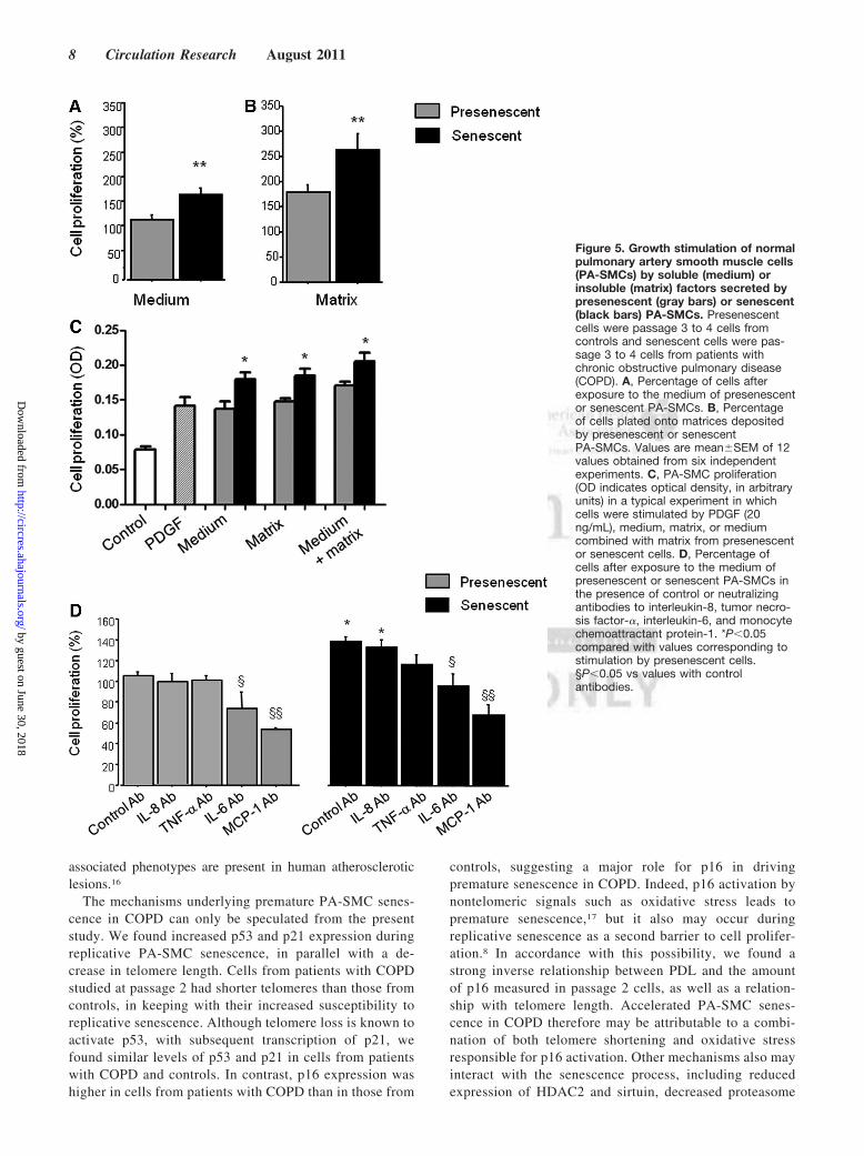
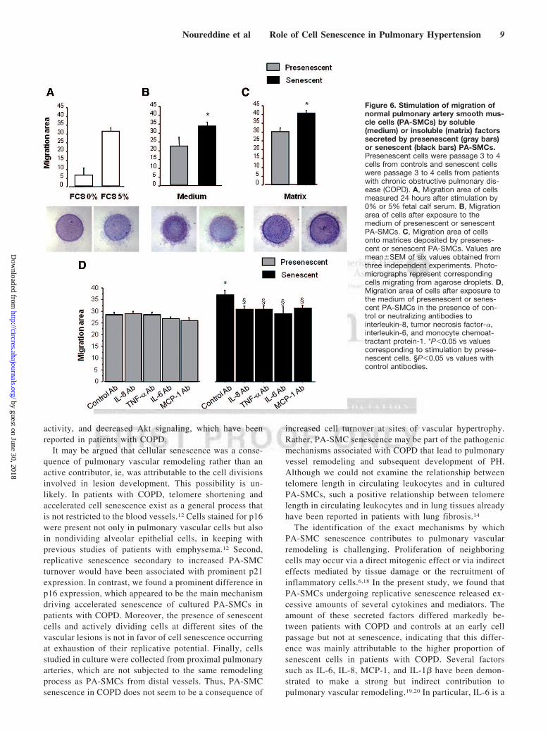
Contribution of Secreted Soluble and InsolubleFactors to PA-SMC Proliferation and MigrationTo investigate whether secretion of soluble factors by senes-cent PA-SMCs affected the function of the target PA-SMCs,we evaluated the proliferation and migration of nonsenescentPA-SMCs treated with media from presenescent and senes-cent PA-SMCs. Growth stimulation of target PA-SMCs wasmore marked with medium of senescent PA-SMCs than withmedium of presenescent cells (Figure 5A, C). Similarly, themedium of senescent PA-SMCs was more potent in stimu-
Figure 2. Immunolocalization of p16-positve, p21-positive, and Ki67-positive cells and collagen accumula-tion visualized by Masson trichromestaining in sections of remodeled pul-monary vessels from patients withchronic obstructive pulmonary dis-ease. Senescent pulmonary arterysmooth muscle cells (PA-SMCs) stainedfor p16 and p21 were virtually confinedto the media, whereas Ki67-stained cellspredominated in the neointima andhypertrophied media. Some p16 andp21 immunostaining was also associatedwith endothelial cells or PA-SMCs nearthe vessel lumen. In Masson trichrome-stained sections, the collagen is blueand the embedded cells are red.
Noureddine et al Role of Cell Senescence in Pulmonary Hypertension 5
by guest on June 30, 2018http://circres.ahajournals.org/
Dow
nloaded from

lating PA-SMC migration than was the medium of presenes-cent cells (Figure 6A, B). Neutralizing antibodies to IL-6 andMCP-1, but not to IL-8 and TNF-�, markedly reducedPA-SMC proliferation induced by PA-SMC culture media(Figure 5D). The stimulatory effects of PA-SMC media fromsenescent and presenescent cells were no longer significantlydifferent in the presence of anti-MCP-1 antibodies or IL-6antibodies (Figure 5D). In contrast, neutralizing antibodies toIL-8, TNF-�, IL-6, or MCP-1 did not affect PA-SMCmigration in response to culture media from nonsenescentcells, whereas they inhibited PA-SMC migration induced byculture media from senescent cells by �25% (Figure 6D).Thus, neutralizing antibodies to IL-8, TNF-�, IL-6, and-MCP-1 abolished the differences in PA-SMC migration inresponse to culture media from senescent vs presenescentcells.
To determine the contribution of secreted matrices orinsoluble factors, we allowed senescent or nonsenescentPA-SMCs to deposit extracellular matrix onto culturedishes for 3 days, after which we removed the cells withoutaltering the matrix and introduced new healthy PA-SMCs,which were assessed for growth or migration. We foundthat target PA-SMCs exhibited faster growth (Figure 5B,
C) and greater migration (Figure 6C) in dishes coated withmatrices secreted by senescent cells compared to nonse-nescent cells.
DiscussionWe show here that PA-SMC senescence is involved in theprocess of pulmonary vessel remodeling that underlies PH inpatients with COPD. After finding a positive correlationbetween telomere shortening and PH severity in a largepopulation of patients with COPD, we investigated thepulmonary vessels and derived cultured PA-SMCs frompatients with or without COPD. We found that remodeledvessels were characterized by PA-SMC senescence and thatcultured PA-SMCs from patients with COPD displayedaccelerated senescence. Our finding that in situ and in vitrocriteria for PA-SMC senescence correlated closely with theseverity of pulmonary vascular remodeling, together with thepresence of senescent cells near actively dividing cells at sitesof vessel wall hypertrophy, strongly suggests a role forsenescent cells in the remodeling process. Moreover, wefound that accelerated PA-SMC senescence in COPD wasassociated with increased expression of soluble and insolublefactors that affected PA-SMC migration and proliferation.
Figure 3. A, Replicative senescence ofpulmonary artery smooth muscle cells(PA-SMCs) from patients with chronicobstructive pulmonary disease (COPD)and controls. Cells were subjected torepeated passages and counted at eachpassage, and the population doublinglevel (PDL) was calculated for patientswith COPD and controls. B, Data aremeans�SEM. *P�0.01 vs controls. C,PA-SMCs were stained for senescence-associated �-galactosidase (�-gal) activ-ity at passage 2 and at senescencewhen cells began to exhibit proliferativearrest. C, Representative photographs ofcells stained for senescence-associated� -gal activity at passage 2 and atsenescence. D, Percentage of �-gal–positive cells. Data are means�SEM.*P�0.01 vs controls. §P�0.05 vs corre-sponding values at passage 2. E, Corre-lations between the vascular wall arearatio and the PDL (r��0.61; P�0.001).
6 Circulation Research August 2011
by guest on June 30, 2018http://circres.ahajournals.org/
Dow
nloaded from

Taken together, these results support a role for PA-SMCsenescence in the process of pulmonary vascular remodelingin COPD.
The role for telomere shortening as a pathogenic mecha-nism was recently highlighted in patients with familialidiopathic fibrosis who harbored a mutation in the telomerasegene.14 Both COPD and pulmonary fibrosis are age-relateddiseases associated with telomere shortening.5,14 Becauseshort telomeres are associated with increased susceptibility toreplicative cellular senescence, one current hypothesis is thatcellular senescence represents one mechanism underlying thepathological alterations seen in these chronic lung diseases.Here, we focused on the process of pulmonary vascularremodeling that underlies PH in patients with COPD. In apopulation of patients with COPD investigated by right heartcatheterization, we found that telomere shortening was asso-
ciated with PH severity independently from the severity ofairflow obstruction, age, and smoking history. To evaluatewhether cellular senescence was present in pulmonary vesselsfrom patients with COPD and reflected a process related topulmonary vascular remodeling, we compared pulmonaryvessels and derived cultured PA-SMCs from patients withCOPD and from sex-matched and age-matched control sub-jects who smoke. Pulmonary vessels from patients withCOPD were characterized by increased wall hypertrophycompared to controls, in keeping with the higher Pap in thepatients with COPD than in the controls. Immunohistochem-ical examination of pulmonary vessels revealed an increasedpercentage of pulmonary vascular cells stained for p21 andp16, including endothelial and smooth muscle cells, inpatients with COPD compared to controls. These cells wereidentified as senescent cells by experiments performed in theproximal pulmonary arteries, in which �-gal–positive cellswere also positive for p16 and p21. We then evaluatedwhether PA-SMCs derived from pulmonary vessels alsoexhibited characteristic features of accelerated senescencewhen studied in vitro. Cultured PA-SMCs from patients withCOPD exhibited premature senescence when compared tothose of controls, with a marked decrease in cumulative PDLand a higher percentage of �-gal–positive cells measured atan early passage.
In studies of pulmonary vessels and derived culturedPA-SMCs from patients with COPD and from sex-matchedand age-matched control subjects who smoke, we foundthat in situ and in vitro criteria for cell senescencecorrelated with the severity of pulmonary vascular wallhypertrophy, suggesting a close relationship between cellsenescence and the pulmonary vascular remodeling pro-cess. The large proportion of senescent cells within thewalls of remodeled pulmonary vessels may seem paradox-ical, because PH is primarily a proliferative disorder andcell senescence is associated with impaired regenerativecapacity in a given tissue. We investigated PA-SMCproliferation in vessels from our patients and found thatremodeled vessels from patients with COPD containedmore proliferating Ki67-stained PA-SMCs and more accu-mulated extracellular matrix than those from controls.Thus, remodeled vessels from patients with COPD wereparadoxically characterized by a combination of elevatedsenescent cell counts with an increased proportion ofproliferating cells and increased extracellular matrix depo-sition. Of note, studies of remodeled vessels at sites ofvascular hypertrophy revealed senescent cells to be virtu-ally confined to the media, with only a few senescent cellsin the neointima, whereas proliferating cells predominatedin the neointima and hypertrophied media. Our resultstherefore support the concept that several PA-SMC subsetsare present in the pulmonary vascular wall of remodeledvessels in COPD and that these subsets work in combina-tion to participate in the remodeling process. Similarresults have been reported in atherosclerotic lesions char-acterized by senescent cells and showing presence in theneointima of actively dividing cells, possibly of monoclonalorigin.11,15 Similarly, endothelial cells with senescence-
Figure 4. Levels of interleukinL-6, interleukin-8, tumornecrosis factor-�, monocyte chemoattractant protein-1, andtransforming growth factor-� in pulmonary artery smoothmuscle cell (PA-SMC) media from the 14 patients withchronic obstructive pulmonary disease (COPD) and 13 con-trols collected at passage 2 and at senescence. Each bar isthe mean�SEM. *P�0.01 compared with values for PA-SMCsfrom controls. §P�0.05 compared with corresponding values forPA-SMCs at passage 2.
Noureddine et al Role of Cell Senescence in Pulmonary Hypertension 7
by guest on June 30, 2018http://circres.ahajournals.org/
Dow
nloaded from
associated phenotypes are present in human atheroscleroticlesions.16
The mechanisms underlying premature PA-SMC senes-cence in COPD can only be speculated from the presentstudy. We found increased p53 and p21 expression duringreplicative PA-SMC senescence, in parallel with a de-crease in telomere length. Cells from patients with COPDstudied at passage 2 had shorter telomeres than those fromcontrols, in keeping with their increased susceptibility toreplicative senescence. Although telomere loss is known toactivate p53, with subsequent transcription of p21, wefound similar levels of p53 and p21 in cells from patientswith COPD and controls. In contrast, p16 expression washigher in cells from patients with COPD than in those from
controls, suggesting a major role for p16 in drivingpremature senescence in COPD. Indeed, p16 activation bynontelomeric signals such as oxidative stress leads topremature senescence,17 but it also may occur duringreplicative senescence as a second barrier to cell prolifer-ation.8 In accordance with this possibility, we found astrong inverse relationship between PDL and the amountof p16 measured in passage 2 cells, as well as a relation-ship with telomere length. Accelerated PA-SMC senes-cence in COPD therefore may be attributable to a combi-nation of both telomere shortening and oxidative stressresponsible for p16 activation. Other mechanisms also mayinteract with the senescence process, including reducedexpression of HDAC2 and sirtuin, decreased proteasome
Figure 5. Growth stimulation of normalpulmonary artery smooth muscle cells(PA-SMCs) by soluble (medium) orinsoluble (matrix) factors secreted bypresenescent (gray bars) or senescent(black bars) PA-SMCs. Presenescentcells were passage 3 to 4 cells fromcontrols and senescent cells were pas-sage 3 to 4 cells from patients withchronic obstructive pulmonary disease(COPD). A, Percentage of cells afterexposure to the medium of presenescentor senescent PA-SMCs. B, Percentageof cells plated onto matrices depositedby presenescent or senescentPA-SMCs. Values are mean�SEM of 12values obtained from six independentexperiments. C, PA-SMC proliferation(OD indicates optical density, in arbitraryunits) in a typical experiment in whichcells were stimulated by PDGF (20ng/mL), medium, matrix, or mediumcombined with matrix from presenescentor senescent cells. D, Percentage ofcells after exposure to the medium ofpresenescent or senescent PA-SMCs inthe presence of control or neutralizingantibodies to interleukin-8, tumor necro-sis factor-�, interleukin-6, and monocytechemoattractant protein-1. *P�0.05compared with values corresponding tostimulation by presenescent cells.§P�0.05 vs values with controlantibodies.
8 Circulation Research August 2011
by guest on June 30, 2018http://circres.ahajournals.org/
Dow
nloaded from
activity, and decreased Akt signaling, which have beenreported in patients with COPD.
It may be argued that cellular senescence was a conse-quence of pulmonary vascular remodeling rather than anactive contributor, ie, was attributable to the cell divisionsinvolved in lesion development. This possibility is un-likely. In patients with COPD, telomere shortening andaccelerated cell senescence exist as a general process thatis not restricted to the blood vessels.12 Cells stained for p16were present not only in pulmonary vascular cells but alsoin nondividing alveolar epithelial cells, in keeping withprevious studies of patients with emphysema.12 Second,replicative senescence secondary to increased PA-SMCturnover would have been associated with prominent p21expression. In contrast, we found a prominent difference inp16 expression, which appeared to be the main mechanismdriving accelerated senescence of cultured PA-SMCs inpatients with COPD. Moreover, the presence of senescentcells and actively dividing cells at different sites of thevascular lesions is not in favor of cell senescence occurringat exhaustion of their replicative potential. Finally, cellsstudied in culture were collected from proximal pulmonaryarteries, which are not subjected to the same remodelingprocess as PA-SMCs from distal vessels. Thus, PA-SMCsenescence in COPD does not seem to be a consequence of
increased cell turnover at sites of vascular hypertrophy.Rather, PA-SMC senescence may be part of the pathogenicmechanisms associated with COPD that lead to pulmonaryvessel remodeling and subsequent development of PH.Although we could not examine the relationship betweentelomere length in circulating leukocytes and in culturedPA-SMCs, such a positive relationship between telomerelength in circulating leukocytes and in lung tissues alreadyhave been reported in patients with lung fibrosis.14
The identification of the exact mechanisms by whichPA-SMC senescence contributes to pulmonary vascularremodeling is challenging. Proliferation of neighboringcells may occur via a direct mitogenic effect or via indirecteffects mediated by tissue damage or the recruitment ofinflammatory cells.6,18 In the present study, we found thatPA-SMCs undergoing replicative senescence released ex-cessive amounts of several cytokines and mediators. Theamount of these secreted factors differed markedly be-tween patients with COPD and controls at an early cellpassage but not at senescence, indicating that this differ-ence was mainly attributable to the higher proportion ofsenescent cells in patients with COPD. Several factorssuch as IL-6, IL-8, MCP-1, and IL-1� have been demon-strated to make a strong but indirect contribution topulmonary vascular remodeling.19,20 In particular, IL-6 is a
Figure 6. Stimulation of migration ofnormal pulmonary artery smooth mus-cle cells (PA-SMCs) by soluble(medium) or insoluble (matrix) factorssecreted by presenescent (gray bars)or senescent (black bars) PA-SMCs.Presenescent cells were passage 3 to 4cells from controls and senescent cellswere passage 3 to 4 cells from patientswith chronic obstructive pulmonary dis-ease (COPD). A, Migration area of cellsmeasured 24 hours after stimulation by0% or 5% fetal calf serum. B, Migrationarea of cells after exposure to themedium of presenescent or senescentPA-SMCs. C, Migration area of cellsonto matrices deposited by presenes-cent or senescent PA-SMCs. Values aremean�SEM of six values obtained fromthree independent experiments. Photo-micrographs represent correspondingcells migrating from agarose droplets. D,Migration area of cells after exposure tothe medium of presenescent or senes-cent PA-SMCs in the presence of con-trol or neutralizing antibodies tointerleukin-8, tumor necrosis factor-�,interleukin-6, and monocyte chemoat-tractant protein-1. *P�0.05 vs valuescorresponding to stimulation by prese-nescent cells. §P�0.05 vs values withcontrol antibodies.
Noureddine et al Role of Cell Senescence in Pulmonary Hypertension 9
by guest on June 30, 2018http://circres.ahajournals.org/
Dow
nloaded from

major contributor to hypoxic PH and is closely linked toPH severity in patients with COPD13 or idiopathic PH.20
To better-investigate the potential interplay betweennonsenescent and senescent cells, we evaluated whethersenescent cells affected the migration and proliferation ofnonsenescent cells in a paracrine manner. We found thatsoluble and insoluble factors released by senescent cellsstimulated the growth and migration of target PA-SMCs. Asimilar finding was obtained previously using senescentfibroblasts and cultured epithelial cells and was taken asevidence that senescent cells promoted cell proliferationand tumor growth.18 This possibility is consistent with ourobservation that actively dividing cells in the neointimallesions of remodeled vessels were surrounded by senescentcells, suggesting cross-talk between the two cell subsets. Inour study, cell proliferation in response to culture mediafrom senescent cells were markedly reduced in the pres-ence of neutralizing antibodies to IL-6 and MCP-1, andcell migration was reduced in the presence of neutralizingantibodies to IL-8, TNF-�, IL-6, and MCP-1. The fact thatthe increased PA-SMC proliferation in response to culturemedia from senescent compared to presenescent cells wasno longer observed in the presence of anti-MCP-1 andanti-IL-6 antibodies suggests an important role for thesecytokines in this process.13,19,20 The fact that anti-IL-6 andanti-MCP-1 antibodies inhibited proliferation under bothbasal and stimulated conditions is also consistent with anautocrine effect of these mediators. Moreover, these resultssuggest that the global action of these soluble factors maybe stimulation of growth and migration and not stimulationof senescence. Thus, senescent cells may create a microen-vironment that facilitates the migration and growth ofnonsenescent cells, thereby inducing neointima formationand vessel remodeling. Whether nonsenescent cells in-volved in neointima formation exhibit a normal or anabnormal phenotype remains to be elucidated.
There are several important limitations to this study. First,cell senescence in COPD is probably a process applying tothe whole body, and it remains unclear whether the observa-tions described here are specific to the pulmonary circulationor apply also to other vascular beds. Second, because wecould not study a third group with severe PH or with otherforms of PH, we do not know whether our data are relevantto various types of PH or specific of diseases associated withtelomere dysfunction. Telomere shortening in circulatingleukocytes is found in patients with COPD and lung fibrosisbut not in patients with idiopathic PH.21 Whether telomereshortening may represent a biomarker of disease severity invarious types of PH also remains an open question. In patientswith COPD, telomere shortening may constitute a biomarkerof overall accelerated aging and, potentially, of its effects,including PH, cardiovascular disease, and cancer, which arethe main causes of morbidity and mortality in patients withCOPD.
Sources of FundingThis study was supported by grants from the INSERM, Delegation ala Recherche Clinique de l’AP-HP, Fondation pour la RechercheMedicale (FRM), and the Carvsen foundation.
DisclosuresNone.
References1. Celli BR, MacNee W, ATS/ERS Task Force. Standards for the diagnosis
and treatment of patients with COPD: a summary of the ATS/ERSposition paper. Eur Respir J. 2004;23:932–946.
2. Chaouat A, Bugnet AS, Kadaoui N, Schott R, Enache I, Ducolone A,Ehrhart M, Kessler R, Weitzenblum E. Severe pulmonary hypertensionand chronic obstructive pulmonary disease. Am J Respir Crit Care Med.2005;172:189–194.
3. Minai OA, Chaouat A, Adnot S. Pulmonary hypertension in COPD:epidemiology, significance, and management: pulmonary vascular dis-ease: the global perspective. Chest. 2010;137:39S–51S.
4. Santos S, Peinado VI, Ramírez J, Melgosa T, Roca J, Rodriguez-Roisin R, Barbera JA. Characterization of pulmonary vascular remod-elling in smokers and patients with mild COPD. Eur Respir J. 2002;19:632– 638.
5. Savale L, Chaouat A, Bastuji-Garin S, Marcos E, Boyer L, Maitre B,Sarni M, Housset B, Weitzenblum E, Matrat M, Le Corvoisier P,Rideau D, Boczkowski J, Dubois-Rande JL, Chouaid C, Adnot S.Shortened telomeres in circulating leukocytes of patients with chronicobstructive pulmonary disease. Am J Respir Crit Care Med. 2009;179:566 –571.
6. Campisi J. Senescent cells, tumor suppression, and organismal aging:good citizens, bad neighbors. Cell. 2005;120:513–522.
7. Mathon NF, Lloyd AC. Cell senescence and cancer. Nat Rev Cancer.2001;1:203–213.
8. Beausejour CM, Krtolica A, Galimi F, Narita M, Lowe SW, Yaswen P,Campisi J. Reversal of human cellular senescence: roles of the p53 andp16 pathways. EMBO J. 2003;22:4212–4222.
9. Kuilman T, Peeper DS. Senescence-messaging secretome: SMS-ingcellular stress. Nat Rev Cancer. 2009;9:81–94.
10. Coppe JP, Desprez PY, Krtolica A, Campisi J. The senescence-associatedsecretory phenotype: the dark side of tumor suppression. Annu RevPathol. 2010;5:99–118.
11. Matthews C, Gorenne I, Scott S, Figg N, Kirkpatrick P, Ritchie A,Goddard M, Bennett M. Vascular smooth muscle cells undergotelomere-based senescence in human atherosclerosis: effects oftelomerase and oxidative stress. Circ Res. 2006;99:156–164.
12. Tsuji T, Aoshiba K, Nagai A. Alveolar cell senescence in patients withpulmonary emphysema. Am J Respir Crit Care Med. 2006;174:886 – 893.
13. Chaouat A, Savale L, Chouaid C, Tu L, Sztrymf B, Canuet M, Maitre B,Housset B, Brandt C, Le Corvoisier P, Weitzenblum E, Eddahibi S, AdnotS. Role for interleukin-6 in COPD-related pulmonary hypertension.Chest. 2009;136:678–687.
14. Alder JK, Chen JJ, Lancaster L, Danoff S, Su SC, Cogan JD, Vulto I, XieM, Qi X, Tuder RM, Phillips JA III, Lansdorp PM, Loyd JE, ArmaniosMY. Short telomeres are a risk factor for idiopathic pulmonary fibrosis.Proc Natl Acad Sci U S A. 2008;105:13051–13056.
15. Murry CE, Gipaya CT, Bartosek T, Benditt EP, Schwartz SM. Monoclo-nality of smooth muscle cells in human atherosclerosis. Am J Pathol.1997;151:697–705.
16. Minamino T, Miyauchi H, Yoshida T, Ishida Y, Yoshida H, KomuroI. Endothelial cell senescence in human atherosclerosis: role oftelomere in endothelial dysfunction. Circulation. 2002;105:1541–1544.
17. Nyunoya T, Monick MM, Klingelhutz AL, Glaser H, Cagley JR,Brown CO, Matsumoto E, Aykin-Burns N, Spitz DR, Oshima J,Hunninghake GW. Cigarette smoke induces cellular senescence viaWerner’s syndrome protein down-regulation. Am J Respir Crit CareMed. 2009;179:279 –287.
18. Krtolica A, Parrinello S, Lockett S, Desprez PY, Campisi J. Senescentfibroblasts promote epithelial cell growth and tumorigenesis: a linkbetween cancer and aging. Proc Natl Acad Sci U S A. 2001;98:12072–12077.
19. Sanchez O, Marcos E, Perros F, Fadel E, Tu L, Humbert M, Dartevelle P,Simonneau G, Adnot S, Eddahibi S. Role of endothelium-derived CC
10 Circulation Research August 2011
by guest on June 30, 2018http://circres.ahajournals.org/
Dow
nloaded from

chemokine ligand 2 in idiopathic pulmonary arterial hypertension. Am JRespir Crit Care Med. 2007;176:1041–1047.
20. Soon E, Holmes AM, Treacy CM, Doughty NJ, Southgate L, MachadoRD, Trembath RC, Jennings S, Barker L, Nicklin P, Walker C, Budd DC,Pepke-Zaba J, Morrell NW. Elevated levels of inflammatory cytokines
predict survival in idiopathic and familial pulmonary arterial hyper-tension. Circulation. 2010;122(9):920–927.
21. Cronkhite JT, Xing C, Raghu G, Chin KM, Torres F, Rosenblatt RL,Garcia CK. Telomere shortening in familial and sporadic pulmonaryfibrosis. Am J Respir Crit Care Med. 2008;178(7):729–737.
Novelty and Significance
What Is Known?
● Chronic obstructive pulmonary disease (COPD) is an age-relateddisease that is among the most common causes of pulmonaryhypertension (PH).
● COPD is associated with telomere shortening, which causes cellsenescence.
● The role for telomere shortening as a pathogenic mechanism in COPDis unknown.
What New Information Does This Article Contribute?
● PH severity is related to telomere shortening in COPD, and remodeledpulmonary vessels are characterized by an increased number ofsenescent pulmonary artery smooth muscle cells (PA-SMCs).
● Senescent PA-SMCs produce soluble and insoluble paracrine factors,which stimulate the growth and migration of normal targetPA-SMCs, thereby contributing to pulmonary vessel remodeling.
COPD is increasingly prevalent in industrialized countries and isamong the most common causes of PH, a condition that has apoor prognosis. COPD is also an age-related disease associated
with telomere shortening in circulating leukocytes and alveolarcells, which causes cell senescence. In 124 patients investi-gated by right heart catheterization, we found that telomereshortening in PA-SMCs was associated with PH severity. Inves-tigations of lung vessels and derived cultured PA-SMCs frompatients with COPD and age-matched and sex-matched controlsubjects who smoke showed increased senescent cell counts inremodeled COPD vessels. Accelerated senescence was inproportion to remodeling severity in derived cultured PA-SMCs.Senescent PA-SMCs were located near actively dividing cells atsites of vessel wall hypertrophy. The senescent cells wereshown in vitro to overexpress soluble and insoluble factors thataffected PA-SMC migration and proliferation. These resultssupport the concept that PA-SMC senescence is a pathogenicmechanism of pulmonary vascular remodeling and PH. Knowl-edge of this new pathophysiological pathway might help toidentify new biomarkers of disease severity and prognosis. Itmay open new therapeutic possibilities targeting cell senes-cence and, potentially, its effects, including PH, which is themain causes of morbidity and mortality in COPD.
Noureddine et al Role of Cell Senescence in Pulmonary Hypertension 11
by guest on June 30, 2018http://circres.ahajournals.org/
Dow
nloaded from
Maitre, A. Chaouat, C. Chouaid, J. L. Dubois-Rande, D. Damotte and S. AdnotH. Noureddine, G. Gary-Bobo, M. Alifano, E. Marcos, M. Saker, N. Vienney, V. Amsellem, B.
Pulmonary Hypertension in Chronic Lung DiseasePulmonary Artery Smooth Muscle Cell Senescence Is a Pathogenic Mechanism for
Print ISSN: 0009-7330. Online ISSN: 1524-4571 Copyright © 2011 American Heart Association, Inc. All rights reserved.is published by the American Heart Association, 7272 Greenville Avenue, Dallas, TX 75231Circulation Research
published online June 30, 2011;Circ Res.
http://circres.ahajournals.org/content/early/2011/06/29/CIRCRESAHA.111.241299World Wide Web at:
The online version of this article, along with updated information and services, is located on the
http://circres.ahajournals.org/content/suppl/2011/06/30/CIRCRESAHA.111.241299.DC1Data Supplement (unedited) at:
http://circres.ahajournals.org//subscriptions/
is online at: Circulation Research Information about subscribing to Subscriptions:
http://www.lww.com/reprints Information about reprints can be found online at: Reprints:
document. Permissions and Rights Question and Answer about this process is available in the
located, click Request Permissions in the middle column of the Web page under Services. Further informationEditorial Office. Once the online version of the published article for which permission is being requested is
can be obtained via RightsLink, a service of the Copyright Clearance Center, not theCirculation Researchin Requests for permissions to reproduce figures, tables, or portions of articles originally publishedPermissions:
by guest on June 30, 2018http://circres.ahajournals.org/
Dow
nloaded from

CIRCRESAHA/2011/241299/R2- 1
SUPPLEMENTAL MATERIAL
Pulmonary Artery Smooth Muscle Cell Senescence Is a Pathogenic Mechanism for Pulmonary
Hypertension in Chronic Lung Disease
Noureddine H1, Gary-Bobo G
1, Alifano M
2, Marcos E
1, Saker M
1, Vienney N
1,
Amsellem V1, Maitre B
1, Chaouat A
3, Chouaid C
4, Dubois-Rande JL
1, Damotte D
2, Adnot S
1
Role of Cell Senescence in Pulmonary Hypertension

CIRCRESAHA/2011/241299/R2- 2
DETAILED METHODS
Study population
We evaluated two groups of patients. The first group consisted of 124 patients with COPD who underwent
right heart catheterization and telomere length measurement. The data from 91 of these patients in whom
inflammatory biomarkers were assayed have been published previously 1 (Table 1). The inclusion criteria
for the patients in this cohort were a history of smoking, an FEV1/FVC ratio <70%, and an arterial partial
pressure of oxygen (PaO2) <80 mm Hg. About 50 of these patients were included in a previously
published prospective study comparing telomere length in patients with COPD and controls 2. Right atrial
pressure, pulmonary artery pressures (systolic, diastolic, and mean), and pulmonary wedge pressures were
measured. Cardiac output (CO) was determined using thermodilution. Derived hemodynamic variables
were calculated using standard formulas: cardiac index (cardiac output/body surface area, CI) as L·min-
1·m
2 and pulmonary vascular resistance (PVR in Wood Units) as mean pulmonary artery pressure (Pap) –
pulmonary wedge pressure /CO.
The second group consisted of 27 patients treated with lung resection surgery and recruited prospectively
at the Hotel-Dieu Teaching Hospital (Paris, France), including 14 with COPD and 13 defined as controls
(Table 2). Most of these patients underwent lobectomy or pneumonectomy for localized lung tumors. In
this group, lung tissue samples and derived cell cultures were studied; pulmonary arteries for the study
were taken at a distance from tumor areas. Systolic pulmonary artery pressure was assessed using
echocardiography. Inclusion criteria for COPD were an at least 10-pack-year history of tobacco smoking
and a forced expiratory volume in 1 second (FEV1)/forced vital capacity (FVC) ratio <70%. Inclusion
criteria for the control smokers were a smoking history greater than 10 pack-years, an FEV1/FVC ratio
greater than 70%, and the absence of chronic cardiovascular, hepatic, and renal disease. None of these
patients had received chemotherapy. This study was approved by the institutional review board of the
Henri Mondor Teaching Hospital. All patients and controls signed an informed consent document before
study inclusion.
Assessment of pulmonary vascular remodeling
The morphologic characteristics of the pulmonary muscular arteries were analyzed in lung tissue sections
stained with hematoxylin-phloxin-saffron 3. Arteries (20 to 30 for each lung) with an external diameter
between 100 to 500 m and complete elastic laminas were evaluated. The areas occupied by the muscular
and intimal layers were analyzed using image J software (http://rsbweb.nih.gov/ij/). Masson trichrome
staining was also performed to identify the extracellular matrix in the vessel wall and quantified using
ImageJ (http://rsbweb.nih.gov) software as the percentage of the wall surface area.
Immunohistochemistry
Paraffin-embedded sections were deparaffinized using xylene and a graded series of ethanol dilutions
then incubated in citrate buffer (0.01 M, pH 6) at 90°C for 20 minutes. Endogenous peroxidase activity
was blocked with 3% H2O2 and 10% methanol in phosphate-buffered saline (PBS) for 10 minutes. Slides
were incubated for 60 minutes in 1% bovine serum albumin and 5% goat serum in PBS then incubated
overnight with anti-p21 mouse antibody (1:50, Cell signaling, Boston MA, USA), anti-p16 mouse
antibody (1:1000, Abcam, Cambridge, MA, USA), and anti-Ki67 rabbit antibody (1:500, Abcam,
Cambridge, MA, USA). We used the ABC Vectastain kit (Vector Labs, Burlingame, CA, USA) to mark
the primary antibodies according to the user’s guide. The staining substrate was DAB (FastDAB, Sigma-
Aldrich, St Louis, MO, USA) and the sections were counterstained with hematoxylin 4.
Paraffin-embedded sections were deparaffinized using xylene and a graded series of ethanol dilutions then
incubated in citrate buffer (0.01 M, pH 6) at 90°C for 20 minutes. Tissue was permeabilized with 0.1%
Triton X-100 in PBS for 10 minutes. Endogenous peroxidase activity was blocked with 3% H2O2 and 10%

CIRCRESAHA/2011/241299/R2- 3
methanol in PBS for 10 minutes. Slides were incubated for 60 minutes in 1% bovine serum albumin and
5% goat serum in PBS. We performed double-label immunohistochemistry in two steps. In step one, the
slides were incubated overnight with anti-p21 mouse antibody (1:50, Cell signaling, Boston MA, USA),
anti-p16 mouse antibody (1:1000, Abcam, Cambridge, MA, USA), or anti-Ki67 rabbit antibody (1:500,
Abcam, Cambridge, MA, USA). We used the ABC Vectastain kit (Vector Labs, Burlingame, CA, USA)
to label the primary antibodies according to the user’s guide. The staining substrate was DAB (FastDAB,
Sigma-Aldrich, St Louis, MO, USA). In step two, slides were incubated for 1 hour with primary
antibodies against smooth muscle actin (SMA) (1:600, Sigma-Aldrich, St Louis, MO, USA) or von
Willebrand Factor (1:1000, Abcam Cambridge, MA, USA). Secondary antibodies were anti-rabbit (1:300,
Dako, Glostrup, Denmark) or anti-mouse (1:300, Dako) coupled to horseradish peroxidase. The staining
substrate was histogreen (Abcys, Paris, France) and the sections were counterstained with hematoxylin 4.
Culture of pulmonary artery smooth muscle cells
PA-SMCs were cultured from explants as previously described 5. To determine the phenotypic
characteristics of cultured PA-SMCs, we assessed the cells from each culture for expression of muscle-
specific contractile and cytoskeletal proteins, including smooth muscle cell -actin and desmin.
Cell replication
After cell outgrowth from the explants, cells were passaged (passage 1), seeded in 25-cm2 flasks, and
cultured to confluence. The cells were then counted and seeded (passage 2) in 75-cm2 culture flasks. The
experiments started at this point and the cells were serially passaged until senescence. The onset of cell
replicative senescence was defined based on cessation of cell division, labeling for SA-beta galactosidase
(-Gal), and cell morphology criteria. At each passage, cells were harvested for quantification of DNA
(telomere length measurement), RNA (real-time RT-PCR), and protein (Western blotting). Cells were also
counted using a hemocytometer, and population-doubling levels (PDL) were calculated as (log10Y –
log10X)/log102, where X is the initial number of seeded cells and Y the final number.
Senescence associated β-galactosidase staining
At each passage, cells were washed twice in PBS, fixed for 10 minutes at 37°C in 4% paraformaldehyde,
washed twice in PBS, and incubated for 24 h at 37°C in SA-βGal staining solution (1 mg/ml X-Gal, 5
mmol/L potassium ferrocyanide, 5 mmol/L potassium ferricyanide, 150 mmol/L NaCl, 2 mmol/L MgCl2,
and 40 mmol/L citrate [titrated to pH 6.0 with NaH2PO4]).
Protein extraction and immunoblotting
For protein extraction, isolated PA-SMCs were washed with PBS and lyzed with RIPA lysis buffer. Base
ingredients (10 mM sodium phosphate, pH 8; 150 mM NaCl; 0.5% SDS; 1% Na-deoxycholate; and 1%
NP40) supplemented with 1 mM phenylmethylsulfonyl fluoride (PMSF), 1 mM NaF, sodium
orthovanadate, and cocktail inhibitors (1/100) were from Sigma Aldrich. For protein analysis using
Western blotting, samples were subjected to electrophoresis in 10% or 15 % polyacrylamide gels under
reducing conditions. After electrophoresis, proteins from the gel were electroblotted onto polyvinylidene
difluoride membranes (Millipore, Molsheim, France) for 2 h. After transfer, the membrane was saturated
with PBS/5% milk. The membrane was then incubated overnight at 4 °C with the appropriate antibodies:
rabbit polyclonal anti-P-p53 (Ser15) antibody was used at 1:1000 dilution (Cell Signaling Technology,
Boston, MA, USA), monoclonal anti-p21Waf1/Cip1 (DCS60) antibody was used at 1:2000 dilution (Cell
Signaling Technology), monoclonal anti-p16 (F-12) (sc-1661) antibody was used at 1:500 dilution (Santa
Cruz Biotechnology, Inc., Santa Cruz, CA, USA), and anti–-actin antibody (Sigma, Saint-Quentin-
Fallavier, France) was used at 1:5000 dilution. Protein expression was reported as the protein/ -actin ratio
and expressed as arbitrary units.

CIRCRESAHA/2011/241299/R2- 4
Measurements of soluble factors by ELISA
Soluble factors were measured in plasma and cell medium. For cell medium determinations, PA-SMCs
from early and late passages were grown to confluence in DMEM containing 15% fetal calf serum (FCS).
The medium was then removed and the cells subjected to growth arrest in medium containing no FCS.
After 48 hours of incubation, the conditioned medium was used for quantitation of IL-6, IL-8, MCP-1,
TNF-, IL-1 and TGF- using Quantikine ELISA kits (R&D systems, Minneapolis, MN, USA)
according to the manufacturer’s instructions. The cells were washed twice with PBS, trypsinized and
counted.
Telomere length assay
Telomere length was assessed using a real-time quantitative polymerase chain reaction (PCR)-
based assay 6. Briefly, the ratio of the telomere repeat copy number over the single-gene copy number
(T/S) was determined using an Applied Biosystems 7900HT thermocycler in a 384-well format, using the
comparative Ct method (T/S = 2 -Ct
). Genomic DNA was extracted from smooth muscle cells using the
QIAamp DNA Kit (Qiagen, Courtaboeuf, France) and quantified using a spectrophotometer. Each sample
was run in triplicate, using the SYBR Green method (Invitrogen, Cergy-Pontoise, France) and 30 ng of
DNA. The sequences and final concentrations of the primers for the telomere and 36B4 (acidic ribosomal
phosphoprotein PO, a single-copy gene for normalization) were as follows: Tel F, 5’-
CGGTTTGTTTGGGTTTGGGTTTGGGTTTGGGTTTGGGTT-3’, 300 nM; Tel R, 5’-
GGCTTGCCTTACCCTTACCCTTACCCTTACCCTTACCCT-3’, 300 nM; 36B4F, 5’-
CAGCAAGTGGGAAGGTGTAATCC-3’,300 nM; and 36B4R, 5’-CCCATTCTATCATCAACGGGTA
CAA-3’, 300 nM. Telomere length is expressed as the ratio of the telomere repeat copy number over the
single-gene copy number (36B4 gene).
Extracellular matrix and soluble factors
After identification, presenescent (4·104) and senescent (6·10
4) PA-SMCs were allowed to attach to 12-
well culture dishes overnight and were incubated in serum and growth factor-free medium for 2 days to
generate similar cell numbers. The media from presenescent and senescent PA-SMCs incubated in serum
and growth factor-free medium for 2 days were used for soluble factor assays and for evaluations of
effects on growth or migration of normal target PA-SMCs previously attached to 12-well culture dishes.
To determine the contribution of secreted matrices, we allowed presenescent and senescent PA-SMCs in
serum-free medium to deposit extracellular matrix onto the culture dishes for 2 days. The cells were then
detached using 2 mM EDTA for 30 min at 37°C. Two brief washes with PBS were then used to remove
the cells. The target PA-SMCs used to study migration or proliferation were incubated in serum-free
medium for 2 days then re-suspended in complete medium and plated on PA-SMC-depleted dishes
prepared as described above.
Cell proliferation assay
Cell proliferation was assessed using the tetrazolium salt (MTT) assay (Sigma, Lyon, France). Briefly,
PA-SMCs were seeded onto 12-well culture dishes pre-coated with extracellular matrix prepared earlier or
in the presence of medium from presenescent or senescent cells or in the presence of 20 ng/mL platelet-
derived growth factor (PDGF). After 72 h, the medium was changed to the same medium supplemented
with 100 g/mL of MTT, and the cells were incubated for 4 h. After washing with saline, 1 ml of
dimethylsulfoxide (DMSO, Sigma)was added and the mixture was shaken for a few minutes to achieve
complete dissolution. Aliquots (200 L) of the resulting solutions were transferred to 96-well plates and
absorbance was recorded at 520 nm using the Microplate Spectrophotometer System.

CIRCRESAHA/2011/241299/R2- 5
Cell migration assay
The cell migration assay was performed as previously described 7. PA-SMCs were subjected to growth
arrest in medium containing no FCS for 48 or 72 h then resuspended at 30·106 cells/mL in culture medium
containing 15% FCS and 0.3% agarose. The cells were maintained at 37° C to prevent setting of the
agarose. Three-microliter drops of the cellular suspension were plated in the center of each well of a 24-
well tissue culture plate. Wells used for the migration assay were precoated with poly-DL-ornithine (0.5
g/ml; Sigma, St. Louis, MO, USA). The preparation was placed at 4°C for 20 min to allow the agarose to
gel. Then, 0.9 ml of medium was added to cover the drops. The preparation was incubated at 37°C in 5%
CO2 for 24 h. Samples were fixed and stained with Diff-Quik kit (Siemens Healthcare Diagnostics, Saint
Denis, France). Images were imported into ImageJ analysis software for calculation of cell migration
under each condition.
Statistical analysis
Data are expressed as mean±SEM. Patients with COPD and controls were compared using the unpaired t-
test for quantitative variables and the chi-square test for categorical variables. Correlations between
variables were evaluated using least-square linear regression techniques. The effects of senescence in cells
from patients with COPD and controls were assessed by using a paired t-test. P values less than 0.05 were
considered significant. Data were analyzed using Stata statistical software (release 8.0; StataCorp, College
Station, TX, USA).

CIRCRESAHA/2011/241299/R2- 6
Online Figure I: Correlations between telomere length, pulmonary artery pressure (r= -0.20, P<0.04) and pulmonary vascular resistance (r= -0.29,
P<0.01) in patients with COPD. Telomere length is expressed as the ratio of the telomere repeat copy number over the single-gene copy number
(36B4 gene).

CIRCRESAHA/2011/241299/R2- 7
Online Fig II: Comparison of pulmonary artery pressure, pulmonary capillary wedge pressure, pulmonary vascular resistance, and right atrial
pressure between patients dichotomized based on the median telomere length. Values are means±SEM. *P<0.05, **P<0.01 compared with values
from subjects with telomere lengths less than or equal to 0.6. Telomere length is expressed as the T/S ratio of the telomere repeat copy number
over the single-gene copy number (36B4 gene)..

CIRCRESAHA/2011/241299/R2- 8
Online Figure III. Telomere length and Western blotting analysis of p16, p53, and p21 protein levels in PA-SMCs from the 14 patients with
COPD and 13 controls determined at passage 2 and at senescence. Each bar is the mean±SEM. *P<0.01 compared with values for PA-SMCs from
controls. § P<0.05 compared with corresponding values for PA-SMCs at passage 2.

CIRCRESAHA/2011/241299/R2- 9
Supplemental references
1. Chaouat A, Savale L, Chouaid C, Tu L, Sztrymf B, Canuet M, Maitre B, Housset B, Brandt
C, Le Corvoisier P, Weitzenblum E, Eddahibi S, Adnot S. Role for Interleukin-6 in COPD-
Related Pulmonary Hypertension. Chest. 2009.
2. Savale L, Chaouat A, Bastuji-Garin S, Marcos E, Boyer L, Maitre B, Sarni M, Housset B,
Weitzenblum E, Matrat M, Le Corvoisier P, Rideau D, Boczkowski J, Dubois-Rande JL,
Chouaid C, Adnot S. Shortened telomeres in circulating leukocytes of patients with chronic
obstructive pulmonary disease. Am J Respir Crit Care Med. 2009;179:566-571.
3. Santos S, Peinado VI, Ramirez J, Melgosa T, Roca J, Rodriguez-Roisin R, Barbera JA.
Characterization of pulmonary vascular remodelling in smokers and patients with mild
COPD. Eur Respir J. 2002;19:632-638.
4. Gary-Bobo G, Houssaini A, Amsellem V, Rideau D, Pacaud P, Perrin A, Bregeon J, Marcos
E, Dubois-Rande JL, Sitbon O, Savale L, Adnot S. Effects of HIV protease inhibitors on
progression of monocrotaline- and hypoxia-induced pulmonary hypertension in rats.
Circulation. 2010;122:1937-1947.
5. Eddahibi S, Humbert M, Fadel E, Raffestin B, Darmon M, Capron F, Simonneau G,
Dartevelle P, Hamon M, Adnot S. Serotonin transporter overexpression is responsible for
pulmonary artery smooth muscle hyperplasia in primary pulmonary hypertension. J Clin
Invest. 2001;108:1141-1150.
6. Cawthon RM. Telomere measurement by quantitative PCR. Nucleic Acids Res.
2002;30:e47.
7. Pascaud M, Eddahibi S, Raffestin B, Yeh P, Griscelli F, Opolon P, Adnot S. Lung
overexpression of the anti-angiogenic factor angiostatin aggravates pulmonary hypertension
in hypoxic mice. American of Journal Respiratory and Critical Care Medicine.
2001;163:A119.