pvd imaging guidelines - med solutions · peripheral vascular disease (pvd) imaging guidelines pvd...
TRANSCRIPT
©2015 MedSolutions, Inc. PVD Imaging Guidelines
PVD IMAGING GUIDELINES Peripheral Vascular Disease (PVD)
Version 17.0; Effective 02-16-2015
MedSolutions, Inc. Clinical Decision Support Tool for Advanced Diagnostic Imaging
Common symptoms and symptom complexes are addressed by this tool. Imaging requests for patients with atypical symptoms or clinical presentations that are not specifically addressed will require physician review. Consultation with the
referring physician may provide additional insight.
This version incorporates MSI accepted revisions prior to 12/31/14
CPT® (Current Procedural Terminology) is a registered trademark of the American Medical Association (AMA). CPT® five digit codes, nomenclature and other data are copyright 2015 American Medical Association. All Rights Reserved. No fee schedules, basic units, relative values or related listings are included in the CPT® book. AMA does not directly or indirectly practice medicine or dispense medical services. AMA assumes no liability for the data contained herein or not contained herein.
MedSolutions, Inc. This tool addresses common symptoms and symptom complexes. Imaging requests for patients with atypicalClinical Decision Support Tool symptoms or clinical presentations that are not specifically addressed will require physician review. Diagnostic Strategies Consultation with the referring physician, specialist and/or patient’s Primary Care Physician (PCP) may provide additional insight.
Version 17.0; Effective 02-16-2015 PVD RETURN 2 of 24
PERIPHERAL VASCULAR DISEASE (PVD) IMAGING GUIDELINES
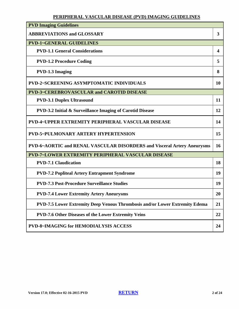
PVD Imaging Guidelines
ABBREVIATIONS and GLOSSARY 3
PVD-1~GENERAL GUIDELINES
PVD-1.1 General Considerations 4
PVD-1.2 Procedure Coding 5
PVD-1.3 Imaging 8
PVD-2~SCREENING ASYMPTOMATIC INDIVIDUALS 10
PVD-3~CEREBROVASCULAR and CAROTID DISEASE
PVD-3.1 Duplex Ultrasound 11
PVD-3.2 Initial & Surveillance Imaging of Carotid Disease 12
PVD-4~UPPER EXTREMITY PERIPHERAL VASCULAR DISEASE 14
PVD-5~PULMONARY ARTERY HYPERTENSION 15
PVD-6~AORTIC and RENAL VASCULAR DISORDERS and Visceral Artery Aneurysms 16
PVD-7~LOWER EXTREMITY PERIPHERAL VASCULAR DISEASE
PVD-7.1 Claudication 18
PVD-7.2 Popliteal Artery Entrapment Syndrome 19
PVD-7.3 Post-Procedure Surveillance Studies 19
PVD-7.4 Lower Extremity Artery Aneurysms 20
PVD-7.5 Lower Extremity Deep Venous Thrombosis and/or Lower Extremity Edema 21
PVD-7.6 Other Diseases of the Lower Extremity Veins 22
PVD-8~IMAGING for HEMODIALYSIS ACCESS 24
Version 17.0; Effective 02-16-2015 PVD RETURN 3 of 24
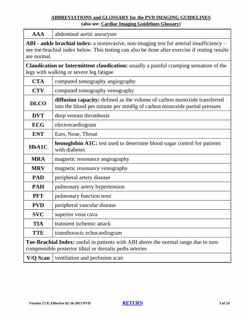
ABBREVIATIONS and GLOSSARY for the PVD IMAGING GUIDELINES (also see: Cardiac Imaging Guidelines Glossary)
AAA abdominal aortic aneurysm
ABI - ankle brachial index: a noninvasive, non-imaging test for arterial insufficiency – see toe-brachial index below. This testing can also be done after exercise if resting results are normal.
Claudication or Intermittent claudication: usually a painful cramping sensation of the legs with walking or severe leg fatigue
CTA computed tomography angiography
CTV computed tomography venography
DLCO diffusion capacity: defined as the volume of carbon monoxide transferred into the blood per minute per mmHg of carbon monoxide partial pressure
DVT deep venous thrombosis
ECG electrocardiogram
ENT Ears, Nose, Throat
HbA1C hemoglobin A1C: test used to determine blood sugar control for patients with diabetes
MRA magnetic resonance angiography
MRV magnetic resonance venography
PAD peripheral artery disease
PAH pulmonary artery hypertension
PFT pulmonary function tests
PVD peripheral vascular disease
SVC superior vena cava
TIA transient ischemic attack
TTE transthoracic echocardiogram
Toe-Brachial Index: useful in patients with ABI above the normal range due to non-compressible posterior tibial or dorsalis pedis arteries
V/Q Scan ventilation and perfusion scan
Version 17.0; Effective 02-16-2015 PVD RETURN 4 of 24
PERIPHERAL VASCULAR DISEASE (PVD) IMAGING GUIDELINES
PVD-1~GENERAL GUIDELINES
PVD-1.1 General Considerations
A current clinical evaluation (within 60 days), including medical treatments, are required prior to considering advanced imaging, which includes: o Relevant history and physical examination and appropriate laboratory studies and
non advanced imaging modalities, such as recent ABIs (within 60 days) after symptoms started or worsened Unless there is documented need for routine imaging that is supported by the
guidelines Other meaningful contact (telephone call, electronic mail or messaging) by an
established patient can substitute for a face-to-face clinical evaluation o The same general risk factors for as coronary disease also apply to vascular
disease. Diabetes is a particularly high risk factor < age 50 with at least one risk factor are considered “at risk” for vascular
disease Erectile dysfunction can be associated with vascular disease*
(See: PV-17~Impotence/Erectile Dysfunction in the Pelvis Imaging Guidelines).
o Simultaneous venous and arterial systems evaluation are unusual but are occasionally needed
o Post angioplasty/reconstruction: Follow-up imaging is principally guided by symptoms. Also see: AB-17~Abdominal Aortic Aneurysm (AAA) Follow Up and Pre-Operative
Evaluation in the Abdomen Imaging Guidelines
AB-18~Abdominal Aortic Aneurysm (AAA)-Post Endovascular or Open Aortic Repair in the Abdomen Imaging Guidelines
CH-30~Thoracic Aorta in the Chest Imaging Guidelines
PVD-7.3 Post-Procedure Surveillance Studies
Version 17.0; Effective 02-16-2015 PVD RETURN 5 of 24
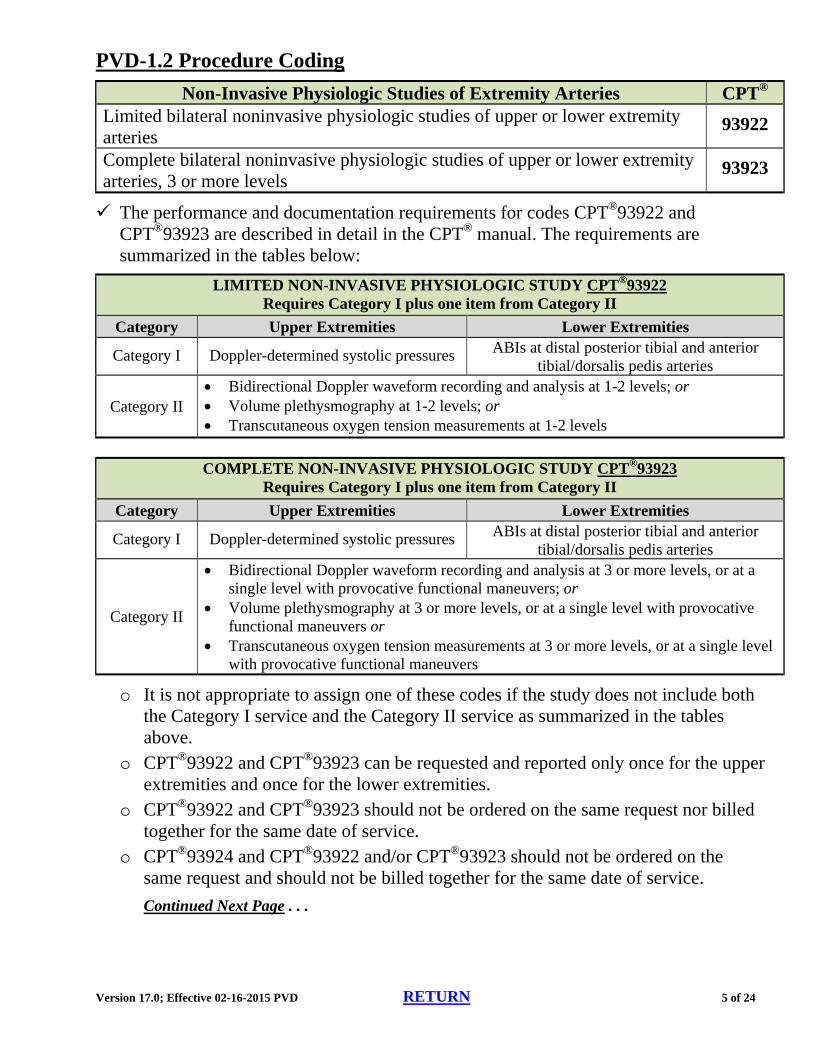
PVD-1.2 Procedure Coding
Non-Invasive Physiologic Studies of Extremity Arteries CPT® Limited bilateral noninvasive physiologic studies of upper or lower extremity arteries
93922
Complete bilateral noninvasive physiologic studies of upper or lower extremity arteries, 3 or more levels
93923
The performance and documentation requirements for codes CPT®93922 and CPT®93923 are described in detail in the CPT® manual. The requirements are summarized in the tables below:
LIMITED NON-INVASIVE PHYSIOLOGIC STUDY CPT®93922 Requires Category I plus one item from Category II
Category Upper Extremities Lower Extremities
Category I Doppler-determined systolic pressures ABIs at distal posterior tibial and anterior tibial/dorsalis pedis arteries
Category II Bidirectional Doppler waveform recording and analysis at 1-2 levels; or Volume plethysmography at 1-2 levels; or Transcutaneous oxygen tension measurements at 1-2 levels
COMPLETE NON-INVASIVE PHYSIOLOGIC STUDY CPT®93923
Requires Category I plus one item from Category II
Category Upper Extremities Lower Extremities
Category I Doppler-determined systolic pressures ABIs at distal posterior tibial and anterior tibial/dorsalis pedis arteries
Category II
Bidirectional Doppler waveform recording and analysis at 3 or more levels, or at a single level with provocative functional maneuvers; or
Volume plethysmography at 3 or more levels, or at a single level with provocative functional maneuvers or
Transcutaneous oxygen tension measurements at 3 or more levels, or at a single level with provocative functional maneuvers
o It is not appropriate to assign one of these codes if the study does not include both the Category I service and the Category II service as summarized in the tables above.
o CPT®93922 and CPT®93923 can be requested and reported only once for the upper extremities and once for the lower extremities.
o CPT®93922 and CPT®93923 should not be ordered on the same request nor billed together for the same date of service.
o CPT®93924 and CPT®93922 and/or CPT®93923 should not be ordered on the same request and should not be billed together for the same date of service.
Continued Next Page . . .
Version 17.0; Effective 02-16-2015 PVD RETURN 6 of 24
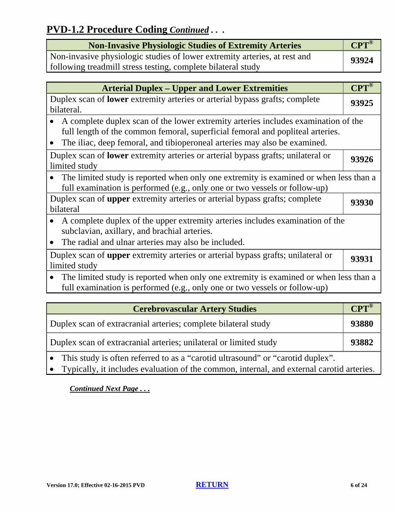
PVD-1.2 Procedure Coding Continued . . .
Non-Invasive Physiologic Studies of Extremity Arteries CPT® Non-invasive physiologic studies of lower extremity arteries, at rest and following treadmill stress testing, complete bilateral study
93924
Arterial Duplex – Upper and Lower Extremities CPT® Duplex scan of lower extremity arteries or arterial bypass grafts; complete bilateral.
93925
A complete duplex scan of the lower extremity arteries includes examination of the full length of the common femoral, superficial femoral and popliteal arteries.
The iliac, deep femoral, and tibioperoneal arteries may also be examined.
Duplex scan of lower extremity arteries or arterial bypass grafts; unilateral or limited study
93926
The limited study is reported when only one extremity is examined or when less than a full examination is performed (e.g., only one or two vessels or follow-up)
Duplex scan of upper extremity arteries or arterial bypass grafts; complete bilateral
93930
A complete duplex of the upper extremity arteries includes examination of the subclavian, axillary, and brachial arteries.
The radial and ulnar arteries may also be included.
Duplex scan of upper extremity arteries or arterial bypass grafts; unilateral or limited study
93931
The limited study is reported when only one extremity is examined or when less than a full examination is performed (e.g., only one or two vessels or follow-up)
Cerebrovascular Artery Studies CPT®
Duplex scan of extracranial arteries; complete bilateral study 93880
Duplex scan of extracranial arteries; unilateral or limited study 93882
This study is often referred to as a “carotid ultrasound” or “carotid duplex”. Typically, it includes evaluation of the common, internal, and external carotid arteries.
Continued Next Page . . .
Version 17.0; Effective 02-16-2015 PVD RETURN 7 of 24
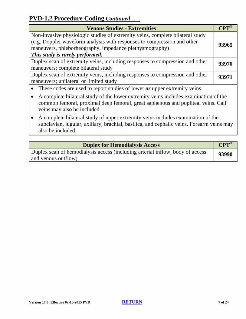
PVD-1.2 Procedure Coding Continued . . .
Venous Studies - Extremities CPT® Non-invasive physiologic studies of extremity veins, complete bilateral study (e.g. Doppler waveform analysis with responses to compression and other maneuvers, phleborheography, impedance plethysmography) This study is rarely performed.
93965
Duplex scan of extremity veins, including responses to compression and other maneuvers; complete bilateral study
93970
Duplex scan of extremity veins, including responses to compression and other maneuvers; unilateral or limited study
93971
These codes are used to report studies of lower or upper extremity veins.
A complete bilateral study of the lower extremity veins includes examination of the common femoral, proximal deep femoral, great saphenous and popliteal veins. Calf veins may also be included.
A complete bilateral study of upper extremity veins includes examination of the subclavian, jugular, axillary, brachial, basilica, and cephalic veins. Forearm veins may also be included.
Duplex for Hemodialysis Access CPT® Duplex scan of hemodialysis access (including arterial inflow, body of access and venous outflow)
93990
Version 17.0; Effective 02-16-2015 PVD RETURN 8 of 24
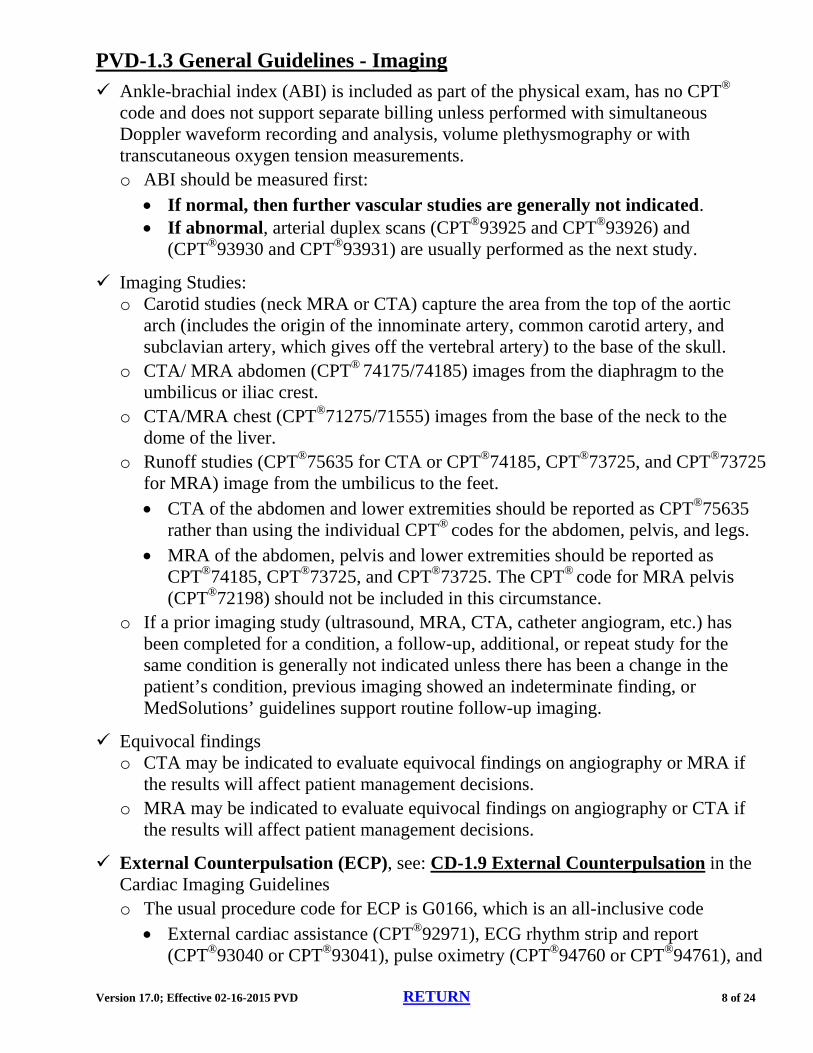
PVD-1.3 General Guidelines - Imaging
Ankle-brachial index (ABI) is included as part of the physical exam, has no CPT® code and does not support separate billing unless performed with simultaneous Doppler waveform recording and analysis, volume plethysmography or with transcutaneous oxygen tension measurements. o ABI should be measured first:
If normal, then further vascular studies are generally not indicated. If abnormal, arterial duplex scans (CPT®93925 and CPT®93926) and
(CPT®93930 and CPT®93931) are usually performed as the next study.
Imaging Studies: o Carotid studies (neck MRA or CTA) capture the area from the top of the aortic
arch (includes the origin of the innominate artery, common carotid artery, and subclavian artery, which gives off the vertebral artery) to the base of the skull.
o CTA/ MRA abdomen (CPT® 74175/74185) images from the diaphragm to the umbilicus or iliac crest.
o CTA/MRA chest (CPT®71275/71555) images from the base of the neck to the dome of the liver.
o Runoff studies (CPT®75635 for CTA or CPT®74185, CPT®73725, and CPT®73725 for MRA) image from the umbilicus to the feet. CTA of the abdomen and lower extremities should be reported as CPT®75635
rather than using the individual CPT® codes for the abdomen, pelvis, and legs. MRA of the abdomen, pelvis and lower extremities should be reported as
CPT®74185, CPT®73725, and CPT®73725. The CPT® code for MRA pelvis (CPT®72198) should not be included in this circumstance.
o If a prior imaging study (ultrasound, MRA, CTA, catheter angiogram, etc.) has been completed for a condition, a follow-up, additional, or repeat study for the same condition is generally not indicated unless there has been a change in the patient’s condition, previous imaging showed an indeterminate finding, or MedSolutions’ guidelines support routine follow-up imaging.
Equivocal findings o CTA may be indicated to evaluate equivocal findings on angiography or MRA if
the results will affect patient management decisions. o MRA may be indicated to evaluate equivocal findings on angiography or CTA if
the results will affect patient management decisions.
External Counterpulsation (ECP), see: CD-1.9 External Counterpulsation in the Cardiac Imaging Guidelines o The usual procedure code for ECP is G0166, which is an all-inclusive code
External cardiac assistance (CPT®92971), ECG rhythm strip and report (CPT®93040 or CPT®93041), pulse oximetry (CPT®94760 or CPT®94761), and
Version 17.0; Effective 02-16-2015 PVD RETURN 9 of 24
plethysmography (CPT®93922 or CPT®93923) should not be separately requested or billed with G0166.
MedSolutions does not currently prior authorize the G0166 code.
References
1. Min JK, Williams KA, Okwuosa TM, et al. Prediction of coronary heart disease by erectile dysfunction in men referred for nuclear stress testing. Arch Intern Med 2006; 166:201-206.
2. CPT® Assistant , June 2012 3. CPT® Assistant , September 2012
Version 17.0; Effective 02-16-2015 PVD RETURN 10 of 24
PERIPHERAL VASCULAR DISEASE (PVD) IMAGING GUIDELINES
PVD-2~Screening Asymptomatic Individuals
PVD-2.1 Screening
Patients without diabetes, SHOULD NOT undergo routine screening. The U.S. Preventive Services Task Force concludes that the harms of routine screening for exceed the benefits.
Patients with diabetes SHOULD undergo screening initially with ABI and limited Doppler US (CPT®93922). PAD is more than twice as common among diabetics, and many are asymptomatic and should undergo screening.
Duplex ultrasound can be performed (CPT®93925 bilateral study or CPT®93926 unilateral study) if ABI is abnormal. o Exercise ABI CPT®93924 may be requested after a normal resting ABI since it is
possible to have a normal or near normal ABI with significant PAD.
References
1. Perlstein TS, Creager MA. The Ankle-Brachial Index as a Biomarker of Cardiovascular Risk: It’s Not Just About the Legs. Circulation, 2009; 120: 2033-2035.
2. Beckman JA, Creager MA, Libby P. Diabetes and atherosclerosis: Epidemiology, pathophysiology, and management. JAMA, 2002; 287: 2570-2581.
3. Saydah SH, Fradkin J, Cowie C. Poor control of risk factors for vascular disease among adults with previously diagnosed diabetes. JAMA, 2004; 291: 335-342.
4. Hennion DR, Siano KA. Diagnosis and treatment of peripheral arterial disease. American Family Physician, 2013; 88: 306-310.
5. Hirsh AT, Criqui MH, Treat-Jacobson D, Regensteiner JG et al. Peripheral arterial disease detection, awareness, and treatment in primary care. JAMA, 2001; 286: 1317-1324.
Version 17.0; Effective 02-16-2015 PVD RETURN 11 of 24
PERIPHERAL VASCULAR DISEASE (PVD) IMAGING GUIDELINES
PVD-3~CEREBROVASCULAR and CAROTID DISEASE
PVD-3.1 Duplex Ultrasound
Duplex Ultrasound (CPT®93880 bilateral or CPT®93882 unilateral is the initial study to evaluate possible carotid artery disease, prior to considering advanced imaging. o If ultrasound shows > 50% occlusion/stenosis of the internal carotid artery, then
Neck MRA with contrast (CPT®70548) or neck CTA (CPT®70498) can be performed.
o Head MRA (CPT®70544) or head CTA (CPT®70496) can be added if carotid intervention is planned.
Other indications for Duplex ultrasound of the carotid arteries (CPT®93880 bilateral study or CPT®93882 unilateral study) as an initial study: o Hemispheric neurologic symptoms, including stroke, TIA, or amaurosis fugax o Non-hemispheric or unexplained neurologic symptoms o Known or suspected retinal arterial emboli o Suspected dissection o Pulsatile neck masses o Carotid bruit o Abnormal findings on physical exam of the carotid arteries (e.g. aneurysm or
absent carotid pulses) o Preoperative evaluation of patients with evidence of severe diffuse atherosclerosis,
scheduled for major cardiovascular surgical procedures o Preoperative evaluation of patients prior to elective coronary artery bypass graft
(CABG) surgery in patients older than 65 years of age and in those with left main coronary stenosis, peripheral artery disease, history of cigarette smoking, history of stroke or TIA, or carotid bruit.
o Suspected Subclavian Steal Syndrome (See: CH-28~Subclavian Steal Syndrome in the Chest Imaging Guidelines)
o Blunt neck trauma o Vasculitis involving carotid arteries o Surveillance imaging once a year for patients with fibromuscular dysplasia of the
extracranial carotid arteries
o Follow-up imaging of known carotid disease (See PVD-3.2)
Duplex Ultrasound – Practice Notes
Carotid intima-media thickness using duplex ultrasound imaging (Category III code 0126T). o Although, outcomes data are lacking, Texas has adopted this method in Texas
Heart Attack Preventive Screening Bill (HR 1290).
Version 17.0; Effective 02-16-2015 PVD RETURN 12 of 24
PVD-3.2 Initial & Surveillance Imaging of Carotid Disease
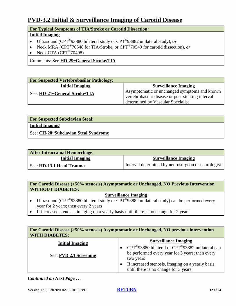
For Typical Symptoms of TIA/Stroke or Carotid Dissection: Initial Imaging
Ultrasound (CPT®93880 bilateral study or CPT®93882 unilateral study), or Neck MRA (CPT®70548 for TIA/Stroke, or CPT®70549 for carotid dissection), or Neck CTA (CPT®70498)
Comments: See HD-29~General Stroke/TIA
For Suspected Vertebrobasilar Pathology:
Initial Imaging
See: HD-21~General Stroke/TIA
Surveillance Imaging Asymptomatic or unchanged symptoms and known vertebrobasilar disease or post-stenting interval determined by Vascular Specialist
For Suspected Subclavian Steal: Initial Imaging
See: CH-28~Subclavian Steal Syndrome
After Intracranial Hemorrhage:
Initial Imaging
See: HD-13.1 Head Trauma
Surveillance Imaging
Interval determined by neurosurgeon or neurologist
For Carotid Disease (>50% stenosis) Asymptomatic or Unchanged, NO Previous Intervention WITHOUT DIABETES:
Surveillance Imaging Ultrasound (CPT®93880 bilateral study or CPT®93882 unilateral study) can be performed every
year for 2 years; then every 2 years If increased stenosis, imaging on a yearly basis until there is no change for 2 years.
For Carotid Disease (>50% stenosis) Asymptomatic or Unchanged, NO previous intervention WITH DIABETES:
Initial Imaging
See: PVD 2.1 Screening
Surveillance Imaging
CPT®93880 bilateral or CPT®93882 unilateral can be performed every year for 3 years; then every two years
If increased stenosis, imaging on a yearly basis until there is no change for 3 years.
Continued on Next Page . . .
Version 17.0; Effective 02-16-2015 PVD RETURN 13 of 24
PVD-3.2 Initial & Surveillance Imaging of Carotid Disease Continued . . .
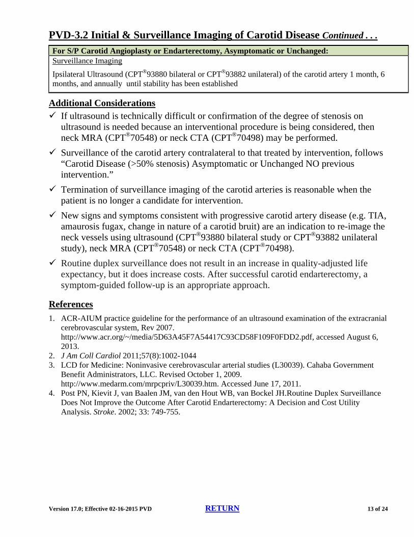
For S/P Carotid Angioplasty or Endarterectomy, Asymptomatic or Unchanged: Surveillance Imaging
Ipsilateral Ultrasound (CPT®93880 bilateral or CPT®93882 unilateral) of the carotid artery 1 month, 6 months, and annually until stability has been established
Additional Considerations If ultrasound is technically difficult or confirmation of the degree of stenosis on
ultrasound is needed because an interventional procedure is being considered, then neck MRA (CPT®70548) or neck CTA (CPT®70498) may be performed.
Surveillance of the carotid artery contralateral to that treated by intervention, follows “Carotid Disease (>50% stenosis) Asymptomatic or Unchanged NO previous intervention.”
Termination of surveillance imaging of the carotid arteries is reasonable when the patient is no longer a candidate for intervention.
New signs and symptoms consistent with progressive carotid artery disease (e.g. TIA, amaurosis fugax, change in nature of a carotid bruit) are an indication to re-image the neck vessels using ultrasound (CPT®93880 bilateral study or CPT®93882 unilateral study), neck MRA (CPT®70548) or neck CTA (CPT®70498).
Routine duplex surveillance does not result in an increase in quality-adjusted life expectancy, but it does increase costs. After successful carotid endarterectomy, a symptom-guided follow-up is an appropriate approach.
References
1. ACR-AIUM practice guideline for the performance of an ultrasound examination of the extracranial cerebrovascular system, Rev 2007. http://www.acr.org/~/media/5D63A45F7A54417C93CD58F109F0FDD2.pdf, accessed August 6, 2013.
2. J Am Coll Cardiol 2011;57(8):1002-1044 3. LCD for Medicine: Noninvasive cerebrovascular arterial studies (L30039). Cahaba Government
Benefit Administrators, LLC. Revised October 1, 2009. http://www.medarm.com/mrpcpriv/L30039.htm. Accessed June 17, 2011.
4. Post PN, Kievit J, van Baalen JM, van den Hout WB, van Bockel JH.Routine Duplex Surveillance Does Not Improve the Outcome After Carotid Endarterectomy: A Decision and Cost Utility Analysis. Stroke. 2002; 33: 749-755.
Version 17.0; Effective 02-16-2015 PVD RETURN 14 of 24
PERIPHERAL VASCULAR DISEASE (PVD) IMAGING GUIDELINES
PVD-4~Upper Extremity Peripheral Vascular Disease
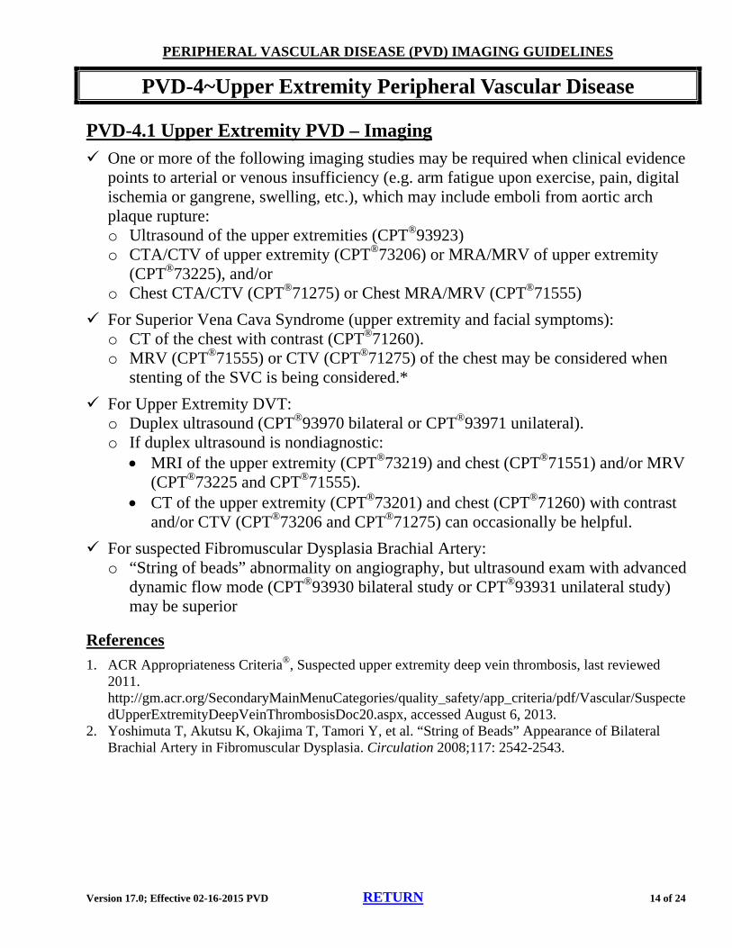
PVD-4.1 Upper Extremity PVD – Imaging
One or more of the following imaging studies may be required when clinical evidence points to arterial or venous insufficiency (e.g. arm fatigue upon exercise, pain, digital ischemia or gangrene, swelling, etc.), which may include emboli from aortic arch plaque rupture: o Ultrasound of the upper extremities (CPT®93923) o CTA/CTV of upper extremity (CPT®73206) or MRA/MRV of upper extremity
(CPT®73225), and/or o Chest CTA/CTV (CPT®71275) or Chest MRA/MRV (CPT®71555)
For Superior Vena Cava Syndrome (upper extremity and facial symptoms): o CT of the chest with contrast (CPT®71260). o MRV (CPT®71555) or CTV (CPT®71275) of the chest may be considered when
stenting of the SVC is being considered.*
For Upper Extremity DVT: o Duplex ultrasound (CPT®93970 bilateral or CPT®93971 unilateral). o If duplex ultrasound is nondiagnostic:
MRI of the upper extremity (CPT®73219) and chest (CPT®71551) and/or MRV (CPT®73225 and CPT®71555).
CT of the upper extremity (CPT®73201) and chest (CPT®71260) with contrast and/or CTV (CPT®73206 and CPT®71275) can occasionally be helpful.
For suspected Fibromuscular Dysplasia Brachial Artery: o “String of beads” abnormality on angiography, but ultrasound exam with advanced
dynamic flow mode (CPT®93930 bilateral study or CPT®93931 unilateral study) may be superior
References
1. ACR Appropriateness Criteria®, Suspected upper extremity deep vein thrombosis, last reviewed 2011. http://gm.acr.org/SecondaryMainMenuCategories/quality_safety/app_criteria/pdf/Vascular/SuspectedUpperExtremityDeepVeinThrombosisDoc20.aspx, accessed August 6, 2013.
2. Yoshimuta T, Akutsu K, Okajima T, Tamori Y, et al. “String of Beads” Appearance of Bilateral Brachial Artery in Fibromuscular Dysplasia. Circulation 2008;117: 2542-2543.
Version 17.0; Effective 02-16-2015 PVD RETURN 15 of 24
PERIPHERAL VASCULAR DISEASE (PVD) IMAGING GUIDELINES
PVD-5~PULMONARY ARTERY HYPERTENSION
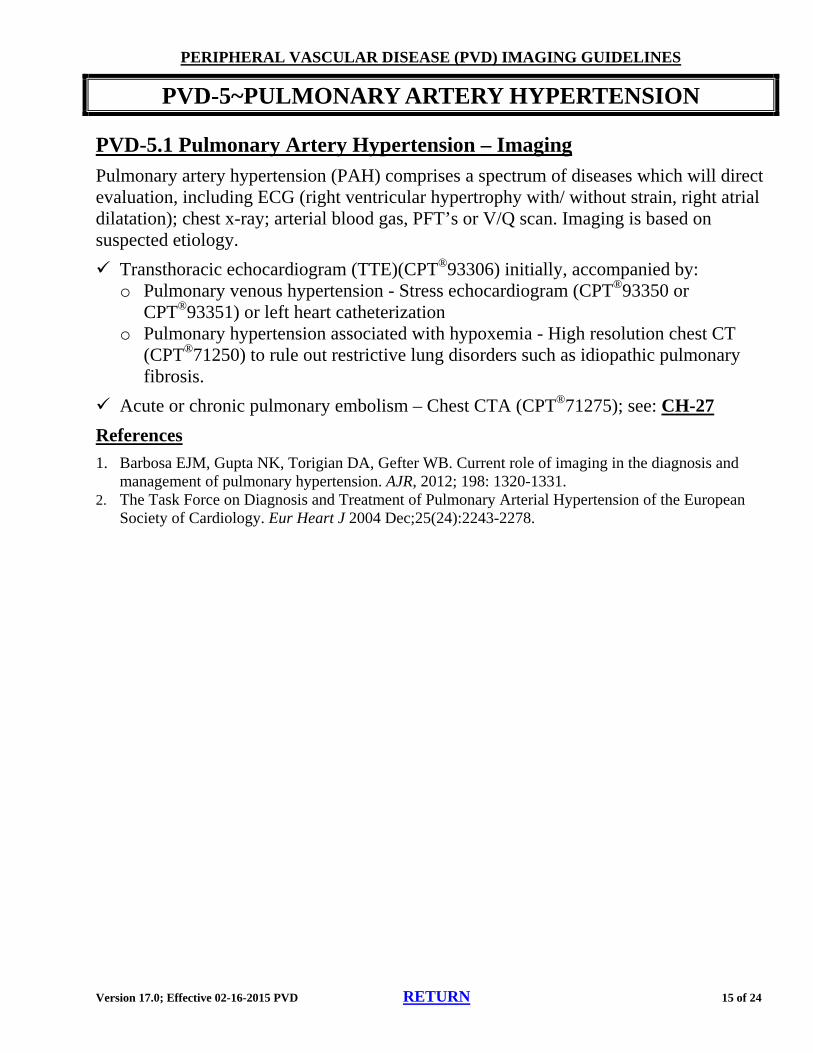
PVD-5.1 Pulmonary Artery Hypertension – Imaging
Pulmonary artery hypertension (PAH) comprises a spectrum of diseases which will direct evaluation, including ECG (right ventricular hypertrophy with/ without strain, right atrial dilatation); chest x-ray; arterial blood gas, PFT’s or V/Q scan. Imaging is based on suspected etiology.
Transthoracic echocardiogram (TTE)(CPT®93306) initially, accompanied by: o Pulmonary venous hypertension - Stress echocardiogram (CPT®93350 or
CPT®93351) or left heart catheterization o Pulmonary hypertension associated with hypoxemia - High resolution chest CT
(CPT®71250) to rule out restrictive lung disorders such as idiopathic pulmonary fibrosis.
Acute or chronic pulmonary embolism – Chest CTA (CPT®71275); see: CH-27
References
1. Barbosa EJM, Gupta NK, Torigian DA, Gefter WB. Current role of imaging in the diagnosis and management of pulmonary hypertension. AJR, 2012; 198: 1320-1331.
2. The Task Force on Diagnosis and Treatment of Pulmonary Arterial Hypertension of the European Society of Cardiology. Eur Heart J 2004 Dec;25(24):2243-2278.
Version 17.0; Effective 02-16-2015 PVD RETURN 16 of 24
PERIPHERAL VASCULAR DISEASE (PVD) IMAGING GUIDELINES
PVD-6~Aortic Disorders and Renal Vascular Disorders and Visceral Artery Aneurysms
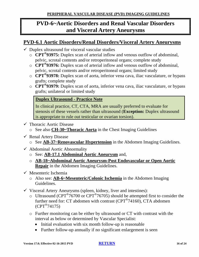
PVD-6.1 Aortic Disorders/Renal Disorders/Visceral Artery Aneurysms
Duplex ultrasound for visceral vascular studies o CPT®93975: Duplex scan of arterial inflow and venous outflow of abdominal,
pelvic, scrotal contents and/or retroperitoneal organs; complete study o CPT®93976: Duplex scan of arterial inflow and venous outflow of abdominal,
pelvic, scrotal contents and/or retroperitoneal organs; limited study o CPT®93978: Duplex scan of aorta, inferior vena cava, iliac vasculature, or bypass
grafts; complete study o CPT®93979: Duplex scan of aorta, inferior vena cava, iliac vasculature, or bypass
grafts; unilateral or limited study
Duplex Ultrasound - Practice Note
In clinical practice, CT, CTA, MRA are usually preferred to evaluate for stenosis of these vessels rather than ultrasound (Exception: Duplex ultrasound is appropriate to rule out testicular or ovarian torsion).
Thoracic Aortic Disease o See also CH-30~Thoracic Aorta in the Chest Imaging Guidelines
Renal Artery Disease o See AB-37~Renovascular Hypertension in the Abdomen Imaging Guidelines.
Abdominal Aortic Abnormality o See: AB-17.1 Abdominal Aortic Aneurysm and, o AB-18~Abdominal Aortic Aneurysm-Post Endovascular or Open Aortic
Repair in the Abdomen Imaging Guidelines.
Mesenteric Ischemia o Also see: AB-6~Mesenteric/Colonic Ischemia in the Abdomen Imaging
Guidelines.
Visceral Artery Aneurysms (spleen, kidney, liver and intestines): o Ultrasound (CPT®76700 or CPT®76705) should be attempted first to consider the
further need for: CT abdomen with contrast (CPT®74160), CTA abdomen (CPT®74175)
o Further monitoring can be either by ultrasound or CT with contrast with the interval as below or determined by Vascular Specialist: Initial evaluation with six month follow-up is reasonable Further follow-up annually if no significant enlargement is seen
Version 17.0; Effective 02-16-2015 PVD RETURN 17 of 24
o Post-stent placement are without guidelines and therefore reasonable to follow the same time table as for endovascular aortic repair: CTA of abdomen (CPT®74175), MRA of abdomen (CPT®74185), or CT abdomen (CPT®74160) at 1 month, 6 months, and 12 months following stent placement, then every year. An additional study can be done at 3 months if there was evidence of endoleak on the 1 month study.
Visceral Artery Aneurysms - Practice Notes
Visceral Artery Aneurysms are defined by an increase of more than 50% of the original arterial diameter.
Vascular specialist consultation is beneficial in order to determine the time-frame to intervention.
May-Thurner Syndrome (Iliac Vein Compression Syndrome) is diagnosed by: o Pelvic MRI, Pelvic MRA(V) or Pelvic CTA(V) are the most common methods
used o Traditional Venography or o Duplex Ultrasound (this technique is very technician-dependent and can be
difficult)
May-Thurner Syndrome - Practice Notes
This is a rare condition caused by compression of the left common iliac vein by the overlying right common iliac artery.
It may cause discomfort and edema of the lower extremity, DVT in the iliofemoral vein or lower extremity DVT which may be recurrent.
References
1. Hirsch AT, Haskal ZJ, Hertzer NR, Bakal CW, et al. ACC/AHA Practice Guidelines: ACC/AHA 2005 Practice Guidelines for the Management of Patients With Peripheral Arterial Disease (Lower Extremity, Renal, Mesenteric, and Abdominal Aortic): A Collaborative Report from the American Association for Vascular Surgery/Society for Vascular Surgery,* Society for Cardiovascular Angiography and Interventions, Society for Vascular Medicine and Biology, Society of Interventional Radiology, and the ACC/AHA Task Force on Practice Guidelines. Circulation.2006;113:e463-e654,doi:10.1161/CIRCULATIONAHA.106.174526.
Version 17.0; Effective 02-16-2015 PVD RETURN 18 of 24
PERIPHERAL VASCULAR DISEASE (PVD) IMAGING GUIDELINES
PVD-7~Lower Extremity Peripheral Vascular Disease
PVD-7.1 Claudication
If resting ABI (CPT®93922) is normal (0.9 – 1.3) and disease is still suspected: o Differentiate from “pseuodoclaudication” (See: SP-9~Lumbar Spinal Stenosis in
the Spine Imaging Guidelines) o Re-measured after exercise (CPT®93924).1 o A toe-brachial index may be used as further screening in patients with ABI’s
greater than 1.3 o Otherwise, advanced imaging is necessary only if there is consideration for
invasive therapy2-5
Duplex ultrasound (CPT®93925 bilateral study or CPT®93926 unilateral study) and Doppler studies are adjuncts to abnormal ABI that may be used: 6,7 o To identify location and extent of disease, and o Prior to considering advanced imaging.
MRA of the aorta, pelvic vessels, and lower extremities (CPT®74185, CPT®73725 and CPT®73725), OR CTA with run off (CPT®75635) and ABI < 0.5 to further evaluate the lower extremity arteries for the following: 2,8 o Intermittent claudication (i.e. non-limb threatening ischemia) and all of the
following: Failed 3 months conservative medical therapy (physician supervised walking
/exercise program) Functional disability (e.g. exercise impairment sufficient to threaten the
patient’s employment or to require significant alterations in the patient’s lifestyle) or
o Potentially limb-threatening vascular disease (such as skin breakdown, non-healing ischemic ulcers, resting leg pain, or gangrene) or
o Blue Toe Syndrome (emboli from aortic plaque or mural thrombus , hyperviscosity syndrome, hypercoagulable states, and vasculitis) Ultrasound (CPT®76775) may be useful to identify a previously unknown
abdominal aortic aneurysm (AAA) but is not required prior to CTA or o Preoperative planning for an invasive procedure (endovascular or open surgery) o NOTE: MRA of the pelvis should not be requested/billed with CPT®74185,
CPT®73725 and CPT®73725
Claudication - Practice Notes
Claudication symptoms usually remain stable (70%-80% of patients) and do not worsen or improve at rapid rates.9
Version 17.0; Effective 02-16-2015 PVD RETURN 19 of 24
PVD-7.2 Popliteal Artery Entrapment Syndrome
Diagnosis of popliteal artery stenosis or occlusion due to compression by adjacent muscle and tendons seen in young men (ages 20-40)10 o Ultrasound (CPT®93926 unilateral study), lower extremity CTA (CPT®73706), or
lower extremity MRA (CPT®73725) o CT or MRI of the lower extremity (contrast as requested) if requested by the
operating surgeon.
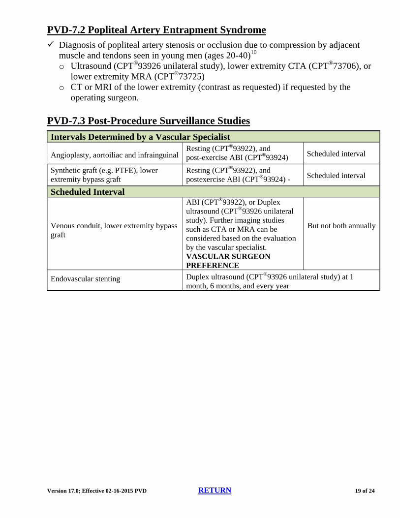
PVD-7.3 Post-Procedure Surveillance Studies
Intervals Determined by a Vascular Specialist
Angioplasty, aortoiliac and infrainguinal Resting (CPT®93922), and post-exercise ABI (CPT®93924) Scheduled interval
Synthetic graft (e.g. PTFE), lower extremity bypass graft
Resting (CPT®93922), and postexercise ABI (CPT®93924) - Scheduled interval
Scheduled Interval
Venous conduit, lower extremity bypass graft
ABI (CPT®93922), or Duplex ultrasound (CPT®93926 unilateral study). Further imaging studies such as CTA or MRA can be considered based on the evaluation by the vascular specialist. VASCULAR SURGEON PREFERENCE
But not both annually
Endovascular stenting Duplex ultrasound (CPT®93926 unilateral study) at 1 month, 6 months, and every year
Version 17.0; Effective 02-16-2015 PVD RETURN 20 of 24
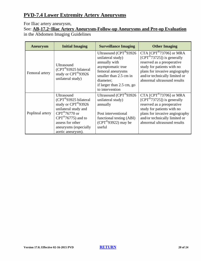
PVD-7.4 Lower Extremity Artery Aneurysms
For Iliac artery aneurysm, See: AB-17.2~Iliac Artery Aneurysm-Follow-up Aneurysms and Pre-op Evaluation in the Abdomen Imaging Guidelines
Aneurysm Initial Imaging Surveillance Imaging Other Imaging
Femoral artery
Ultrasound (CPT®93925 bilateral study or CPT®93926 unilateral study)
Ultrasound (CPT®93926 unilateral study) annually with asymptomatic true femoral aneurysms smaller than 2.5 cm in diameter; if larger than 2.5 cm, go to intervention
CTA [CPT®73706] or MRA [CPT®73725]) is generally reserved as a preoperative study for patients with no plans for invasive angiography and/or technically limited or abnormal ultrasound results
Popliteal artery
Ultrasound (CPT®93925 bilateral study or CPT®93926 unilateral study and CPT®76770 or CPT®76775) and to assess for other aneurysms (especially aortic aneurysm).
Ultrasound (CPT®93926 unilateral study) annually Post interventional functional testing (ABI) (CPT®93922) may be useful
CTA [CPT®73706] or MRA [CPT®73725]) is generally reserved as a preoperative study for patients with no plans for invasive angiography and/or technically limited or abnormal ultrasound results
Version 17.0; Effective 02-16-2015 PVD RETURN 21 of 24
PVD-7.5 Lower Extremity Deep Venous Thrombosis (DVT) and/or Lower Extremity Edema
Deep venous thrombosis can present with swelling, pain, warmth, erythema, and pain with dorsiflexion of the foot (Homan’s Sign) or with progression, such as phlegmasia cerulea dolens. However, 1/3 of all cases are asymptomatic. Symptoms are often not apparent until there is involvement above the knee.
Risk factors for DVT include inactivity, posture, obstruction as well as those outlines outlined in CH-29.
Venous duplex study (CPT®93970 bilateral or CPT®93971 unilateral) is the initial imaging study to evaluate for deep venous thrombosis (DVT)
Further imaging is determined by suspected disease or venous duplex findings: o Concomitant arterial disease
ABI (CPT®93922) (see: PVD-7.1 Claudication)
Duplex ultrasound (CPT®93970 bilateral study or CPT®93971 unilateral study) is the initial imaging study for any suspected DVT o If Duplex ultrasound is normal, repeat Duplex ultrasound testing is not supported
Unilateral or bilateral calf edema with negative or indeterminate venous duplex study o Abdomen and Pelvic Ultrasound (CPT®76700 and/or CPT®76856 and/or
CPT®76830 [transvaginal]), and if still negative: Pelvis CT with contrast (CPT®72193) or Abdomen and Pelvis CT with contrast
(CPT® 74177), or MRV or CTV of the pelvis or abdomen and pelvis (CPT®74185 and
CPT®72198 or CPT®74175 and CPT®72191) and if the extent of thrombosis needs more detailed assessment and
CT or MRI of the lower extremity without contrast (CPT®73700 or CPT®73718)
o May-Thurner Syndrome (Iliac Vein Compression Syndrome) suspected - is a rare condition of left common iliac vein compression by the overlying right common iliac artery best diagnosed with Pelvis MRI, Pelvis MRA(V) or Pelvis CTA(V) and traditional venography
o Popliteal (Baker’s) Cyst suspected - dedicated ultrasound of the popliteal fossa (CPT®76882)
o Diabetic muscle necrosis suspected - MRI of the extremity (contrast as requested) o Chronic venous insufficiency - advanced imaging is not routinely indicated, unless
suspected thigh or abdominal/pelvic clot(s) or masses o Phlegmasia cerulea dolens can be evaluated by MRV, CTV or CTA with run off to
assess the arterial system. MRA (CPT®74185, CPT®73725, and CPT®73725) may also be required for this problem, which can reflect both arterial and venous compromise and produce substantial lower extremity edema.

Version 17.0; Effective 02-16-2015 PVD RETURN 22 of 24
Generally not considered: o Impedance plethysmography (IPG)— CPT®93965) may be useful but is currently
uncommonly utilized o Venography is more accurate but carries the risk of phlebitis o Superficial venous thrombosis should not require advanced imaging. o There is insufficient data at this time to justify routinely performing CTA-CTV,
including CTV of the pelvis and lower extremities. o Duplex study of the arteries (CPT®93925 bilateral study or CPT®93926 unilateral
study) is not indicated unless there is evidence of arterial insufficiency (See: PVD-7.1 Claudication)
Follow-up imaging of known DVT:
Duplex ultrasound (CPT®93970 bilateral study or CPT®93971unilateral study) can be repeated in order to rule out proximal extension of the clot: o One week after the initial diagnosis o Serial imaging (up to 3 studies) over the first two weeks if calf DVT is not treated
Imaging during or to terminate long term anticoagulation therapy to determine venous recanalization.is not supported by evidence.
PVD-7.6 Other Diseases of the Lower Extremity Veins
Venous duplex scan (CPT®93970 bilateral study or CPT®93971 unilateral study) can be performed in patients who are candidates for anticoagulation or invasive therapeutic procedures for the following: o Post-thrombotic (post-phlebitic) syndrome o Confirm the diagnosis of venous insufficiency/valvular incompetence in patients
with signs and symptoms of this disease (ulceration, thickening, and skin discoloration)
o Venous mapping prior to autologous vein graft harvesting (e.g. for cardiac bypass surgery)
o Following radiofrequency ablation of varicosities when the greater saphenous vein was closed (not indicated if only superficial veins underwent ablation), for DVT surveillance performed between 6 – 12 weeks. (CPT®93971 unilateral study)
References
1. Hirsch AT, Haskal ZJ, Hertzer NR, Bakal CW, et al. ACC/AHA Practice Guidelines: ACC/AHA 2005 Practice Guidelines for the Management of Patients With Peripheral Arterial Disease (Lower Extremity, Renal, Mesenteric, and Abdominal Aortic): A Collaborative Report from the American Association for Vascular Surgery/Society for Vascular Surgery,* Society for Cardiovascular Angiography and Interventions, Society for Vascular Medicine and Biology, Society of Interventional Radiology, and the ACC/AHA Task Force on Practice Guidelines. Circulation.2006;113:e463-e654,doi:10.1161/CIRCULATIONAHA.106.174526.
Version 17.0; Effective 02-16-2015 PVD RETURN 23 of 24
2. Dill KE, Rybicki FJ, Desjardins B, Flamm SD, et al. Expert Panel on Vascular Imaging. ACR Appropriateness Criteria® claudication -- suspected vascular etiology. American College of Radiology (ACR); 2012.
3. Fowkes FG, Murray GD, Butcher I, Heald CL et al. Ankle brachial index combined with Framingham Risk Score to predict cardiovascular events and mortality: a meta-analysis. JAMA 2008;300(2):197-208
4. Rutherford RB, Lowenstein DH, Klein MF. Combining segmental systolic pressures and plethysmography to diagnose arterial occlusive disease of the legs. Am J Surg. 1979 Aug;138(2):211–218.
5. Van De Water JM, Laska ED, Ciniero WV. Patient and operation selectivity. The peripheral vascular laboratory. Ann Surg. 1979 Feb;189(2):143–146.
6. Federman DG, Kravetz JD, Bravata DM, Kirsner RS. Peripheral artery disease: A marker of morbidity and mortality. Postgrad Med 2006;119(2):21-27
7. Willmann JK, Baumert B, Schertler T, Wildermuth S, et al. Aortoiliac and Lower Extremity Arteries Assessed with 16–Detector Row CT Angiography: Prospective Comparison with Digital Subtraction Angiography1. Radiology 2005;236:1083-1093 and1094-1103
8. Allison MA, Hiatt WR, Hirsch AT, et al: A high ankle-brachial index is associated with increased cardiovascular disease morbidity and lower quality of life. J Am Coll Cardiol 2008;51:1292–1298.
9. Weitz JI, Byrne J, Clagett GP, Farkouh ME, et al. Diagnosis and Treatment of Chronic Arterial Insufficiency of the Lower Extremities: A Critical Review. Circulation 1996;94:3026-3049.
10. Macedo TA, Johnson M, Hallett JW, Breen JF.Popliteal Artery Entrapment Syndrome: Role of Imaging in the Diagnosis. AJR 2003;181:1259-1265.
Version 17.0; Effective 02-16-2015 PVD RETURN 24 of 24
PERIPHERAL VASCULAR DISEASE (PVD) IMAGING GUIDELINES
PVD-8~IMAGING for HEMODIALYSIS ACCESS
PVD-8.1 Preoperative Arterial Evaluation and Venous Mapping Prior to AV Fistula Creation
There is a Level II HCPCS code for vessel mapping prior to AV fistula creation that does not have a CPT® Level I equivalent, {HCPCS code G0365 [Vessel mapping of vessels for hemodialysis access (Services for preoperative vessel mapping prior to creation of hemodialysis access using an autogenous hemodialysis conduit, including arterial inflow and venous outflow)]}. Therefore, CPT® codes for duplex venous and arterial are used for this purpose.
Arterial evaluation to assess arterial suitability (size, degree of stenosis and calcification) prior to AV fistula creation may be appropriate. o CPT®93930 or CPT®93931 can be used to report upper extremity arterial
evaluation. o Venous Mapping to assess venous suitability prior to AV fistula creation may be
appropriate. CPT®93970 or CPT®93971 can be used to report venous mapping.
Indications for Duplex Ultrasound (CPT®93990) of hemodialysis access include but are not limited to: o Patients with decreased flow rates during hemodialysis o Development of arm swelling or discomfort after access placement surgery or a
hemodialysis session o Prolonged immaturity of a surgically created AV fistula o Suspected pseudoaneurysm o Suspected AV fistula or graft stenosis o Known or suspected fluid collection adjacent to an AV fistula or graft o Though it is, generally, not needed, one duplex US (CPT®93990) can be performed
after a surgically created AV fistula for assessment.