radiation protection and radiation measurement issues with non
TRANSCRIPT

THE UNIVERSITY OF NEW MEXICO HEALTH SCIENCES CENTER
COLLEGE OF PHARMACY ALBUQUERQUE, NEW MEXICO
Correspondence Continuing Education Courses for Nuclear Pharmacists and Nuclear
Medicine Professionals
VOLUME 11, LESSON 5
Radiation Protection and Radiation Measurement Issues with Non-Traditional Radiopharmaceuticals
By
A. Michael Zimmer, NPh, Ph.D.
Director, Nuclear Pharmacy Radiation Safety Officer
Northwestern Memorial Hospital Professor of Radiology
Northwestern University Medical Center Chicago, IL
The University of New Mexico Health Sciences Center College of Pharmacy is accredited by the Accredication Council for Pharmacy Education as a provider of continuing pharmaceutical education. Program No. 039-000-04-003-H04. 2.5 Contact Hours or .25 CEUs. Expires 05/25/2007.

2

3
Radiation Protection and Radiation Measurement Issues with Non-Traditional Radiopharmaceuticals
By:
A. Michael Zimmer, NPh, Ph.D.
Coordinating Editor and Director of Pharmacy Continuing Education William B. Hladik III, MS, RPh, FASHP, FAPhA
College of Pharmacy University of New Mexico Health Sciences Center
Managing Editor
Julliana Newman, ELS Wellman Publishing, Inc.
Albuquerque, New Mexico
Editorial Board George H. Hinkle, MS, RPh, BCNP, FASHP, FAPhA David Lawrence Laven, NPh, CRPh, FASHP, FAPhA
Jeffrey P. Norenberg, MS, PharmD, BCNP, FASHP, FAPha Neil A. Petry, MS, RPh, BCNP, FAPhA
Timothy M. Quinton, PharmD, MS, RPh, BCNP, FAPhA
Guest Reviewer Kenneth T. Cheng, PhD, RPh, BCNP, FAPhA
Associate Professor, Director Nuclear Pharmacy Training Program Nuclear Pharmacy
Medical University of South Carolina 171 Ashley Avenue
Charleston, South Carolina 29425
While the advice and information in this publication are believed to be true and accurate at press time, the author(s), editors, or the publisher cannot accept any legal responsibility for any errors or omissions that may be made. The publisher makes no war-
ranty, expressed or implied, with respect to the material contained herein.
Copyright 2004 University of New Mexico Health Sciences Center
Pharmacy Continuing Education Albuquerque, New Mexico
4
RADIATION PROTECTION AND RADIATION MEASUREMENT ISSUES WITH NON-TRADITIONAL RADIOPHARMACEUTICALS
STATEMENT OF OBJECTIVES
The purpose of this continuing education lesson is to provide an update of alpha-emitting and beta-emitting therapeutic radiopharmaceuticals including radiation protection and radiation measurement issues. This lesson describes specific alpha and beta radionuclides used for labeling radiopharmaceuticals, their advantages and the radiation protection issues associated with each. Specific alpha- and beta-emitting radiopharmaceuticals are described with an emphasis on ra-tionale for use, radiolabeling methodology, pharmacokinetics, clinical protocols and radiation protection issues. Radiation dose measurement issues specifically related to therapeutic beta-emitting radiophar-maceuticals, and their gamma imaging analogs, are discussed. Effects of geometry and attenua-tion on dose measurements of radiopharmaceuticals are addressed. Specific step-by-step instruc-tions are provided in calibrating dose calibrators for specific beta-emitting radiopharmaceuticals. Alternate calibration methods are discussed. Upon completion of this lesson, the reader should be able to:
1. List specific alpha-emitting radionuclides used in targeted therapies.
2. Identify specific alpha-emitting therapeutic radiopharmaceuticals, their properties and clinical use.
3. Describe advantages and disadvantages of alpha-emitting radiopharmaceuticals.
4. Elaborate on radiation protection issues related to alpha-emitting radiopharmaceuticals.
5. List specific beta-emitting radionuclides, their properties and applications in targeted
therapy.
6. Summarize specific beta radionuclides conjugated to beta-emitting therapeutic radio-pharmaceuticals, including advantages and disadvantages of each.
7. Identify specific beta-emitting therapeutic radiopharmaceuticals used in clinical practice
including properties and pharmacokinetics.
8. Describe radiation protection issues related to beta-emitting radiopharmaceuticals includ-ing radiation protection related to radiopharmaceutical formulation, dispensing and pa-tient administration.
9. Describe radiation protection issues related to patients receiving therapeutic beta-emitting
radiopharmaceuticals.
5
10. Understand the issues and specific procedures related to radiation dose measurements of therapeutic radiopharmaceuticals.

6
COURSE OUTLINE
I. INTRODUCTION
II. ALPHA-EMITTING RADIOPHARMACEUTICALS
A. Rationale for Use
B. Astatine-211 Radiopharmaceuticals
C. Bismuth-212 Radiopharmaceuticals
D. Bismuth-213 Radiopharmaceuticals
E. Actinium-225 Radiopharmaceuticals
F. Radiation Protection Issues
III. BETA-EMITTING RADIONUCLIDES
A. Classification of Beta Radionuclides and Tumor Therapy Rationale
B. Clinical Radionuclide Therapies
C. Radioimmunotherapy- Non-Hodgkin’s Lymphoma
D. Y-90 Ibritumomab Tiuxetan
E. I-131 Tositumomab
F. Radiolabeled Peptides for Therapy
G. Y-90-DOTA-Octreotide
H. Y-90 Microspheres
IV. CONCLUSION
V. REFERENCES
VI. QUESTIONS
7
RADIATION PROTECTION AND RADIATION MEASUREMENT ISSUES WITH NON-TRADITIONAL RADIOPHARMACEUTICALS
By
A. Michael Zimmer, NPh, Ph.D. Northwestern University Medical Center
I. INTRODUCTION Within the last decade, radionuclide ther-
apy has become increasingly more important in nuclear medicine. Specific radiopharma-ceuticals used for therapies include radioio-dine, P-32 sodium phosphate and chromic phosphate, radiolabeled peptides, radio-labeled particles or microspheres, radiophar-maceuticals for palliation of painful bone me-tastases and radiopharmaceuticals for radio-immunotherapy. Newer targeted therapies use radiopharmaceutical labeled with alpha-emitting and beta-emitting radionuclides. Some of these specific newer targeted ra-dionuclide therapies and therapeutic radio-pharmaceuticals will be discussed. In addi-tion, whenever appropriate, specific issues related to dose measurement of therapeutic radiopharmaceuticals will be discussed. Also, radiation safety issues of alpha and beta-emitting radionuclides, including shielding requirements and radiation exposures to ra-diation workers and to members of the gen-eral public, will also be discussed.
For radionuclide therapy, the likelihood of treatment success depends on a number of parameters. These include the following: the concentration of the radiopharmaceutical in tumor; the radiation absorbed dose in tumor; the delivery of the radiation dose (dose rate and fractionation); the sensitivity and re-sponse of tumor cells to the specific radia-tion; and tumor size compared to the type of radionuclide emission and the energy of emission.1,2 For beta-emitting radionuclides, there appears to be a relationship between the mean energy emitted from beta radionuclides and the optimal tumor size which results in
radiocurability (1). Optimal tumor cure di-ameters range from less than 1 mm for short-range beta emitters such as Lu-177 and Au-199 to several centimeters for long-range beta-emitters such as Y-90 and P-32. As a result, future radionuclide therapies may in-clude tailoring specific beta-emitting ra-dionuclides to specific tumor sizes, including the use of a radionuclide therapy “cocktail” consisting of several different beta-emitting radionuclides for more effective tumor treat-ment.1
When selecting a specific radionuclide for radionuclide therapy, a number of factors should be considered. One factor is tumor size, as mentioned previously, and another is tumor heterogeneity. The availability and cost of the radionuclide is an important fac-tor. In addition, an acceptable nuclear decay of the specific radionuclide, a physical half-life conducive to radiopharmaceutical prepa-ration and tumor pharmacokinetics, fairly rapid and stable attachment of the radionu-clide to the desired chemical species and ap-propriate gamma ray emissions for imaging to aid in biodistribution and imaging are also important.
II. ALPHA-EMITTING RADIO-PHARMACEUTICALS
A. Rationale for Use Alpha-emitting radionuclides have physi-
cal properties that are useful for cancer treatment. Alpha-emitting radionuclides pro-duce high densities of ionization. Energetic alpha emitters have a tissue range of 40–80 microns (µm) as opposed to tissue ranges of

8
3000–6000 µm for energetic beta-emitting radionuclides.3 As a result, the effective treatment radius is several cell diameters, thus making alpha-emitters ideal for thera-peutic treatment at a cellular or micrometas-tases level.4 The short tissue path length also reduces non-specific irradiation of distant tissue. Alpha-emitting radionuclides also have a high linear energy transfer (LET). High LET of alpha radiation results in effec-tive cell killing by limiting the ability of cells to repair damage to DNA. The effectiveness of alpha particles in cell killing can be dem-onstrated by the fact that the number of beta particles required to elicit the same effect is more than 1000 times higher as compared to alpha particles. Additional advantages of al-pha emitters in targeted therapy include their independence of cell cycle position and also the cell hypoxic state.4,5
There are limitations associated with al-pha-emitting radionuclides for therapy and, as such, may hamper their widespread clini-cal use. Once conjugated to the specific chemical moiety, release of unbound alpha-emitting radionuclides may result in severe toxicities. The complexity of conjugation, using either bifunctional chelating agents or attachment to non-activated aromatic rings, is another limitation. As alpha radionuclides decay, bifunctional chelating agent may not appropriately bind daughter radionuclides ultimately leading to the release of the un-bound daughter radionuclide. Because alpha radionuclide decay results in a deposition of high radiation within a very small volume,
radiolysis may also be significant. Radiolysis can result in protein degradation, including protein fragmentation, and, in the case of monoclonal antibodies, may result in loss of antibody radioimmunoreactivity.6 Other limi-tations of alpha-emitters include the lack of commercial availability and unfamiliarity in regards to radiation safety issues.
Alpha-emitting radionuclides are pro-duced by cyclotron or reactor irradiation. Some alpha radionuclides may be incorpo-rated into a generator system, which is sub-sequently eluted prior to use. Some common alpha-emitting radionuclides used for ra-dionuclide therapy are listed in Table 1. As shown in the table, many alpha-emitting ra-dionuclides also emit beta and gamma rays. Some gamma ray emission energies from alpha-emitting radionuclides are appropriate for biodistribution and imaging studies, which may be performed prior to initiation of therapy.
B. Astatine-211 Radiopharmaceuticals Astatine-211 is the heaviest halogen with
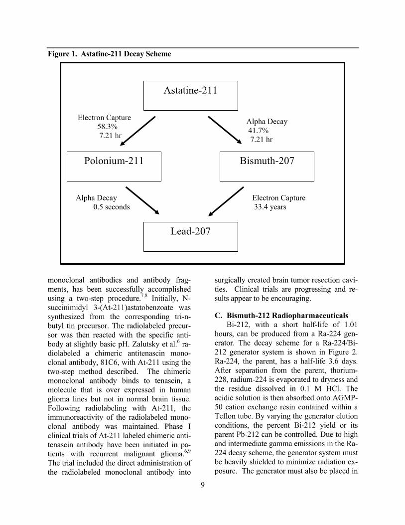
a 7.2-hour half-life. The Astatine-211 decay scheme is shown in Figure 1. It decays by a double-branch pathway with alpha energies of 5.9 and 7.5 MeV. The short range in tis-sue, 55–70 µm, and high LET of the alpha particles make the radionuclide attractive for radionuclide therapy. During Astatine-211 decay, x-rays of 77 keV and 97 keV are pro-duced, allowing for external gamma imaging and biodistribution studies. Radiolabeling of proteins with Astatine-211, including
Table 1. Physical Properties of Some Common Alpha-Emitting Radionuclides for Radionuclide Therapy
Nuclide Half-life (hours) Energyα (MeV) Energyβ (MeV) Energyγ (MeV) At-211 7.21 5.9, 7.5 ----- 0.070, 0.080, 0.570, 1.0 Bi-212 1.01 6.04, 6.08, 9.0 0.492, 0.56 0.51, 0.58, 2.60 Bi-213 0.76 5.9 0.444 0.440 Ac-225 240 6.0 ----- 0.218 Ra-223 273.6 7.02 ----- 0.08, 0.270
9
Figure 1. Astatine-211 Decay Scheme
Astatine-211
Polonium-211 Bismuth-207
Lead-207
Electron Capture 58.3% 7.21 hr
Alpha Decay 41.7% 7.21 hr
Alpha Decay 0.5 seconds
Electron Capture 33.4 years
monoclonal antibodies and antibody frag-ments, has been successfully accomplished using a two-step procedure.7,8 Initially, N-succinimidyl 3-(At-211)astatobenzoate was synthesized from the corresponding tri-n-butyl tin precursor. The radiolabeled precur-sor was then reacted with the specific anti-body at slightly basic pH. Zalutsky et al.6 ra-diolabeled a chimeric antitenascin mono-clonal antibody, 81C6, with At-211 using the two-step method described. The chimeric monoclonal antibody binds to tenascin, a molecule that is over expressed in human glioma lines but not in normal brain tissue. Following radiolabeling with At-211, the immunoreactivity of the radiolabeled mono-clonal antibody was maintained. Phase I clinical trials of At-211 labeled chimeric anti-tenascin antibody have been initiated in pa-tients with recurrent malignant glioma.6,9 The trial included the direct administration of the radiolabeled monoclonal antibody into
surgically created brain tumor resection cavi-ties. Clinical trials are progressing and re-sults appear to be encouraging.
C. Bismuth-212 Radiopharmaceuticals Bi-212, with a short half-life of 1.01
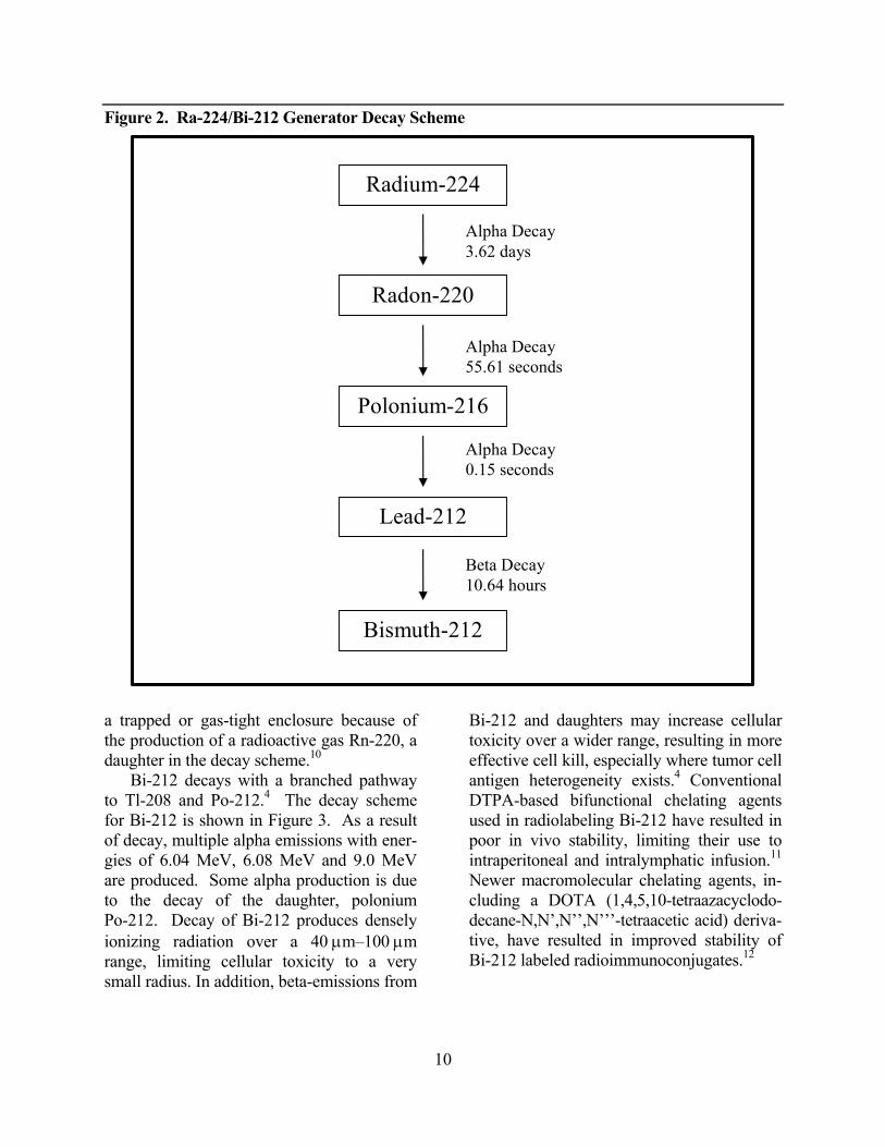
hours, can be produced from a Ra-224 gen-erator. The decay scheme for a Ra-224/Bi-212 generator system is shown in Figure 2. Ra-224, the parent, has a half-life 3.6 days. After separation from the parent, thorium-228, radium-224 is evaporated to dryness and the residue dissolved in 0.1 M HCl. The acidic solution is then absorbed onto AGMP-50 cation exchange resin contained within a Teflon tube. By varying the generator elution conditions, the percent Bi-212 yield or its parent Pb-212 can be controlled. Due to high and intermediate gamma emissions in the Ra-224 decay scheme, the generator system must be heavily shielded to minimize radiation ex-posure. The generator must also be placed in
10
Figure 2. Ra-224/Bi-212 Generator Decay Scheme
Radium-224
Radon-220
Polonium-216
Lead-212
Bismuth-212
Alpha Decay 3.62 days
Alpha Decay 55.61 seconds
Alpha Decay 0.15 seconds
Beta Decay 10.64 hours
a trapped or gas-tight enclosure because of the production of a radioactive gas Rn-220, a daughter in the decay scheme.10
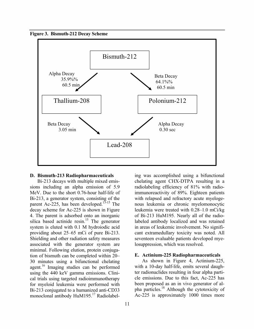
Bi-212 decays with a branched pathway to Tl-208 and Po-212.4 The decay scheme for Bi-212 is shown in Figure 3. As a result of decay, multiple alpha emissions with ener-gies of 6.04 MeV, 6.08 MeV and 9.0 MeV are produced. Some alpha production is due to the decay of the daughter, polonium Po-212. Decay of Bi-212 produces densely ionizing radiation over a 40 µm–100 µm range, limiting cellular toxicity to a very small radius. In addition, beta-emissions from
Bi-212 and daughters may increase cellular toxicity over a wider range, resulting in more effective cell kill, especially where tumor cell antigen heterogeneity exists.4 Conventional DTPA-based bifunctional chelating agents used in radiolabeling Bi-212 have resulted in poor in vivo stability, limiting their use to intraperitoneal and intralymphatic infusion.11 Newer macromolecular chelating agents, in-cluding a DOTA (1,4,5,10-tetraazacyclodo-decane-N,N’,N’’,N’’’-tetraacetic acid) deriva-tive, have resulted in improved stability of Bi-212 labeled radioimmunoconjugates.12
11
Figure 3. Bismuth-212 Decay Scheme
Bismuth-212
Thallium-208 Polonium-212
Lead-208
Alpha Decay 35.9%% 60.5 min
Beta Decay 64.1%% 60.5 min
Beta Decay 3.05 min
Alpha Decay 0.30 sec
D. Bismuth-213 Radiopharmaceuticals Bi-213 decays with multiple mixed emis-
sions including an alpha emission of 5.9 MeV. Due to the short 0.76-hour half-life of Bi-213, a generator system, consisting of the parent Ac-225, has been developed.13-15 The decay scheme for Ac-225 is shown in Figure 4. The parent is adsorbed onto an inorganic silica based actinide resin.15 The generator system is eluted with 0.1 M hydroiodic acid providing about 25–65 mCi of pure Bi-213. Shielding and other radiation safety measures associated with the generator system are minimal. Following elution, protein conjuga-tion of bismuth can be completed within 20–30 minutes using a bifunctional chelating agent.16 Imaging studies can be performed using the 440 keV gamma emissions. Clini-cal trials using targeted radioimmunotherapy for myeloid leukemia were performed with Bi-213 conjugated to a humanized anti-CD33 monoclonal antibody HuM195.17 Radiolabel-
ing was accomplished using a bifunctional chelating agent CHX-DTPA resulting in a radiolabeling efficiency of 81% with radio-immunoreactivity of 89%. Eighteen patients with relapsed and refractory acute myeloge-nous leukemia or chronic myelomonocytic leukemia were treated with 0.28–1.0 mCi/kg of Bi-213 HuM195. Nearly all of the radio-labeled antibody localized and was retained in areas of leukemic involvement. No signifi-cant extramedullary toxicity was noted. All seventeen evaluable patients developed mye-losuppression, which was resolved.
E. Actinium-225 Radiopharmaceuticals As shown in Figure 4, Actinium-225,
with a 10-day half-life, emits several daugh-ter radionuclides resulting in four alpha parti-cle emissions. Due to this fact, Ac-225 has been proposed as an in vivo generator of al-pha particles.18 Although the cytotoxicity of Ac-225 is approximately 1000 times more

12
Figure 4. Actinium Ac-225 Decay Scheme
potent on a mCi basis than Bi-213, the varied chemical periodicity of the daughters of Ac-225 decay make conjugation to monoclonal antibodies extremely difficult. This is be-cause no single chelating agent will effec-tively bind all the daughters of Ac-225. It has been proposed that an appropriate chelating agent could stably bind Ac-225 to the anti-body, so as to deliver the radioimmunoconju-gate to the cellular site, where other mecha-nism would cause internalization of the radio-labeled monoclonal antibody. Once internal-ized, the daughters of Ac-225 would remain within the cell.18
Preclinical trials to assess dosimetry and toxicity of a specific Ac-225 labeled mono-clonal antibody in primates have been pub-lished. Actinium-225 was conjugated to HuM195, an anti-CD33 monoclonal antibody using a bifunctional chelating agent (DOTA derivative). In one experiment, monkeys re-ceived a 28 kBq/kg (0.75 µCi/kg) single in-jection of Ac-225 HuM195. In another study,
monkeys received a dose escalation schedule (3 doses) with a cumulative activity of 377 kBq/kg (10.2 µCi/kg). Prolonged blood re-tention (t1/2 = 12 days) was observed. The prolonged blood retention was attributed to the fact that monkeys do not express CD33 antigens. No toxicity at the single dose of 28 kBq/kg was observed at 6 months after dos-ing whereas, with the cumulative dosing schedule, severe toxicity was observed in-cluding renal toxicity and anemia. Human studies using Ac-225 HuM195 are planned beginning at lower dose levels of 28 kBq/kg (0.75 µCi/kg).19
F. Radiation Protection Issues Due to the unfamiliarity in dealing with
alpha-emitting radionuclides, radiation safety aspects are of concern. Because alpha emit-ters are limited in their ability to penetrate matter, the dead outer layers of the skin will absorb all alpha radiation from external ra-dioactive sources. As a result, alpha radiation
Ac-225
Fr-221
At-217
Bi-213
Tl-209
Po-213
Pb-209 Bi-209
α, 10.0 days
α, 4.8 minutes
α, 32.3 minutes
β
α, 45.6 minutes
β, 2.2 minutes
α, 4.2 usec
β, 3.25 hours

13
does not pose an external radiation hazard.20 However, if internalized, the shielding effect of the dead layer of skin is absent, and the alpha energy is deposited in living tissue. Epidemiological studies have indicated an association between internal exposure of al-pha emitters and cancer, specifically between exposure to radon gas and the incidence of lung cancer.21 Due to the internalization risk, allowable removable contamination levels for alpha-emitting radionuclides are significantly less than for beta/gamma emitting radionu-clides. In addition, special monitoring equipment and facilities may be needed to limit or prevent contamination or airborne release of alpha emitting radionuclides during handling and/or storage.5
III. BETA-EMITTING RADIONU-CLIDES
A. Classification of Beta Radionuclides and Tumor Therapy Rationale Beta-emitting radionuclides have been
widely used in targeted radionuclide therapy. A list of some common beta-emitting ra-dionuclides used for therapy is found in Ta-ble 2. The listed beta emitting radionuclides are divided into three distinct groups depend-ing on the tissue range of each beta particle. Specific groups include beta emitting ra-dionuclides with mean tissue penetration ranges of less than 200 microns, beta emit-ting radionuclides with mean tissue penetra-tion ranges of 200–1000 microns and beta emitters with greater than 1000 micron pene-tration range. As stated previously, for treatment effectiveness, there appears to be a correlation between tumor size and beta en-ergy emitted.1 Beta-emitting radionuclides with relatively low energies are more effec-tive in treating micrometastases whereas beta-emitting radionuclides with higher beta energies are more effective in treating larger tumors.
Table 2. Physical Properties of Some Beta-Emitting Radionuclides used in Targeted Therapy
Nuclide Half-Life Mean Beta Energy
Beta-Emitting Radionuclides with Mean Tissue Penetration Range Less than 200 microns
Lu-177 6.71 days 147 keV Au-199 3.13 days 142 keV
Beta-Emitting Radionuclides with Mean Tissue Penetration Range between 200 microns and 1 mm
Cu-67 2.58 days 154.1 keV I-131 8.02 days 192.3 keV Re-186 3.78 days 340.8 keV Sm-153 1.95 days 269 keV
Beta-Emitting Radionuclides with Mean Tissue Penetration Range Greater than 1 mm
P-32 14.28 days 695 keV Y-90 2.67 days 939 keV Re-188 0.71 days 778 keV Ho-166 1.12 days 694 keV Sr-89 50.5 days 580 keV
B. Clinical Radionuclide Therapies Common therapeutic beta-emitting radio-
pharmaceuticals include I-131 sodium iodide for the treatment of thyroid disease and car-cinoma; P-32 sodium phosphate for treating polycythemia vera; P-32 chromic phosphate for intracavitary therapies; and P-32 ortho-phosphate, Sr-89 chloride and Sm 153 lexidronam for palliation of painful bone me-tastases. Newer beta emitting radiopharma-ceuticals for targeted radionuclide therapy include beta-emitting radionuclides bound to microspheres, radiolabeled peptides for ther-apy and radiommunotherapeutic agents.
C. Radioimmunotherapy- Non-Hodgkin’s Lymphoma Recently, two radioimmunotherapy
agents, I-131 tositumomab and Y-90 ibritu-momab tiuxetan, have been introduced for the treatment of non-Hodgkin’s lymphoma. Advantages of using radiolabeled mono-
14
clonal antibodies in the treatment of non-Hodgkin’s lymphoma include:
1. Radiolabeled monoclonal antibodies tar-get radiation to tumor sites limiting ef-fects to non-antigen bearing normal cells.
2. Tumor cells distant from the bound anti-body can be killed by ionizing radiation from beta-emitting radionuclides, which is especially important in bulky or poorly vascularized tumors. This is called the crossfire effect.
3. Lymphoma cells are inherently sensitive to radiotherapy.
4. Specific monoclonal antibodies recog-nize and react specifically with antigens present on tumor are commercially available.
D. Y-90 Ibritumomab Tiuxetan Y-90 Zevalin (ibritumomab tiuxetan) is a
pure beta-emitting radioimmunotherapy agent used for the treatment of relapsed or refractory non-Hodgkin’s lymphoma. Zevalin consists of ibritumomab, an anti CD20 mur-ine monoclonal antibody, and the chelator tiuxetan (MX-DTPA), which stably binds radiometals including In-111 and Y-90. A kit for radiolabeling the monoclonal antibody is provided by the manufacturer. Radiolabeling of ibritumomab is accomplished by adding the radiometal, either In-111 or Y-90, to so-dium acetate. After mixing, ibritumomab tiuxetan is added and the solution is incu-bated at room temperature for either 5 or 30 minutes, depending on the specific radiome-tal. Formulation buffer is then added to ter-minate the reaction and also to stabilize the radiolabeled monoclonal antibody prepara-tion. Using the method outlined above, our nuclear medicine department has radio-labeled over 200 In-111- and Y-90-Zevalin preparations with radiolabeling efficiencies consistently greater than 95%.22
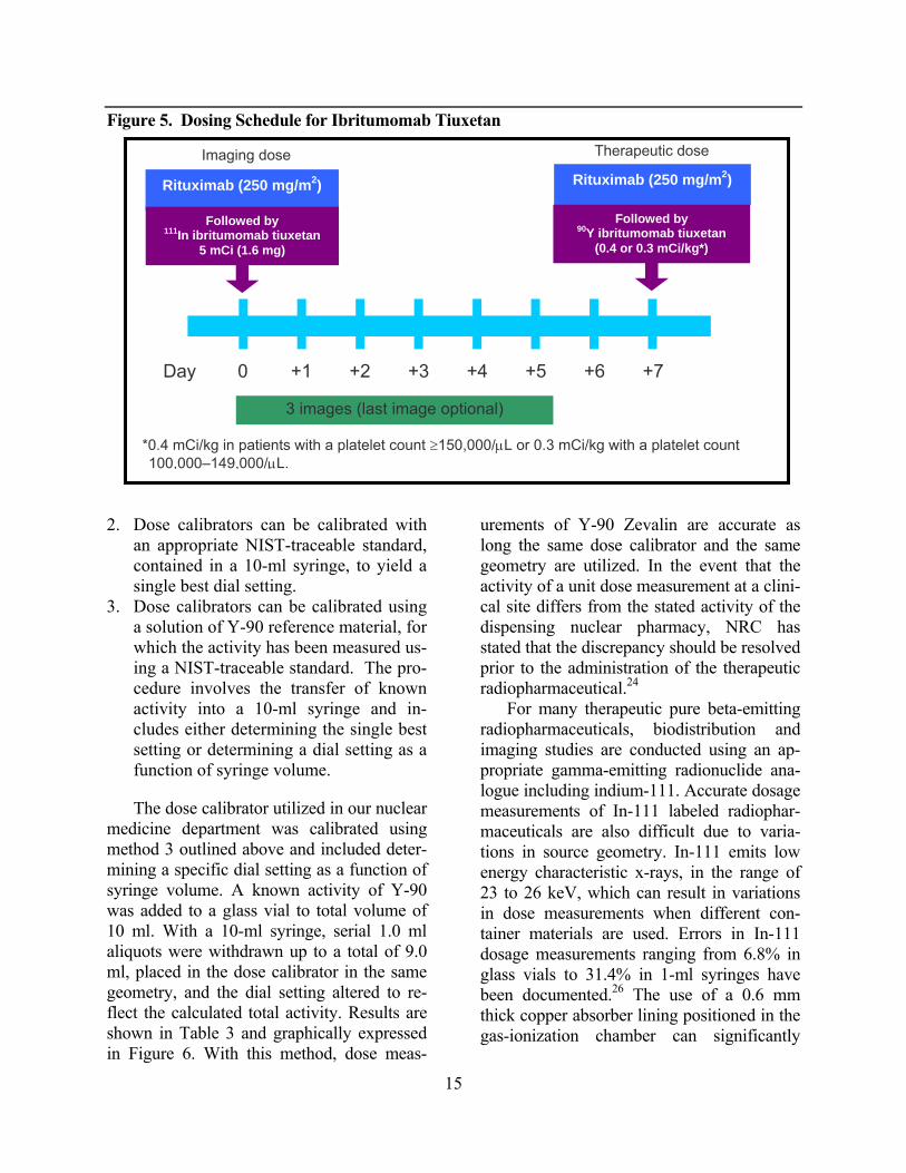
The dosing schedule for radiolabeled ibri-tumomab is illustrated in Figure 5. Patients
receive an infusion of rituximab (Rituxan®), a chimeric construct of ibritumomab, at 250 mg/m2, to optimize biodistribution of the ra-diolabeled antibody, followed immediately by an imaging dose of In-111 Zevalin (5 mCi on 1.6 mg of ibritumomab) by slow IV injec-tion over 10 minutes. Imaging is then per-formed, to assess altered biodistribution, at two or three different days after injection. One week after In-111 injection, patients without an altered biodistribution receive a second infusion of rituximab (250 mg/m2) immediately followed by a slow IV injection of Y-90 Zevalin over a 10-minute time period at a dose of either 0.3 mCi/kg or 0.4 mCi/kg, depending on platelet count, with a maximum injection dose of 32 mCi.23
An accurate measurement of pure beta-emitting radionuclides with a well-type gas-ionization chamber or a dose calibrator is dif-ficult due to source geometry. The special shape of a source can result in differences in self-absorption and also different containers can result in different degrees of attenuation. In addition, altering the volume of source ma-terial can significantly alter the response of the dose calibrator. Because the primary ra-diation measured for Y-90 is bremsstrahlung, effects of various containers may be opposite to that expected. Increases in the atomic number of the material in the container may increase the response of the dose calibrator due to the increased bremsstrahlung produc-tion.24 For example, use of lead containers as opposed to aluminum or plastic containers will increase bremsstrahlung production from less than one percent to approximately 6%,25 resulting in a higher response from a dose calibrator. Proposed methods for the accurate measurement of Y-90 Zevalin in dose cali-brators include the following:
1. Dose calibrator settings, or a range of settings, can be obtained from the manu-facturer or from a qualified expert who has performed and documented calibra-tions for the dose calibrator in question.
15
Figure 5. Dosing Schedule for Ibritumomab Tiuxetan
*0.4 mCi/kg in patients with a platelet count ≥150,000/µL or 0.3 mCi/kg with a platelet count 100,000–149,000/µL.
0 +1 +2 +3 +4 +5 +6
Rituximab (250 mg/m2)
3 images (last image optional)
Day +7
Followed by 111In ibritumomab tiuxetan
5 mCi (1.6 mg)
Rituximab (250 mg/m2)
Followed by 90Y ibritumomab tiuxetan
(0.4 or 0.3 mCi/kg*)
Imaging dose Therapeutic dose
2. Dose calibrators can be calibrated with
an appropriate NIST-traceable standard, contained in a 10-ml syringe, to yield a single best dial setting.
3. Dose calibrators can be calibrated using a solution of Y-90 reference material, for which the activity has been measured us-ing a NIST-traceable standard. The pro-cedure involves the transfer of known activity into a 10-ml syringe and in-cludes either determining the single best setting or determining a dial setting as a function of syringe volume.
The dose calibrator utilized in our nuclear medicine department was calibrated using method 3 outlined above and included deter-mining a specific dial setting as a function of syringe volume. A known activity of Y-90 was added to a glass vial to total volume of 10 ml. With a 10-ml syringe, serial 1.0 ml aliquots were withdrawn up to a total of 9.0 ml, placed in the dose calibrator in the same geometry, and the dial setting altered to re-flect the calculated total activity. Results are shown in Table 3 and graphically expressed in Figure 6. With this method, dose meas-
urements of Y-90 Zevalin are accurate as long the same dose calibrator and the same geometry are utilized. In the event that the activity of a unit dose measurement at a clini-cal site differs from the stated activity of the dispensing nuclear pharmacy, NRC has stated that the discrepancy should be resolved prior to the administration of the therapeutic radiopharmaceutical.24
For many therapeutic pure beta-emitting radiopharmaceuticals, biodistribution and imaging studies are conducted using an ap-propriate gamma-emitting radionuclide ana-logue including indium-111. Accurate dosage measurements of In-111 labeled radiophar-maceuticals are also difficult due to varia-tions in source geometry. In-111 emits low energy characteristic x-rays, in the range of 23 to 26 keV, which can result in variations in dose measurements when different con-tainer materials are used. Errors in In-111 dosage measurements ranging from 6.8% in glass vials to 31.4% in 1-ml syringes have been documented.26 The use of a 0.6 mm thick copper absorber lining positioned in the gas-ionization chamber can significantly

16
Table 3. Calibration Setting for Y-90 Using a 10-ml Syringe Volume
(mL) Calculated Activity
Concentration (mCi/mL) Calculated Total
Activity (mCi) Dial Setting
1.0 3.91 3.91 56 2.0 3.91 7.82 55 3.0 3.91 11.73 55 4.0 3.91 15.64 53 5.0 3.91 19.55 48 6.0 3.91 23.46 48 7.0 3.91 27.37 47 8.0 3.91 31.28 47 9.0 3.91 35.19 48
Figure 6. Dose Calibrator Response Using 40 mCi Y-90 Zevalin in a 10-ml Syringe
Example: Response Using 40 mCi Source in 10 mL Syringe
10
15
20
25
30
35
40
10.0 9.0 8.0 7.0 6.0 5.0 4.0 3.0 2.0 1.0 0.0APPROXIMATE VOLUME (mL)
DIA
L SE
TTIN
G x
10
reduce the low energy x-ray emissions from In-111 resulting in accurate dose calibrator measurements regardless of container geome-try.27 When using the copper filter, dose cali-brator setting for In-111 must be recalibrated. A copper filter for dose calibrators is cur-rently commercially available.
Radiation precaution issues related to Y-90 Zevalin include radionuclide handling,
radiopharmaceutical preparation and patient injection. In addition, patient radiation pre-cautions and radiation precautions related to potential exposures to members of the family and members of the public are of concern. Due to bremsstrahlung production, shielding requirements for beta-emitting radionuclides require materials with a low atomic number. As shown by the formula in Figure 7, the
17
fraction of incident beta energy converted to bremsstrahlung is a function of the atomic number of the absorbing or shielding material and the energy of the beta-emitting radionu-clide. For beta-emitting radionuclides, shield-ing with a low atomic number, such as acrylic or plastic, or composite shielding, such as aluminum/lead, is essential in mini-mizing bremsstrahlung production. For Y-90 Zevalin, with an energetic 2.3 MeV beta-emission, effective shielding requirements are necessary for vial shields, syringe shields and transport shields. Our department has evaluated the effectiveness of a number of shielding devices using thermoluminescent dosimeters.28 High surface dose rates were recorded from an unshielded glass vial and plastic syringe containing clinical activities of Y-90 Zevalin (366 rads/hr and 1287 rads/hr, respectively). Use of an acrylic vial shield or a composite (aluminum/lead) vial shield reduced the radiation levels by a factor of 1800–3600. Using an acrylic or composite (acrylic/lead) syringe shield also reduced ra-diation levels by a factor of 4000–6000.
Figure 7. Bremsstrahlung Production as a Function of Atomic Number of Absorber and Beta-Emitting Radionuclide Energy
ƒ = 3.5 × 10⁻⁴ ZE ƒ = the fraction of the incident beta energy con-
verted into photons Z = atomic number of the absorber E = maximum energy of the beta particle in MeV
Patient dose administration of Y-90
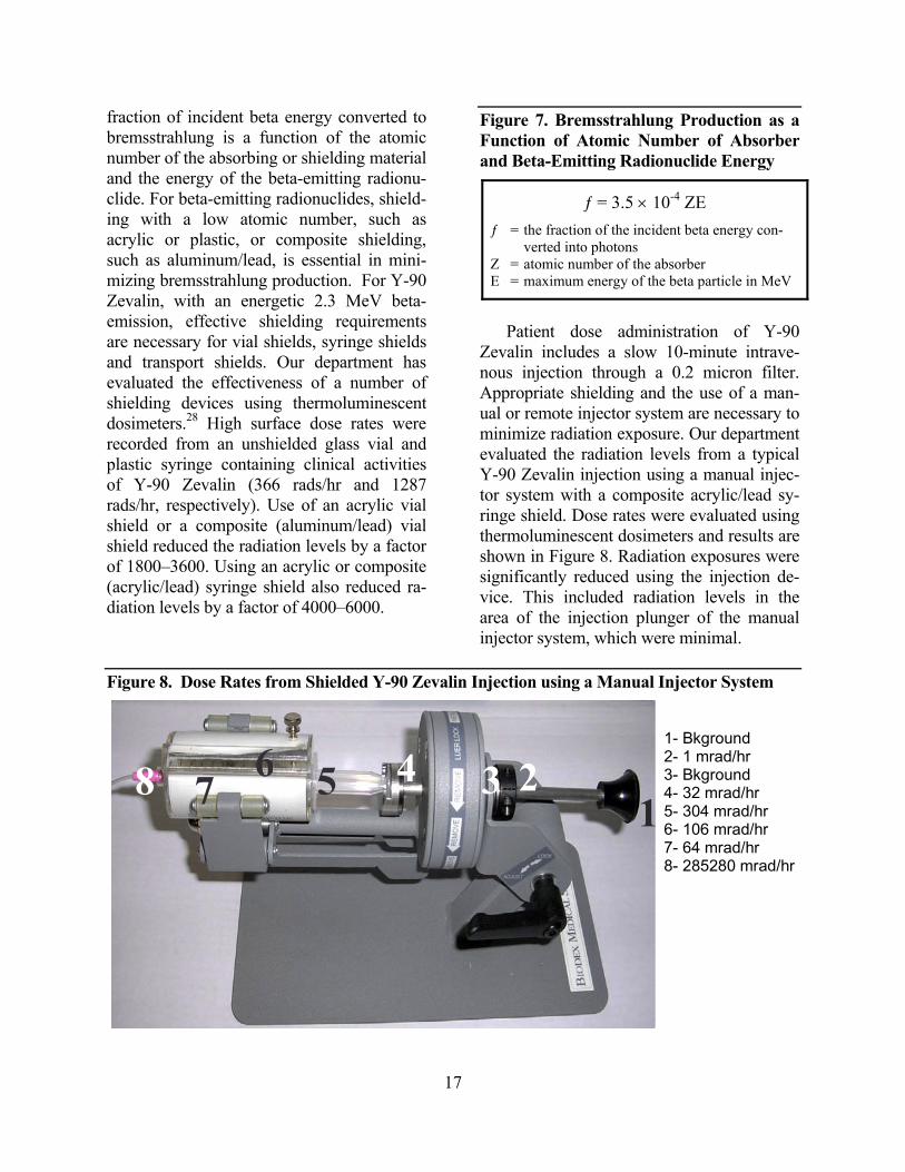
Zevalin includes a slow 10-minute intrave-nous injection through a 0.2 micron filter. Appropriate shielding and the use of a man-ual or remote injector system are necessary to minimize radiation exposure. Our department evaluated the radiation levels from a typical Y-90 Zevalin injection using a manual injec-tor system with a composite acrylic/lead sy-ringe shield. Dose rates were evaluated using thermoluminescent dosimeters and results are shown in Figure 8. Radiation exposures were significantly reduced using the injection de-vice. This included radiation levels in the area of the injection plunger of the manual injector system, which were minimal.
Figure 8. Dose Rates from Shielded Y-90 Zevalin Injection using a Manual Injector System
1- Bkground 2- 1 mrad/hr 3- Bkground 4- 32 mrad/hr 5- 304 mrad/hr 6- 106 mrad/hr 7- 64 mrad/hr 8- 285280 mrad/hr
16 5
48 7 34 2

18
Radioimmunotherapy treatments with Y-90 Zevalin can be performed on an outpa-tient basis. Immediately following injection, dose rates at 1meter from patients receiving Y-90 Zevalin are less than 0.5 mR/hr. As a result, patient instructions following Y-90 Zevalin administration are minimal and are found in Table 4. Instructions deal primarily with possible radiation contamination prob-lems related to urine and body fluids and generally extend for a period of three days. In addition, the use of condoms for sexual rela-tions is encouraged for a period of 7 days.29
Table 4. Patient Release Instructions Fol-lowing Y-90 Zevalin Administration
3 days Clean Spilled Urine and Dispose of Any Fluid Contaminated Material
7 days Use Condoms for Sexual Relations
Following release of patients receiving Y-90 Zevalin therapy, radiation exposures to members of the public and especially mem-bers of the immediate family are of concern. In a study conducted by Wiseman et al.,30 family members of patients receiving Y-90 Zevalin, with unrestricted access to the pa-tient, were issued radiation monitoring de-vices for a period of 7 days following ther-apy. Results showed that radiation exposures to family members were minimal, ranging
from 1.4–7.9 mrem, for a seven-day period. As a result, no radiation precautions to family members of patients receiving Y-90 Zevalin are warranted.
E. I-131 Tositumomab Another radioimmunotherapy agent for
the treatment of non-Hodgkin’s lymphoma is I-131 tositumomab (Bexxar), a murine IgG2 monoclonal antibody that binds to CD20 an-tigen on lymphoma cells. For this radioim-munotherapy agent, therapeutic doses are customized to the individual patient based on whole body clearances. Prior to treatment, patients receive supersaturated potassium io-dide (SSKI) to block thyroid uptake. The treatment regimen, as outlined in Figure 9, consists of an initial dosimetric dose of I-131 tositumomab (5 mCi) preceded by a 1-hour administration of unlabeled tositumomab (450 mg). Total body counts, usually per-formed with a gamma scintillation camera, are obtained three times over a 6–7 day pe-riod to determine residence time of the radio-labeled monoclonal antibody. A therapeutic dose is then calculated from the residence time so that the patient receives a whole body absorbed dose of 75 cGy. The therapeutic activity of I-131 tositumomab is usually in the range of 33–161 mCi.31,32
Figure 9. Treatment Schema for I-131 Tositumomab
Day 0 Day 7-14

19
Radioimmunotherapy using I-131 tositu-momab may require hospitalization, depend-ing on the regulatory requirements of the re-spective states. NRC regulated states have amended regulations related to the criteria for the release of patients administered radioac-tive materials. The new criteria for patient release may be based on limiting the total effective dose equivalent (TEDE) to less than 500 mrem for the maximally exposed indi-vidual. Siegel et al.33 developed a methodol-ogy for the release of patients administered I-131 tositumomab. As a result, radioimmuno-therapy with I-131 tositumomab could be performed on an outpatient basis in NRC regulated states.
Shielding during I-131 tositumomab for-mulation, dispensing and patient injections is important in reducing radiation exposure to occupational radiation workers. I-131 is a mixed emitter, with the major beta-emission of 606 keV (86%) and the major gamma-emission of 364 keV (81%). As a result, shielding requirements must be appropriate for both types of emissions. The range of beta-emission of I-131 in lead is 0.17 mm and the fraction of beta-emissions converted to bremsstrahlung in lead is low (1.7%). In addition, only about 1% of the x-rays gener-ated from bremsstrahlung have energies greater than 360 keV.20 From the data, it would appear that lead shielding for the gamma component of I-131 is also effective in shielding the beta component.
Radiation safety issues related to I-131 tositumomab administration deal with radia-tion doses to the patient as well as radiation exposure to family members and members of the general public. Immediately following therapeutic administration of I-131 tositu-momab, the mean measured dose rate at 1 meter from patients was 10.9 mrem/hr with a range of 4–24 mrem/hr.33 Radiation levels decreased with increasing patient body size. In order to assure compliance to dose limiting criteria, radiation exposures to family mem-
bers of patients receiving I-131 tositumomab were measured directly with appropriate ra-diation dosimeters.34 The mean radiation ex-posure to family members was 144 mrem, with a range of 10–354 mrem, which was well below the dose limit allowed to mem-bers of the general public (500 mrem).
In order to minimize radiation exposures to others, patient radiation safety instructions must be issued following radioimmunother-apy treatment with I-131 tositumomab. In-structions are based on total body residence times and measured patient dose rates. Typi-cal instructions for a patient receiving I-131 tositumomab with a total effective dose equivalent (TEDE) of 365 mrem would in-clude the following:33
1. Sleep in separate bed (at least 2 meter separation) for 8 days.
2. Do not take long trips (more than 4 hrs) sitting near others for 1 day.
3. Stay at least two meters from children and pregnant women for 7 days.
4. Minimize time spent in close contact with others and delay return to work for 4 days.
F. Radiolabeled Peptides for Therapy The use of radiolabeled peptides for
therapeutic applications is a new and exciting area of clinical nuclear medicine research. Because peptides generally consist of less than 50 amino acids, they have a low molecu-lar weight. As such, they tend to have fast clearances and rapid tissue and tumor accu-mulation. A wide range of peptides, having an affinity for specific receptors over ex-pressed on a large number of tumor cell types, have been identified. Some interesting peptides under clinical investigation include somatostatin and analogues of somatostatin, bombesin, gastrin, and epidermal growth fac-tor.33
Although radiolabeled peptides have gen-erated great interest in imaging and therapy, there are concerns regarding their clinical

20
use. As the size of peptides increase, they tend to more slowly diffuse into tumors and are also highly susceptible to proteolysis. Some endogenous peptides may also have unwanted physiological side effects. Radio-labeling of peptides usually involves the in-corporation of a bifunctional chelating agent onto the peptide. With its relatively high mo-lecular weight of the chelator, the molecular weight of the peptide complex may distort peptide conformation resulting in altered tar-get binding.35
A number of beta-emitting radionuclides have been utilized for peptide receptor ra-dionuclide therapy, including Y-90, Lu-177 and Cu-6436-38 Coupling to peptides is usu-ally accomplished via a peptide bond using bifunctional chelating agents such as DOTA (1,4,5,10-tetraazacyclododecane-N,N’,N’’,N’’’-tetraacetic acid) and TETA (1,4,8,11-tetraazacyclotetradecane-N’,N’’,N’’’-tetraacetic acid).
G. Y-90-DOTA-Octreotide Clinical trials in peptide receptor ra-
dionuclide therapy have been initiated using a somatostatin analogue, DOTA-DPhe1-Tyr3-Octreotide (DOTATOC) (36). With this con-jugated peptide, stable Y-90 incorporation was achieved. The schematic presentation of Y-90 DOTATOC is found in Figure 10. DO-TATOC is an 8 amino acid structure in which DOTA is bound to phenylalanine. Clinical studies in patients with histologically con-firmed subtype 2 somatostatin tumor recep-tors (sst2) have been initiated using Y-90 DOTATOC. Besides neuroendocrine tumors, somatostatin receptors have been identified in cancers of the central nervous system, breast, lung, and lymphatic tissue. Patients received either a number of cycles of Y-90 DOTA-TOC, with an 8-week interval between treat-ments. Y-90 DOTATOC was, in some cases, administered in a dose-escalating manner, ranging between 24–150 mCi per treatment, with a cumulated dose of 260–576 mCi. Ad-ministration of Y-90 DOTATOC involved a
slow intravenous infusion of 50-100 ml over 20 minutes. Some patients received an amino acid infusion, consisting of lysine and argin-ine, prior to radionuclide therapy in order to the reduce radiation dose to kidneys. The maximum tolerated dose per cycle was de-fined as 150 mCi Y-90 DOTATOC. Objec-tive therapeutic responses, either partial or complete, were documented in more than 20% of the treated patients. No major acute reactions were noted after peptide therapy administration. Moderate gastrointestinal tox-icity, including nausea and/or vomiting, and some transient hematologic toxicity were ob-served in some patients.
Figure 10. Schematic Presentation of Y-90 Labeled DOTA-DPhe1-Tyr3-Octreotide (DOTATOC)
Dose measurement issues related to Y-90 are similar to those stated previously. Due to differences in source geometry, especially volume differences, dose calibrators must be recalibrated as previously described.
Shielding requirements for Y-90 DOTA-TOC are similar to those described for other Y-90 labeled radiopharmaceuticals. Initially, bremsstrahlung production is minimized by use of shielding materials with a low atomic number, such as glass, acrylic or aluminum. Bremsstrahlung, or energetic x-ray emis-sions, can then be effectively attenuated us-ing lead or other materials with a high atomic number. Radiation safety issues related to radiation exposure to ancillary personnel, in-cluding family members, must be considered in light of regulatory constraints. In order to assess radiation exposures from beta-emitting radionuclides, a “specific bremsstrahlung

21
constant”, with units of R-cm2/mCi-h, has been proposed.25 This constant is somewhat analogous to a specific gamma-ray constant. Using the specific bremsstrahlung constant, it would appear that patients could be treated on an outpatient basis with activities of sev-eral thousand mCi Y-90 radiopharmaceuti-cals, based on regulatory constraints of 0.5 rem total dose equivalent.25
H. Y-90 Microspheres Y-90 microspheres are classified as de-
vices intended for regional radionuclide brachytherapy. As a radionuclide brachyther-apy source, two specifications must be met: 1) The device must minimize the leakage of possibly hazardous radiation, and 2) the en-capsulated material must be biocompatible and not cause significant adverse tissue reac-tion.39
The intrahepatic arterial administration of Y-90 microspheres includes the use of cathe-ters and an administration apparatus. An ap-propriate catheter is accurately positioned within the right or left hepatic arteries corre-sponding to the desired treatment target area. Y-90 microspheres are then administered through an administration apparatus consist-ing of a variety of sterile apyrogenic compo-nents including water for injection, saline, sealed tubing, acrylic shields, inlet and outlet lines and three-way connection system.40
One of the limitations associated with the clinical use of Y-90 microspheres is arterio-venous shunting which can result in in-creased radiation doses to ancillary organs or tissues. In order to limit radiation doses to ancillary organs, especially the lungs, the de-gree of shunting must be determined prior to radionuclide therapy using Tc-99m macroag-gregated albumin. Following administration of Tc-99m MAA (2–6 mCi), perfusion imag-ing is performed to evaluate the degree of pulmonary and gastric tract shunting. Patients may only be treated with Y-90 microspheres if the total cumulative lung dose does not ex-ceed 3000 rads.40
At the present time, there are two Y-90 microsphere preparations in clinical use, SIR-Spheres (Sirtex Medical) and TheraSphere (MSD Nordion). SIR-Spheres consists of biocompatible microspheres with a mean di-ameter of 32 µm and a range of 20–40 µm. SIR-Spheres is commercially available in glass vials containing 3 GBy (81 mCi) ± 10% per 5 ml of Y-90 microspheres (40–80 mil-lion particles) suspended in water for injec-tion. Each sterile, pyrogen-free vial is crimp sealed to allow individual patient doses to be withdrawn. SIR-Spheres are indicated for the treatment of unresectable metastatic liver tu-mors from primary colorectal cancer with adjuvant intra-hepatic artery chemotherapy of Floxuridine.41
Y-90 microspheres (TheraSphere) consist of insoluble glass microspheres in which Y-90 is imbedded. The microspheres have a mean diameter of 25 µm ±10 µm (standard deviation), with less than 5% smaller than 15 µm and less than 10% larger than 35 µm. Each milligram contains between 22,000 and 73,000 microspheres. Y-90 microspheres are supplied in 0.05 ml of sterile, pyrogen-free, water that is contained in a dose vial shielded with polystyrene. Y-90 microspheres are commercially available in six dose sizes of 81 mCi, 135 mCi, 189 mCi, 270 mCi, 405 mCi and 540 mCi. Y-90 microspheres (TheraSphere) are currently approved by the U.S. Food and Drug Administration under the provisions of a “Humanitarian Device Exemption) (HDE No. H9800006) which includes unique restrictions on the medical use of the device. TheraSpheres is indicated for use in radiation treatment of patients with unresectable hepatocellular carcinoma (HCC).39
Patients with unresectable hepatocellular carcinoma have been successfully treated with intrahepatic arterial administrations of Y-90 microspheres.40,42 Total activity of Y-90 microspheres administered is based on liver volume and the desired radiation dose
22
delivered to the liver, usually between 100–150 Gy (10,000–15000 rad). After catheter placement in left or right hepatic arteries cor-responding to the desired treatment area, Y-90 microspheres are injected via a specific administration system, as described previ-ously.
Results of Phase I and Phase II studies with Y-90 microspheres have been encourag-ing; both in stabilizing the disease process and also in increased survival benefit.39 Ra-dionuclide therapy with Y-90 microspheres has been well tolerated with low toxicities, including mild fatigue and fever. Some gas-trointestinal toxicities have been observed in a limited amount of patients.39
Radiation safety issues related to the clinical use of Y-90 microspheres are impor-tant and are an integral part of the therapy procedure. Administration of Y-90 micro-spheres is generally performed in interven-tional radiology suites. Control measures to limit radiation contamination are essential. Dose measurements of the administration device, prior-to and following Y-90 micro-sphere injection, are important determining the total administered activity. Patient moni-toring is important in assessing radiation doses to members of the public and also in meeting release criteria.
IV. CONCLUSION This review has attempted to provide an
update of therapeutic radiopharmaceuticals that are in current clinical use or are under investigation. It is important to note that sev-eral therapeutic radiopharmaceuticals for ra-dioimmunotherapy, including Y-90 Ibtritu-momab Tiuxan and I-131 Tositumomab, have been approved for general use. In addi-tion, research endeavors related to alpha-emitting radiopharmaceuticals are increasing due to the attractive therapeutic properties of specific alpha radionuclides. Although some radionuclide therapeutic agents have been classified as brachytherapy devices, including Y-90 microspheres, radiopharmaceutical dis-
pensing is still required. In addition, radiation safety issues related to unsealed sources must be considered.
V. REFERENCES 1. O’Donoghue JA, Bardies M, Wheldon
TE. Relationship between tumor size and curability for uniformly targeted therapy with beta-emitting radionu-clides. J Nucl Med 1995;36:1902-1909.
2. Siegel JA, Stabin MG. Absorbed frac-tions for electrons and beta particles in spheres of various sizes. J Nucl Med 1994;35:152-156.
3. Humm JL. Dosimetric aspects of radio-labeled antibodies for tumor therapy. J Nucl Med 1986;27:1491-1497.
4. Imam SK. Advancements in cancer therapy with alpha-emitters: a review. Int J Radiation Oncology Biol Phys 2001;51:271-278.
5. Laven DL, Hinkle GH. Use of alpha- and beta-emitting radionuclides for the treatment of cancer. In Correspondence Continuing Education Courses for Nu-clear Pharmacists and Nuclear Medicine Professionals. 1995;6:1-25.
6. Zalutsky MR, Zhao XG, Alston KL, Bigner D. High-level production of α-particel-emitting At-211 and preparation of At-211-labeled antibodies for clinical use. J Nucl Med 2001;42:1508-1515.
7. Zalutsky, MR, Garg PK, Friedman HS, et al. Labeling Mabs and F(ab’)2 frag-ments with the alpha particle emitting nuclide At-211: Preservation of immu-noreactivity and in vivo localizing ca-pacity. Proc Natl Acad Sci USA 1989: 86:7149-7153.
8. Zalutsky MR, Narula AS. Astatination of protein using an N-succimidyl tri-n-

23
butylstannyl benzoate intermediate. Appl Radiat Isot 1988;39:227-232.
9. Zalutsky MR, Cokgor I, Akabani G, et al. Phase I trial of alpha-particle-emitting astatine-211 labeled chimeric anti-tenascin antibody in recurrent ma-lignant glioma patients. Proc Am Assoc Cancer Res 2000;41:544.
10. Atcher RN, Friedman AM, Hines JJ. An improved generator for the production of 212Pb and 212Bi from 224Ra. Appl Radiat Isot, Int J Radiat Appl Instrum (A) 1988;39:283-286.
11. Ruegg CL, Anderson-Berg WT, Bre-chbiel MW et al. Improved in vivo sta-bility and tumor targeting of bismuth-labeled antibody. Cancer Res 1990;50: 4221-4226.
12. Junghans RP, Dobbs D. Brechbiel MW et al. Pharmacokinetics and bioactivity of DOTA-bismuth-conjugated anti-Tac antibody for α-emitter (Bi-212) therapy. Cancer Res 1993;53:5683-5689.
13. McDevitt MR, Nijula TN, Finn RD, et al. Bismuth labeled antibodies for ther-apy of leukemias, lymphomas and car-cinomas: Pre-clinical studies. Tumor Targeting 1996;2:182.
14. Finn RD, McDevitt MR, Scheinberg DA, et al. Refinements and improve-ments for Bi-213 production and use as a targeted therapeutic radiopharmaceuti-cal. J Lab Comp Radiopharm 1997;40: 293.
15. McDevitt MR, Finn RD, Sgouros G, et al. An Ac-225/Bi213 generator system for therapeutic clinical application: Con-struction and operation. Appl Rad Iso-topes 1999;50:895-904.
16. Brechbiel MW, Gansow OA. Synthesis of C-functionalized CHX-DTPA for la-beling of MAbs with the Bi-121 α-particle emitter. J Chem Soc Perkin Trans 1992;1:1173-1178.
17. Jurcic JG, Larson, SM, Sgouros, G. et al. Targeted α particle immonothrapy for myeloid leukemia. Blood 2002; 1001233-1239.
18. McDevitt MR, Scheinberg DA. Ac-225 and the daughters: the many faces of Shiva. Cell Death and Diff 2002;9:593-594.
19. Miederer M, McDevitt MR, Sgouros G, et al. Pharmacokinetics, dosimetry, and toxicity of the targetable atomic genera-tor, 225Ac-HuM195, in nonhuman pri-mates. J Nucl Med 2003;45:129-137.
20. Cember H. Introduction to health phys-ics. McGraw-Hill, New York, NY. 1992.
21. Zhou H, Suzuki M, Randers-Pehrson G, et al. Radiation risk to low fluences of α particles may be greater than we thought. PNAS 2001;98:14410-14415.
22. Zimmer AM, Chinn PC, Morena BA, et al. Clinical experience with preparation of In-111 and Y-90 Zevalin: radio-chemical purity assessment. Eur J Nucl Med 2001;28:1261.
23. Witzig TE, White CA, Wiseman GA, et al. Phase I/II trail of IDEC-Y2B8 radio-immunotherapy for treatment of re-lapsed or refractory CD20+ B-cell non-Hodgkin’s lymphoma. J Clin Oncol 1999;17:3793-3803.
24. NRC Information Notice 02-017. Unites States Nuclear Regulatory Commission, Washington DC, May 2002.

24
25. Zanzonico PB, Binkert BL, Goldsmith SJ. Bremsstrahlung radiation exposure from pure b-ray emitters. J Nucl Med 1999;40:1024-1028.
26. Kowalsly RJ, Johnston RE. Dose cali-brator assay of iodine-123 and indium-111 with a copper filter. J Nucl Med Tech 1998;26:94-98.
27. Yundt, KL, Zimmer, AM, Cutrera, PF, et. al. Calibrator Assay of In-111 using a commercially available copper filter. J Nucl Med Tech 2004;32:130.
28. Zimmer AM, Carey AM, Spies SM. Ef-fectiveness of specific vial and syringe shields in reducing radiation exposures from Y-90 Zevalin. J Nucl Med 2002; 43:45P.
29. Wagner HN Jr, Leigh B, Witzig T, et al. Administration guidelines for radioim-munotherapy of non-Hodgkin’s lym-phoma with Y-90 anti-CD20 mono-clonal antibody. J Nucl Med 2002;43: 267-272.
30. Wiseman G, Leigh B, Witzig T, et al. Radiation exposure is very low to the family members of patients treated with yttrium-90 Zevalin anti-CD20 mono-clonal antibody therapy for lymphoma. Eur J Nucl Med 2001;28:1198.
31. Kaminski MS, Estes J, Zasadny KR, et al. Radioimmunotherapy with iodine I-131 tositumomab for relapsed or refrac-tory B-cell lymphoma: updated results and long-term follow-up of the Univer-sity of Michigan experience. Blood 2000;96:1259-1266.
32. Wahl RL, Kroll S, Zasadny KR, et al. Patient-specific whole-body dosimetry: principles and a simplified method for
clinical implementation. J Nucl Med 1998;39 (supp.):14S-20S.
33. Siegel JA, Drol S, Regan D, et al. A practical methodology for patient release after tositumomab and I-131 tositumo-mab therapy. J Nucl Med 2002;43:354-363.
34. Rutar FJ, Augustine SC, Colcher D, et al. Outpatient treatment with I-131 anti-B1 antibody: radiation exposure to fam-ily members. J Nucl Med 2001;42:907-915.
35. Weiner RE, Thakur ML. Radiolabeled peptides in the diagnosis and therapy of oncological diseases. Appl Rad and Iso-topes 2002;57:749-763.
36. Chinol M, Bodei L, Cremonesi M, et al. Receptor-mediated radiotherapy with Y-90 DOTA- DPhe1-Tyr3-Octreotide: The experience of the European Instute of Oncology Group. Sem Nucl Med 2002; 32:141-147.
37. Kwekkeboom WH, Bakker WH, Kooij PPM, et al. (Lu-177 DOTA, Tyr) oc-treotide: comparison with (In-111-DTPA octreotide) in patients. Eur J Nucl Med 2001;28:1319-1325.
38. Anderson CJ, Dehdashti F, Cutler PD, et al. Cu-64 TETA-octreotide a PET imag-ing agent for patients with neuroendo-crine tumors. J Nucl Med 2001;42:213-221.
39. Package Insert. TheraSphere Yttrium-90 Glass Microspheres. MDS Nordion, Kanata, ON, Canada.
40. Salem R, Thurston KG, Carr BI, et al. Yttrium-90 microspheres: radiation therapy for unresectable liver cancer. JVIR 2002;13:S223-S229.

25
41. Package Insert. SIR-Spheres Yttrium-90 Micrspheres. Sirtex Medical, Lake Forrest, IL.
42. Dancey JE, Shepherd FA, Paul K, et al. Treatment of nonresectable hepatocellu-lar carcinoma with intrahepatic Y-90-microspheres. J Nucl Med 2000;41: 1673-1681.
VI. QUESTIONS
1. Which of the following parameters is not important for radionuclide therapy suc-cess? a. Absorbed radiation dose to tumor b. Concentration of radiopharmaceuti-
cal in tumor c. The degree of disease progression d. The sensitivity of tumor to radiation
2. Which alpha-emitting radionuclide uses a generator system in which the parent is Ac-225? a. At-211 b. Bi-212 c. Bi-213 d. Ra-226
3. Which of the following statements is not true for Ac-225? a. Ac-225 can stably bind to mono-
clonal antibodies b. Ac-225 emits four alpha particles c. Ac-225 has a 10-day half-life d. Ac-225 has been proposed as an in
vivo alpha generator
4. Which of the following statements is not true in radiation protection of alpha-emitting radionuclides? a. Alpha-emitters are stopped by the
dead outer layers of the skin b. Alpha-emitters pose a danger if in-
gested c. Alpha-emitters pose a significant ex-
ternal radiation hazard
d. Alpha-emitter pose a danger if in-haled
5. Which of the following beta-emitting radionuclides is ideal for treating tumors with diameters less than 1 mm? a. P-32 b. I-132 c. Lu-177 d. Y-90
6. Which monoclonal antibody is used for the treatment of Non-Hodgkin’s lym-phoma? a. anti-tenascin antibody b. ibtritumomab c. HuM195 d. T101
7. Which therapeutic radionuclide is conju-gated to ibritumomab? a. Y-90 b. I-131 c. Re-186 d. In-111
8. For dose measurement issues related to In-111, inaccurate measurements are due to which of the following? a. Attenuation of energetic gamma
emissions b. Attenuation of low energy x-rays c. Attenuation by the plastic insert d. Attenuation of radionuclidic impuri-
ties
9. I-131 tositumomab is a radioimmuno-therapy agent used to treat what disease? a. Ovarian cancer b. Non-Hodgkin’s lymphoma c. T-cell lymphoma d. Prostate cancer

26
10. Following I-131 tositumomab admini-stration, which of the following state-ments is not true? a. Patients can be treated with I-131 to-
situmomab on an outpatient basis in NRC regulated states.
b. Patients must is issued radiation safety precautions after therapy.
c. Patient release criteria are based on limiting the total effective dose equivalent to less than 500 mrem for the maximally exposed individual
d. Patients must be hospitalized follow-ing therapy.
11. Radiolabeled peptides for therapy in-clude which of the following? a. Y-90 microspheres b. Y-90 Octreotide c. In-111 Octreotide d. Y-90 Ibritumomab
12. Beta-emitting radionuclides used for peptide imaging include all of the fol-lowing except? a. Y-90 b. Lu-177 c. P-32 d. Cu-64
13. Y-90 TheraSpheres consist of which of the following? a. Y-90 labeled to macroaggregated al-
bumin b. Y-90 labeled to human albumin mi-
crospheres c. Y-90 imbedded insoluble glass mi-
crospheres d. Y-90 biocompatible microspheres
14. For Y-90 microsphere therapy, what is the desired radiation dose delivered to the liver? a. 25-50 Gy b. 50-100 Gy c. 100-150 Gy d. 150-200 Gy
15. For Y-90 Ibritumomab radioimmuno-therapy, what is the maximum dose ad-ministered to patients? a. 32 mCi b. 40 mCi c. 0.4 mCi/kg body weight d. 0.3 mCi/kg body weight
16. Beta-emitting radionuclides with mean tissue penetration range greater than 1 mm include all of the following except one? a. Sr-89 b. I-131 c. Y-90 d. P-32
17. Following an imaging dose of In-111 ibritumomab, how many images must be taken to assess biodistribution? a. 1 image b. 2 images c. 3 images d. 4 images
18. Bremsstrahlung production from beta-emitting radionuclides depends on all the following parameters except one? a. range of beta-emitting radionuclide b. energy of beta-emitting radionuclide c. atomic number of the shielding ma-
terial d. the atomic number of the container
19. A remote injection device is needed for Y-90 patient administration due to the following? a. inject large volume of radiopharma-
ceutical b. reduce radiation exposure to person-
nel c. allow the slow infusion of radio-
pharmaceutical d. needed due to 0.2 micron filter
27
20. The formulation buffer is added when radiolabeling Y-90 ibritumomab for all of the following except one? a. to terminate the reaction sequence b. to stabilize the radiolabeled mono-
clonal antibody preparation c. to increase the radiolabeling effi-
ciency
21. Y-90 SIR-Spheres consist of the follow-ing? a. Y-90 labeled to macroaggregated al-
bumin b. Y-90 labeled to human albumin mi-
crospheres c. Y-90 imbedded insoluble glass mi-
crospheres d. Y-90 biocompatible microspheres
22. Y-90 TheraSpheres microsphere parti-cles have a mean diameter of? a. 10 µm b. 25 µm c. 35 µm d. 50 µm
23. Accurate dosage measurements of In-111 labeled radiopharmaceuticals in a dose calibrator are difficult due to? a. attenuation of gamma emissions b. attenuation of low energy x-ray
emissions c. attenuation of beta emissions d. all of the above
24. Which of the following describe the spe-cific bremsstrahlung constant? a. has units of R-cm2/mCi-h b. is analogous to a specific gamma-ray
constant c. describes the radiation exposures
from beta-emitting radionuclides d. all of the above
25. Rituximab is a monoclonal antibody with the following characteristics? a. is a chimeric construct of ibritumo-
mab b. is a chimeric construct of tositumo-
mab c. is a humanized construct of ibritu-
momab d. is a murine construct of tositumomab